Communication

Synthesis of 1H-3-{4-[(3-Dimethylaminopropyl)aminomethyl]phenyl}-2-phenylindole and Evaluation of Its Antiprotozoal Activity

1
Laboratoire ARNA, INSERM U1212/UMR CNRS 5320, UFR des Sciences Pharmaceutiques, Univ. Bordeaux, 146 rue Léo Saignat, F-33076 Bordeaux CEDEX, France
2
INSERM U1094, Tropical Neuroepidemiology, Institute of Neuroepidemiology and Tropical Neurology, Université de Limoges, F-87025 Limoges, France
3
IHU Méditerranée Infection, UMR VITROME, Aix-Marseille Univ., 19-21 Boulevard Jean Moulin, 13005 Marseille, France
4
CNRS, Univ. Bordeaux, Bordeaux INP, ICMCB, UMR 5026, F-33600 Pessac, France
5
AGIR (Agents Infectieux, Résistance et chimiothérapie), EA 4294, UFR de Pharmacie, Université de Picardie Jules Verne, F-80037 Amiens, France
*
Author to whom correspondence should be addressed.
Molbank 2019, 2019(2), M1060; https://doi.org/10.3390/M1060
Submission received: 9 April 2019 / Revised: 6 May 2019 / Accepted: 7 May 2019 / Published: 9 May 2019
(This article belongs to the Section Organic Synthesis and Biosynthesis)

Abstract

:
1H-3-{4-[(3-Dimethylaminopropyl)aminomethyl]phenyl}-2-phenylindole was synthesized via a multi-step pathway starting from 2-iodoaniline. Structure characterization of this new indole compound was achieved by 1H-NMR, 13C-NMR and ESI-MS spectral analysis. The title compound was screened in vitro against three protozoan parasites (Plasmodium falciparum, Leishmania donovani and Trypanosoma brucei brucei). Biological results showed antiparasitic activity with IC50 values in the μM range.

1. Introduction

Numerous indole-containing synthetic derivatives and natural products demonstrate pharmacological activities of importance to human disease. Thus, indoles constitute the basis of an important class of compounds with interesting and promising biological activities [1,2,3,4,5]. This nitrogen heterocyclic scaffold has gained much interest due to its wide range of biological activity upon modifications, including antiviral, antibacterial, anticancer, anti-Alzheimer’s disease and antiparasitic properties [6,7,8,9,10,11]. In this last field, the indole compounds are considered as attractive candidates for antiprotozoal therapy [11,12,13,14]. In the course of our work devoted to discovering new original heterocyclic candidates for use in antiparasitic chemotherapy [15,16,17], and as an extension of our work on the development of new antimalarial indole heterocyclic drugs, we have identified 1H-3-{4-[(3-dimethylaminopropyl)aminomethyl]phenyl}-2-phenylindole and found that it is endowed with interesting activity against three protozoan parasites. Thus, we report herein on the synthesis and structural identification of this new indole scaffold, which was then screened in vitro against three protozoan parasites (Plasmodium falciparum, Leishmania donovani and Trypanosoma brucei brucei).

2. Results and Discussion

2.1. 1H-3-{4-[(3-Ddimethylaminopropyl)aminomethyl]phenyl}-2-phenylindole

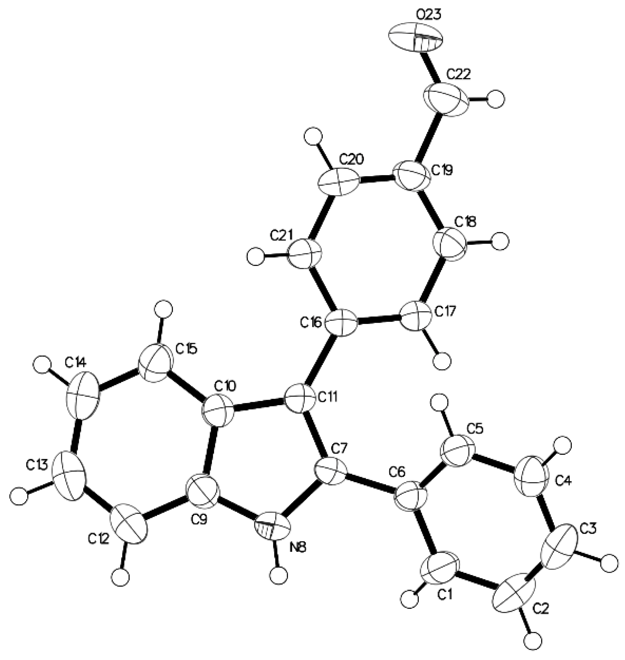
The synthesis of this new substituted indole compound 4 was achieved starting from the commercially available 2-iodoaniline (1) (Scheme 1). The key intermediate 2-phenyl-3-(4-formylphenyl)indole (2) was prepared from 2-iodoaniline (1) via a Sonogashira cross-coupling reaction, followed by a protection step and then by a palladium-catalyzed reaction, as previously described [18,19,20]. The 3D structural determination of disubstituted indole 2 was established by X-ray crystallography (Figure 1) [21] and confirmed the structure in the solid state as anticipated on the basis of NMR data [19,20]. Condensation of 3-dimethylaminopropylamine with the aldehyde 2 led to the unstable imine 3, which was immediately reduced into the 1H-3-{4-[(3-dimethylaminopropyl)aminomethyl]phenyl}-2-phenylindole (4) using sodium borohydride as a reductive agent in refluxing methanol as previously described by our team [16,17]. The structure of this new synthesized derivative 4 was then confirmed by FTIR, 1H/13C-NMR and ESI-MS analyses (see Supplementary Materials).

2.2. Antiprotozoal Activity

This new substituted indole 4 was evaluated for its antimalarial activity in vitro by incubation with P. falciparum chloroquine (CQ)-resistant strain W2 (IC50 CQ = 0.40 μM, IC50 MQ = 0.016 μM) and the strain 3D7, which is CQ sensitive and which has decreased sensitivity to mefloquine (MQ) decrease (IC50 CQ = 0.11 μM, MQ = 0.06 μM). As shown in Table 1, this derivative 4 was active on the W2 strain with an IC50 of 1.80 μM. Moreover, indole 4 also showed significant activity against the CQ-sensitive 3D7 strain (IC50 = 1.10 μM).
As P. falciparum belongs to the coccidian protozoan parasite family, in vitro activity against the flagellate protozoan parasite L. donovani was also evaluated (Table 1). The reference drugs amphotericin B and pentamidine had IC50 values of 0.25 µM and 5.50 µM, respectively, against L. donovani. Compound 4 was found to be active against the promastigote forms of L. donovani with an IC50 of 3.03 μM, slightly more potent than pentamidine, one of the reference compounds (IC50 = 5.5 μM).
This newly synthesized indole heterocyclic derivative 4 was then evaluated against T. brucei brucei. In this test, pentamidine, suramine, fexinidazole, and eflornithine were used as reference compounds. The screening data are presented in Table 1. The 1H-3-{4-[(3-dimethylaminopropyl)aminomethyl]phenyl}-2-phenylindole (4) was found to be active against T. brucei brucei with an IC50 of 0.22 μM.
To assess selectivity of action, the cytotoxicity of this new antiparasitic heterocyclic indole 4 was evaluated in vitro against the human cell line HepG2, which is a commonly used human-derived hepatocarcinoma cell line. Indole 4 showed a significant level of cytotoxicity against the HepG2 cell line (IC50 = 1.14 μM). The aim of this assay using HepG2 cells was to evaluate the impact of metabolic activation of the tested compound on cell viability. The cytotoxic concentration of 50% (CC50) was determined, and selectivity indexes (SI), defined as the ratios of cytotoxic to antiparasitic activities (SI = CC50/IC50) were calculated. Thus, this substituted indole 4 produced an interesting finding with SI = 5.18 on the T. brucei brucei strain.

3. Materials and Methods

Commercially available reagents were used as received without additional purification. Melting points were determined with an SM-LUX-POL Leitz hot-stage microscope (Leitz GMBH, Midland, ON, USA) and are uncorrected. The IR spectrum was recorded on a NICOLET 380FT-IR spectrophotometer (Thermo Electron Scientific Instruments LLC, Madison, WI, USA). The UV-vis spectrum was recorded on a GENESYS 10S UV-Vis spectrophotometer (Fisher Scientific, Illkirch, France). NMR spectra were recorded with tetramethylsilane as an internal standard using a BRUKER AVANCE 300 spectrometer (Bruker BioSpin, Wissembourg, France). Splitting patterns were reported as follows: s = singlet; bs = broad singlet; d = doublet; t = triplet; dd = double doublet; ddd = double double doublet; qt = quintuplet; m = multiplet. Analytical TLC was carried out on 0.25 pre-coated silica gel plates (POLYGRAM SIL G/UV254) and visualization of compounds after UV light irradiation. Silica gel 60 (70–230 mesh) was used for column chromatography. An ESI Orbitrap LTQ Velos from Thermo Fisher Scientific (Waltham, MA, USA) was used for MS analysis. Elemental analyses were found to be within ±0.4% of the theoretical values.

3.1. 1H-3-{4-[(3-Dimethylaminopropyl)iminomethyl]phenyl}-2-phenylindole (3)

To a solution of 3-dimethylaminopropylamine (36 mg, 0.337 mmol) in ethanol (5 mL) was added 2-phenyl-3-(4-formylphenyl)indole 2 (100 mg, 0.353 mmol). The reaction mixture was then heated under reflux for 5 h and then evaporated to dryness under reduced pressure. After cooling, the residue was extracted with dichloromethane (20 mL). The organic layer was dried over sodium sulfate and evaporated to dryness. The obtained product 3 was then used without further purification. Colorless powder (125 mg, 98%), m.p. 134–136 °C (EtOH); 1H-NMR (δ, ppm, CDCl3, 300 MHz): 8.74 (bs, 1H, NH), 8.34 (s, 1H, CH=N), 7.75 (d, 2H, J = 8.40 Hz, H-2” and H-6”), 7.72 (d, 1H, J = 7.60 Hz, H-4 indole), 7.51 (d, 2H, J = 8.40 Hz, H-3” and H-5”), 7.47–7.43 (m, 3H, H-7 indole and H phenyl), 7.36–7.29 (m, 3H, H phenyl), 7.27 (ddd, 1H, J = 8.10, 6.90 and 1.20 Hz, H-5 indole), 7.18 (ddd, 1H, J = 8.10, 6.90 and 1.20 Hz, H-6 indole), 3.68 (t, 2H, J = 7.20 Hz, NCH2), 2.40 (t, 2H, J = 7.20 Hz, NCH2), 2.27 (s, 6H, N(CH3)2), 1.91 (qt, 2H, J = 7.20 Hz, CH2).

3.2. 1H-3-{4-[(3-Dimethylaminopropyl)aminomethyl]phenyl}-2-phenylindole (4)

To a solution of compound 3 (120 mg, 0.315 mmol) in methanol (8 mL) was added portion-wise at 0 °C sodium borohydride (50 mg, 1.26 mmol; 4 equivalent.). The reaction mixture was stirred for 1 h at room temperature, heated under reflux for 1 h and then evaporated to dryness under reduced pressure. After cooling, the residue was triturated in water and extracted with dichloromethane (15 mL). The organic layer was separated, dried over sodium sulfate, and evaporated to dryness. The residue was then purified by column chromatography on silica gel using dichloromethane/methanol (90/10, v/v) as an eluent to give the pure product 4: pale-yellow oil (102 mg, 85%). IR (KBr) 3401 and 3149 (NH), 2938, 2858 2820 and 2770 (CH and CH2), 1604 (C=C), 1552, 1516, 1453, 1263, 1097, 1016, 804, 734, 697. UV λmax (EtOH)/nm 220 (log ε 4.43), 305 (log ε 4.93). 1H-NMR (δ, ppm, CDCl3, 300 MHz): 8.80 (bs, 1H, NH), 7.71 (d, 1H, J = 7.80 Hz, H-4 indole), 7.47-7.42 (m, 3H, H-7 indole and H phenyl), 7.43 (d, 2H, J = 7.80 Hz, H-2” and H-6”), 7.35 (d, 2H, J = 7.80 Hz, H-3” and H-5”), 7.34-7.29 (m, 3H, H phenyl), 7.26 (ddd, 1H, J = 8.10, 6.90 and 1.20 Hz, H-5 indole), 7.17 (ddd, 1H, J = 8.10, 6.90 and 1.20 Hz, H-6 indole), 3.84 (s, 2H, NCH2), 2.75 (t, 2H, J = 7.20 Hz, NCH2), 2.37 (t, 2H, J = 7.20 Hz, NCH2), 2.25 (s, 6H, N(CH3)2), 1.82 (bs, 1H, NH), 1.73 (qt, 2H, J = 7.20 Hz, CH2). 13C-NMR (CDCl3) δ: 139.5 (C-4” and C-7a), 137.4 (C-2), 135.64 (C-1”), 135.1 (C-3), 134.25 (C-1’), 131.5 (C-3’ and C-5’), 130.0 (C-3” and C-5”), 129.7 (C-2”, C-6’, C-2” and C-6”), 129.0 (C-4’), 123.97 (C-5), 121.7 (C-6), 121.06 (C-4), 116.1 (C-3a), 112.4 (C-7), 59.5 (NCH2), 55.4 (NCH2), 49.5 (NCH2), 46.9 (N(CH3)2), 29.4 (CH2). ESI-MS m/z [M + H]+ Calcd for C26H30N3: 384.244, Found: 384.243. Anal. Calcd. for C26H29N3: C, 81.42; H, 7.62; N, 10.96. Found: C, 81.57; H, 7.74; N, 11.08.

3.3. X-ray Data

The structure of compound 2 was established by X-ray crystallography (Figure 1). A colorless single crystal of 2 was obtained by slow evaporation from a methanol/chloroform solution (v/v: 20/80): triclinic, space group P-1, a = 9.9997(13) Å, b = 10.0457(11) Å, c = 10.1872(11) Å, α = 62.019(7)°, β = 69.259(8)°, γ = 62.451(8)°, V = 788.86(18)Å3, Z = 2, δ(calcd) = 1.252 Mg·m−3, FW = 297.34 for C21H15NO, F(000) = 312. Full crystallographic results have been deposited at the Cambridge Crystallographic Data Centre (CCDC-1903043), UK, as Supplementary Materials [21]. The data were corrected for Lorentz and polarization effects and for empirical absorption [22]. The structure was solved by direct methods Shelx 2013 [23] and refined using the Shelx 2013 [23] suite of programs.

3.4. In Vitro Antiplasmodial Activity

The in vitro antiplasmodial activities were tested over concentrations ranging from 39 nM to 40 µM against culture-adapted Plasmodium falciparum reference strains 3D7 and W2. The former strain is susceptible to chloroquine (CQ) but displays a decreased susceptibility to mefloquine (MQ) while the latter is considered resistant to CQ. The parasites were cultivated in RPMI medium (Sigma-Aldrich, Lyon, France) supplemented with 0.5% Albumax I (Life Technologies Corporation, Paisley, UK), hypoxanthine (Sigma-Aldrich), and gentamicin (Sigma-Aldrich) with human erythrocytes and were incubated at 37 °C in a candle jar, as described previously [24]. The P. falciparum drug susceptibility test was carried out in 96-well flat bottom sterile plates in a final volume of 250 µL. After a 48 h incubation with the drugs, quantities of DNA in treated and control cultures of parasites in human erythrocytes were quantified using the SYBR Green I (Sigma-Aldrich) fluorescence-based method [25,26]. Briefly, after incubation, plates were frozen at −20 °C until use. Plates were then thawed for 2 h at room temperature, and 100 µL of each homogenized culture was transferred to a well of a 96-well flat bottom sterile black plate (Thermo Fisher Scientific Inc., Illkirch, France) that contained 100 µL of the SYBR Green I lysis buffer (2xSYBR Green, 20 mM Tris base pH 7.5, 5 mM EDTA, 0.008% w/v saponin, 0.08% w/v Triton X-100). A negative control, controls treated with solvent (typically DMSO or H2O), and positive controls (CQ and MQ) were added to each set of experiments. Plates were incubated for 1 h at room temperature and then read on a fluorescence plate reader (Tecan, Austria) using excitation and emission wavelengths of 485 and 535 nm, respectively. Concentrations inhibiting 50% of the parasite’s growth (half maximal inhibitory concentration or IC50 values) were then calculated from the obtained experimental results using a previously described regression program [27].

3.5. In Vitro Antileishmanial Activity

The L. donovani (MHOM/IN/00/DEVI) used in this study was provided by the CNR Leishmania (Montpellier, France). The effects of the tested compound on the growth of L. donovani (MHOM/IN/00/DEVI) promastigotes were assessed by MTT assay [28]. Briefly, promastigotes in log-phase in Schneider’s medium supplemented with 20% fetal calf serum (FCS), 2 mM l-glutamine and antibiotics (100 U/mL penicillin and 100 µg/mL streptomycin), were incubated at an average density of 106 parasites/mL in sterile 96-well plates with various concentrations of compounds dissolved in DMSO (final concentration less than 0.5% v/v), in duplicate. Appropriate controls treated with DMSO and pentamidine or amphotericin B (reference drugs purchased from Sigma-Aldrich) were added to each set of experiments. After a 72 h incubation period at 27 °C, parasite metabolic activity was determined. Each well was microscopically examined for precipitate formation. To each well was added 20 µL of a 5 mg/mL MTT [3-(4,5-dimethylthiazol-2-yl)-2,5-diphenyltetrazolium bromide)] solution followed by incubation for another 4 h. The enzyme reaction was stopped by addition of 100 µL of 50% isopropanol/10% sodium dodecyl sulfate [29]. Plates were shaken vigorously (300 rpm) for 10 min, and the absorbance measured in a plate reader at 570 nm in a BIO-TEK ELx808 Absorbance Microplate Reader. The IC50 was defined as the concentration of drug required to inhibit by 50% the metabolic activity of L. donovani promastigotes compared to the control. IC50 values were calculated by non-linear regression analysis of data from dose-response curves, using TableCurve 2D V5.0 software (Systat Software, San Jose, CA, USA). IC50 values are reported as means calculated from three independent experiments.

3.6. In Vitro Antitrypanosomal Activity

The effect of the tested compound on the growth of T. brucei brucei was assessed using an Alamar Blue® assay as described by Räz et al. [30]. T. brucei brucei AnTat 1.9 (IMTA, Antwerpen, Belgium) was cultured in MEM with Earle’s salts, supplemented according to the protocol of Baltz et al. [31], with the following modifications, 0.5 mM mercaptoethanol (Sigma Aldrich), 1.5 mM l-cysteine (Sigma Aldrich), 0.05 mM bathocuproine sulfate (Sigma Aldrich), and 20% heat-inactivated horse serum (Gibco, France) at 37 °C and 5% CO2. Samples were incubated at an average density of 2000 parasites/well in sterile 96-well plates (Fisher, Illkirch, France) with various concentrations of compounds dissolved in 0.9% NaCl. All doses were tested in duplicate. Appropriate controls treated with solvents 0.9% NaCl or DMSO or with suramin, pentamidine, eflornithine, and fexinidazole (reference drugs purchased from Sigma Aldrich and Fluorochem, UK) were added to each set of experiments. After a 69-h incubation at 37 °C, 10 µL of the viability marker Alamar Blue (Fisher) was added to each well, and the plates were incubated for 5 h. The plates were read in a PerkinElmer ENSPIRE (Waltham, MA, USA) microplate reader using an excitation wavelength of 530 nm and an emission wavelength of 590 nm. The IC50 was defined as the concentration of drug necessary to inhibit by 50% the activity of T. brucei brucei compared to the control. IC50 values were calculated using a nonlinear regression analysis of dose-response curves performed using GraphPad Prism software (GraphPad Software, CA, USA). The IC50 value was calculated from three independent experiments.

3.7. Cytotoxicity Evaluation

A cytotoxicity evaluation was performed using the method reported by Mosmann [28], with slight modifications, to determine the cytotoxic concentrations 50% (CC50) and using doxorubicin as a cytotoxic reference compound. These assays were performed in human HepG2 cells. HepG2 were purchased from ATCC (ref HB-8065). These cells are a commonly used human hepatocarcinoma-derived cell line that has characteristics similar to those of primary hepatocytes. These cells express many hepatocyte-specific metabolic enzymes, thus enabling the cytotoxicity of tested product metabolites to be evaluated. Briefly, cells in 100 µL of complete RPMI medium, (RPMI supplemented with 10% FCS, 1% l-glutamine (200 mM), penicillin (100 U/mL), and streptomycin (100 µg/mL)) were inoculated into each well of 96-well plates and incubated at 37 °C in a humidified chamber in 6% CO2. After 24 h, 100 µL of medium with test compounds at various concentrations dissolved in DMSO (final concentration less than 0.5% v/v) were added, and the plates were incubated for 72 h at 37 °C. Duplicate assays were performed for each sample. Each well was microscopically examined for precipitate formation before the medium was aspirated from the wells. After aspiration, 100 µL of MTT solution (0.5 mg/mL in medium without FCS) was then added to each well. Cells were incubated for 2 h at 37 °C. The MTT solution was removed, and DMSO (100 µL) was added to dissolve the resulting blue formazan crystals. The plates were shaken vigorously (300 rpm) for 5 min. The absorbance was measured at 570 nm with 630 nm as the reference wavelength in a BIO-TEK ELx808 Absorbance Microplate Reader. DMSO was used as the blank and doxorubicin (Sigma Aldrich) as the positive control. Cell viability was calculated as the percentage of control (cells incubated without compound). The CC50 was determined from the dose-response curve using the TableCurve 2D V5.0 software (Systat Software).

4. Conclusions

In summary, by taking into account our experiences using the indole template, we designed and synthesized the new 1H-3-{4-[(3-dimethylaminopropyl)aminomethyl]phenyl}-2-phenylindole 4 and then evaluated its antiprotozoal activities in vitro against three protozoan parasites (Plasmodium falciparum, Leishmania donovani and Trypanosoma brucei brucei). These new biological data demonstrate that indole 4 could be a promising scaffold for the design and synthesis of new bioactive antiparasitic derivatives. This indole compound showing interesting antiprotozoal properties may constitute a suitable candidate for further pharmacomodulations and pharmacological studies in the fight against parasitic diseases.

Supplementary Materials

1H-NMR spectrum of imine 3; and 1H-NMR, 13C-NMR, JMOD-NMR, and HRMS spectra of title compound 4 are available online. Figure S1: 1H-NMR spectrum of compound 3. Figure S2: 1H-NMR spectrum of compound 4. Figure S3: JMOD-NMR spectrum of compound 4. Figure S4: MALDI-TOF MS data for compound 4. Figure S5: FT-IR spectrum of compound 4. Figure S6: UV-vis spectrum of compound 4.

Author Contributions

J.G. performed the synthesis, and prepared and revised the manuscript; S.S. and V.M. carried out the experiments; S.R. helped in the analysis of the compounds; M.M. carried out the crystallographic experiments; C.B. and B.C. conducted the in vitro tests against Trypanosoma brucei brucei; A.C. and N.A. conducted the in vitro antileishmanial tests; C.M. and P.S. conducted the in vitro antimalarial activity tests.

Funding

This publication was supported by a DGA grant (project ANR-12-ASTR-003).

Acknowledgments

The authors thank Philippe Grellier for supplying 3D7 and W2 strains (Muséum National d’Histoire Naturelle collection, Paris, France).

Conflicts of Interest

The authors declare no conflicts of interest.

References and Notes

  1. Norwood, V.M.; Huigens, R.W. Harnessing the chemistry of the indole heterocycle to drive discoveries in biology and medicine. ChemBioChem 2019. [Google Scholar] [CrossRef] [PubMed]
  2. Shafakat Alia, N.A.; Darab, B.A.; Pradhana, V.; Farooquia, M. Chemistry and Biology of indoles and Indazoles: A mini-review. Mini-Rev. Med. Chem. 2013, 13, 1792–1800. [Google Scholar] [CrossRef]
  3. Kaushik, N.K.; Kaushik, N.; Attri, P.; Kumar, N.; Kim, C.H.; Verma, A.K.; Ha Choi, E. Biomedical Importance of Indoles. Molecules 2013, 18, 6620–6662. [Google Scholar] [CrossRef] [Green Version]
  4. Biswal, S.; Sahoo, U.; Sethy, S.; Kumar, H.K.S.; Banerjee, M. Indole: The molecule of diverse biological activities. Asian J. Pharm. Clin. Res. 2012, 5, 1–6. [Google Scholar]
  5. Johansson, H.; Bøgeløv Jørgensen, T.; Gloriam, D.E.; Braüner-Osborne, H.; Sejer Pedersen, D. 3-Substituted 2-phenyl-indoles: privileged structures for medicinal chemistry. RSC Adv. 2013, 3, 945–960. [Google Scholar] [CrossRef]
  6. Zhang, M.Z.; Chen, Q.; Yang, G.F. A review on recent developments of indole-containing antiviral agents. Eur. J. Med. Chem. 2015, 89, 421–441. [Google Scholar] [CrossRef]
  7. Kim, J.; Park, W. Indole: A signaling molecule or a mere metabolic byproduct that alters bacterial physiology at a high concentration? J. Microbiol. 2015, 53, 421–428. [Google Scholar] [CrossRef]
  8. Melander, R.J.; Minvielle, M.J.; Melander, C. Controlling bacterial behavior with indole-containing natural products and derivatives. Tetrahedron 2014, 70, 6363–6372. [Google Scholar] [CrossRef] [PubMed] [Green Version]
  9. Goyal, D.; Kaur, A.; Goyal, B. Benzofuran and Indole: Promising Scaffolds for Drug Development in Alzheimer’s Disease. ChemMedChem 2018, 13, 1275–1299. [Google Scholar] [CrossRef] [PubMed]
  10. Ahmad, A.; Sakr, W.A.; Rahman, K.M. Anticancer properties of indole compounds: mechanism of apoptosis induction and role in chemotherapy. Curr. Drug Targets 2010, 11, 652–666. [Google Scholar] [CrossRef] [PubMed]
  11. Kalaria, P.N.; Karad, S.C.; Raval, D.K. A review on diverse heterocyclic compounds as the privileged scaffolds in antimalarial drug discovery. Eur. J. Med. Chem. 2018, 158, 917–936. [Google Scholar] [CrossRef] [PubMed]
  12. Jampilek, J. Design of antimalarial agents based on natural products. Curr. Org. Chem. 2017, 21, 1824–1846. [Google Scholar] [CrossRef]
  13. Xu, Y.J.; Pieters, L. Recent developments in antimalarial natural products isolated from medicinal plants. Mini-Rev. Med. Chem. 2013, 13, 1056–1072. [Google Scholar] [CrossRef]
  14. Barnett, D.S.; Guy, R.K. Antimalarials in Development in 2014. Chem. Rev. 2014, 114, 11221–11241. [Google Scholar] [CrossRef] [PubMed]
  15. Guillon, J.; Grellier, P.; Labaied, M.; Sonnet, P.; Léger, J.M.; Déprez-Poulain, R.; Forfar-Bares, I.; Dallemagne, P.; Lemaître, N.; Péhourcq, F.; et al. Synthesis, antimalarial activity and molecular modeling of new pyrrolo[1,2-a]quinoxalines, bispyrrolo[1,2-a]quinoxalines, bispyrido[3,2-e]pyrrolo[1,2-a]pyrazines and bispyrrolo[1,2-a]thieno[3,2-e]pyrazines. J. Med. Chem. 2004, 47, 1997–2009. [Google Scholar] [CrossRef] [PubMed]
  16. Guillon, J.; Cohen, A.; Gueddouda, N.M.; Das, R.N.; Moreau, S.; Ronga, L.; Savrimoutou, S.; Basmaciyan, L.; Monnier, A.; Monget, M.; et al. Design, synthesis and antimalarial activity of novel bis{N-[(pyrrolo[1,2-a]quinoxalin-4-yl)benzyl]-3-aminopropyl}amine derivatives. J. Enzym. Inhib. Med. Chem. 2017, 32, 547–563. [Google Scholar] [CrossRef]
  17. Guillon, J.; Cohen, A.; Nath Das, R.; Boudot, C.; Meriem Gueddouda, N.; Moreau, S.; Ronga, L.; Savrimoutou, S.; Basmaciyan, L.; Tisnerat, C.; et al. Design, synthesis, and antiprotozoal evaluation of new 2,9-bis[(substituted-aminomethyl)phenyl]-1,10-phenanthroline derivatives. Chem. Biol. Drug Des. 2018, 91, 974–995. [Google Scholar] [CrossRef]
  18. Arcadia, A.; Cacchi, S.; Marinelli, F. A versatile approach to 2,3-disubstituted indoles through the palladium-catalysed cyclization of o-alkynyltrifluoroacetanilides with vinyl triflates and aryl halides. Tetrahedron Lett. 1992, 33, 3915–3918. [Google Scholar]
  19. Cacchi, S.; Fabrizi, G.; Lamba, D.; Marinelli, F.; Parisi, L.M. 2-Subsitutedd 3-aryl- and 3-heteroarylindoles by the palladium-catalyzed reaction of o-trifluoroacetanilides with aryl bromides and triflates? Synthesis 2003, 728–734. [Google Scholar] [CrossRef]
  20. Cacchi, S.; Fabrizi, G.; Parisi, L.M. Preparation of indoles from o-alkynyltrifluoroacetanilides through the aminopalladium-reductive elimination process. Synthesis 2004, 2004, 1889–1894. [Google Scholar] [CrossRef]
  21. Supplementary X-ray crystallographic data: Cambridge Crystallographic Data Centre, University Chemical Lab, Lensfield Road, Cambridge, CB2 1EW, UK; E-mail: [email protected].
  22. Sheldrick, G.M. (1996) SADABS, University of Göttingen, Germany.
  23. Sheldrick, G.M. A short history of SHELX. Acta Crystallogr. Sect. A 2008, 64, 112–122. [Google Scholar] [CrossRef] [PubMed]
  24. Desjardins, R.E.; Canfield, C.J.; Haynes, J.D.; Chulay, J.D. Quantitative assessment of antimalarial activity in vitro by a semiautomated microdilution technique. Antimicrob. Agents Chemother. 1979, 16, 710–718. [Google Scholar] [CrossRef]
  25. Bennett, T.N.; Paguio, M.; Gligorijevic, B.; Seudieu, C.; Kosar, A.D.; Davidson, E.; Roepe, P.D. Novel, rapid, and inexpensive cell-based quantification of antimalarial drug efficacy. Antimicrob. Agents Chemother. 2004, 48, 1807–1810. [Google Scholar] [CrossRef] [PubMed]
  26. Bacon, D.J.; Latour, C.; Lucas, C.; Colina, O.; Ringwald, P.; Picot, S. Comparison of a SYBR green I-based assay with a histidine-rich protein II enzyme-linked immunosorbent assay for in vitro antimalarial drug efficacy testing and application to clinical isolates. Antimicrob. Agents Chemother. 2007, 51, 1172–1178. [Google Scholar] [CrossRef]
  27. Kaddouri, H.; Nakache, S.; Houzé, S.; Mentré, F.; Le Bras, J. Assessment of the drug susceptibility of Plasmodium falciparum clinical isolates from africa by using a Plasmodium lactate dehydrogenase immunodetection assay and an inhibitory maximum effect model for precise measurement of the 50-percent inhibitory concentration. Antimicrob. Agents Chemother. 2006, 50, 3343–3349. [Google Scholar] [PubMed]
  28. Mosmann, T. Rapid colorimetric assay for cellular growth and survival: Application to proliferation and cytotoxicity assays. J. Immunol. Methods 1983, 65, 55–63. [Google Scholar] [CrossRef]
  29. Emami, S.A.; Zamanai Taghizadeh Rabe, S.; Ahi, A.; Mahmoudi, M. Inhibitory Activity of Eleven Artemisia Species from Iran against Leishmania Major Parasites. Iran J. Basic Med. Sci. 2012, 15, 807–811. [Google Scholar]
  30. Räz, B.; Iten, M.; Grether-Bühler, Y.; Kaminsky, R.; Brun, R. The Alamar Blue assay to determine drug sensitivity of African trypanosomes (T.b. rhodesiense and T.b. gambiense) in vitro. Acta Trop. 1997, 68, 139–147. [Google Scholar] [CrossRef]
  31. Baltz, T.; Baltz, D.; Giroud, C. Cultivation in a semi-defined medium of animal infective forms of Trypanosoma brucei, T. equiperdum, T. evansi, T. rhodesiense and T. gambiense. EMBO J. 1985, 4, 1273–1277. [Google Scholar] [CrossRef] [PubMed]
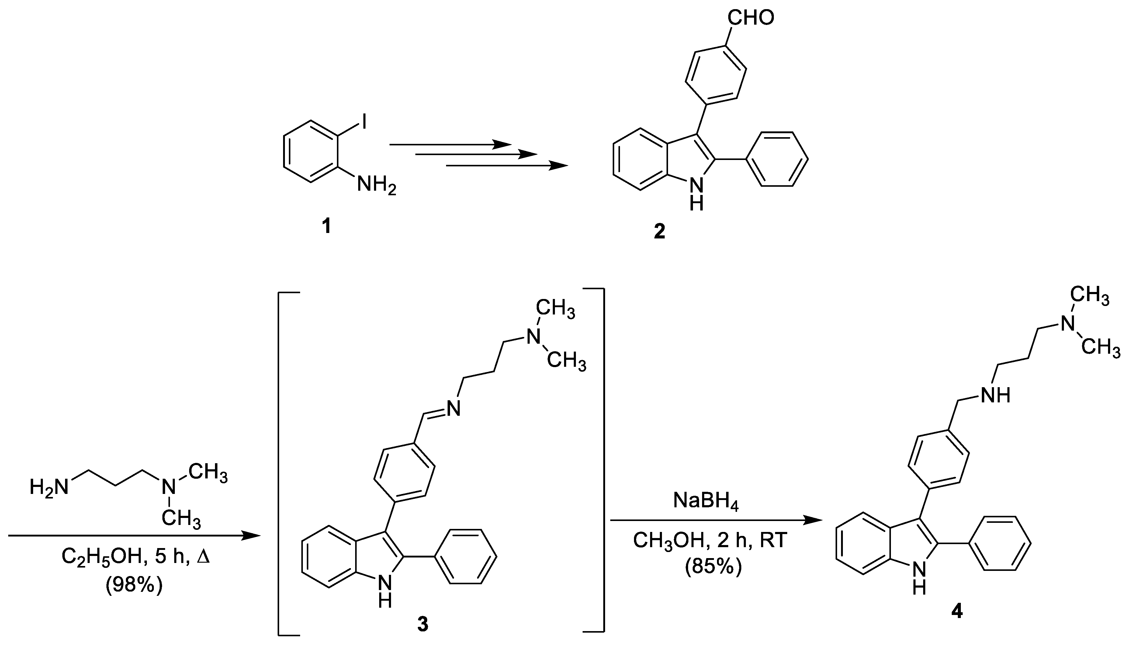
Scheme 1. Synthesis of 1H-3-{4-[(3-dimethylaminopropyl)aminomethyl]phenyl}-2-phenylindole (4).
Molbank 2019 m1060 sch001
Figure 1. The ORTEP drawing of the 2-phenyl-3-(4-formylphenyl)indole (2) with thermal ellipsoids at 30% level.
Molbank 2019 m1060 g001
Table 1. In vitro sensitivity of Plasmodium falciparum, Leishmania donovani and Trypanosoma brucei brucei strains to compound 4 and cytotoxicity of these compounds in HepG2 cells.
CompoundP. falciparum strains
IC50 Values (μM) a
L. donovani
IC50 Values (μM) b
Trypanosoma brucei brucei
Strain Antat 1.9
IC50 Values (μM) c
Cytotoxicity to HepG2 Cells
CC50 Values (μM) d
W23D7
Chloroquine e0.40 ± 0.040.11 ± 0.01n.d. hn.d. h30
Mefloquine e0.016 ± 0.0020.06 ± 0.003n.d. hn.d. hn.d. h
Pentamidine fn.d. hn.d. h5.5 ± 0.80.0002 ± 0.000062.3 ± 0.5
Amphotericin B fn.d. hn.d. h0.25 ± 0.01n.d. h8.8 ± 0.6
Suramine gn.d. hn.d. hn.d. h0.03 ± 0.003n.d. h
Fexinidazole gn.d. hn.d. hn.d. h0.59 ± 0.039n.d. h
Eflornithine gn.d. hn.d. hn.d. h15.19 ± 0.64n.d. h
Doxorubicinn.d. hn.d. hn.d. hn.d. h0.17 ± 0.03
41.80 ± 0.101.10 ± 0.123.03 ± 0.200.22 ± 0.031.14 ± 0.10
a Values were measured against the chloroquine-resistant and mefloquine-sensitive strain W2 and the chloroquine-sensitive and mefloquine-resistant strain 3D7. b IC50 values were measured against the promastigotes of the Leishmania donovani strain. The IC50 (µM) values correspond to the means +/− standard deviations from three independent experiments. c IC50 values were measured against the slender bloodstream trypomastigotes of the Trypanosoma brucei brucei AnTat 1.9 strain. The IC50 (µM) values correspond to the means +/− standard deviations from three independent experiments with each concentration tested in duplicate in all experiments. d CC50 values were measured against HepG2 cells. The CC50 (µM) values correspond to the means +/− standard deviations from three independent experiments. e Chloroquine and mefloquine were used as antiplasmodial compounds of reference. f Pentamidine and amphotericin B were used as antileishmanial compounds of reference. g Suramine, pentamidine, fexinidazole and eflornithine were used as antitrypanosomal compounds of reference. h n.d.: not done.