Review

Comparative Molecular Insights and Computational Modeling of Multiple Myeloma and Osteosarcoma

by
Alina Ioana Ghiță
1,2,
Vadim V. Silberschmidt
1 and
Mariana Ioniță
1,3,4,*
1
Faculty of Medical Engineering, National University of Science and Technology Politehnica Bucharest, Gheorghe Polizu 1-7, 011061 Bucharest, Romania
2
Faculty of Veterinary Medicine, Spiru Haret University, Basarabia 256, 031121 Bucharest, Romania
3
Advanced Polymer Materials Group, National University of Science and Technology Politehnica Bucharest, Gheorghe Polizu 1-7, 011061 Bucharest, Romania
4
Ebio-Hub Research Center, National University of Science and Technology Politehnica Bucharest-Campus, Iuliu Maniu 6, 061344 Bucharest, Romania
*
Author to whom correspondence should be addressed.
Int. J. Mol. Sci. 2026, 27(8), 3611; https://doi.org/10.3390/ijms27083611
Submission received: 12 March 2026 / Revised: 9 April 2026 / Accepted: 16 April 2026 / Published: 18 April 2026
(This article belongs to the Section Molecular Oncology)

Abstract

Multiple myeloma (MM) and osteosarcoma (OS) are two biologically distinct osseous malignancies with similar molecular networks that present translational challenges for their computational modeling. This comparative research analyzes MM and OS biology relevant to in silico approaches, focusing on PI3K-AKT-mTOR signaling, the RANK-RANKL-OPG axis, angiogenic factors (VEGF, TGFs), and immune mediators in MM, alongside the transcription factors (SOX9, RUNX2), signaling pathways (PI3K-AKT-mTOR, NOTCH), immune cell state (TAM2), and interleukins in OS. Based on this pathophysiologic foundation, the review outlines five computational paradigms: (i) mechanistic models; (ii) data-driven/machine learning schemes; (iii) hybrid mechanistic approaches; (iv) digital twins/virtual cohorts, and (v) MIDD/PBPK models for real-world applications. A cross-cancer comparison section summarizes common and distinct biological axes and their computational translation as well as the overlapping features from the bone microenvironment. For both MM and OS, the research assesses strengths, limitations, and data needs of current models, outlining the strategic objectives for next-generation multiscale, AI-enabled models providing a roadmap for tissue engineers, oncology scientists, and translational researchers to design clinically relevant preclinical tests and accelerate safer, more effective strategies for tumor-affected bones. The differences between MM and OS impose distinct biological constraints, so their comparisons are rare. Combining all these features with artificial intelligence capabilities will underpin a promising transition in the development of in silico adaptive and learning models.

1. Introduction

The spatial disposition and dynamic arrangement of cells govern their response to various factors and are fundamental to eliciting their functions [1]. For instance, bones are responsible for mechanical support, and also act as the cellular repository, being the source of both hematopoietic and osteogenic cells [2]. Bone marrow hosts hematopoietic stem cells that can develop into blood lineage cells and mesenchymal stem cells that generate osteogenic, chondrogenic, and adipogenic cell lineages while also supporting the marrow microenvironment [3]. Additionally, bones elicit some of the most fundamental immunological mechanisms that modulate homeostasis and engage in the pathophysiology of specific disorders. Stemming from their high plasticity and fast adaptive response, bones usually fulfill biological requirements, except under disease-related stimuli [4]. Still, regardless of their regenerative capability, bones remain a frequent site for cancerous conversion, demonstrating a delicate balance of their architectural, renewal, and immunological activities and their susceptibility to malignancy [5].
Considering the latest tactics of disease detection and therapy, bone cancers are still a continuing challenge for orthopedics and hematology–oncology [6]. Their tropism for either matrix or marrow is due to the abundance of cellular and molecular resources that sustain their proliferation and growth. Malignant development alters the homeostasis between bone resorption and regeneration, resulting in multifaceted neoplastic foci, notably lytic, blastic, or concurrent variants [7].
In oncology, in silico strategies are progressively establishing themselves as a cohesive connection among biological data, mechanistic understanding, and clinical decisional making, facilitating multiscale modeling, virtual trials, and precision therapeutics that underpin complex computational frameworks discussed in this research [8].
Multiple myeloma (MM) and osteosarcoma (OS) were selected among bone malignancies due to their significant clinical burden and persistent unmet need in medical practice. MM is the second most prevalent hematological cancer [9], with a rising global incidence and an incurable nature, as most patients experience relapses or develop resistance to the treatment [10]. More than 80% of patients suffer from osteolytic lesions, and approximately 50% get fractures, leading to a high mortality rate (between 20 and 24% of the cases) [11]. Although OS is less common than MM, it is the most frequent primary bone tumor and is associated with a 5-year overall survival rate that can be as low as 30% in patients with metastatic diseases, despite substantial therapeutic advances over recent decades [12,13]. Notwithstanding their distinct cellular origins, MM and OS demonstrate interconnected aspects of bone cancer, both characterized by bone tropism and osseous destruction, which highlight their molecular complementarity and malignant bone–microenvironment crosstalk [14,15]. By examining both malignancies concurrently, this review aims to identify computational opportunities by elucidating the overlapping mechanisms that drive tumor–bone interactions, thereby facilitating the discovery of emerging preclinical strategies.
Although other clinically significant bone malignancies (e.g., Ewing sarcoma [16], and chondrosarcoma [17]) along with hematological cancers (acute myeloid leukemia [18], chronic lymphocytic leukemia [19], and non-Hodgkin lymphoma [20]), are included in the large oncological landscape, our current focus is on MM and OS.
Thus, the aim of this research is to present a combined, computational perspective on multiple myeloma (MM) and osteosarcoma (OS), two biologically related but unevenly explored bone malignancies, by (i) comparing their molecular and microenvironmental characteristics, including signaling pathways and immune crosstalk; (ii) methodically assessing mechanistic, data-driven/machine learning (ML), hybrid, digital-twin/virtual cohort, and MIDD/PBPK frameworks developed for each cancer; and (iii) using cross-cancer comparison to recognize the shared and distinct biological and in silico axes. By structuring current models for MM and OS, bone-specific affinity, and common pathways, this research seeks to clarify the areas where the present strategies already reinforce oncology and where more comprehensive approaches are required. The present work constitutes a structured narrative comparative review.

2. Pathophysiology of MM and OS Relevant to In Silico Modeling

Bone cancers, namely, multiple myeloma (MM) and osteosarcoma (OS), exhibit dynamic mechanisms, shaping their behaviors and clinical outcomes. These two malignancies represent a molecularly heterogenous group of cancers unified by their mutual bone involvement and their dependence on the osseous microenvironment, yet they are markedly distinct in their cellular origin and clinical behavior [20]. In computational modeling, this variability presents both challenges and opportunities. The complexity of tumor biology requires multiscale in silico methods, while common pathological pathways across different diseases underpin the feasibility of generalizable modeling approaches [21]. Aberrant bone reorganization through central signaling pathways, dysregulated intracellular signaling cascades, cell survival and differentiation, the microenvironment dynamic crosstalk of bone marrow, and genomic instability are among the key pathophysiological features relevant to computational modeling [22,23]. In this context, MM and OS appear as two complementary entities with a largely distinct yet partially overlapping molecular landscape: MM as a plasma cell-derived hematological cancer and OS as a mesenchymal-origin solid tumor. This strongly associated dependence on the bone marrow niche and aggressive bone destruction makes them suitable models for comparative computational frameworks [24,25].

2.1. Multiple Myeloma

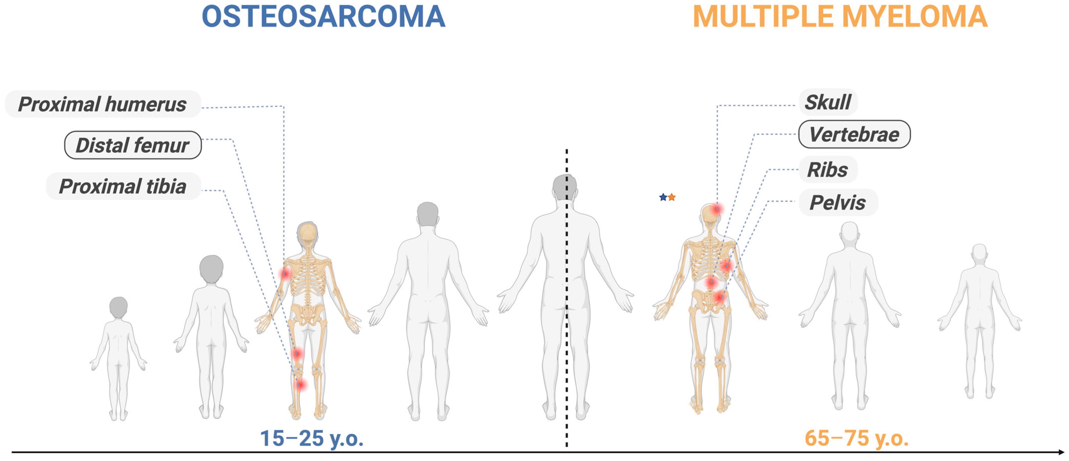
While comprising nearly 1% of all malignancies [26], MM accounts for approximately 10% of hematologic tumors [27], exposing its overrepresented impact and clinical relevance. Worldwide, MM impacts men at a higher frequency than women (55–57% of instances) and predominantly emerges in adults. The central age tendency is around 65–70-year-olds, followed closely by those aged 75 years, with a notable growth after 40 years [9]. The common sites of MM are the vertebrae and the pelvis area [10].
Multiple myeloma is characterized as a genetically heterogeneous neoplasm triggered in B cells [28]. The deviant clonal cellular expansion of plasma cells [29] resident in the bone marrow causes an overproduction of monoclonal immunoglobulin [30]. This phenomenon leads to organ malfunction along with bone lesions and additional anemia [31]. Atypical genomic rearrangements and hypermutations are driven by activation-induced cytidine deaminase (AICDA) [32]; this elicits DNA fragmentation that leads to instability and malignant transformation of B cells [10]. A substantial proportion of all MM cases evolve from an asymptomatic pre-malignant phase named monoclonal gammopathy of undetermined significance (MGUS) [33] or smoldering multiple myeloma (SMM) [34]. Some of the secondary injuries are related to chromosome alteration, notably deletion of 17p [del(17p)] or addition of 1q [gain(91q)] [35]. The sum of these abnormalities defines the therapeutic response and prognosis [9]. MM is influenced by both recurring activation of key intracellular pathways and cytokine signaling. Wingless/integrated (Wnt) signaling [36] and the phosphoinositide 3-kinase (PI3K) [37], protein kinase B (AKT) [38], and mechanistic target of rapamycin (mTOR) signaling axis pathways [39] are used to maintain survival, growth, and treatment tolerance [40].
The pathophysiology of MM is characterized by abnormal osteoclast activity [41]. Myeloma cells change the microenvironment, enhancing bone resorption while suppressing the osteoblast function. The receptor activator of nuclear factor κb (RANK)/receptor activator of nuclear factor κb ligand (RANKL)/osteoprotegerin (OPG) (RANK-RANKL-OPG) axis [23], the central regulatory axis of skeletal rehabilitation, drives osteoclast activation while osteoblast differentiation is impeded due to sclerostin, activin, and wnt-β-catenin pathway inhibition [42].
Pro-angiogenic growth factors (PAGFs) [43], such as vascular endothelial growth factors (VEGFs), fibroblastic GF (FGFs), and hepatocyte growth factors (HGFs), are synthesized and released in MM, supporting clonal expansion and therapy tolerance [44]. Finally, proinflammatory interleukins (ILs) [45] and transforming growth factors (TGF-ß) [46] amplify the imbalance between bone formation and bone resorption, leading to the genetic reprogramming of osteogenic mesenchymal progenitors [10]. In MM, cancerous plasma cells reset the marrow niche [39]. This pattern is characterized by aberrant natural killer (NK) cells, regulatory T-cell proliferation, T-cell fatigue, an increase in myeloid-derived suppressor cells, and macrophage polarization into malignant phenotypes [47].

2.2. Osteosarcoma

Osteosarcoma (OS) is a bone malignancy characterized by mesenchymal lineage conversion, in which abnormal osteogenic differentiation initiates aberrant tumorigenic osteoid production [48]. OS is the most prevalent primary osseous neoplasm [1]. With a 35% incidence of all malignant skeletal tumors, it displays two peaks in the age distributions. One peak coincides with fast skeletal development and pubertal hormonal fluctuations in adolescence (10–30 years old). The following peak is in late adulthood (60–65 years old) [49]. As in MM, this tumor has a male predominance [6]. Almost 75% of the affected individuals are under 25 years old. The most common sites of tumors are the metaphysis of long bones, especially around the knee and proximal humerus [50].
An illustration of the temporal mapping of selected neoplasms across the human ontogeny is depicted in Figure 1.
Intrinsic genetic shifts and extrinsic microenvironmental signals activate tumorigenesis through osteoblast proliferation [51]. While the steps of mesenchymal differentiation remain less clearly defined than those in the hematopoietic system, several transcription factors are now recognized as key regulators of mesenchymal cell fate. A SRY-box transcription factor 9 (SOX9) [52], osterix, and a runt-related transcription factor 2 (RUNX2) [53] are fundamental factors that indicate the osteogenic cellular profile. Their presence and dynamics are directly governed by tumor suppressors and oncogenes, such as tumor protein 53 (TP53) [54], MYC proto-oncogene [55], and bHLH transcription factor [56]. Additionally, major neoplastic pathways such as PI3K-AKT-mTOR [57], Hedgehog [58], wnt/β-catenin [59], Janus kinase-signal transducer and activator of transcription (JAK-STAT) [60], and NOTCH [61] are heterogeneously activated in OS. They vary greatly among cancers and constitute an integrated system that controls differentiation, stress adaptation, and growth [62]. A specific category of OS stem-like cells supported by NOTCH, Hedgehog, and wnt/β-catenin signaling are identified by the presence of SOX2, octamer-binding transcription factor 4 (OCT4) [58], nanog homeobox transcription factor (NANOG) [63], and Kruppel-like factor 4 (KLF4) [64], involved in therapeutic resistance and tumor relapse [65].
The tumor evolves from a multipotent mesenchymal stem cell and is characterized by malignant cells that generate an unmineralized bone matrix, with a vast population displaying adipogenic, fibroblastic, and chondroblastic phenotypes [48]. The interplay between cells and their milieu is crucial for neoplasm development and involves direct contact between vascular, mesenchymal, and immune cells. In the early stages of the disease, the tumor growth impairs the osteoblast–osteoclast balance, mobilizing pro-tumorigenic factors [66]. OS cells with coexisting marrow populations reshape the architecture of the entire bone, producing the carcinogenic osteoid. The altered osteoid extracellular matrix stimulates malignant growth, invasion, and drug tolerance, promoting metastatic spread [67]. In this area, regardless of progress in therapy, outcomes stagnated. Yet, new approaches to tumor biology yield some promising results through molecular pathways and antigens [68]. OS has a substantial predilection for pulmonary metastasis. Its microenvironment is abundant in M2-like tumor-associated macrophages (TAM2) [69], coupled with C-C motif chemokine ligand 18 (CCL18) [70] and cyclooxygenase-2 phosphorylated signaling (COX2/p-STAT3) [71], correlated with cancer dissemination and a guarded prognosis [72]. This is an essential determinant of patient survival and remains a clinical challenge. Malignant cells guide mesenchymal stem cells (MSCs) via extracellular vesicles with tumor growth factor β (TGF-β), triggering Interleukin-6 (IL-6) production and activating a cascade that initiates metastatic lesions in the lung [73]. Extracellular vesicles from OS cells reset myofibroblasts and stem cells to metastatic seeding [74].

2.3. Shared and Distinct Biological Axes

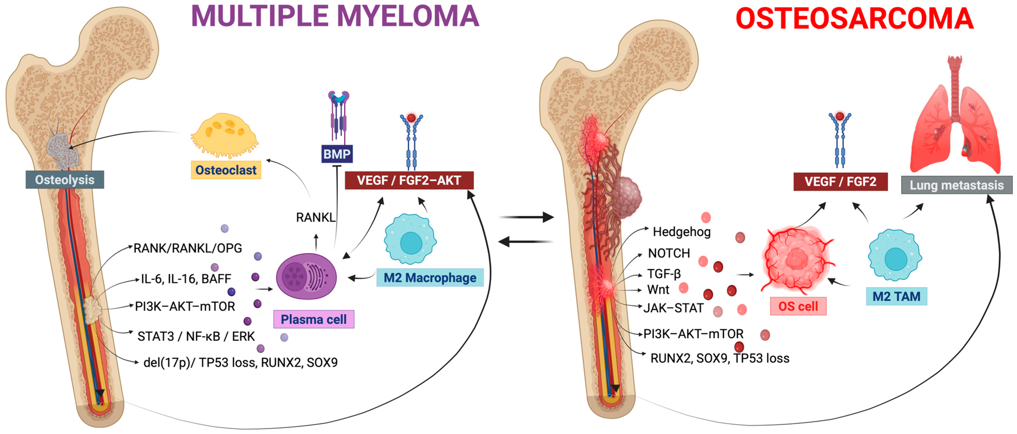
Although MM and OS are distinct, they share some common features, including RANK/RANKL/OPG activity, the PI3K-AKT-mTOR pathway, Wnt signaling, TGF-β tone, and IL-6/STAT3. MM is additionally marked by del(17p) and RUNX2 imbalance, while OS reveals TP53 loss, SOX9, JAK-STAT, NOTCH, and Hedgehog aberrant activation [75,76].
MM is characterized by a gradual genomic heterogeneity, with successive AID mutations, chymotrypsin rearrangement, and MYC activation [77]. OS features chromosomal instability, with TP53 and RB1 variations and chymotrypsin rearrangement [76]. In terms of genomic instability, these two malignancies share genomic instability via distinct patterns.
From an evolutionary point, MM commonly arises from MGUS/SMM. This early transcriptional alteration enables ribonucleic acid sequencing (RNA-seq), indicating clonal expansion [77]. In OS, the RNA-seq quantifies gene expression and can be used to infer cancerous stem cell-like (CSC) states through stemness-associated transcriptional signatures, in a temporal pattern [78]. Therefore, both cancers share evolutionary variation with stem-cell-like properties originating from mesenchymal or plasma-cell populations.
In the tumor microenvironment (TME) and immune reprogramming, MM actively reorganizes the bone marrow (BM) through CD8 cytotoxic T lymphocytes (CD8 T-cells), macrophages, and ligand–receptor pathways, such as HGF, macrophage migration inhibitory factor (MIF), IL-15, and B-cell activating factor (BAFF), from MGUS to MM [79]. OS development is induced by M2 TAMs triggered by hypoxia, STAT3/NF-kB/ERK signaling, and VEGF/FGF2-AKT angiogenesis. Single-cell transcriptomics indicates neoplastic clusters that release Wnt, TGF-β, and bone morphogenetic protein (BMP) ligands [80]. Across MM and OS, neoplastic dynamics are mediated by TME. Vascular pathways and stress stimuli govern the conditions, although they arise in different bone niches.
Figure 2 depicts the MM and OS biological networks and data context.

3. In Silico Modeling Frameworks for MM and OS

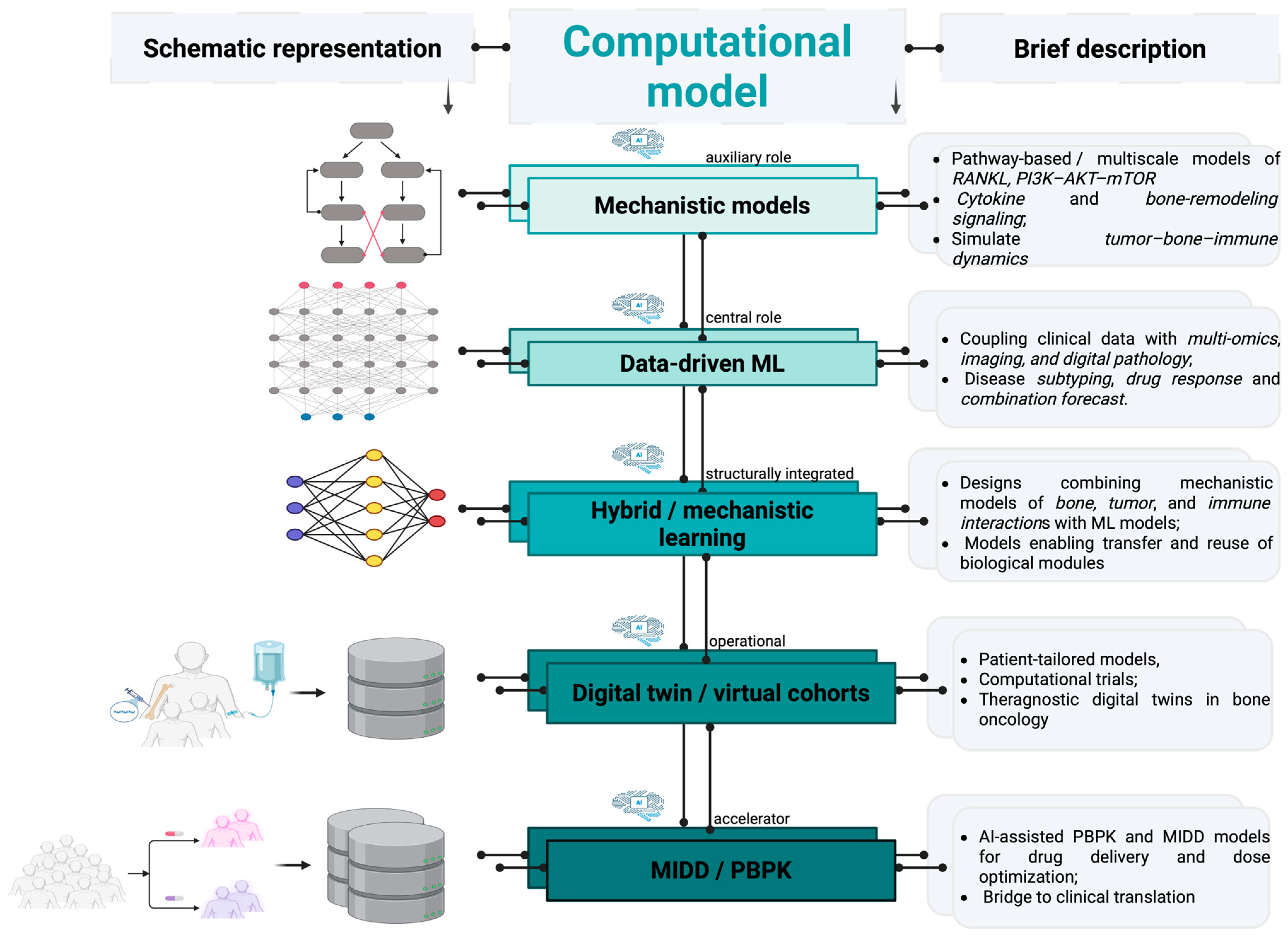
Considering the interconnected heterogeneity of MM and OS, namely, clonal dynamics, altered bone reorganization, and interconnected tumor–bone–immune niches, computational frameworks provide a safe strategy to assess challenging hypotheses for both preclinical (in silico, in vitro, ex vivo, and in vivo) and clinical scenarios [81,82]. The following section addresses five computational paradigms applied to MM and OS within the cross-disease framework developed in this review.

3.1. Mechanistic Models

Mechanistic models emphasize present biological, chemical, or physical processes, e.g., tumor growth dynamics or pharmacological modulation, as equations, enabling a personalized, patient-specific approach [83]. These frameworks combine deterministic or stochastic representations rooted in biological architecture, such as quantitative systems pharmacology (QSP), and models employing ordinary differential equations (ODEs) and partial differential equations (PDEs) for neoplastic expansion, osseous reorganization, or immune-mediated cancer interactions. Mechanistic frameworks are implemented for causal explanation or hypothesis synthesis. In MM and OS, such models were employed to address bone–tumor crosstalk, such as RANK-RANKL-OPG signaling, clonal proliferation, therapeutic outcome, or growth factors in the bone marrow milieu [82]. The models developed for MM address marrow-level kinetics and cytokine feedback, along with the progression from precursor stages to active MM, justifying their inclusion in this classification [84,85,86]. Alternatively, mechanistic hypotheses of osteoblast–osteoclast dynamics, osteoid spatial proliferation, and metastatic spread are extensively validated for OS [87,88].

3.2. Data-Driven/Machine-Learning (ML) Models

In these models, extensive scientific data resources and predictive algorithms are implemented to identify patterns and generate predictions with context-dependent mechanical assumptions [89]. Data-driven frameworks, e.g., classical ML and deep learning, are implemented to facilitate quantitative prediction but lack causal interpretability. Machine and statistical learning, pharmacokinetics (PK), and response-predictive designs are compatible with this modeling type. Lacking a clear mechanistic design, these schemes instantly recognize patterns in clinical, imaging, molecular, or histopathological data. Each of the two considered malignancies provides representative examples in therapeutic responses, outcome forecasting, and multi-omics subgrouping [90]. For MM, this approach can predict the overall survival time or therapeutic outcome from the real-world data. For OS, besides forecasting, ML frameworks support radiomics-based diagnosis, staging, and metastasis occurrence [91].

3.3. Hybrid Mechanistic-Learning Models

This approach employs a hybrid or mechanistic-learning framework that combines the strengths of a mechanistic design with elements of data-driven schemes to optimize forecast efficiency, for example, for bone remodeling and tumor growth [92]. The core mechanistic components are expressly defined, quantitatively specified and data-constrained, or expanded by learning-based models; insufficient data availability can affect their predictive capability. The hybrid design results in a distinct modeling paradigm. Both shallow and deep integration can be used as modeling approaches. In the former framework, ML and mechanistic designs are merged gradually, while in the latter, the computational data are integrated directly or collaboratively refined [93]. Hybrid MM models commonly integrate mechanistic features of osseous-tumor dynamics with statistical data obtained from patients.
Such frameworks are implemented for causal explanation or hypothesis synthesis. For instance, the mathematical ODE framework (a computational model that describes the capability of a system to change over time based on tracking the state variables) characterizes how malignant cells and osteoclast crosstalk are calibrated to individual biomarker specificity [94]. An OS framework comprises osseous remodeling equations enriched with data-driven features to assess signaling pathways [95].

3.4. Digital-Twin/Virtual Cohort/“In Silico Clinical Trial” Models

This type of models builds by formulating a virtual patient or cohort trial to simulate a clinical trial result, thereby reducing reliance on experimental studies [96]. This approach is not based on a model but on a dynamically recalibrated, patient-tailored parameterization method enhanced with longitudinal data inputs [97]. To the best of our knowledge, such frameworks are currently relatively uncommon and conceptually different from static models. For MM, such models were implemented to a greater extent and combine longitudinal measurements of M-protein levels, therapy graphics, or skeletal markers to assess patterns of disease progression and relapse. For OS, this approach is mostly conceptual, using limited pilot studies. Some designs employ patient-tailored neoplastic growth models parametrized to imaging data with the aim to predict chemotherapeutic responses [98]. A virtual comparison between MM and OS on overlapping pathways, including RANK-RANKL-OPG signaling and PI3K/AKT/mTOR activation, can demonstrate how bone–tumor networks react to equivalent therapies [99,100].

3.5. Model-Informed Drug Development (MIDD)/Physiologically Based Pharmacokinetic-Type (PBPK) Models

Models of this type rely on computational frameworks (commonly pharmacokinetics/pharmacodynamics or patient-oriented) to establish doses, risk avoidance, and efficient early developmental decisions [101]. MIDD designs are defined by collective application context, namely, regulatory applicability, decision assistance, or translational relevance, rather than mathematical structure. In modern models, QST (quantitative systems toxicology), QSP, and ODE systems, pharmacokinetic/pharmacodynamic (PK/PD) designs, Bayesian frameworks (computational schemes that rely on applying probability to describe uncertainty while refining their data whenever novel knowledge arises), and ML methods can be used simultaneously [102]. PBPK designs define therapeutic absorption, drug metabolism, and clearance employing bone-tailored, anatomically accurate sites [103]. Together, they can calibrate the drug dosage, drug–drug interactions, assess risk–benefit outcomes, or design robust clinical trials [104,105,106].
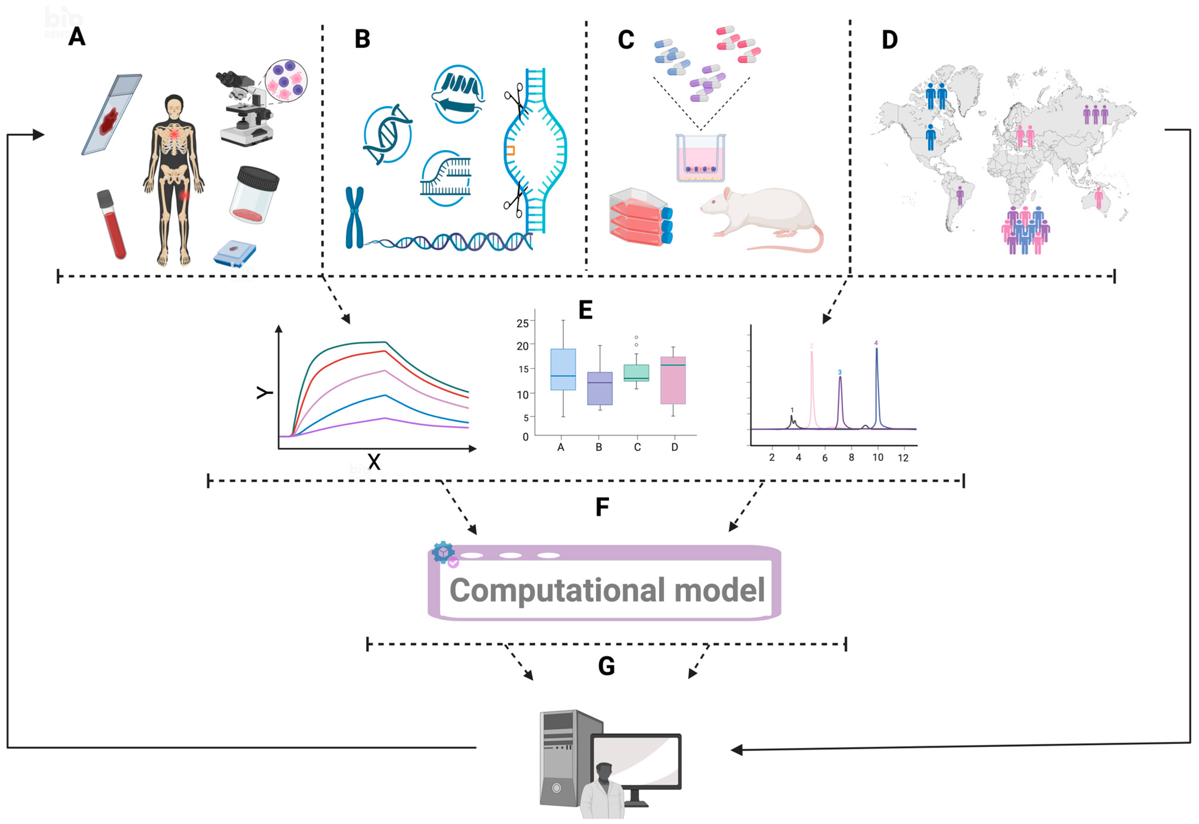
A conceptual framework that clarifies the integration and propagation of knowledge across the scales is necessary to justify the input of multicomponent data into computational disease modeling. Figure 3 presents such a system for in silico models.

4. Perspective of Cross-Cancer Comparison: MM Versus OS

Cross-cancer comparison augments the comprehension of the diseases’ patterns and therapeutic approaches. It should be stated here that the computational paradigms addressed above are disease-agnostic and analyze two physiologically different osseous neoplasms. MM and OS are typically approached, parameterized, and treated as independent entities. They vary in cell origin, clinical pathway, and microenvironmental interplay. In silico models addressed are, therefore, an opportunity to recognize the features that might be illustrated for these tumors without assuming a singular disease narrative.
After examining each modeling paradigm separately in Section 3, this part summarizes the cross-disease computational insights that arise through comparative application of the unified framework across MM and OS.

Computational Translations and Cross-Disease Insights

Mechanistic models of cancers add value through their causal structure encoding neoplastic and milieu frameworks that are optimal for RANKL-pathway osteolysis characterization, angiogenesis, or PI3K-AKT-mTOR signaling [107]. MM benefits from robust data on the bone marrow niche, enabling detailed multiscale tumor–bone–immune designs. This serves as the foundation for digital-twin models of MM [100]. In OS, the strength of such models stems from the well-defined pathways of knowledge, allowing analogous multiscale frameworks. One constraint of this scheme for OS is that the bone marrow compartment and immune modulators are poorly developed [108].
Data-driven and ML designs provide pathway patterns from clinical, omics (genomics, transcriptomics, proteomics, and epigenomics), and imaging knowledge without detailing the underlying software implementation [67,109]. For example, in MM, gene-expression-framed ML metrics categorize the subjects and predict the outcomes based on del(17p) and NK-kB biology. For OS, the strengths of this approach lie in radiomics based on computed tomography (CT), magnetic resonance imaging (MRI), positron emission tomography (PET), and deep learning. It forecasts therapeutic outcomes, diagnoses, or metastasis by merging VEGF/FGF2-AKT, BMP, and TME reorganization detected in imaging [110]. The limitation of this model are reduced cohorts, especially for OS, diminished interpretability, and low connection to explicit pathways.
Hybrid mechanistic-learning models provide a feasible strategy for ML to optimize bone-niche QSP frameworks, utilizing biological models, such as M2 TAMS or ILs, to predict multi-omics and therapeutic outcomes for patients [107]. For OS, the potential emerges from the reutilization of MM modules while allowing ML optimization of OS-specific patterns [111].
Digital twins and virtual cohorts offer value by supporting cross-oncology evidence [110]. In terms of MM, the strength is thanks to robust longitudinal data and validated QSP designs [96]. For OS, the rarity and heterogeneity of the disease provide an amenable background for digital-twin frameworks. One specific example may be the combination of this design and pan-cancer histopathology foundation approaches, such as Virchow’s. The Virchow model is an AI tool that explores limited data availability in rare cancers [112].
MIDD/PBPK models, employing AI-enhanced pharmacometric designs, enrich the computational approaches by translating data from niche biology and pathways to therapeutic outcomes, dose parameterization, or exposure responses. The value of this approach is its demonstration that ML models can support derivation of the PK/PD variables and ability to reinforce digital-twin models in terms of treatment-related toxicity and clinical efficacy across oncologic diseases [103].
The recent data [113] indicate that MM has an extended single-cell immunological database and longitudinal multi-omics, incorporating also large cohorts. Multi-omics and single-cell integrative research have also become more prevalent for OS, including integrative modeling that combines omics elements. Modern computational schemes supported by AI, such as large language models (LLMs) (data-driven neural frameworks used in probabilistic language formulation), can leverage these similarities to promote cross-disease optimization [107,114].
Figure 4 illustrates computational models integrating inputs from multiple myeloma and osteosarcoma.
Table 1 summarizes the common and distinct features relevant to computational modeling of MM and OS.
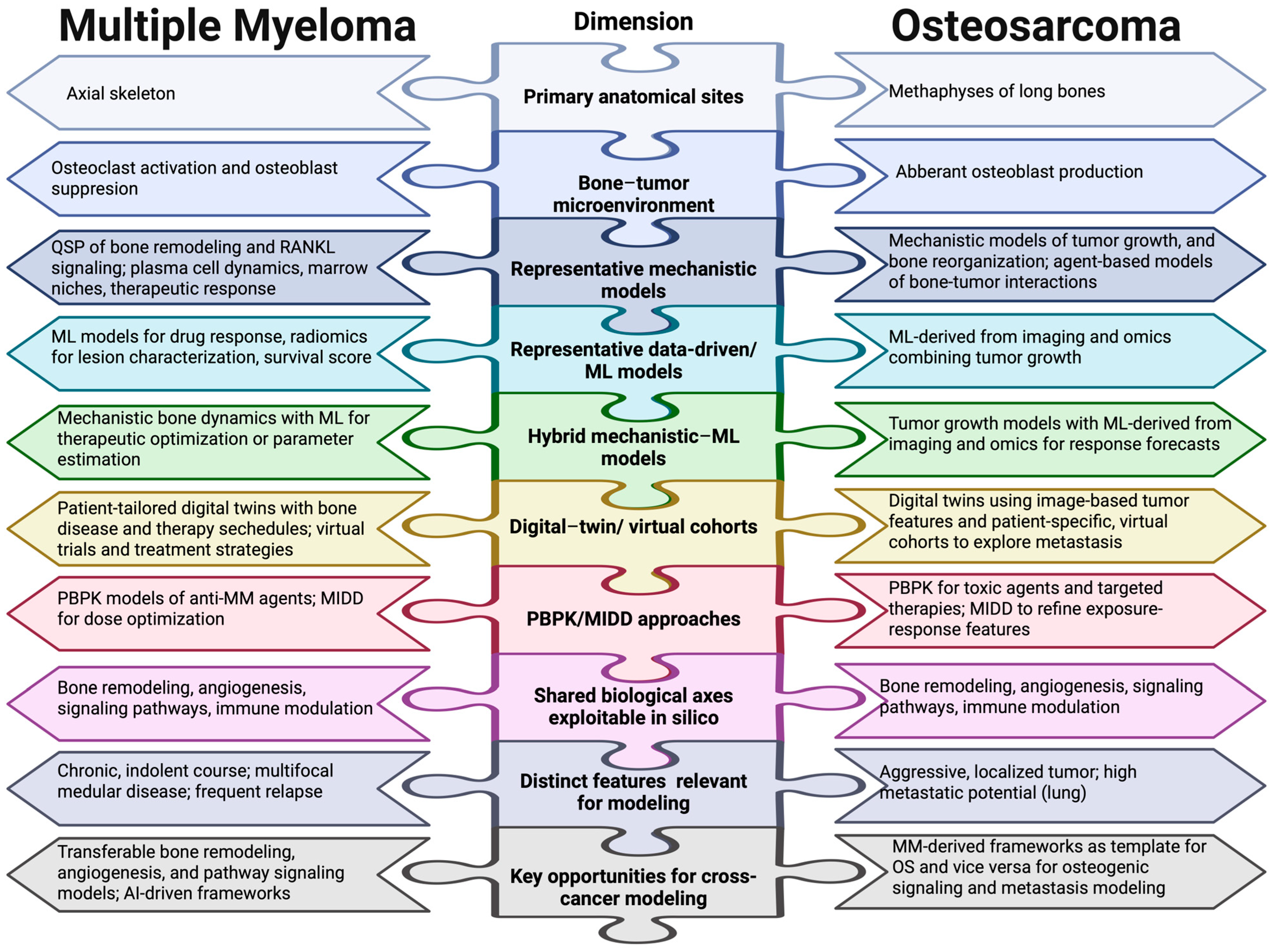
Table 1 provides a summary of features and approaches for MM aligned with OS among essential biological and computational dimensions. It correlates oncogenic networks, bone niches, immune and cytokine microenvironments, and datasets with their specific characteristics and current limitations. Figure 5 offers an illustration of the same dimensions for two distinct malignancies.
This schematic representation offers a side-by-side overview of MM and OS across ten key areas with high relevance for computational modeling. Pathophysiologically, osteoclast activation and osteoblast inhibition are the main pathways in which MM impacts the bone, affecting the axial skeleton [23]. Conversely, abnormal osteoblast synthesis triggers OS to arise in the metaphysis of long bones [22]. These malignancies represent two types of osseous dysregulation that are mechanistically different yet computationally convergent. From a modeling perspective, illustrative mechanistic frameworks for MM consist of Quantitative Systems Pharmacology models of bone reorganization and RANKL signaling [86]. Agent-based approaches of bone–tumor dynamics and tumor progression were used to address OS [88]. Therapeutic response prediction and radiomics-based lesion characterization in MM both benefited from the application of data-driven and ML models [90], as did imaging and omics integration for the modeling of OS [89]. Hybrid mechanistic–ML models, digital-twin approaches, and PBPK/MIDD strategies were established independently for each malignancy [96,136]. Most significantly, bone remodeling, angiogenesis, immunological regulation, and signaling pathways are exploitable axes shared by both illnesses, serving as the common computational ground for cross-disease modeling [21]. As previously mentioned, their unique features—the aggressive, localized, highly metastatic behavior of OS against the chronic, multifocal, relapsing course of MM—define the parameters of this comparison and influence the transferability of modeling frameworks between the two illnesses.

5. Discussion

In silico models represent a novel, methodological advance approach for simulation and assessment of tumor–bone interplay, prediction of therapeutic outcomes, and optimization of preclinical framework design. Such computational studies can deliver quantitative insights into illness progression, microenvironment dynamics, and therapeutic efficacy while reducing reliance on conventional in vitro and in vivo experimental models [137]. These designs offer a safe, cost-effective, and time-efficient alternative that minimizes the need for animal or human testing.
In the current research landscape, artificial intelligence (AI) has evolved as a cognitive platform, redefining how in silico models are developed. Still, AI does not establish new models; its impact is on the mechanistic function of the current frameworks by redefining interconnections between the model’s design, behavior, and reliability [98,138,139].

5.1. Critical Appraisal of Current Computational Approaches for MM and OS

Mechanistic, data-driven, and hybrid approaches have evolved significantly in oncologic research. Still, MM and OS continue to expose critical knowledge gaps. Mechanistic ODE/QSP/PKPD models include the established signaling cascades and drug–tumor interplay. For instance, QSP and PBPK designs regularly encode PI3K-AKT-mTOR and associated tumorigenic networks and are substantially used in MIDD. They can forecast efficacy and dose parameterization as well as simulate digital trials in bone malignancies [40,140]. For MM, computational and 3D models of the bone marrow niche simulate stromal adhesion, hypoxia, cytokine dynamics, and osteoblast–osteoclast interplay, incorporating the role of the PI3K inhibitory effect on bone resorption and remodeling [141]. Mechanistic immuno-oncology QSP frameworks consider elements of the tumor immunity cycle and are employed as a key checkpoint regulating T-cell activity in programmed death-1 (PD-(L)1) combinations among numerous cancers [100]. In 2023, Urdeitx et al. [142] described an agent-based/finite-element approach that integrated cell motility, proliferation, apoptosis, and signaling interactions of original bone tissue and stromal cells but in the MM-like context. Existing Wnt-mediated stromal cell signaling, and a detailed mechanistic description of the skeletal microenvironment hypothesized by Belik et al. [143] in the MM context are increasingly integrated into bone cancer models, namely, for OS. These frameworks outperform pathway-based mechanistic reasoning and experimental tests exploring inhibition strategies or niche-targeted regimens before the clinical assessment [144,145].
Despite their advanced complexity, important limitations persist in the use of mechanistic models. Immune-oncologic QSP frameworks face constraints with intra-tumoral heterogeneity of T-cell density and cytokines [146]. Comparative research demonstrates that ODE-based models can recreate dynamics yet fail to address spatial organization. The lack of these features is critical in immune checkpoint inhibitors that agent-based models may consider [147]. Clonal evolution and therapy tolerance reveal heterogeneous trajectories in MM with dysregulated metabolism and immune evasion post-treatment [148]. Furthermore, 3D myeloma frameworks identify distinct microenvironmental sensitivities in terms of extramedullary malignancy versus bone-marrow-restricted clones. Most models use simplistic interactions and cannot quantify robustly the involved mechanisms [149]. Regardless of their biological interpretability and capability to formulate testable hypotheses, numerous models were validated computationally, with limited independent experimental or clinical validation, which reduces confidence in their predictive accuracy [85,87], raising questions regarding repeatability and generalizability [107].
In contrast, data-driven ML and AI approaches have a significant value in analysis of skeletal cancers. ML designs, calibrated on radiomics, histopathology, or multi-omics, provide important data on biomarker discovery or immune milieu characteristics from digital slides. However, they rely strongly on very large, well-annotated datasets while being affected by limited interpretability [140]. These models have difficulty representing the pathway structures, such as RANK-RANKL-OPG, Wnt, or NOTCH, and extrapolating beyond the observed therapeutic regimens [150]. Liquid-biopsy-driven ML approaches describe circulating cell-free tumor DNA kinetics, yet they do not integrate the bone niche reorganization. These strategies are pan-cancer approaches applicable to solid tumors, such as OS [151]. Although ML frameworks for both malignancies showed promising discriminative outcomes, most of them were developed on small, single-institution cohorts. Without external validation and insufficient compliance with predetermined reporting guidelines, their reproducibility remains limited [152,153].
Analytical reviews reveal a small number of real hybrid models, where mechanistic frameworks and ML are highly interconnected [82], particularly those that combine MM and OS. ML enables estimation of, and introduction of constraints for, parameters with limited measurability, such as tumor immune infiltration or cell-state fractions from imaging data or single-cell knowledge. This information is further integrated into a mechanistic model, and the therapy simulation is initiated [154]. Mechanistic designs can also derive digital cohorts or virtual patients to amplify the limited clinical datasets for training of ML predictors [155]. Despite this, hybrid models continue to be technically demanding, constrained by data harmonization, with limited prospects of their validation [156]. The degree to which the mechanistic component restricts model behavior instead of serving as a regularization layer is rarely evaluated through ablation analysis. A standardized validation structure for this class of models in oncology has yet to be clearly defined [82,111].
Digital-twin and virtual-patient frameworks represent an evolving paradigm, remaining mostly experimental. In 2024, an evidence mapping review in oncology identified only 30 digital-twin studies [157]. Most of them utilized synthetic or low-sample datasets, with limited clinical validation. A real-time knowledge or ML-driven customization was scarcely observed [158]. Theoretical and regulatory systems highlight their anticipated value in terms of trial parameterization and support of patient decision-making. However, they have deficiencies in data integration and credibility assessment [159]. Although conceptually promising, numerous digital-twin models for MM addressed here remain at the proof-of-concept stage. They are validated retrospectively on small patient cohorts without prospective evaluation against real clinical results. For OS, a dedicated digital-twin framework remains underdeveloped, underscoring the gap between their computational readiness and clinical requirements [100,138].
MIDD, PBPK, and semi-mechanistic PK-PD models provide a foundation for dose selection and schedule optimization in oncology research. They characterize the organ-level drug distribution and time exposure [107,160]. These designs usually address the biology and development of tumors in a simple manner [161], being insufficiently focused on MM or OS niche dynamics or immune modulation. With limited effectiveness for long-term tolerance patterns, high-level system models are required [162]. PBPK and MIDD approaches remain some of the most direct clinical translations among the frameworks addressed in this research. Still, they also have some limitations: MM models rely on pharmacokinetic parameters derived from early-phase datasets and may not reflect all patients’ outcomes [163]. Meanwhile, in OS, methotrexate models were developed for pediatric cohorts with limited applicability to adult patients [107].
Considering that MM and OS display an osteoclast-mediated bone loss, mechanistic and quantitative frameworks based on the RANK/RANKL/OPG-axis established for MM offer some of the most readily transferable approaches from MM to OS [164]. Similarly, PBPK and MIDD approaches validated for MM anti-resorptive agents may be adapted to optimize dosing in OS, where such frameworks remain at an early stage of development [165], particularly in addressing the pharmacokinetic and pharmacodynamic challenges presented by pediatric patients.
Radiomics-based ML frameworks for response predictions can be transferred from OS to MM thanks to the similar nature of osteolytic lesions and the increasing use of imaging for disease monitoring. Regarding neoadjuvant chemotherapy responses in OS, these processes achieved good accuracy. As a result, they may be included in models for MM imaging responses [166,167]. Additionally, current studies are focused on the use of 3D, agent-based, or spatial models to replicate the OS tumor microenvironment architecture. The development of MM bone marrow niche frameworks can be influenced by these models. This can be especially important to illustrate the spatial dynamic among immune populations, stromal components, and plasma cells [146,168].
Overall, for MM and OS, current computational models robustly encode signaling pathways and the bone microenvironment. Still, they underrepresent complex immune cell dynamics, exemplified through Treg cells, myeloid populations, or CD8+ T cells [116,169], or therapy-driven trajectory and metastatic transitions [170,171,172].

5.2. Integrating AI and Multimodal Data Across MM and OS

Recent available data on multimodal deep learning demonstrate that MM and OS frameworks can shift from single-mechanism paradigms by merging omics, pathology, medical imaging, and clinical data into a unified patient representation [173]. Recent reviews of the use of artificial intelligence for biomarker identification and immune checkpoint inhibitors (ICIs) stress that single dataset types, namely, proteomics, genomics, and radiomics, partially represent tumor heterogeneity [174]. Recent models can support MM and OS approaches with rich multimodal AI layers [175].
Large-scale foundation frameworks, namely, Virchow [113] or PathOrchestra, indicate that self-supervised training of a substantial number of whole-slide images produces generalizable slide embeddings for pan-cancer or rare-cancer detection, biomarker forecasting, and reconstruction of gene expression profiles [113]. The transformer-based pathology Image and Text Alignment Network (TITAN) [176] expands this knowledge to the multimodal pathology–image–text alignment, facilitating identification of rare cancers, survival forecasts, and report generation with limited labeled data. Key features like these may be adopted in MM/OS digital twins as pathology encoders, modeling bone microenvironments and immune patterns that recent mechanistic models represent with limited fidelity.
Throughout oncology, ML systems are commonly used to guide prognostic and predictive biomarkers. Multi-level knowledge outperforms single omics but requires complex ML tools [177]. Frameworks such as ML-based consensus prognostic signature (MLPS) [178] or AI-driven multi-omics profiling [179] could identify niche-dependent mechanisms for common constraints. Flexynesis [180], a deep learning tool for precision oncology, lies at the foundation of advanced models. In MM, radiomics, AI, PET/CT and MRI previously linked imaging with bone tumor mechanisms to improve forecasts and therapeutic outcomes and to create powerful digital-twin models [181]. Radiomics and AI also supported the OS models to predict neoadjuvant chemotherapy and patient outcomes [166]. In OS, the Artificial Intelligence-Derived Prognostic Index (AIDPI) [182] provides an efficient example of AI integration that can identify abnormal metabolic and signaling pathways, namely PI3K-AKT-mTOR, supporting the generation of mechanism-informed therapeutic hypotheses.

5.3. Positioning Digital Twins and Virtual Cohorts for MM and OS

Population (pop) PK/PD and PBPK frameworks mostly quantify the absorption, distribution, metabolism, and excretion (ADME) processes [183] without considering specific individuals. Based on them, popPK/PD designs for MM [163] and OS [160] play an established role. According to this research, virtual cohorts/patients correspond to ensembles of mechanistically customized individuals. Their features can be depicted from clinical or omics datasets [184]. For individual patients, digital twins provide dynamic, continuous, and bidirectional connections with a physical person optimized with multimodal disease data, such as CERTAINTY [185], a pioneering virtual twin used for personalized cancer treatment in CAR-T-cell therapy for MM. Theragnostic digital twins (TDTs) [186] provide an example of bidirectional updating by integrating PBPK approaches with radiopharmaceutical features into a patient-tailored framework that allows optimization of agent selection. This type of refined concept can also be implemented in MM and OS modeling.
MM and OS are promising illnesses for evaluation of these methods thanks to their adequate anatomical characterization and medical imaging, allowing strong analysis at the population level. At the same time, MM is a heterogeneous disease, while OS is a rare malignancy, leading to their compatibility in digital-twin and virtual-cohort modeling.

5.4. Limitations of Current AI and Hybrid Models for MM and OS

Most AI cancer models are developed on limited, single-centered, retrospective cohorts, with limited population-based diversity, resulting in model overfitting and a lack of external validity [187]. For MM, radiomics and AI prognostic models were trained on less than 100 patients, so the outcomes were considered preliminary and not yet applicable in standard practice [188]. MRI-based 3D convolutional neural network (3D-CNN) risk frameworks are constrained to modest single-country datasets, highlighting concerns of unreliable calibration [189]. For OS, several radiomic ML models are developed on small cohorts, demanding multicenter validation prior to clinical implementation [182].
Deep learning frameworks for oncologic imaging and biomarker identification commonly lack transparency or biological plausibility. This methodological opacity raises concerns about safety, including unnecessary therapeutic augmentation for MM or high-toxicity protocols for OS [190,191].
The conceptual and methodological inability to accurately distinguish between prognostic and predictive signals is another issue that is addressed. Numerous studies explore therapeutic cohorts before employing trial-like methods, then considering any result-associated trait as “predictive”, even if it could just represent baseline risk [166,192].
As discussed, several established models exhibit methodological flaws, especially in validation and reporting, which might compromise their therapeutic usefulness, according to the available research [193,194]. Analysis of advantages and limitations of computational models is given in Table 2.

5.5. Toward Next-Generation MM and OS AI

Recent MM and OS approaches are moving toward AI-driven, multi-omics-integrated precision oncology, supporting more accurate prognosis and treatment selection [134,199,206].
Transparent Reporting of a Multivariable Prediction Model for Individual Prognosis or Diagnosis-AI (TRIPOD + AI) [153], Consolidated Standards of Reporting Trials-AI (CONSORT-AI) [154], and Standard Protocol Items: Recommendations for Interventional Trials-AI (SPIRIT-AI) [207] represent three international reporting standards integrating artificial intelligence in clinical research. Application of these reporting guidelines and validation criteria to MM and OS can be critical for bias mitigation and performance assurance, uncovering nonlinear configurations that remain invisible to conventional models.
COMpass [208], a geometric graph neural network (GGNN), provides another next-generation computational model utilized for MM that surpasses established methods. This approach adds value by integrating multi-omics data with pathway networks to predict survival.
Mistral-7B + retrieval-augmented generation (RAG) [209], a specific model built for MM, integrates RNA-seq, genetics, and clinical knowledge, refining patient-tailored therapeutic models.
In the area of OS, one specific AI-driven pathway-anchored model is the AIDPI framework, based on multi-algorithm ML, that was suggested to expose metabolic vulnerabilities and provide pathway-optimized therapies [182].

5.6. Limitations of This Review

To adequately contextualize the future perspectives and conclusions with the rapidly evolving research on bone cancers and computational oncology, it is necessary to indicate some of the limitations encountered in this review.
First, biological knowledge, molecular features, and pathophysiological frameworks relate specifically to MM and OS. Therefore, the generalizability of the findings to other bone malignancies cannot be assumed without disease-tailored validation.
Second, the computational approaches were reviewed in the context of MM and OS. This constraint reflects a deliberate attempt to maintain analytical coherence within a unified computational approach. As a result, other primary bone malignancies or hematological cancers may differ and require independent assessment before extrapolation.
A third limitation reflects the imbalanced availability and maturity of data resources. While MM is supported by high-dimensional omics data and harmonized clinical-genomic cohorts, OS datasets remain relatively small, heterogeneous, and less standardized. Consequently, certain aspects of the comparative approaches remain better suitable for MM than OS. This also represents the main justification for this cross-disease comparative study and a topic for additional research in addressing the computational and biological differences between MM and OS.

6. Future Perspectives

This rapidly developing field still faces considerable challenges that will define its future progress. In mechanistic modeling, there is still limited data availability, particularly for OS. Hybrid data fusion with ML and AI can optimize the performance of models in terms of parameter estimation, calibration, and predictive reliability [83].
With respect to the data-driven/ML models, multimodal deep learning combining omics with imaging and pathological features may be a novel approach [109].
Digital twins and virtual cohorts face a limitation in terms of standardized multi-omics, imaging, or epidemiologic datasets. Ethical and privacy concerns should be also addressed, along with validation constraints. Future developments may take into consideration a dynamic framework with data fusion that provides a recurrent parameter updating [210].
For MIDD and PBPK models with AI capabilities, LLMs may offer an assistive approach throughout pharmacometrics modeling designs, consolidating a hybrid mechanistic–human–AI direction [211,212].
From another perspective, MM and OS models can be re-architected from distinct pathologies. One example may be bladder cancer models [213], by creating an analogous application of a graph-based multimodal framework integrating a hematoxylin and eosin (H&E) histopathological stain (for cellular and tumor morphology) and RNA sequencing (to quantify gene expression). Another example may be from HPV-positive oropharyngeal cancer [214]. Despite being validated for another tumor, it may permit the transfer of disease-tailored features from CT/MRI radiomics and histopathological examination of the tumor while retaining cross-modal features.
A highly promising area of inquiry for MM and OS is the integration of spatial transcriptomics (ST) data into in silico modeling. This ST approach maintains tissue architecture and cell–cell dynamics, the key features of tumorigenesis and progression [215,216]. By incorporating such spatially resolved multi-omics, a feasible approach may clearly encode tumor–microenvironment heterogeneity or metastasis interconnections in MM and OS.
Given the fact that MM and OS have smaller cohort sizes than typical epithelial malignancies, the development of foundation models pre-trained on extensive multi-omics and imaging data may be pertinent. Through transfer learning, these self-supervised models built on large data may be tailored to illnesses, allowing for reliable predictions and biological understanding even in malignancies with limited datasets [217,218,219].
A relatively understudied yet feasible direction for OS is cross-species computational approaches that couple human, canine, and murine transcriptomic datasets. A rationale for adopting such models is the similarity of genetics, histology, and metastatic patterns in both species while canine OS is more common than human OS [220,221]. Embedding these cross-species cellular and microenvironmental datasets into computational approaches may provide a key method to validate spontaneous OS from dog models and genetically engineered murine OS with human models [222].
A potential yet understudied strategy for MM and OS may be the generation of a multiscale digital-twin model that integrates patient-tailored, tissue-level bone biomechanics with molecular and cellular disease models. Both malignancies are associated with pathological fractures resulting from the changing mechanical competency of osseous tissues driven by bone remodeling [223,224]. Integrating such approaches in schemes for MM and OS by combining mechanobiological models with oncologic digital-twin designs may enable clinical predictions, such as local fracture risks or mechanical antiresorptive effects of implemented therapies [111,225].
Considering the limited incidence of MM and OS, individual centers rarely provide enough patient numbers for training robust in silico models. Federated learning (FL) frameworks trained locally at each institution may address the limitation by enabling multi-institutional modeling [226,227]. Applied to MM and OS, FL infrastructures may build and validate models that generalize across institutions, health systems, and demographic groups [228].

7. Materials and Methods

This study presents a structured narrative review aiming to provide the first integrated comparative analysis of MM and OS through a computational modeling lens. As a structured narrative review, it does not follow a formal systematic review framework. Additionally, it does not aim for exhaustive or unbiased literature coverage.
This review was carried out to summarize the recent literature on computational modeling of bone cancers, with a specific focus on the cross-pathology transferability of multiple myeloma and osteosarcoma. The authors primarily considered references from 2020 to 2026 to ensure the relevance of the findings. The research was conducted using Google Scholar, Web of Science, PubMed, Scopus, and other resources. The focus was on bone cancer, multiple myeloma, osteosarcoma, computational models, and artificial intelligence. Specific search terms, or a combination of the following keywords were used to find pertinent research articles: “bone microenvironment”, “bone cancer”, “bone tumor”, “oncology”, “multiple myeloma”, “osteosarcoma”, “computational”, “in silico”, “artificial intelligence”, “mechanistic model”, “machine learning”, “digital twin”, “virtual cohort”, “PBPK”, “MIDD”, “RANK/RANKL/OPG”, “Wnt signaling”, “PI3K/AKT/mTOR”, “cross-cancer comparison”.
For each study, the focus was on the effects of disease pathophysiology on the mechanism generation and its results as well as the way to translate it in computational approaches. The reviewed computational models were classified according to their structure (mechanistic, ML, and hybrid), role (digital-twin and virtual cohort), and pharmacology context (MIDD/PBPK), permitting both MM and OS frameworks to be associated with multiple tags, e.g., mechanistic-QSP and digital twin.
The criteria for inclusion and exclusion into this research were based on the applicability of the studies to the molecular characterization, disease mechanism, diagnostic classification, or therapeutic approach of MM and OS. Regarding tumor types, the inclusion criteria were limited to MM and OS. These two malignancies benefit from extensive multi-omics characterization and well-known dependence on the bone microenvironment [229,230,231]. Articles that employed in silico modeling, molecular profiling, computational bioinformatics, and AI-driven methods were prioritized, in addition to other methodologies tailored to these two cancers. Studies were considered eligible if they met the following criteria: (i) peer-reviewed original research articles, reviews, or methodological papers published in English; (ii) addressing the pathophysiology, molecular biology, genomics, or tumor microenvironment of MM or OS; (iii) describing, applying, or evaluating computational, mathematical, or in silico modeling approaches in the context of MM, OS, or bone malignancies.
Publications were excluded based on the following criteria: (i) if they were published before 2020; (ii) if they addressed other malignancies than MM or OS (for example, Ewing sarcoma or chondrosarcoma were excluded due to their multi-omics and integration deficit, limiting the development of robust and replicable in silico approaches. Additionally, they are defined by key molecular factors that describe more lineage-specific oncogenic pathways, such as EWSR1::FLI1 fusion oncoprotein in Ewing sarcoma [232] and IDH1/IDH2 mutations in chondrosarcoma [233]. These molecular features are not directly comparable with the pathway aberrations of MM and OS. In a similar way, other hematological cancers, including acute myeloid leukemia, chronic lymphocytic leukemia, or non-Hodgkin lymphoma, were excluded since they do not primarily involve bone destruction or remodeling, the main biological axis underlying this comparative study); and (iii) if they described computational models that were not linked to bone diseases.
Next, the included studies were divided into distinct themes based on the tumor type and computational method. An additional focus was on common biological features and computational strategies that can be transferred from one disease to another. Strengths and limitations of the reviewed studies were also addressed, although there is some imbalance in the depth of available knowledge between the two malignancies. This work employs a qualitative, conceptually driven synthesis organized by theme across the manuscript.

8. Conclusions

This review provides a critical comparison of links between computational models, human cancer research, and the clinical pathophysiology of a disease for multiple myeloma and osteosarcoma. They are two distinct malignancies that share common features, providing a unique opportunity to enable the transfer and integration of approaches across cancer domains. From this point, it can reinforce both joint analysis and disease-specific research, which, in turn, can advance the understanding of each oncologic entity. To the best of our knowledge, this review represents the first structured comparative approach to computational strategies, specifically designed to bridge MM and OS, developing a conceptual and methodological basis for future in silico cross-disease oncology research.
MM can be considered as having a well-defined mechanistic basis regarding bone niche dynamics or pathway signaling, while OS is more considered with AI- and radiomics-based designs. Cross-disease comparative modeling can allow OS studies to integrate the validated bone marrow and immune designs originating from MM. At the same time, MM research can benefit from ML and imaging-informed frameworks implemented for OS. Hybrid mechanistic–ML approaches and digital twins may also be used as a convergence point since both cancers share the same pathways, for example, RANK/RANKL/OPG or PI3K-AKT-mTOR, becoming reusable modular computational components. A key observation from this comparative study is the significant computational asymmetry between these two malignancies. MM is supported by a more mature in silico modeling infrastructure, while OS has limited digital-twin approaches and validated ML frameworks. This gap in knowledge may benefit from cross-disease knowledge transfer.
Artificial intelligence, such as LLMs or foundational models, represents a powerful tool, not to replace any of the computational models, but to enhance capabilities to promote a deep, multimodal, and real-time update of current knowledge and support the advances towards precise forecasting in bone cancers. Nevertheless, their incorporation into MM and OS clinical procedures is still a developing approach that needs prospective confirmation prior to clinical use.
Most of the cross-disease computational frameworks addressed above are still in the proof-of-the-concept or early developmental stages. Their clinical translation will necessitate interdisciplinary cooperation among computational sciences, oncologists, and regulatory agencies as well as prospective validation and standardized multi-omics datasets. The concept that a unified cross-disease in silico approach to MM and OS has the potential to speed up treatment development and improve precision oncology for both cancers is supported by these data taken together.

Author Contributions

Conceptualization, A.I.G.; methodology, A.I.G., V.V.S. and M.I.; investigation, A.I.G.; visualization, A.I.G., V.V.S. and M.I.; resources, V.V.S. and M.I.; data curation, V.V.S. and M.I.; validation, V.V.S. and M.I.; supervision, V.V.S. and M.I.; project administration, V.V.S. and M.I.; funding acquisition, V.V.S. and M.I.; writing—original draft preparation, A.I.G.; writing—review and editing, V.V.S. and M.I. All authors have read and agreed to the published version of the manuscript.

Funding

This work was funded by EU’s NextGenerationEU instrument through the National Recovery and Resilience Plan of Romania—Pillar III-C9-18, managed by the Ministry of Research, Innovation, and Digitalization, within the project entitled “Advanced & personalized solutions for bone regeneration and complications associated with multiple myeloma”, contract no. 760093/23.05.2023, code CF 213/29.11.2022.

Data Availability Statement

No new data were created or analyzed in this study. Data sharing is not applicable to this article.

Acknowledgments

The figures in this review were created using BioRender.com under an academic license. The authors used ChatGPT (GPT-4o) by OpenAI and Claude (Sonnet 3.5) by Anthropic, to improve the organization, readability, and language of the manuscript.

Conflicts of Interest

The authors declare no conflicts of interest.

References

  1. Brar, G.S.; Schmidt, A.A.; Willams, L.R.; Wakefield, M.R.; Fang, Y. Osteosarcoma: Current insights and advances. Explor. Target. Anti-Tumor Ther. 2025, 6, 1002324. [Google Scholar] [CrossRef]
  2. Lecka-Czernik, B.; Rosen, C.J.; Napoli, N. The role of bone in whole-body energy metabolism. Nat. Rev. Endocrinol. 2025, 21, 743–756. [Google Scholar] [CrossRef]
  3. Yuan, G.; Lin, X.; Liu, Y.; Greenblatt, M.B.; Xu, R. Skeletal stem cells in bone development, homeostasis, and disease. Protein Cell 2024, 15, 421–438. [Google Scholar] [CrossRef]
  4. Ardelean, A.I.; Mârza, S.M.; Marica, R.; Dragomir, M.F.; Rusu-Moldovan, A.O.; Moldovan, M.; Pașca, P.M.; Oana, L. Evaluation of Biocomposite Cements for Bone Defect Repair in Rat Models. Life 2024, 14, 1097. [Google Scholar] [CrossRef]
  5. Rai, V.; Munazzam, S.W.; Wazir, N.U.; Javaid, I. Revolutionizing bone tumor management: Cutting-edge breakthroughs in limb-saving treatments. Eur. J. Orthop. Surg. Traumatol. 2024, 34, 1741–1748, Erratum in Eur. J. Orthop. Surg. Traumatol. 2024, 34, 3405. https://doi.org/10.1007/s00590-024-04026-1. [Google Scholar] [CrossRef]
  6. Hosseini, H.; Heydari, S.; Hushmandi, K.; Daneshi, S.; Raesi, R. Bone tumors: A systematic review of prevalence, risk determinants, and survival patterns. BMC Cancer 2025, 25, 321. [Google Scholar] [CrossRef] [PubMed]
  7. Choi, J.H.; Ro, J.Y. The 2020 WHO Classification of Tumors of Bone: An Updated Review. Adv. Anat. Pathol. 2021, 28, 119–138. [Google Scholar] [CrossRef] [PubMed]
  8. Kolla, L.; Gruber, F.K.; Khalid, O.; Hill, C.; Parikh, R.B. The case for AI-driven cancer clinical trials—The efficacy arm in silico. Biochim. Biophys. Acta (BBA)-Rev. Cancer 2021, 1876, 188572. [Google Scholar] [CrossRef]
  9. Rajkumar, S.V. Multiple myeloma: 2024 update on diagnosis, risk-stratification, and management. Am. J. Hematol. 2024, 99, 1802–1824. [Google Scholar] [CrossRef]
  10. Malard, F.; Neri, P.; Bahlis, N.J.; Terpos, E.; Moukalled, N.; Hungria, V.T.M.; Manier, S.; Mohty, M. Multiple myeloma. Nat. Rev. Dis. Prim. 2024, 10, 45. [Google Scholar] [CrossRef] [PubMed]
  11. Mukkamalla, S.K.R.; Malipeddi, D. Myeloma bone disease: A comprehensive review. Int. J. Mol. Sci. 2021, 22, 6208. [Google Scholar] [CrossRef]
  12. Yan, L.; Dong, X.; Chen, S. Advances in osteosarcoma research: Pathogenesis and emerging therapeutic strategies. Crit. Rev. Oncol. Hematol. 2026, 222, 105252. [Google Scholar] [CrossRef]
  13. Albany, F.; Farmer, S.H.; O’brien, A.C.; Feustel, P.J.; Dicaprio, M.R. Time to treatment initiation and overall survival in osteosarcoma: A national cancer database analysis. Bone Jt. Open 2023, 4, 522–530. [Google Scholar]
  14. Tian, H.; Cao, J.; Li, B.; Nice, E.C.; Mao, H.; Zhang, Y.; Huang, C. Managing the immune microenvironment of osteosarcoma: The outlook for osteosarcoma treatment. Bone Res. 2023, 11, 11. [Google Scholar] [CrossRef]
  15. Fotiou, D.; Katodritou, E. From Biology to Clinical Practice: The Bone Marrow Microenvironment in Multiple Myeloma. J. Clin. Med. 2025, 14, 327. [Google Scholar] [CrossRef] [PubMed]
  16. Dupuy, M.; Lamoureux, F.; Mullard, M.; Postec, A.; Regnier, L.; Baud’huin, M.; Georges, S.; Royer, B.B.-L.; Ory, B.; Rédini, F.; et al. Ewing sarcoma from molecular biology to the clinic. Front. Cell Dev. Biol. 2023, 11, 1248753. [Google Scholar] [CrossRef] [PubMed]
  17. Agulnik, M.; Wilky, B.A.; Thorpe, S.W.; Zuckerman, L.M. Chondrosarcoma: Clinical behavior, molecular mechanisms, and emerging therapeutic strategies. Crit. Rev. Oncol. Hematol. 2026, 218, 105075. [Google Scholar] [CrossRef] [PubMed]
  18. Shimony, S.; Stahl, M.; Stone, R.M. Acute myeloid leukemia: 2023 update on diagnosis, risk-stratification, and management. Am. J. Hematol. 2023, 98, 502–526. [Google Scholar] [CrossRef]
  19. Hallek, M. Chronic Lymphocytic Leukemia: 2025 Update on the Epidemiology, Pathogenesis, Diagnosis, and Therapy. Am. J. Hematol. 2025, 100, 450–480. [Google Scholar] [CrossRef]
  20. Guan, Y.; Zhang, W.; Mao, Y.; Li, S. Nanoparticles, and bone microenvironment: A comprehensive review for malignant bone tumor diagnosis and treatment. Mol. Cancer 2024, 2, 246. [Google Scholar] [CrossRef]
  21. Ramtani, S.; Sánchez, J.F.; Boucetta, A.; Kraft, R.; Vaca-González, J.J.; Garzón-Alvarado, D.A. A coupled mathematical model between bone remodeling and tumors: A study of different scenarios using Komarova’s model. Biomech. Model. Mechanobiol. 2023, 22, 925–945. [Google Scholar] [CrossRef] [PubMed]
  22. Yu, Y.; Li, K.; Peng, Y.; Zhang, Z.; Pu, F.; Shao, Z.; Wu, W. Tumor microenvironment in osteosarcoma: From cellular mechanism to clinical therapy. Genes Dis. 2025, 12, 101569. [Google Scholar] [CrossRef]
  23. Lungu, O.; Toscani, D.; Giuliani, N. Mechanistic insights into bone destruction in multiple myeloma: Cellular and molecular perspectives. J. Bone Oncol. 2025, 51, 100668. [Google Scholar] [CrossRef] [PubMed]
  24. Chen, X.; Tian, B.; Wang, Y.; Zheng, J.; Kang, X. Harnessing multi-omics to revolutionize understanding and management of osteosarcoma: A pathway to precision medicine (Review). Int. J. Mol. Med. 2025, 55, 92. [Google Scholar] [CrossRef] [PubMed]
  25. Wang, S.; Wang, S.J.; Pan, W.; Yi, Y.Y.; Lu, J. Construct prognostic models of multiple myeloma with pathway information incorporated. PLoS Comput. Biol. 2024, 20, e1012444. [Google Scholar] [CrossRef]
  26. Elbezanti, W.O.; Challagundla, K.B.; Jonnalagadda, S.C.; Budak-Alpdogan, T.; Pandey, M.K. Past, Present, and a Glance into the Future of Multiple Myeloma Treatment. Pharmaceuticals 2023, 16, 415. [Google Scholar] [CrossRef]
  27. Yu, T.; Jiao, J.H.; Wu, M.F. CAR-T cells in the treatment of multiple myeloma: An encouraging cell therapy. Front. Immunol. 2025, 16, 1499590. [Google Scholar] [CrossRef]
  28. Petcov, T.E.; Silberschmidt, V.V.; Pandele, M.A.; Chiticaru, E.A.; Ioniță, M.; Manole, M. Nanostructures: An efficient drug delivery platform for therapy of multiple myeloma. Eur. J. Med. Chem. Rep. 2025, 14, 100263. [Google Scholar] [CrossRef]
  29. Liang, Y.; He, H.; Wang, W.; Wang, H.; Mo, S.; Fu, R.; Liu, X.; Song, Q.; Xia, Z.; Wang, L. Malignant clonal evolution drives multiple myeloma cellular ecological diversity and microenvironment reprogramming. Mol. Cancer 2022, 21, 182. [Google Scholar] [CrossRef]
  30. Guedes, A.; Becker, R.G.; Teixeira, L.E.M. Multiple Myeloma (Part 1)—Update on Epidemiology, Diagnostic Criteria, Systemic Treatment and Prognosis. Rev. Bras. Ortop. 2023, 58, 361–367. [Google Scholar] [CrossRef]
  31. Charliński, G.; Vesole, D.H.; Jurczyszyn, A. Multiple myeloma in 2026-current guidelines and new therapies. Nowotw. J. Oncol. 2026, 76, 108723. [Google Scholar] [CrossRef]
  32. Kuliński, T.M.; Gewartowska, O.; Mahé, M.; Kasztelan, K.; Durys, J.; Stroynowska-Czerwińska, A.; Jedynak-Slyvka, M.; Owczarek, E.P.; Chaudhury, D.; Nowotny, M.; et al. Multiple Myeloma associated DIS3 mutations drive AID-dependent IGH Translocations. bioRxiv 2023. [Google Scholar] [CrossRef]
  33. Liu, Y.; Parks, A.L. Diagnosis and Management of Monoclonal Gammopathy of Undetermined Significance: A Review. JAMA Intern. Med. 2025, 185, 450–456. [Google Scholar] [CrossRef]
  34. Zanwar, S.; Rajkumar, S.V. Current risk stratification and staging of multiple myeloma and related clonal plasma cell disorders. Leukemia 2025, 39, 2610–2617. [Google Scholar] [CrossRef] [PubMed]
  35. De Novellis, D.; Scala, P.; Giudice, V.; Selleri, C. High-Risk Genetic Multiple Myeloma: From Molecular Classification to Innovative Treatment with Monoclonal Antibodies and T-Cell Redirecting Therapies. Cells 2025, 14, 776. [Google Scholar] [CrossRef] [PubMed]
  36. Wu, Y.; Luo, J.; Zhou, Y.; Lin, J.; Wu, Y.; Zheng, S.; Chen, J.; Che, F.; Wang, Q.; Zhong, L. Nestin in multiple myeloma: Emerging insights into a potential therapeutic target. Front. Oncol. 2025, 15, 1596928. [Google Scholar] [CrossRef]
  37. Janfada, M.; Vahdat, S.; Kaviani, S. PI3K Signaling Pathway Inhibitor Affects Myeloma Cells in A Culture-Dependent Manner. Adv. Pharm. Bull. 2025, 15, 440–452. [Google Scholar] [CrossRef]
  38. Muñoz, L.G.; Luna, S.P.; Chamorro, A.F. Plasma Cell Myeloma: Biochemical Insights into Diagnosis, Treatment, and Smart Nanocarrier-Based Therapeutic Development. Pharmaceutics 2025, 17, 1570. [Google Scholar] [CrossRef]
  39. Xiaoling, Z.; Qi, C.; Tingting, Y.; Di, H.; Meijia, Y. Mechanisms and intervention strategies of microenvironment-mediated drug resistance in multiple myeloma. Cancer Cell Int. 2026, 26, 100. [Google Scholar] [CrossRef] [PubMed]
  40. Glaviano, A.; Foo, A.S.C.; Lam, H.Y.; Yap, K.C.H.; Jacot, W.; Jones, R.H.; Eng, H.; Nair, M.G.; Makvandi, P.; Geoerger, B.; et al. PI3K/AKT/mTOR signaling transduction pathway and targeted therapies in cancer. Mol. Cancer 2023, 22, 138. [Google Scholar] [CrossRef]
  41. Mehdi, S.H.; Nafees, S.; Mehdi, S.J.; Morris, C.A.; Mashouri, L.; Yoon, D. Animal Models of Multiple Myeloma Bone Disease. Front. Genet. 2021, 12, 640954. [Google Scholar] [CrossRef] [PubMed]
  42. Roux, S.; Debiais, F.; Vieillard, M.H. Tackling myeloma bone disease: From pathophysiology to cutting-edge therapies. Blood Rev. 2025, 74, 101305. [Google Scholar] [CrossRef]
  43. Ribatti, D. Angiogenesis in Multiple Myeloma: 25 Years of Research in This Field. Eur. J. Haematol. 2025, 115, 516–532. [Google Scholar] [CrossRef]
  44. Melaccio, A.; Reale, A.; Saltarella, I.; Desantis, V.; Lamanuzzi, A.; Cicco, S.; Frassanito, M.A.; Vacca, A.; Ria, R. Pathways of Angiogenic and Inflammatory Cytokines in Multiple Myeloma: Role in Plasma Cell Clonal Expansion and Drug Resistance. J. Clin. Med. 2022, 11, 6491. [Google Scholar] [CrossRef]
  45. Luo, M.; Qin, L.; Li, Y.; Mei, Q.; Wu, Q.; Feng, X. Inflammatory markers from routine blood tests predict survival in multiple myeloma: A Systematic Review and meta-analysis. Front. Immunol. 2025, 16, 1669878. [Google Scholar] [CrossRef]
  46. Li, R.; Wei, F.; Yang, M. TGF-β in hematologic malignancies: Molecular functions and clinical applications. Front. Immunol. 2026, 17, 1728730. [Google Scholar] [CrossRef]
  47. Lopes, R.; Caetano, J.; Ferreira, B.; Barahona, F.; Carneiro, E.A.; João, C. The immune microenvironment in multiple myeloma: Friend or foe? Cancers 2021, 13, 625. [Google Scholar] [CrossRef]
  48. El Motassime, A.; Vitiello, R.; Comodo, R.M.; Capece, G.; Bocchino, G.; Bocchi, M.B.; Maccauro, G.; Meschini, C. Osteosarcoma: A Comprehensive Morphological and Molecular Review with Prognostic Implications. Biology 2025, 14, 1407. [Google Scholar] [CrossRef]
  49. Kim, C.; Davis, L.E.; Albert, C.M.; Samuels, B.; Roberts, J.L.; Wagner, M.J. Osteosarcoma in Pediatric and Adult Populations: Are Adults Just Big Kids? Cancers 2023, 15, 5044. [Google Scholar] [CrossRef] [PubMed]
  50. Robbins, G.; Vue, Y.; Rahrmann, E.P.; Moriarity, B. Osteosarcoma: A comprehensive review of model systems and experimental therapies. Med. Res. Arch. 2024, 12, 6000. [Google Scholar] [CrossRef] [PubMed]
  51. Chang, X.; Ma, Z.; Zhu, G.; Lu, Y.; Yang, J. New perspective into mesenchymal stem cells: Molecular mechanisms regulating osteosarcoma. J. Bone Oncol. 2021, 29, 100372. [Google Scholar] [CrossRef] [PubMed]
  52. Nakov, K.M.; Gething, M.E.; Kassis, T.K.; Rutt, G.J.; Ho, B.; Bhandari, N.; Collignon, T.E.; Banerjee, S.; Bishayee, A. Targeting signaling cascades by bioactive phytocompounds in osteosarcoma: A novel therapeutic approach. Pharmacol. Ther. 2026, 109014. [Google Scholar] [CrossRef]
  53. Magar, A.G.; Morya, V.K.; Noh, K.C. Molecular Crosstalk Between RUNX2 and HIF-1α in Osteosarcoma: Implications for Angiogenesis, Metastasis, and Therapy Resistance. Int. J. Mol. Sci. 2025, 26, 7642. [Google Scholar] [CrossRef]
  54. Shen, Y.; Huang, S.; Chen, G.; Wang, G.; Sui, L. Involvement of TP53 in osteosarcoma-challenges and prospects. Front. Oncol. 2025, 15, 1605080. [Google Scholar] [CrossRef]
  55. Tian, K.; Jiang, Y.; Sun, M.; Lv, Y.; Fan, J.; Sun, C.; Zhu, Y.; Yu, Y.; Qiang, X.; Peng, P.; et al. Targeting high-risk MYC-overexpressed osteosarcoma with an Aurora kinase inhibitor: -Results from a pilot umbrella trial. NPJ Precis Oncol. 2026, 10, 24. [Google Scholar] [CrossRef]
  56. Saba, K.H.; Difilippo, V.; Kovac, M.; Cornmark, L.; Magnusson, L.; Nilsson, J.; Bos, H.v.D.; Spierings, D.C.; Bidgoli, M.; Jonson, T.; et al. Disruption of the TP53 locus in osteosarcoma leads to TP53 promoter gene fusions and restoration of parts of the TP53 signalling pathway. J. Pathol. 2024, 262, 147–160. [Google Scholar] [CrossRef]
  57. Hu, D.; Yu, X.; Xu, J.; Li, B.; Ou, X.; Shi, S. Innovative gene targeted treatments for osteosarcoma: A mini review of current clinical evidence and prospects. Front. Med. 2025, 12, 1699287. [Google Scholar] [CrossRef]
  58. Zhra, M.; Akhund, S.A.; Mohammad, K.S. Advancements in Osteosarcoma Therapy: Overcoming Chemotherapy Resistance and Exploring Novel Pharmacological Strategies. Pharmaceuticals 2025, 18, 520. [Google Scholar] [CrossRef] [PubMed]
  59. Ding, Y.; Chen, Q. Wnt/β-catenin signaling pathway: An attractive potential therapeutic target in osteosarcoma. Front. Oncol. 2025, 14, 1456959. [Google Scholar] [CrossRef]
  60. Bashir, A.; Ismail, A.; Mavadia, A.; Ghose, A.; Ovsepian, S.V.; Boussios, S. Pathobiology and Molecular Pathways Implicated in Osteosarcoma Lung Metastasis: A Scoping Review. Technol. Cancer Res. Treat. 2025, 24, 1–18. [Google Scholar] [CrossRef] [PubMed]
  61. Tan, Y.; Gao, J. METTL16 promotes osteosarcoma progression by inducing m6A methylation of the UBE3A and Notch signaling pathway. Electron. J. Biotechnol. 2025, 78, 86–95. [Google Scholar] [CrossRef]
  62. Sun, Y.; Zhang, C.; Fang, Q.; Zhang, W.; Liu, W. Abnormal signal pathways and tumor heterogeneity in osteosarcoma. J. Transl. Med. 2023, 21, 99. [Google Scholar] [CrossRef]
  63. Yang, H.; Ji, J. SHP2 promotes osteosarcoma via regulating STAT3/TET3/HOXB2 signaling. Sci. Rep. 2026, 16, 6158. [Google Scholar] [CrossRef]
  64. Yadav, S.S.; Kalia, P.; Kaur, N.; Sharma, S.; Thakur, S.; Kumari, S.; Nair, R.R. KLF4 in cancer chemoresistance: Molecular mechanisms and therapeutic implications. Discov. Oncol. 2025, 16, 1690. [Google Scholar] [CrossRef]
  65. Martins-Neves, S.R.; Sampaio-Ribeiro, G.; Gomes, C.M.F. Self-Renewal and Pluripotency in Osteosarcoma Stem Cells’ Chemoresistance: Notch, Hedgehog, and Wnt/β-Catenin Interplay with Embryonic Markers. Int. J. Mol. Sci. 2023, 24, 8401. [Google Scholar] [CrossRef]
  66. Kottmann, V.; Nienhaus, M.; Drees, P.; Gercek, E.; Ritz, U. From bone homeostasis to skeletal metastasis and osteosarcoma: Insights into osteoclast and osteoblast roles in bone remodelling and cancer. Biochim. Biophys. Acta (BBA)-Rev. Cancer 2026, 1881, 189551. [Google Scholar] [CrossRef]
  67. Yuan, Z.; Li, Y.; Zhang, S.; Wang, X.; Dou, H.; Yu, X.; Zhang, Z.; Yang, S.; Xiao, M. Extracellular matrix remodeling in tumor progression and immune escape: From mechanisms to treatments. Mol. Cancer 2023, 22, 48. [Google Scholar] [CrossRef] [PubMed]
  68. Zhao, X.; Wu, Q.; Gong, X.; Liu, J.; Ma, Y. Osteosarcoma: A review of current and future therapeutic approaches. Biomed. Eng. Online 2021, 20, 24. [Google Scholar] [CrossRef]
  69. Shi, T.; Kang, J.; Wang, Q.; Song, J.; He, X. Advancements in research regarding the influence of the tumor microenvironment on the proliferation and metastasis of osteosarcoma (Review). Oncol. Lett. 2025, 31. [Google Scholar] [CrossRef] [PubMed]
  70. Li, Z.; Lin, D.; Wu, L.; Xu, W.; Zhang, H.; Zhuang, Z.; Chen, J.; Su, Z. Macrophage-centered immunotherapy for osteosarcoma: Mechanisms, repolarization, and translational strategies. World J. Surg. Oncol. 2026, 24, 165. [Google Scholar] [CrossRef]
  71. Schmidt, A.A.; Prasad, A.; Huisman, A.R.; Wakefield, M.R.; Fang, Y. Exploring the Tumor Microenvironment in Osteosarcoma: Driver of Resistance and Progression. Cancers 2025, 17, 3106. [Google Scholar] [CrossRef] [PubMed]
  72. Bull, E.C.; Singh, A.; Harden, A.M.; Soanes, K.; Habash, H.; Toracchio, L.; Carrabotta, M.; Schreck, C.; Shah, K.M.; Riestra, P.V.; et al. Targeting metastasis in paediatric bone sarcomas. Mol. Cancer 2025, 24, 153. [Google Scholar] [CrossRef]
  73. Su, Z.; Fang, X.; Duan, H. The paradoxical role of stem cells in osteosarcoma: From pathogenesis to therapeutic breakthroughs. Front. Oncol. 2025, 15, 1643491. [Google Scholar] [CrossRef] [PubMed]
  74. Beird, H.C.; Bielack, S.S.; Flanagan, A.M.; Gill, J.; Heymann, D.; Janeway, K.A.; Livingston, J.A.; Roberts, R.D.; Strauss, S.J.; Gorlick, R. Osteosarcoma. Nat. Rev. Dis. Prim. 2022, 8, 77. [Google Scholar] [CrossRef] [PubMed]
  75. Terpos, E.; Ntanasis-Stathopoulos, I.; Gavriatopoulou, M.; Dimopoulos, M.A. Pathogenesis of bone disease in multiple myeloma: From bench to bedside. Blood Cancer J. 2018, 8, 7. [Google Scholar] [CrossRef]
  76. Nirala, B.K.; Yamamichi, T.; Yustein, J.T. Deciphering the Signaling Mechanisms of Osteosarcoma Tumorigenesis. Int. J. Mol. Sci. 2023, 24, 11367. [Google Scholar] [CrossRef]
  77. Dang, M.; Wang, R.; Lee, H.C.; Patel, K.K.; Becnel, M.R.; Han, G.; Wang, R.; Thomas, S.K.; Hao, D.; Chu, Y.; et al. Single cell clonotypic and transcriptional evolution of multiple myeloma precursor disease. Cancer Cell 2023, 41, 1032–1047.e4. [Google Scholar] [CrossRef]
  78. Zhang, H.; Wang, T.; Gong, H.; Jiang, R.; Zhou, W.; Sun, H.; Huang, R.; Wang, Y.; Wu, Z.; Xu, W.; et al. A novel molecular classification method for osteosarcoma based on tumor cell differentiation trajectories. Bone Res. 2023, 11, 1. [Google Scholar] [CrossRef]
  79. Bergiers, I.; Köse, M.C.; Skerget, S.; Malfait, M.; Fourneau, N.; Ellis, J.C.; Vanhoof, G.; Smets, T.; Verbist, B.; De Maeyer, D.; et al. Immunophenotypic changes in the tumor and tumor microenvironment during progression to multiple myeloma. PLoS Genet. 2025, 21, e1011848. [Google Scholar] [CrossRef]
  80. Liu, F.; Zhang, T.; Yang, Y.; Wang, K.; Wei, J.; Shi, J.H.; Zhang, D.; Sheng, X.; Zhang, Y.; Zhou, J.; et al. Integrated analysis of single-cell and bulk transcriptomics reveals cellular subtypes and molecular features associated with osteosarcoma prognosis. BMC Cancer 2025, 25, 280. [Google Scholar] [CrossRef]
  81. Marques, L.; Costa, B.; Pereira, M.; Silva, A.; Santos, J.; Saldanha, L.; Silva, I.; Magalhães, P.; Schmidt, S.; Vale, N. Advancing Precision Medicine: A Review of Innovative In Silico Approaches for Drug Development, Clinical Pharmacology and Personalized Healthcare. Pharmaceutics 2024, 16, 332. [Google Scholar] [CrossRef] [PubMed]
  82. Procopio, A.; Cesarelli, G.; Donisi, L.; Merola, A.; Amato, F.; Cosentino, C. Combined mechanistic modeling and machine-learning approaches in systems biology—A systematic literature review. Comput. Methods Programs Biomed. 2023, 240, 107681. [Google Scholar] [CrossRef] [PubMed]
  83. Vera-Siguenza, E.; Escribano-Gonzalez, C.; Serrano-Gonzalo, I.; Eskla, K.L.; Spill, F.; Tennant, D. Mathematical reconstruction of the metabolic network in an in-vitro multiple myeloma model. PLoS Comput. Biol. 2023, 19, e1011374. [Google Scholar] [CrossRef]
  84. Skelding, K.A.; Barry, D.L.; Lincz, L.F. Modeling the Bone Marrow Microenvironment to Better Understand the Pathogenesis, Progression, and Treatment of Hematological Cancers. Cancers 2025, 17, 2571. [Google Scholar] [CrossRef]
  85. Cook, C.V.; Lighty, A.M.; Smith, B.J.; Ford Versypt, A.N. A review of mathematical modeling of bone remodeling from a systems biology perspective. Front. Syst. Biol. 2024, 4, 1368555. [Google Scholar] [CrossRef]
  86. Bishop, R.T.; Miller, A.K.; Froid, M.; Nerlakanti, N.; Li, T.; Frieling, J.S.; Nasr, M.M.; Nyman, K.J.; Sudalagunta, P.R.; Canevarolo, R.R.; et al. The bone ecosystem facilitates multiple myeloma relapse and the evolution of heterogeneous drug-resistant disease. Nat. Commun. 2024, 15, 2458. [Google Scholar] [CrossRef]
  87. Debnath, G.; Vasu, B.; Gorla, R.; Beg, O.A.; Beg, T.A. Integrating Mathematical Models in Clinical Oncology: Enhancing Therapeutic Strategies. Arch. Pharmacol. Ther. 2025, 7, 1–27. [Google Scholar] [CrossRef]
  88. Nave, O. Integral invariant manifold method applied to a mathematical model of osteosarcoma. Results Control Optim. 2025, 18, 100529. [Google Scholar] [CrossRef]
  89. Zhao, Q.; Hu, W.; Xia, Y.; Dai, S.; Wu, X.; Chen, J.; Yuan, X.; Zhong, T.; Xi, X.; Wang, Q. Feasibility of machine learning–based modeling and prediction to assess osteosarcoma outcomes. Sci. Rep. 2025, 15, 17386. [Google Scholar] [CrossRef]
  90. Mosquera Orgueira, A.; Gonzalez Perez, M.S.; D’Agostino, M.; Cairns, D.A.; Larocca, A.; Palacios, J.J.L.; Wester, R.; Bertsch, U.; Waage, A.; Zamagni, E.; et al. Machine learning risk stratification strategy for multiple myeloma: Insights from the EMN–HARMONY Alliance platform. Hemasphere 2025, 9, e70228. [Google Scholar] [CrossRef] [PubMed]
  91. Feuerriegel, S.; Frauen, D.; Melnychuk, V.; Schweisthal, J.; Hess, K.; Curth, A.; Bauer, S.; Kilbertus, N.; Kohane, I.S.; van der Schaar, M. Causal machine learning for predicting treatment outcomes. Nat. Med. 2024, 30, 958–968. [Google Scholar] [CrossRef]
  92. Bouchnita, A.; Belmaati, F.E.; Aboulaich, R.; Koury, M.J.; Volpert, V. A hybrid computation model to describe the progression of multiple myeloma and its intra-clonal heterogeneity. Computation 2017, 5, 16. [Google Scholar] [CrossRef]
  93. Lan, Y.; Shin, S.Y.; Nguyen, L.K. From shallow to deep: The evolution of machine learning and mechanistic model integration in cancer research. Curr. Opin. Syst. Biol. 2025, 40, 100541. [Google Scholar] [CrossRef]
  94. Otani, Y.; Zhao, Y.; Wang, G.; Labotka, R.; Rogge, M.; Gupta, N.; Vakilynejad, M.; Bottino, D.; Tanigawara, Y. Modeling serum M-protein response for early detection of biochemical relapse in myeloma patients treated with bortezomib, lenalidomide and dexamethasone. CPT Pharmacomet. Syst. Pharmacol. 2024, 13, 2124–2136. [Google Scholar] [CrossRef] [PubMed]
  95. Romero Rodríguez, M.I.; Vargas Pino, J.C.; Sierra-Ballén, E.L. Tumor Growth, Proliferation and Diffusion in Osteosarcoma. Acta Biotheor. 2025, 73, 4. [Google Scholar] [CrossRef]
  96. Grieb, N.; Schmierer, L.; Kim, H.U.; Strobel, S.; Schulz, C.; Meschke, T.; Kubasch, A.S.; Brioli, A.; Platzbecker, U.; Neumuth, T.; et al. A digital twin model for evidence-based clinical decision support in multiple myeloma treatment. Front. Digit. Health 2023, 5, 1324453. [Google Scholar] [CrossRef]
  97. Akbarialiabad, H.; Pasdar, A.; Murrell, D.F.; Mostafavi, M.; Shakil, F.; Safaee, E.; Leachman, S.A.; Haghighi, A.; Tarbox, M.; Bunick, C.G.; et al. Enhancing randomized clinical trials with digital twins. NPJ Syst. Biol. Appl. 2025, 11, 110, Erratum in NPJ Syst. Biol. Appl. 2025, 11, 124. https://doi.org/10.1038/s41540-025-00609-8. [Google Scholar] [CrossRef]
  98. D’Orsi, L.; Capasso, B.; Lamacchia, G.; Pizzichini, P.; Ferranti, S.; Liverani, A.; Fontana, C.; Panunzi, S.; De Gaetano, A.; Presti, E.L. Recent Advances in Artificial Intelligence to Improve Immunotherapy and the Use of Digital Twins to Identify Prognosis of Patients with Solid Tumors. Int. J. Mol. Sci. 2024, 25, 11588. [Google Scholar] [CrossRef]
  99. Sadée, C.; Testa, S.; Barba, T.; Hartmann, K.; Schuessler, M.; Thieme, A.; Church, G.M.; Okoye, I.; Hernandez-Boussard, T.; Hood, L.; et al. Medical digital twins: Enabling precision medicine and medical artificial intelligence. Lancet Digit. Health 2025, 7, 100864. [Google Scholar] [CrossRef] [PubMed]
  100. Wang, H.; Arulraj, T.; Ippolito, A.; Popel, A.S. From virtual patients to digital twins in immuno-oncology: Lessons learned from mechanistic quantitative systems pharmacology modeling. NPJ Digit. Med. 2024, 7, 189. [Google Scholar] [CrossRef]
  101. Andrean, D.; Da Ros, F.; Mazzucato, M.; Pedersen, M.G.; Visentin, R. Doxorubicin PK/PD modeling in multiple myeloma: Towards in silico trials. Biol. Direct 2025, 20, 33. [Google Scholar] [CrossRef]
  102. Yang, K.; Gonzalez, D.; Woodhead, J.L.; Bhargava, P.; Ramanathan, M. Leveraging In Silico and Artificial Intelligence Models to Advance Drug Disposition and Response Predictions Across the Lifespan. Clin. Transl. Sci. 2025, 18, e70272. [Google Scholar] [CrossRef]
  103. Tosca, E.M.; Aiello, L.; De Carlo, A.; Magni, P. Pharmacometrics in the Age of Large Language Models: A Vision of the Future. Pharmaceutics 2025, 17, 1274. [Google Scholar] [CrossRef]
  104. Liu, H.; Ibrahim, E.I.K.; Centanni, M.; Sarr, C.; Venkatakrishnan, K.; Friberg, L.E. Integrated modeling of biomarkers, survival, and safety in clinical oncology drug development. Adv. Drug Deliv. Rev. 2025, 216, 115476. [Google Scholar] [CrossRef] [PubMed]
  105. Olivo LBen de Oliveira Henz, P.; Wermann, S.; Dias, B.B.; Porto, G.O.; Pinhatti, A.V.; Martins, M.D.; Gregianin, L.J.; Costa, T.D.; de Araújo, B.V. Anticipating Leucovorin Rescue Therapy in Patients with Osteosarcoma through Methotrexate Population Pharmacokinetic Model. Pharmaceutics 2024, 16, 1180. [Google Scholar] [CrossRef] [PubMed]
  106. Cheng, Y.; Zhang, Y.; Zhang, Y.; Liu, M.; Zhao, L. Population pharmacokinetic analyses of methotrexate in pediatric patients: A systematic review. Eur. J. Clin. Pharmacol. 2024, 80, 965–982. [Google Scholar] [CrossRef]
  107. Baumgartner, C. Computational modeling and simulation in oncology. Clin. Transl. Med. 2025, 15, e70456. [Google Scholar] [CrossRef]
  108. Steyaert, S.; Pizurica, M.; Nagaraj, D.; Khandelwal, P.; Hernandez-Boussard, T.; Gentles, A.J.; Gevaert, O. Multimodal data fusion for cancer biomarker discovery with deep learning. Nat. Mach. Intell. 2023, 5, 351–362. [Google Scholar] [CrossRef] [PubMed]
  109. Lorenzo, G.; Rakin Ahmed, S.; Hormuth, I.I.D.A.; Vaughn, B.; Kalpathy-Cramer, J.; Solorio, L.; E Yankeelov, T.; Gomez, H. Patient-Specific, Mechanistic Models of Tumor Growth Incorporating Artificial Intelligence and Big Data. Annu. Rev. Biomed. Eng. 2026, 29, 56. [Google Scholar] [CrossRef]
  110. Shen, S.; Qi, W.; Liu, X.; Zeng, J.; Li, S.; Zhu, X.; Dong, C.; Wang, B.; Shi, Y.; Yao, J.; et al. From virtual to reality: Innovative practices of digital twins in tumor therapy. J. Transl. Med. 2025, 23, 348. [Google Scholar] [CrossRef]
  111. Fochesato, A.; Brooks, L.; Bazgir, O.; Pierrillas, P.B.; Jamois, C.; Lu, J.; Mercier, F. Building Hybrid Pharmacometric-Machine Learning Models in Oncology Drug Development: Current State and Recommendations. CPT Pharmacomet. Syst. Pharmacol. 2026, 15, 70113. [Google Scholar] [CrossRef] [PubMed]
  112. Vorontsov, E.; Bozkurt, A.; Casson, A.; Shaikovski, G.; Zelechowski, M.; Severson, K.; Zimmermann, E.; Hall, J.; Tenenholtz, N.; Fusi, N.; et al. A foundation model for clinical-grade computational pathology and rare cancers detection. Nat. Med. 2024, 30, 2924–2935. [Google Scholar] [CrossRef] [PubMed]
  113. Pilcher, W.C.; Yao, L.; Gonzalez-Kozlova, E.; Pita-Juarez, Y.; Karagkouni, D.; Acharya, C.R.; Michaud, M.E.; Hamilton, M.; Nanda, S.; Song, Y.; et al. A single-cell atlas characterizes dysregulation of the bone marrow immune microenvironment associated with outcomes in multiple myeloma. Nat. Cancer 2026, 7, 224–246. [Google Scholar] [CrossRef]
  114. Fountzilas, E.; Pearce, T.; Baysal, M.A.; Chakraborty, A.; Tsimberidou, A.M. Convergence of evolving artificial intelligence and machine learning techniques in precision oncology. NPJ Digit. Med. 2025, 8, 75. [Google Scholar] [CrossRef] [PubMed]
  115. Larrayoz, M.; Garcia-Barchino, M.J.; Celay, J.; Etxebeste, A.; Jimenez, M.; Perez, C.; Ordoñez, R.; Cobaleda, C.; Botta, C.; Fresquet, V.; et al. Preclinical models for prediction of immunotherapy outcomes and immune evasion mechanisms in genetically heterogeneous multiple myeloma. Nat. Med. 2023, 29, 632–645. [Google Scholar] [CrossRef]
  116. Wiese, W.; Barczuk, J.; Racinska, O.; Siwecka, N.; Rozpedek-Kaminska, W.; Slupianek, A.; Sierpinski, R.; Majsterek, I. PI3K/Akt/mTOR Signaling Pathway in Blood Malignancies—New Therapeutic Possibilities. Cancers 2023, 15, 5297. [Google Scholar] [CrossRef]
  117. Lim, S.H.; Lee, H.; Lee, H.J.; Kim, K.; Choi, J.; Han, J.M.; Min, D.S. PLD1 is a key player in cancer stemness and chemoresistance: Therapeutic targeting of crosstalk between the PI3K/Akt and Wnt/β-catenin pathways. Exp. Mol. Med. 2024, 56, 1479–1487. [Google Scholar] [CrossRef]
  118. Pei, G.; Liu, Y.; Wang, L. Spatially resolving cancer: From cell states to therapy. Trends Cancer 2025, 12, 20–33. [Google Scholar] [CrossRef]
  119. Wang, C.; Tan, J.Y.M.; Chitkara, N.; Bhatt, S. TP53 Mutation-Mediated Immune Evasion in Cancer: Mechanisms and Therapeutic Implications. Cancers 2024, 16, 3069. [Google Scholar] [CrossRef]
  120. Anloague, A.; Sabol, H.M.; Kaur, J.; Khan, S.; Ashby, C.; Schinke, C.; Barnes, C.L.; Alturkmani, F.; Ambrogini, E.; Gundesen, M.T.; et al. A novel CCL3-HMGB1 signaling axis regulating osteocyte RANKL expression in multiple myeloma. Haematologica 2025, 110, 952–966. [Google Scholar] [CrossRef]
  121. Toscani, D.; Lungu, O.; Chiu, M.; Maccari, C.; Raimondi, V.; Taurino, G.; Bianchi, M.G.; Scita, M.; Palma, B.D.; Iannozzi, N.T.; et al. High glutamate levels in the bone marrow of multiple myeloma patients promote osteoclast formation: A novel target for osteolytic bone disease. Leukemia 2025, 39, 2492–2503. [Google Scholar] [CrossRef]
  122. Almeida, S.F.F.; Santos, L.; Sampaio-Ribeiro, G.; Ferreira, H.R.S.; Lima, N.; Caetano, R.; Abreu, M.; Zuzarte, M.; Ribeiro, A.S.; Paiva, A.; et al. Unveiling the role of osteosarcoma-derived secretome in premetastatic lung remodelling. J. Exp. Clin. Cancer Res. 2023, 42, 328. [Google Scholar] [CrossRef]
  123. Tang, F.; Tie, Y.; Lan, T.X.; Yang, J.Y.; Hong, W.Q.; Chen, S.Y.; Shi, H.; Li, L.; Zeng, H.; Min, L.; et al. Surgical Treatment of Osteosarcoma Induced Distant Pre-Metastatic Niche in Lung to Facilitate the Colonization of Circulating Tumor Cells. Adv. Sci. 2023, 10, e2207518. [Google Scholar] [CrossRef]
  124. Yan, M.; Tsukasaki, M.; Muro, R.; Ando, Y.; Nakamura, K.; Komatsu, N.; Nitta, T.; Okamura, T.; Okamoto, K.; Takayanagi, H. Identification of an intronic enhancer regulating RANKL expression in osteocytic cells. Bone Res. 2023, 11, 43. [Google Scholar] [CrossRef] [PubMed]
  125. Lu, W.; Huang, H.; Xu, Z.; Xu, S.; Zhao, K.; Xiao, M. MiR-27a inhibits the growth and metastasis of multiple myeloma through regulating Th17/Treg balance. PLoS ONE 2024, 19, e0311419. [Google Scholar] [CrossRef]
  126. Mazumdar, A.; Urdinez, J.; Boro, A.; Migliavacca, J.; Arlt, M.J.E.; Muff, R.; Fuchs, B.; Snedeker, J.G.; Gvozdenovic, A. Osteosarcoma-derived extracellular vesicles induce lung fibroblast reprogramming. Int. J. Mol. Sci. 2020, 21, 5451. [Google Scholar] [CrossRef] [PubMed]
  127. Wolf-Dennen, K.; Gordon, N.; Kleinerman, E.S. Exosomal communication by metastatic osteosarcoma cells modulates alveolar macrophages to an M2 tumor-promoting phenotype and inhibits tumoricidal functions. Oncoimmunology 2020, 9, 1747677. [Google Scholar] [CrossRef]
  128. Mao, X.; Xu, J.; Wang, W.; Liang, C.; Hua, J.; Liu, J.; Zhang, B.; Meng, Q.; Yu, X.; Shi, S. Crosstalk between cancer-associated fibroblasts and immune cells in the tumor microenvironment: New findings and future perspectives. Mol. Cancer 2021, 20, 131. [Google Scholar] [CrossRef]
  129. Tai, Y.T.; Lin, L.; Xing, L.; Cho, S.F.; Yu, T.; Acharya, C.; Wen, K.; Hsieh, P.A.; Dulos, J.; van Elsas, A.; et al. APRIL signaling via TACI mediates immunosuppression by T regulatory cells in multiple myeloma: Therapeutic implications. Leukemia 2019, 33, 426–438. [Google Scholar] [CrossRef] [PubMed]
  130. Ju, F.E.; Huang, B.H.; Wu, H.; Zou, B.; Chen, S.N.; Sang, X.Y.; Liang, W.-Y.; Liu, Z.-X.; Zhang, Z.-X.; Yang, Z.-Y.; et al. Integrative analysis of bulk and single-cell gene expression profiles to identify bone marrow mesenchymal cell heterogeneity and prognostic significance in multiple myeloma. J. Transl. Med. 2025, 23, 659. [Google Scholar] [CrossRef]
  131. Maura, F.; Rajanna, A.R.; Ziccheddu, B.; Poos, A.M.; Derkach, A.; MacLachlan, K.; Durante, M.; Diamond, B.; Papadimitriou, M.; Davies, F.; et al. Genomic Classification and Individualized Prognosis in Multiple Myeloma. J. Clin. Oncol. 2024, 42, 1229–1240. [Google Scholar] [CrossRef] [PubMed]
  132. Cui, L.; Zhao, S.; Teng Hlong Yang, B.; Liu, Q.; Qin, A. Integrins identified as potential prognostic markers in osteosarcoma through multi-omics and multi-dataset analysis. NPJ Precis. Oncol. 2025, 9, 19. [Google Scholar] [CrossRef]
  133. Jiang, Y.; Wang, J.; Sun, M.; Zuo, D.; Wang, H.; Shen, J.; Jiang, W.; Mu, H.; Ma, X.; Yin, F.; et al. multi-omics analysis identifies osteosarcoma subtypes with distinct prognosis indicating stratified treatment. Nat. Commun. 2022, 13, 7207. [Google Scholar] [CrossRef]
  134. Alberge, J.B.; Dutta, A.K.; Poletti, A.; Coorens, T.H.H.; Lightbody, E.D.; Toenges, R.; Loinaz, X.; Wallin, S.; Dunford, A.; Priebe, O.; et al. Genomic landscape of multiple myeloma and its precursor conditions. Nat. Genet. 2025, 57, 1493–1503. [Google Scholar] [CrossRef]
  135. Dutour, A.; Pasello, M.; Farrow, L.; Amer, M.H.; Entz-Werlé, N.; Nathrath, M.; Scotlandi, K.; Mittnacht, S.; Gomez-Mascard, A. Microenvironment matters: Insights from the FOSTER consortium on microenvironment-driven approaches to osteosarcoma therapy. Cancer Metastasis Rev. 2025, 44, 44. [Google Scholar] [CrossRef]
  136. Borji, A.; Kronreif, G.; Angermayr, B.; Hatamikia, S. Advanced hybrid deep learning model for enhanced evaluation of osteosarcoma histopathology images. Front. Med. 2025, 12, 1555907. [Google Scholar] [CrossRef]
  137. Viceconti, M.; Pappalardo, F.; Rodriguez, B.; Horner, M.; Bischoff, J.; Musuamba Tshinanu, F. In silico trials: Verification, validation and uncertainty quantification of predictive models used in the regulatory evaluation of biomedical products. Methods 2021, 185, 120–127. [Google Scholar] [CrossRef] [PubMed]
  138. Romero, M.; Mosquera Orgueira, A.; Mejía Saldarriaga, M. How artificial intelligence revolutionizes the world of multiple myeloma. Front. Hematol. 2024, 3, 1331109. [Google Scholar] [CrossRef]
  139. Subhan, A.; Manoharan, G. Advancing cancer care through artificial intelligence: From innovative models to clinical decision-making and regulatory integration. Clin. Cancer Bull. 2025, 4, 23. [Google Scholar] [CrossRef]
  140. Hassan, J.; Saeed, S.M.; Deka, L.; Uddin, M.J.; Das, D.B. Applications of Machine Learning (ML) and Mathematical Modeling (MM) in Healthcare with Special Focus on Cancer Prognosis and Anticancer Therapy: Current Status and Challenges. Pharmaceutics 2024, 16, 260. [Google Scholar] [CrossRef]
  141. Kikuchi, H.; Amofa, E.; Mcenery, M.; Schey, S.A.; Ramasamy, K.; Farzaneh, F.; Calle, Y. Inhibition of PI3K Class IA Kinases Using GDC-0941 Overcomes Cytoprotection of Multiple Myeloma Cells in the Osteoclastic Bone Marrow Microenvironment Enhancing the Efficacy of Current Clinical Therapeutics. Cancers 2023, 15, 462. [Google Scholar] [CrossRef]
  142. Urdeitx, P.; Mousavi, S.J.; Avril, S.; Doweidar, M.H. Computational modeling of multiple myeloma interactions with resident bone marrow cells. Comput. Biol. Med. 2023, 153, 106458. [Google Scholar] [CrossRef]
  143. Belik, L.A.; Enukashvily, N.I.; Semenova, N.Y.; Ostromyshenskii, D.I.; Motyko, E.V.; Kirienko, A.N.; Kustova, D.V.; Bessmeltsev, S.S.; Sidorkevich, S.V.; Martynkevich, I.S. Expression of WNT Family Genes in Mesenchymal Stromal Cells of the Hematopoietic Niche in Patients with Different Responses to Multiple Myeloma Treatment. Int. J. Mol. Sci. 2025, 26, 6236. [Google Scholar] [CrossRef] [PubMed]
  144. Masalaci, I.; Akdogan, Y.; Mutlu, O.; Eyvaz, H.; Kiraz, Y. In Silico Approach for Identification of PI3K/mTOR Dual Inhibitors for Multiple Myeloma Treatment. Eur. J. Biol. 2023, 82, 1178214. [Google Scholar] [CrossRef]
  145. Le, T.; Su, S.; Kirshtein, A.; Shahriyari, L. Data-driven mathematical model of osteosarcoma. Cancers 2021, 13, 2367. [Google Scholar] [CrossRef]
  146. Wang, H.; Arulraj, T.; Ippolito, A.; Popel, A.S. Quantitative Systems Pharmacology Modeling in Immuno-Oncology: Hypothesis Testing, Dose Optimization, and Efficacy Prediction. In Handbook of Experimental Pharmacology; Springer: Cham, Switzerland, 2025; pp. 261–284. [Google Scholar] [CrossRef]
  147. Wang, Y.; Bergman, D.R.; Trujillo, E.; Fernald, A.A.; Li, L.; Pearson, A.T.; Sweis, R.F.; Jackson, T.L. Agent-Based Modeling of Virtual Tumors Reveals the Critical Influence of Microenvironmental Complexity on Immunotherapy Efficacy. Cancers 2024, 16, 2942. [Google Scholar] [CrossRef] [PubMed]
  148. Cui, J.; Li, X.; Deng, S.; Du, C.; Fan, H.; Yan, W.; Xu, J.; Li, X.; Yu, T.; Zhang, S.; et al. Identification of Therapy-Induced Clonal Evolution and Resistance Pathways in Minimal Residual Clones in Multiple Myeloma through Single-Cell Sequencing. Clin. Cancer Res. 2024, 30, 3919–3936. [Google Scholar] [CrossRef]
  149. Verbruggen, S.W.; Freeman, C.L.; Freeman, F.E. Utilizing 3D Models to Unravel the Dynamics of Myeloma Plasma Cells’ Escape from the Bone Marrow Microenvironment. Cancers 2024, 16, 889. [Google Scholar] [CrossRef] [PubMed]
  150. Zhang, Q.; Wang, Y.; Chen, Q.; Ma, J.; Lu, C.; Chen, Y.; Shi, Y.; Shi, W.; He, Z.; Yu, L.; et al. The prognostic value of POD24 for multiple myeloma: A comprehensive analysis based on traditional statistics and machine learning. BMC Cancer 2025, 25, 1652. [Google Scholar] [CrossRef]
  151. Sivapalan, L.; Murray, J.C.; Canzoniero, J.V.; Landon, B.; Jackson, J.; Scott, S.; Lam, V.; Levy, B.P.; Sausen, M.; Anagnostou, V. Liquid biopsy approaches to capture tumor evolution and clinical outcomes during cancer immunotherapy. J. Immunother. Cancer 2023, 11, e005924. [Google Scholar] [CrossRef]
  152. Collins, G.S.; Moons, K.G.M.; Dhiman, P.; Riley, R.D.; Beam, A.L.; Van Calster, B.; Ghassemi, M.; Liu, X.; Reitsma, J.B.; van Smeden, M.; et al. TRIPOD+AI statement: Updated guidance for reporting clinical prediction models that use regression or machine learning methods. BMJ 2024, 385, q902, Erratum in BMJ 2024, 385, e078378. https://doi.org/10.1136/bmj-2023-078378. [Google Scholar] [CrossRef]
  153. Chen, D.; Arnold, K.; Sukhdeo, R.; Alla, J.F.; Raman, S. Concordance with CONSORT-AI guidelines in reporting of randomised controlled trials investigating artificial intelligence in oncology: A systematic review. BMJ Oncol. 2025, 4, e000733. [Google Scholar] [CrossRef]
  154. Pan, S.; Hu, W.; Xie, P.; Zhang, Z.; Ma, J.; Wang, C. Single-cell and multi-omics integrative modeling identifies mitochondrial gene HSPE1 as a therapeutic target in osteosarcoma. J. Transl. Med. 2026, 24, 142. [Google Scholar] [CrossRef]
  155. Ferle, M.; Grieb, N.; Kreuz, M.; Ader, J.; Goldschmidt, H.; Mai, E.K.; Bertsch, U.; Platzbecker, U.; Neumuth, T.; Reiche, K.; et al. Predicting progression events in multiple myeloma from routine blood work. NPJ Digit. Med. 2025, 8, 231, Erratum in NPJ Digit. Med. 2025, 8, 316. https://doi.org/10.1038/s41746-025-01719-7. [Google Scholar] [CrossRef]
  156. Alharthi, S. AI-powered in silico twins: Redefining precision medicine through simulation, personalization, and predictive healthcare. Saudi Pharm. J. 2026, 34, 1. [Google Scholar] [CrossRef] [PubMed]
  157. Ștefănigă, S.A.; Cordoș, A.A.; Ivascu, T.; Feier, C.V.I.; Muntean, C.; Stupinean, C.V.; Călinici, T.; Aluaș, M.; Bolboacă, S.D. Advancing Precision Oncology with Digital and Virtual Twins: A Scoping Review. Cancers 2024, 16, 3817. [Google Scholar] [CrossRef]
  158. Moingeon, P.; Chenel, M.; Rousseau, C.; Voisin, E.; Guedj, M. Virtual patients, digital twins and causal disease models: Paving the ground for in silico clinical trials. Drug Discov. Today 2023, 28, 103605. [Google Scholar] [CrossRef] [PubMed]
  159. Zhang, Y.; Qi, X.; Huang, X.; Liu, X.; Liu, Y.; Rui, J.; Yin, Q.; Wu, S.; Zhou, G. An interactive dose optimizer based on population pharmacokinetic study to guide dosing of methotrexate in Chinese patients with osteosarcoma. Cancer Chemother. Pharmacol. 2024, 94, 733–745. [Google Scholar] [CrossRef] [PubMed]
  160. Koiwai, K.; El-Cheikh, R.; Thai, H.T.; Brillac, C.; Fau, J.B.; Veyrat-Follet, C.; Risse, M.; van de Velde, H.; Semiond, D.; Nguyen, L. PK/PD modeling analysis for dosing regimen selection of isatuximab as single agent and in combination therapy in patients with multiple myeloma. CPT Pharmacomet. Syst. Pharmacol. 2021, 10, 928–940. [Google Scholar] [CrossRef]
  161. Sychterz, C.; Chen, P.; Saxena, A.; Lin, H.; Chen, L.; Li, Y.; Gaohua, L.; Cheng, Y. Model-informed Drug Development (MIDD) Approach to Support Biopharmaceutical Development of Iberdomide. AAPS J. 2025, 27, 85. [Google Scholar] [CrossRef]
  162. Papathanasiou, T.; Kaullen, J.; Polireddy, K.; Chen, X.; Ho, Y.L.; Taylor, A.; Struemper, H.; Carreño, F.; Ferron-Brady, G. Population Pharmacokinetics for Belantamab Mafodotin Monotherapy and Combination Therapies in Patients with Relapsed/Refractory Multiple Myeloma. Clin. Pharmacokinet. 2025, 64, 925–942. [Google Scholar] [CrossRef]
  163. De Leon-Oliva, D.; Barrena-Blázquez, S.; Jiménez-Álvarez, L.; Fraile-Martinez, O.; García-Montero, C.; López-González, L.; Torres-Carranza, D.; García-Puente, L.M.; Carranza, S.T.; Álvarez-Mon, M.Á.; et al. The RANK–RANKL–OPG System: A Multifaceted Regulator of Homeostasis, Immunity, and Cancer. Medicina 2023, 59, 1752. [Google Scholar] [CrossRef] [PubMed]
  164. Rowland Yeo, K.; Gil Berglund, E.; Chen, Y. Dose Optimization Informed by PBPK Modeling: State-of-the Art and Future. Clin. Pharmacol. Ther. 2024, 116, 563–576. [Google Scholar] [CrossRef]
  165. Salimi, M.; Houshi, S.; Gholamrezanezhad, A.; Vadipour, P.; Seifi, S. Radiomics-based machine learning in prediction of response to neoadjuvant chemotherapy in osteosarcoma: A systematic review and meta-analysis. Clin. Imaging 2025, 123, 110494. [Google Scholar] [CrossRef] [PubMed]
  166. Zhong, J.; Zhang, C.; Hu, Y.; Zhang, J.; Liu, Y.; Si, L.; Xing, Y.; Ding, D.; Geng, J.; Jiao, Q.; et al. Automated prediction of the neoadjuvant chemotherapy response in osteosarcoma with deep learning and an MRI-based radiomics nomogram. Eur. Radiol. 2022, 32, 6196–6206. [Google Scholar] [CrossRef]
  167. Bottaro, A.; Nasso, M.E.; Stagno, F.; Fazio, M.; Allegra, A. Modeling the Bone Marrow Niche in Multiple Myeloma: From 2D Cultures to 3D Systems. Int. J. Mol. Sci. 2025, 26, 6229. [Google Scholar] [CrossRef]
  168. Cai, J.; Qiu, S.; Sun, B.; Ge, J.; Yu, Z.; Wang, C. Targeting the osteosarcoma immune microenvironment for improved immunotherapy and translational applications. Discov. Oncol. 2025, 17, 148. [Google Scholar] [CrossRef]
  169. Stribbling, S.M.; Beach, C.; Ryan, A.J. Orthotopic and metastatic tumour models in preclinical cancer research. Pharmacol. Ther. 2024, 257, 108631. [Google Scholar] [CrossRef]
  170. Li, W.; Liu, Y.; Liu, W.; Tang, Z.R.; Dong, S.; Li, W.; Zhang, K.; Xu, C.; Hu, Z.; Wang, H.; et al. Machine Learning-Based Prediction of Lymph Node Metastasis Among Osteosarcoma Patients. Front. Oncol. 2022, 12, 797103. [Google Scholar] [CrossRef] [PubMed]
  171. Bai, B.L.; Wu, Z.Y.; Weng, S.J.; Yang, Q. Application of interpretable machine learning algorithms to predict distant metastasis in osteosarcoma. Cancer Med. 2023, 12, 5025–5034. [Google Scholar] [CrossRef]
  172. Sang, H.; Lin, T.; Luo, L.; Liu, M.; Li, J.; Luo, X.; Shen, J.; Zhong, S.; Xu, L.; Huang, W. Multimodal deep learning for bone tumor diagnosis with clinical imaging, pathology, and blood biomarkers. J. Bone Oncol. 2025, 55, 100718. [Google Scholar] [CrossRef]
  173. Prelaj, A.; Miskovic, V.; Zanitti, M.; Trovo, F.; Genova, C.; Viscardi, G.; Rebuzzi, S.; Mazzeo, L.; Provenzano, L.; Kosta, S.; et al. Artificial intelligence for predictive biomarker discovery in immuno-oncology: A systematic review. Ann. Oncol. 2024, 35, 29–65. [Google Scholar] [CrossRef]
  174. Nikolaou, N.; Salazar, D.; RaviPrakash, H.; Gonçalves, M.; Mulla, R.; Burlutskiy, N.; Markuzon, N.; Jacob, E. A machine learning approach for multimodal data fusion for survival prediction in cancer patients. NPJ Precis. Oncol. 2025, 9, 128. [Google Scholar] [CrossRef]
  175. Ding, T.; Wagner, S.J.; Song, A.H.; Chen, R.J.; Lu, M.Y.; Zhang, A.; Vaidya, A.J.; Jaume, G.; Shaban, M.; Kim, A.; et al. A multimodal whole-slide foundation model for pathology. Nat. Med. 2025, 31, 3749–3761. [Google Scholar] [CrossRef]
  176. Al-Tashi, Q.; Saad, M.B.; Muneer, A.; Qureshi, R.; Mirjalili, S.; Sheshadri, A.; Le, X.; Vokes, N.I.; Zhang, J.; Wu, J. Machine Learning Models for the Identification of Prognostic and Predictive Cancer Biomarkers: A Systematic Review. Int. J. Mol. Sci. 2023, 2023, 7781. [Google Scholar] [CrossRef] [PubMed]
  177. Chen, L.; Wu, H.; Ren, R.; Zhang, Y.; Zhu, Z.; Chen, X.; Wang, L.; Gan, X.; Kang, H.; Pu, H.; et al. Construction and clinical validation of a machine-learning-based consensus prognostic signature (MLPS) for osteosarcoma via multi-cohort data integration. Int. Immunopharmacol. 2026, 168, 115831. [Google Scholar] [CrossRef]
  178. Wu, Y.; Zhang, D.; Jiang, J.; Zheng, L.; Zhou, Z.; Zhang, Z.; Nouri, S. Multi-omics profiling and AI-driven clinically deployable risk models in MGUS and smoldering myeloma. Clin. Exp. Med. 2026, 26, 92. [Google Scholar] [CrossRef]
  179. Uyar, B.; Savchyn, T.; Naghsh Nilchi, A.; Sarigun, A.; Wurmus, R.; Shaik, M.M.; Grüning, B.; Franke, V.; Akalin, A. Flexynesis: A deep learning toolkit for bulk multi-omics data integration for precision oncology and beyond. Nat. Commun. 2025, 16, 8261. [Google Scholar] [CrossRef]
  180. Li, F.; Li, F.; Xu, X.; Wang, X.; Yu, Q.; Duan, G.; Yan, J.; Jiang, B.; Sun, H.; Xu, S.; et al. Application of radiomics model based on FDG-PET/CT for the assessment of therapeutic effect in patients with newly diagnosed multiple myeloma. Front. Oncol. 2025, 15, 1647730. [Google Scholar] [CrossRef] [PubMed]
  181. Wang, Y.; Ma, X.; Xu, E.; Huang, Z.; Yang, C.; Zhu, K.; Dong, Y.; Zhang, C. Identifying squalene epoxidase as a metabolic vulnerability in high-risk osteosarcoma using an artificial intelligence-derived prognostic index. Clin. Transl. Med. 2024, 14, e1586. [Google Scholar] [CrossRef] [PubMed]
  182. Sánchez-Dengra, B.; González-Álvarez, I. Development of Physiologically Based Pharmacokinetic (PBPK) Modeling. Pharmaceutics 2026, 18, 238. [Google Scholar] [CrossRef]
  183. Wang, H.; Arulraj, T.; Kimko, H.; Popel, A.S. Generating immunogenomic data-guided virtual patients using a QSP model to predict response of advanced NSCLC to PD-L1 inhibition. NPJ Precis. Oncol. 2023, 7, 55. [Google Scholar] [CrossRef] [PubMed]
  184. Rade, M.; Fandrei, D.; Kreuz, M.; Seiffert, S.; Grahnert, A.; Friedrich, M.; Wiemers, T.; Born, P.; Fischer, L.; Weidner, H.; et al. A longitudinal single-cell atlas to predict outcome and toxicity after BCMA-directed CAR T cell therapy in multiple myeloma. Cancer Cell 2025, 44, 586–603.e9. [Google Scholar] [CrossRef] [PubMed]
  185. Strigari, L.; Schwarz, J.; Bradshaw, T.; Brosch-Lenz, J.; Currie, G.; El-Fakhri, G.; Jha, A.K.; Mežinska, S.; Pandit-Taskar, N.; Roncali, E.; et al. Computational Nuclear Oncology Toward Precision Radiopharmaceutical Therapies: Ethical, Regulatory, and Socioeconomic Dimensions of Theranostic Digital Twins. J. Nucl. Med. 2025, 66, 748–756. [Google Scholar] [CrossRef]
  186. Pallumeera, M.; Giang, J.C.; Singh, R.; Pracha, N.S.; Makary, M.S. Evolving and Novel Applications of Artificial Intelligence in Cancer Imaging. Cancers 2025, 17, 1510. [Google Scholar] [CrossRef]
  187. Ni, B.; Huang, G.; Huang, H.; Wang, T.; Han, X.; Shen, L.; Chen, Y.; Hou, J. Machine Learning Model Based on Optimized Radiomics Feature from 18F-FDG-PET/CT and Clinical Characteristics Predicts Prognosis of Multiple Myeloma: A Preliminary Study. J. Clin. Med. 2023, 12, 2280. [Google Scholar] [CrossRef]
  188. Michalska-Foryszewska, A.; Rogowska, A.; Kwiatkowska-Miernik, A.; Sklinda, K.; Mruk, B.; Hus, I.; Walecki, J. Role of Imaging in Multiple Myeloma: A Potential Opportunity for Quantitative Imaging and Radiomics? Cancers 2024, 16, 4099. [Google Scholar] [CrossRef]
  189. Sachpekidis, C.; Goldschmidt, H.; Edenbrandt, L.; Dimitrakopoulou-Strauss, A. Radiomics and Artificial Intelligence Landscape for [18F] FDG PET/CT in Multiple Myeloma. Semin. Nucl. Med. 2025, 55, 387–395. [Google Scholar] [CrossRef] [PubMed]
  190. Hunter, B.; Hindocha, S.; Lee, R.W. The Role of Artificial Intelligence in Early Cancer Diagnosis. Cancers 2022, 14, 1524. [Google Scholar] [CrossRef]
  191. Manco, L.; Albano, D.; Urso, L.; Arnaboldi, M.; Castellani, M.; Florimonte, L.; Guidi, G.; Turra, A.; Castello, A.; Panareo, S. Positron Emission Tomography-Derived Radiomics and Artificial Intelligence in Multiple Myeloma: State-of-the-Art. J. Clin. Med. 2023, 12, 7669. [Google Scholar] [CrossRef]
  192. Collins, G.S.; Dhiman, P.; Ma, J.; Schlussel, M.M.; Archer, L.; Van Calster, B.; E Harrell, F.; Martin, G.P.; Moons, K.G.M.; van Smeden, M.; et al. Evaluation of clinical prediction models (part 1): From development to external validation. BMJ 2024, 384, e074819. [Google Scholar] [CrossRef]
  193. Efthimiou, O.; Seo, M.; Chalkou, K.; Debray, T.; Egger, M.; Salanti, G. Developing clinical prediction models: A step-by-step guide. BMJ 2024, 386, e078276. [Google Scholar] [CrossRef]
  194. Lu, Q.; Yang, D.; Li, H.; Niu, T.; Tong, A. Multiple myeloma: Signaling pathways and targeted therapy. Mol. Biomed. 2024, 5, 25. [Google Scholar] [CrossRef]
  195. Molinari, V.; Gitto, S.; Serpi, F.; Fusco, S.; Albano, D.; Messina, C.; Del Fabbro, M.; Peretti, G.; Sconfienza, L. Radiomics for bone tumour diagnosis and management. Clin. Radiol. 2025, 91, 10706. [Google Scholar] [CrossRef] [PubMed]
  196. Lu, J.; Xu, L.; Wei, Y.; Ma, L.; Gao, Y.; Wang, Y.; Zhang, H.; Wei, S. An Intelligent Prediction Method for Osteosarcoma Metastasis Based on a Multi-Modal Fusion Model. J. Mech. Med. Biol. 2025, 25, 25400615. [Google Scholar] [CrossRef]
  197. Qi, T.; Liao, X.; Cao, Y. Development of bispecific T cell engagers: Harnessing quantitative systems pharmacology. Trends Pharmacol. Sci. 2023, 44, 880–890. [Google Scholar] [CrossRef] [PubMed]
  198. Ren, L.; Xu, B.; Xu, J.; Li, J.; Jiang, J.; Ren, Y.; Liu, P. A Machine Learning Model to Predict Survival and Therapeutic Responses in Multiple Myeloma. Int. J. Mol. Sci. 2023, 24, 6683. [Google Scholar] [CrossRef]
  199. Tudor, B.H.; Shargo, R.; Gray, G.M.; Fierstein, J.L.; Kuo, F.H.; Burton, R.; Johnson, J.T.; Scully, B.B.; Asante-Korang, A.; Rehman, M.A.; et al. A scoping review of human digital twins in healthcare applications and usage patterns. NPJ Digit. Med. 2025, 8, 587. [Google Scholar] [CrossRef]
  200. Poels, K.E.; Elmeliegy, M.; Hibma, J.; Wang, D.; Musante, C.J.; Shtylla, B. Leveraging quantitative systems pharmacology modeling for elranatamab regimen optimization in relapsed or refractory multiple myeloma. NPJ Syst. Biol. Appl. 2025, 11, 102. [Google Scholar] [CrossRef] [PubMed]
  201. Zhang, F.F.; Wu, M.J.; Yu, Z.W.; Lin, Y.L.; Huang, P.F.; Liu, M.M. A point-of-care testing biosensing platform integrated with physiologically based pharmacokinetic model for on-site therapeutic drug monitoring of methotrexate. Chem. Eng. J. 2025, 524, 169430. [Google Scholar] [CrossRef]
  202. Kortam, S.; Lu, Z.; Zreiqat, H. Recent advances in drug delivery systems for osteosarcoma therapy and bone regeneration. Commun. Mater. 2024, 5, 168. [Google Scholar] [CrossRef]
  203. Oprea, M.; Voicu, S.I.; Ionita, M. Recent advances in nanoparticle-based approaches for the treatment of multiple myeloma. Results Chem. 2025, 18, 102849. [Google Scholar] [CrossRef]
  204. Chang, H.P.; Shah, D.K. A translational physiologically based pharmacokinetic model for MMAE-based antibody-drug conjugates. J. Pharmacokinet. Pharmacodyn. 2025, 52, 27. [Google Scholar] [CrossRef]
  205. Hussain, Z.; De Brouwer, E.; Boiarsky, R.; Setty, S.; Gupta, N.; Liu, G.; Li, C.; Srimani, J.; Zhang, J.; Labotka, R.; et al. Joint AI-driven event prediction and longitudinal modeling in newly diagnosed and relapsed multiple myeloma. NPJ Digit. Med. 2024, 7, 200. [Google Scholar] [CrossRef]
  206. Cruz Rivera, S.; Liu, X.; Chan, A.W.; Denniston, A.K.; Calvert, M.J.; The SPIRIT-AI and CONSORT-AI Working Group; SPIRIT-AI and CONSORT-AI Steering Group; SPIRIT-AI and CONSORT-AI Consensus Group. Guidelines for clinical trial protocols for interventions involving artificial intelligence: The SPIRIT-AI extension. Nat. Med. 2020, 26, 1351–1363. [Google Scholar] [CrossRef] [PubMed]
  207. Zhu, J.; Oh, J.H.; Simhal, A.K.; Elkin, R.; Norton, L.; Deasy, J.O.; Tannenbaum, A. Geometric graph neural networks on multi-omics data to predict cancer survival outcomes. Comput. Biol. Med. 2023, 163, 107117. [Google Scholar] [CrossRef] [PubMed]
  208. Quidwai, M.A.; Lagana, A. A RAG Chatbot for Precision Medicine of Multiple Myeloma. medRxiv 2024. [Google Scholar] [CrossRef]
  209. Vallée, A. Digital Twins for Personalized Medicine Require Epidemiological Data and Mathematical Modeling: Viewpoint. J. Med. Internet Res. 2025, 27, e72411. [Google Scholar] [CrossRef]
  210. Zhai, G.; Bar, M.; Cowan, A.J.; Rubinstein, S.; Shi, Q.; Zhang, N.; Xie, E.; Ma, W. AI for evidence-based treatment recommendation in oncology: A blinded evaluation of large language models and agentic workflows. Front Artif Intell 2025, 8, 1683322. [Google Scholar] [CrossRef]
  211. Bar-Natan, O.; Harris, Y.; Sason, H.; Shamay, Y. Functional personalized complex combination nano therapy for osteosarcoma. Sci. Rep. 2025, 15, 36227. [Google Scholar] [CrossRef]
  212. Bai, Z.; Osman, M.; Brendel, M.; Tangen, C.M.; Flaig, T.W.; Thompson, I.M.; Plets, M.; Lucia, M.S.; Theodorescu, D.; Gustafson, D.; et al. Predicting response to neoadjuvant chemotherapy in muscle-invasive bladder cancer via interpretable multimodal deep learning. NPJ Digit. Med. 2025, 8, 174. [Google Scholar] [CrossRef]
  213. Song, B.; Leroy, A.; Yang, K.; Dam, T.; Wang, X.; Maurya, H.; Pathak, T.; Lee, J.; Stock, S.; Li, X.T.; et al. Deep learning informed multimodal fusion of radiology and pathology to predict outcomes in HPV-associated oropharyngeal squamous cell carcinoma. Lancet Digit. Health 2024, 6, e188–e199. [Google Scholar] [CrossRef] [PubMed]
  214. Qiu, G.; Tang, Y.; Zuo, J.; Wu, H.; Wan, Y. Deciphering spatially confined immune evasion niches in osteosarcoma with 3-D spatial transcriptomics: A literature review. Front. Oncol. 2025, 15, 1640645. [Google Scholar] [CrossRef] [PubMed]
  215. Cilento, M.A.; Sweeney, C.J.; Butler, L.M. Spatial transcriptomics in cancer research and potential clinical impact: A narrative review. J. Cancer Res. Clin. Oncol. 2024, 150, 296. [Google Scholar] [CrossRef]
  216. Moor, M.; Banerjee, O.; Abad, Z.S.H.; Krumholz, H.M.; Leskovec, J.; Topol, E.J.; Rajpurkar, P. Foundation models for generalist medical artificial intelligence. Nature 2023, 616, 259–265. [Google Scholar] [CrossRef]
  217. Pai, S.; Bontempi, D.; Hadzic, I.; Prudente, V.; Sokač, M.; Chaunzwa, T.L.; Bernatz, S.; Hosny, A.; Mak, R.H.; Birkbak, N.J.; et al. Foundation model for cancer imaging biomarkers. Nat. Mach. Intell. 2024, 6, 354–367. [Google Scholar] [CrossRef]
  218. Khan, W.; Leem, S.; See, K.B.; Wong, J.K.; Zhang, S.; Fang, R. A Comprehensive Survey of Foundation Models in Medicine. arXiv 2025, arXiv:2406.10729. [Google Scholar] [CrossRef]
  219. Beck, J.; Ren, L.; Huang, S.; Berger, E.; Bardales, K.; Mannheimer, J.; Mazcko, C.; LeBlanc, A. Canine and murine models of osteosarcoma. Vet. Pathol. 2022, 59, 399–414. [Google Scholar] [CrossRef] [PubMed]
  220. Scott, M.C.; Temiz, N.A.; Sarver, A.E.; LaRue, R.S.; Rathe, S.K.; Varshney, J.; Wolf, N.K.; Moriarity, B.S.; O’BRien, T.D.; Spector, L.G.; et al. Comparative transcriptome analysis quantifies immune cell transcript levels, metastatic progression, and survival in osteosarcoma. Cancer Res. 2018, 78, 326–337. [Google Scholar] [CrossRef]
  221. Fan, T.M.; Roberts, R.D.; Lizardo, M.M. Understanding and Modeling Metastasis Biology to Improve Therapeutic Strategies for Combating Osteosarcoma Progression. Front. Oncol. 2020, 10, 13. [Google Scholar] [CrossRef]
  222. Visconti, R.J.; Kolaja, K.; Cottrell, J.A. A functional three-dimensional microphysiological human model of myeloma bone disease. J. Bone Miner. Res. 2021, 36, 1914–1930. [Google Scholar] [CrossRef]
  223. Guha, I.; Zhang, X.; Rajapakse, C.S.; Chang, G.; Saha, P.K. Finite element analysis of trabecular bone microstructure using CT imaging and continuum mechanical modeling. Med. Phys. 2022, 49, 3886–3899. [Google Scholar] [CrossRef] [PubMed]
  224. Kemkar, S.; Tao, M.; Ghosh, A.; Stamatakos, G.; Graf, N.; Poorey, K.; Balakrishnan, U.; Trask, N.; Radhakrishnan, R. Towards verifiable cancer digital twins: Tissue level modeling protocol for precision medicine. Front. Physiol. 2024, 15, 1473125. [Google Scholar] [CrossRef] [PubMed]
  225. Crowson, M.G.; Moukheiber, D.; Arévalo, A.R.; Lam, B.D.; Mantena, S.; Rana, A.; Goss, D.; Bates, D.W.; Celi, L.A. A systematic review of federated learning applications for biomedical data. PLoS Digit. Health 2022, 1, e0000033. [Google Scholar] [CrossRef] [PubMed]
  226. Sarma, K.V.; Harmon, S.; Sanford, T.; Roth, H.R.; Xu, Z.; Tetreault, J.; Xu, D.; Flores, M.G.; Raman, A.G.; Kulkarni, R.; et al. Federated learning improves site performance in multicenter deep learning without data sharing. J. Am. Med. Inform. Assoc. 2021, 28, 1259–1264. [Google Scholar] [CrossRef]
  227. Cremonesi, F.; Planat, V.; Kalokyri, V.; Kondylakis, H.; Sanavia, T.; Resinas, V.M.M.; Singh, B.; Uribe, S. The need for multimodal health data modeling: A practical approach for a federated-learning healthcare platform. J. Biomed. Inform. 2023, 141, 104338. [Google Scholar] [CrossRef]
  228. Alhamrani, S.Q.; Ball, G.R.; El-Sherif, A.A.; Ahmed, S.; Mousa, N.O.; Alghorayed, S.A.; Alatawi, N.A.; Ali, A.M.; Alqahtani, F.A.; Gabre, R.M. Machine Learning for Multi-Omics Characterization of Blood Cancers: A Systematic Review. Cells 2025, 14, 1385. [Google Scholar] [CrossRef]
  229. Yang, Y.; Tang, X.; Liu, Z. Multi-omics Analysis of Histone-related Genes in Osteosarcoma: A Multidimensional Integrated Study Revealing Drug Sensitivity and Immune Microenvironment Characteristics. Technol. Cancer Res. Treat. 2025, 24, 15330338251336275. [Google Scholar] [CrossRef]
  230. Zhou, X.; Gui, R.; Liu, J.; Gao, M. Construction of a treatment response prediction model for multiple myeloma based on multi-omics and machine learning. J. Cent. South Univ. (Med. Sci.) 2025, 50, 531–544. [Google Scholar] [CrossRef]
  231. McGinnis, J.H.; Enriquez, A.B.; Vandiver, F.; Bai, X.; Kim, J.; Kilgore, J.; Saha, P.; O’Hara, R.; Xie, Y.; Banaszynski, L.A.; et al. Endogenous EWSR1-FLI1 degron alleles enable control of fusion oncoprotein expression in tumor cell lines and xenografts. bioRxiv 2024. [Google Scholar] [CrossRef]
  232. Deshmukh, S.; Kelly, C.; Tinoco, G. IDH1/2 Mutations in Cancer: Unifying Insights and Unlocking Therapeutic Potential for Chondrosarcoma. Target. Oncol. 2025, 20, 13–25. [Google Scholar] [CrossRef] [PubMed]
  233. Alaggio, R.; Amador, C.; Anagnostopoulos, I.; Attygalle, A.D.; Araujo , I.B.d.O.; Berti, E.; Bhagat, G.; Borges, A.M.; Boyer, D.; Calaminici, M.; et al. The 5th edition of the World Health Organization Classification of Haematolymphoid Tumours: Lymphoid Neoplasms. Leukemia 2022, 36, 1720–1748, Erratum in Leukemia 2023, 37, 1944–1951. https://doi.org/10.1038/s41375-023-01962-5. [Google Scholar] [CrossRef] [PubMed]
Figure 1. The evolutionary trajectory of an individual across the lifespan. Each malignancy has a color designation, with OS depicted in blue and MM in orange. The two neoplasms are situated on a timeline according to the age at which they most frequently arise throughout an individual’s existence. The red circle marks indicate the most common sites of each disease. The two asterisks (blue and orange) indicate that, to date, only three cases with the simultaneous emergence of MM and OS have been reported. Image created in BioRender. Ghita, A. (2026) https://BioRender.com/nqf64i2.
Ijms 27 03611 g001
Figure 2. Schematic illustration of bone-centered disease dynamics for MM and OS with common and specific features. MM develops from malignant plasma cells within the hematological niche and is driven by tumor–bone–immune interaction, such as RANK/RANKL-mediated osteolysis, cytokine signaling, and NF-kB pathways. OS originates from osteoblastic cells in the bone matrix and is controlled by oncogenic pathways such as NOTCH, Hedgehog, or JAK–STAT. Both diseases share PI3K-AKT-mTOR and pro-angiogenic factors such as VEGF/FHF2 that sustain the tumor growth and bone remodeling. TP53, RUNX2, and SOX9 are present in both malignancies, with disease-specific roles. M2 tumor-associated macrophages occur in MM and OS, creating a pro-tumoral microenvironment. In MM they support plasma-cell survival with altered bone morphogenetic protein signaling (BMP), tumorigenesis, and osteolysis, while in OS they enhance angiogenesis and metastatic dissemination. This figure provides key features that might motivate common computational and AI modeling strategies. Image created in BioRender. Ghita, A. (2026) https://BioRender.com/4qfrcqs.
Ijms 27 03611 g002
Figure 3. Integrated preclinical and clinical dataset for generation of multidimensional in silico records for OS and MM. It illustrates the data integration into the construction of computational models for MM and OS. Such research originates from the comprehensive collection of tumor-specific information, including localization, clinical evolution, imaging results, and histopathological characteristics (A). These data are merged with higher-resolution molecular profiles generated with genomics, transcriptomics, proteomics, and related platforms (B). Once integrated, they guide preclinical experimentation in representative in vitro systems and in vivo models to validate the biological relevance of the initial observations (C). The final step involves global consolidation of these datasets and their translation into in silico frameworks, allowing quantitative reconstruction of tumor dynamics and prediction of therapeutic responses (D). The process remains iterative: each dataset reshapes a new model (E), which, in turn, guides further data collection (F), creating a cycle of refinement (G) rather than a finite endpoint. Image created in BioRender. Ghita, A. (2026) https://BioRender.com/avsf1on.
Ijms 27 03611 g003
Figure 4. Computational frameworks for MM and OS with shared features. Mechanistic models address pathway-based and multiscale tumor–bone–immunity interplay, with AI serving an auxiliary role. Data-driven machine-learning frameworks incorporate multi-omics data with imaging and digital pathology while also simulating drug outcomes. Hybrid mechanistic–ML designs couple mechanistic features with machine learning datasets in terms of tumor dynamics, bone interplay, and immune interactions. AI is structurally integrated in these models and can transfer and reuse biological modules between MM and OS. Digital-twin and virtual cohorts implement patient-tailored models for trials and theragnostic applications, with AI having an operational role. PKPB and MIDD approaches collect data from patients, enhance the pharmacokinetics, optimize dosage, and create a link between preclinical and clinical research. Image created in BioRender. Ghita, A. (2026) https://BioRender.com/n96g9td.
Ijms 27 03611 g004
Figure 5. Comparative summary of MM and OS and their computational frameworks. The overview outlines biological features as well as in silico commonalities and distinctions between the two malignancies, highlighting their applicability for computational modeling. The tumors differ significantly in the age of onset, skeletal distribution, and disease dynamics. Nevertheless, a few common features are identified, namely, angiogenesis, pathway signaling, and bone remodeling. Shared dependency provides a common biological feature that may be also exploited. Image created in BioRender. Ghita, A. (2026) https://BioRender.com/ju9ddcm.
Ijms 27 03611 g005
Table 1. Common and individual key features of MM and OS linking biology with computational modeling.
FocusSpecific FeaturesCommon FeaturesFrameworkLimitations
MMOS
Oncogenic networkdel(17p)/TP53 loss, MAPK-MYC axis [115]
PI3K-AKT-mTOR [116]
PI3K-AKT-mTOR, Wnt/β-catenin, Hedgehog, Notch [117]
SOX2, NANOG, OCT4 [65]
Sustained proliferation,
impeded apoptosis and immune resistance
Mechanistic/QSP
Data-driven/ML
Over-simplified signaling [40]
Lack of spatial detail [118]
Immune evasion [119]
Bone nicheRANKL-osteoclastogenesis [120]
High glutamate and RANKL via NF-kB-NFATc1 [121]
Osteoid and pre-metastatic niche activation [122]
CXCL14-integrin-TGF-β, IL-1 [123]
Bone–immune–stromal signaling,
bone cells remodeling
Mechanistic/QSP
Hybrid/Mechanistic learning
Digital twin
Low osteocyte-specific regulation and metabolic inputs [120]
Simplified bone–lung correlation
Lack of spatial detail [124]
Immune and cytokine milieuTGF- β1 and IL-10 [125]
CCL3-HMGB1-RANKL [120]
IL-1 β, IL-6, TNF, CXCL, TGF-β1 [126]
M2-like TAM2 [127]
Immunosuppressive microenvironments
Cytokine release
Hybrid/Mechanistic learning
Digital twin/Virtual cohort
PBPK/MIDD
Low integration of multiple cytokines [128]
Rarely modeled multiple tumor-secretor factors [129]
DatasetscRNA-seq [130] and MSC profiling [131]scRNA-seq [132] and secretome identification [133] MM with dense data landscape
OS high-value multi-omics
Hybrid/Mechanistic learning
Digital twin/Virtual cohort
MM with limited radiomics and spatial omics of bone lesions [134]
Insufficient data for OS [135]
Table 2. Advantages, limitations and common features of computational models with implications for MM and OS.
ModelAdvantages LimitationsCommon Features
MechanisticInterpretability;
Ability to simulate drug schedules, combinations, and resistance mechanisms [60,87]
High model complexity;
Poorly constrained parameters for bone biology and immune crosstalk;
Lack of disease-specific calibration data [86]
Cellular signaling pathways [77,195]
Data-driven/machine learning (ML)Enhanced prognostic and therapeutic outcome predictions;
Multi-modal integration;
Transferability features [173]
Potentially limited external validation of AI studies;
Data imbalance: MM > OS [115]
Inter- and intra-tumoral heterogeneity across molecular, imaging and clinical features [196]
Hybrid/mechanistic learningPotential for integration of AI/ML into patient-tailored strategies with continuous update [188]High model complexity with dependence on datasets [197,198] Mechanistic core with data-driven observation layer [137,199]
Digital-twin/virtual cohortProvision of in silico virtual single-patient-specific or cohort trials for precision medicine [101,178]Lack of standardized clinical workflows;
Ethical and legal concerns;
Data imbalance: MM > OS [200]
Common digital-twin backbone [97]
MIDD/PBPKSupport for trial frameworks and linking dosing, exposure and systemic toxicity [201,202]Constraint for accurate representation of micro-scale bone architecture and dynamic reorganization [203,204]Shared pharmacology backbone, AI-reinforced [205]
Disclaimer/Publisher’s Note: The statements, opinions and data contained in all publications are solely those of the individual author(s) and contributor(s) and not of MDPI and/or the editor(s). MDPI and/or the editor(s) disclaim responsibility for any injury to people or property resulting from any ideas, methods, instructions or products referred to in the content.