The Prognostic Value of Pre-Procedural and Post-Procedural Inflammatory–Oxidative Stress Biomarkers in Acute Coronary Patients Undergoing Percutaneous Coronary Intervention: A Systematic Review and Meta-Analysis
 , , , , ,
Abstract
1. Introduction
2. Methods
2.1. Study Design
- Population: Adult patients (≥18 years) diagnosed with acute coronary syndrome (ACS) who underwent percutaneous coronary intervention (PCI).
- Exposure (E): Elevated concentrations of six specific inflammatory and oxidative stress biomarkers: sST2, GDF-15, OPG, sLOX-1, H-FABP, and Galectin-3. “Elevated” is defined by study-specific thresholds, such as the highest quartile or tertile or a predefined clinical cut-off point.
- Comparator: Baseline or lower concentrations of the respective biomarkers (e.g., lower quartiles/tertiles or levels below the clinical cut-off) within the same study population.
- Outcome: Prognostic endpoints including major adverse cardiovascular events (MACE), MACCE, cardiovascular mortality, all-cause mortality, heart failure hospitalization, and recurrent ACS.
- Timing: Biomarker sampling was stratified into two specific timeframes, pre-procedural (baseline) and post-procedural (acute phase following PCI), with no restriction on follow-up duration.
- Setting: Cohort, case–control, or cross-sectional studies conducted in clinical settings involving ACS patients managed with PCI.
2.2. Search Strategy
- Biomarkers: “soluble lectin-like oxidized low-density lipoprotein receptor-1” OR “sLOX-1” OR “soluble LOX-1” OR “Heart-type Fatty Acid-Binding Protein” OR “H-FABP” OR “FABP3” OR “Osteoprotegerin” OR “OPG” OR “Soluble ST2” OR “sST2” OR “Growth Differentiation Factor-15” OR “GDF-15” OR “Galectin-3”.
- Disease: “Acute Coronary Syndrome” OR “ACS” OR “Myocardial Infarction” OR “STEMI” OR “NSTEMI” OR “Unstable Angina”.
- Intervention: “Percutaneous Coronary Intervention” OR “PCI.”
- Prognosis and Outcomes: “Prognosis” OR “Major Adverse Cardiac Events” OR “MACE” OR “Mortality” OR “Death” OR “Rehospitalization” OR “Recurrence.”
2.3. Eligibility Criteria
2.4. Definition of Clinical Outcomes and Sampling Timepoints
- Pre-procedural (Pre-PCI): Defined as blood samples collected at hospital admission or immediately prior to the start of the PCI procedure (baseline).
- Post-procedural (Post-PCI): Defined as blood samples collected immediately after the procedure with no restriction on follow-up duration to capture the peri-procedural inflammatory response.
2.5. Quality Assessment and Certainty of Evidence
2.6. Data Synthesis and Statistical Analysis
3. Results
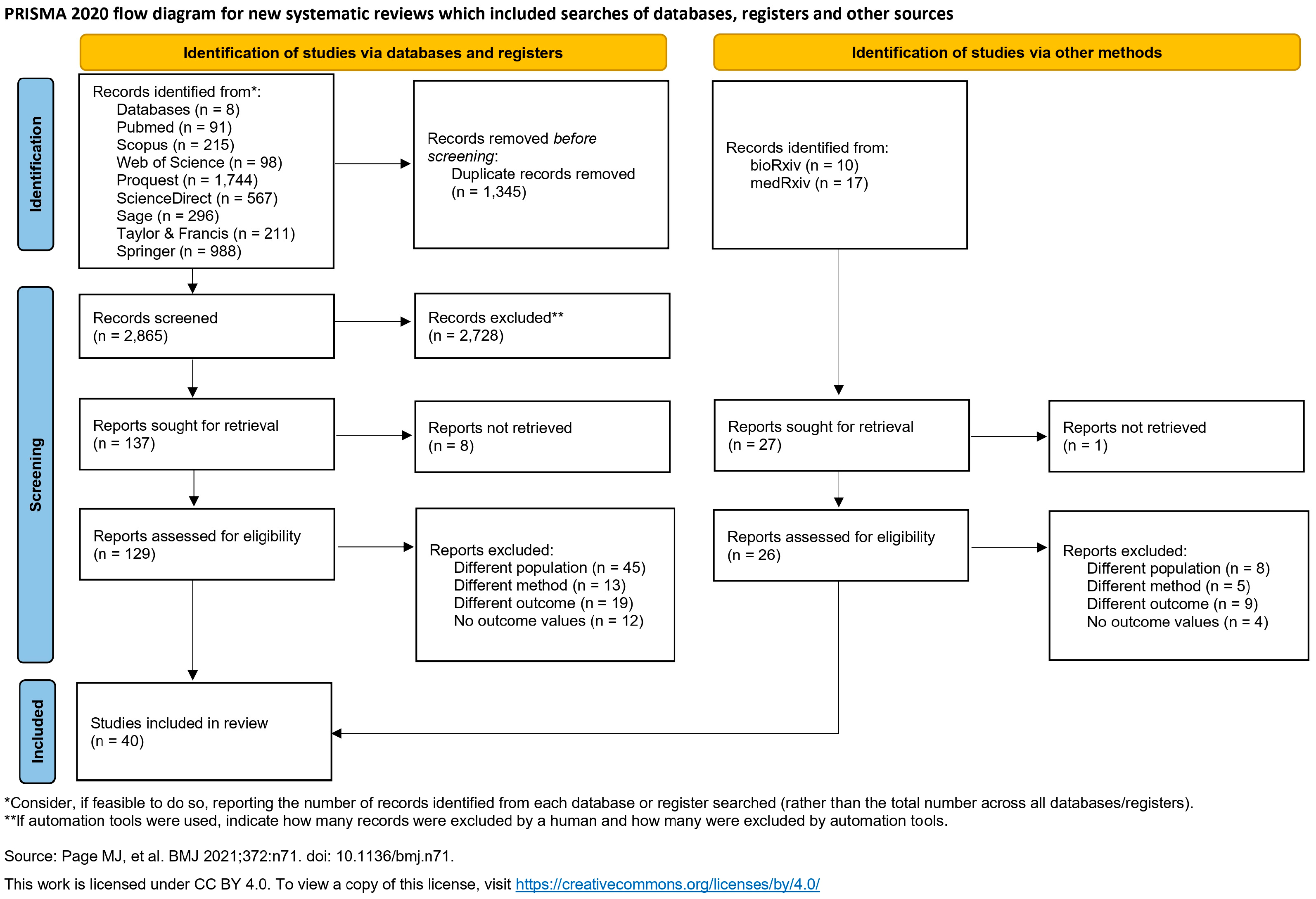
3.1. Study Selection
3.2. Study Characteristics

| Author (Year) | Marker | Study Location | Population Age | Sample Size | Disease Spectrum | PCI Rate (%) | Clinical Outcome | Sampling Timepoint | Follow-Up | Biomarker Concentration (Mean ± SD) | Hazard Ratio (95% CI) | Measurement Unit | ||
|---|---|---|---|---|---|---|---|---|---|---|---|---|---|---|
| Events | Non-Events | Events | Non-Events | |||||||||||
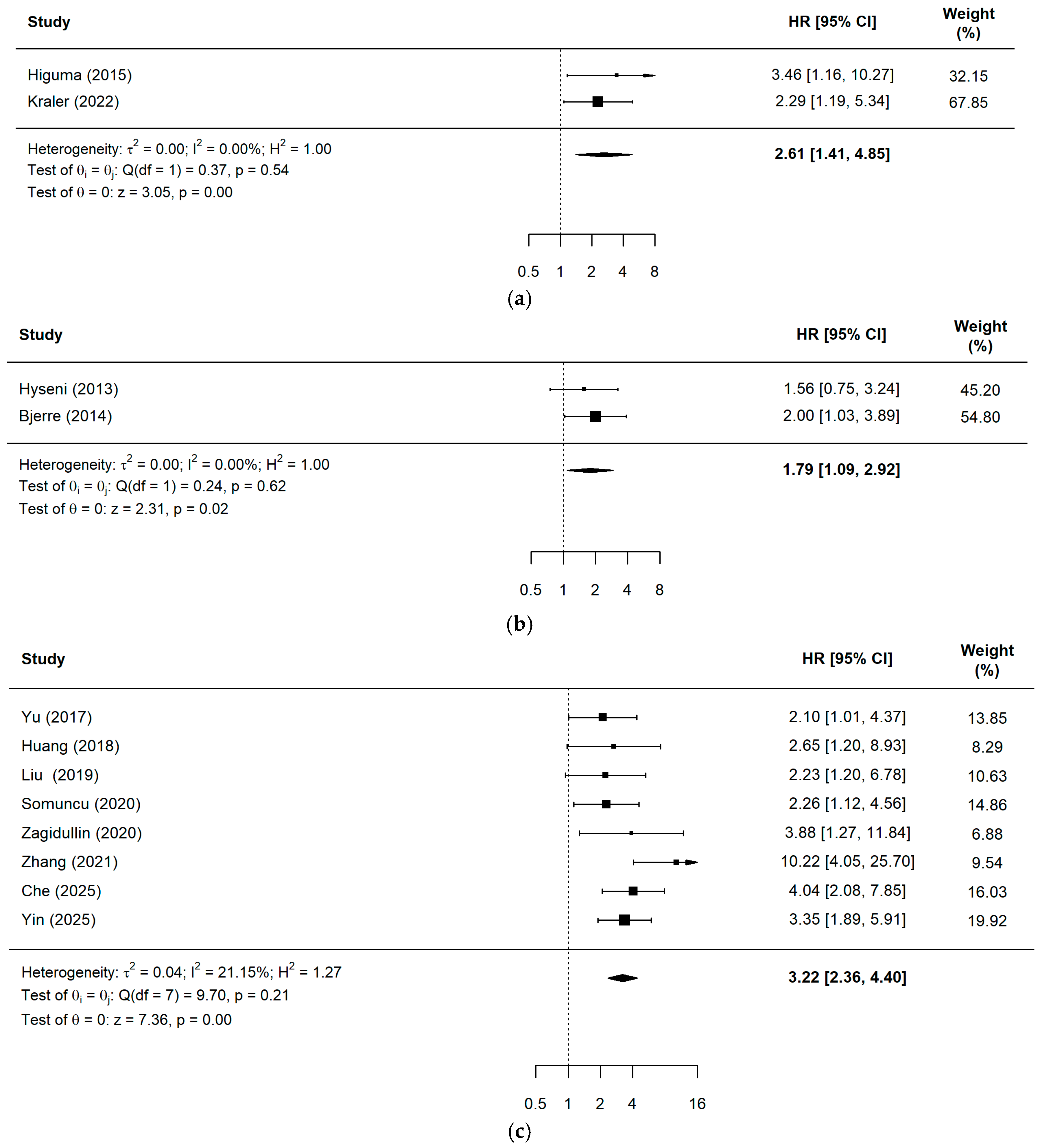
| Higuma (2015) [21] | sLOX-1 | Japan | 67 ± 12 | 20 | 133 | STEMI | 94.1% | MACE | Pre-PCI | 3 years | N/A | N/A | 3.46 (1.16–10.27) | ng/mL |
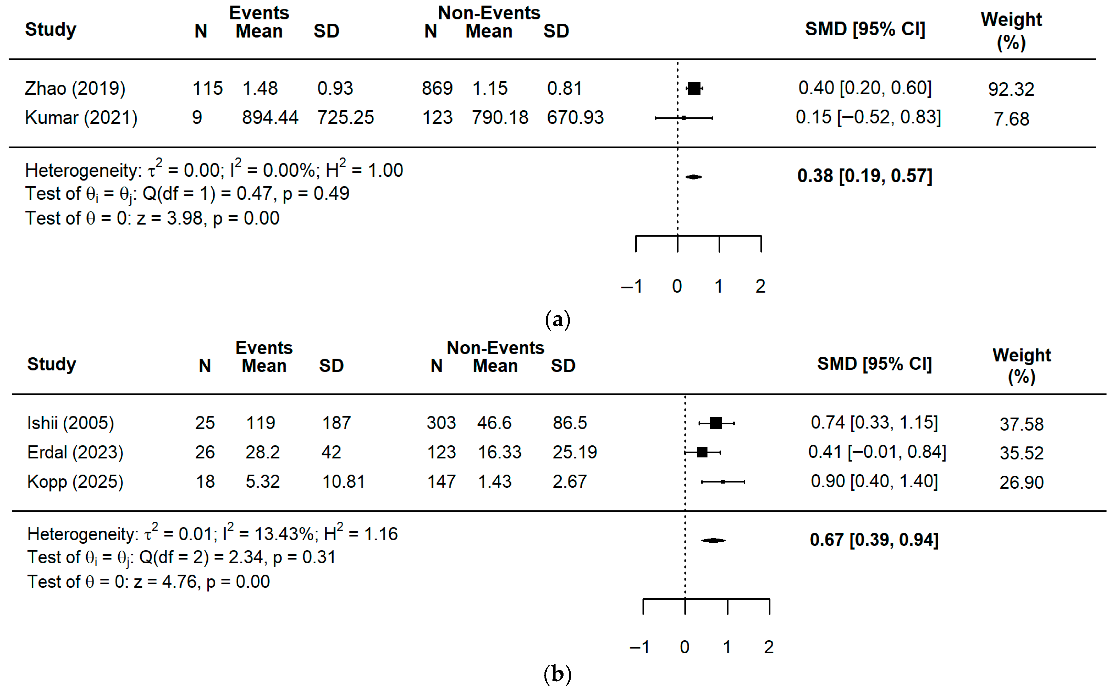
| Zhao (2019) [22] | sLOX-1 | China | MACCE: 67 (59–74) Non-MACCE: 69 (59–76) | 115 | 869 | Mixed (78.05% ACS) | 100% | MACCE | Pre-PCI | 2 years | 1.48 ± 0.93 | 1.15 ± 0.81 | N/A | ng/mL |
| Kumar (2021) [23] | sLOX-1 | India | 47.94 ± 14.30 | 9 | 123 | Mixed (48.5% ACS) | 100% | Stroke, recurrent myocardial infarction (MI) | Pre-PCI and post-PCI | 1 year | Pre-PCI: 894.44 ± 725.25 Post-PCI: 2086.22 ± 1079.43 | Pre-PCI: 790.18 ± 670.93 Post-PCI: 1332.81 ± 967.50 | N/A | ng/mL |
| Kraler (2022) [24] | sLOX-1 | Multicenter | 63.7 ± 10.27 | 107 | 2.532 | ACS | 100% | CVD | Pre-PCI | 1 year | N/A | N/A | 2.29 (1.19–5.34) | ng/mL |
| Ishii (2005) [25] | H-FABP | Japan | 64.9 ± 10.4 | 25 | 303 | ACS | 55.5% | CVD, non-fatal AMI | Pre-PCI | 6 months | 119 ± 187 | 46.6 ± 86.5 | 8.96 (2.64–30.4) | µg/L |
| Erdal (2023) [26] | H-FABP | Turkey | 54.6 ± 12.4 | 26 | 123 | STEMI | 100% | CVD | Pre-PCI and post-PCI 12 h after the start of symptoms | 6 years | Pre-PCI: 28.20 ± 42.00 Post-PCI: 43.00 ± 53.33 | Pre-PCI: 16.33 ± 25.19 Post-PCI: 26.33 ± 43.70 | N/A | ng/mL |
| Kopp (2025) [17] | H-FABP | Russia | 63.0 (53.5–70.0) | 18 | 147 | STEMI | 97.8% | CVD | Pre-PCI | 3 years | 5.32 ± 10.81 | 1.43 ± 2.67 | N/A | ng/mL |
| sST2 | 15.53 ± 9.47 | 8.63 ± 12.52 | ||||||||||||
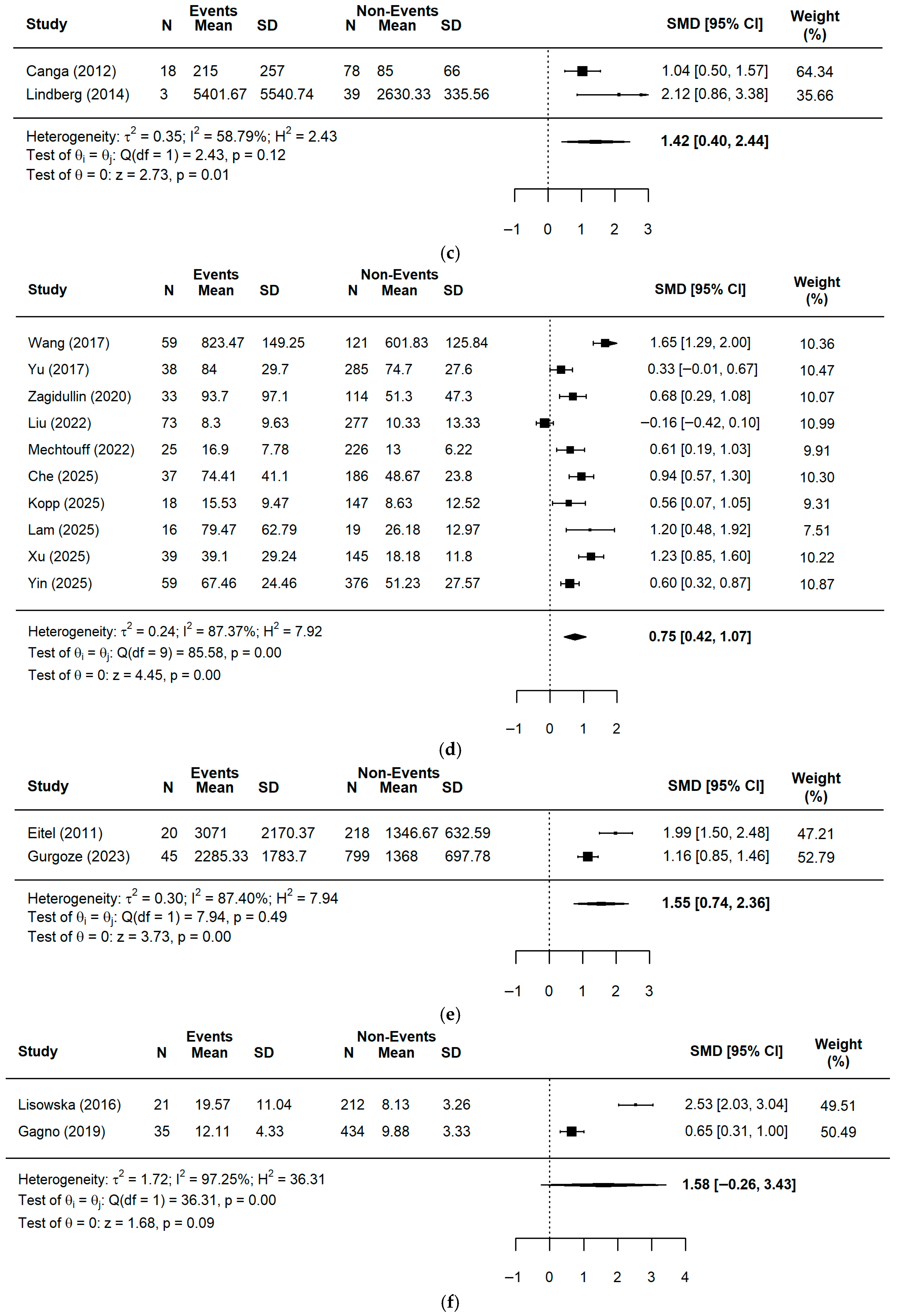
| Canga (2012) [27] | OPG | Turkey | MACE: 66 ± 10 tahun Non-MACE: 60 ± 13 tahun | 18 | 78 | STEMI | 100% | MACE | Pre-PCI | In hospital | 215 ± 257 | 85 ± 66 | N/A | pg/mL |
| Hyseni (2013) [28] | OPG | Netherlands and Germany | Survivors = 67.5 (59.5–73.4) Non-survivors = 76.8 (68.9–83.4) | 46 | 293 | ACS | 100% | All-cause mortality | Pre-PCI | 4 years | N/A | N/A | 1.56 (0.75–3.24) | μg/mL |
| Bjerre (2014) [29] | OPG | Denmark | Low OPG = 60.3 ± 10.5 High OPG = 66.9 ± 10.8 | 47 | 172 | STEMI | 100% | MACCE | Pre-PCI | 45 (38–48) months | N/A | N/A | 2.0 (1.03–3.89) | ng/L |
| Fuernau (2014) [18] | OPG | Germany | 71 (58–79) | 78 | 112 | AMI with cardiogenic shock | 95.8% | All-cause mortality | Immediate post-PCI | 30 days | 1274.33 ± 1547.41 | 690.33 ± 627.41 | N/A | ng/L |
| GDF-15 | 100% | 10,590.67 ± 6217.78 | 6327.33 ± 5382.96 | |||||||||||
| Lindberg (2014) [30] | OPG | Denmark | 60 ± 13 | 3 | 39 | STEMI with total occlusion of the left coronary artery branch | 100% | All-cause mortality, HF hospitalization | Pre-PCI | 1 year | 5401.67 ± 5540.74 | 2630.33 ± 335.56 | N/A | ng/L |
| Wang (2017) [31] | sST2 | China | 61.41 ± 8.90 | 59 | 121 | AMI | 100% | MACE | Pre-PCI | 1 year | 823.47 ± 149.25 | 601.83 ± 125.84 | N/A | ng/mL |
| Yu (2017) [32] | sST2 | South Korea | 59.1 ± 13.1 | 38 | 285 | STEMI | 100% | MACCE | Pre-PCI | 1 year | 84.0 ± 29.7 | 74.7 ± 27.6 | 2.098 (1.008–4.367) | ng/mL |
| Huang (2018) [33] | sST2 | China | 68.5 (30–72) | 21 | 157 | STEMI | 71.9% | MACE | Pre-PCI | 1 year | N/A | N/A | 2.653 (1.201–8.929) | ng/mL |
| Liu (2019) [34] | sST2 | China | 60.2 ± 10.8 | 19 | 276 | STEMI | 100% | MACE | Pre-PCI | 1 year | N/A | N/A | 2.23 (1.20–6.78) | ng/mL |
| Tyminska (2019) [19] | sST2 | Poland | 61.0 (50.5–67.0) | 9 | 95 | STEMI | 100% | CVD, HF hospitalization | Post-PCI 72–96 h after admission | 1 year | N/A | N/A | 11.79 (1.52–91.26) | ng/mL |
| Galectin-3 | 14.51 (1.46–143.95) | ng/mL | ||||||||||||
| Somuncu (2020) [35] | sST2 | Turkey | Low sST2: 60.0 ± 12.1 High sST2: 60.5 ± 11.6 | 68 | 312 | AMI | 75% | MACE | Pre-PCI | 1 year | N/A | N/A | 2.263 (1.124–4.557) | ng/mL |
| Zagidullin (2020) [36] | sST2 | Russia | 60.9 ± 12.1 | 33 | 114 | STEMI | 88.4% | CVD | Pre-PCI | 2 years | 93.7 ± 97.1 | 51.3 ± 47.3 | 3.88 (1.27–11.84) | ng/mL |
| Hongisto (2021) [20] | sST2 | Multicenter | 81 ± 4 | 26 | 30 | Mixed (80.82% ACS with cardiogenic shock) | 69.64% | All-cause mortality | Post-PCI 12 h after admission | In hospital | 1008.00 ± 763.70 | 572.00 ± 453.33 | N/A | ng/mL |
| GDF-15 | 27,276.00 ± 24,171.11 | 8040.67 ± 6968.89 | ng/L | |||||||||||
| Zhang (2021) [37] | sST2 | China | Low sST2: 61.13 ± 9.67 High sST2: 63.71 ± 10.02 | 25 | 180 | NSTEMI, UA | 100% | MACCE | Pre-PCI | 1 year | N/A | N/A | 10.22 (4.05–25.7) | ng/mL |
| Liu (2022) [38] | sST2 | China | 62 (55–70) | 73 | 277 | STEMI | 100% | MACE | Pre-PCI and post-PCI 24 h after admission | 1 year | Pre-PCI: 8.30 ± 9.63 Post-PCI: 17.07 ± 19.19 | Pre-PCI: 10.33 ± 13.33 Post-PCI: 13.97 ± 16.96 | N/A | ng/mL |
| Mechtouff (2022) [39] | sST2 | France | 59 ± 12 | 25 | 226 | STEMI | 100% | MACE | Pre-PCI and post-PCI 24 h after admission | 1 year | Pre-PCI: 16.90 ± 7.78 Post-PCI: 40.00 ± 32.96 | Pre-PCI: 13.00 ± 6.22 Post-PCI: 25.70 ± 17.19 | Post PCI: 2.4 (1.03–5.5) | ng/mL |
| Che (2025) [40] | sST2 | China | 60.77 ± 12.61 | 37 | 186 | STEMI | 100% | MACCE | Pre-PCI | 630 ± 433 days | 74.41 ± 41.10 | 48.67 ± 23.80 | 4.038 (2.077–7.852) | ng/mL |
| Lam (2025) [41] | sST2 | Vietnam | 70.35 ± 11.50 | 16 | 19 | STEMI | 100% | Mortality, HF | Pre-PCI | 30 days | 79.47 ± 62.79 | 26.18 ± 12.97 | N/A | ng/mL |
| Xu (2025) [42] | sST2 | China | MACE: 65.26 ± 14.54 Non-MACE: 59.16 ± 12.81 | 39 | 145 | STEMI | 100% | MACE | Pre-PCI and post-PCI 24 h after admission | 6 months | Pre-PCI: 39.10 ± 29.24 Post-PCI: 31.63 ± 27.99 | Pre-PCI: 18.18 ± 11.80 Post-PCI: 47.40 ± 39.30 | N/A | ng/mL |
| Yin (2025) [43] | sST2 | China | MACE: 60.54 ± 11.17 Non-MACE: 57.73 ± 7.62 | 59 | 376 | NSTEMI | 100% | MACE | Pre-PCI | 1 year | 67.46 ± 24.46 | 51.23 ± 27.57 | 3.35 (1.894–5.914) | ng/mL |
| Eitel (2011) [44] | GDF-15 | Germany | 67 (56–75) | 20 | 218 | STEMI | 100% | All-cause mortality | Pre-PCI | 6 months | 3071.00 ± 2170.37 | 1346.67 ± 632.59 | N/A | ng/L |
| Velders (2015) [45] | GDF-15 | Multicenter | 59 (IQR: 15) | 345 | 5040 | STEMI | 100% | CVD | Pre-PCI | 282 (IQR:180) days | N/A | N/A | 2.27(1.32–4.09) | ng/L |
| Sun (2018) [46] | GDF-15 | China | CIN: 71.6 ± 13.0 Non-CIN: 63.8 ± 11.7 | N/A | N/A | AMI | 100% | MACE | Pre-PCI | 30 days | N/A | N/A | 3.562 (1.658–7.652) | ng/L |
| Bodde (2019) [47] | GDF-15 | Netherlands | 59.0 ± 11.5 | 37 | 253 | STEMI | 100% | All-cause mortality | Pre-PCI | 9.4 (8.8–10.0) years | N/A | N/A | 2.453 (1.064–5.658) | pmol/L |
| Peiro (2019) [48] | GDF-15 | Spain | 64.8 (55.6–74.3) | 101 | 257 | ACS | 69.3% | MACE | Pre-PCI or peri-procedural | 4.9 (4.2–5.8) years | N/A | N/A | 2.48 (1.41–4.34) | ng/L |
| Gurgoze (2023) [49] | GDF-15 | Netherlands | 62.5 (54.3–70.2) | 45 | 799 | ACS | 86% | CVD, recurrent ACS | Pre-PCI | 1 year | 2285.33 ± 1783.70 | 1368.00 ± 697.78 | N/A | pg/mL |
| Peiro (2025) [50] | GDF-15 | Spain | NHRHF: 59.7 (51.4–69.3) HRHF: 72.7 (64.0–79.2) | 49 | 226 | AMI | 74.18% | All-cause mortality, HF hospitalization | Pre-PCI or peri-procedural | Median 4.9 years | N/A | N/A | 6.3 (2.9–13.3) | ng/L |
| Lisowska (2016) [51] | Galectin-3 | Poland | STEMI: 63.3 ± 9.9 NSTEMI: 63.5 ± 11.8 | 21 | 212 | STEMI, NSTEMI | 100% | All-cause mortality | Pre-PCI | Mean 2.8 years | 19.57 ± 11.04 | 8.13 ± 3.26 | N/A | ng/mL |
| Di Tano (2017) [52] | Galectin-3 | Italy | 65 (56–76) | 20 | 83 | STEMI | 100% | All-cause mortality, HF hospitalization | Post-PCI 24 h after admission | 22 (3–30) months | 26.60 ± 18.59 | 12.77 ± 3.19 | N/A | ng/mL |
| Mosleh (2018) [53] | Galectin-3 | United States | 60.94 ± 13.53 | 12 | 69 | STEMI | 100% | MACE | Post-PCI 12–48 h after admission | 1 year | 19.07 ± 11.10 | 11.75 ± 5.64 | N/A | ng/mL |
| Gagno (2019) [54] | Galectin-3 | Italy | 67.63 ± 11.29 | 35 | 434 | STEMI, NSTEMI | 70.6% | CVD | Pre-PCI | 1 year | 12.11 ± 4.33 | 9.88 ± 3.33 | N/A | ng/mL |
| Swiecki (2020) [55] | Galectin-3 | Poland | STEMI: 62.4 ± 9.7 NSTEMI: 64.2 ± 10.1 | 67 | 43 | STEMI, NSTEMI | 100% | Cardiac disorder rehospitalization | Post-PCI 24 h after admission | 41.3 ± 9.6 months | 13.7 ± 7.3 | 8.5 ± 4.5 | N/A | ng/mL |
| Choi (2025) [56] | Galectin-3 | South Korea | Low Galectin-3: 60.7 ± 10.0 High Galectin-3: 65.0 ± 10.8 | 92 | 847 | Mixed (51.18% ACS, 43.77% SA, 5.22% SI) | 100% | All-cause mortality, AMI, stroke | Pre-PCI | 997 (766–1.264) days | N/A | N/A | 1.670 (1.014–2.751) | ng/mL |
3.3. Methodological Quality of Included Studies
3.4. Quantitative Data Synthesis
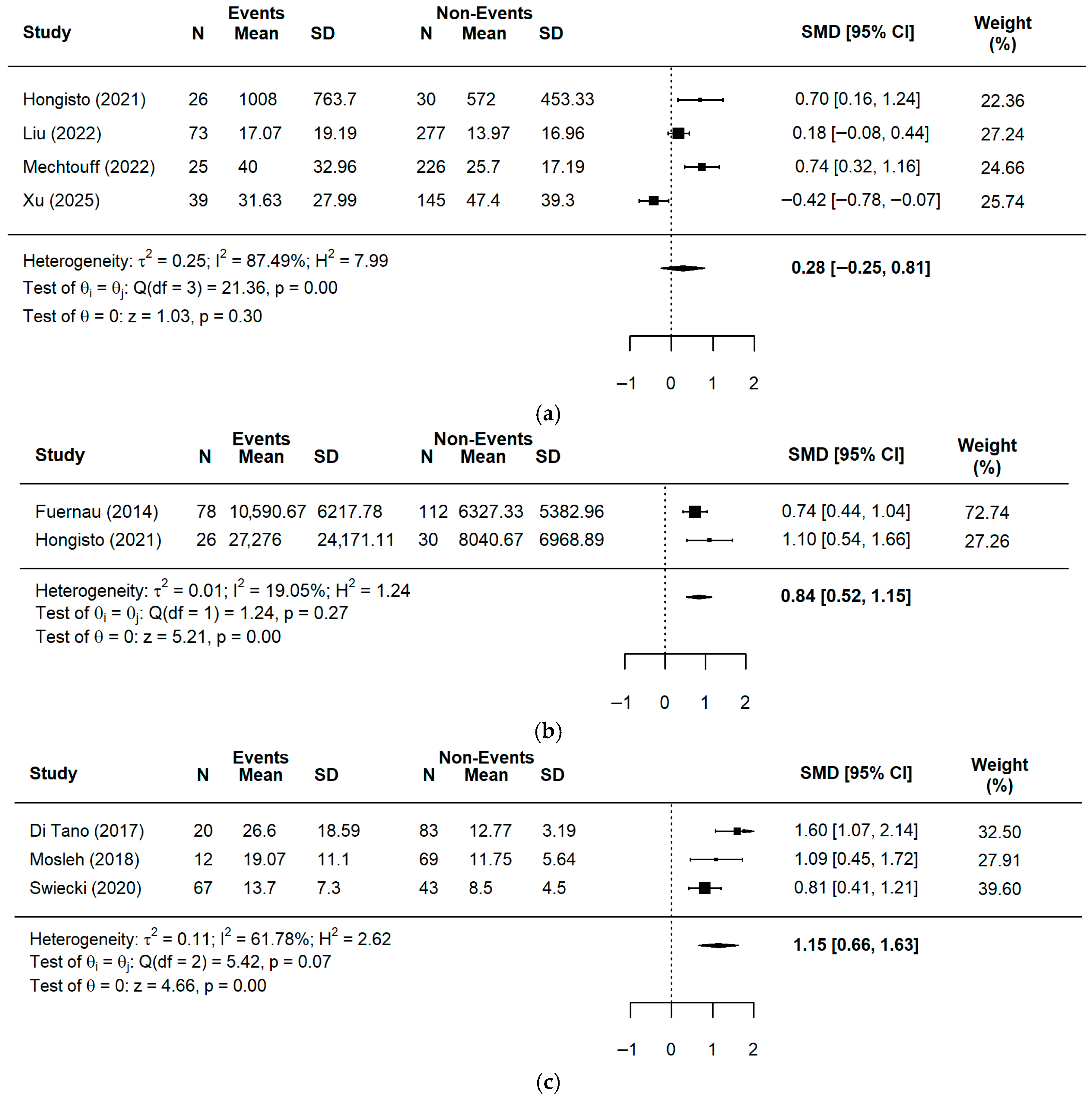
3.4.1. Biomarker Levels and Clinical Outcomes (SMD Analysis)
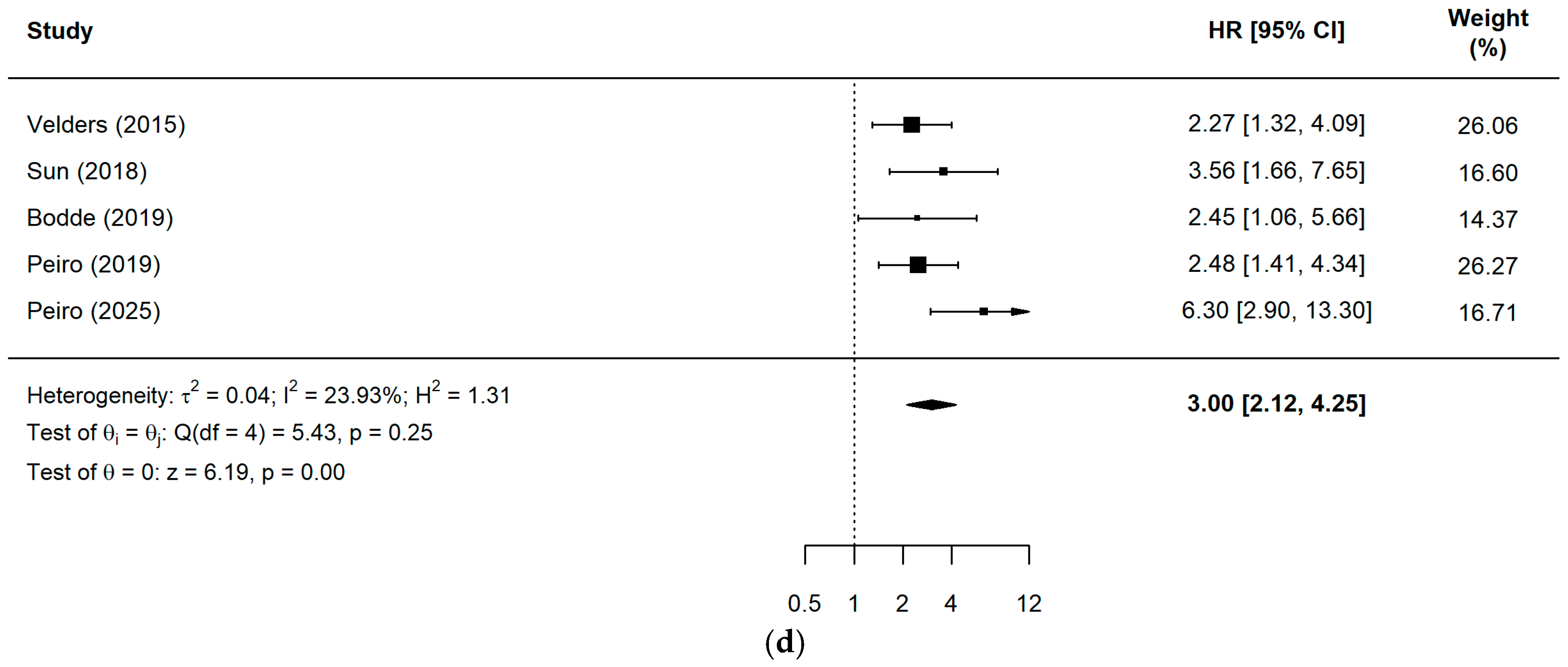
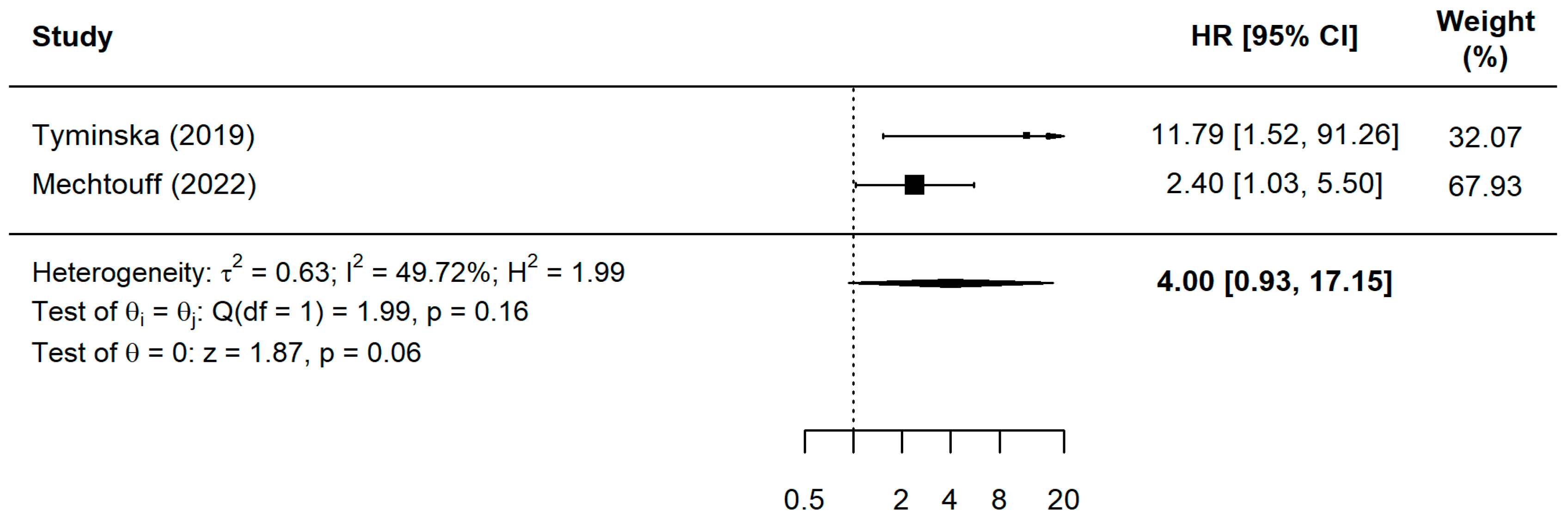
3.4.2. Prognostic Value for Adverse Events (Hazard Ratio Analysis)
3.5. Qualitative Synthesis of Single Studies
3.6. Subgroup Analysis
3.7. Heterogeneity Exploration
3.8. Certainty of Evidence (GRADE)
3.9. Sensitivity Analysis and Publication Bias
4. Discussion
5. Conclusions
Supplementary Materials
Author Contributions
Funding
Institutional Review Board Statement
Informed Consent Statement
Data Availability Statement
Acknowledgments
Conflicts of Interest
References
- World Health Organization. Cardiovascular Diseases (CVDs); World Health Organization: Geneva, Switzerland, 2022. [Google Scholar]
- Collet, J.P.; Thiele, H.; Barbato, E.; Bauersachs, J.; Dendale, P.; Edvardsen, T.; Folliguet, T.; Gale, C.; Gilard, M.; Jobs, A.; et al. 2020 ESC Guidelines for the management of acute coronary syndromes in patients presenting without persistent ST-segment elevation: The Task Force for the management of acute coronary syndromes in patients presenting without persistent ST-segment elevation of the European Society of Cardiology (ESC). Eur. Heart J. 2021, 42, 1289–1367, Erratum in Eur. Heart J. 2021, 42, 1908. https://doi.org/10.1093/eurheartj/ehaa895. Erratum in Eur. Heart J. 2021, 42, 1925. https://doi.org/10.1093/eurheartj/ehab088. Erratum in Eur. Heart J. 2021, 42, 2298. https://doi.org/10.1093/eurheartj/ehab285. Erratum in Eur Heart J. 2024, 45, 404–405. https://doi.org/10.1093/eurheartj/ehad879. [Google Scholar] [CrossRef] [Scilit] [PubMed]
- Madhavan, M.V.; Kirtane, A.J.; Redfors, B.; Généreux, P.; Ben-Yehuda, O.; Palmerini, T.; Benedetto, U.; Biondi-Zoccai, G.; Smits, P.; von Birgelen, C.; et al. Stent-Related Adverse Events >1 Year After Percutaneous Coronary Intervention. J. Am. Coll. Cardiol. 2020, 75, 590–604. [Google Scholar] [CrossRef] [Scilit] [PubMed]
- Mueller, T.; Leitner, I.; Egger, M.; Haltmayer, M.; Dieplinger, B. Association of the biomarkers soluble ST2, galectin-3 and growth-differentiation factor-15 with heart failure and other non-cardiac diseases. Clin. Chim. Acta 2015, 445, 155–160. [Google Scholar] [CrossRef] [Scilit] [PubMed]
- De Boer, R.A.; Voors, A.A.; Muntendam, P.; Van Gilst, W.H.; Van Veldhuisen, D.J. Galectin-3: A novel mediator of heart failure development and progression. Eur. J. Heart Fail. 2009, 11, 811–817. [Google Scholar] [CrossRef] [Scilit] [PubMed]
- Wollert, K.C.; Kempf, T.; Wallentin, L. Growth differentiation factor 15 as a biomarker in cardiovascular disease. Clin. Chem. 2017, 63, 140–151. [Google Scholar] [CrossRef] [Scilit] [PubMed]
- Venuraju, S.M.; Yerramasu, A.; Corder, R.; Lahiri, A. Osteoprotegerin as a Predictor of Coronary Artery Disease and Cardiovascular Mortality and Morbidity. J. Am. Coll. Cardiol. 2010, 55, 2049–2061. [Google Scholar] [CrossRef] [Scilit] [PubMed]
- Hayashida, K.; Kume, N.; Murase, T.; Minami, M.; Nakagawa, D.; Inada, T.; Tanaka, M.; Ueda, A.; Kominami, G.; Kambara, H.; et al. Serum soluble lectin-like oxidized low-density lipoprotein receptor-1 levels are elevated in acute coronary syndrome a novel marker for early diagnosis. Circulation 2005, 112, 812–818. [Google Scholar] [CrossRef] [Scilit] [PubMed]
- Ye, X.D.; He, Y.; Wang, S.; Wong, G.T.; Irwin, M.G.; Xia, Z. Heart-Type Fatty Acid Binding Protein (H-FABP) as a Biomarker for Acute Myocardial Injury and Long-Term Post-Ischemic Prognosis. Acta Pharmacologica Sinica. 2018. Available online: www.chinaphar.com (accessed on 2 March 2026).
- Piccolo, R.; Leone, A.; Avvedimento, M.; Simonetti, F.; Ippolito, D.; Angellotti, D.; Verde, N.; Manzi, L.; Cirillo, P.; Serafino, L.; et al. Impact of biomarker type on periprocedural myocardial infarction in patients undergoing elective PCI. Eur. Heart J. Qual. Care Clin. Outcomes 2023, 9, 680–690. [Google Scholar] [CrossRef] [Scilit] [PubMed]
- Bhatty, A.; Wilkinson, C.; Sydes, M.; Gale, C.P. Defining the need for cardiovascular event definitions. Eur. Heart J.-Qual. Care Clin. Outcomes 2024, 10, 105–107. [Google Scholar] [CrossRef] [Scilit] [PubMed]
- Wells, G.; Shea, B.; O’Connell, D.; Peterson, J.; Welch, V.; Losos, M. The Newcastle-Ottawa Scale (NOS) for Assessing the Quality of Nonrandomised Studies in Meta-Analyses. 2021. Available online: http://www.ohri.ca/programs/clinical_epidemiology/nosgen.pdf (accessed on 5 March 2026).
- Foroutan, F.; Guyatt, G.; Zuk, V.; Vandvik, P.O.; Alba, A.C.; Mustafa, R.; Vernooij, R.; Arevalo-Rodriguez, I.; Munn, Z.; Roshanov, P.; et al. GRADE Guidelines 28: Use of GRADE for the assessment of evidence about prognostic factors: Rating certainty in identification of groups of patients with different absolute risks. J. Clin. Epidemiol. 2020, 121, 62–70. [Google Scholar] [CrossRef] [Scilit] [PubMed]
- Wan, X.; Wang, W.; Liu, J.; Tong, T. Estimating the Sample Mean and Standard Deviation from the Sample Size, Median, Range and/or Interquartile Range. BMC Med. Res. Methodol. 2014, 14, 135. [Google Scholar] [CrossRef] [Scilit]
- Andrade, C. Mean Difference, Standardized Mean Difference (SMD), and Their Use in Meta-Analysis: As Simple as It Gets. J. Clin. Psychiatry 2020, 81, 20f13681. [Google Scholar] [CrossRef] [Scilit] [PubMed]
- Page, M.J.; McKenzie, J.E.; Bossuyt, P.M.; Boutron, I.; Hoffmann, T.C.; Mulrow, C.D.; Shamseer, L.; Tetzlaff, J.M.; Akl, E.A.; Brennan, S.E.; et al. The PRISMA 2020 statement: An updated guideline for reporting systematic reviews. BMJ 2021, 372, n71. [Google Scholar] [CrossRef] [Scilit] [PubMed]
- Kopp, K.; Lichtenauer, M.; Paar, V.; Hoppe, U.C.; Rakhimova, R.F.; Badykova, E.A.; Agletdinov, E.; Grishaev, D.; Cheremisina, K.; Baraboshkina, A.; et al. Diagnostic Biomarkers for Risk Estimation of In-Hospital and Post-Discharge Cardiovascular Mortality in ST-Segment Elevation Myocardial Infarction (STEMI) Patients. J. Clin. Med. 2025, 14, 6632. [Google Scholar] [CrossRef] [Scilit] [PubMed]
- Fuernau, G.; Poenisch, C.; Eitel, I.; De Waha, S.; Desch, S.; Schuler, G.; Adams, V.; Werdan, K.; Zeymer, U.; Thiele, H. Growth-differentiation factor 15 and osteoprotegerin in acute myocardial infarction complicated by cardiogenic shock: A biomarker substudy of the IABP-SHOCK II-trial. Eur. J. Heart Fail. 2014, 16, 880–887. [Google Scholar] [CrossRef] [Scilit] [PubMed]
- Tymińska, A.; Kapłon-Cieślicka, A.; Ozierański, K.; Budnik, M.; Wancerz, A.; Sypień, P.; Peller, M.; Maksym, J.; Balsam, P.; Opolski, G.; et al. Association of galectin-3 and soluble ST2 with in-hospital and 1-year outcomes in patients with ST-segment elevation myocardial infarction treated with primary percutaneous coronary intervention. Pol. Arch. Intern. Med. 2019, 129, 770–780. [Google Scholar] [CrossRef] [Scilit] [PubMed]
- Hongisto, M.; Lassus, J.; Tarvasmäki, T.; Sionis, A.; Sans-Rosello, J.; Tolppanen, H.; Kataja, A.; Jäntti, T.; Sabell, T.; Lindholm, M.; et al. Mortality risk prediction in elderly patients with cardiogenic shock: Results from the CardShock study. ESC Heart Fail. 2021, 8, 1398–1407. [Google Scholar] [CrossRef] [Scilit] [PubMed]
- Higuma, T.; Abe, N.; Tateyama, S.; Endo, T.; Shibutani, S.; Yokoyama, H.; Hanada, K.; Yamada, M.; Tomita, H.; Hanada, H.; et al. Plasma soluble lectin-like oxidized low-density lipoprotein receptor-1 as a novel prognostic biomarker in patients with ST-segment elevation acute myocardial infarction. Circ. J. 2015, 79, 641–648. [Google Scholar] [CrossRef] [Scilit] [PubMed]
- Zhao, Z.W.; Xu, Y.W.; Li, S.M.; Guo, J.J.; Sun, J.M.; Hong, J.C.; Chen, L. Baseline Serum sLOX-1 Concentrations Are Associated with 2-Year Major Adverse Cardiovascular and Cerebrovascular Events in Patients after Percutaneous Coronary Intervention. Dis. Markers 2019, 2019, 4925767. [Google Scholar] [CrossRef] [Scilit] [PubMed]
- Kumar, S.; Ali, W.; Mishra, S.; Pradhan, A.; Sethi, R.; Kushwaha, R.; Singh, U.; Perrone, M. Circulating soluble lectin-like oxidized low-density lipoprotein receptor-1 (sLOX-1): A diagnostic indicator across the spectrum of acute coronary syndrome. J. Clin. Med. 2021, 10, 5567. [Google Scholar] [CrossRef] [Scilit]
- Kraler, S.; Wenzl, F.A.; Georgiopoulos, G.; Obeid, S.; Liberale, L.; von Eckardstein, A.; Muller, O.; Mach, F.; Räber, L.; Losdat, S.; et al. Soluble lectin-like oxidized low-density lipoprotein receptor-1 predicts premature death in acute coronary syndromes. Eur. Heart J. 2022, 43, 1849–1860. [Google Scholar] [CrossRef] [Scilit]
- Ishii, J.; Ozaki, Y.; Lu, J.; Kitagawa, F.; Kuno, T.; Nakano, T.; Nakamura, Y.; Naruse, H.; Mori, Y.; Matsui, S.; et al. Prognostic value of serum concentration of heart-type fatty acid-binding protein relative to cardiac troponin T on admission in the early hours of acute coronary syndrome. Clin. Chem. 2005, 51, 1397–1404. [Google Scholar] [CrossRef] [Scilit] [PubMed]
- Şahingöz Erdal, G.; Karabulut, D.; Yıldız, C.; Turhan Çağlar, F.; Satılmışoğlu, M.; Koser, M.; Kasapoğlu, P.; Işıksaçan, N. The role of h-FABP and Myoglobin in Determining Disease Severity and Prognosis in STEMI. Istanb. Med. J. 2023, 24, 149–154. [Google Scholar] [CrossRef] [Scilit]
- Çanga, A.; Durakoĝlugil, M.E.; Erdoĝan, T.; Kirbaş, A.; Yilmaz, A.; Çiçek, Y.; Ergül, E.; Çetin, M.; Kocaman, S. Elevated serum osteoprotegerin levels predict in-hospital major adverse cardiac events in patients with ST elevation myocardial infarction. J. Cardiol. 2012, 60, 355–360. [Google Scholar] [CrossRef] [Scilit] [PubMed]
- Hyseni, A.; Roest, M.; Braun, S.L.; Barendrecht, A.D.; De Groot, P.G.; Ndrepepa, G.; Kastrati, A. Chronic dysfunction of the endothelium is associated with mortality in acute coronary syndrome patients. Thromb. Res. 2013, 131, 198–203. [Google Scholar] [CrossRef] [Scilit] [PubMed]
- Bjerre, M.; Munk, K.; Sloth, A.D.; Nielsen, S.S.; Flyvbjerg, A.; Botker, H.E. High osteoprotegerin levels predict MACCE in STEMI patients, but are not associated with myocardial salvage. Scand. Cardiovasc. J. 2014, 48, 209–215. [Google Scholar] [CrossRef] [Scilit] [PubMed]
- Lindberg, S.; Jensen, J.S.; Hoffmann, S.; Iversen, A.Z.; Pedersen, S.H.; Mogelvang, R.; Galatius, S.; Flyvbjerg, A.; Bjerre, M. Osteoprotegerin Levels Change During STEMI and Reflect Cardiac Function. Can. J. Cardiol. 2014, 30, 1523–1528. [Google Scholar] [CrossRef] [Scilit] [PubMed]
- Wang, Y.P.; Wang, J.H.; Wang, X.L.; Liu, J.Y.; Jiang, F.Y.; Huang, X.L.; Qin, W.; Ma, S.; Zhang, J.; Yuan, M.; et al. Roles of ST2, IL-33 and BNP in predicting major adverse cardiovascular events in acute myocardial infarction after percutaneous coronary intervention. J. Cell. Mol. Med. 2017, 21, 2677–2684. [Google Scholar] [CrossRef] [Scilit] [PubMed]
- Yu, J.; Oh, P.C.; Kim, M.; Moon, J.; Park, Y.M.; Lee, K.; Suh, S.; Han, S.; Byun, K.; Ahn, T.; et al. Improved early risk stratification of patients with ST-segment elevation myocardial infarction undergoing primary percutaneous coronary intervention using a combination of serum soluble ST2 and NT-proBNP. PLoS ONE 2017, 12, e0182829. [Google Scholar] [CrossRef] [Scilit] [PubMed]
- Huang, W.P.; Zheng, X.; He, L.; Su, X.; Liu, C.W.; Wu, M.X. Role of Soluble ST2 Levels and Beta-Blockers Dosage on Cardiovascular Events of Patients with Unselected ST-Segment Elevation Myocardial Infarction. Chin. Med. J. 2018, 131, 1282–1288. [Google Scholar] [CrossRef] [Scilit] [PubMed]
- Liu, X.; Hu, Y.; Huang, W.; Zhang, G.; Cao, S.; Yan, X.; Li, L.; Zhang, L.; Zheng, X. Soluble ST2 for prediction of clinical outcomes in patients with ST-segment elevation myocardial infarction receiving primary PCI. Int. Heart J. 2019, 60, 19–26. [Google Scholar] [CrossRef] [Scilit] [PubMed]
- Somuncu, M.U.; Kalayci, B.; Avci, A.; Akgun, T.; Karakurt, H.; Demir, A.R.; Avci, Y.; Can, M. Predicting long-term cardiovascular outcomes of patients with acute myocardial infarction using soluble ST2. Horm. Mol. Biol. Clin. Investig. 2020, 41, 20190062. [Google Scholar] [CrossRef] [Scilit] [PubMed]
- Zagidullin, N.; Motloch, L.J.; Gareeva, D.; Hamitova, A.; Lakman, I.; Krioni, I.; Popov, D.; Zulkarneev, R.; Paar, V.; Kopp, K.; et al. Combining novel biomarkers for risk stratification of two-year cardiovascular mortality in patients with ST-elevation myocardial infarction. J. Clin. Med. 2020, 9, 550. [Google Scholar] [CrossRef] [Scilit] [PubMed]
- Zhang, Q.; Hu, M.; Ma, S. Association of soluble suppression of tumorigenicity with no-reflow phenomenon and long-term prognosis in patients with non-st-segment elevation acute coronary syndrome after percutaneous coronary intervention. J. Atheroscler. Thromb. 2021, 28, 1289–1297. [Google Scholar] [CrossRef] [Scilit] [PubMed]
- Liu, L.; Li, S.; Ding, X.; Wang, D.; Li, W.; Li, H. Dynamic changes in soluble suppression of tumorigenicity 2 levels predict major adverse cardiovascular events in patients with ST-segment elevation myocardial infarction. Pol. Arch. Intern. Med. 2022, 132, 16317. [Google Scholar] [CrossRef] [Scilit] [PubMed]
- Mechtouff, L.; Paccalet, A.; Crola Da Silva, C.; Buisson, M.; Mewton, N.; Amaz, C.; Bonnefoy-Cudraz, E.; Leboube, S.; Cho, T.; Nighoghossian, N.; et al. Prognosis value of serum soluble ST2 level in acute ischemic stroke and STEMI patients in the era of mechanical reperfusion therapy. J. Neurol. 2022, 269, 2641–2648. [Google Scholar] [CrossRef] [Scilit] [PubMed]
- Che, W.; Jin, Y.; Chang, S.; Sun, Y.; Hou, A.; Wang, C. Prediction of myocardial ischemia–reperfusion injury post-PCI: Role of sST2 levels in STEMI patients. BMC Cardiovasc. Disord. 2025, 25, 280. [Google Scholar] [CrossRef] [Scilit] [PubMed]
- Lam, N.V.; Nghia, D.T.; Tien, H.A. Combining SST2 and NT-proBNP for improved risk stratification in STEMI patients post-PCI. Rom. Med. J. 2025, 72, 434–439. [Google Scholar] [CrossRef] [Scilit]
- Xu, Y.; Li, L.; Lin, Z.; Hu, Y.; Liu, Y.; Zhou, T. Predictive value of dynamic changes in RvD1, sST2, and PIV levels for the prognosis of STEMI patients after PCI: A prospective study. Front. Cardiovasc. Med. 2025, 12, 1705808. [Google Scholar] [CrossRef] [Scilit]
- Yin, J.; Qiu, B.; Li, T.; Tao, Y.; Gu, X. NSTEMI Risk Prediction with the Combination of the Biomarkers Epicardial Adipose Tissue and Soluble Suppression of Tumorigenicity 2. Med. Sci. Monit. 2025, 31, e947019. [Google Scholar] [CrossRef] [Scilit] [PubMed]
- Eitel, I.; Blase, P.; Adams, V.; Hildebrand, L.; Desch, S.; Schuler, G.; Thiele, H. Growth-differentiation factor 15 as predictor of mortality in acute reperfused ST-elevation myocardial infarction: Insights from cardiovascular magnetic resonance. Heart 2011, 97, 632–640. [Google Scholar] [CrossRef] [Scilit] [PubMed]
- Velders, M.A.; Wallentin, L.; Becker, R.C.; Van Boven, A.J.; Himmelmann, A.; Husted, S.; Katus, H.; Lindholm, D.; Morais, J.; Siegbahn, A.; et al. Biomarkers for risk stratification of patients with ST-elevation myocardial infarction treated with primary percutaneous coronary intervention: Insights from the Platelet Inhibition and Patient Outcomes trial. Am. Heart J. 2015, 169, 879–889.e7. [Google Scholar] [CrossRef] [Scilit] [PubMed]
- Sun, L.; Zhou, X.; Jiang, J.; Zang, X.; Chen, X.; Li, H.; Cao, H.; Wang, Q. Growth differentiation factor-15 levels and the risk of contrast induced nephropathy in patients with acute myocardial infarction undergoing percutaneous coronary intervention: A retrospective observation study. PLoS ONE 2018, 13, e0197609. [Google Scholar] [CrossRef] [Scilit] [PubMed]
- Bodde, M.C.; Hermans, M.P.J.; van der Laarse, A.; Mertens, B.; Romijn, F.P.H.T.M.; Schalij, M.J.; Cobbaert, C.M. Growth Differentiation Factor-15 Levels at Admission Provide Incremental Prognostic Information on All-Cause Long-term Mortality in ST-Segment Elevation Myocardial Infarction Patients Treated with Primary Percutaneous Coronary Intervention. Cardiol. Ther. 2019, 8, 29–41. [Google Scholar] [CrossRef] [Scilit]
- Peiró, Ó.M.; García-Osuna, Á.; Ordóñez-Llanos, J.; Cediel, G.; Bonet, G.; Rojas, S.; Quintern, V.; Bardají, A. Long-term prognostic value of growth differentiation factor-15 in acute coronary syndromes. Clin. Biochem. 2019, 73, 62–69. [Google Scholar] [CrossRef] [Scilit] [PubMed]
- Gürgöze, M.T.; Akkerhuis, K.M.; Oemrawsingh, R.M.; Umans, V.A.W.M.; Kietselaer, B.; Schotborgh, C.E.; Lenderink, T.; Aksoy, I.; Van Der Harst, P.; Asselbergs, F.; et al. Serially measured high-sensitivity cardiac troponin T, N-terminal-pro-B-type natriuretic peptide, high-sensitivity C-reactive protein, and growth differentiation factor 15 for risk assessment after acute coronary syndrome: The BIOMArCS cohort. Eur. Heart J. Acute Cardiovasc. Care 2023, 12, 451–461. [Google Scholar] [CrossRef] [Scilit] [PubMed]
- Peiró, Ó.M.; Bardají, A.; Carrasquer, A.; Cediel, G.; Serrano, I.; Bonet, G.; Ferrero, M.; Del-Moral-Ronda, V.; Rocamora-Horrach, M.; Ferreiro, J. Growth differentiation factor 15: A biomarker to guide empagliflozin treatment in acute myocardial infarction? Int. J. Cardiol. 2025, 429, 133167. [Google Scholar] [CrossRef] [Scilit] [PubMed]
- Lisowska, A.; Knapp, M.; Tycińska, A.; Motybel, E.; Kamiński, K.; Świecki, P.; Musiał, W.; Dymicka-Piekarska, V. Predictive value of Galectin-3 for the occurrence of coronary artery disease and prognosis after myocardial infarction and its association with carotid IMT values in these patients: A mid-term prospective cohort study. Atherosclerosis 2016, 246, 309–317. [Google Scholar] [CrossRef] [Scilit] [PubMed]
- Di Tano, G.; Caretta, G.; De Maria, R.; Bettari, L.; Parolini, M.; Testa, S.; Pirelli, S. Galectin-3 and outcomes after anterior-wall myocardial infarction treated by primary percutaneous coronary intervention. Biomark. Med. 2018, 12, 21–26. [Google Scholar] [CrossRef] [Scilit] [PubMed]
- Mosleh, W.; Elango, K.; Shah, T.; Chaudhari, M.; Gandhi, S.; Kattel, S.; Karki, R.; Khalil, C.; Frodey, K.; Dahal, S.; et al. Elevated end-diastolic wall stress after acute myocardial infarction predicts adverse cardiovascular outcomes and longer hospital length of stay. Echocardiography 2018, 35, 1721–1728. [Google Scholar] [CrossRef] [Scilit] [PubMed]
- Gagno, G.; Padoan, L.; Stenner, E.; Beleù, A.; Ziberna, F.; Hiche, C.; Paldino, A.; Barbati, G.; Biolo, G.; Fiotti, N.; et al. Galectin 3 and galectin 3 binding protein improve the risk stratification after myocardial infarction. J. Clin. Med. 2019, 8, 570. [Google Scholar] [CrossRef] [Scilit]
- Święcki, P.; Sawicki, R.; Knapp, M.; Kamiński, K.A.; Ptaszyńska-Kopczyńska, K.; Sobkowicz, B.; Lisowska, A. Galectin-3 as the prognostic factor of adverse cardiovascular events in long-term follow up in patients after myocardial infarction—A pilot study. J. Clin. Med. 2020, 9, 1640. [Google Scholar] [CrossRef] [Scilit] [PubMed]
- Choi, Y.J.; Seo, S.M. Prognostic significance of serum galectin-3 in predicting cardiovascular outcomes after percutaneous coronary intervention with drug-eluting stents. Front. Cardiovasc. Med. 2025, 12, 1563068. [Google Scholar] [CrossRef] [Scilit]
- Mair, J.; Jaffe, A.; Lindahl, B.; Mills, N.; Möckel, M.; Cullen, L.; Giannitsis, E.; Hammarsten, O.; Huber, K.; Krychtiuk, K.; et al. The clinical approach to diagnosing peri-procedural myocardial infarction after percutaneous coronary interventions according to the fourth universal definition of myocardial infarction–from the study group on biomarkers of the European Society of Cardiology (ESC) Association for Acute CardioVascular Care (ACVC). Biomarkers 2022, 27, 407–417. [Google Scholar] [CrossRef] [Scilit] [PubMed]
- Eggers, K.M.; Armstrong, P.W.; Califf, R.M.; Simoons, M.L.; Venge, P.; Wallentin, L.; James, S. ST2 and mortality in non-ST-segment elevation acute coronary syndrome. Am. Heart J. 2010, 159, 788–794. [Google Scholar] [CrossRef] [Scilit] [PubMed]
- Sanada, S.; Hakuno, D.; Higgins, L.J.; Schreiter, E.R.; McKenzie, A.N.J.; Lee, R.T. IL-33 and ST2 comprise a critical biomechanically induced and cardioprotective signaling system. J. Clin. Investig. 2007, 117, 1538–1549. [Google Scholar] [CrossRef] [Scilit] [PubMed]
- Tucker, B.; Vaidya, K.; Cochran, B.J.; Patel, S. Inflammation during percutaneous coronary intervention— prognostic value, mechanisms and therapeutic targets. Cells 2021, 10, 1391. [Google Scholar] [CrossRef] [Scilit] [PubMed]
- Nakata, T.; Hashimoto, A.; Hase, M.; Tsuchihashi, K.; Shimamoto, K. Human heart-type fatty acid-binding protein as an early diagnostic and prognostic marker in acute coronary syndrome. Cardiology 2003, 99, 96–104. [Google Scholar] [CrossRef] [Scilit] [PubMed]
- Kilcullen, N.; Viswanathan, K.; Das, R.; Morrell, C.; Farrin, A.; Barth, J.H.; Hall, A. Heart-Type Fatty Acid-Binding Protein Predicts Long-Term Mortality After Acute Coronary Syndrome and Identifies High-Risk Patients Across the Range of Troponin Values. J. Am. Coll. Cardiol. 2007, 50, 2061–2067. [Google Scholar] [CrossRef] [Scilit] [PubMed]
- Viswanathan, K.; Kilcullen, N.; Morrell, C.; Thistlethwaite, S.J.; Sivananthan, M.U.; Hassan, T.B.; Barth, J.; Hall, A. Heart-Type Fatty Acid-Binding Protein Predicts Long-Term Mortality and Re-Infarction in Consecutive Patients With Suspected Acute Coronary Syndrome Who Are Troponin-Negative. J. Am. Coll. Cardiol. 2010, 55, 2590–2598. [Google Scholar] [CrossRef] [Scilit] [PubMed]
- O’Donoghue, M.; De Lemos, J.A.; Morrow, D.A.; Murphy, S.A.; Buros, J.L.; Cannon, C.P.; Sabatine, M. Prognostic utility of heart-type fatty acid binding protein in patients with acute coronary syndromes. Circulation 2006, 114, 550–557. [Google Scholar] [CrossRef] [Scilit] [PubMed]
- Schiopu, A.; Svedlund, S.; Narasimhan, G.; Juin Loong, B.; Yndigegn, T.; Varma, V.; Ongstad, E.; Goncalves, I.; Collén, A.; Nilsson, J.; et al. Circulating soluble LOX-1 and patient prognosis after an acute coronary syndrome. Heart, 2025; epub ahead of print. [CrossRef] [Scilit] [PubMed]
- Suthahar, N.; Meijers, W.C.; Silljé, H.H.W.; Ho, J.E.; Liu, F.T.; de Boer, R.A. Galectin-3 activation and inhibition in heart failure and cardiovascular disease: An update. Theranostics 2018, 8, 593–609. [Google Scholar] [CrossRef] [Scilit] [PubMed]
- Tsai, T.H.; Sung, P.H.; Chang, L.T.; Sun, C.K.; Yeh, K.H.; Chung, S.Y.; Chua, S.; Chen, Y.; Wu, C.; Chang, H.; et al. Value and Level of Galectin-3 in Acute Myocardial Infarction Patients Undergoing Primary Percutaneous Coronary Intervention. J. Atheroscler. Thromb. 2012, 19, 1073–1082. [Google Scholar] [CrossRef] [Scilit] [PubMed]
- Granger, C.B.; Goldberg, R.J.; Dabbous, O.; Pieper, K.S.; Eagle, K.A.; Cannon, C.P.; Van De Werf, F.; Avezum, A.; Goodman, S.; Flather, M.; et al. Predictors of Hospital Mortality in the Global Registry of Acute Coronary Events. Available online: http://archinte.jamanetwork.com/ (accessed on 1 March 2026).
- Antman, E.M.; Cohen, M.; Bernink, P.J.L.M.; Mccabe, C.H.; Horacek, T.; Papuchis, G.; Mautner, B.; Corbalan, R.; Radley, D.; Braunwald, E. The TIMI Risk Score for Unstable Angina/Non-ST Elevation MI A Method for Prognostication and Therapeutic Decision Making. Available online: http://jama.jamanetwork.com/ (accessed on 2 March 2026).




| Marker | Sampling Timepoint | Subgroup Analysis Variable | Number of Studies per Subgroup | Measurement Effect | 95% Cl | p-Value |
|---|---|---|---|---|---|---|
| sST2 | Pre-PCI | Follow-up (short-term) | 2 | SMD = 1.21 | 0.42–2.01 | 0.003 * |
| Follow-up (medium-term) | 6 | SMD = 0.65 | 0.22–1.07 | 0.003 * | ||
| Follow-up (long-term) | 2 | SMD = 0.62 | (−0.14)–1.38 | 0.11 | ||
| Clinical outcome (MACE) | 5 | SMD = 0.77 | 0.25–1.30 | 0.004 * | ||
| Clinical outcome (MACCE) | 2 | SMD = 0.63 | (−0.20)–1.47 | 0.14 | ||
| Clinical outcome (CVD) | 2 | SMD = 0.62 | (−0.23)–1.48 | 0.15 | ||
| Clinical outcome (mortality/HF) | 1 | SMD = 1.20 | (−0.14)–2.54 | 0.08 | ||
| Continent (Asian) | 7 | SMD = 0.80 | 0.39–1.21 | 0.0001 * | ||
| Continent (European) | 3 | SMD = 0.62 | (−0.02)–1.25 | 0.06 | ||
| Disease (STEMI) | 8 | SMD = 0.64 | 0.31–0.97 | 0.0001 * | ||
| Disease (NSTEMI) | 1 | SMD = 0.60 | (−0.28)–1.48 | 0.18 | ||
| Disease (AMI) | 1 | SMD = 1.65 | 0.74–2.56 | 0.0004 * | ||
| Follow-up (medium-term) | 7 | HR = 3.18 | 2.26–4.49 | <0.0001 * | ||
| Follow-up (long-term) | 1 | HR = 3.88 | 1.14–13.20 | 0.03 * | ||
| Clinical outcome (MACE) | 4 | HR = 2.66 | 1.66–4.25 | <0.0001 * | ||
| Clinical outcome (MACCE) | 3 | HR = 4.07 | 2.36–7.02 | <0.0001 * | ||
| Clinical outcome (CVD) | 1 | HR = 3.88 | 1.11–13.51 | 0.03 * | ||
| Disease (STEMI) | 5 | HR = 2.88 | 1.98–4.17 | <0.0001 * | ||
| Disease (NSTEMI) | 1 | HR = 3.35 | 1.90–5.92 | <0.0001 * | ||
| Disease (NSTEMI, UA) | 1 | HR = 10.22 | 4.06–25.74 | <0.0001 * | ||
| Disease (AMI) | 1 | HR = 2.26 | 1.98–4.17 | 0.02 * | ||
| Continent (Asian) | 7 | HR = 3.18 | 2.26–4.49 | <0.0001 * | ||
| Continent (European) | 1 | HR = 3.88 | 1.14–13.20 | 0.03 * | ||
| Post-PCI | Follow-up (short-term) | 2 | SMD = 0.11 | (−0.76)–0.97 | 0.81 | |
| Follow-up (medium-term) | 2 | SMD = 0.45 | (−0.39)–(1.29) | 0.30 | ||
| Clinical outcome (MACE) | 3 | SMD = 0.1581 | (−0.48)–0.80 | 0.63 | ||
| Clinical outcome (all-cause mortality) | 1 | SMD = 0.6971 | (−0.49)–1.88 | 0.25 | ||
| Disease (STEMI) | 3 | SMD = 0.16 | (−0.48)–0.80 | 0.63 | ||
| Disease (ACS with cardiogenic shock) | 1 | SMD = 0.70 | (−0.49)–1.88 | 0.25 | ||
| Continent (European) | 1 | SMD = 0.74 | (−0.14)–1.62 | 0.10 | ||
| Continent (Asian) | 2 | SMD = −0.11 | (−0.70)–0.48 | 0.71 | ||
| Continent (multicenter) | 1 | SMD = 0.70 | (−0.24)–1.64 | 0.15 | ||
| GDF-15 | Pre-PCI | Follow-up (short-term) | 2 | HR = 2.74 | 1.48–5.07 | 0.001 * |
| Follow-up (long-term) | 3 | HR = 3.28 | 1.94–5.55 | <0.0001 * | ||
| Clinical outcome (MACE) | 2 | HR = 2.82 | 1.79–4.43 | <0.0001 * | ||
| Clinical outcome (CVD) | 1 | HR = 2.27 | 1.29–4.00 | 0.005 * | ||
| Clinical outcome (all-cause mortality) | 1 | HR = 2.45 | 1.06–5.66 | 0.04 * | ||
| Clinical outcome (all-cause mortality, HF hospitalization) | 1 | HR = 6.30 | 2.94–13.49 | <0.0001 * | ||
| Continent (European) | 3 | HR = 3.31 | 1.22–10.40 | <0.0001 * | ||
| Continent (Asian) | 1 | HR = 3.56 | 1.82–6.01 | 0.02 * | ||
| Continent (multicenter) | 1 | HR = 2.27 | 0.89–5.81 | 0.09 | ||
| Disease (ACS) | 1 | HR = 2.48 | 1.41–4.35 | 0.0015 * | ||
| Disease (AMI) | 2 | HR = 4.74 | 2.76–8.14 | <0.0001 * | ||
| Disease (STEMI) | 2 | HR = 2.33 | 1.46–3.72 | 0.0004 * | ||
| H-FABP | Pre-PCI | Follow-up (short-term) | 1 | SMD = 0.74 | 0.10–1.38 | 0.02 * |
| Follow-up (long-term) | 2 | SMD = 0.64 | 0.16–1.11 | 0.01 * | ||
| Clinical outcome (CVD) | 2 | SMD = 0.64 | 0.16–1.11 | 0.01 * | ||
| Clinical outcome (CVD, non-fatal AMI) | 1 | SMD = 0.74 | 0.10–1.38 | 0.02 * | ||
| Continent (European) | 1 | SMD = 0.90 | 0.337–1.43 | 0.0009 * | ||
| Continent (Asian) | 2 | SMD = 0.58 | 0.26–0.90 | 0.0004 * | ||
| Disease (STEMI) | 2 | SMD = 0.64 | 0.16–1.11 | 0.01 * | ||
| Disease (ACS) | 1 | SMD = 0.74 | 0.10–1.38 | 0.02 * | ||
| Galectin-3 | Post-PCI | Continent (European) | 2 | SMD = 1.19 | 0.41–1.96 | 0.003 * |
| Continent (American) | 1 | SMD = 1.09 | (−0.09)–2.26 | 0.07 | ||
| Disease (STEMI) | 2 | SMD = 1.37 | 0.87–1.88 | <0.0001 * | ||
| Disease (AMI) | 1 | SMD = 0.81 | 0.24–1.38 | 0.005 * |
| Outcome | GRADE Domains | Pooled Effect | Reasoning | Final GRADE Decision | Overall Certainty | |||||
|---|---|---|---|---|---|---|---|---|---|---|
| Study Design | Risk of Bias | Inconsistency | Indirectness | Imprecision | Other | |||||
| Pre-PCI sLOX-1 (SMD unit) | Non-randomized study | Not serious | Not serious | Not serious | Not serious | None | SMD = 0.38 (0.19–0.57) | None | None | ⨁⨁⨁⨁ High |
| Pre-PCI H-FABP (SMD unit) | Non-randomized study | Not serious | Not serious | Not serious | Not serious | None | SMD = 0.67 (0.39–0.94) | None | None | ⨁⨁⨁⨁ High |
| Pre-PCI OPG (SMD unit) | Non-randomized study | Not serious | Serious | Not serious | Not serious | Very large effect | SMD = 1.42 (0.40–2.44) | Substantial heterogeneity (I2 = 58.79%); very large effect size (SMD > 1.2) | Downgraded 1 level for inconsistency and upgraded 2 level for very large effect | ⨁⨁⨁⨁ High |
| Pre-PCI sST2 (SMD unit) | Non-randomized study | Not serious | Serious | Not serious | Not serious | None | SMD = 0.75 (0.42–1.07) | Substantial heterogeneity (I2 = 87.37%) | Downgraded 1 level for inconsistency | ⨁⨁⨁◯ Moderate |
| Pre-PCI GDF-15 (SMD unit) | Non-randomized study | Not serious | Serious | Not serious | Not serious | Very large effect | SMD = 1.55 (0.74–2.36) | Substantial heterogeneity (I2 = 87.40%); very large effect (SMD > 1.2) | Downgraded 1 level for inconsistency and upgraded 2 level for very large effect | ⨁⨁⨁⨁ High |
| Pre-PCI Galectin-3 (SMD unit) | Non-randomized study | Not serious | Serious | Not serious | Serious | None | SMD = 1.58 (−0.26–3.43) | Substantial heterogeneity (I2 = 97.25%); crosses null value | Downgraded 2 level for inconsistency and imprecision | ⨁⨁◯◯ Low |
| Post-PCI sST2 (SMD unit) | Non-randomized study | Not serious | Serious | Not serious | Serious | None | SMD = 0.28 (−0.25–0.81) | Substantial heterogeneity (I2 = 87.49%); crosses null value | Downgraded 2 level for inconsistency and imprecision | ⨁⨁◯◯ Low |
| Post-PCI GDF-15 (SMD unit) | Non-randomized study | Not serious | Not serious | Not serious | Not serious | Large effect | SMD = 0.84 (0.52–1.15) | None | None | ⨁⨁⨁⨁ High |
| Post-PCI Galectin-3 (SMD unit) | Non-randomized study | Not serious | Serious | Not serious | Not serious | Large effect | SMD = 1.15 (0.66–1.63) | Substantial heterogeneity (I2 = 61.78%); large effect (SMD > 0.8) | Downgraded 1 level for inconsistency and upgraded 1 level for large effect | ⨁⨁⨁⨁ High |
| Pre-PCI sLOX-1 (HR unit) | Non-randomized study | Not serious | Not serious | Not serious | Not serious | Large effect | HR = 2.61 (1.41–4.85) | Large effect (HR > 2) | Upgraded 1 level for large effect | ⨁⨁⨁⨁ High |
| Pre-PCI OPG (HR unit) | Non-randomized study | Not serious | Not serious | Not serious | Not serious | None | HR = 1.79 (1.09–2.92) | None | None | ⨁⨁⨁⨁ High |
| Pre-PCI sST2 (HR unit) | Non-randomized study | Not serious | Not serious | Not serious | Not serious | Large effect | HR = 3.22 (2.36–4.40) | Large effect (HR > 2) | Upgraded 1 level for large effect | ⨁⨁⨁⨁ High |
| Pre-PCI GDF-15 (HR unit) | Non-randomized study | Not serious | Not serious | Not serious | Not serious | Large effect | HR = 3.00 (2.12–4.25) | Large effect (HR > 2) | Upgraded 1 level for large effect | ⨁⨁⨁⨁ High |
| Post-PCI sST2 (HR unit) | Non-randomized study | Not serious | Not serious | Not serious | Very serious | None | HR = 4.00 (0.93–17.15) | Crosses null value and wide confidence intervals | Downgraded 2 level for imprecision | ⨁⨁◯◯ Low |
Disclaimer/Publisher’s Note: The statements, opinions and data contained in all publications are solely those of the individual author(s) and contributor(s) and not of MDPI and/or the editor(s). MDPI and/or the editor(s) disclaim responsibility for any injury to people or property resulting from any ideas, methods, instructions or products referred to in the content. |
© 2026 by the authors. Licensee MDPI, Basel, Switzerland. This article is an open access article distributed under the terms and conditions of the Creative Commons Attribution (CC BY) license.
Share and Cite
Tambunan, J.S.M.; Wungu, C.D.K.; Susilo, H.; Santoso, A.B.Z.; Fauzana, A.A.; Hernandi, P.D.; Purnama, A.S.; Waskito, L.A.; Amin, I.M.; Pratama, N.R. The Prognostic Value of Pre-Procedural and Post-Procedural Inflammatory–Oxidative Stress Biomarkers in Acute Coronary Patients Undergoing Percutaneous Coronary Intervention: A Systematic Review and Meta-Analysis. Int. J. Mol. Sci. 2026, 27, 3389. https://doi.org/10.3390/ijms27083389
Tambunan JSM, Wungu CDK, Susilo H, Santoso ABZ, Fauzana AA, Hernandi PD, Purnama AS, Waskito LA, Amin IM, Pratama NR. The Prognostic Value of Pre-Procedural and Post-Procedural Inflammatory–Oxidative Stress Biomarkers in Acute Coronary Patients Undergoing Percutaneous Coronary Intervention: A Systematic Review and Meta-Analysis. International Journal of Molecular Sciences. 2026; 27(8):3389. https://doi.org/10.3390/ijms27083389
Chicago/Turabian StyleTambunan, Jonathan Samuel Matogu, Citrawati Dyah Kencono Wungu, Hendri Susilo, Azizah Bonitha Zahrah Santoso, Anindita Azkia Fauzana, Pramudya Dhafa Hernandi, Albert Steven Purnama, Langgeng Agung Waskito, Indah Mohd Amin, and Nando Reza Pratama. 2026. "The Prognostic Value of Pre-Procedural and Post-Procedural Inflammatory–Oxidative Stress Biomarkers in Acute Coronary Patients Undergoing Percutaneous Coronary Intervention: A Systematic Review and Meta-Analysis" International Journal of Molecular Sciences 27, no. 8: 3389. https://doi.org/10.3390/ijms27083389
APA StyleTambunan, J. S. M., Wungu, C. D. K., Susilo, H., Santoso, A. B. Z., Fauzana, A. A., Hernandi, P. D., Purnama, A. S., Waskito, L. A., Amin, I. M., & Pratama, N. R. (2026). The Prognostic Value of Pre-Procedural and Post-Procedural Inflammatory–Oxidative Stress Biomarkers in Acute Coronary Patients Undergoing Percutaneous Coronary Intervention: A Systematic Review and Meta-Analysis. International Journal of Molecular Sciences, 27(8), 3389. https://doi.org/10.3390/ijms27083389