Stage-Associated Cellular and Molecular Signatures in Diabetic Retinopathy Identified Through Integrated Bulk and Single-Cell Transcriptomic Analysis


Abstract
1. Introduction
2. Results
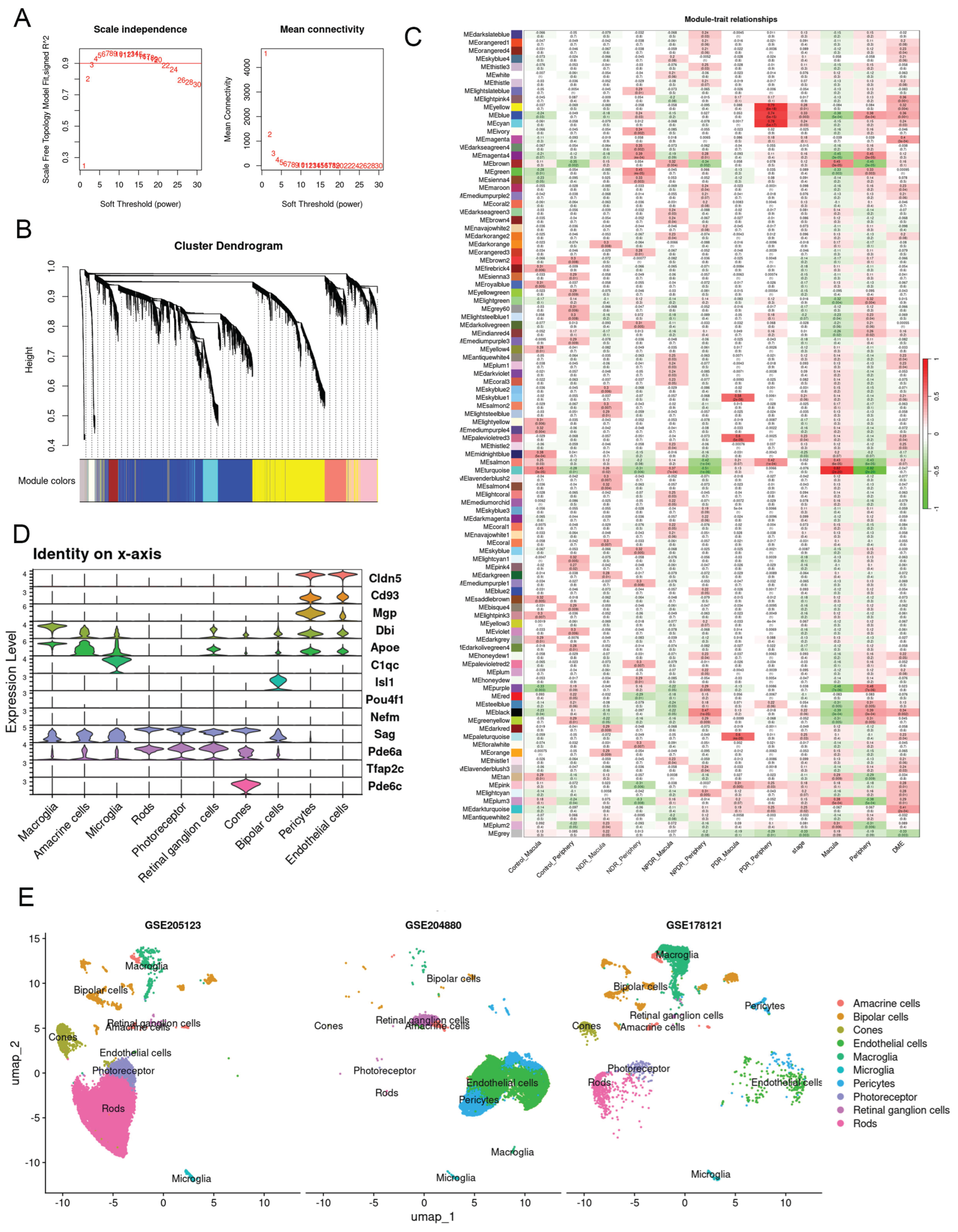
2.1. Identification of Key Gene Modules and Their Mapping to Single-Cell Transcriptomes
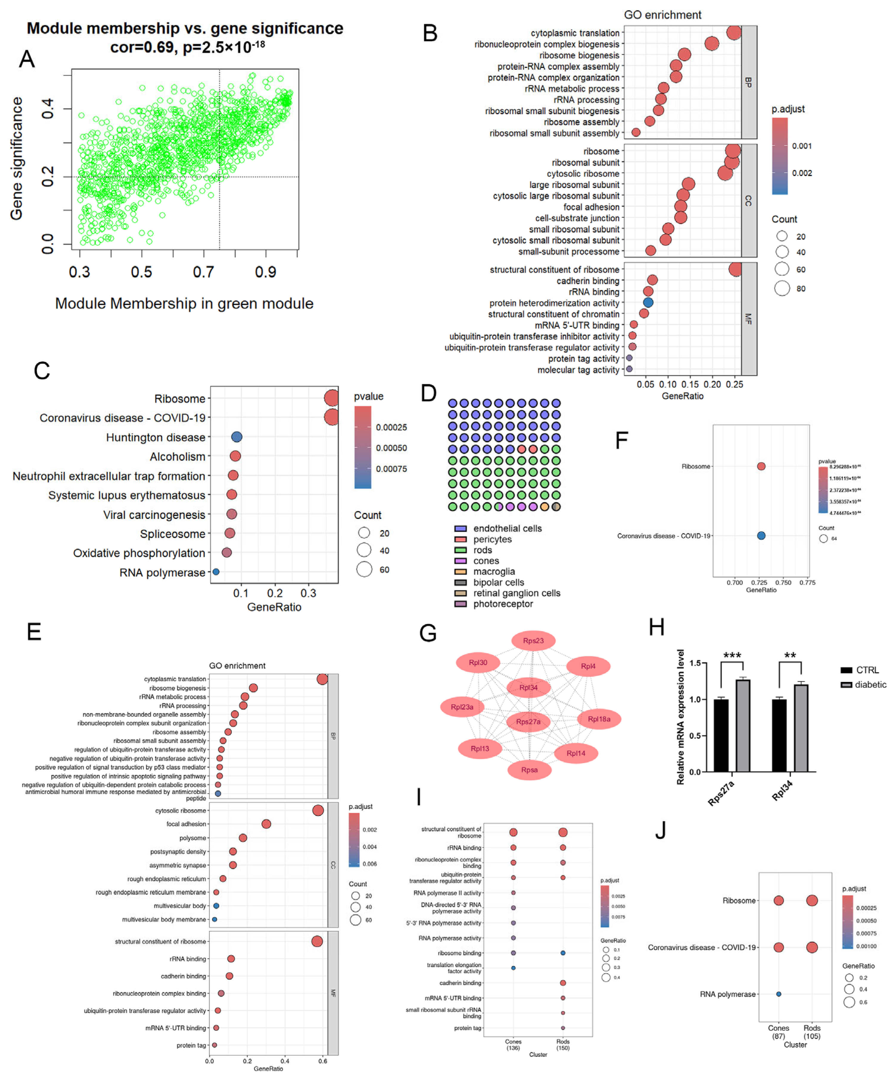
2.2. Cell Type-Specific Transcriptomic Alterations During the NDR Stage
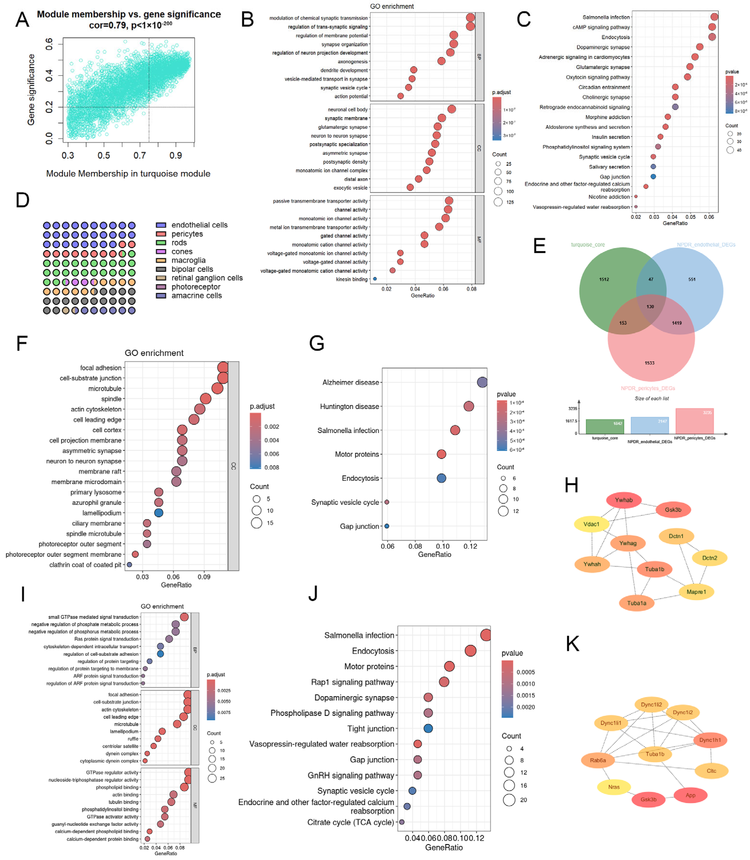
2.3. Cell Type-Specific Transcriptomic Alterations During the NPDR Stage
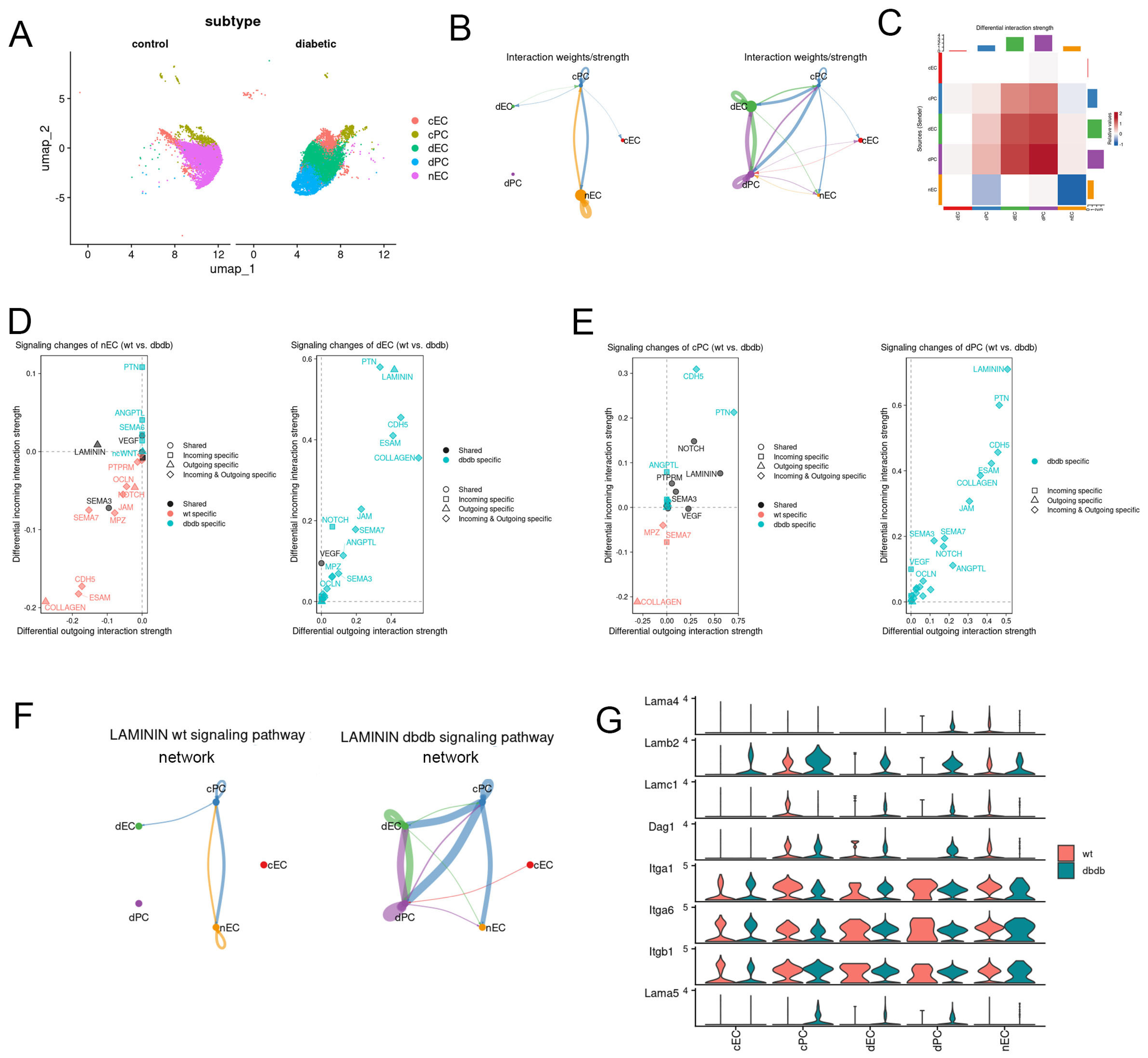
2.4. Cell Type-Specific Transcriptomic Alterations During the PDR Stage
2.5. Temporal Gene Expression Changes in Macroglia During Diabetic Retinopathy
3. Discussion
3.1. Experimental Design: Integration of Species and Sequencing Platforms
3.2. Early Transcriptional Alterations in Photoreceptors Suggest Ribosomal Response in NDR Retina
3.3. Endothelial and Pericyte Transcriptomic Remodeling Characterizes the Intermediate Stage of Diabetic Retinopathy
3.4. Activation of ANGPTL and LAMININ Signaling Indicates Multicellular Remodeling and ECM Dysregulation in Proliferative Diabetic Retinopathy
3.5. The Critical Role of Macroglia Cells in Diabetic Retinopathy and Their Associated Gene Signatures
3.6. Limitations
4. Materials and Methods
4.1. Access to GEO Datasets
4.2. Animals and Ethical Statement
4.3. Animal Model of Diabetic Retinopathy
4.4. Quantitative Real-Time PCR
4.5. Weighted Gene Co-Expression Network Analysis
4.6. Single-Cell RNA Sequencing Analysis
4.7. Subcluster Analysis
4.8. Differential Analysis
4.9. Identification of Cell Types Enriched for WGCNA Modules
4.10. Pathway Analysis
4.11. Protein–Protein Interaction (PPI) Network Construction and Analysis
4.12. CellChat Analysis
4.13. Analysis of the Impact of Hyperglycemia Duration on Macroglia Gene Expression
4.14. Software and Data Analysis Tools
5. Conclusions
Supplementary Materials
Author Contributions
Funding
Institutional Review Board Statement
Informed Consent Statement
Data Availability Statement
Acknowledgments
Conflicts of Interest
Abbreviations
| DR | diabetic retinopathy |
| NDR | no clinically apparent diabetic retinopathy |
| NPDR | non-proliferative diabetic retinopathy |
| PDR | proliferative diabetic retinopathy |
| RGC | retinal ganglion cell |
| scRNA-seq | single-cell RNA sequencing |
| WGCNA | weighted gene co-expression network analysis |
| bulk RNA-seq | bulk RNA sequencing |
| ME | module eigengene |
| MM | module membership |
| GS | gene significance |
| RPCA | reciprocal PCA |
| ECs | endothelial cells |
| PCs | pericytes |
| GO | gene ontology |
| KEGG | Kyoto Encyclopedia of Genes and Genomes |
| FDR | false discovery rate |
| MCC | maximal clique centrality |
| SD | standard deviation |
| BP | biological process |
| CC | cellular component |
| MF | molecular function |
| PPI | protein–protein interaction |
| nECs | normal ECs |
| dECs | diabetic ECs |
| cECs | common ECs |
| dPCs | divided into diabetic PCs |
| cPCs | common PCs |
| ECM | extracellular matrix |
| NTN4 | netrin-4 |
| LAMA1 | laminin subunit alpha-1 |
References
- Cheung, N.; Mitchell, P.; Wong, T.Y. Diabetic retinopathy. Lancet 2010, 376, 124–136. [Google Scholar] [CrossRef] [PubMed]
- Bryl, A.; Mrugacz, M.; Falkowski, M.; Zorena, K. The Effect of Diet and Lifestyle on the Course of Diabetic Retinopathy—A Review of the Literature. Nutrients 2022, 14, 1252. [Google Scholar] [CrossRef]
- Teo, Z.L.; Tham, Y.-C.; Yu, M.; Chee, M.L.; Rim, T.H.; Cheung, N.; Bikbov, M.M.; Wang, Y.X.; Tang, Y.; Lu, Y.; et al. Global Prevalence of Diabetic Retinopathy and Projection of Burden through 2045. Ophthalmology 2021, 128, 1580–1591. [Google Scholar] [CrossRef]
- Kim, I.M.; Radgoudarzi, N.; Chen, E.M.; Liu, J.; Moussa, K.; Shantha, J.G.; Tsui, E.; Gore, C.; Baxter, S.L.; Porco, T.C.; et al. Time to Progression to Proliferative Diabetic Retinopathy in Patients with Type 2 Diabetes. JAMA Netw. Open 2025, 8, e2521150. [Google Scholar] [CrossRef]
- Yue, T.; Shi, Y.; Luo, S.; Weng, J.; Wu, Y.; Zheng, X. The role of inflammation in immune system of diabetic retinopathy: Molecular mechanisms, pathogenetic role and therapeutic implications. Front. Immunol. 2022, 13, 1055087. [Google Scholar] [CrossRef] [PubMed]
- Antonetti, D.A.; Silva, P.S.; Stitt, A.W. Current understanding of the molecular and cellular pathology of diabetic retinopathy. Nat. Rev. Endocrinol. 2021, 17, 195–206, Correction in Nat. Rev. Endocrinol. 2025, 21, 62. [Google Scholar] [CrossRef]
- Peng, H.; Hu, Q.; Zhang, X.; Huang, J.; Luo, S.; Zhang, Y.; Jiang, B.; Sun, D. Identifying Therapeutic Targets and Potential Drugs for Diabetic Retinopathy: Focus on Oxidative Stress and Immune Infiltration. J. Inflamm. Res. 2025, 18, 2205–2227. [Google Scholar] [CrossRef]
- Sachdeva, M.M. Retinal Neurodegeneration in Diabetes: An Emerging Concept in Diabetic Retinopathy. Curr. Diabetes Rep. 2021, 21, 65. [Google Scholar] [CrossRef]
- Ren, J.; Zhang, S.; Pan, Y.; Jin, M.; Li, J.; Luo, Y.; Sun, X.; Li, G. Diabetic retinopathy: Involved cells, biomarkers, and treatments. Front. Pharmacol. 2022, 13, 953691. [Google Scholar] [CrossRef] [PubMed]
- Fu, D.; Yu, J.Y.; Yang, S.; Wu, M.; Hammad, S.M.; Connell, A.R.; Du, M.; Chen, J.; Lyons, T.J. Survival or death: A dual role for autophagy in stress-induced pericyte loss in diabetic retinopathy. Diabetologia 2016, 59, 2251–2261. [Google Scholar] [CrossRef]
- Du, M.; Zhao, X.; Guo, M.; Wang, X.; Zhang, Y.; Yang, L.; Liu, S.; Sun, L.; Liao, M.; Dong, X.; et al. Drugless peptide-based nanohybrids alleviate diabetic retinopathy by suppressing microglial activation and endothelial inflammation. Theranostics 2025, 15, 3943–3960. [Google Scholar] [CrossRef] [PubMed]
- Qiu, A.-W.; Huang, D.-R.; Li, B.; Fang, Y.; Zhang, W.-W.; Liu, Q.-H. IL-17A injury to retinal ganglion cells is mediated by retinal Muller cells in diabetic retinopathy. Cell Death Dis. 2021, 12, 1057. [Google Scholar] [CrossRef]
- Becker, K.; Klein, H.; Simon, E.; Viollet, C.; Haslinger, C.; Leparc, G.; Schultheis, C.; Chong, V.; Kuehn, M.H.; Fernandez-Albert, F.; et al. In-depth transcriptomic analysis of human retina reveals molecular mechanisms underlying diabetic retinopathy. Sci. Rep. 2021, 11, 10494. [Google Scholar] [CrossRef]
- Xu, Y.; Xiang, Z.; E, W.; Lang, Y.; Huang, S.; Qin, W.; Yang, J.; Chen, Z.; Liu, Z. Single-cell transcriptomes reveal a molecular link between diabetic kidney and retinal lesions. Commun. Biol. 2023, 6, 912. [Google Scholar] [CrossRef]
- Sun, L.; Wang, R.; Hu, G.; Liu, H.; Lv, K.; Duan, Y.; Shen, N.; Wu, J.; Hu, J.; Liu, Y.; et al. Single cell RNA sequencing (scRNA-Seq) deciphering pathological alterations in streptozotocin-induced diabetic retinas. Exp. Eye Res. 2021, 210, 108718. [Google Scholar] [CrossRef]
- Olivares, A.M.; Althoff, K.; Chen, G.F.; Wu, S.; Morrisson, M.A.; DeAngelis, M.M.; Haider, N. Animal Models of Diabetic Retinopathy. Curr. Diab. Rep. 2017, 17, 93. [Google Scholar] [CrossRef]
- Su, L.; Ji, J.; Bian, J.; Fu, Y.; Ge, Y.; Yuan, Z. Tacrolimus (FK506) prevents early retinal neovascularization in streptozotocin-induced diabetic mice. Int. Immunopharmacol. 2012, 14, 606–612. [Google Scholar] [CrossRef]
- Hernández, C.; Bogdanov, P.; Corraliza, L.; García-Ramírez, M.; Solà-Adell, C.; Arranz, J.A.; Arroba, A.I.; Valverde, A.M.; Simó, R. Topical Administration of GLP-1 Receptor Agonists Prevents Retinal Neurodegeneration in Experimental Diabetes. Diabetes 2016, 65, 172–187. [Google Scholar] [CrossRef]
- Sohn, E.H.; van Dijk, H.W.; Jiao, C.; Kok, P.H.B.; Jeong, W.; Demirkaya, N.; Garmager, A.; Wit, F.; Kucukevcilioglu, M.; van Velthoven, M.E.J.; et al. Retinal neurodegeneration may precede microvascular changes characteristic of diabetic retinopathy in diabetes mellitus. Proc. Natl. Acad. Sci. USA 2016, 113, E2655–E2664. [Google Scholar] [CrossRef] [PubMed]
- Otsuka, K.; Morita, A.; Kashihara, T.; Nakahara, T. Pharmacological depletion of pericytes induces diabetic retinopathy-like abnormal blood vessels in neonatal rat retina. Exp. Eye Res. 2025, 251, 110243. [Google Scholar] [CrossRef] [PubMed]
- Li, J.; Wang, J.J.; Yu, Q.; Chen, K.; Mahadev, K.; Zhang, S.X. Inhibition of Reactive Oxygen Species by Lovastatin Downregulates Vascular Endothelial Growth Factor Expression and Ameliorates Blood-Retinal Barrier Breakdown in db/db Mice. Diabetes 2010, 59, 1528–1538. [Google Scholar] [CrossRef]
- Sivaprasad, S.; Prevost, A.T.; Vasconcelos, J.C.; Riddell, A.; Murphy, C.; Kelly, J.; Bainbridge, J.; Tudor-Edwards, R.; Hopkins, D.; Hykin, P.; et al. Clinical efficacy of intravitreal aflibercept versus panretinal photocoagulation for best corrected visual acuity in patients with proliferative diabetic retinopathy at 52 weeks (CLARITY): A multicentre, single-blinded, randomised, controlled, phase 2b, non-inferiority trial. Lancet 2017, 389, 2193–2203. [Google Scholar] [PubMed]
- Corano Scheri, K.; Lavine, J.A.; Tedeschi, T.; Thomson, B.R.; Fawzi, A.A. Single-cell transcriptomics analysis of proliferative diabetic retinopathy fibrovascular membranes reveals AEBP1 as fibrogenesis modulator. JCI Insight 2023, 8, e172062. [Google Scholar] [CrossRef] [PubMed]
- Yan, W.; Peng, Y.-R.; van Zyl, T.; Regev, A.; Shekhar, K.; Juric, D.; Sanes, J.R. Cell Atlas of The Human Fovea and Peripheral Retina. Sci. Rep. 2020, 10, 9802. [Google Scholar] [CrossRef]
- Peng, Y.R.; Shekhar, K.; Yan, W.; Herrmann, D.; Sappington, A.; Bryman, G.S.; van Zyl, T.; Do, M.T.H.; Regev, A.; Sanes, J.R. Molecular Classification and Comparative Taxonomics of Foveal and Peripheral Cells in Primate Retina. Cell 2019, 176, 1222–1237.e22. [Google Scholar] [CrossRef] [PubMed]
- Lu, Y.; Shiau, F.; Yi, W.; Lu, S.; Wu, Q.; Pearson, J.D.; Kallman, A.; Zhong, S.; Hoang, T.; Zuo, Z.; et al. Single-Cell Analysis of Human Retina Identifies Evolutionarily Conserved and Species-Specific Mechanisms Controlling Development. Dev. Cell 2020, 53, 473–491.e9. [Google Scholar] [CrossRef]
- Stitt, A.W.; Curtis, T.M.; Chen, M.; Medina, R.J.; McKay, G.J.; Jenkins, A.; Gardiner, T.A.; Lyons, T.J.; Hammes, H.-P.; Simó, R.; et al. The progress in understanding and treatment of diabetic retinopathy. Prog. Retin. Eye Res. 2016, 51, 156–186. [Google Scholar] [CrossRef]
- Gava, G.P.; McHugh, S.B.; Lefèvre, L.; Lopes-Dos-Santos, V.; Trouche, S.; El-Gaby, M.; Schultz, S.R.; Dupret, D. Integrating new memories into the hippocampal network activity space. Nat. Neurosci. 2021, 24, 326–330. [Google Scholar] [CrossRef]
- Hammes, H.P. Diabetic retinopathy: Hyperglycaemia, oxidative stress and beyond. Diabetologia 2018, 61, 29–38. [Google Scholar] [CrossRef]
- Heimberg, G.; Bhatnagar, R.; El-Samad, H.; Thomson, M. Low Dimensionality in Gene Expression Data Enables the Accurate Extraction of Transcriptional Programs from Shallow Sequencing. Cell Syst. 2016, 2, 239–250. [Google Scholar] [CrossRef]
- Luo, S.; Alwattar, B.; Li, Q.; Bora, K.; Blomfield, A.K.; Lin, J.; Fulton, A.; Chen, J.; Agrawal, P.B. HBS1L deficiency causes retinal dystrophy in a child and in a mouse model associated with defective development of photoreceptor cells. Dis. Model. Mech. 2024, 17, dmm050557. [Google Scholar] [CrossRef]
- Zhang, R.; Sun, J.; Xie, Y.; Zhu, W.; Tao, M.; Chen, Y.; Xie, W.; Bade, R.; Jiang, S.; Liu, X.; et al. Mutant kri1l causes abnormal retinal development via cell cycle arrest and apoptosis induction. Cell Death Discov. 2024, 10, 251. [Google Scholar] [CrossRef] [PubMed]
- Han, R.; Jin, M.; Xu, G.; He, J. Progressively Decreased HCN1 Channels Results in Cone Morphological Defects in Diabetic Retinopathy. J. Neurosci. 2022, 42, 8200–8212. [Google Scholar] [CrossRef]
- Moore-Dotson, J.M.; Beckman, J.J.; Mazade, R.E.; Hoon, M.; Bernstein, A.S.; Romero-Aleshire, M.J.; Brooks, H.L.; Eggers, E.D. Early Retinal Neuronal Dysfunction in Diabetic Mice: Reduced Light-Evoked Inhibition Increases Rod Pathway Signaling. Investig. Opthalmology Vis. Sci. 2016, 57, 1418–1430. [Google Scholar] [CrossRef] [PubMed]
- McLaughlin, T.; Wang, G.; Medina, A.; Perkins, J.; Nihlawi, R.; Seyfried, D.; Hu, Z.; Wang, J.J.; Zhang, S.X. Essential Role of XBP1 in Maintaining Photoreceptor Synaptic Integrity in Early Diabetic Retinopathy. Investig. Opthalmology Vis. Sci. 2023, 64, 40. [Google Scholar] [CrossRef] [PubMed]
- Liu, H.; Tang, J.; Du, Y.; Saadane, A.; Samuels, I.; Veenstra, A.; Kiser, J.Z.; Palczewski, K.; Kern, T.S. Transducin1, Phototransduction and the Development of Early Diabetic Retinopathy. Investig. Opthalmology Vis. Sci. 2019, 60, 1538–1546. [Google Scholar] [CrossRef]
- McAnany, J.J.; Park, J.C. Rod photoreceptor activation and deactivation in early-stage diabetic eye disease. Doc. Ophthalmol. 2023, 146, 229–239. [Google Scholar] [CrossRef]
- McAnany, J.J.; Park, J.C. Cone Photoreceptor Dysfunction in Early-Stage Diabetic Retinopathy: Association Between the Activation Phase of Cone Phototransduction and the Flicker Electroretinogram. Investig. Opthalmology Vis. Sci. 2019, 60, 64–72. [Google Scholar] [CrossRef]
- Montellese, C.; van den Heuvel, J.; Ashiono, C.; Dörner, K.; Melnik, A.; Jonas, S.; Zemp, I.; Picotti, P.; Gillet, L.C.; Kutay, U. USP16 counteracts mono-ubiquitination of RPS27a and promotes maturation of the 40S ribosomal subunit. Elife 2020, 9, e54435. [Google Scholar] [CrossRef] [PubMed]
- Xia, M.; Jiao, L.; Wang, X.-H.; Tong, M.; Yao, M.-D.; Li, X.-M.; Yao, J.; Li, D.; Zhao, P.-Q.; Yan, B. Single-cell RNA sequencing reveals a unique pericyte type associated with capillary dysfunction. Theranostics 2023, 13, 2515–2530. [Google Scholar] [CrossRef]
- Yao, X.; Zhao, Z.; Zhang, W.; Liu, R.; Ni, T.; Cui, B.; Lei, Y.; Du, J.; Ai, D.; Jiang, H.; et al. Specialized Retinal Endothelial Cells Modulate Blood-Retina Barrier in Diabetic Retinopathy. Diabetes 2024, 73, 225–236. [Google Scholar] [CrossRef] [PubMed]
- Zhang, X.; Liu, D.; Qiu, Y.; Hu, R.; Chen, S.; Xu, Y.; Qian, C.; Du, L.; Yuan, J.; Li, X. Recent Advancements in Understanding the Role and Mechanisms of Angiopoietin-like Proteins in Diabetic Retinopathy. Metabolites 2025, 15, 352. [Google Scholar] [CrossRef] [PubMed]
- Khanani, A.M.; Russell, M.W.; A Aziz, A.; Danzig, C.J.; Weng, C.Y.; A Eichenbaum, D.; Singh, R.P. Angiopoietins as Potential Targets in Management of Retinal Disease. Clin. Ophthalmol. 2021, 15, 3747–3755. [Google Scholar] [CrossRef]
- Lu, Q.; Zou, W.; Chen, B.; Zou, C.; Zhao, M.; Zheng, Z. ANGPTL-4 correlates with vascular endothelial growth factor in patients with proliferative diabetic retinopathy. Graefes Arch. Clin. Exp. Ophthalmol. 2016, 254, 1281–1288. [Google Scholar] [CrossRef]
- Xu, Q.; Gong, C.; Qiao, L.; Feng, R.; Liu, H.; Liu, Y.; Ji, S.; Zhang, Y.; Wu, S.; Li, S. Aqueous Level of ANGPTL4 Correlates with the OCTA Metrics of Diabetic Macular Edema in NPDR. J. Diabetes Res. 2022, 2022, 8435603. [Google Scholar] [CrossRef]
- Aryal, B.; Price, N.L.; Suarez, Y.; Fernández-Hernando, C. ANGPTL4 in Metabolic and Cardiovascular Disease. Trends Mol. Med. 2019, 25, 723–734. [Google Scholar] [CrossRef]
- Kwon, S.H.; Shin, J.P.; Kim, I.T.; Park, D.H. Aqueous Levels of Angiopoietin-like 4 and Semaphorin 3E Correlate with Nonperfusion Area and Macular Volume in Diabetic Retinopathy. Ophthalmology 2015, 122, 968–975. [Google Scholar] [CrossRef] [PubMed]
- Crespo-Garcia, S.; Reichhart, N.; Kociok, N.; Skosyrski, S.; Joussen, A.M. Anti-Inflammatory Role of Netrin-4 in Diabetic Retinopathy. Int. J. Mol. Sci. 2021, 22, 4481. [Google Scholar] [CrossRef]
- Song, G.; Lin, D.; Bao, L.; Jiang, Q.; Zhang, Y.; Zheng, H.; Gao, Q. Effects of High Glucose on the Expression of LAMA1 and Biological Behavior of Choroid Retinal Endothelial Cells. J. Diabetes Res. 2018, 2018, 7504614. [Google Scholar] [CrossRef]
- Thompson, K.; Chen, J.; Luo, Q.; Xiao, Y.; Cummins, T.R.; Bhatwadekar, A.D. Advanced glycation end (AGE) product modification of laminin downregulates Kir4.1 in retinal Müller cells. PLoS ONE 2018, 13, e0193280. [Google Scholar] [CrossRef]
- Jin, S.; Guerrero-Juarez, C.F.; Zhang, L.; Chang, I.; Ramos, R.; Kuan, C.-H.; Myung, P.; Plikus, M.V.; Nie, Q. Inference and analysis of cell-cell communication using CellChat. Nat. Commun. 2021, 12, 1088. [Google Scholar] [CrossRef]
- Padovani-Claudio, D.A.; Ramos, C.J.; Capozzi, M.E.; Penn, J.S. Elucidating glial responses to products of diabetes-associated systemic dyshomeostasis. Prog. Retin. Eye Res. 2023, 94, 101151. [Google Scholar] [CrossRef]
- Van Hove, I.; De Groef, L.; Boeckx, B.; Modave, E.; Hu, T.T.; Beets, K.; Etienne, I.; Van Bergen, T.; Lambrechts, D.; Moons, L.; et al. Single-cell transcriptome analysis of the Akimba mouse retina reveals cell-type-specific insights into the pathobiology of diabetic retinopathy. Diabetologia 2020, 63, 2235–2248. [Google Scholar] [CrossRef]
- Xu, H.; Zhang, L.-B.; Luo, Y.-Y.; Wang, L.; Zhang, Y.-P.; Chen, P.-Q.; Ba, X.-Y.; Han, J.; Luo, H. Synaptotagmins family affect glucose transport in retinal pigment epithelial cells through their ubiquitination-mediated degradation and glucose transporter-1 regulation. World J. Diabetes 2024, 15, 958–976. [Google Scholar] [CrossRef]
- Mealer, R.G.; Williams, S.E.; Daly, M.J.; Scolnick, E.M.; Cummings, R.D.; Smoller, J.W. Glycobiology and schizophrenia: A biological hypothesis emerging from genomic research. Mol. Psychiatry 2020, 25, 3129–3139. [Google Scholar] [CrossRef]
- Morita, I.; Kakuda, S.; Takeuchi, Y.; Kawasaki, T.; Oka, S. HNK-1 (human natural killer-1) glyco-epitope is essential for normal spine morphogenesis in developing hippocampal neurons. Neuroscience 2009, 164, 1685–1694. [Google Scholar] [CrossRef]
- Hoppa, M.B.; Lana, B.; Margas, W.; Dolphin, A.C.; Ryan, T.A. α2δ expression sets presynaptic calcium channel abundance and release probability. Nature 2012, 486, 122–125. [Google Scholar] [CrossRef]
- Kapur, M.; Ganguly, A.; Nagy, G.; Adamson, S.I.; Chuang, J.H.; Frankel, W.N.; Ackerman, S.L. Expression of the Neuronal tRNA n-Tr20 Regulates Synaptic Transmission and Seizure Susceptibility. Neuron 2020, 108, 193–208.e9. [Google Scholar] [CrossRef]
- Liu, N.; Li, R.; Cao, J.; Song, X.; Ma, W.; Liu, T.; Wang, L.; Zou, J.; Zhang, B.; Liu, Z.; et al. The inhibition of FKBP5 protects β-cell survival under inflammation stress via AKT/FOXO1 signaling. Cell Death Discov. 2023, 9, 247. [Google Scholar] [CrossRef]
- Reed, M.J.; Meszaros, K.; Entes, L.; Claypool, M.; Pinkett, J.; Gadbois, T.; Reaven, G. A new rat model of type 2 diabetes: The fat-fed, streptozotocin-treated rat. Metabolism 2000, 49, 1390–1394. [Google Scholar] [CrossRef]
- Augustine-Wofford, K.; Connaughton, V.P.; McCarthy, E. Are Hyperglycemia-Induced Changes in the Retina Associated with Diabetes-Correlated Changes in the Brain? A Review from Zebrafish and Rodent Type 2 Diabetes Models. Biology 2024, 13, 477. [Google Scholar] [CrossRef] [PubMed]
- Yu, G.; Wang, L.-G.; Han, Y.; He, Q.-Y. clusterProfiler: An R Package for Comparing Biological Themes Among Gene Clusters. OMICS J. Integr. Biol. 2012, 16, 284–287. [Google Scholar] [CrossRef] [PubMed]
- Bardou, P.; Mariette, J.; Escudié, F.; Djemiel, C.; Klopp, C. jvenn: An interactive Venn diagram viewer. BMC Bioinform. 2014, 15, 293. [Google Scholar] [CrossRef] [PubMed]



| GEO Accession | Species | Sequencing Platforms | Conditions/Treatments | Duration of Hyperglycemia | Cell Numbers | References |
|---|---|---|---|---|---|---|
| GSE160306 | Homo sapiens | bulk sequencing | ctrl/NDR/NPDR/PDR | - | - | [13] |
| GSE205123 | Mus musculus | single-cell sequencing | db/db | 13-6 weeks | 18,170 | none |
| GSE204880 | Mus musculus | single-cell sequencing | db/db | 21-6 weeks | 15,210 | [14] |
| GSE178121 | Mus musculus | single-cell sequencing | STZ | 25 weeks | 6359 | [15] |
| GEO Accession | Antibody-Based Sorting | Cell/Tissue Type Composition |
|---|---|---|
| GSE160306 | none | macula and retinal periphery |
| GSE205123 | none | a diverse composition of retinal cell types |
| GSE204880 | PECAM1 and CD104b to enrich EC and PC | ECs and PCs of glomerular and retinal |
| GSE178121 | CD73 to reduce rods | a diverse composition of retinal cell types with low proportion of rods |
Disclaimer/Publisher’s Note: The statements, opinions and data contained in all publications are solely those of the individual author(s) and contributor(s) and not of MDPI and/or the editor(s). MDPI and/or the editor(s) disclaim responsibility for any injury to people or property resulting from any ideas, methods, instructions or products referred to in the content. |
© 2026 by the authors. Licensee MDPI, Basel, Switzerland. This article is an open access article distributed under the terms and conditions of the Creative Commons Attribution (CC BY) license.
Share and Cite
Li, Y.; Liu, L.; Zhang, Y.; Ouyang, L.; Chen, X.; Huang, J.; Ke, M. Stage-Associated Cellular and Molecular Signatures in Diabetic Retinopathy Identified Through Integrated Bulk and Single-Cell Transcriptomic Analysis. Int. J. Mol. Sci. 2026, 27, 2775. https://doi.org/10.3390/ijms27062775
Li Y, Liu L, Zhang Y, Ouyang L, Chen X, Huang J, Ke M. Stage-Associated Cellular and Molecular Signatures in Diabetic Retinopathy Identified Through Integrated Bulk and Single-Cell Transcriptomic Analysis. International Journal of Molecular Sciences. 2026; 27(6):2775. https://doi.org/10.3390/ijms27062775
Chicago/Turabian StyleLi, Ying, Lian Liu, Yuan Zhang, Lingyi Ouyang, Xiaomin Chen, Jingqiu Huang, and Min Ke. 2026. "Stage-Associated Cellular and Molecular Signatures in Diabetic Retinopathy Identified Through Integrated Bulk and Single-Cell Transcriptomic Analysis" International Journal of Molecular Sciences 27, no. 6: 2775. https://doi.org/10.3390/ijms27062775
APA StyleLi, Y., Liu, L., Zhang, Y., Ouyang, L., Chen, X., Huang, J., & Ke, M. (2026). Stage-Associated Cellular and Molecular Signatures in Diabetic Retinopathy Identified Through Integrated Bulk and Single-Cell Transcriptomic Analysis. International Journal of Molecular Sciences, 27(6), 2775. https://doi.org/10.3390/ijms27062775