High-Throughput Sequencing Decodes tsRNA Landscapes: Insights into Cancer Biomarkers and Therapeutic Targets
 ,
,
Abstract
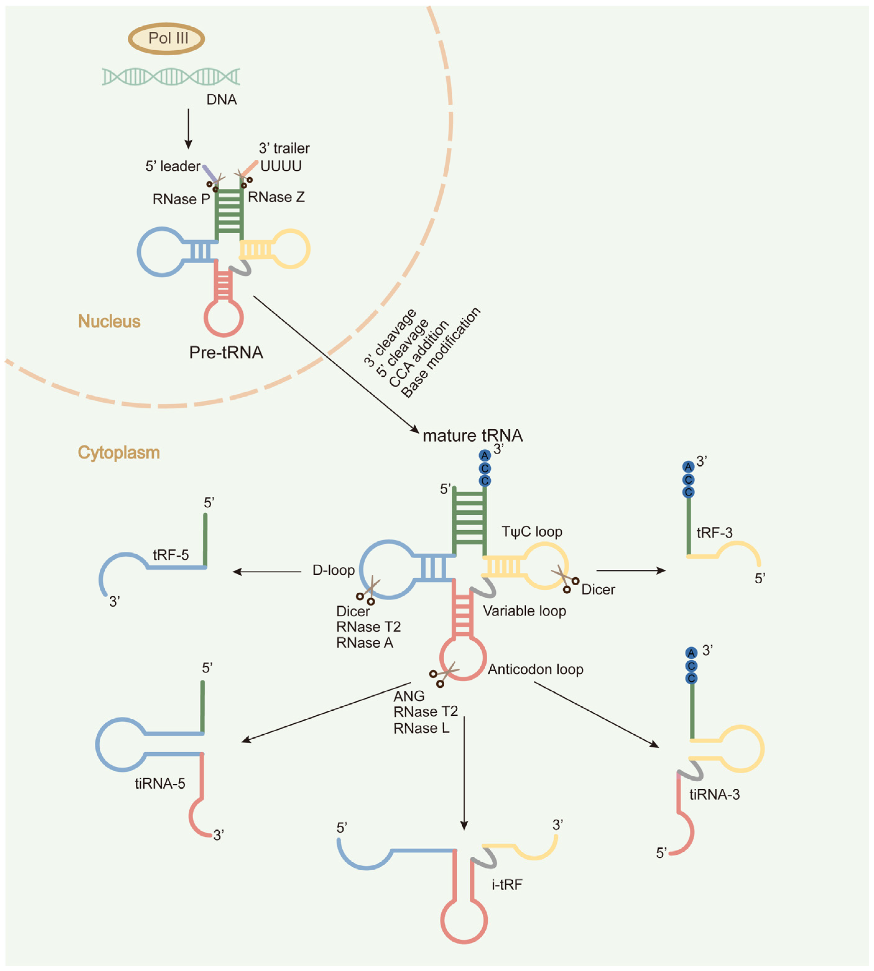
1. Introduction
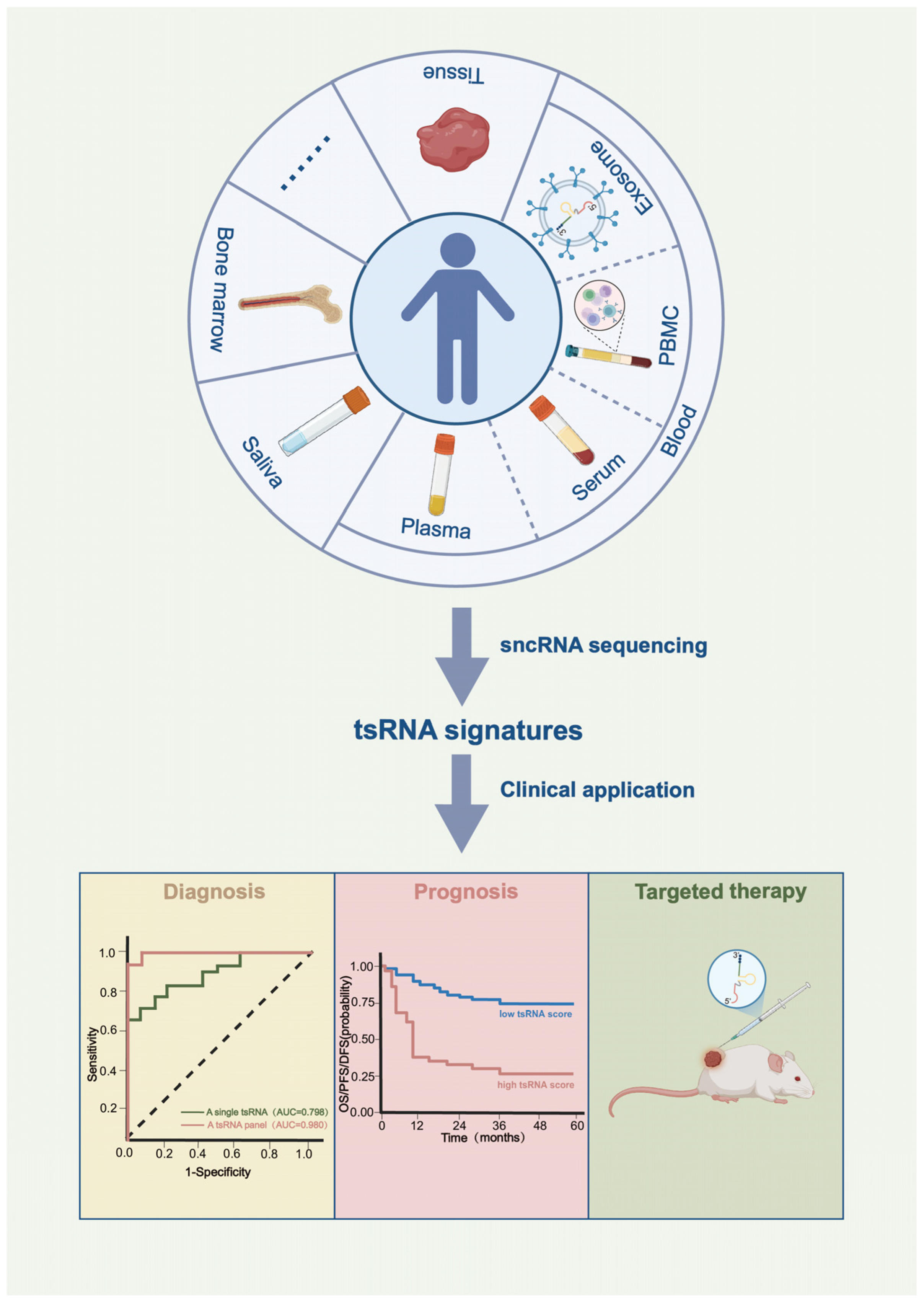
2. tsRNAs as Potential Diagnostic Biomarkers for Cancer
2.1. Tissue Samples
2.2. Peripheral Blood Samples
2.3. Others


| Cancer Type | Sequencing Sample Type | Sequencing Method | Potential Clinical Utility | References | ||
|---|---|---|---|---|---|---|
| Diagnosis | Prognosis | Targeted Therapy | ||||
| HCC | Tissue | Optimized-seq | √ | √ | [43] | |
| Tissue | Traditional-seq | √ | [76,77] | |||
| Tissue | Optimized-seq | √ | [78] | |||
| Serum | Traditional-seq | √ | √ | [79] | ||
| Plasma exosome | Traditional-seq | √ | [68] | |||
| Lung cancer | Tissue | Traditional-seq | √ | [31] | ||
| Plasma | Optimized-seq | √ | √ | [61] | ||
| Plasma | SLiPiR-seq | √ | [62] | |||
| Serum | Optimized-seq | √ | [80] | |||
| PBMC | Traditional-seq | √ | [73] | |||
| CRC | Tissue | Optimized-seq | √ | √ | [39,81] | |
| Tissue | Optimized-seq | √ | √ | √ | [82] | |
| Tissue Plasma exosome | Traditional-seq | √ | √ | [32] | ||
| Plasma | Optimized-seq | √ | [63] | |||
| Breast cancer | Tissue | Optimized-seq | √ | [23,44] | ||
| Plasma | Optimized-seq | √ | √ | [64] | ||
| Cell line | Traditional-seq | √ | [20,29] | |||
| Gastric cancer | Tissue | Traditional-seq | √ | √ | [22] | |
| Tissue | Optimized-seq | √ | √ | [42] | ||
| Thyroid cancer | Tissue | Optimized-seq | √ | √ | [40,45] | |
| Pancreatic | Tissue | Optimized-seq | √ | √ | √ | [17] |
| Serum | Optimized-seq | √ | √ | √ | [60] | |
| RCC | Tissues | Traditional-seq | √ | [33] | ||
| Tissue Plasma | Optimized-seq | √ | [83] | |||
| ESCC | Saliva exosome | Traditional-seq | √ | √ | [74] | |
| Ovarian cancer | Serum | Traditional-seq | √ | [65] | ||
| Prostate cancer | Tissue | Traditional-seq | √ | √ | [84] | |
| LSCC | Tissue | Optimized-seq | √ | √ | [41] | |
| Bladder cancer | Plasma | Traditional-seq | √ | [85] | ||
| AML | Serum Bone marrow | Traditional-seq | √ | √ | [75] | |
| MM | Serum exosome | Optimized-seq | √ | [86] | ||
3. tsRNAs as Potential Prognostic Biomarkers for Cancer
4. tsRNAs as Potential Therapeutic Targets for Cancer
5. Conclusions and Prospects
Author Contributions
Funding
Data Availability Statement
Conflicts of Interest
Abbreviations
| AML | Acute myeloid leukemia |
| AUC | Area under the curve |
| CA19-9 | Carbohydrate Antigen 19-9 |
| CACNA1d | Calcium voltage-gated channel subunit alpha1 D |
| CEA | Carcinoembryonic Antigen |
| CLL | Chronic lymphocytic leukemia |
| C-MYC | MYC proto-oncogene, bHLH transcription factor |
| CRC | Colorectal cancer |
| CT | Computed Tomography |
| DFS | Disease-free survival |
| EEF1A2 | Eukaryotic translation elongation factor 1 alpha 2 |
| ESCC | Esophageal squamous cell carcinoma |
| EVs | Extracellular vesicles |
| FOXK1 | Forkhead box K1 |
| FUBP1 | Far upstream element binding protein 1 |
| HCC | Hepatocellular carcinoma |
| LATS2 | Large Tumor Suppressor Kinase 2 |
| LGALS1 | Galectin 1 |
| LNA | Locked nucleic acid |
| LSCC | Laryngeal squamous cell carcinoma |
| MDM2 | MDM2 proto-oncogene |
| miRNA | microRNA |
| MM | Multiple myeloma |
| mRNA | Messenger RNA |
| NSCLC | Non-small cell lung cancer |
| OS | Overall survival |
| PBMCs | Peripheral blood mononuclear cells |
| PDX | Patient-derived xenograft |
| PDXO | Patient-derived xenograft organoid |
| PFS | Progression-free survival |
| pre-tRNAs | Precursors tRNAs |
| qRT-PCR | quantitative reverse-transcription PCR |
| RBM17 | RNA binding motif protein 17 |
| RCC | Renal cell carcinoma |
| RG4 | RNA G-quadruplex |
| RSP | Prognostic risk score |
| sncRNAs | Small non-coding RNAs |
| TCL1 | T-Cell leukemia/lymphoma 1 |
| tRNAs | Transfer RNAs |
| tsRNAs | Transfer RNA-derived small RNAs |
| UTR | Untranslated region |
| YBX-1 | Y-box binding protein 1 |
References
- Schimmel, P. The emerging complexity of the tRNA world: Mammalian tRNAs beyond protein synthesis. Nat. Rev. Mol. Cell Biol. 2018, 19, 45–58. [Google Scholar] [CrossRef] [Scilit]
- Li, Y.; Luo, J.; Zhou, H.; Liao, J.Y.; Ma, L.M.; Chen, Y.Q.; Qu, L.H. Stress-induced tRNA-derived RNAs: A novel class of small RNAs in the primitive eukaryote Giardia lamblia. Nucleic Acids Res. 2008, 36, 6048–6055. [Google Scholar] [CrossRef] [Scilit]
- Yamasaki, S.; Ivanov, P.; Hu, G.F.; Anderson, P. Angiogenin cleaves tRNA and promotes stress-induced translational repression. J. Cell Biol. 2009, 185, 35–42. [Google Scholar] [CrossRef] [Scilit] [PubMed]
- Lee, Y.S.; Shibata, Y.; Malhotra, A.; Dutta, A. A novel class of small RNAs: tRNA-derived RNA fragments (tRFs). Genes Dev. 2009, 23, 2639–2649. [Google Scholar] [CrossRef] [Scilit] [PubMed]
- Haussecker, D.; Huang, Y.; Lau, A.; Parameswaran, P.; Fire, A.Z.; Kay, M.A. Human tRNA-derived small RNAs in the global regulation of RNA silencing. RNA 2010, 16, 673–695. [Google Scholar] [CrossRef] [Scilit] [PubMed]
- Liu, B.; Cao, J.; Wang, X.; Guo, C.; Liu, Y.; Wang, T. Deciphering the tRNA-derived small RNAs: Origin, development, and future. Cell Death Dis. 2021, 13, 24. [Google Scholar] [CrossRef] [Scilit]
- Torres, A.G.; Reina, O.; Stephan-Otto Attolini, C.; Ribas de Pouplana, L. Differential expression of human tRNA genes drives the abundance of tRNA-derived fragments. Proc. Natl. Acad. Sci. USA 2019, 116, 8451–8456. [Google Scholar] [CrossRef] [Scilit]
- Chen, Q.; Zhang, X.; Shi, J.; Yan, M.; Zhou, T. Origins and evolving functionalities of tRNA-derived small RNAs. Trends Biochem. Sci. 2021, 46, 790–804. [Google Scholar] [CrossRef] [Scilit]
- Shi, J.; Zhou, T.; Chen, Q. Exploring the expanding universe of small RNAs. Nat. Cell Biol. 2022, 24, 415–423. [Google Scholar] [CrossRef] [Scilit]
- Chen, Q.; Zhou, T. Emerging functional principles of tRNA-derived small RNAs and other regulatory small RNAs. J. Biol. Chem. 2023, 299, 105225. [Google Scholar] [CrossRef] [Scilit]
- Kuhle, B.; Chen, Q.; Schimmel, P. tRNA renovatio: Rebirth through fragmentation. Mol. Cell 2023, 83, 3953–3971. [Google Scholar] [CrossRef] [Scilit]
- Xu, D.; Qiao, D.; Lei, Y.; Zhang, C.; Bu, Y.; Zhang, Y. Transfer RNA-derived small RNAs (tsRNAs): Versatile regulators in cancer. Cancer Lett. 2022, 546, 215842. [Google Scholar] [CrossRef] [Scilit]
- Kumar, P.; Kuscu, C.; Dutta, A. Biogenesis and Function of Transfer RNA-Related Fragments (tRFs). Trends Biochem. Sci. 2016, 41, 679–689. [Google Scholar] [CrossRef] [Scilit] [PubMed]
- Balatti, V.; Rizzotto, L.; Miller, C.; Palamarchuk, A.; Fadda, P.; Pandolfo, R.; Rassenti, L.Z.; Hertlein, E.; Ruppert, A.S.; Lozanski, A.; et al. TCL1 targeting miR-3676 is codeleted with tumor protein p53 in chronic lymphocytic leukemia. Proc. Natl. Acad. Sci. USA 2015, 112, 2169–2174. [Google Scholar] [CrossRef] [Scilit] [PubMed]
- Pekarsky, Y.; Balatti, V.; Palamarchuk, A.; Rizzotto, L.; Veneziano, D.; Nigita, G.; Rassenti, L.Z.; Pass, H.I.; Kipps, T.J.; Liu, C.G.; et al. Dysregulation of a family of short noncoding RNAs, tsRNAs, in human cancer. Proc. Natl. Acad. Sci. USA 2016, 113, 5071–5076. [Google Scholar] [CrossRef] [Scilit] [PubMed]
- Balatti, V.; Nigita, G.; Veneziano, D.; Drusco, A.; Stein, G.S.; Messier, T.L.; Farina, N.H.; Lian, J.B.; Tomasello, L.; Liu, C.G.; et al. tsRNA signatures in cancer. Proc. Natl. Acad. Sci. USA 2017, 114, 8071–8076. [Google Scholar] [CrossRef] [Scilit]
- Xiong, Q.; Zhang, Y.; Xu, Y.; Yang, Y.; Zhang, Z.; Zhou, Y.; Zhang, S.; Zhou, L.; Wan, X.; Yang, X.; et al. tiRNA-Val-CAC-2 interacts with FUBP1 to promote pancreatic cancer metastasis by activating c-MYC transcription. Oncogene 2024, 43, 1274–1287. [Google Scholar] [CrossRef] [Scilit]
- Yu, M.; Yi, J.; Qiu, Q.; Yao, D.; Li, J.; Yang, J.; Mi, C.; Zhou, L.; Lu, B.; Lu, W.; et al. Pan-cancer tRNA-derived fragment CAT1 coordinates RBPMS to stabilize NOTCH2 mRNA to promote tumorigenesis. Cell Rep. 2023, 42, 113408. [Google Scholar] [CrossRef] [Scilit]
- Su, Z.; Monshaugen, I.; Wilson, B.; Wang, F.; Klungland, A.; Ougland, R.; Dutta, A. TRMT6/61A-dependent base methylation of tRNA-derived fragments regulates gene-silencing activity and the unfolded protein response in bladder cancer. Nat. Commun. 2022, 13, 2165. [Google Scholar] [CrossRef] [Scilit]
- Liu, X.; Mei, W.; Padmanaban, V.; Alwaseem, H.; Molina, H.; Passarelli, M.C.; Tavora, B.; Tavazoie, S.F. A pro-metastatic tRNA fragment drives Nucleolin oligomerization and stabilization of its bound metabolic mRNAs. Mol. Cell 2022, 82, 2604–2617.e2608. [Google Scholar] [CrossRef] [Scilit]
- Guzzi, N.; Muthukumar, S.; Cieśla, M.; Todisco, G.; Ngoc, P.C.T.; Madej, M.; Munita, R.; Fazio, S.; Ekström, S.; Mortera-Blanco, T.; et al. Pseudouridine-modified tRNA fragments repress aberrant protein synthesis and predict leukaemic progression in myelodysplastic syndrome. Nat. Cell Biol. 2022, 24, 299–306. [Google Scholar] [CrossRef] [Scilit]
- Cui, H.; Li, H.; Wu, H.; Du, F.; Xie, X.; Zeng, S.; Zhang, Z.; Dong, K.; Shang, L.; Jing, C.; et al. A novel 3′tRNA-derived fragment tRF-Val promotes proliferation and inhibits apoptosis by targeting EEF1A1 in gastric cancer. Cell Death Dis. 2022, 13, 471. [Google Scholar] [CrossRef] [Scilit] [PubMed]
- Mo, D.; Jiang, P.; Yang, Y.; Mao, X.; Tan, X.; Tang, X.; Wei, D.; Li, B.; Wang, X.; Tang, L.; et al. A tRNA fragment, 5′-tiRNA(Val), suppresses the Wnt/β-catenin signaling pathway by targeting FZD3 in breast cancer. Cancer Lett. 2019, 457, 60–73. [Google Scholar] [CrossRef] [Scilit] [PubMed]
- Falconi, M.; Giangrossi, M.; Zabaleta, M.E.; Wang, J.; Gambini, V.; Tilio, M.; Bencardino, D.; Occhipinti, S.; Belletti, B.; Laudadio, E.; et al. A novel 3′-tRNA(Glu)-derived fragment acts as a tumor suppressor in breast cancer by targeting nucleolin. FASEB J. 2019, 33, 13228–13240. [Google Scholar] [CrossRef] [Scilit]
- Chen, Z.; Qi, M.; Shen, B.; Luo, G.; Wu, Y.; Li, J.; Lu, Z.; Zheng, Z.; Dai, Q.; Wang, H. Transfer RNA demethylase ALKBH3 promotes cancer progression via induction of tRNA-derived small RNAs. Nucleic Acids Res. 2019, 47, 2533–2545. [Google Scholar] [CrossRef] [Scilit] [PubMed]
- Kim, H.K.; Fuchs, G.; Wang, S.; Wei, W.; Zhang, Y.; Park, H.; Roy-Chaudhuri, B.; Li, P.; Xu, J.; Chu, K.; et al. A transfer-RNA-derived small RNA regulates ribosome biogenesis. Nature 2017, 552, 57–62. [Google Scholar] [CrossRef] [Scilit]
- Huang, B.; Yang, H.; Cheng, X.; Wang, D.; Fu, S.; Shen, W.; Zhang, Q.; Zhang, L.; Xue, Z.; Li, Y.; et al. tRF/miR-1280 Suppresses Stem Cell-like Cells and Metastasis in Colorectal Cancer. Cancer Res. 2017, 77, 3194–3206. [Google Scholar] [CrossRef] [Scilit]
- Honda, S.; Loher, P.; Shigematsu, M.; Palazzo, J.P.; Suzuki, R.; Imoto, I.; Rigoutsos, I.; Kirino, Y. Sex hormone-dependent tRNA halves enhance cell proliferation in breast and prostate cancers. Proc. Natl. Acad. Sci. USA 2015, 112, E3816–E3825. [Google Scholar] [CrossRef] [Scilit]
- Goodarzi, H.; Liu, X.; Nguyen, H.C.; Zhang, S.; Fish, L.; Tavazoie, S.F. Endogenous tRNA-Derived Fragments Suppress Breast Cancer Progression via YBX1 Displacement. Cell 2015, 161, 790–802. [Google Scholar] [CrossRef] [Scilit]
- Wang, J.; Shao, E.L.; Gao, Z. Emerging trends and hotspots of tRNA-derived small RNAs in tumours: A bibliometric analysis via VOSviewer and CiteSpace. Discov. Oncol. 2025, 16, 767. [Google Scholar] [CrossRef] [Scilit]
- Wang, Q.; Song, X.; Zhao, F.; Chen, Q.; Xia, W.; Dong, G.; Xu, L.; Mao, Q.; Jiang, F. Noninvasive diagnosis of pulmonary nodules using a circulating tsRNA-based nomogram. Cancer Sci. 2023, 114, 4607–4621. [Google Scholar] [CrossRef] [Scilit] [PubMed]
- Lu, S.; Wei, X.; Tao, L.; Dong, D.; Hu, W.; Zhang, Q.; Tao, Y.; Yu, C.; Sun, D.; Cheng, H. A novel tRNA-derived fragment tRF-3022b modulates cell apoptosis and M2 macrophage polarization via binding to cytokines in colorectal cancer. J. Hematol. Oncol. 2022, 15, 176. [Google Scholar] [CrossRef] [Scilit]
- Nientiedt, M.; Deng, M.; Schmidt, D.; Perner, S.; Müller, S.C.; Ellinger, J. Identification of aberrant tRNA-halves expression patterns in clear cell renal cell carcinoma. Sci. Rep. 2016, 6, 37158. [Google Scholar] [CrossRef] [Scilit] [PubMed]
- Cui, H.; Liu, Z.; Peng, L.; Liu, L.; Xie, X.; Zhang, Y.; Gao, Z.; Zhang, C.; Yu, X.; Hu, Y.; et al. A novel 5′tRNA-derived fragment tRF-Tyr inhibits tumor progression by targeting hnRNPD in gastric cancer. Cell Commun. Signal. 2025, 23, 88. [Google Scholar] [CrossRef] [Scilit]
- Shi, J.; Zhang, Y.; Tan, D.; Zhang, X.; Yan, M.; Zhang, Y.; Franklin, R.; Shahbazi, M.; Mackinlay, K.; Liu, S.; et al. PANDORA-seq expands the repertoire of regulatory small RNAs by overcoming RNA modifications. Nat. Cell Biol. 2021, 23, 424–436, Erratum in Nat. Cell Biol. 2021, 23, 676. [Google Scholar] [CrossRef] [Scilit] [PubMed]
- Wang, H.; Huang, R.; Li, L.; Zhu, J.; Li, Z.; Peng, C.; Zhuang, X.; Lin, H.; Shi, S.; Huang, P. CPA-seq reveals small ncRNAs with methylated nucleosides and diverse termini. Cell Discov. 2021, 7, 25. [Google Scholar] [CrossRef] [Scilit]
- Cozen, A.E.; Quartley, E.; Holmes, A.D.; Hrabeta-Robinson, E.; Phizicky, E.M.; Lowe, T.M. ARM-seq: AlkB-facilitated RNA methylation sequencing reveals a complex landscape of modified tRNA fragments. Nat. Methods 2015, 12, 879–884. [Google Scholar] [CrossRef] [Scilit]
- Zheng, G.; Qin, Y.; Clark, W.C.; Dai, Q.; Yi, C.; He, C.; Lambowitz, A.M.; Pan, T. Efficient and quantitative high-throughput tRNA sequencing. Nat. Methods 2015, 12, 835–837. [Google Scholar] [CrossRef] [Scilit]
- Tao, E.W.; Wang, H.L.; Cheng, W.Y.; Liu, Q.Q.; Chen, Y.X.; Gao, Q.Y. A specific tRNA half, 5′tiRNA-His-GTG, responds to hypoxia via the HIF1α/ANG axis and promotes colorectal cancer progression by regulating LATS2. J. Exp. Clin. Cancer Res. 2021, 40, 67. [Google Scholar] [CrossRef] [Scilit]
- Han, L.; Lai, H.; Yang, Y.; Hu, J.; Li, Z.; Ma, B.; Xu, W.; Liu, W.; Wei, W.; Li, D.; et al. A 5′-tRNA halve, tiRNA-Gly promotes cell proliferation and migration via binding to RBM17 and inducing alternative splicing in papillary thyroid cancer. J. Exp. Clin. Cancer Res. 2021, 40, 222. [Google Scholar] [CrossRef] [Scilit]
- Zhao, R.; Yang, Z.; Zhao, B.; Li, W.; Liu, Y.; Chen, X.; Cao, J.; Zhang, J.; Guo, Y.; Xu, L.; et al. A novel tyrosine tRNA-derived fragment, tRF(Tyr), induces oncogenesis and lactate accumulation in LSCC by interacting with LDHA. Cell. Mol. Biol. Lett. 2023, 28, 49. [Google Scholar] [CrossRef] [Scilit] [PubMed]
- Xu, W.; Zheng, J.; Wang, X.; Zhou, B.; Chen, H.; Li, G.; Yan, F. tRF-Val-CAC-016 modulates the transduction of CACNA1d-mediated MAPK signaling pathways to suppress the proliferation of gastric carcinoma. Cell Commun. Signal. 2022, 20, 68. [Google Scholar] [CrossRef] [Scilit] [PubMed]
- Wu, C.; Liu, D.; Zhang, L.; Wang, J.; Ding, Y.; Sun, Z.; Wang, W. 5′-tiRNA-Gln inhibits hepatocellular carcinoma progression by repressing translation through the interaction with eukaryotic initiation factor 4A-I. Front. Med. 2023, 17, 476–492. [Google Scholar] [CrossRef] [Scilit] [PubMed]
- Zhu, P.; Lu, J.; Zhi, X.; Zhou, Y.; Wang, X.; Wang, C.; Gao, Y.; Zhang, X.; Yu, J.; Sun, Y.; et al. tRNA-derived fragment tRFLys-CTT-010 promotes triple-negative breast cancer progression by regulating glucose metabolism via G6PC. Carcinogenesis 2021, 42, 1196–1207, Erratum in Carcinogenesis. 2022, 43, 813.. [Google Scholar] [CrossRef] [Scilit]
- Fu, B.; Lou, Y.; Lu, X.; Wu, Z.; Ni, J.; Jin, C.; Wu, P.; Xu, C. tRF-1:30-Gly-CCC-3 inhibits thyroid cancer via binding to PC and modulating metabolic reprogramming. Life Sci. Alliance 2024, 7, e202302285. [Google Scholar] [CrossRef] [Scilit]
- Pu, M.; Shi, L.; Ma, H.; Tao, C.; Zhang, Y.; Bu, Y.; Ye, J. PANDORA-Seq Unveils the Hidden Small Non-Coding RNA Landscape in Hypopharyngeal Carcinoma. Int. J. Mol. Sci. 2025, 26, 5972. [Google Scholar] [CrossRef] [Scilit]
- Yang, Y.; Liu, H.; Chen, Y.; Xiao, N.; Zheng, Z.; Liu, H.; Wan, J. Liquid biopsy on the horizon in immunotherapy of non-small cell lung cancer: Current status, challenges, and perspectives. Cell Death Dis. 2023, 14, 230. [Google Scholar] [CrossRef] [Scilit]
- Nikanjam, M.; Kato, S.; Kurzrock, R. Liquid biopsy: Current technology and clinical applications. J. Hematol. Oncol. 2022, 15, 131. [Google Scholar] [CrossRef] [Scilit]
- Li, W.; Liu, J.B.; Hou, L.K.; Yu, F.; Zhang, J.; Wu, W.; Tang, X.M.; Sun, F.; Lu, H.M.; Deng, J.; et al. Liquid biopsy in lung cancer: Significance in diagnostics, prediction, and treatment monitoring. Mol. Cancer 2022, 21, 25. [Google Scholar] [CrossRef] [Scilit]
- Zhang, X.; Cozen, A.E.; Liu, Y.; Chen, Q.; Lowe, T.M. Small RNA Modifications: Integral to Function and Disease. Trends Mol. Med. 2016, 22, 1025–1034. [Google Scholar] [CrossRef] [Scilit]
- Tosar, J.P.; Cayota, A. Extracellular tRNAs and tRNA-derived fragments. RNA Biol. 2020, 17, 1149–1167. [Google Scholar] [CrossRef] [Scilit]
- Zhang, Y.; Zhang, Y.; Shi, J.; Zhang, H.; Cao, Z.; Gao, X.; Ren, W.; Ning, Y.; Ning, L.; Cao, Y.; et al. Identification and characterization of an ancient class of small RNAs enriched in serum associating with active infection. J. Mol. Cell Biol. 2014, 6, 172–174. [Google Scholar] [CrossRef] [Scilit] [PubMed]
- Costa, B.; Li Calzi, M.; Castellano, M.; Blanco, V.; Cuevasanta, E.; Litvan, I.; Ivanov, P.; Witwer, K.; Cayota, A.; Tosar, J.P. Nicked tRNAs are stable reservoirs of tRNA halves in cells and biofluids. Proc. Natl. Acad. Sci. USA 2023, 120, e2216330120. [Google Scholar] [CrossRef] [Scilit] [PubMed]
- Tosar, J.P.; Gámbaro, F.; Darré, L.; Pantano, S.; Westhof, E.; Cayota, A. Dimerization confers increased stability to nucleases in 5′ halves from glycine and glutamic acid tRNAs. Nucleic Acids Res. 2018, 46, 9081–9093. [Google Scholar] [CrossRef] [Scilit] [PubMed]
- Chen, Q.; Yan, M.; Cao, Z.; Li, X.; Zhang, Y.; Shi, J.; Feng, G.H.; Peng, H.; Zhang, X.; Zhang, Y.; et al. Sperm tsRNAs contribute to intergenerational inheritance of an acquired metabolic disorder. Science 2016, 351, 397–400. [Google Scholar] [CrossRef] [Scilit]
- Sharma, U.; Conine, C.C.; Shea, J.M.; Boskovic, A.; Derr, A.G.; Bing, X.Y.; Belleannee, C.; Kucukural, A.; Serra, R.W.; Sun, F.; et al. Biogenesis and function of tRNA fragments during sperm maturation and fertilization in mammals. Science 2016, 351, 391–396. [Google Scholar] [CrossRef] [Scilit]
- Sharma, U.; Sun, F.; Conine, C.C.; Reichholf, B.; Kukreja, S.; Herzog, V.A.; Ameres, S.L.; Rando, O.J. Small RNAs Are Trafficked from the Epididymis to Developing Mammalian Sperm. Dev. Cell 2018, 46, 481–494.e486. [Google Scholar] [CrossRef] [Scilit]
- Boskovic, A.; Bing, X.Y.; Kaymak, E.; Rando, O.J. Control of noncoding RNA production and histone levels by a 5′ tRNA fragment. Genes Dev. 2020, 34, 118–131, Erratum in Genes Dev. 2020, 34, 462. [Google Scholar] [CrossRef] [Scilit]
- Chiou, N.T.; Kageyama, R.; Ansel, K.M. Selective Export into Extracellular Vesicles and Function of tRNA Fragments during T Cell Activation. Cell Rep. 2018, 25, 3356–3370.e3354. [Google Scholar] [CrossRef] [Scilit]
- Jin, F.; Yang, L.; Wang, W.; Yuan, N.; Zhan, S.; Yang, P.; Chen, X.; Ma, T.; Wang, Y. A novel class of tsRNA signatures as biomarkers for diagnosis and prognosis of pancreatic cancer. Mol. Cancer 2021, 20, 95. [Google Scholar] [CrossRef] [Scilit]
- Yang, W.; Gao, K.; Qian, Y.; Huang, Y.; Xiang, Q.; Chen, C.; Chen, Q.; Wang, Y.; Fang, F.; He, Q.; et al. A novel tRNA-derived fragment AS-tDR-007333 promotes the malignancy of NSCLC via the HSPB1/MED29 and ELK4/MED29 axes. J. Hematol. Oncol. 2022, 15, 53. [Google Scholar] [CrossRef] [Scilit]
- Wang, J.; Huang, J.; Hu, Y.; Guo, Q.; Zhang, S.; Tian, J.; Niu, Y.; Ji, L.; Xu, Y.; Tang, P.; et al. Terminal modifications independent cell-free RNA sequencing enables sensitive early cancer detection and classification. Nat. Commun. 2024, 15, 156. [Google Scholar] [CrossRef] [Scilit]
- Wu, Y.; Yang, X.; Jiang, G.; Zhang, H.; Ge, L.; Chen, F.; Li, J.; Liu, H.; Wang, H. 5′-tRF-GlyGCC: A tRNA-derived small RNA as a novel biomarker for colorectal cancer diagnosis. Genome Med. 2021, 13, 20. [Google Scholar] [CrossRef] [Scilit] [PubMed]
- Wang, J.; Ma, G.; Ge, H.; Han, X.; Mao, X.; Wang, X.; Veeramootoo, J.S.; Xia, T.; Liu, X.; Wang, S. Circulating tRNA-derived small RNAs (tsRNAs) signature for the diagnosis and prognosis of breast cancer. NPJ Breast Cancer 2021, 7, 4. [Google Scholar] [CrossRef] [Scilit] [PubMed]
- Zhang, M.; Li, F.; Wang, J.; He, W.; Li, Y.; Li, H.; Wei, Z.; Cao, Y. tRNA-derived fragment tRF-03357 promotes cell proliferation, migration and invasion in high-grade serous ovarian cancer. OncoTargets Ther. 2019, 12, 6371–6383. [Google Scholar] [CrossRef] [Scilit] [PubMed]
- Kalluri, R.; LeBleu, V.S. The biology, function, and biomedical applications of exosomes. Science 2020, 367, eaau6977. [Google Scholar] [CrossRef] [Scilit]
- Kok, V.C.; Yu, C.C. Cancer-Derived Exosomes: Their Role in Cancer Biology and Biomarker Development. Int. J. Nanomed. 2020, 15, 8019–8036. [Google Scholar] [CrossRef] [Scilit]
- Zhu, L.; Li, J.; Gong, Y.; Wu, Q.; Tan, S.; Sun, D.; Xu, X.; Zuo, Y.; Zhao, Y.; Wei, Y.Q.; et al. Exosomal tRNA-derived small RNA as a promising biomarker for cancer diagnosis. Mol. Cancer 2019, 18, 74. [Google Scholar] [CrossRef] [Scilit]
- Zhang, Y.; Zhou, X.; Zhong, Y.; Chen, X.; Li, Z.; Li, R.; Qin, P.; Wang, S.; Yin, J.; Liu, S.; et al. Pan-cancer scRNA-seq analysis reveals immunological and diagnostic significance of the peripheral blood mononuclear cells. Hum. Mol. Genet. 2024, 33, 342–354. [Google Scholar] [CrossRef] [Scilit]
- Hiam-Galvez, K.J.; Allen, B.M.; Spitzer, M.H. Systemic immunity in cancer. Nat. Rev. Cancer 2021, 21, 345–359. [Google Scholar] [CrossRef] [Scilit]
- Sathe, A.; Grimes, S.M.; Lau, B.T.; Chen, J.; Suarez, C.; Huang, R.J.; Poultsides, G.; Ji, H.P. Single-Cell Genomic Characterization Reveals the Cellular Reprogramming of the Gastric Tumor Microenvironment. Clin. Cancer Res. 2020, 26, 2640–2653. [Google Scholar] [CrossRef] [Scilit] [PubMed]
- Ma, R.; Zhou, X.; Zhai, X.; Wang, C.; Hu, R.; Chen, Y.; Shi, L.; Fang, X.; Liao, Y.; Ma, L.; et al. Single-cell RNA sequencing reveals immune cell dysfunction in the peripheral blood of patients with highly aggressive gastric cancer. Cell Prolif. 2024, 57, e13591. [Google Scholar] [CrossRef] [Scilit] [PubMed]
- Gu, W.; Shi, J.; Liu, H.; Zhang, X.; Zhou, J.J.; Li, M.; Zhou, D.; Li, R.; Lv, J.; Wen, G.; et al. Peripheral blood non-canonical small non-coding RNAs as novel biomarkers in lung cancer. Mol. Cancer 2020, 19, 159. [Google Scholar] [CrossRef] [Scilit] [PubMed]
- Li, K.; Lin, Y.; Luo, Y.; Xiong, X.; Wang, L.; Durante, K.; Li, J.; Zhou, F.; Guo, Y.; Chen, S.; et al. A signature of saliva-derived exosomal small RNAs as predicting biomarker for esophageal carcinoma: A multicenter prospective study. Mol. Cancer 2022, 21, 21. [Google Scholar] [CrossRef] [Scilit]
- Xia, L.; Guo, H.; Wu, X.; Xu, Y.; Zhao, P.; Yan, B.; Zeng, Y.; He, Y.; Chen, D.; Gale, R.P.; et al. Human circulating small non-coding RNA signature as a non-invasive biomarker in clinical diagnosis of acute myeloid leukaemia. Theranostics 2023, 13, 1289–1301. [Google Scholar] [CrossRef] [Scilit]
- Selitsky, S.R.; Baran-Gale, J.; Honda, M.; Yamane, D.; Masaki, T.; Fannin, E.E.; Guerra, B.; Shirasaki, T.; Shimakami, T.; Kaneko, S.; et al. Small tRNA-derived RNAs are increased and more abundant than microRNAs in chronic hepatitis B and C. Sci. Rep. 2015, 5, 7675. [Google Scholar] [CrossRef] [Scilit]
- Xu, T.; Hua, H.; Song, F.; Zhang, N.; Gao, C.; Chen, Z. tRF-34-P4R8YP9LON4VHM Promotes Hepatocellular Carcinoma Progression and Tumour Cell-Induced Angiogenesis via the MEK/ERK Pathway. J. Cell. Mol. Med. 2025, 29, e70560. [Google Scholar] [CrossRef] [Scilit]
- Si, J.; Zou, Y.; Gao, Y.; Chen, J.; Jiang, W.; Shen, X.; Zhu, C.; Yao, Q. tRF-3a-Pro: A Transfer RNA-Derived Small RNA as a Novel Biomarker for Diagnosis of Hepatitis B Virus-Related Hepatocellular Carcinoma. Cell Prolif. 2025, 58, e70006. [Google Scholar] [CrossRef] [Scilit]
- Li, X.; Li, Y.; Yuan, J.; Zhang, W.; Xu, T.; Jing, R.; Ju, S. Serum tRF-33-RZYQHQ9M739P0J as a novel biomarker for auxiliary diagnosis and disease course monitoring of hepatocellular carcinoma. Heliyon 2024, 10, e30084. [Google Scholar] [CrossRef] [Scilit]
- Li, C.; Zhong, S.; Chen, J.; Mu, X. TsRNA-49-73-Glu-CTC: A promising serum biomarker in non-small cell lung cancer. PLoS ONE 2025, 20, e0320187. [Google Scholar] [CrossRef] [Scilit]
- Li, S.; Shi, X.; Chen, M.; Xu, N.; Sun, D.; Bai, R.; Chen, H.; Ding, K.; Sheng, J.; Xu, Z. Angiogenin promotes colorectal cancer metastasis via tiRNA production. Int. J. Cancer 2019, 145, 1395–1407. [Google Scholar] [CrossRef] [Scilit] [PubMed]
- Han, Y.; Peng, Y.; Liu, S.; Wang, X.; Cai, C.; Guo, C.; Chen, Y.; Gao, L.; Huang, Q.; He, M.; et al. tRF3008A suppresses the progression and metastasis of colorectal cancer by destabilizing FOXK1 in an AGO-dependent manner. J. Exp. Clin. Cancer Res. 2022, 41, 32. [Google Scholar] [CrossRef] [Scilit] [PubMed]
- Ding, M.; Zhou, W.; Chen, W.; Mo, W.; Guo, X.; Li, Y.; Ji, C.; Liu, G.; Diao, W.; Guo, H. Plasma tsRNAs as novel diagnostic biomarkers for renal cell carcinoma. Clin. Transl. Med. 2024, 14, e1575. [Google Scholar] [CrossRef] [Scilit] [PubMed]
- Olvedy, M.; Scaravilli, M.; Hoogstrate, Y.; Visakorpi, T.; Jenster, G.; Martens-Uzunova, E.S. A comprehensive repertoire of tRNA-derived fragments in prostate cancer. Oncotarget 2016, 7, 24766–24777. [Google Scholar] [CrossRef] [Scilit]
- Xu, X.; Chen, J.; Bai, M.; Liu, T.; Zhan, S.; Li, J.; Ma, Y.; Zhang, Y.; Wu, L.; Zhao, Z.; et al. Plasma tsRNA Signatures Serve as a Novel Biomarker for Bladder Cancer. Cancer Sci. 2025, 116, 1255–1267. [Google Scholar] [CrossRef] [Scilit]
- Fu, Y.; Sang, J.; Zhang, F.; Jiang, S.; Li, F.; Liang, T.; Xu, C. Exosomal tRF-1003 induces angiogenesis via regulating the HIF1α/VEGF signaling in multiple myeloma. Int. Immunopharmacol. 2025, 146, 113862. [Google Scholar] [CrossRef] [Scilit]
- Zhu, Q.; Kirby, J.A.; Chu, C.; Gou, L.T. Small Noncoding RNAs in Reproduction and Infertility. Biomedicines 2021, 9, 1884. [Google Scholar] [CrossRef] [Scilit]
- Feng, W.; Li, Y.; Chu, J.; Li, J.; Zhang, Y.; Ding, X.; Fu, Z.; Li, W.; Huang, X.; Yin, Y. Identification of tRNA-derived small noncoding RNAs as potential biomarkers for prediction of recurrence in triple-negative breast cancer. Cancer Med. 2018, 7, 5130–5144. [Google Scholar] [CrossRef] [Scilit]
- Slack, F.J. Tackling Tumors with Small RNAs Derived from Transfer RNA. N. Engl. J. Med. 2018, 378, 1842–1843. [Google Scholar] [CrossRef] [Scilit]
- Liang, Y.; Zhang, X.; Peng, J.; Liu, J.; Chen, H.; Guo, S. Vitamin D-mediated tsRNA-07804 triggers mitochondrial dysfunction and suppresses non-small cell lung cancer progression by targeting CRKL. J. Cancer Res. Clin. Oncol. 2024, 150, 51. [Google Scholar] [CrossRef] [Scilit]
- Mo, D.; Tang, X.; Ma, Y.; Chen, D.; Xu, W.; Jiang, N.; Zheng, J.; Yan, F. tRNA-derived fragment 3′tRF-AlaAGC modulates cell chemoresistance and M2 macrophage polarization via binding to TRADD in breast cancer. J. Transl. Med. 2024, 22, 706. [Google Scholar] [CrossRef] [Scilit]
- Xu, R.; Du, A.; Deng, X.; Du, W.; Zhang, K.; Li, J.; Lu, Y.; Wei, X.; Yang, Q.; Tang, H. tsRNA-GlyGCC promotes colorectal cancer progression and 5-FU resistance by regulating SPIB. J. Exp. Clin. Cancer Res. 2024, 43, 230. [Google Scholar] [CrossRef] [Scilit] [PubMed]
- Chen, Z.; Zhang, Y.; Yan, F.; Zhao, G.; Wang, Y. tsRNA-08614 inhibits glycolysis and histone lactylation by ALDH1A3 to confer oxaliplatin sensitivity in colorectal cancer. Transl. Oncol. 2025, 58, 102427. [Google Scholar] [CrossRef] [Scilit] [PubMed]
- Huang, T.; Chen, C.; Du, J.; Zheng, Z.; Ye, S.; Fang, S.; Liu, K. A tRF-5a fragment that regulates radiation resistance of colorectal cancer cells by targeting MKNK1. J. Cell. Mol. Med. 2023, 27, 4021–4033. [Google Scholar] [CrossRef] [Scilit] [PubMed]
- Watkins, C.P.; Zhang, W.; Wylder, A.C.; Katanski, C.D.; Pan, T. A multiplex platform for small RNA sequencing elucidates multifaceted tRNA stress response and translational regulation. Nat. Commun. 2022, 13, 2491. [Google Scholar] [CrossRef] [Scilit] [PubMed]
- Hu, J.F.; Yim, D.; Ma, D.; Huber, S.M.; Davis, N.; Bacusmo, J.M.; Vermeulen, S.; Zhou, J.; Begley, T.J.; DeMott, M.S.; et al. Quantitative mapping of the cellular small RNA landscape with AQRNA-seq. Nat. Biotechnol. 2021, 39, 978–988. [Google Scholar] [CrossRef] [Scilit]
- Shi, J.; Zhang, Y.; Li, Y.; Zhang, L.; Zhang, X.; Yan, M.; Chen, Q.; Zhang, Y. Optimized identification and characterization of small RNAs with PANDORA-seq. Nat. Protoc. 2025, 20, 3314–3338. [Google Scholar] [CrossRef] [Scilit] [PubMed]
- Zheng, L.L.; Xu, W.L.; Liu, S.; Sun, W.J.; Li, J.H.; Wu, J.; Yang, J.H.; Qu, L.H. tRF2Cancer: A web server to detect tRNA-derived small RNA fragments (tRFs) and their expression in multiple cancers. Nucleic Acids Res. 2016, 44, W185–W193. [Google Scholar] [CrossRef] [Scilit]
- Pliatsika, V.; Loher, P.; Magee, R.; Telonis, A.G.; Londin, E.; Shigematsu, M.; Kirino, Y.; Rigoutsos, I. MINTbase v2.0: A comprehensive database for tRNA-derived fragments that includes nuclear and mitochondrial fragments from all The Cancer Genome Atlas projects. Nucleic Acids Res. 2018, 46, D152–D159. [Google Scholar] [CrossRef] [Scilit]
- La Ferlita, A.; Alaimo, S.; Veneziano, D.; Nigita, G.; Balatti, V.; Croce, C.M.; Ferro, A.; Pulvirenti, A. Identification of tRNA-derived ncRNAs in TCGA and NCI-60 panel cell lines and development of the public database tRFexplorer. Database 2019, 2019, baz115. [Google Scholar] [CrossRef] [Scilit]
- Zuo, Y.; Zhu, L.; Guo, Z.; Liu, W.; Zhang, J.; Zeng, Z.; Wu, Q.; Cheng, J.; Fu, X.; Jin, Y.; et al. tsRBase: A comprehensive database for expression and function of tsRNAs in multiple species. Nucleic Acids Res. 2021, 49, D1038–D1045. [Google Scholar] [CrossRef] [Scilit]
- Cai, L.; Xuan, J.; Lin, Q.; Wang, J.; Liu, S.; Xie, F.; Zheng, L.; Li, B.; Qu, L.; Yang, J. Pol3Base: A resource for decoding the interactome, expression, evolution, epitranscriptome and disease variations of Pol III-transcribed ncRNAs. Nucleic Acids Res. 2022, 50, D279–D286. [Google Scholar] [CrossRef] [Scilit]
- Wang, J.H.; Chen, W.X.; Mei, S.Q.; Yang, Y.D.; Yang, J.H.; Qu, L.H.; Zheng, L.L. tsRFun: A comprehensive platform for decoding human tsRNA expression, functions and prognostic value by high-throughput small RNA-Seq and CLIP-Seq data. Nucleic Acids Res. 2022, 50, D421–D431. [Google Scholar] [CrossRef] [Scilit]
- Yang, H.; Zhu, S.; Wei, H.; Huang, W.; Chen, Q.; He, Y.; Lv, K.; Yang, Z. tsRNADisease: A manually curated database of tsRNAs associated with human disease. J. Genet. Genom. 2025, S1673-8527(25)00223-1. [Google Scholar] [CrossRef] [Scilit]

Disclaimer/Publisher’s Note: The statements, opinions and data contained in all publications are solely those of the individual author(s) and contributor(s) and not of MDPI and/or the editor(s). MDPI and/or the editor(s) disclaim responsibility for any injury to people or property resulting from any ideas, methods, instructions or products referred to in the content. |
© 2026 by the authors. Licensee MDPI, Basel, Switzerland. This article is an open access article distributed under the terms and conditions of the Creative Commons Attribution (CC BY) license.
Share and Cite
Pu, M.; Shi, L.; Shen, C.; Cheng, H.; Ding, W.; Tian, J.; Ye, J.; Bu, Y.; Zhang, Y. High-Throughput Sequencing Decodes tsRNA Landscapes: Insights into Cancer Biomarkers and Therapeutic Targets. Int. J. Mol. Sci. 2026, 27, 1949. https://doi.org/10.3390/ijms27041949
Pu M, Shi L, Shen C, Cheng H, Ding W, Tian J, Ye J, Bu Y, Zhang Y. High-Throughput Sequencing Decodes tsRNA Landscapes: Insights into Cancer Biomarkers and Therapeutic Targets. International Journal of Molecular Sciences. 2026; 27(4):1949. https://doi.org/10.3390/ijms27041949
Chicago/Turabian StylePu, Miaoyan, Luyu Shi, Chuanlin Shen, Haimei Cheng, Weijie Ding, Jiaxin Tian, Junhong Ye, Youquan Bu, and Ying Zhang. 2026. "High-Throughput Sequencing Decodes tsRNA Landscapes: Insights into Cancer Biomarkers and Therapeutic Targets" International Journal of Molecular Sciences 27, no. 4: 1949. https://doi.org/10.3390/ijms27041949
APA StylePu, M., Shi, L., Shen, C., Cheng, H., Ding, W., Tian, J., Ye, J., Bu, Y., & Zhang, Y. (2026). High-Throughput Sequencing Decodes tsRNA Landscapes: Insights into Cancer Biomarkers and Therapeutic Targets. International Journal of Molecular Sciences, 27(4), 1949. https://doi.org/10.3390/ijms27041949