Article

Expanding the Phenotypic Spectrum of NDUFS6-Related Disease: From Neonatal Mitochondrial Encephalopathy to Childhood-Onset Axonal Neuropathy


1
Department of Medical Genetics, Aydın Maternity and Children’s Hospital, Aydin 09020, Türkiye
2
Department of Pediatric Neurology, Dicle University, Diyarbakır 21280, Türkiye
3
Gelisim Medical Laboratories, Division of Genetics, Istanbul 34100, Türkiye
4
Department of Molecular Biology and Genetics, Koc University, Istanbul 34450, Türkiye
*
Author to whom correspondence should be addressed.
Int. J. Mol. Sci. 2026, 27(3), 1375; https://doi.org/10.3390/ijms27031375
Submission received: 9 January 2026 / Revised: 24 January 2026 / Accepted: 27 January 2026 / Published: 29 January 2026
(This article belongs to the Section Molecular Genetics and Genomics)

Abstract

Biallelic variants in NDUFS6, encoding an accessory subunit of mitochondrial complex I, were initially associated with lethal neonatal mitochondrial encephalopathy and Leigh syndrome. Recent studies have demonstrated that NDUFS6 variants can also cause childhood- or adolescent-onset axonal neuropathy and Charcot–Marie–Tooth (CMT)-like phenotypes, indicating marked clinical heterogeneity. Here, we report a patient with a novel homozygous truncating NDUFS6 variant presenting with a neuropathy-predominant phenotype accompanied by epilepsy, in the absence of neonatal metabolic decompensation. The patient presented with childhood-onset progressive gait abnormality, pes cavus deformity, distal weakness requiring Achilles tendon-release surgery, pyramidal signs, urinary incontinence, and focal epileptiform EEG findings. Brain MRI showed bilateral lenticular nucleus abnormalities. Whole-exome sequencing identified a novel homozygous NDUFS6 nonsense variant (c.130C>T, p.Gln44*). While neuropathy has previously been reported primarily in association with the recurrent splice-site variant c.309+5G>A, our findings demonstrate that truncating NDUFS6 mutations can also underlie a neuropathy-predominant phenotype. Together with previously published cases, our findings support a phenotypic heterogeneity ranging from lethal encephalopathy to neuropathy and reinforce the role of NDUFS6 as a disease-causing gene for inherited peripheral neuropathy. These data support inclusion of NDUFS6 among established neuropathy and Charcot–Marie–Tooth genes.

1. Introduction

NDUFS6 is a nuclear-encoded mitochondrial protein that localizes to the matrix arm of respiratory chain complex I (CI). Complex I, also known as NADH:ubiquinone oxidoreductase, EC 1.6.5.3, is the largest and most intricate enzyme of the oxidative phosphorylation (OXPHOS) system and plays a central role in cellular energy metabolism. Composed of 45 subunits, including 14 catalytic core subunits and multiple accessory subunits, complex I is essential not only for electron transfer but also for maintaining mitochondrial structural stability and assembly integrity [1,2]. Clinically, complex I deficiency is associated with Leigh syndrome; mitochondrial encephalomyopathy, lactic acidosis, and stroke-like episodes (MELAS); Leber hereditary optic neuropathy (LHON); and cardiomyopathies. Patients showed marked phenotypic heterogeneity, with presentations ranging from fatal infantile lactic acidosis to more moderate phenotypes, including spastic quadriplegia with lactic acidosis, cardiomyopathy with cataracts, hepatopathy, and renal tubulopathy, as well as milder manifestations accompanied by chronic lactic acidemia [3,4].
Peripheral neurons are particularly vulnerable to complex I dysfunction because of their high energetic demands, long axonal architecture, and reliance on efficient mitochondrial transport and ATP production. Consequently, defects in nuclear-encoded complex I subunits—including NDUFS2 (OMIM 602985), NDUFS6 (OMIM 603848), NDUFS7 (OMIM 601825), NDUFV2 (OMIM 600532), and NDUFA1 (OMIM 300078)—have increasingly been recognized as an important cause of inherited neuropathy and Charcot–Marie–Tooth (CMT)-like disease [5,6,7].
NDUFS6 encodes a nuclear-encoded accessory subunit located in the matrix arm of complex I and contributes to stabilization of the N-module [8]. Mutations in the NDUFS6 gene have been primarily associated with severe disease, most commonly reported as lethal infantile mitochondrial disease (LMID) or Leigh syndrome (LS), with onset typically before 6 months of age and a rapidly progressive clinical course. In early reports, affected patients died within the first days to weeks of life, due to central hypoventilation or refractory lactic acidosis despite treatment [9,10,11,12,13,14,15].
Initial reports of NDUFS6 pathogenic variants described neonates with severe lactic acidosis, respiratory failure, and early death, implying the gene as a cause of lethal infantile mitochondrial disease. However, recent reports have suggested that NDUFS6-related disease may not be restricted to neonatal encephalopathy. A childhood-onset axonal neuropathy with optic atrophy was reported in a male patient with homozygous c.309+5G>A mutation demonstrating prolonged survival and predominant peripheral nervous system involvement [7]. Additionally, Armirola-Ricaurte et al. [16] identified multiple families with the same c.309+5G>A homozygous NDUFS6 splice variants presenting with axonal Charcot–Marie–Tooth (CMT) disease, pes cavus deformity, and slow progression, often without significant metabolic derangement.
These observations indicate that NDUFS6 deficiency encompasses a broader phenotypic spectrum than previously appreciated. Here, we report a new patient with a novel homozygous truncating NDUFS6 variant and neuropathy-dominant features and present a review of all published cases to refine the clinical and phenotypic boundaries of NDUFS6-related disease.

2. Results

The patient was a 13-year-old girl referred for evaluation of progressive gait abnormalities. The patient was born by the non-cesarean vaginal route. Early neurodevelopment was normal until three years of age, when toe walking and gait instability became apparent. At six years of age, she underwent Achilles tendon-release surgery due to severe distal contractures. Additional history included urinary incontinence. Distant parental consanguinity was present. Family history was notable for epilepsy in cousins, frequent falls and persistent W-sitting in a sibling, and exertional leg pain and early fatigue in the mother.
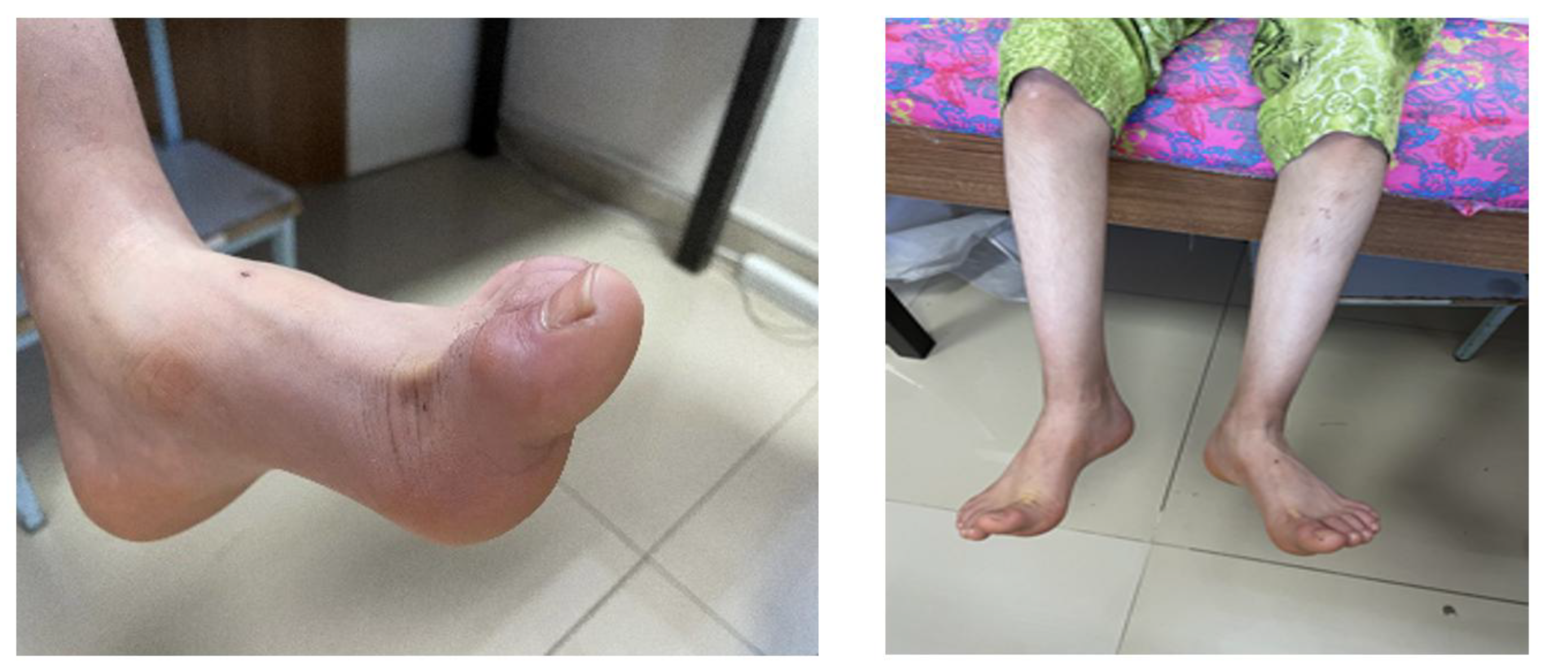
At physical examination, the patient was mentally well. Muscle strength was moderately reduced (3–5/5). Mild scoliosis was observed. Deep tendon reflexes were increased, more prominently in the lower extremities. Gait examination revealed a knee-flexion gait with impaired heel and tandem walking, consistent with distal motor involvement and pyramidal tract signs. Knee-fracture gait was present, heel and tandem gait was impaired, and pes cavus deformity and a surgical scar on the right Achilles tendon were observed (Figure 1).
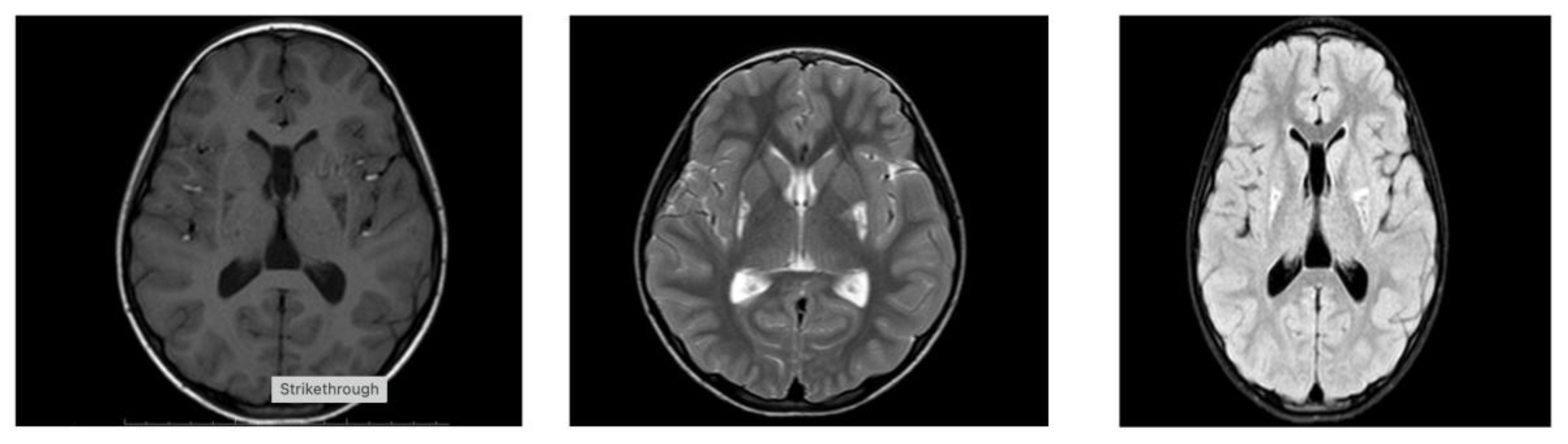
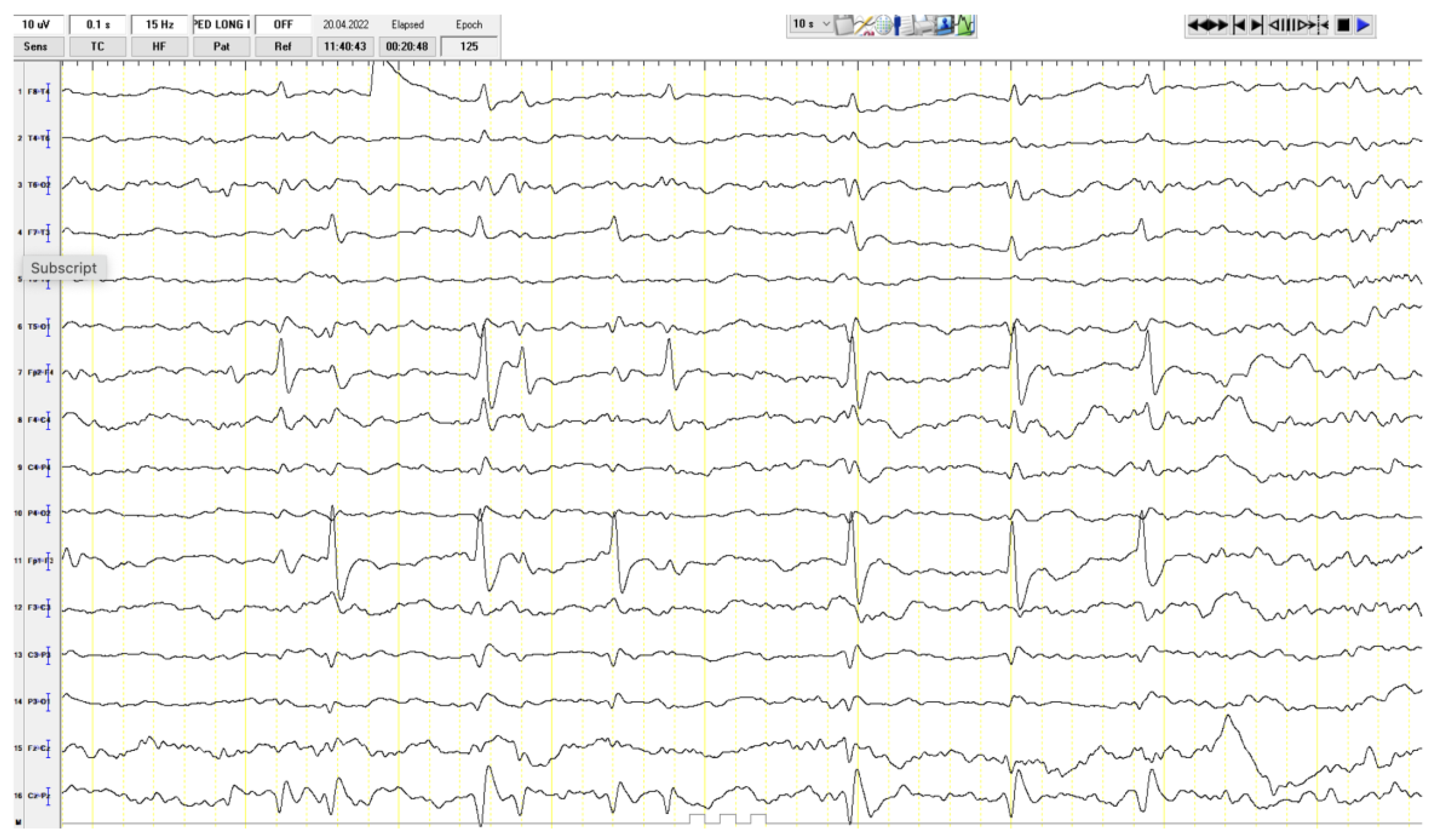
Among the laboratory tests, her lactate level was 2.3 mmol/L (normal value is 1.5–1.63 mmol/L), while other values were within normal limits. Eye examination, hearing tests, and EMG were normal. A focal epileptiform finding was present at EEG (Figure 2). The patient’s abdominal ultrasound, along with cardiac examination, were assessed as normal. Hypointense involvement on T1 and hyperintensive involvement on T2 FLAIR images were observed in the lower half of the bilateral lenticular nuclei on cerebral MRI (Figure 3). The patient was started on mitochondrial cocktail therapy, and it was decided to continue with physical therapy.
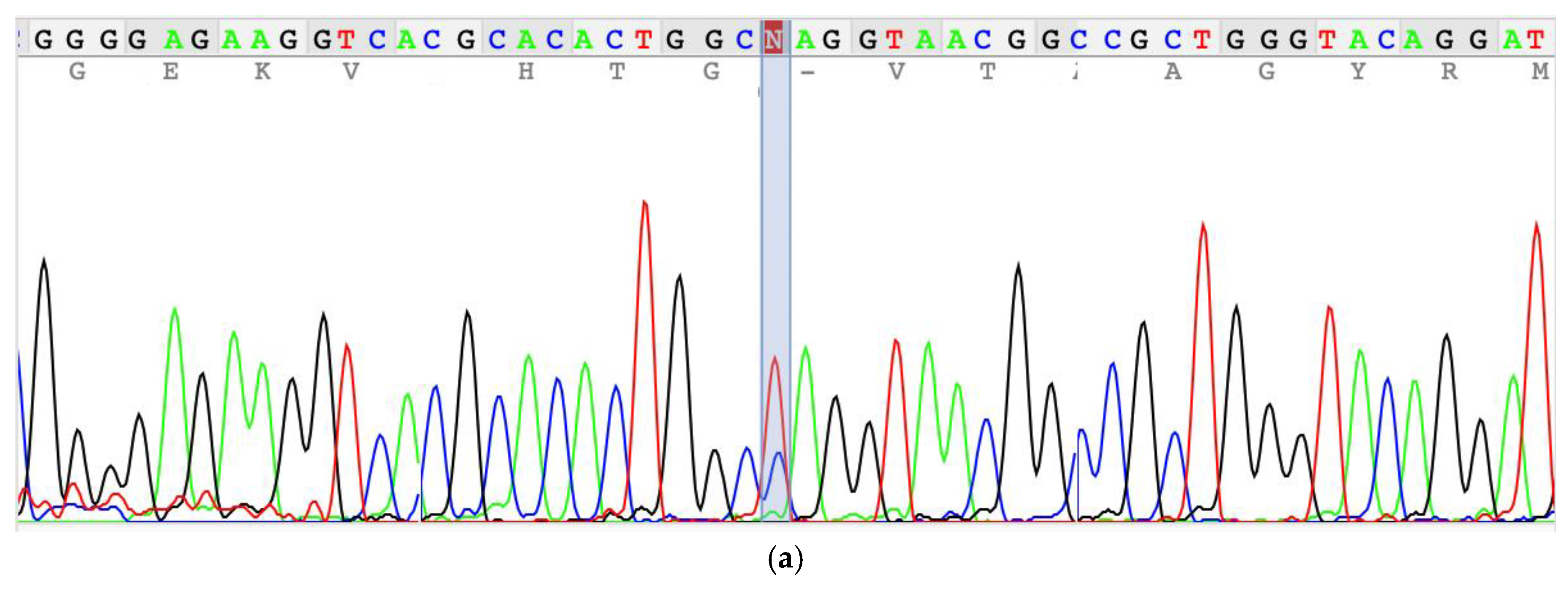
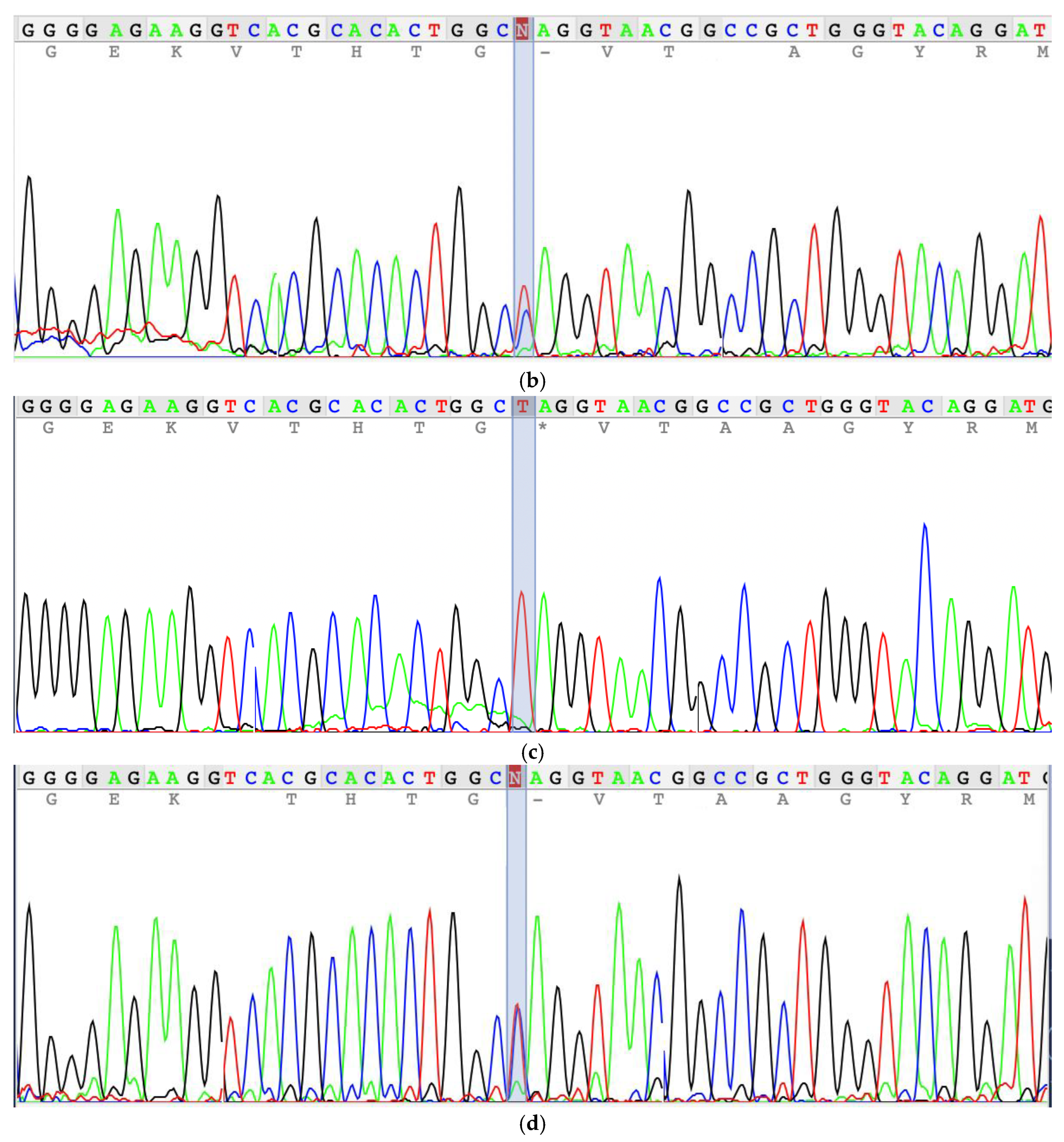
Whole-exome sequencing identified a novel homozygous nonsense variant in NDUFS6 (NM_004553.6:c.130C>T; p.Gln44*). The variant is predicted to cause premature termination at position 44 of the 124–amino acid NADH dehydrogenase [ubiquinone] iron–sulfur protein 6, mitochondrial (NDUFS6) protein encoded by the NDUFS6 gene. As a result, the truncated protein is expected to lack an 81–amino acid region including the zinc-finger domain, leading to a substantial impairment of protein function (NDUFS6_HUMAN, https://www.uniprot.org/uniprot/O75380, accessed on 29 January 2026). Although this variant has not been previously reported in the literature, its predicted truncating effect suggest that it is likely to be clinically significant, consistent with the pathogenicity observed for other reported NDUFS6 variants. NDUFS6 encodes a 13 kDa nuclear-encoded accessory subunit of mitochondrial complex I that localizes to the matrix arm and contributes to stabilization of the N-module. Functional studies with the previously published c.186+2T>A (p.Val63Glufs*9), the missense variant c.344G>A (p.Cys115Tyr), and the truncating variant c.352C>T (p.Gln118*) showed that NDUFS6 results in dissociation of most of the N-module and destabilization of complex I [1]. Consistent with this model, the variant was classified as likely pathogenic based on PVS1 (null variant in a gene with established loss-of-function disease mechanism) and PM2 (absent or very rare in public databases) according to ACMG/AMP guidelines [17]. Segregation analysis confirmed the autosomal recessive inheritance with heterozygosity in healthy parents and her sister (Figure 4).
The patient underwent comprehensive genetic analysis, including evaluation for both single-nucleotide variants (SNVs) and copy-number variants (CNVs). No additional pathogenic or likely pathogenic variants, including structural variations, were identified that could account for the observed phenotype. The absence of other candidate variants supports the conclusion that the identified NDUFS6 variation represents the most plausible genetic cause of the patient’s clinical presentation.

3. Discussion

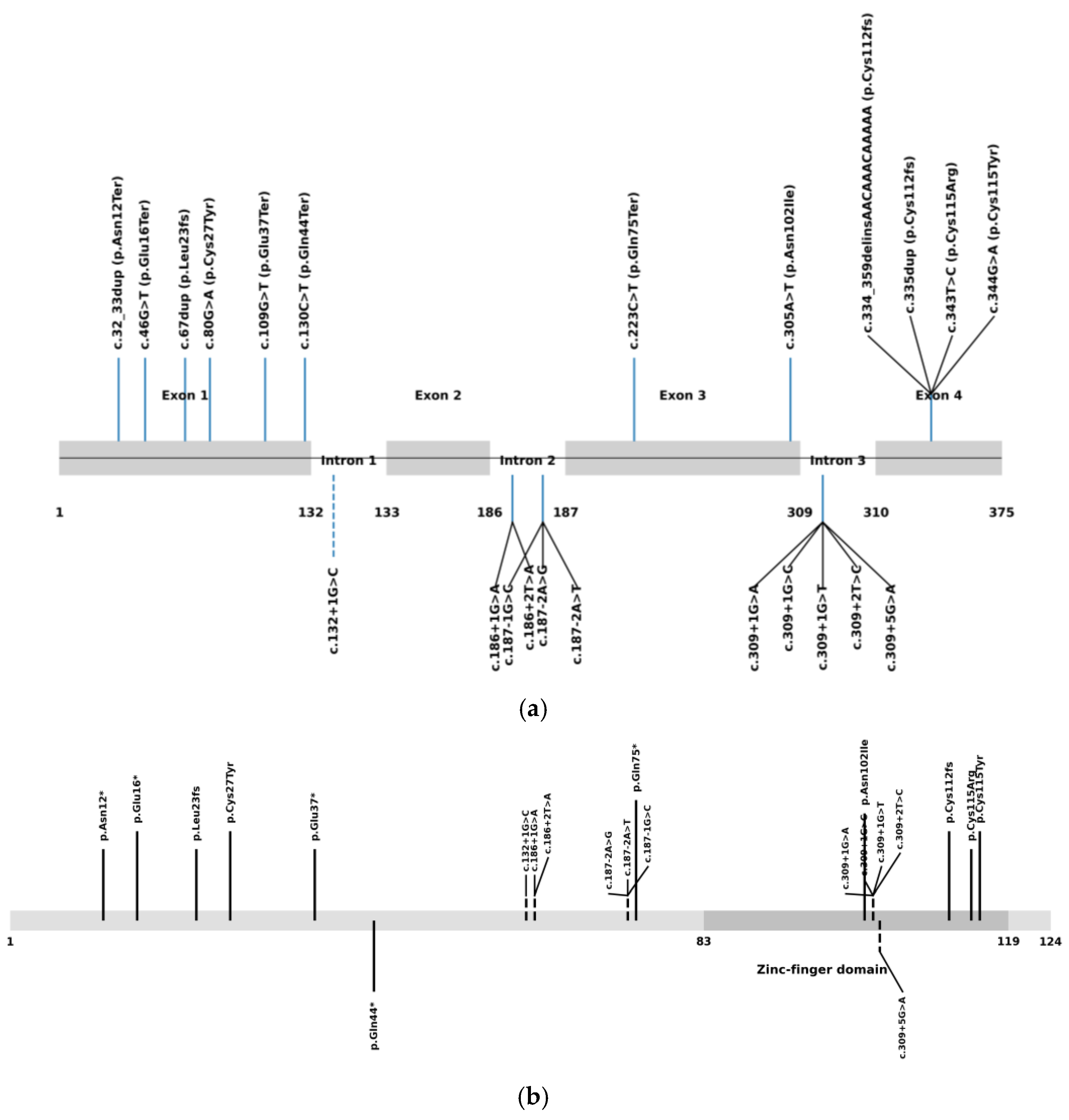
Comprehensive analysis of all published patients with biallelic NDUFS6 variants reveals that NDUFS6-related disease constitutes a phenotypic continuum rather than a single clinical entity. As of December 2025, 22 pathogenic or likely pathogenic variants were reported in the Pubmed and ClinVar databases (Figure 5). Across all reported cases, biallelic NDUFS6 variants give rise to a broad clinical spectrum ranging from neonatal lethal mitochondrial disease to childhood- or adolescent-onset axonal neuropathy/Charcot–Marie–Tooth (CMT)-like phenotypes (Table 1). Early reports predominantly described neonates presenting within the first days of life with profound hypotonia, severe lactic acidosis, and rapid clinical deterioration leading to death, often in association with truncating or frameshift variants and minimal residual complex I function. A subset of patients presented later in infancy with Leigh syndrome or Leigh-like disease, characterized by developmental regression, characteristic basal ganglia involvement on neuroimaging, elevated lactate levels, and poor outcomes [9,10,11,12,13,14,15].
In contrast, recent studies reported a distinct neuropathy-predominant phenotype, associated with the recurrent splice-region variant c.309+5G>A [7,16]. Six patients in two studies were found to carry c.309+5G>A mutation and manifest axonal sensorimotor neuropathy with pes cavus, steppage gait, and minimal disease progression, often with normal or near-normal brain MRI and only mildly elevated or normal lactate levels. Importantly, epilepsy has traditionally been associated with the encephalopathic end of the NDUFS6 disease spectrum. However, Armirola-Ricaurte et al. [16] reported epilepsy in patient II of family 3, who predominantly exhibited a neuropathy-dominant phenotype. The presence of epilepsy in our patient provides independent confirmation that seizures can occur within NDUFS6-associated neuropathy. Collectively, these observations imply a phenotypic spectrum of NDUFS6-related disease, in which partial complex I dysfunction variably affects both the peripheral and central nervous systems.
NDUFS6 encodes a 13 Kda nuclear-encoded accessory subunit located in the matrix arm of complex I and contributes to stabilization of the N-module. Functional studies provide a mechanistic framework to explain this variability. Experimental knockout of NDUFS6, as reported by Stroud et al. [1], demonstrated that complete loss of NDUFS6 leads to dissociation of most of the N-module and destabilization of mitochondrial complex I. Importantly, despite this pronounced structural defect, complex I enzymatic activity and overall respiratory capacity were not severely impaired, indicating that NDUFS6 is critical for structural stability and assembly, rather than for the catalytic function of the enzyme. Interestingly, pathogenic NDUFS6 variants reported in patients—including the splice-site variant c.186+2T>A (p.Val63Glufs*9), the missense variant c.344G>A (p.Cys115Tyr), and the truncating variant c.352C>T (p.Gln118*)—have been clinically associated with severe neonatal phenotypes, yet remain compatible with partial complex I function.
Functional experiments were not performed in the present study due to limited experimental facilities. Nevertheless, inference regarding the functional impact of the identified variant is supported by previously published studies demonstrating that truncating NDUFS6 variants result in loss of protein function and destabilization of mitochondrial complex I. In particular, functional analyses by Stroud et al. [1] showed that complete loss of NDUFS6 leads to dissociation of the N-module and impaired assembly of complex I without abolishing catalytic activity, indicating a primary role in structural stability and assembly rather than enzymatic function. Similar truncating NDUFS6 variants reported in patients with severe mitochondrial disease have also been shown to be associated with reduced protein levels and complex I instability. Accordingly, we infer that our novel homozygous NDUFS6 nonsense variant (c.130C>T, p.Gln44*) is likewise expected to cause loss of NDUFS6 function and impaired complex I assembly.
The recurrent neuropathy-associated splice variant c.309+5G>A disrupts the canonical exon 3 donor site, leading to aberrant splicing and markedly reduced levels of the full-length transcript, while still allowing expression of partially functional isoforms that retain the critical Cys115 residue within the zinc-finger domain [16]. This residual complex I function provides a mechanistic explanation for survival beyond infancy and accounts for the emergence of later-onset, neuropathy-predominant, or Charcot–Marie–Tooth-like phenotypes, occasionally accompanied by central nervous system involvement such as epilepsy. Here, we have presented that neuropathy is not restricted to the recurrent splice-site variant c.309+5G>A. By reporting a patient with a novel homozygous truncating NDUFS6 variant and a neuropathy-predominant phenotype, our study shows that mutations at different positions within the NDUFS6 gene may be associated with peripheral neuropathy, further underscoring the phenotypic heterogeneity of NDUFS6-related disease.
Current evidence does not support a clear genotype–phenotype correlation for pathogenic NDUFS6 variants. Both truncating and splice-site mutations in NDUFS6 have been associated with markedly different clinical outcomes, ranging from neonatal lethal mitochondrial disease and Leigh syndrome to later-onset axonal neuropathy and Charcot–Marie–Tooth-like phenotypes. Notably, mutations predicted to cause complete loss of protein function, including nonsense, frameshift, and canonical splice-site variants, have been reported in patients with both severe encephalopathic presentations and comparatively mild neuropathy-predominant disease. Additionally, no clear genotype–phenotype correlation is observed between patients carrying homozygous or compound heterozygous NDUFS6 mutations. This phenotypic variability is further supported by functional data from Stroud et al. [1], who demonstrated that complete knockout of NDUFS6 leads to dissociation of the N-module and destabilization of mitochondrial complex I without severely impairing enzymatic activity or respiratory capacity. These findings indicate that NDUFS6 is essential for the structural stability and assembly of complex I rather than for its catalytic function, and that substantial residual complex I activity may persist even in the context of truncating variants. Consequently, clinical severity in NDUFS6-related disease is unlikely to be determined solely by mutation type and is more plausibly influenced by additional factors such as residual transcript expression, tissue-specific vulnerability, mitochondrial compensatory mechanisms, and genetic or environmental modifiers.
Our patient clearly aligns with this neuropathy-dominant spectrum. Despite carrying a truncating NDUFS6 variant predicted to cause loss of protein function, she did not develop neonatal metabolic decompensation. Instead, she manifested a slowly progressive motor neuropathy with orthopedic complications, pyramidal signs, and mild basal ganglia involvement. The presence of urinary incontinence suggests possible autonomic involvement, further expanding the neurological manifestations associated with NDUFS6 deficiency.

4. Materials and Methods

Written informed consent was obtained from the parents of the patient. A comprehensive clinical history was obtained, and neurological examinations were performed for all family members. Genomic DNA was extracted from peripheral blood and whole-exome sequencing (WES) was performed by capture of the coding regions and splice sites of targeted genes using the Twist Human Core Exome (Twist Bioscience, South San Francisco, CA 94080 USA). After library enrichment and quality control, the samples were sequenced using the Illumina HiSeq4000 (Illumina, Inc. San Diego, CA 92122 USA) instrument with 100 bp paired-end reads at an average sequencing depth of 100×. An average read depth of 20× and 95% coverage, including exon–intron junction boundaries (±10 bp), were evaluated. Variant filtering considered allele frequency, predicted impact, inheritance pattern, and phenotypic relevance [18]. The reference human genome (hg19/GRCh37) was used for the analysis. Human Phenotype Ontology was used for phenotypic filters, and Online Mendelian Inheritance in Man (OMIM, https://www.omim.org/, accessed on 2 December 2025) was used for gene sets. The raw data was uploaded and analyzed on Franklin (https://franklin.genoox.com, accessed on 2 December 2025). Both single-nucleotide variants (SNVs) and copy-number variants (CNVs) were investigated. The variants were classified according to the American College of Medical Genetics and Genomics (ACMG) criteria [17]. Confirmation of the identified variant within the family was performed using Sanger sequencing on an ABI PRISM 3130 Genetic Analyzer (Applied Biosystems, Thermo Fisher Scientific, Tokyo, Japan).
Clinical evaluation included detailed neurological examination, gait assessment, strength testing, and review of autonomic symptoms. Electroencephalography (EEG), metabolic laboratory studies, electromyography (EMG), and ophthalmologic and audiologic evaluations were obtained. Brain magnetic resonance imaging (MRI) included T1-weighted, T2-weighted, and Fluid-Attenuated Inversion Recovery (FLAIR) sequences.
A comprehensive review of all published patients with NDUFS6 variants was performed, and the main findings from the previously reported cases, alongside the present index cases, are summarized in Table 1.

5. Conclusions

This study identifies a novel homozygous truncating NDUFS6 variant associated with childhood-onset axonal neuropathy accompanied by pyramidal signs, further expanding the phenotypic spectrum of NDUFS6-related disease. While neuropathy has previously been reported primarily in association with the recurrent splice-site variant c.309+5G>A, our findings demonstrate that truncating NDUFS6 mutations can also underlie a neuropathy-predominant phenotype. Integration of all published cases highlights the marked clinical heterogeneity associated with NDUFS6 deficiency, encompassing presentations that range from lethal neonatal mitochondrial encephalopathy to slowly progressive hereditary neuropathy.
These observations support the inclusion of NDUFS6 in the diagnostic evaluation of neuropathy, Charcot–Marie–Tooth-like phenotypes, and unexplained pes cavus deformity, particularly in consanguineous families or in patients with subtle or atypical mitochondrial features. Collectively, the present study provides further evidence that NDUFS6 is a bona fide neuropathy-associated gene and justifies its inclusion in diagnostic gene panels for inherited peripheral neuropathies and Charcot–Marie–Tooth disease.

Author Contributions

Conceptualization, S.B. and I.B.; Methodology, S.T.B. and I.B.; Data curation, S.B., R.I. and S.T.B.; Writing—review and editing, S.B., R.I., S.T.B. and I.B.; Supervision, I.B. All authors have read and agreed to the published version of the manuscript.

Funding

This research received no external funding.

Institutional Review Board Statement

Ethical approval was not required for this case report as it involved the retrospective presentation of anonymized data obtained during routine diagnostic procedures.

Informed Consent Statement

Written informed consent for publication was obtained from the patient’s parents.

Data Availability Statement

The original contributions presented in this study are included in the article. Further inquiries can be directed to the corresponding author.

Conflicts of Interest

The authors declare no conflicts of interest.

References

  1. Stroud, D.A.; Surgenor, E.E.; Formosa, L.E.; Reljic, B.; Frazier, A.E.; Dibley, M.G.; Osellame, L.D.; Stait, T.; Beilharz, T.H.; Thorburn, D.R.; et al. Accessory subunits are integral for assembly and function of human mitochondrial complex I. Nature 2016, 538, 123–126. [Google Scholar] [CrossRef] [PubMed]
  2. Sazanov, L.A. From the ‘black box’ to ‘domino effect’ mechanism: What have we learned from the structures of respiratory complex I. Biochem. J. 2023, 480, 319–333. [Google Scholar] [CrossRef] [PubMed]
  3. Kirby, D.M.; Crawford, M.; Cleary, M.A.; Dahl, H.H.; Dennett, X.; Thorburn, D.R. Respiratory chain complex I deficiency: An underdiagnosed energy generation disorder. Neurology 1999, 52, 1255–1264. [Google Scholar] [CrossRef] [PubMed]
  4. Loeffen, J.L.; Smeitink, J.A.; Trijbels, J.M.; Janssen, A.J.; Triepels, R.H.; Sengers, R.C.; van den Heuvel, L.P. Isolated complex I deficiency in children: Clinical, biochemical and genetic aspects. Hum. Mutat. 2000, 15, 123–134. [Google Scholar] [CrossRef] [PubMed]
  5. Bugiani, M.; Invernizzi, F.; Alberio, S.; Briem, E.; Lamantea, E.; Carrara, F.; Moroni, I.; Farina, L.; Spada, M.; Donati, M.A.; et al. Clinical and molecular findings in children with complex I deficiency. Biochim. Biophys. Acta. 2004, 1659, 136–147. [Google Scholar] [CrossRef] [PubMed]
  6. Fiorini, C.; Jurkute, N.; Torraco, A.; La Morgia, C.; Ghezzi, D.; Tioli, G.; Rigobello, L.; Ormanbekova, D.; Berghella, A.; Pietro Pasti, A.; et al. Recessive variants in mitochondrial Complex I nuclear subunits are an underrated cause of optic atrophy. Brain 2025, awaf422. [Google Scholar] [CrossRef] [PubMed]
  7. Gangfuß, A.; Rating, P.; Ferreira, T.; Hentschel, A.; Marina, A.D.; Kölbel, H.; Sickmann, A.; Abicht, A.; Kraft, F.; Ruck, T.; et al. A Homozygous NDUFS6 Variant Associated with Neuropathy and Optic Atrophy. J. Neuromuscul. Dis. 2024, 11, 485–491. [Google Scholar] [CrossRef] [PubMed] [PubMed Central]
  8. Zhu, J.; Vinothkumar, K.R.; Hirst, J. Structure of mammalian respiratory complex I. Nature 2016, 536, 354–358. [Google Scholar] [CrossRef] [PubMed]
  9. Kirby, D.M.; Salemi, R.; Sugiana, C.; Ohtake, A.; Parry, L.; Bell, K.M.; Kirk, E.P.; Boneh, A.; Taylor, R.W.; Dahl, H.H.; et al. NDUFS6 mutations are a novel cause of lethal neonatal mitochondrial complex I deficiency. J. Clin. Investig. 2004, 114, 837–845. [Google Scholar] [CrossRef] [PubMed] [PubMed Central]
  10. Spiegel, R.; Shaag, A.; Mandel, H.; Reich, D.; Penyakov, M.; Hujeirat, Y.; Saada, A.; Elpeleg, O.; Shalev, S.A. Mutated NDUFS6 is the cause of fatal neonatal lactic acidemia in Caucasus Jews. Eur. J. Hum. Genet. 2009, 17, 1200–1203. [Google Scholar] [CrossRef] [PubMed] [PubMed Central]
  11. Haack, T.B.; Madignier, F.; Herzer, M.; Lamantea, E.; Danhauser, K.; Invernizzi, F.; Koch, J.; Freitag, M.; Drost, R.; Hillier, I.; et al. Mutation screening of 75 candidate genes in 152 complex I deficiency cases identifies pathogenic variants in 16 genes including NDUFB9. J. Med. Genet. 2012, 49, 83–89. [Google Scholar] [CrossRef] [PubMed]
  12. Pronicka, E.; Piekutowska-Abramczuk, D.; Ciara, E.; Trubicka, J.; Rokicki, D.; Karkucińska-Więckowska, A.; Pajdowska, M.; Jurkiewicz, E.; Halat, P.; Kosińska, J.; et al. New perspective in diagnostics of mitochondrial disorders: Two years’ experience with whole-exome sequencing at a national paediatric centre. J. Transl. Med. 2016, 14, 174. [Google Scholar] [CrossRef] [PubMed] [PubMed Central]
  13. Ogawa, E.; Shimura, M.; Fushimi, T.; Tajika, M.; Ichimoto, K.; Matsunaga, A.; Tsuruoka, T.; Ishige, M.; Fuchigami, T.; Yamazaki, T.; et al. Clinical validity of biochemical and molecular analysis in diagnosing Leigh syndrome: A study of 106 Japanese patients. J. Inherit. Metab. Dis. 2017, 40, 685–693. [Google Scholar] [CrossRef] [PubMed] [PubMed Central]
  14. Rouzier, C.; Chaussenot, A.; Fragaki, K.; Serre, V.; Ait-El-Mkadem, S.; Richelme, C.; Paquis-Flucklinger, V.; Bannwarth, S. NDUFS6 related Leigh syndrome: A case report and review of the literature. J. Hum. Genet. 2019, 64, 637–645. [Google Scholar] [CrossRef] [PubMed]
  15. Li, Y.; Zhang, Y.; Jiang, G.; Wang, Y.; He, C.; Zhao, X.; Liu, L.; Li, L. Case report: Novel mutations of NDUFS6 and NHLRC2 genes potentially cause the quick postnatal death of a Chinese Hani minority neonate with mitochondrial complex I deficiency and FINCA syndrome. Medicine 2022, 101, e29239. [Google Scholar] [CrossRef] [PubMed] [PubMed Central]
  16. Armirola-Ricaurte, C.; Zonnekein, N.; Koutsis, G.; Amor-Barris, S.; Pelayo-Negro, A.L.; Atkinson, D.; Efthymiou, S.; Turchetti, V.; Dinopoulos, A.; Garcia, A.; et al. Alternative splicing expands the clinical spectrum of NDUFS6-related mitochondrial disorders. Genet. Med. 2024, 26, 101117. [Google Scholar] [CrossRef] [PubMed] [PubMed Central]
  17. Richards, S.; Aziz, N.; Bale, S.; Bick, D.; Das, S.; Gastier-Foster, J.; Grody, W.W.; Hegde, M.; Lyon, E.; Spector, E.; et al. ACMG Laboratory Quality Assurance Committee. Standards and guidelines for the interpretation of sequence variants: A joint consensus recommendation of the American College of Medical Genetics and Genomics and the Association for Molecular Pathology. Genet. Med. 2015, 17, 405–424. [Google Scholar] [CrossRef] [PubMed] [PubMed Central]
  18. Unsel-Bolat, G.; Bolat, H.; Citli, S.; Ozdemir, O.; Baris, I. Novel RORA Variants Reveal Genotype-Phenotype Diversity and Variable Expressivity in Neurodevelopmental Disorders. Clin. Genet. 2025. [Google Scholar] [CrossRef] [PubMed]
Figure 1. Bilateral pes cavus deformity in patient.
Ijms 27 01375 g001
Figure 2. Interictal EEG recording of the patient. Representative segment of EEG, recorded during wakefulness using a longitudinal bipolar montage, showing focal high-amplitude epileptiform activity.
Ijms 27 01375 g002
Figure 3. Hypointensity on T1 and hyperintensity on T2 and T2 FLAIR sections in the lower half of the bilateral lenticular nuclei.
Ijms 27 01375 g003
Figure 4. Sequencing electrogram showing the c.130C>T (p.Gln44*) variation in mother (a), father (b), index patient (c), and sister (d). The position of the variation is highlighted.
Ijms 27 01375 g004aIjms 27 01375 g004b
Figure 5. Published pathogenic NDUFS6 variants shown at the gene (a) and protein (b) levels. The NDUFS6 protein is shown with the zinc-finger domain highlighted (amino acids 83–119). Coding variants are indicated by solid lines, while splice-site variants are shown with dashed lines and branched connectors when multiple variants affect the same splice junction. Variants associated with Leigh syndrome or Leigh-like disease are shown above the protein, whereas neuropathy/Charcot–Marie–Tooth-associated variants are shown below.
Ijms 27 01375 g005
Table 1. Clinical features of patients with NDUFS6 variant in the present report and previous reports.
ReferenceVariationPhenotypeOnsetBrain MRIBlood LactateOutcome
Kirby et al., 2004 [9]deletion: chr5:g.1,865,477_1,869,744delNeonatal lethal mitochondrial disease,
profoundly hypotonic and drowsy, abnormal, slowly drifting eye
movements, rolling nystagmus
Neonatal-12.0 mmol/L (normal < 2.5)Died: 6 d; Central hypoventilation
Kirby et al., 2004 [9]c.186+2T>A (p.V63Efs*9)Neonatal lethal mitochondrial disease,
profoundly hypotonic and drowsy, abnormal, slowly drifting eye
movements, rolling nystagmus
NeonatalCerebral CT scan normal6.4 mmol/L (normal < 2.5)Died: 11 d; Central hypoventilation
Kirby et al., 2004 [9]c.186+2T>A (p.V63Efs*9)Neonatal lethal mitochondrial disease, profoundly hypotonic and drowsyNeonatalCerebral CT scan normal6.7 mmol/L (normal < 2.5)Died: 6 d; Central hypoventilation
Spiegel et al., 2009 [10]c.344G>A (p.Cys115Tyr)Neonatal lethal mitochondrial disease,
encephalopathy, severe lactic acidosis
2 d--Died/Outcome: 8 d
Spiegel et al., 2009 [10]c.344G>A (p.Cys115Tyr)Neonatal lethal mitochondrial disease,
lethargic, drowsy, refused to eat
2 dPostnatal brain ultrasound normal 6.0–11.2 mmol/L (normal < 2.0)Died: <10 d; Severe metabolic acidosis
Spiegel et al., 2009 [10]c.344G>A (p.Cys115Tyr)Neonatal lethal mitochondrial disease,
apathy, tachypnea, severe metabolic
acidosis
6 d--Died: 6 d; Severe metabolic
acidosis
Spiegel et al., 2009 [10]c.344G>A (p.Cys115Tyr)Neonatal lethal mitochondrial disease,
pale, tachypneic,
severely hypotonic, unresponsive
6 d-16.8 mmol/L (normal < 2.0)Died/Outcome: 8 d
Haack et al., 2012 [11]c.352C>T (p.Gln118*)Other/unspecified<6 moNormalElevatedAlive
Pronicka et al., 2016 [12]c.313_315delAAAG and c.334_359del126ins13 (p.104Lys_106Thrfs and p.Glu112fs)Neonatal lethal mitochondrial diseaseNeonatalNormal-Not reported
Ogawa et al., 2017 [13]c.309+5G>A (p.?) and c.343T>C (p.Cys115Arg)Leigh syndrome/Leigh-like---Not reported
Rouzier et al., 2019 [14]c.309+5G>A (p.?) and c.343T>C (p.Cys115Arg)Leigh syndrome/Leigh-like4 moConsistent with Leigh syndrome4,74 mmol/L (normal < 2.5)Died/Outcome: 11 months
Li et al., 2022 [15]c.344G >T (p.Cys115Phe)Neonatal lethal mitochondrial disease13 d-ElevatedDied: 27 d; Ineffective treatment
Gangfuß et al., 2024 [7]c.309+5G>A (p.?)Axonal neuropathy/CMT,
abnormal gait with frequent falls, pronounced axonal, sensory, and motor neuropathy (reduced latency and amplitudes, mostly normal conduction velocities)
7 yCerebral MRI at 10 yr normal2.5–2.8 mmol/L (normal 0.5–1.6)Alive at 10 y
Armirola-Ricaurte et al., 2024 [16]c.309+5G>A (p.?)Axonal neuropathy/CMT,
distal weakness/atrophy, pes cavus, steppage gait, nystagmus
1 yNormal1.9 mmol/LAlive/Not reported
Armirola-Ricaurte et al., 2024 [16]c.309+5G>A (p.?)Axonal neuropathy/CMT,
distal weakness, pes cavus, steppage gait, nystagmus, minimal progression, intellectual disability
10 yNormal1.03 mmol/LAlive/Not reported
Armirola-Ricaurte et al., 2024 [16]c.309+5G>A (p.?)Axonal neuropathy/CMT,
unsteady gait/falls, pes cavus, steppage gait, minimal progression, involuntary movements
10 yEye-of-the-tiger sign reported (basal ganglia)2.02 mmol/LAlive/Not reported
Armirola-Ricaurte et al., 2024 [16]c.309+5G>A (p.?)Axonal neuropathy/CMT,
unsteady gait/falls, pes cavus, steppage gait, minimal progression, Rolandic epilepsy, involuntary movements
10 y----Alive/Not reported
Present studyc.130C>T (p.Gln44*)Axonal neuropathy,
childhood-onset motor-neuropathy-like phenotype with pes cavus, knee-flexion gait, impaired heel/tandem gait, tendon-release surgery, urinary incontinence, focal epileptiform EEG
3 yBilateral lenticular nuclei T1 hypointensity and T2/T2-FLAIR hyperintensity2.3 mmol/LAlive
Disclaimer/Publisher’s Note: The statements, opinions and data contained in all publications are solely those of the individual author(s) and contributor(s) and not of MDPI and/or the editor(s). MDPI and/or the editor(s) disclaim responsibility for any injury to people or property resulting from any ideas, methods, instructions or products referred to in the content.