Review

Genetic and Immunologic Interactions Driving STAT3 Signaling and Macrophage Polarization in Acute Myeloid Leukemia

by
Kvan Omar Ahmed
1,2,*,
Measer Abdullah Ahmed
1,
Goran Othman
3,4 and
Shukur Wasman Smail
5,6,7,*
1
Department of Biology, College of Education for Pure Science, Tikrit University, Tikrit 34001, Iraq
2
Department of Medical Microbiology, College of Medicine, University of Kirkuk, Kirkuk 36001, Iraq
3
Department of Medical Laboratory Technology, Erbil Health and Medical Technical College, Erbil Polytechnic University, Erbil 44001, Kurdistan Region, Iraq
4
Department of Biology, College of Science, Al-Qalam University, Kirkuk 36001, Iraq
5
College of Pharmacy, Cihan University-Erbil, Erbil 44001, Kurdistan Region, Iraq
6
Department of Biology, College of Science, Salahaddin University-Erbil, Erbil 44001, Kurdistan Region, Iraq
7
Department of Medical Science, Respiratory Medicine, and Allergology, Uppsala University and University Hospital, 75185 Uppsala, Sweden
*
Authors to whom correspondence should be addressed.
Int. J. Mol. Sci. 2026, 27(20), 8957; https://doi.org/10.3390/ijms27208957 (registering DOI)
Submission received: 1 August 2026 / Revised: 2 October 2026 / Accepted: 6 October 2026 / Published: 9 October 2026

Abstract

Acute myeloid leukemia (AML) is an increasingly recognized malignancy sustained by a dysregulated inflammatory niche, in which genetic alterations and microenvironmental signaling converge to drive pathogenesis. This review synthesizes emerging evidence linking genetic mutations and immune dysregulation in AML, proposing an integrated mutation-macrophage-signal transducer and activator of transcription 3 (STAT3) axis as a conceptual framework of disease progression. It proposes that mutations in CCAAT/enhancer-binding protein alpha (CEBPA) and Runt-related transcription factor 1 (RUNX1), despite conferring divergent genetic risk, converge on a common pathway of inflammatory cytokine overproduction, including interleukin-1 beta (IL-1β), tumor necrosis factor alpha (TNF-α), interleukin-6 (IL-6), and interleukin-10 (IL-10), as well as C-C motif chemokine ligand 2 (CCL2)-mediated recruitment of tumor-associated macrophages. These educated macrophages are proposed to undergo M2-like polarization, releasing oncostatin M (OSM) and IL-6, which are positioned to activate the Janus kinase/signal transducer and activator of transcription 3 (JAK/STAT3) survival pathway in leukemic blasts. Concurrently, IL-10 produced by regulatory T cells, stromal cells, and M2-like polarized macrophages within the leukemic niche signals through its cognate receptor to potently activate STAT3, suppressing antigen-presenting cell function, downregulating major histocompatibility complex class II (MHC class II) expression, and impairing effective anti-tumor T-cell immunity, thereby potentially reinforcing the immunosuppressive architecture that shields leukemic blasts from immune surveillance. This proposed feed-forward loop is associated with transcriptional upregulation of the anti-apoptotic factor BCL2-like 1 (BCL2L1) and the negative-feedback regulator suppressor of cytokine signaling 3 (SOCS3), a pattern consistent with chemoresistance and impaired negative-feedback control of STAT3 signaling. Translational biomarkers, including OSM, IL-6, IL-10, and SOCS3/BCL2L1 transcripts, may quantify this axis, offering a dynamic, functional complement to static genetic risk stratification. Ultimately, this proposed triad suggests that aberrant inflammation may represent a shared therapeutic vulnerability; targeting the IL-6/oncostatin M receptor (OSMR)/STAT3 signaling nodes or reprogramming macrophage polarization could, if validated, disrupt this circuit in selected mutational subtypes.

1. Introduction

Acute myeloid leukemia (AML) is an aggressive heterogeneous malignant clonal disorder characterized by uncontrolled myeloid blast (progenitor cell) proliferation within the bone marrow accompanied by a block in differentiation that impairs effective hematopoiesis, resulting in clinically significant cytopenia [1]. AML incidence rises with age. The median age at diagnosis is 68 years, and two-thirds of cases occur in patients older than 55 years [2]. Leukemia ranks 13th in incidence among cancers worldwide, with an estimated 487,000 new cases in 2022, and 10th in cancer-related mortality, with an estimated 305,000 deaths [3]. Long-term burden analysis from 1990 to 2021 demonstrates an 82.25% increase in global leukemia incidence over this period [4]. Overall, five-year survival in AML is approximately 31.9%, decreasing to 11.2% among patients aged >65 years. These outcomes revealed a disproportionate burden of this disease in older adults [5].
AML constitutes a heterogeneous hematologic malignancy with multifactorial pathogenesis which includes accumulated mutations, genome instability, immune dysregulation, epigenetic reprogramming, resistance to apoptosis by various mechanisms, disruption of metabolic pathways, sustained proliferative signaling, activation of inflammatory cascades and immunosuppression of bone marrow microenvironment [6]. AML originates within the bone marrow and then blasts may enter the peripheral bloodstream. It is found to reach other parts of the body such as lymph nodes, spleen and some other organs. The term “acute” reflects the aggressive course of the disease, which arises from genetic and epigenetic alterations in hematopoietic stem cells (HSCs) and their myeloid progenitors; these generate immature white blood cells (blasts) that proliferate abnormally and become malignant leukemic cells within the bone marrow microenvironment (BMM) [7].
Recurring AML-associated mutations accumulate progressively in hematopoietic progenitor cells with age driving clonal expansion but without leukemic development, known as clonal hematopoiesis of indeterminate potential (CHIP) phenomenon [8]. Notably, clonal hematopoiesis progresses to overt AML at an estimated rate of approximately 0.5–1% per year [9]. Signal transducer and activator of transcription 3 (STAT3) signaling has been implicated in the protective bone marrow microenvironment of AML and in blast survival, chemoresistance and immune evasion. STAT3 is constitutively activated in many, though not all, leukemia subtypes and has been proposed as an oncogenic driver. Its sustained activation has been linked to leukemic cell survival, proliferation and resistance to apoptosis, and is frequently associated with poor prognosis; targeting STAT3 is therefore under investigation as a strategy to overcome resistance and improve outcomes in AML [10].
The present investigation aims to integrate current evidence into a cohesive model of AML pathogenesis, emphasizing the interrelationship between specific genetic mutations and the immunologic tumor microenvironment. We will critically examine the proposed biologic mechanisms through which mutations in the CCAAT/enhancer-binding protein alpha (CEBPA) and runt-related transcription factor 1 (RUNX1) genes, despite their divergent molecular functions, may converge to dysregulate inflammatory cytokine signaling. A core focus is to clarify how mutation-initiated cascades may involve the recruitment of tumor-associated macrophages (TAMs) and their M2-like polarization. Additionally, this review will examine how educated macrophages, through sustained production and release of cytokines such as oncostatin M (OSM) and interleukin-6 (IL-6), amplify oncogenic signaling through the oncostatin M receptor (OSMR)-STAT3 axis in leukemic blasts. Ultimately, by integrating molecular and immunological perspectives, this review proposes the “Mutation–Macrophage–STAT3 Triad” as a conceptual axis linking genetic alteration to immune signaling modulation.
Given that direct mechanistic evidence linking CEBPA and RUNX1 mutations to macrophage polarization and STAT3 pathway activation remains limited within AML specifically, this review integrates findings derived from other hematologic malignancies and solid-tumor systems into a biologically plausible, testable conceptual model. Findings extrapolated from non-AML systems should be interpreted as hypothesis-generating rather than as evidence validated in AML. The critical contribution of this review is therefore not the identification of a previously unknown molecular interaction, but the construction of an evidence-ranked framework that separates three levels of support: (i) AML-specific clinical or experimental evidence, (ii) mechanistic evidence from non-AML systems that is biologically plausible but not yet validated in AML, and (iii) explicit hypotheses generated by integrating these levels. Unlike prior accounts of isolated TAM-STAT3 or cytokine-survival circuits, the proposed triad links mutation-defined AML subgroups to the cellular source, receiving compartment, and measurable outputs of STAT3 signaling. Its value lies in generating discriminating predictions, including whether CEBPA- and RUNX1-altered AML differ in macrophage-state composition, whether stromal OSMR rather than blast OSMR predicts pathway dependence, and whether cytokine blockade loses efficacy when parallel niche programs remain active. The model should be judged by these testable predictions rather than treated as proof of a unitary pathway.

2. STAT3 Signaling and the Cytokine Feedback Loop

2.1. Overview of STAT3 Activation via IL-6/JAK/gp130 Pathway

STAT3 belongs to a family of cytoplasmic transcription factors. The Src homology 2 (SH2) domain plays a critical role in activation and mediates receptor recruitment following tyrosine phosphorylation at Tyr705, which promotes homodimerization [11]. Consequently, the active dimer translocates to the nucleus, where its DNA-binding domain (DBD) binds specific promoter sequences to regulate genes responsible for proliferation [12].
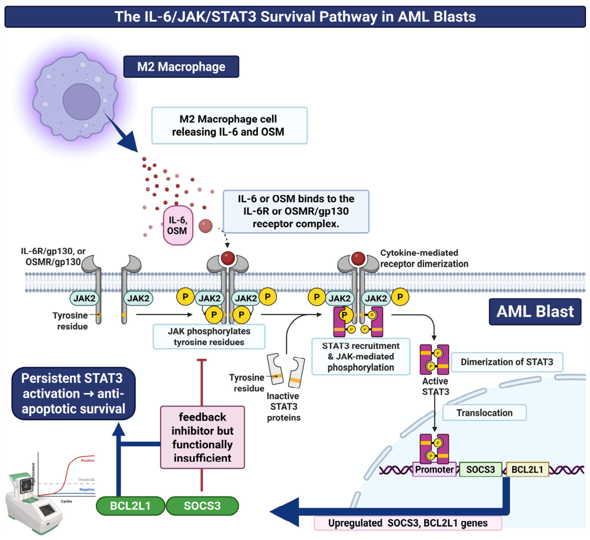
STAT3 is primarily activated by IL-6, a member of a cytokine family released by immune cells and critical for inflammation and cell survival, through a highly regulated, multistep process initiated by the assembly of a receptor complex at the cell membrane [13]. These phospho-tyrosine motifs serve as docking sites for the SH2 domains of inactive STAT3 monomers in the cytosol. Once recruited, JAKs phosphorylate STAT3 at the critical tyrosine residue Tyr705. The phospho-Tyr705 of one STAT3 molecule engages the SH2 domain of another STAT3 molecule, generating a stable homodimer complex [14]. The phosphorylated dimer subsequently translocates into the nucleus, where it binds to gamma-activated sequences at the promoter of target genes, resulting in transcription of genes that contribute to cell proliferation and survival in lung adenocarcinoma [15].

2.2. Downstream Transcriptional Targets: SOCS3 and BCL2L1

STAT3 directly regulates the expression of several genes critical for cancer cell fate. Among these, BCL2-like 1 (BCL2L1) (encoding Bcl-xL) is a well-established anti-apoptotic factor. STAT3-mediated upregulation of BCL2L1 enhances tumor cell survival by inhibiting programmed cell death, contributing to resistance against chemotherapy and other stressors. Suppressor of cytokine signaling 3 (SOCS3), another STAT3 target, acts as a negative feedback regulator of cytokine signaling. In the tumor context, however, this feedback loop can become functionally insufficient, so that SOCS3 upregulation may be more appropriately regarded as an indicator of dysregulated or impaired pathway control rather than SOCS3 itself acting as a direct oncogenic driver [16].
SOCS3 represents an essential mediator within the SOCS family that enforces negative feedback on the Janus kinase (JAK)/STAT axis, a function demonstrated in contexts including human immunodeficiency virus (HIV)-infected cell models, where SOCS3 dampens cytokine-induced signal transduction [17] via SH2-domain engagement of the receptor phosphotyrosine and kinase inhibitory region (KIR)-mediated occlusion of the JAK2 substrate-binding groove [18].

2.3. STAT3 Upregulates BCL2L1

STAT3 directly upregulates BCL2L1, encoding the anti-apoptotic protein Bcl-xL, which inhibits programmed cell death and supports tumor cell survival across multiple cancer types [12]. Inhibition of STAT3 reduces Bcl-xL expression, leading to increased apoptosis and reduced tumor growth [19].

2.4. Functional Outcomes: Survival, Proliferation, Immune Evasion

SOCS3 and BCL2L1 are both transcriptional targets of the IL-6/JAK/STAT3 axis, but they play functionally opposite roles; BCL2L1 is a direct pro-survival effector, whereas SOCS3 is a negative-feedback regulator whose elevation is best interpreted as a marker of recent/ongoing STAT3 activation rather than an independent pathogenic driver [20]. STAT3 activation induces transcription of anti-apoptotic genes such as BCL2L1 (Bcl-xL), myeloid cell leukemia 1 (MCL1), and survivin, which mediate resistance to apoptosis and enhance tumor cell survival and proliferation [12,19]. SOCS3 regulates immune cell infiltration and tumor immune evasion by constraining cytokine-dependent signaling networks that shape myeloid cell differentiation, specifically the expansion and suppressive activity of myeloid-derived suppressor cells (MDSCs). Its downregulation amplifies IL-6-driven JAK/STAT3 signaling, promoting an immunosuppressive tumor microenvironment that accelerates malignant progression and diminishes the efficacy of immune checkpoint blockade [21].

2.5. Clinical Correlates of IL-6/STAT3 Activation

Clinical evidence, largely from solid tumors, links the dysregulation of the IL-6 and STAT3 axis to aggressive tumor behavior and adverse clinical outcomes. Elevated levels of IL-6 and activated STAT3 within the tumor microenvironment serve as potent, stage-independent prognostic biomarkers, consistently associated with reduced overall survival, decreased disease-free survival, and a higher relapse rate in several solid tumors. Moreover, IL-6 overexpression in the tumor microenvironment directly promotes tumor cell proliferation and survival through activation of STAT3 in solid tumors such as oral cancer [22]. Clinically, elevated IL-6 and transcription of STAT3 downstream targets consistently predict aggressive disease, high relapse risk, and highlight this axis as both a prognostic biomarker and a rational therapeutic target [23], as shown in Figure 1.

3. Macrophage Polarization and OSMR Signaling

3.1. M1-like vs. M2-like Macrophage Phenotypes in AML Bone Marrow

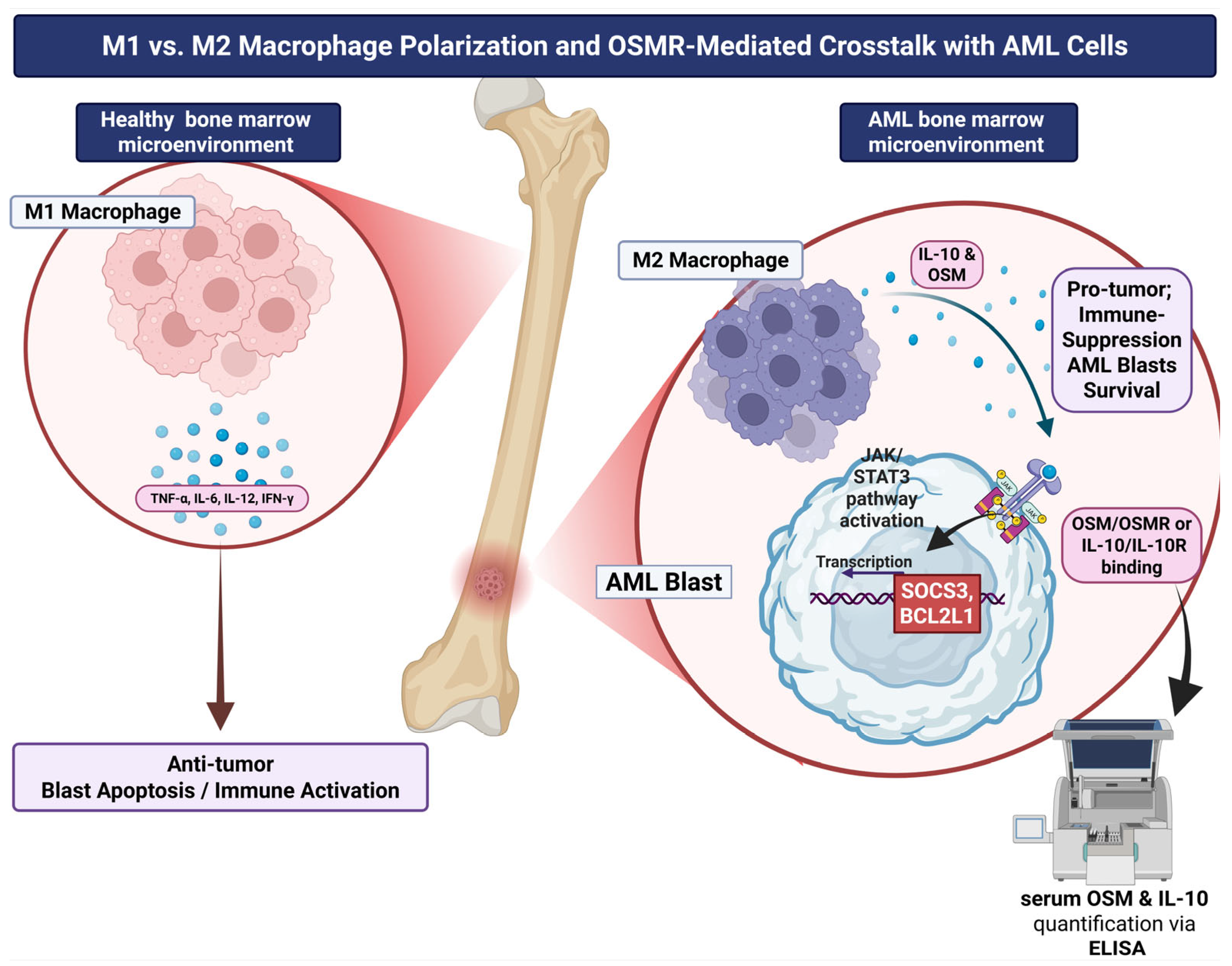
The BMM in AML constitutes a sophisticated immunological niche, within which macrophages, in addition to their various physiological and pathological roles, assume a key regulatory role in pathogenesis and immune modulation of the tumor. Macrophages within the BMM are broadly subclassified into anti-tumor (M1-like) and pro-tumor (M2-like) polarization states, with the M2-like phenotype promoting tumor progression and immune suppression within the tumor microenvironment (TME) [24]. This M1-like/M2-like phenotype dichotomy is a simplified heuristic; in vivo, macrophages occupy a continuum of context-dependent activation states rather than two fixed, mutually exclusive populations, and this terminology is used throughout the review for conceptual clarity rather than to imply discrete cellular subsets. In hepatocellular carcinoma, tumor-associated macrophages expressing high levels of programmed death-ligand 1 (PD-L1) have been shown to directly induce CD8+ T-lymphocyte exhaustion, characterized by upregulation of exhaustion markers and suppressed cytotoxic function leading to impaired immune response and reduced cytokine production [25]. Moreover, M2-like polarized macrophages release IL-10 and TGF-β, which attenuate cytotoxic T cells and promote regulatory T-cell differentiation [26].
Importantly, AML-associated macrophages cannot be reduced to a binary M1-like/M2-like switch. Single-cell analyses identify multiple macrophage populations and activation trajectories, including Fc fragment of IgA receptor (FCAR)-positive and Fc gamma receptor IIIA (FCGR3A)-positive states enriched in patients who fail to achieve complete remission, with distinct metabolic and intercellular-communication programs [27]. These states may coexist within the same marrow and change after chemotherapy, reflecting recruitment, local differentiation, and reversible niche education rather than fixed terminal polarization. Spatial position is also likely to matter: macrophages adjacent to vascular, stromal, adipocytic, or blast-rich regions may receive different combinations of IL-6, IL-10, hypoxic, and metabolic signals. Accordingly, “M1-like phenotype” and “M2-like phenotype” are retained only as broad functional descriptors; future studies should define TAMs by transcriptomic program, surface phenotype, anatomical neighborhood, and temporal state [28].

3.2. OSM–OSMR Signaling Promotes M2-like Immune Suppression

OSM is an IL-6 family cytokine that signals through its receptor (OSMR) and has been proposed to influence macrophage polarization, although this has not been demonstrated in AML. Upon OSM/OSMR engagement, OSMR pairs with the co-receptor gp130, initiating intracellular signaling and thereby activating JAK1/JAK2 kinases, which results in phosphorylation and translocation of STAT3 into the nucleus to upregulate its downstream target genes [29]. Activation of the OSM/OSMR/JAK/STAT3 pathway within macrophages induces transcription of genes that contribute to an M2-like phenotype, such as IL-10, arginase 1 (ARG1), cluster of differentiation 163 (CD163) and CD206, an effect demonstrated in glioblastoma-associated macrophages and not yet confirmed in the AML bone-marrow niche [30]. Consequently, this phenotypic remodeling may contribute to tumor progression and reduced anti-tumor immunity. Furthermore, in non-AML systems OSM/OSMR signaling has been linked to proliferation, survival and apoptosis resistance through cell cycle and anti-apoptotic genes such as BCL2, MYC and MCL1 [31]; in AML, OSMR appears to be expressed mainly by stromal cells rather than blasts. Serum OSM and soluble OSMR have been proposed as candidate-independent biomarkers of induction response and early mortality in a retrospective AML cohort [32]. These findings suggest that the OSM-JAK/STAT3 axis may merit investigation as a therapeutic target; whether interventions can disrupt OSM-dependent signaling in AML remains to be tested.

3.3. Macrophage-Driven STAT3 Feed-Forward Signaling

The amplification loop operates via autocrine and paracrine signaling; IL-6 and OSM secreted by M2-like polarized macrophages bind receptor complexes on neighboring macrophages and leukemic blasts, thus activating gp130/JAK complexes and inducing STAT3 phosphorylation, dimerization and nuclear translocation. Activated STAT3 then upregulates transcriptional programs that promote additional cytokine production and expression of survival and immunomodulatory effectors [33]. An analogous STAT3/IL-10/IL-6 feedback circuit regulating macrophage phenotype has been described in a non-malignant hepatic-injury model [34]; this is cited as evidence that such a macrophage-intrinsic STAT3 feedback circuit can operate independently of infectious or malignant triggers rather than as direct evidence of this circuit within the AML niche.

3.4. Soluble OSMR and IL-10 as Macrophage-Activity Biomarkers

A soluble form of OSMR (sOSMR) has been identified and molecularly characterized [35]. Quantifying serum sOSMR via ELISA offers a means to evaluate OSM/OSMR pathway engagement, and its clinical relevance in malignancies, including AML, has subsequently been investigated [32,36]. Circulating sOSMR has been proposed to reflect OSM/sOSMR axis engagement and, indirectly, macrophage activation; its evaluation may therefore provide an indicator of immunoregulatory activity. Elevated sOSMR concentrations have been associated with adverse clinical outcomes across several malignancies, including AML [36], as shown in Figure 2.
IL-10 within the AML bone marrow niche does not act uniformly as an oncogenic signal. In regulatory T cells, stromal cells, and M2-like polarized macrophages, IL-10 reinforces an immunosuppressive microenvironment by suppressing antigen-presenting cell function and dampening cytotoxic T-cell activity via STAT3 signaling. However, several studies report that IL-10 can directly inhibit AML blast proliferation and is paradoxically associated with improved survival and remission rates in some cohorts, indicating a tumor-suppressive action on the leukemic clone itself even as it supports leukemic stem cell persistence through niche-level immunosuppression. IL-10 receptor (IL-10R) is reported to be overexpressed on AML blasts, positioning the IL-10 axis as a potential target for future immunotherapeutic strategies [37]. These findings indicate that IL-10’s net effect in AML is determined by the responding cell type and disease stage, and this should be considered when interpreting circulating IL-10 as a single biomarker of disease activity [38].
The apparently opposing clinical associations of IL-10 are therefore not necessarily mutually exclusive. High circulating IL-10 may mark an anti-inflammatory response or direct restraint of bulk blast proliferation in some disease phases, while local IL-10 signaling in macrophages, regulatory T cells, antigen-presenting cells, or leukemia stem-cell niches may still suppress immune clearance. Differences in sampling compartment, treatment phase, cellular receptor expression, AML subtype, and endpoint can consequently reverse the observed association. For this reason, total serum IL-10 should not be interpreted as a unidirectional surrogate of immunosuppression, and therapeutic inhibition would require compartment-resolved evidence that the immune-suppressive effect outweighs any blast-intrinsic growth restraint [39].

4. Genetic Drivers of Inflammatory Signaling

4.1. CEBPA Mutations

AML is a heterogeneous group of hematologic malignancies defined by arrested myeloid differentiation and clonal outgrowth of myeloid progenitor cells. Although somatic genetic alterations are major contributors to leukemogenesis, accumulating evidence implicates inflammatory signaling and cytokine network disruption as central contributors to disease initiation and progression [40]. Notably, CEBPA and RUNX1 are both pivotal regulators of myeloid differentiation and inflammatory homeostasis, and their mutations represent prominent genetic determinants of inflammatory dysregulation in AML. During granulopoiesis, CEBPA is expressed in HSCs and myeloid-restricted progenitors, where it induces transcription of lineage-defining genes such as those encoding granule constituents and growth-factor receptors crucial for granulocytic specification [41].
CEBPA encodes C/EBPα, a basic leucine zipper (bZIP) transcription factor that functions as a master regulator of myeloid lineage commitment and granulocytic differentiation, directing lineage specification and driving cell-cycle arrest during hematopoietic maturation. Loss-of-function CEBPA mutations found in AML, typically involving the N-terminal transactivation domain or C-terminal bZIP region, impair granulocytic maturation and generate progenitors that remain locked in a proliferative and undifferentiated state. Several lines of evidence indicate that these mutations also dismantle key transcriptional restraint on inflammatory gene expression. Under sustained IL-1β stimulation, HSCs initiate a C/EBPα-dependent differentiation program. However, CEBPA-deficient HSCs and progenitor cells fail to execute this response and instead retain stem cell-associated transcriptional programs, thereby conferring a selective growth advantage in an inflammatory microenvironment [42,43].
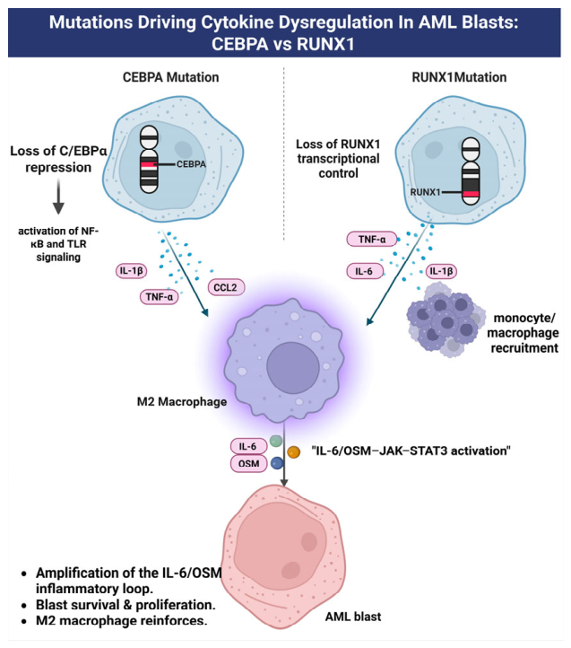
At the molecular level, the loss of C/EBPα removes repressive occupancy from regulatory regions of cytokine loci normally maintained in a silenced state during granulopoiesis. Loss of normal C/EBPα transcriptional control may contribute to dysregulated inflammatory gene expression through interactions with NF-κB-dependent pathways [44]. The specific effects of CEBPA mutation on individual cytokines and chemokines, including IL-1β, TNF-α, and CCL2, remain to be directly established in AML. At the molecular level, C/EBPα participates in the transcriptional regulation of inflammatory responses. Interactions between C/EBPα and NF-κB signaling have been implicated in inflammatory cytokine-driven myeloid differentiation [44]. Thus, disruption of normal C/EBPα function may alter inflammatory transcriptional programs in AML; however, the specific cytokine and chemokine consequences of CEBPA mutation, including effects on CCL2-mediated macrophage recruitment, require further direct experimental validation. Collectively, CEBPA mutations deregulate cytokine repression by eliminating CCAAT/enhancer-binding protein alpha (C/EBPα)’s transcriptional control, enabling chronic activation of inflammatory signaling networks and remodeling of the AML microenvironment. CCL2 is frequently upregulated in the leukemic microenvironment and is proposed to drive tumor-associated macrophage recruitment.

4.2. RUNX1 Mutations

RUNX1 is considered a master transcription factor essential for definitive hemato-poiesis and for controlling gene expression programs that direct normal blood cell development and myeloid cell functions. RUNX1 forms a functional heterodimer with core-binding factor β (CBFβ) essential for stable DNA binding and transcriptional activity [45]. Mutations in RUNX1 are linked to diverse hematologic disorders and myeloid cancers. Under homeostatic conditions, RUNX1 constrains toll-like receptor-driven pathways by repressing Toll-like receptor (TLR)1/2 and TLR4 signaling outputs. This restraint is achieved through the suppression of the NF-κB branch of myeloid differentiation primary response protein 88 (MyD88) and Toll/interleukin-1 receptor domain-containing adaptor protein (TIRAP) cascade, partially mediated by its interaction with the IκB kinase (IKK) complex to stabilize IκBα and prevent its degradation. The loss of RUNX1 disrupts this inhibitory mechanism, leading to accelerated IκBα turnover, unchecked NF-κB activation, and induction of inflammatory gene programs that are normally repressed [46].
RUNX1 is frequently mutated in AML, and these mutations result in either loss of function or dominant-negative activity. They are characteristically associated with immature cellular morphological features and are enriched in secondary AMLs that arise from prior myelodysplastic syndromes (MDSs) [47]. These AML-associated loss-of-function and dominant-negative point mutations are molecularly distinct from the RUNX1::RUNX1T1 fusion protein generated by t(8;21), a structurally and prognostically separate class of RUNX1 alteration [48], and from RUNX1 overexpression as reported in non-AML solid-tumor contexts; the three should not be treated as functionally interchangeable when interpreting downstream inflammatory or macrophage-polarization effects. RUNX1 mutations result in derepression of pro-inflammatory cytokines, mainly TNF-α, through enhanced TLR signaling [46]. However, persistent STAT3 signaling and direct M2-like polarization via IL-6/IL-1β/STAT3 are not clearly established in myeloid cells. In solid tumors, RUNX1 can promote M2-like polarization through alternative pathways. It is important to note that this M2-like polarizing activity has been characterized specifically in the context of RUNX1 overexpression in solid tumors and has not been demonstrated for the loss-of-function or dominant-negative RUNX1 alterations that predominate in AML; solid-tumor findings are presented as an illustrative alternative biological context rather than as direct evidence for the consequences of RUNX1 loss-of-function in the AML microenvironment. RUNX1 overexpression in solid tumors (such as colorectal cancer) can recruit and polarize macrophages toward the M2-like phenotype, mainly via CCL2 and Hedgehog pathway activation rather than direct IL-6/STAT3. RUNX1 overexpression can elevate CCL2 as a chemokine that recruits macrophages to the tumor microenvironment and drives their M2-like polarization. In parallel, it activates the Hedgehog signaling pathway, further reinforcing M2-like macrophage skewing and tumor-promoting functions, and platelet-derived growth factor-BB (PDGF-BB) released by M2-like macrophages was found to upregulate RUNX1 in tumor cells, establishing a feed-forward loop that sustains M2-like polarization and tumor progression [49,50].
M2-like macrophage polarization is associated with immunosuppression and tumor progression, but the mechanism in myeloid malignancies with RUNX1 mutation remains less clear. To date, evidence does not support sustained STAT3 activation as a direct downstream consequence of RUNX1 mutation within myeloid malignancies. Instead, RUNX1 loss primarily reconfigures type 1 interferon signaling networks, manifesting as augmented STAT1 and STAT2 activity rather than induction of STAT3 transcriptional programs [51]. Although IL-6-mediated STAT3 activation drives M2-like macrophage polarization in some malignancies, this pathway has not been mechanistically linked to RUNX1-mutant hematopoietic compartments based on currently available data. The clinical consequences of RUNX1-driven inflammatory and immune alterations are substantial: RUNX1-mutant AML is characterized by unique clinicopathological attributes and poorer therapeutic outcomes. Evidence also indicates that RUNX1 mutation defines an adverse clinical entity with reduced treatment responsiveness. In older patients and in monocytic AML (M4/M5), it supports classification as a distinct, high-risk entity requiring intensified clinical surveillance and risk-adapted therapeutic strategies. Moreover, RUNX1 mutation in AML is associated with poorer survival outcomes, reinforcing its potential role as an adverse prognostic marker [47], as illustrated in Figure 3.
In contrast to CEBPA and RUNX1, which are proposed to influence STAT3 signaling indirectly through cytokine production and macrophage crosstalk, JAK2 mutation activates the JAK/STAT pathway independent of upstream receptor-ligand engagement. The canonical JAK2 V617F mutation is a rare event in de novo AML but occurs at an appreciable frequency in AML arising secondary to a pre-existing JAK2-mutant myeloproliferative neoplasm (MPN), where it is associated with a distinct clinicopathological profile, including higher JAK2 V617F variant allele frequency, MPN-like megakaryocytic morphology, and a complex karyotype, relative to JAK2-mutant AML arising de novo [52]. Notably, even in de novo AML cohorts where JAK2 V617F itself is uncommon, phosphorylated STAT3 is frequently detected across AML subtypes, although less often in core-binding-factor leukemias, indicating that STAT3 activation in AML is largely JAK2-mutation-independent and driven by alternative upstream mechanisms [53]. This observation is consistent with the paracrine, macrophage/cytokine-driven route to STAT3 activation proposed above for CEBPA- and RUNX1-mutant AML, and positions JAK2-mutant (predominantly secondary) AML as a cell-intrinsic, mutation-driven complement to—rather than the principal explanation for STAT3 pathway engagement across AML more broadly. Bone marrow mesenchymal stromal cells from de novo AML show broader cytokine dysregulation: elevated IL-1β, IL-6, IL-32, vascular endothelial growth factor A (VEGFA), C-X-C motif chemokine ligand 12 (CXCL12), and decreased IL-10, compared with those from secondary AML with myelodysplasia-related changes, where the increase is largely restricted to IL-6 [54]. Since IL-6 is a principal glycoprotein 130 (gp130)/JAK/STAT3 activator, this suggests secondary AML may depend on a narrower, IL-6-centered route to STAT3 engagement, while de novo AML sustains it through a broader inflammatory milieu, a hypothesis warranting direct testing with paired cytokine/STAT3-target expression data stratified by AML ontogeny.

5. The Mutation–Macrophage–STAT3 Triad

5.1. Convergent Downstream Signaling from Distinct Mutations

Mutations in transcription factors such as CEBPA and RUNX1 influence myeloid differentiation; understanding the molecular mechanisms by which these distinct mutations lead to similar phenotypic outcomes provides critical insights into AML pathogenesis and therapeutic vulnerability. CEBPA and RUNX1 mutations converge on the same myeloid transcriptional circuitry: RUNX1 directly controls CEBPA via promoter and distal enhancer binding, while both cooperate with purine-rich box 1 (PU.1), a transcription factor crucial for the development and differentiation of immune cells, to activate lineage-specific enhancers; mutations in either factor disrupt enhancer activation and block monocyte/macrophage differentiation [55]. CEBPA and RUNX1 are cooperative master regulators of macrophage specification whose mutations converge to disrupt enhancer-dependent chromatin programs required for the monocyte–macrophage transition. CEBPA shapes the chromatin landscape, while RUNX1 controls monocytic fate and inflammatory pathways; disruption of either factor can impair macrophage maturation, alter inflammatory responsiveness, and promote leukemic persistence [56].

5.2. Proposed Convergence of CEBPA and RUNX1 Alterations on Macrophage-Associated STAT3 Signaling

STAT3 signaling is essential for hematopoietic stem cell self-renewal and for directing macrophage polarization, and its dysregulation is broadly implicated in AML pathogenesis [57]. Consistent with the previously described CEBPA- and RUNX1-associated cytokine profiles and TLR/NF-κB signaling mechanisms, we propose that CEBPA and RUNX1 mutations converge, through distinct upstream mechanisms, on disruption of this same STAT3 signaling axis. A functional RUNX1-STAT3 signaling intersection, involving regulation of negative feedback regulators, has been reported in epithelial stem cell biology [58]; this is offered here only as an analogy, since neither this circuit nor any direct physical RUNX1-STAT3 interaction has been demonstrated in RUNX1-mutant AML specifically. On the other hand, mutant CEBPA blocks the enhancer-dependent differentiation response, producing a highly proliferative yet differentiation-arrested state that is sensitive to JAK/STAT inhibition [59]. Functionally, STAT3 has been associated with immunoregulatory macrophage polarization, including M2-like functional states [33], and its aberrant activation in AML may therefore promote leukemic self-renewal, suppress differentiation, and support tumor-permissive microenvironments [60].
We propose that the CEBPA-mutant leukemic niche is characterized by marked macrophage recruitment accompanied by elevated IL-6-family cytokines, particularly IL-6 and OSM, which may cooperate with IL-1β and TNF-α to reinforce inflammatory signaling in a feed-forward amplification loop. Mechanistically, engagement of the IL-6 receptor-gp130 complex is well established to activate JAKs and induce STAT3 phosphorylation at Tyr705, driving its nuclear accumulation and transcription of survival- and inflammation-associated genes [12,14]. Whether CEBPA loss specifically reshapes the AML microenvironment to sustain this IL-6/OSM-JAK-STAT3 signaling axis in vivo, and thereby favors a persistent inflammatory state supporting leukemic cell maintenance, has not, to our knowledge, been directly demonstrated, and should be regarded as a proposed mechanism warranting experimental validation.
IL-6 and OSM activate STAT3 in macrophages and leukemic cells via gp130/JAK signaling, sustaining an IL-6/STAT3/CEBPβ autocrine loop that drives M2-like polarization in lung adenocarcinoma [15]. CEBPA p30 mutants blunt inflammatory gene induction and cytokine secretion, but co-occurring activating colony-stimulating factor 3 receptor (CSF3R) mutations promote ligand-independent JAK/STAT3 activation that engages enhancers, including E2F transcription factor 2 (E2F2), to fuel proliferation. In this setting, mutant CEBPA sustains an enhancer-dependent differentiation-arrested leukemic state, while TAM-derived IL-6 and intercellular crosstalk further reinforce leukemic persistence, potentially increasing sensitivity to JAK/STAT pathway inhibition [59]. OSM has been reported to induce RUNX1 expression and activate STAT3 in non-AML systems, suggesting a possible feed-forward loop. Whether loss or mutation of RUNX1 perturbs this circuit in AML, and whether this produces dominant-negative-like dysregulation and OSM/G-CSF hypersensitivity, is a hypothesis proposed by this review rather than a demonstrated finding. Whether RUNX1-mutant AML is characterized by enhanced OSM/OSMR–STAT3 signaling remains unresolved. Evidence from non-AML models demonstrates that macrophage STAT3/IL-10/IL-6 feedback loops can operate [34], but whether RUNX1 mutation directly induces macrophage OSM secretion within the AML bone marrow niche has not been established.

5.3. Feedback Between Macrophages and Blasts Sustaining Survival Signaling

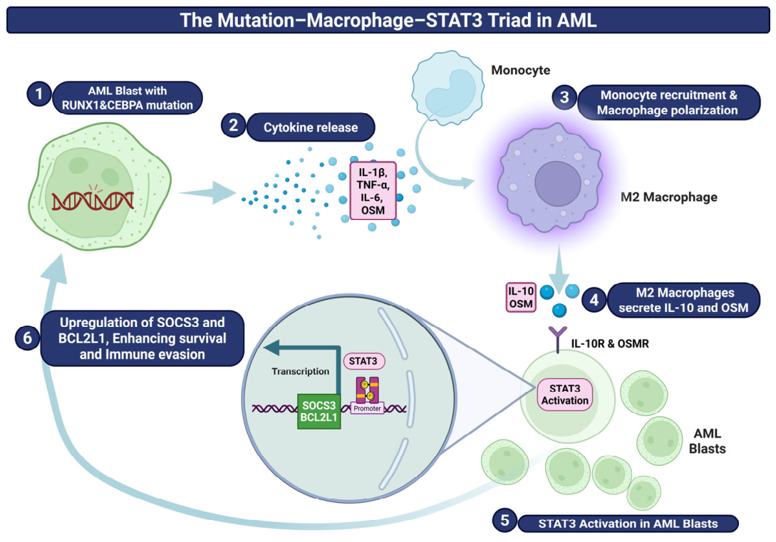
The convergence of CEBPA and RUNX1 mutations on macrophage-driven STAT3 activation is proposed to constitute a triad of mutant transcription factors, TAMs and STAT3 signaling that could function as a self-reinforcing circuit sustaining AML progression [61]. This circuit is mediated by blast-derived extracellular vesicles and cytokines that remodel the bone marrow into an inflammatory niche, recruiting and polarizing macrophages toward a pro-tumoral M2-like phenotype. Educated TAMs, in turn, produce IL-6 and OSM, resulting in the activation of STAT3 in blasts to enhance their survival, proliferation, and further niche-modifying capacity, while STAT3 signaling within TAMs is proposed to reinforce their M2-like polarization through a positive feedback loop. CEBPA mutations confer STAT3 hypersensitivity and a differentiation block, while RUNX1 mutations impair STAT3 attenuation and reduce CEBPA expression. We propose that CEBPA- and RUNX1-mutant AML converge on a common macrophage-STAT3 axis, a model that, if validated, would represent a disease-sustaining circuit persisting under therapeutic pressure [62], as presented in Figure 4 and Table 1.
The triad consequently extends earlier descriptions of self-reinforcing TAM-STAT3 circuits in two specific ways. First, it places mutation-defined leukemic states upstream of niche remodeling and explicitly distinguishes the blast, macrophage, and stromal compartments instead of treating STAT3 activation as a single tumor cell autonomous event. Second, it converts the synthesis into falsifiable conditional predictions: RUNX1-associated convergence is expected to be weaker and more context-dependent than CEBPA-associated convergence; OSMR dependence should track stromal OSMR abundance and spatial proximity rather than blast genotype alone; and combined pathway inhibition should be most effective in samples showing concurrent macrophage-state enrichment, cytokine elevation, and blast p-STAT3. These elements are the manuscript’s original integration. They do not establish causality and should not be interpreted as replacing prior TAM-STAT3 models [15].

6. Translational Biomarkers and Clinical Implications

6.1. Measurable Indicators: Serum OSM and SOCS3, BCL2L1 Expression

Translational biomarkers permit accurate risk classification and dynamic monitoring to inform targeted therapeutic intervention. OSM is a promising, noninvasively measurable biomarker in de novo AML. In a recent retrospective proteomic cohort, serum OSM emerged as a candidate-independent predictor of overall survival, early mortality, and response to induction chemotherapy [32]; this finding awaits prospective, multi-cohort validation.
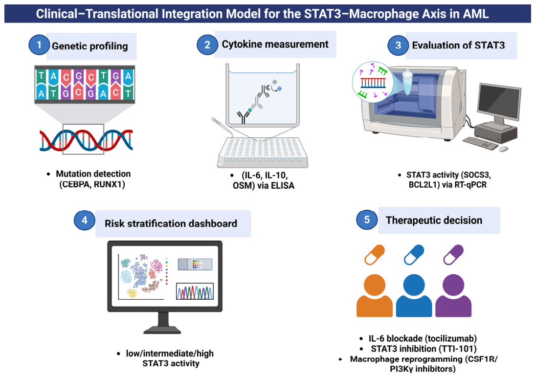
Clinically, serum OSM has been proposed as a candidate-independent predictor of overall survival, early mortality and induction response, outperforming the European LeukemiaNet (ELN) 2022 risk model. Patients with high OSM levels have substantially lower complete remission rates after both intensive and non-intensive induction. OSMR is principally expressed by bone marrow mesenchymal stromal cells rather than leukemic blasts, consistent with a paracrine paradigm in which OSMR/gp130 heterodimerization, following OSM binding, elicits JAK1/JAK2 cross-phosphorylation and downstream activation of STAT3, PI3K and MAPK cascades that contribute to chemoresistance, survival and disease progression [36]. IL6, SOCS3, and BCL2L1 transcript abundance in AML blasts can be quantified using reverse transcription quantitative PCR (RT-qPCR) [63], providing high sensitivity and specificity for evaluating pathway activation. Elevated IL-6 and dysregulated SOCS3 reflect perturbed IL-6/STAT3 signaling [64], while increased BCL2L1 (Bcl-xL) reflects anti-apoptotic activity and treatment resistance [10].

6.2. Integration with Mutation Status for Risk Assessment

The cellular source and receiving compartment are decisive for therapeutic interpretation. Current AML data support OSM/OSMR primarily as an outcome-associated blood biomarker and suggest substantial OSMR expression in mesenchymal/stromal cells, whereas direct dependence of primary AML blasts on TAM-derived OSM remains insufficiently demonstrated. A stromal-dominant circuit would predict that OSMR blockade modifies niche support indirectly and may require preserved drug penetration and combination with blast-directed therapy. By contrast, demonstrable blast-intrinsic OSMR expression and OSM-dependent viability would provide a stronger rationale for direct pathway addiction. These alternatives should be resolved using paired marrow plasma, sorted macrophages, stromal cells, and blasts, together with ligand-neutralization and cell-type-specific OSMR perturbation [32,36].
Integrating IL-6/OSM/SOCS3/BCL2L1 biomarker data with conventional mutational profiling (such as CEBPA and RUNX1) yields a dynamic, functionally informed risk framework that complements static genotypic classification (e.g., European LeukemiaNet 2022). A molecular-functional model would combine the genetic mutation profile as the disease baseline (CEBPA and RUNX1) with an inflammatory microenvironment readout (IL-6, OSM, SOCS3 and BCL2L1) [65]. Consequently, combining these biomarker axes with mutational context is proposed as a future translational risk-stratification framework, capable in principle of identifying high-risk patients and guiding mechanism-based therapeutic decisions once its component biomarkers have undergone prospective validation, as summarized in Table 2.

6.3. Potential Therapeutic Strategies: STAT3 Inhibitors, IL-6/OSMR Blockade, Macrophage Reprogramming

STAT3 represents a central mediator of oncogenic signaling, inflammation, and immune evasion, and its constitutive activation is associated with poor prognosis in multiple cancer types; pharmaceutical STAT3 inhibition is therefore proposed to disrupt tumor-promoting pathways and sensitize tumors to therapy [65]. In AML, several classes of STAT3 inhibitors have been described, targeting distinct points in the pathway: Src homology 2 (SH2) domain inhibitors that prevent STAT3 dimerization; DNA-binding domain inhibitors that block transcriptional activity; upstream JAK2 kinase inhibitors; and indirect agents that affect STAT3 protein stability or post-translational modifications [66]. Small-molecule inhibitors such as nifuroxazide, static and SH-4-54 have reduced tumor cell proliferation and enhanced apoptosis in preclinical cancer models. Combining STAT3 inhibitors with chemotherapy has shown synergistic effects in preclinical bladder-cancer models [67].
Blockade of the IL-6/OSMR signaling axis represents an alternative, microenvironment-directed strategy for inhibiting STAT3 activation in the inflammatory niche proposed to sustain AML. Two therapeutic modalities target this axis: anti-IL-6 monoclonal antibodies that sequester the cytokine ligand, and anti-IL-6 receptor antibodies that block receptor engagement. Tocilizumab, an IL-6 receptor antagonist, blocks both soluble and membrane-bound IL-6R, producing durable inhibition of IL-6-driven JAK/STAT3 signaling and reducing STAT3 phosphorylation through sustained receptor occupancy [68]. Tocilizumab has not been tested in AML directly; evidence that IL-6 blockade favorably modulates outcomes in solid-tumor patients receiving immune checkpoint blockade [69] suggests, by extrapolation, that in the subset of AML patients with IL-6-dependent STAT3 activation, tocilizumab could plausibly disrupt stromal survival signaling and enhance chemosensitivity, a hypothesis that would require dedicated AML clinical investigation. In preclinical ovarian cancer models, the anti-OSMR monoclonal antibody B21 abrogates OSMR-driven STAT3 signaling, reducing cancer cell proliferation and suppressing tumor growth and cisplatin resistance [70]; this agent has not yet been evaluated in AML. Given that OSMR is predominantly expressed on bone-marrow stromal rather than leukemic cells, OSMR blockade in AML would be expected to act on the microenvironment, disrupting paracrine OSM/OSMR-gp130 signaling proposed to sustain blast survival and chemoresistance.
Reprogramming TAMs from a pro-tumor (M2-like phenotype) to an anti-tumor (M1-like phenotype) is a promising therapeutic strategy under active clinical investigation across solid and hematologic cancers [71], with proof-of-concept preclinical support from a murine ascites carcinoma model [72]. TLR agonists, notably TLR4 ligands such as lipopolysaccharide or monophosphoryl lipid A, and TLR7/8 agonists such as imiquimod or resiquimod, engage NF-κB and interferon-regulatory factor (IRF) signaling to drive macrophages toward an M1-like, pro-inflammatory phenotype; imiquimod is FDA-approved as a topical agent for superficial basal cell carcinoma, whereas other TLR agonists remain investigational. Additional strategies described in the solid-tumor macrophage-reprogramming literature include CCL2/CCR2 blockade to inhibit monocyte recruitment, physical TAM depletion, and metabolic reprogramming via phosphoinositide 3-kinase gamma (PI3Kγ) or AMP-activated protein kinase (AMPK) modulation to skew polarization toward M1-like phenotypes [73]. None of these reprogramming strategies has yet been evaluated in AML, and translating them to the bone-marrow niche presents specific challenges: this niche is a distinct, multicellular environment comprising stromal cells, osteoblasts, endothelium, and hematopoietic elements, in which systemic macrophage reprogramming may carry a risk of inflammatory toxicity such as cytokine-release syndrome.
Among the topics discussed here, only OSM/OSMR has AML-cohort data, and these come from a single retrospective proteomic study awaiting prospective validation; it is a prognostic biomarker, not a validated therapeutic target. STAT3 small-molecule inhibitors, IL-6/IL-6R blockade, anti-OSMR antibodies, and macrophage-reprogramming agonists remain, with respect to AML specifically, at the preclinical or early-phase/cross-tumor investigational stage, and should be interpreted as therapeutic hypotheses generated by this review rather than established treatment options.

7. Future Perspectives

7.1. Need for Multi-Omics Studies Combining Genetics, Cytokines, and Immune Phenotyping

The cellular and molecular heterogeneity of the AML bone-marrow niche mandates a shift from single-modality profiling to integrated, mechanistic multi-omics frameworks. Current prognostic schemas for AML remain dominated by static genomic lesions and cytogenetic aberrations, such as FMS-like tyrosine kinase 3 (FLT3) and nucleophosmin 1 (NPM1) mutations. Yet these DNA-level readouts alone fail to represent dynamic functional states driven by regulated proteomic and cytokine signaling networks (IL-6/JAK/STAT3, Phosphoinositide 3-Kinase (PI3K)/Protein Kinase B (AKT) and NF-κB) that support blast survival and chemoresistance. Integrative approaches that combine genomic backbones with proteomic and cytokine profiling and deep immune phenotyping are therefore essential to map the dynamic blast-niche interactome underlying therapeutic failure and to reshape how immune function and therapeutic response are understood across cancer systems [74]. Parallel advances in single-cell and spatial technologies now illuminate the spatial organization of immunoregulatory networks within the bone-marrow niche. Integrating single-cell RNA sequencing (scRNA-seq) with spatial transcriptomic platforms (e.g., CosMx and Visium) enables reconstruction of discrete ‘immune neighborhood’ localized clusters of macrophages, stromal populations, T cells, and leukemic blasts responsible for immunosuppressive circuits [75].
The IL-6/OSM/STAT3 axis proposed in this review does not operate in isolation and is best understood as one of several coexisting, and potentially interacting, AML bone-marrow survival programs. The CXCL12/CXCR4 axis retains leukemic and normal hematopoietic progenitors within protective perivascular niches and activates PI3K/AKT and NF-κB signaling in leukemic cells; because NF-κB signaling also governs the pro-inflammatory cytokine output, CXCR4-mediated niche retention may act upstream of, or in parallel with, the cytokine milieu that drives OSM/IL-6-STAT3 engagement, rather than operating through an independent mechanism [76].
Bone-marrow adipocytes and MSCs constitute a second, metabolically distinct survival program, supplying free fatty acids that sustain fatty-acid oxidation-dependent chemoresistance in leukemic blasts; because these same stromal and adipocytic populations are also a principal cellular source of niche IL-6, metabolic support from adipocytes/MSCs and the cytokine-STAT3 axis proposed here likely share a common cellular origin within the niche rather than representing fully separable circuits [77].
IL-1β/NLRP3 inflammasome activation bears the most direct mechanistic relationship to the model proposed in this review: inflammasome-dependent caspase-1 cleavage generates the mature IL-1β already implicated here as part of the CEBPA-mutant inflammatory cytokine signature and the broader macrophage-driven feed-forward loop, positioning NLRP3 inflammasome activity as a plausible upstream contributor to, rather than a competing alternative to, IL-6/OSM-driven STAT3 signaling in the AML niche [78].
Direct experimental dissection of whether these programs are convergent, compensatory, or independently sufficient for leukemic survival has not, to our knowledge, been undertaken in AML and represents an important direction for future work; from a therapeutic standpoint, however, the possibility of convergence or compensation is itself actionable, since agents targeting the IL-6/OSM/STAT3 axis in isolation may prove insufficient if CXCR4-, adipocyte/MSC-, or inflammasome-driven survival signaling remains active in parallel.
Recent AML single-cell studies further argue for replacing bulk TAM abundance with state-resolved and spatial measurements. Macrophage subpopulations associated with non-complete remission show distinct FCAR/FCGR3A and metabolic programs [27], while contemporary single-cell reviews emphasize that blast, immune, and stromal states cannot be inferred reliably from bulk profiles [79]. Therapeutically, colony-stimulating factor 1 receptor (CSF1R), PI3Kγ, MER proto-oncogene tyrosine kinase (MerTK), AXL receptor tyrosine kinase (Axl), triggering receptor expressed on myeloid cells 2 (TREM2), leukocyte immunoglobulin-like receptor B (LILRB), V-domain immunoglobulin suppressor of T-cell activation (VISTA), and CD40 are increasingly discussed as myeloid-reprogramming or myeloid-checkpoint targets, but most evidence remains cross-tumor or preclinical rather than AML-specific [80]. In AML, checkpoint-directed strategies should therefore be framed as subtype- and biomarker-dependent experiments, not as established extensions of the proposed triad. These developments strengthen the rationale for measuring macrophage state and spatial context while also underscoring the present translational gap.

7.2. Validation of OSMR and STAT3 Activity as Predictive Tools

Although serum OSMR expression and phosphorylation of intracellular STAT3 have surfaced as promising biomarkers, their implementation in clinical practice mandates stringent validation. The designation of OSMR as a routine, evidence-based biomarker will require large-scale, prospective validation in independent patient cohorts comprising multi-ethnic populations and heterogeneous treatment regimens, together with standardized assay methodology and predefined performance thresholds to demonstrate clinical relevance and reliability [32].
Establishing the clinical validity of inflammatory biomarkers will also require rigorous standardization of analytical procedures. In contrast to categorical genetic lesions, these biomarkers function as continuous quantitative variables and therefore demand precisely defined, clinically validated threshold values. This issue is particularly pertinent for STAT3, as phospho-STAT3 measurements alone offer an incomplete assessment of pathway activity and exhibit substantial variability across cytogenetic subgroups; phospho-STAT3 is typically elevated in normal-karyotype AML while infrequent in core-binding factor leukemias [64]. The expression levels of canonical STAT3-responsive genes, including BCL2L1, SOCS3, and ICAM1, provide a more functionally robust indicator of pathway activation than static protein abundance [81]. Developing standardized assays, establishing analytical performance characteristics, and defining clinically actionable cut-points will be essential steps toward integrating these biomarkers into routine diagnostic practice. Additionally, integrating inflammatory biomarkers into established minimal residual disease (MRD) frameworks represents an essential translational advance. Rather than conceptualizing mutation clearance and inflammatory resolution as discrete pathways, future prognostic models should quantify an “inflammatory residual disease” state, defined by IL-6/OSMR-driven microenvironmental activation despite morphological or molecular remission as a biologically grounded predictor of early relapse [82].

7.3. Exploring Combined Immuno-Targeted Therapies

The future of cancer therapy lies in rationally designed combinations that target both oncogenic pathways and the immune microenvironment. Combining immune checkpoint inhibitors (ICIs) with targeted therapies such as kinase inhibitors, anti-angiogenic agents, or OSMR/STAT3 pathway inhibitors has shown synergistic effects in clinical studies of solid tumors [83]. The redundancy of cytokine signaling networks in AML suggests that monotherapies targeting the IL-6/STAT3 axis may be insufficient, necessitating the exploration of rational combination strategies. A promising avenue lies in the dual targeting of inflammatory pathways and immune checkpoints. Preclinical data in AML cells indicate that STAT3 signaling facilitates immune evasion and is associated with upregulation of PD-L1 expression [84]. Consequently, combining STAT3 inhibition with PD-1/PD-L1 blockade could theoretically release the brakes on T-cell immunity while simultaneously sensitizing leukemic blasts to immune surveillance.
Integrated targeted agents with immune checkpoint inhibitors are supported by mechanistic and translational data, in which targeted drugs can remodel the tumor microenvironment and enhance neoantigen exposure and promote effector-cell infiltration, thereby potentiating ICI activity [85]. Cytokine-driven activation of the OSMR-STAT3 signaling axis has been shown to mediate resistance to chemotherapy in ovarian cancer with antibody blockade of OSMR restoring chemosensitivity [70], while IL-6 blockade has separately been shown to favorably modulate outcomes in cancer patients receiving immune checkpoint blockade [69]; whether concurrent OSMR/IL-6-STAT3 pathway blockade would similarly restore treatment sensitivity in AML has not been verified. Ultimately, clinical development across solid and hematologic malignancies is increasingly focused on biomarker-driven trials evaluating rational combinations of immune checkpoint inhibitors with targeted microenvironmental or kinase inhibitors to test synergistic efficacy [86], as depicted in Figure 5 and detailed in Table 3.

7.4. Complexity and Heterogeneity of STAT3 Signaling in AML Immunity

The evidence reviewed above emphasizes a pro-leukemic, immunosuppressive function for STAT3 within the AML niche; however, this framing does not capture the full immunological complexity of STAT3 signaling in AML. STAT3 activity intrinsic to leukemic blasts has also been reported to be required for their recognition and elimination by natural killer (NK) cells, such that STAT3 loss impaired, rather than enhanced, NK-cell-mediated surveillance of leukemic cells [81]. This finding is not readily accommodated by a model in which STAT3 activation uniformly favors immune evasion, and instead indicates that the net immunological consequence of STAT3 signaling is compartment-dependent, varying with the cellular source of STAT3 activity (blast-intrinsic versus macrophage-intrinsic), the immune effector population under consideration, and disease stage. A comparable context-dependence has already been noted for IL-10 within the same niche: IL-10/STAT3 signaling in macrophages and regulatory T cells reinforces immunosuppression, whereas IL-10 can directly restrain AML blast proliferation and has been associated with improved survival in some cohorts. Taken together, these observations argue against treating STAT3 pathway activation as a uniformly unfavorable biological readout and instead support a compartment- and context-specific interpretive framework.
STAT3 pathway engagement is similarly unlikely to be uniform across AML molecular and cytogenetic subtypes. Phospho-STAT3 positivity itself varies by cytogenetic subgroup, being frequently detected in normal-karyotype AML but comparatively infrequent in core-binding-factor leukemias [64]. Disease ontogeny appears to contribute an additional layer of heterogeneity: bone marrow mesenchymal stromal cells from de novo AML display broader cytokine dysregulation (elevated IL-1β, IL-6, IL-32, VEGFA, and CXCL12) than those from secondary AML with myelodysplasia-related changes, in which the increase is largely restricted to IL-6 [54]. Genetic risk and inflammatory cytokine burden may also dissociate, as illustrated by CEBPA-mutant AML in Table 1, where an otherwise favorable intrinsic genetic risk profile may be offset by a high cytokine burden driving inflammation-dependent relapse. Collectively, this heterogeneity suggests that the proposed Mutation–Macrophage–STAT3 axis is unlikely to apply uniformly across AML and may instead be most relevant to specific molecular or cytogenetic subgroups, a distinction that should be tested directly in subtype-stratified cohorts rather than assumed.

7.5. A Testable Translational Roadmap for the Proposed Axis

A first validation study should enroll newly diagnosed adults prospectively and stratify them by CEBPA alteration, RUNX1 alteration, AML ontogeny, and ELN risk, with a parallel validation cohort receiving the same treatment backbone. At diagnosis, day 14, remission assessment, and relapse, paired marrow and blood should undergo cytokine profiling, phospho-flow for blast and myeloid p-STAT3, scRNA-seq/cellular Indexing of Transcriptomes and Epitopes by Sequencing (CITE-seq), and spatial profiling of OSM/OSMR-expressing macrophage–stromal–blast neighborhoods. The prespecified primary mechanistic endpoint should be concordance between macrophage-state abundance, stromal or blast OSMR localization, and compartment-specific STAT3 activity. Secondary endpoints should include complete remission, measurable residual disease clearance, event-free survival, and treatment-related inflammatory toxicity [75,97].
A subsequent biomarker-enriched window-of-opportunity study could recruit patients with high OSM/IL-6, an OSMR-positive stromal niche, and elevated blast p-STAT3. Short exposure to a pathway-directed agent before standard therapy would test pharmacodynamic suppression of p-STAT3 and downstream BCL2L1 without initially relying on response rate as proof of mechanism. Randomization to standard therapy with or without the investigational agent should occur only after demonstrating target engagement. Safety monitoring should prioritize infection, cytopenias, hepatic toxicity, impaired normal myelopoiesis, and cytokine-release or rebound inflammation. Because broad macrophage depletion may damage normal marrow defense and repair, state-selective reprogramming or temporally limited blockade may offer a more feasible strategy than continuous pan-macrophage suppression [98,99].

8. Conclusions

This review proposes the Mutation–Macrophage–STAT3 axis as an emerging conceptual model of AML pathogenesis, in which CEBPA and RUNX1 mutations, despite divergent prognostic weight, are each independently associated with overproduction of IL-1β, TNF-α, IL-6, and CCL2, a pattern consistent with remodeling of the bone marrow into a chronically inflamed niche. CCL2-recruited monocytes are proposed to polarize toward M2-like tumor-associated macrophages that reciprocally produce OSM and IL-6, a process that, if confirmed, would activate gp130/JAK-dependent STAT3 phosphorylation in leukemic blasts and upregulate BCL2L1 and SOCS3, contributing to survival and chemoresistance. IL-10 from macrophages and stroma has additionally been reported, in related contexts, to further activate STAT3 and suppress antigen-presenting cell function and cytotoxic T-cell responses, suggesting a potential self-reinforcing circuit of immune evasion that we propose may operate largely independent of the initiating genetic lesion. Translationally, integrating mutational profiling with serum OSM and IL-10 quantification alongside RT-qPCR measurement of SOCS3 and BCL2L1 transcripts generates a dynamic risk framework, positioning aberrant inflammation as a candidate prognostic determinant and a hypothesis-generating target for IL-6/OSMR/STAT3-directed or macrophage-reprogramming therapies in selected AML subtypes.

Author Contributions

Conceptualization, K.O.A., M.A.A. and S.W.S.; methodology, K.O.A., M.A.A. and G.O.; software, K.O.A.; validation, K.O.A., M.A.A., G.O. and S.W.S.; formal analysis, K.O.A. and G.O.; investigation, K.O.A.; resources, M.A.A. and S.W.S.; data curation, K.O.A.; writing—original draft preparation, K.O.A.; writing—review and editing, M.A.A., G.O. and S.W.S.; visualization, K.O.A.; supervision, S.W.S.; project administration, S.W.S.; funding acquisition, S.W.S. All authors have read and agreed to the published version of the manuscript.

Funding

This research received no external funding.

Data Availability Statement

No new data were created or analyzed in this study. Data sharing is not applicable to this article.

Acknowledgments

The authors gratefully acknowledge Uppsala University, Sweden, for providing funding support to cover the article processing charge (APC) for this publication.

Conflicts of Interest

The authors declare no conflicts of interest.

Declaration of Generative AI

We used Microsoft Copilot (GPT-5-based version available through Microsoft Copilot at the time of manuscript preparation) to assist with language editing and readability improvements only. No AI tools were used for data analysis, interpretation, or generation of scientific conclusions.

Abbreviations

The following abbreviations are used in this manuscript:
AKTProtein kinase B
AMLAcute myeloid leukemia
AMPKAMP-activated protein kinase
AP-1Activator protein 1
ARG1Arginase 1
BCL2B-cell lymphoma 2
BCL2L1BCL2-like 1 (gene; encodes Bcl-xL)
Bcl-xLB-cell lymphoma-extra large
BM-MSCBone marrow mesenchymal stromal cell
BMMBone marrow microenvironment
bZIPBasic leucine zipper
CAR-TChimeric antigen receptor T cell
CBFCore-binding factor
CBFβCore-binding factor beta
CCL2C-C motif chemokine ligand 2
CCR2C-C chemokine receptor type 2
CD8Cluster of differentiation 8
CD34Cluster of differentiation 34
CD40Cluster of differentiation 40
CD163Cluster of differentiation 163
CD206Cluster of differentiation 206
C/EBPαCCAAT/enhancer-binding protein alpha (protein)
CEBPACCAAT/enhancer-binding protein alpha (gene)
C/EBPβCCAAT/enhancer-binding protein beta
CHIPClonal hematopoiesis of indeterminate potential
CITE-seqCellular indexing of transcriptomes and epitopes by sequencing
CMMLChronic myelomonocytic leukemia
CSF1RColony-stimulating factor 1 receptor
CSF3RColony-stimulating factor 3 receptor
CXCL12C-X-C motif chemokine ligand 12
CXCR4C-X-C chemokine receptor type 4
DBDDNA-binding domain
E2F2E2F transcription factor 2
ELISAEnzyme-linked immunosorbent assay
ELNEuropean LeukemiaNet
ERK1/2Extracellular signal-regulated kinases 1 and 2
FCARFc alpha receptor (CD89)
FCGR3AFc gamma receptor IIIA (CD16A)
FDAFood and Drug Administration
FLT3FMS-like tyrosine kinase 3
G-CSFGranulocyte colony-stimulating factor
GATA2GATA-binding protein 2
gp130Glycoprotein 130
HIVHuman immunodeficiency virus
HSCHematopoietic stem cell
ICAM1Intercellular adhesion molecule 1
ICIImmune checkpoint inhibitor
IDHIsocitrate dehydrogenase
IFN-γInterferon gamma
IKKIκB kinase
ILInterleukin
IL-1βInterleukin-1 beta
IL-5Interleukin-5
IL-6Interleukin-6
IL-6RInterleukin-6 receptor
IL-10Interleukin-10
IL-10RInterleukin-10 receptor
IL-12Interleukin-12
IL-32Interleukin-32
IκBαInhibitor of kappa B alpha
iPSCInduced pluripotent stem cell
IRFInterferon-regulatory factor
JAKJanus kinase
JAK1Janus kinase 1
JAK2Janus kinase 2
KIRKinase inhibitory region
LILRBLeukocyte immunoglobulin-like receptor B
LPSLipopolysaccharide
LSCLeukemic stem cell
M1-likeClassically activated macrophage state
M2-likeAlternatively activated macrophage state
MAPKMitogen-activated protein kinase
MCL1Myeloid cell leukemia 1
MDSMyelodysplastic syndrome
MDSCMyeloid-derived suppressor cell
MerTKMER proto-oncogene tyrosine kinase
MHCMajor histocompatibility complex
miR-9MicroRNA-9
MPNMyeloproliferative neoplasm
mRNAMessenger RNA
MRDMeasurable residual disease
MSCMesenchymal stromal cell
MyD88Myeloid differentiation primary response 88
NF-κBNuclear factor kappa B
NKNatural killer cell
NLRP3NOD-, LRR- and pyrin domain-containing protein 3
NPM1Nucleophosmin 1
OSMOncostatin M
OSMROncostatin M receptor
PD-1Programmed cell death protein 1
PD-L1Programmed death-ligand 1
PDGF-BBPlatelet-derived growth factor-BB
PI3KPhosphoinositide 3-kinase
PI3KγPhosphoinositide 3-kinase gamma
p-STAT3Phosphorylated STAT3
PU.1PU.1 transcription factor
RT-qPCRReverse transcription quantitative polymerase chain reaction
RUNX1Runt-related transcription factor 1
scRNA-seqSingle-cell RNA sequencing
SH2Src homology 2
SOCS3Suppressor of cytokine signaling 3
sOSMRSoluble oncostatin M receptor
STAT1Signal transducer and activator of transcription 1
STAT2Signal transducer and activator of transcription 2
STAT3Signal transducer and activator of transcription 3
TAMTumor-associated macrophage
TGF-βTransforming growth factor beta
TIRAPToll/interleukin-1 receptor domain-containing adaptor protein
TLRToll-like receptor
TMETumor microenvironment
TNF-αTumor necrosis factor alpha
TP53Tumor protein p53
TREM2Triggering receptor expressed on myeloid cells 2
VEGFVascular endothelial growth factor
VEGFAVascular endothelial growth factor A
VISTAV-domain immunoglobulin suppressor of T-cell activation

References

  1. Song, F.; Lin, S.; Xu, T.; Yang, C.; Sharavyn, B.; Naranmandura, H.; Zhang, Y.; Huang, P. Targeted therapy in acute myeloid leukemia: Resistance and overcoming strategy. Drug Resist. Updat. 2025, 83, 101286. [Google Scholar] [CrossRef] [Scilit] [PubMed]
  2. DiNardo, C.D.; Erba, H.P.; Freeman, S.D.; Wei, A.H. Acute myeloid leukaemia. Lancet 2023, 401, 2073–2086. [Google Scholar] [CrossRef] [Scilit] [PubMed]
  3. Bray, F.; Laversanne, M.; Sung, H.; Ferlay, J.; Siegel, R.L.; Soerjomataram, I.; Jemal, A. Global cancer statistics 2022: GLOBOCAN estimates of incidence and mortality worldwide for 36 cancers in 185 countries. CA Cancer J. Clin. 2024, 74, 229–263. [Google Scholar] [CrossRef] [Scilit] [PubMed]
  4. Ni, H.; Shi, Y.; Wang, M.; Ji, C. Analysis of global, regional, and national burden and attributable risk factors of acute lymphoblastic leukemia and acute myeloid leukemia from 1990 to 2021. PLoS ONE 2025, 20, e0330479. [Google Scholar] [CrossRef] [Scilit] [PubMed]
  5. Pan, Q.; Mahato, R.I. Recent advances in drug delivery and treatment strategies for acute myeloid leukemia. Int. J. Pharm. 2025, 683, 126078. [Google Scholar] [CrossRef] [Scilit] [PubMed]
  6. Liu, M.; Yang, M.; Qi, Y.; Ma, Y.; Guo, Q.; Guo, L.; Liu, C.; Liu, W.; Xiao, L.; Yang, Y. Immunosuppressive cells in acute myeloid leukemia: Mechanisms and therapeutic target. Front. Immunol. 2025, 16, 1627161. [Google Scholar] [CrossRef] [Scilit] [PubMed]
  7. Chatterjee, M.; Gupta, S.; Kumar, U.; Parashar, D.; Maitra, A.; Das, K. Extracellular vesicles in acute myeloid leukemia: The role in disease pathogenesis, potential biomarker, and application in clinical settings. Crit. Rev. Oncol. Hematol. 2025, 211, 104743. [Google Scholar] [CrossRef] [Scilit] [PubMed]
  8. Jaiswal, S.; Fontanillas, P.; Flannick, J.; Manning, A.; Grauman, P.V.; Mar, B.G.; Lindsley, R.C.; Mermel, C.H.; Burtt, N.; Chavez, A.; et al. Age-related clonal hematopoiesis associated with adverse outcomes. N. Engl. J. Med. 2014, 371, 2488–2498. [Google Scholar] [CrossRef] [Scilit] [PubMed]
  9. Steensma, D.P.; Bejar, R.; Jaiswal, S.; Lindsley, R.C.; Sekeres, M.A.; Hasserjian, R.P.; Ebert, B.L. Clonal hematopoiesis of indeterminate potential and its distinction from myelodysplastic syndromes. Blood 2015, 126, 9–16. [Google Scholar] [CrossRef] [Scilit] [PubMed]
  10. Shi, Y.; Zhang, Z.; Qu, X.; Zhu, X.; Zhao, L.; Wei, R.; Guo, Q.; Sun, L.; Yin, X.; Zhang, Y.; et al. Roles of STAT3 in leukemia (Review). Int. J. Oncol. 2018, 53, 7–20. [Google Scholar] [CrossRef] [Scilit] [PubMed]
  11. Li, W.; Liu, Q.; Xiong, W.; Zhong, X.; Tian, L. Decoding STAT3: A new frontier in understanding and treating hyperoxic lung injury. Front. Immunol. 2025, 16, 1657823. [Google Scholar] [CrossRef] [Scilit] [PubMed]
  12. Samad, M.A.; Ahmad, I.; Hasan, A.; Alhashmi, M.H.; Ayub, A.; Al-Abbasi, F.A.; Kumer, A.; Tabrez, S. STAT3 Signaling Pathway in Health and Disease. MedComm 2025, 6, e70152. [Google Scholar] [CrossRef] [Scilit] [PubMed]
  13. Hu, Y.; Dong, Z.; Liu, K. Unraveling the complexity of STAT3 in cancer: Molecular understanding and drug discovery. J. Exp. Clin. Cancer Res. 2024, 43, 23. [Google Scholar] [CrossRef] [Scilit] [PubMed]
  14. Becker, S.; Groner, B.; Muller, C.W. Three-dimensional structure of the Stat3β homodimer bound to DNA. Nature 1998, 394, 145–151. [Google Scholar] [CrossRef] [Scilit] [PubMed]
  15. Hu, Z.; Sui, Q.; Jin, X.; Shan, G.; Huang, Y.; Yi, Y.; Zeng, D.; Zhao, M.; Zhan, C.; Wang, Q.; et al. IL6-STAT3-C/EBPβ-IL6 positive feedback loop in tumor-associated macrophages promotes the EMT and metastasis of lung adenocarcinoma. J. Exp. Clin. Cancer Res. 2024, 43, 63. [Google Scholar] [CrossRef] [Scilit] [PubMed]
  16. Yu, H.; Liu, Y.; McFarland, B.C.; Deshane, J.S.; Hurst, D.R.; Ponnazhagan, S.; Benveniste, E.N.; Qin, H. SOCS3 Deficiency in Myeloid Cells Promotes Tumor Development: Involvement of STAT3 Activation and Myeloid-Derived Suppressor Cells. Cancer Immunol. Res. 2015, 3, 727–740. [Google Scholar] [CrossRef] [Scilit] [PubMed]
  17. Sood, V.; Lata, S.; Ramachandran, V.G.; Banerjea, A.C. Suppressor of Cytokine Signaling 3 (SOCS3) Degrades p65 and Regulate HIV-1 Replication. Front. Microbiol. 2019, 10, 114. [Google Scholar] [CrossRef] [Scilit] [PubMed]
  18. Kershaw, N.J.; Murphy, J.M.; Liau, N.P.; Varghese, L.N.; Laktyushin, A.; Whitlock, E.L.; Lucet, I.S.; Nicola, N.A.; Babon, J.J. SOCS3 binds specific receptor-JAK complexes to control cytokine signaling by direct kinase inhibition. Nat. Struct. Mol. Biol. 2013, 20, 469–476. [Google Scholar] [CrossRef] [Scilit] [PubMed]
  19. Tolomeo, M.; Cascio, A. The Multifaced Role of STAT3 in Cancer and Its Implication for Anticancer Therapy. Int. J. Mol. Sci. 2021, 22, 603. [Google Scholar] [CrossRef] [Scilit] [PubMed]
  20. Manore, S.G.; Doheny, D.L.; Wong, G.L.; Lo, H.W. IL-6/JAK/STAT3 Signaling in Breast Cancer Metastasis: Biology and Treatment. Front. Oncol. 2022, 12, 866014. [Google Scholar] [CrossRef] [Scilit] [PubMed]
  21. Dai, L.; Tao, Y.; Shi, Z.; Liang, W.; Hu, W.; Xing, Z.; Zhou, S.; Guo, X.; Fu, X.; Wang, X. SOCS3 Acts as an Onco-immunological Biomarker With Value in Assessing the Tumor Microenvironment, Pathological Staging, Histological Subtypes, Therapeutic Effect, and Prognoses of Several Types of Cancer. Front. Oncol. 2022, 12, 881801. [Google Scholar] [CrossRef] [Scilit] [PubMed]
  22. Wei, L.Y.; Lin, H.C.; Tsai, F.C.; Ko, J.Y.; Kok, S.H.; Cheng, S.J.; Lee, J.J.; Chia, J.S. Effects of Interleukin-6 on STAT3-regulated signaling in oral cancer and as a prognosticator of patient survival. Oral Oncol. 2022, 124, 105665. [Google Scholar] [CrossRef] [Scilit] [PubMed]
  23. Mir, M.A.; Bashir, M.; Jan, N. The Role of Interleukin (IL)-6/IL-6 Receptor Axis in Cancer. In Cytokine and Chemokine Networks in Cancer; Springer: Singapore, 2023; pp. 137–164. [Google Scholar] [CrossRef] [Scilit]
  24. Bai, X.; Guo, Y.R.; Zhao, Z.M.; Li, X.Y.; Dai, D.Q.; Zhang, J.K.; Li, Y.S.; Zhang, C.D. Macrophage polarization in cancer and beyond: From inflammatory signaling pathways to potential therapeutic strategies. Cancer Lett. 2025, 625, 217772. [Google Scholar] [CrossRef] [Scilit] [PubMed]
  25. Nosaka, T.; Ohtani, M.; Yamashita, J.; Murata, Y.; Akazawa, Y.; Tanaka, T.; Takahashi, K.; Naito, T.; Imamura, Y.; Koneri, K.; et al. PD-L1(+) tumor-associated macrophages induce CD8(+) T Cell exhaustion in hepatocellular carcinoma. Neoplasia 2025, 69, 101234. [Google Scholar] [CrossRef] [Scilit] [PubMed]
  26. Chen, S.; Saeed, A.; Liu, Q.; Jiang, Q.; Xu, H.; Xiao, G.G.; Rao, L.; Duo, Y. Macrophages in immunoregulation and therapeutics. Signal Transduct. Target. Ther. 2023, 8, 207. [Google Scholar] [CrossRef] [Scilit] [PubMed]
  27. Gao, J.; Yan, X.; Fan, D.; Li, Y. Single-cell data revealed the function of natural killer cells and macrophage cells in chemotherapy tolerance in acute myeloid leukemia. PeerJ 2024, 12, e18521. [Google Scholar] [CrossRef] [Scilit] [PubMed]
  28. Kovaleva, O.; Gratchev, A. Macrophage Plasticity in Cancer Therapy: Function, Timing, and Tradeoffs. Int. J. Mol. Sci. 2026, 27, 6916. [Google Scholar] [CrossRef] [Scilit] [PubMed]
  29. Persson, E.; Souza, P.P.C.; Floriano-Marcelino, T.; Conaway, H.H.; Henning, P.; Lerner, U.H. Activation of Shc1 Allows Oncostatin M to Induce RANKL and Osteoclast Formation More Effectively Than Leukemia Inhibitory Factor. Front. Immunol. 2019, 10, 1164. [Google Scholar] [CrossRef] [Scilit] [PubMed]
  30. Xiao, C.; Tan, L.; Liu, X.; Zhou, M.; Chen, P.; Wang, Z.; Wang, B. OSMR induces M2 polarization of glioblastoma associated macrophages through JAK/STAT3 signaling pathway. Front. Oncol. 2025, 15, 1538649. [Google Scholar] [CrossRef] [Scilit] [PubMed]
  31. Aggarwal, B.B.; Kunnumakkara, A.B.; Harikumar, K.B.; Gupta, S.R.; Tharakan, S.T.; Koca, C.; Dey, S.; Sung, B. Signal transducer and activator of transcription-3, inflammation, and cancer: How intimate is the relationship? Ann. N. Y. Acad. Sci. 2009, 1171, 59–76. [Google Scholar] [CrossRef] [Scilit] [PubMed]
  32. Reville, P.K.; Wang, B.; Marvin-Peek, J.; Yuan, B.; Kuo, Y.A.; Garza, A.; Root, J.; Qiao, W.; Arruda, A.; Veletic, I.; et al. Blood-based proteomic profiling identifies OSMR as a novel biomarker of AML outcomes. Blood 2025, 145, 3015–3029. [Google Scholar] [CrossRef] [Scilit] [PubMed]
  33. Xia, T.; Zhang, M.; Lei, W.; Yang, R.; Fu, S.; Fan, Z.; Yang, Y.; Zhang, T. Advances in the role of STAT3 in macrophage polarization. Front. Immunol. 2023, 14, 1160719. [Google Scholar] [CrossRef] [Scilit] [PubMed]
  34. Campana, L.; Starkey Lewis, P.J.; Pellicoro, A.; Aucott, R.L.; Man, J.; O’Duibhir, E.; Mok, S.E.; Ferreira-Gonzalez, S.; Livingstone, E.; Greenhalgh, S.N.; et al. The STAT3-IL-10-IL-6 Pathway Is a Novel Regulator of Macrophage Efferocytosis and Phenotypic Conversion in Sterile Liver Injury. J. Immunol. 2018, 200, 1169–1187. [Google Scholar] [CrossRef] [Scilit] [PubMed]
  35. Diveu, C.; Venereau, E.; Froger, J.; Ravon, E.; Grimaud, L.; Rousseau, F.; Chevalier, S.; Gascan, H. Molecular and functional characterization of a soluble form of oncostatin M/interleukin-31 shared receptor. J. Biol. Chem. 2006, 281, 36673–36682. [Google Scholar] [CrossRef] [Scilit] [PubMed]
  36. Levesque, J.P.; Bisht, K.; Alexander, K.A.; Winkler, I.G. Role and prognostic value of oncostatin M and its receptor OSMR in acute myeloid leukemia, myeloproliferative neoplasms and non-hematological malignancies. Front. Oncol. 2025, 15, 1636570. [Google Scholar] [CrossRef] [Scilit] [PubMed]
  37. Luciano, M.; Krenn, P.W.; Horejs-Hoeck, J. The cytokine network in acute myeloid leukemia. Front. Immunol. 2022, 13, 1000996. [Google Scholar] [CrossRef] [Scilit] [PubMed]
  38. Naji, N.S.; Sathish, M.; Karantanos, T. Inflammation and Related Signaling Pathways in Acute Myeloid Leukemia. Cancers 2024, 16, 3974. [Google Scholar] [CrossRef] [Scilit] [PubMed]
  39. Zhang, M.; Li, Q.-J.; Lam, K.P.; Xu, S. IL-10’s contextual role in cytotoxic antitumor immunity: Evidence, controversies, and therapeutic translation. Front. Immunol. 2026, 17, 1898790. [Google Scholar] [CrossRef] [Scilit] [PubMed]
  40. Bhowmik, S.; Bose, A.; Sengupta, A. Innate immune-inflammatory signaling milieu in myeloid leukemia and aging-associated clonal hematopoiesis pathologies. Front. Immunol. 2025, 16, 1660709. [Google Scholar] [CrossRef] [Scilit] [PubMed]
  41. Theilgaard-Monch, K.; Pundhir, S.; Reckzeh, K.; Su, J.; Tapia, M.; Furtwangler, B.; Jendholm, J.; Jakobsen, J.S.; Hasemann, M.S.; Knudsen, K.J.; et al. Transcription factor-driven coordination of cell cycle exit and lineage-specification in vivo during granulocytic differentiation: In memoriam Professor Niels Borregaard. Nat. Commun. 2022, 13, 3595. [Google Scholar] [CrossRef] [Scilit] [PubMed]
  42. Saluja, S.; Bansal, I.; Bhardwaj, R.; Beg, M.S.; Palanichamy, J.K. Inflammation as a driver of hematological malignancies. Front. Oncol. 2024, 14, 1347402. [Google Scholar] [CrossRef] [Scilit] [PubMed]
  43. Santoso, C.S.; Li, Z.; Lal, S.; Yuan, S.; Gan, K.A.; Agosto, L.M.; Liu, X.; Pro, S.C.; Sewell, J.A.; Henderson, A.; et al. Comprehensive mapping of the human cytokine gene regulatory network. Nucleic Acids Res. 2020, 48, 12055–12073. [Google Scholar] [CrossRef] [Scilit] [PubMed]
  44. Wang, D.; Paz-Priel, I.; Friedman, A.D. NF-κB p50 regulates C/EBPα expression and inflammatory cytokine-induced neutrophil production. J. Immunol. 2009, 182, 5757–5762. [Google Scholar] [CrossRef] [Scilit] [PubMed]
  45. Bellissimo, D.C.; Speck, N.A. RUNX1 Mutations in Inherited and Sporadic Leukemia. Front. Cell Dev. Biol. 2017, 5, 111. [Google Scholar] [CrossRef] [Scilit] [PubMed]
  46. Bellissimo, D.C.; Chen, C.H.; Zhu, Q.; Bagga, S.; Lee, C.T.; He, B.; Wertheim, G.B.; Jordan, M.; Tan, K.; Worthen, G.S.; et al. Runx1 negatively regulates inflammatory cytokine production by neutrophils in response to Toll-like receptor signaling. Blood Adv. 2020, 4, 1145–1158. [Google Scholar] [CrossRef] [Scilit] [PubMed]
  47. Rinaldi, I.; Arwanih, E.Y.; Winston, K.; Adibah, F.F.; Shufiyani, Y.M.; Salma, R.A. RUNX1 alterations and survival outcomes in AML: Leukocyte dynamics and thrombocytosis insights from an Indonesian cohort. Blood Res. 2025, 60, 49. [Google Scholar] [CrossRef] [Scilit] [PubMed]
  48. Kellaway, S.G.; Potluri, S.; Keane, P.; Blair, H.J.; Ames, L.; Worker, A.; Chin, P.S.; Ptasinska, A.; Derevyanko, P.K.; Adamo, A.; et al. Leukemic stem cells activate lineage inappropriate signalling pathways to promote their growth. Nat. Commun. 2024, 15, 1359. [Google Scholar] [CrossRef] [Scilit] [PubMed]
  49. Boutilier, A.J.; Elsawa, S.F. Macrophage Polarization States in the Tumor Microenvironment. Int. J. Mol. Sci. 2021, 22, 6995. [Google Scholar] [CrossRef] [Scilit] [PubMed]
  50. Guo, X.; Zhang, H.; He, C.; Qin, K.; Lai, Q.; Fang, Y.; Chen, Q.; Li, W.; Wang, Y.; Wang, X.; et al. RUNX1 promotes angiogenesis in colorectal cancer by regulating the crosstalk between tumor cells and tumor associated macrophages. Biomark. Res. 2024, 12, 29. [Google Scholar] [CrossRef] [Scilit] [PubMed]
  51. Zezulin, A.U.; Yen, D.; Ye, D.; Howell, E.D.; Bresciani, E.; Diemer, J.; Ren, J.G.; Ahmad, M.H.; Castilla, L.H.; Touw, I.P.; et al. RUNX1 is required in granulocyte-monocyte progenitors to attenuate inflammatory cytokine production by neutrophils. Genes Dev. 2023, 37, 605–620. [Google Scholar] [CrossRef] [Scilit] [PubMed]
  52. Aynardi, J.; Manur, R.; Hess, P.R.; Chekol, S.; Morrissette, J.J.D.; Babushok, D.; Hexner, E.; Rogers, H.J.; Hsi, E.D.; Margolskee, E.; et al. JAK2 V617F-positive acute myeloid leukaemia (AML): A comparison between de novo AML and secondary AML transformed from an underlying myeloproliferative neoplasm. A study from the Bone Marrow Pathology Group. Br. J. Haematol. 2018, 182, 78–85. [Google Scholar] [CrossRef] [Scilit] [PubMed]
  53. Steensma, D.P.; McClure, R.F.; Karp, J.E.; Tefferi, A.; Lasho, T.L.; Powell, H.L.; DeWald, G.W.; Kaufmann, S.H. JAK2 V617F is a rare finding in de novo acute myeloid leukemia, but STAT3 activation is common and remains unexplained. Leukemia 2006, 20, 971–978. [Google Scholar] [CrossRef] [Scilit] [PubMed]
  54. Lopes, M.R.; Pereira, J.K.; de Melo Campos, P.; Machado-Neto, J.A.; Traina, F.; Saad, S.T.; Favaro, P. De novo AML exhibits greater microenvironment dysregulation compared to AML with myelodysplasia-related changes. Sci. Rep. 2017, 7, 40707. [Google Scholar] [CrossRef] [Scilit] [PubMed]
  55. Gu, X.; Hu, Z.; Ebrahem, Q.; Crabb, J.S.; Mahfouz, R.Z.; Radivoyevitch, T.; Crabb, J.W.; Saunthararajah, Y. Runx1 regulation of Pu.1 corepressor/coactivator exchange identifies specific molecular targets for leukemia differentiation therapy. J. Biol. Chem. 2014, 289, 14881–14895. [Google Scholar] [CrossRef] [Scilit] [PubMed]
  56. Zhu, Y.P.; Thomas, G.D.; Hedrick, C.C. 2014 Jeffrey M. Hoeg Award Lecture: Transcriptional Control of Monocyte Development. Arterioscler. Thromb. Vasc. Biol. 2016, 36, 1722–1733. [Google Scholar] [CrossRef] [Scilit] [PubMed]
  57. King, Z.; Desai, S.R.; Frank, D.A.; Shastri, A. STAT signaling in the pathogenesis and therapy of acute myeloid leukemia and myelodysplastic syndromes. Neoplasia 2025, 61, 101137. [Google Scholar] [CrossRef] [Scilit] [PubMed]
  58. Scheitz, C.J.; Lee, T.S.; McDermitt, D.J.; Tumbar, T. Defining a tissue stem cell-driven Runx1/Stat3 signalling axis in epithelial cancer. EMBO J. 2012, 31, 4124–4139. [Google Scholar] [CrossRef] [Scilit] [PubMed]
  59. Braun, T.P.; Okhovat, M.; Coblentz, C.; Carratt, S.A.; Foley, A.; Schonrock, Z.; Curtiss, B.M.; Nevonen, K.; Davis, B.; Garcia, B.; et al. Myeloid lineage enhancers drive oncogene synergy in CEBPA/CSF3R mutant acute myeloid leukemia. Nat. Commun. 2019, 10, 5455. [Google Scholar] [CrossRef] [Scilit] [PubMed]
  60. Moser, B.; Edtmayer, S.; Witalisz-Siepracka, A.; Stoiber, D. The Ups and Downs of STAT Inhibition in Acute Myeloid Leukemia. Biomedicines 2021, 9, 1051. [Google Scholar] [CrossRef] [Scilit] [PubMed]
  61. Chen, D.W.; Fan, J.M.; Schrey, J.M.; Mitchell, D.V.; Jung, S.K.; Hurwitz, S.N.; Perez, E.B.; Muraro, M.J.; Carroll, M.; Taylor, D.M.; et al. Inflammatory recruitment of healthy hematopoietic stem and progenitor cells in the acute myeloid leukemia niche. Leukemia 2024, 38, 741–750. [Google Scholar] [CrossRef] [Scilit] [PubMed]
  62. Tettamanti, S.; Pievani, A.; Biondi, A.; Dotti, G.; Serafini, M. Catch me if you can: How AML and its niche escape immunotherapy. Leukemia 2022, 36, 13–22. [Google Scholar] [CrossRef] [Scilit] [PubMed]
  63. Maurillo, L.; Bassan, R.; Cascavilla, N.; Ciceri, F. Quality of Response in Acute Myeloid Leukemia: The Role of Minimal Residual Disease. Cancers 2019, 11, 1417. [Google Scholar] [CrossRef] [Scilit] [PubMed]
  64. Redell, M.S.; Ruiz, M.J.; Alonzo, T.A.; Gerbing, R.B.; Tweardy, D.J. Stat3 signaling in acute myeloid leukemia: Ligand-dependent and -independent activation and induction of apoptosis by a novel small-molecule Stat3 inhibitor. Blood 2011, 117, 5701–5709. [Google Scholar] [CrossRef] [Scilit] [PubMed]
  65. Thilakasiri, P.S.; Dmello, R.S.; Nero, T.L.; Parker, M.W.; Ernst, M.; Chand, A.L. Repurposing of drugs as STAT3 inhibitors for cancer therapy. Semin. Cancer Biol. 2021, 68, 31–46. [Google Scholar] [CrossRef] [Scilit] [PubMed]
  66. Cheng, H.; Chen, L.; Huang, C. Advances of signal transducer and activator of transcription 3 inhibitors in acute myeloid leukemia (Review). Oncol. Lett. 2025, 29, 134. [Google Scholar] [CrossRef] [Scilit] [PubMed]
  67. Hindupur, S.V.; Schmid, S.C.; Koch, J.A.; Youssef, A.; Baur, E.M.; Wang, D.; Horn, T.; Slotta-Huspenina, J.; Gschwend, J.E.; Holm, P.S.; et al. STAT3/5 Inhibitors Suppress Proliferation in Bladder Cancer and Enhance Oncolytic Adenovirus Therapy. Int. J. Mol. Sci. 2020, 21, 1106. [Google Scholar] [CrossRef] [Scilit] [PubMed]
  68. Jordan, S.C.; Choi, J.; Kim, I.; Wu, G.; Toyoda, M.; Shin, B.; Vo, A. Interleukin-6, A Cytokine Critical to Mediation of Inflammation, Autoimmunity and Allograft Rejection: Therapeutic Implications of IL-6 Receptor Blockade. Transplantation 2017, 101, 32–44. [Google Scholar] [CrossRef] [Scilit] [PubMed]
  69. Delyon, J.; Lebbe, C. IL-6 blockade in cancer patients treated with immune checkpoint blockade: A win-win strategy. Cancer Cell 2022, 40, 450–451. [Google Scholar] [CrossRef] [Scilit] [PubMed]
  70. Geethadevi, A.; Ku, Z.; Tsaih, S.W.; Parashar, D.; Kadamberi, I.P.; Xiong, W.; Deng, H.; George, J.; Kumar, S.; Mittal, S.; et al. Blocking Oncostatin M receptor abrogates STAT3 mediated integrin signaling and overcomes chemoresistance in ovarian cancer. npj Precis. Oncol. 2024, 8, 127. [Google Scholar] [CrossRef] [Scilit] [PubMed]
  71. Rannikko, J.H.; Hollmen, M. Clinical landscape of macrophage-reprogramming cancer immunotherapies. Br. J. Cancer 2024, 131, 627–640. [Google Scholar] [CrossRef] [Scilit] [PubMed]
  72. Kalish, S.V.; Lyamina, S.V.; Usanova, E.A.; Manukhina, E.B.; Larionov, N.P.; Malyshev, I.Y. Macrophages Reprogrammed In Vitro Towards the M1 Phenotype and Activated with LPS Extend Lifespan of Mice with Ehrlich Ascites Carcinoma. Med. Sci. Monit. Basic Res. 2015, 21, 226–234. [Google Scholar] [CrossRef] [Scilit] [PubMed]
  73. Genard, G.; Lucas, S.; Michiels, C. Reprogramming of Tumor-Associated Macrophages with Anticancer Therapies: Radiotherapy versus Chemo- and Immunotherapies. Front. Immunol. 2017, 8, 828. [Google Scholar] [CrossRef] [Scilit] [PubMed]
  74. Murray, H.C.; Sillar, J.; Chambers, M.; Verrills, N.M. Proteogenomic profiling of acute myeloid leukemia to identify therapeutic targets. Expert Rev. Proteom. 2024, 21, 515–528. [Google Scholar] [CrossRef] [Scilit] [PubMed]
  75. Gui, G.; Bingham, M.A.; Herzog, J.R.; Wong-Rolle, A.; Dillon, L.W.; Goswami, M.; Martin, E.; Reeves, J.; Kim, S.; Bahrami, A.; et al. Single-cell spatial transcriptomics reveals immunotherapy-driven bone marrow niche remodeling in AML. Sci. Adv. 2025, 11, eadw4871. [Google Scholar] [CrossRef] [Scilit] [PubMed]
  76. Shah, A.; Trivedi, A.K.; Chaturvedi, C.P. Crosstalk of factors underlying bone marrow niche-mediated chemoprotection in acute myeloid leukemia. Front. Hematol. 2026, 5, 1850831. [Google Scholar] [CrossRef] [Scilit]
  77. Tabe, Y.; Konopleva, M.; Andreeff, M. Fatty Acid Metabolism, Bone Marrow Adipocytes, and AML. Front. Oncol. 2020, 10, 155. [Google Scholar] [CrossRef] [Scilit] [PubMed]
  78. Fayad, S.H.; Salah, A.M.; Jaffa, A.A.; Darwiche, N. Targeting the NLRP3 inflammasome signalling in acute myeloid leukemia: Mechanisms, therapeutics, and future directions. Biochim. Biophys. Acta Mol. Basis Dis. 2026, 1872, 168143. [Google Scholar] [CrossRef] [Scilit] [PubMed]
  79. Zhou, J.; Chng, W.J. Unveiling novel insights in acute myeloid leukemia through single-cell RNA sequencing. Front. Oncol. 2024, 14, 1365330. [Google Scholar] [CrossRef] [Scilit] [PubMed]
  80. Chen, H.; Xu, Z.; Varner, J. Targeting myeloid cells to improve cancer immune therapy. Front. Immunol. 2025, 16, 1623436. [Google Scholar] [CrossRef] [Scilit] [PubMed]
  81. Witalisz-Siepracka, A.; Denk, C.M.; Zdarsky, B.; Hofmann, L.; Edtmayer, S.; Harm, T.; Weiss, S.; Heindl, K.; Hessenberger, M.; Summer, S.; et al. STAT3 in acute myeloid leukemia facilitates natural killer cell-mediated surveillance. Front. Immunol. 2024, 15, 1374068. [Google Scholar] [CrossRef] [Scilit] [PubMed]
  82. Peterlin, P.; Gaschet, J.; Guillaume, T.; Garnier, A.; Eveillard, M.; Le Bourgeois, A.; Cherel, M.; Debord, C.; Le Bris, Y.; Theisen, O.; et al. A new cytokine-based dynamic stratification during induction is highly predictive of survivals in acute myeloid leukemia. Cancer Med. 2021, 10, 642–648. [Google Scholar] [CrossRef] [Scilit] [PubMed]
  83. Li, B.; Jin, J.; Guo, D.; Tao, Z.; Hu, X. Immune Checkpoint Inhibitors Combined with Targeted Therapy: The Recent Advances and Future Potentials. Cancers 2023, 15, 2858. [Google Scholar] [CrossRef] [Scilit] [PubMed]
  84. Koranova, T.; Brodska, B.; Ptacek, A.; Otevrelova, P.; Jedlicka, M.; Musil, J.; Gasova, Z.; Valka, J.; Kuzelova, K. PD-L1 Expression in Acute Myeloid Leukemia Cells: Associations With Cell Metabolism. J. Immunol. Res. 2026, 2026, 1427790. [Google Scholar] [CrossRef] [Scilit] [PubMed]
  85. Gotwals, P.; Cameron, S.; Cipolletta, D.; Cremasco, V.; Crystal, A.; Hewes, B.; Mueller, B.; Quaratino, S.; Sabatos-Peyton, C.; Petruzzelli, L.; et al. Prospects for combining targeted and conventional cancer therapy with immunotherapy. Nat. Rev. Cancer 2017, 17, 286–301. [Google Scholar] [CrossRef] [Scilit] [PubMed]
  86. Nicolo, E.; Giugliano, F.; Ascione, L.; Tarantino, P.; Corti, C.; Tolaney, S.M.; Cristofanilli, M.; Curigliano, G. Combining antibody-drug conjugates with immunotherapy in solid tumors: Current landscape and future perspectives. Cancer Treat. Rev. 2022, 106, 102395. [Google Scholar] [CrossRef] [Scilit] [PubMed]
  87. Almaghrabi, R.; Alyahyawi, Y.; Keane, P.; Mian, S.A.; Habel, K.; Atkinson, A.; Ward, C.; Bayley, R.; Sargas, C.; Menendez, P.; et al. A heterozygous CEBPA mutation disrupting the bZIP domain in a RUNX1 and SRSF2 mutational background causes MDS disease progression. Nat. Commun. 2025, 16, 5489. [Google Scholar] [CrossRef] [Scilit] [PubMed]
  88. Kao, H.W.; Kuo, M.C.; Ou, C.W.; Huang, T.Y.; Chang, H.; Lin, T.L.; Hung, Y.S.; Wu, J.H.; Shih, L.Y. Clonal dynamics of chronic myelomonocytic leukemia progression: Paired-sample comparison. J. Pathol. 2025, 265, 437–447. [Google Scholar] [CrossRef] [Scilit] [PubMed]
  89. Shimony, S.; Stahl, M.; Stone, R.M. Acute Myeloid Leukemia: 2025 Update on Diagnosis, Risk-Stratification, and Management. Am. J. Hematol. 2025, 100, 860–891. [Google Scholar] [CrossRef] [Scilit] [PubMed]
  90. Goel, H.; Tanwar, P. Abstract A050: Unraveling Immune Dysregulation in Acute Myeloid Leukemia: Insights from Whole Exome and Transcriptome Analysis. Cancer Immunol. Res. 2024, 12, A050. [Google Scholar] [CrossRef] [Scilit]
  91. Dhiman, S.; Uddin, M.H.; Dhillon, V.; Aguilar, J.J.; Aboukameel, A.; Khan, H.; Bannoura, S.; Yang, J.; Dyson, G.; Buck, S.; et al. Combined Use of Ziftomenib and Selinexor Is Effective in NPM1 Mutant Acute Myeloid Leukemia. Blood 2024, 144, 2768. [Google Scholar] [CrossRef] [Scilit]
  92. Wang, L.Y.; Gao, H.T.; Fu, Q.; Jiang, Q.; Jiang, H.; Wang, Y.; Xu, L.P.; Zhang, X.H.; Huang, X.J.; Tang, F.F. Decoding the molecular drivers of TP53-mutant acute myeloid leukaemia: Clinical implications and prognostic insights. Br. J. Haematol. 2026, 208, 579–588. [Google Scholar] [CrossRef] [Scilit] [PubMed]
  93. Liu, W.; Liu, Y.; Lai, A.; Gu, R.; Wang, Y.; Rao, Q.; Mi, Y.; Wei, H.; Qiu, S.; Wang, M.; et al. The Chromatin Accessibility Landscape and Regulatory Network of CD34 Positive Cells in Acute Myeloid Leukemia. Blood 2024, 144, 2927. [Google Scholar] [CrossRef] [Scilit]
  94. Dohner, H.; Pratz, K.W.; DiNardo, C.D.; Wei, A.H.; Jonas, B.A.; Pullarkat, V.A.; Thirman, M.J.; Recher, C.; Schuh, A.C.; Babu, S.; et al. Genetic risk stratification and outcomes among treatment-naive patients with AML treated with venetoclax and azacitidine. Blood 2024, 144, 2211–2222. [Google Scholar] [CrossRef] [Scilit] [PubMed]
  95. Kantarjian, H.M.; DiNardo, C.D.; Kadia, T.M.; Daver, N.G.; Altman, J.K.; Stein, E.M.; Jabbour, E.; Schiffer, C.A.; Lang, A.; Ravandi, F. Acute myeloid leukemia management and research in 2025. CA Cancer J. Clin. 2025, 75, 46–67. [Google Scholar] [CrossRef] [Scilit] [PubMed]
  96. Chen, N.; Xu, Y.; Mou, J.; Rao, Q.; Xing, H.; Tian, Z.; Tang, K.; Wang, M.; Wang, J. Targeting of IL-10R on acute myeloid leukemia blasts with chimeric antigen receptor-expressing T cells. Blood Cancer J. 2021, 11, 144. [Google Scholar] [CrossRef] [Scilit] [PubMed]
  97. Dohner, H.; Wei, A.H.; Appelbaum, F.R.; Craddock, C.; DiNardo, C.D.; Dombret, H.; Ebert, B.L.; Fenaux, P.; Godley, L.A.; Hasserjian, R.P.; et al. Diagnosis and management of AML in adults: 2022 recommendations from an international expert panel on behalf of the ELN. Blood 2022, 140, 1345–1377. [Google Scholar] [CrossRef] [Scilit] [PubMed]
  98. Smith, S.; Olszewski, A.; Starodub, A.N.; Stevens, D.A.; Feldman, T.; Porcu, P.; Epstein-Peterson, Z.; Huen, A.; Pinter-Brown, L.; Mattour, A.; et al. Phase 1 trial of KT-333, a STAT3 degrader, in patients with relapsed or refractory lymphomas, large granular lymphocytic leukemia and solid tumors. Hematol. Oncol. 2023, 41, 567–568. [Google Scholar] [CrossRef] [Scilit]
  99. Wellbrock, J.; Latuske, E.; Köhler, J.; Wagner, K.; Stamm, H.; Vettorazzi, E.; Vohwinkel, G.; Klokow, M.; Uibeleisen, R.; Ehm, P. CSF1R Inhibitors Exhibit Antitumor Activity in Acute Myeloid Leukemia by Blocking Paracrine Signals from Support Cells. Blood 2019, 133, 588–599. [Google Scholar] [CrossRef] [Scilit] [PubMed]
Figure 1. IL-6/JAK/STAT3 survival signaling in AML blasts. M2-like macrophage-associated IL-6 and OSM bind IL-6R/gp130 or OSMR/gp130 receptor complexes on leukemic blasts, inducing JAK2-mediated STAT3 phosphorylation, homodimerization, and nuclear translocation. Activated STAT3 drives transcription of BCL2L1 (Bcl-xL; anti-apoptotic survival) and SOCS3 (negative feedback inhibitor). Persistent SOCS3 overexpression reflects functionally insufficient feedback attenuation, sustaining constitutive STAT3 activation and chemoresistance. BCL2L1 transcript levels are quantified by RT-qPCR (inset). Arrows, activating signals; flat-headed lines, inhibitory interactions; P, phosphorylation. AML, acute myeloid leukemia; BCL2L1, B-cell lymphoma 2-like protein 1; IL-6, interleukin-6; IL-6R, IL-6 receptor; JAK2, Janus kinase 2; OSM, oncostatin M; OSMR, OSM receptor; RT-qPCR, reverse transcription quantitative PCR; SOCS3, suppressor of cytokine signaling 3; STAT3, signal transducer and activator of transcription 3. Created in BioRender. Zangana, K. (2026) https://BioRender.com/3kq206y, accessed on 21 May 2026.
Ijms 27 08957 g001
Figure 2. Macrophage polarization and OSMR-mediated crosstalk with AML blasts in the bone marrow microenvironment. In the healthy marrow (left), classically activated M1-like macrophages release TNF-α, IL-6, IL-12, and IFN-γ, exerting tumoricidal activity and promoting blast apoptosis. In the AML niche (right), alternatively activated M2-like macrophages secrete IL-10 and OSM, which engage OSMR/gp130 or IL-10R on leukemic blasts to activate JAK/STAT3 signaling. Nuclear STAT3 drives transcription of SOCS3 and BCL2L1, sustaining blast survival, immune suppression, and chemoresistance. Serum OSM and IL-10 are quantifiable by ELISA as surrogate biomarkers of M2-like macrophage activity and STAT3 pathway engagement. Arrows, activating signals. AML, acute myeloid leukemia; BCL2L1, B-cell lymphoma 2-like protein 1; ELISA, enzyme-linked immunosorbent assay; IFN-γ, interferon-gamma; IL, interleukin; IL-10R, IL-10 receptor; JAK, Janus kinase; OSM, oncostatin M; OSMR, OSM receptor; SOCS3, suppressor of cytokine signaling 3; STAT3, signal transducer and activator of transcription 3; TNF-α, tumor necrosis factor-alpha. Created in BioRender. Zangana, K. (2026) https://BioRender.com/fwiiom3, accessed on 21 May 2026.
Ijms 27 08957 g002
Figure 3. Proposed convergent cytokine dysregulation associated with CEBPA and RUNX1 mutations in AML. (Left) Loss of C/EBPα transcriptional repression activates NF-κB and TLR signaling, driving overproduction of IL-1β, TNF-α, and CCL2, which recruit and polarize monocytes toward an M2-like macrophage phenotype. (Right) Loss of RUNX1 transcriptional control derepresses TLR/NF-κB pathways, elevating IL-6, IL-1β, and TNF-α and promoting monocyte/macrophage recruitment. Despite divergent upstream mechanisms, both mutations are proposed to converge on M2-like macrophage-associated IL-6 and OSM secretion, which is hypothesized to activate the IL-6/OSM–JAK–STAT3 axis in AML blasts, amplifying the inflammatory loop and sustaining blast survival, proliferation, and M2-like polarization reinforcement. Arrows, activating signals. AML, acute myeloid leukemia; CCL2, C-C motif chemokine ligand 2; IL, interleukin; JAK, Janus kinase; NF-κB, nuclear factor kappa B; OSM, oncostatin M; STAT3, signal transducer and activator of transcription 3; TLR, toll-like receptor; TNF-α, tumor necrosis factor-alpha. Created in BioRender. Zangana, K. (2026) https://BioRender.com/psqjql5, accessed on 21 May 2026.
Ijms 27 08957 g003
Figure 4. The Mutation–Macrophage–STAT3 triad as a self-reinforcing pathogenic circuit in AML. (1) In the proposed model, RUNX1- and CEBPA-mutant leukemic states are hypothesized to promote inflammatory cytokine dysregulation (2), which may contribute to CCL2-mediated monocyte recruitment and M2-like macrophage polarization (3). (4) Educated M2-like macrophages are proposed to secrete IL-10 and OSM, which would engage IL-10R and OSMR on leukemic blasts to activate STAT3 (5). Nuclear STAT3 is hypothesized to drive transcription of SOCS3 and BCL2L1, potentially enhancing blast survival and immune evasion (6), in a proposed self-reinforcing circuit rather than an established sequence of events. This feed-forward loop constitutes a central therapeutic vulnerability amenable to combined molecular and immunologic targeting. Arrows, activating signals. AML, acute myeloid leukemia; BCL2L1, B-cell lymphoma 2-like protein 1; CCL2, C-C motif chemokine ligand 2; IL, interleukin; IL-10R, IL-10 receptor; OSM, oncostatin M; OSMR, OSM receptor; SOCS3, suppressor of cytokine signaling 3; STAT3, signal transducer and activator of transcription 3; TNF-α, tumor necrosis factor-alpha. Created in BioRender. Zangana, K. (2026) https://BioRender.com/4ouolsq, accessed on 21 May 2026.
Ijms 27 08957 g004
Figure 5. Proposed hypothesis-generating conceptual framework integrating genetic, cytokine, and STAT3 pathway profiling into a clinical-translational workflow for biomarker-guided management of AML. (1) Molecular genetic profiling detects CEBPA and RUNX1 mutations to establish baseline genomic risk. (2) Serum cytokine quantification of IL-6, IL-10, and OSM by ELISA defines the inflammatory microenvironmental state. (3) RT-qPCR measurement of SOCS3 and BCL2L1 transcripts in bone marrow blasts evaluates downstream STAT3 pathway activation. (4) Integrated multi-dimensional data are used to stratify patients into low, intermediate, or high STAT3 activity risk categories. (5) The proposed risk categories could, after prospective validation, inform investigation of mechanism-based therapeutic strategies, including IL-6 blockade, STAT3 inhibition, or macrophage-directed approaches. AML, acute myeloid leukemia; BCL2L1, B-cell lymphoma 2-like protein 1; CSF1R, colony-stimulating factor 1 receptor; ELISA, enzyme-linked immunosorbent assay; IL, interleukin; OSM, oncostatin M; PI3Kγ, phosphoinositide 3-kinase gamma; RT-qPCR, reverse transcription quantitative PCR; SOCS3, suppressor of cytokine signaling 3; STAT3, signal transducer and activator of transcription 3; TTI-101, STAT3 small-molecule inhibitor. Created in BioRender. Zangana, K. (2026) https://BioRender.com/20vmzj2, accessed on 21 May 2026.
Ijms 27 08957 g005
Table 1. Comparative immunomodulatory profile and STAT3 activation in CEBPA- versus RUNX1-mutant AML.
Genetic LesionPathogenic Cytokine SignatureTumor Microenvironment Response (Macrophage Phenotype)Downstream STAT3 Pathway ActivationClinical Correlation and Mechanistic Implication
CEBPA mutation↑ IL-1β, TNF-α (pro-inflammatory); ↑ CCL2 (chemotactic)Proposed monocyte recruitment and M2-like skewing; hypothesized OSM secretion fueling paracrine STAT3 activationProposed STAT3 activation; hypothesized increases in downstream STAT3-responsive targets, including SOCS3 and BCL2L1Favorable genetic risk overall; hypothesized that high cytokine burden may contribute to inflammation-dependent relapse risk
RUNX1 mutation↑ IL-6, IL-1β, TNF-α (pro-inflammatory and autocrine)Hypothesized M2-like skewing; candidate increase in OSMR expression and IL-10 secretionHypothesized STAT3 pathway activation; direct evidence linking RUNX1 mutation to elevated p-STAT3 and downstream targets in AML remains limitedAdverse prognosis associated with RUNX1-mutant AML; whether STAT3 activation contributes mechanistically to this outcome remains unestablished.
Convergent outcomeInflammatory cytokine overproductionRecruitment and polarization of pro-tumorigenic macrophagesPersistent STAT3 pathway activationAberrant inflammation as a shared therapeutic vulnerability, despite divergent genetic risk
AML, acute myeloid leukemia; BCL2L1, B-cell lymphoma 2-like protein 1; CCL2, C-C motif chemokine ligand 2; IL, interleukin; IL-6, interleukin-6 gene; OSM, oncostatin M; OSMR, OSM receptor gene; p-STAT3, phosphorylated STAT3; SOCS3, suppressor of cytokine signaling 3; STAT3, signal transducer and activator of transcription 3; TNF-α, tumor necrosis factor-alpha. ↑ indicates increased expression or activity.
Table 2. Soluble and transcriptional biomarkers of the inflammatory STAT3 axis in AML.
BiomarkerDetection MethodBiological Role in the PathwayClinical Interpretation
IL-6, IL-10, and OSMSerum/plasma ELISAPrimary upstream cytokines driving JAK/STAT3 activation in leukemic blasts and microenvironmental cellsElevated concentrations indicate altered inflammatory signaling; prognostic interpretation is analyte- and context-dependent, particularly for IL-10
Soluble OSMRSerum/plasma ELISA (circulating); RT-qPCR/flow cytometry (cellular)Candidate circulating marker of OSM/OSMR pathway activity; cellular source and link to macrophage state require further validationElevated circulating sOSMR has been associated with adverse clinical outcomes in AML; its relationship with the immunosuppressive tumor microenvironment requires further validation
SOCS3 mRNART-qPCR (bone marrow blasts)Immediate-early STAT3 transcriptional target; persistent overexpression indicates failed negative feedback inhibition of JAK signalingTranscriptional signature of sustained STAT3 activation and evidence of failed negative-feedback attenuation
BCL2L1 mRNART-qPCR (bone marrow blasts)Key anti-apoptotic effector gene under direct STAT3 transcriptional control; encodes Bcl-xL, which blocks programmed cell deathElevated expression reflects a STAT3-driven survival advantage and intrinsic resistance to chemotherapy
AML, acute myeloid leukemia; BCL2L1, B-cell lymphoma 2-like protein 1; Bcl-xL, B-cell lymphoma extra-large; ELISA, enzyme-linked immunosorbent assay; IL, interleukin; JAK, Janus kinase; mRNA, messenger RNA; OSM, oncostatin M; OSMR, OSM receptor; RT-qPCR, reverse transcription quantitative polymerase chain reaction; SOCS3, suppressor of cytokine signaling 3; STAT3, signal transducer and activator of transcription 3. Note: OSMR appears to be expressed predominantly by bone marrow stromal/mesenchymal cells rather than leukemic blasts, supporting a primarily paracrine microenvironmental role.
Table 3. Previous Studies on the Roles of RUNX1, CEBPA, IL-10/OSM in AML.
ObjectiveOutcomeConclusionsReference
1To determine if CEBPA mutations are causative for AML development in the context of RUNX1/SRSF2 mutations using patient-derived iPSCs.Introduction of a CEBPA bZIP domain mutation on a RUNX1/SRSF2 background promoted disease progression by blocking myeloid differentiation and increasing progenitor self-renewal.Mutant CEBPA is causative for MDS progression toward AML, especially when co-occurring with RUNX1 mutations.[87]
2To examine how t(8;21) (RUNX1::ETO) LSCs activate signaling pathways to promote growth and relapse.t(8;21) LSCs aberrantly activate VEGF and IL-5 pathways via a circuit involving RUNX1::ETO, AP-1, and GATA2; CEBPA double mutant AML activates alternative cytokine receptors.Aberrant pathway activation by RUNX1/CEBPA mutations supports LSC survival and relapse risk in AML subtypes.[48]
3To study clonal evolution during CMML progression to secondary AML focusing on transcription factor gene dynamics (including RUNX1/CEBPA).RUNX1 mutations often arose as late events during transformation; acquisition of cytogenetic changes or additional TF gene mutations (including CEBPA) marked disease progression risk.Dynamic changes in TF genes like RUNX1/CEBPA are critical for leukemic transformation risk stratification.[88]
4To summarize updates in diagnosis, risk stratification, and management of AML, including the role of molecular markers like RUNX1/CEBPA.New biological insights have led to improved classification systems incorporating molecular findings such as RUNX1/CEBPA status for prognosis and therapy selection.Integration of molecular data (including RUNX1/CEBPA) is essential for personalized AML management strategies.[89]
5To elucidate genetic and transcriptomic alterations in AML through whole-exome sequencing and transcriptome analysis, focusing on regulatory networks involving RUNX1/CEBPA.Key transcription factors including RUNX1 and CEBPA were frequently mutated/dysregulated, affecting cell cycle, differentiation, apoptosis, and immune regulation pathways in AML cells.Disruptions in regulatory networks involving RUNX1/CEBPA contribute to AML pathogenesis and offer potential therapeutic targets.[90]
6To investigate synergy between ziftomenib (menin inhibitor) and selinexor (nuclear export inhibitor) in NPM1-mutant AML models affecting PU.1–CEBPA/RUNX axis function.Combination therapy synergistically inhibited growth of NPM1-mutant AML cells by restoring nuclear localization of NPM1c with PU.1–CEBPA/RUNX axis activation, leading to differentiation/apoptosis without toxicity to normal cells.Targeting the PU.1–CEBPA/RUNX axis via combination therapy may be an effective strategy for NPM1-mutant AML treatment beyond KMT2A-r models.[91]
7To investigate clinical/molecular features of TP53-mutant AML, including the impact of CEBPA/RUNX alterations on prognosis/outcomes.TP53-mutant patients with CEBPA bZIP mutations or RUNX1::RUNX1T1 fusion had better survival; isolated RUNX1 mutation was associated with worse prognosis among TP53-mutants.Molecular context (e.g., co-mutations with CEBPA/RUNX family genes) significantly influences prognosis in TP53-mutant AML patients; personalized approaches needed based on mutational landscape.[92]
8To map chromatin accessibility/regulatory networks across genetically defined subtypes including those defined by double-mutant CEBPA or RUNX1::RUNX1T1 fusions.Distinct epigenomic/transcriptomic clusters corresponded to genetic subtypes (e.g., dmCEBPA, RUNX1::RUNX1T1), reflecting unique regulatory mechanisms affecting differentiation blockades.Subtype-specific regulatory networks driven by CEBPA/RUNX1T1 define heterogeneity in AML; these may inform targeted therapies.[93]
9To analyze how ELN 2017/2022 genetic risk classifications perform among older/unfit patients treated with venetoclax-azacitidine, focusing on outcomes by mutational status including RUNX1 mutations.ELN criteria poorly discriminated outcomes after venetoclax-azacitidine; however, presence of certain mutations (e.g., favorable outcomes seen with some cases harboring NPM1 or RUNX1 mutations).New molecular signatures beyond current ELN criteria are needed to guide therapy selection among older/unfit patients; mutational context matters.[94]
10To review frontline/later-line therapies and research directions in modern AML management—including roles for targeted therapies against molecular drivers like CEBPA/RUNX1::RUNX1T1.Since 2017, multiple targeted agents have been approved or investigated based on underlying genetics such as FLT3/NPM1/IDH/CBF fusions/double-mutant CEBPA, shifting standard-of-care paradigms.Ongoing research into novel agents targeting specific genetic drivers will continue to reshape induction and consolidation strategies; precision medicine is central.[95]
11To review cytokine networks in AML, focusing on IL-10’s dual role in disease progression and immune modulation.High plasma IL-10 correlates with better survival/remission; IL-10 inhibits AML blast proliferation but may also support leukemic stem cell (LSC) survival via immunosuppression.IL-10 has context-dependent effects: it can suppress AML blasts but also create an immunosuppressive niche supporting LSCs; targeting IL-10 signaling could enhance chemotherapy efficacy.[37]
12To investigate IL-10 receptor (IL-10R) as a therapeutic target for CAR-T cell therapy in AML.IL-10R is overexpressed on most AML cells; CAR-T cells targeting IL-10R show potent cytotoxicity against AML in vitro/in vivo with minimal off-target effects.IL-10R is a promising immunotherapy target for AML; ligand-based CAR-T therapy may improve prognosis.[96]
13To summarize inflammatory cytokine signaling in AML, including anti-inflammatory mediators like IL-10.Elevated IL-10 was detected in AML patients; associated with both improved survival and increased immunosuppression by bone marrow stromal cells.The role of IL-10 is complex—while it can inhibit leukemic growth, it may also foster immune escape; understanding this balance is key for new therapies.[38]
AML, acute myeloid leukemia; CAR-T, chimeric antigen receptor T-cell; CBF, core-binding factor; CEBPA, CCAAT/enhancer-binding protein alpha; CMML, chronic myelomonocytic leukemia; ELN, European LeukemiaNet; FLT3, Fms-like tyrosine kinase 3; GATA2, GATA-binding protein 2; IDH, isocitrate dehydrogenase; IL, interleukin; IL-10R, interleukin-10 receptor; iPSC, induced pluripotent stem cell; KMT2A-r, KMT2A-rearranged; LSC, leukemic stem cell; MDS, myelodysplastic syndrome; NPM1, nucleophosmin 1; OSM, oncostatin M; PU.1, Spi-1 proto-oncogene transcription factor; RUNX1, runt-related transcription factor 1; RUNX1::ETO (RUNX1::RUNX1T1), RUNX1-RUNX1T1 fusion protein; SRSF2, serine and arginine rich splicing factor 2; TF, transcription factor; TP53, tumor protein p53; VEGF, vascular endothelial growth factor. Note: This table summarizes representative studies describing the biological, prognostic, and therapeutic relevance of RUNX1, CEBPA, and IL-10/OSM-associated signaling in AML. Findings should be interpreted within the context of study design, patient population, molecular subtype, and disease stage, as the clinical impact of these biomarkers may vary across AML subgroups.
Disclaimer/Publisher’s Note: The statements, opinions and data contained in all publications are solely those of the individual author(s) and contributor(s) and not of MDPI and/or the editor(s). MDPI and/or the editor(s) disclaim responsibility for any injury to people or property resulting from any ideas, methods, instructions or products referred to in the content.