Hereditary Ataxias: From Pathogenesis and Clinical Features to Neuroimaging, Fluid, and Digital Biomarkers—A Scoping Review

 , , , and
Abstract
1. Introduction

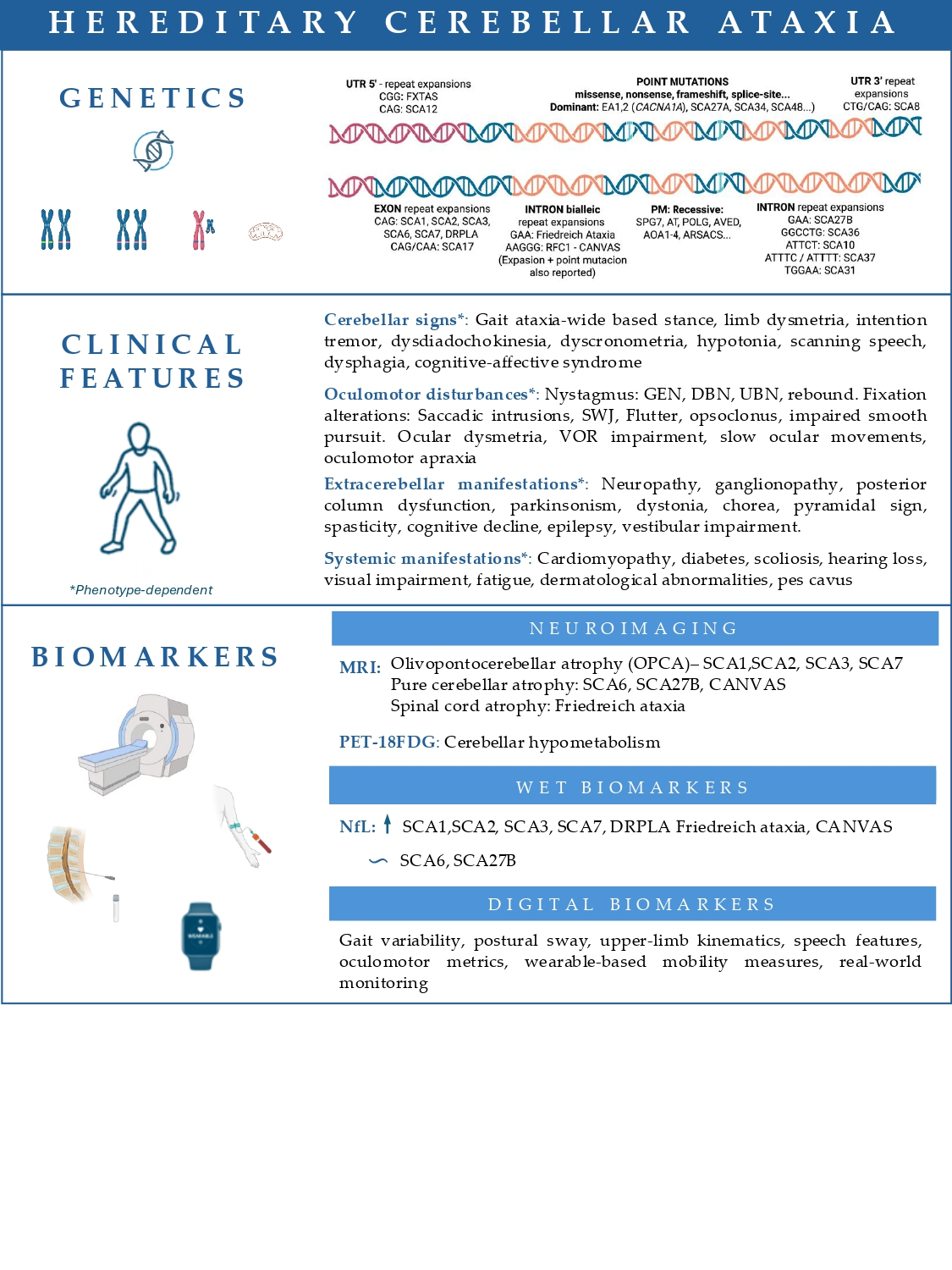
| Clinical Features | Associated Ataxias |
|---|---|
| Pure cerebellar ataxia | SCA 5, 6 *, 11, 14, 15, 16, 22 |
| Ocular/Oculomotor/Vestibular | |
| Ophthalmoparesis | SCA 1, 2, 3, 7, SPG7, mitochondrial *, NPC |
| Slow saccades | SCA 1, 2 *, 3, 7 |
| Oculomotor apraxia | AT, AOA1-4 *, SCA 7 |
| SWJ | FRDA *, RFC1, SCA 1, 2, 3 |
| DBN | SCA 1, 3, 6 *, 27B *, RFC1 |
| GEN | SCA 1, 3, 6, 27B *, 34, RFC1 |
| VOR impairment | SCA 6, 27B, RFC1 * |
| Retinitis pigmentosa | SCA 7 *, mitochondrial |
| Hearing loss | SCA36 *, mitochondrial * |
| Neuromuscular | |
| Spasticity | SCA 1, 3, 7, FRDA, SPG7 *, ARSACS *, AVED, SYNE1 |
| Peripheral neuropathy | SCA 1, 2, 3, 4, RFC1 *, FRDA *, AVED, FXTAS, mitochondrial |
| Myopathy | SCA 28, mitochondrial * |
| Fasciculations | SCA 36 * |
| Movement disorders | |
| Chorea | SCA 1, 2, 3, 17 *, 48, DRPLA *, AT |
| Myoclonus | SCA 2, 17, 19, DRPLA |
| Tremor | SCA 2 *, 8, 12, 15, 27A |
| Parkinsonism | SCA 2, 3 *, 12, 17, 48, FXTAS * |
| Dystonia | SCA 3, 17, AT, DRPLA |
| Cognitive/Psychiatric/Other | |
| Cognitive impairment | SCA 1 *, 2, 13, 17, 19, 21, 48 *, DRPLA, FXTAS *, mitochondrial |
| Behavioral disturbances | SCA 1, 17, 48, DRPLA, FXTAS * |
| Psychosis | SCA 17 *, 27A, DRPLA * |
| Epilepsy | SCA 10 *, 17, DRPLA, mitochondrial * |
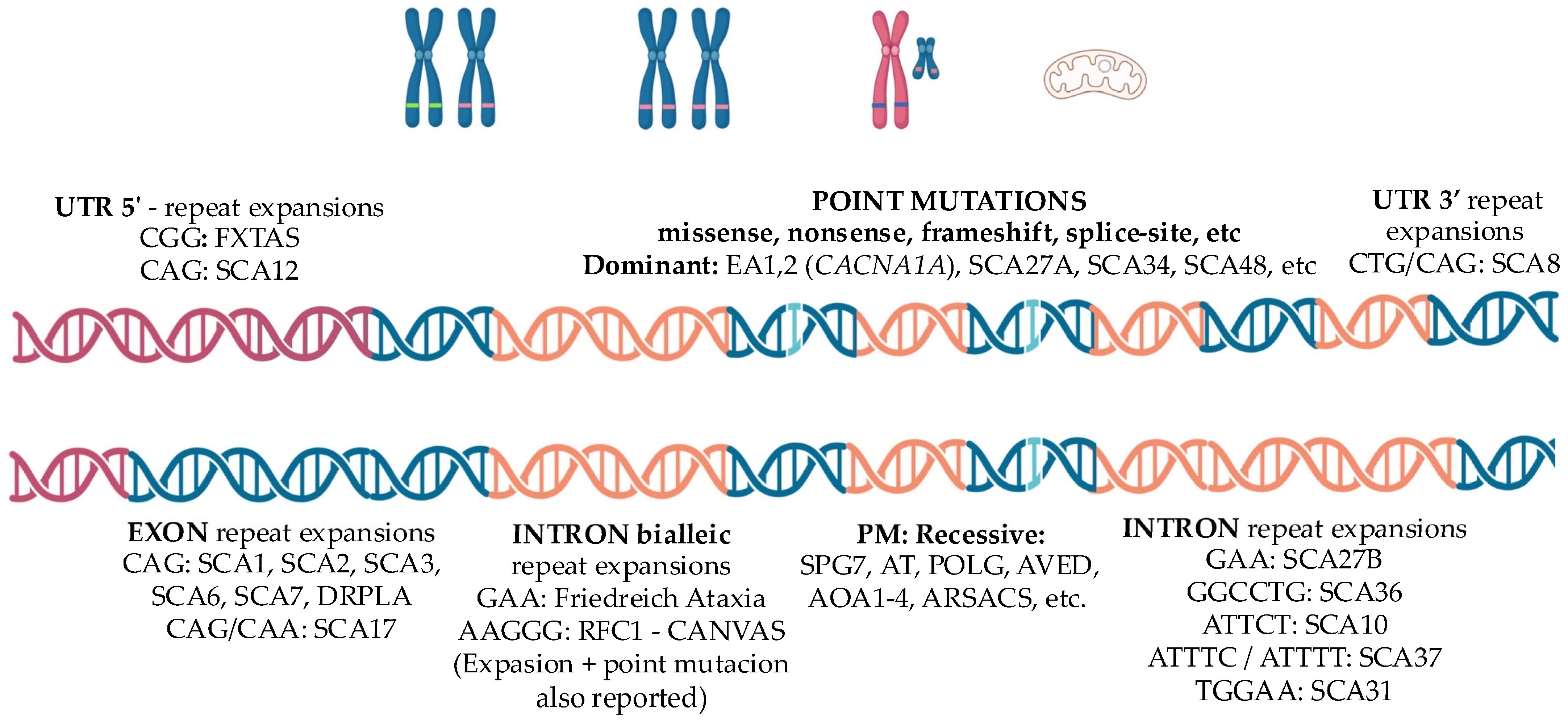
| Disorder | Gene | Genetics | Pathogenesis/Topography | Main/Distinguishing Features |
|---|---|---|---|---|
| SCA1 [2,43] | ATXN1 AD | CAG expansion ≥ 39 correlates with onset/severity | Toxic polyglutamine accumulation, Progressive Purkinje cell degeneration (cerebellar cortex). Brainstem, frontotemporal cortex | Hyperreflexia, pyramidal signs, cognitive impairment |
| SCA2 [2,44] | ATXN2 AD | CAG expansion ≥ 33 correlates with onset/severity; intermediate alleles (IA) linked to ALS spectrum | Toxic polyglutamine accumulation. Progressive Purkinje cell degeneration. Brainstem (pontocerebellar nuclei, inferior olive, cranial nerve nuclei) substantia nigra, dorsal root ganglion | Slowness of saccadic eye movements Hyporeflexia Tremor |
| SCA3 [2,10] | ATXN3 AD | Largest CAG expansions among SCAs ≥ 52; predictive of onset | Toxic polyglutamine accumulation. Dentate nucleus, substantia nigra, Brainstem motor nuclei, Basal ganglia. | Progressive external ophthalmoplegia, Parkinsonism, dystonia, pyramidal signs |
| SCA6 [2,45] | CACNA1A AD | Most stable and smallest CAG-expansion ≥ 20 | Toxic polyglutamine accumulation Progressive Purkinje cell degeneration Calcium channel dysfunction | Pure cerebellar ataxia Down beat nystagmus |
| SCA7 [46,47] | ATXN7 AD | Massive unstable CAG expansion ≥ 36 | Toxic polyglutamine accumulation Mitochondrial dysfunction Progressive Purkinje cell and retinal photoreceptors degeneration | Vision loss due retinal dystrophy Slowness of saccadic eye movements, oculomotor apraxia Spasticity |
| SCA17 [48,49] | TBP AD | CAG/CAA expansions ≥ 43 STUB1 mutations may modulate phenotype in IA (41–42) | Toxic polyglutamine accumulation Purkinje cells, striatum (caudate nucleus and putamen), thalamus, cerebral cortex (mainly frontal, temporal, parietal, and occipital areas), Limbic system (parahippocampus and cingulate gyrus). | HD-Like phenotype Chorea, dystonia, parkinsonism Psychiatric disturbances, cognitive impairment. |
| SCA27B [1,4] | FGF14 AD | Intronic GAA repeat expansion ≥ 250 | Abnormal gene transcription, reduced FGF14 mRNA and protein levels Purkinje and granule cells | Late onset episodic cerebellar ataxia Pure cerebellar ataxia Down beat nystagmus |
| DRPLA [5,48,50] | ATN1 AD | Massive unstable CAG expansion ≥ 48 | Toxic polyglutamine accumulation Dentate nucleus, globus pallidus, caudate, putamen, subthalamic nucleus of Luys, and cerebral cortex | HD-like phenotype: Chorea, parkinsonism, dystonia, cognitive impairment, psychiatric disturbances Juvenile onset: PME |
| Friedrich Ataxia [51] | FXN AR | Biallelic GAA intronic repeats expansions ≥ 66 correlate with onset | Decreased gene transcription, Mitochondrial frataxin deficiency Dorsal root ganglia, cerebellar dentate nucleus, Clarke’s column, gracile and cuneate nuclei, and corticospinal tracts. | Areflexia, Babinski sign, square waves, scoliosis, cardiomyopathy, diabetes |
| RFC1 related ataxia CANVAS [28] | RFC1 AR | Biallelic pentanucleotide intronic expansions ≥ 400; motif heterogeneity (AAGGG, ACAGG) | RNA expansion–mediated toxicity. Dorsal root ganglia, Purkinje and granular cells of the cerebellum (particularly in the vermis), vestibular ganglia | Sensitive neuronopathy with preserved reflexes Vestibular areflexia Down beat nystagmus Spasmodic dry cough |
| SPG7 [52] | SPG7 AR | Biallelic pathogenic variants (point mutations) | Mitochondrial dysfunction due to paraplegin deficiency Corticospinal tracts, cerebellar (dentate nucleus), brainstem, optic nerve neurons | Cerebellar ataxia (predominant in 1/3 cases) Spastic paraplegia Ophthalmoplegia, optic neuropathy, dystonia |
| FXTAS [35,53] | FMR1 X-linked | Premutation (55–200 CGG) (>200 CGG lead to X fragile syndrome) in 5′UTR | Predominant astroglial RNA expansion–mediated toxicity White matter of middle cerebellar peduncle, cerebellum, hippocampus, striatum, cortex, corpus callosum | Postural and intentional tremor, parkinsonism, cognitive impairment, neuropsychiatric disturbances Females: milder phenotype, premature ovarian failure. |
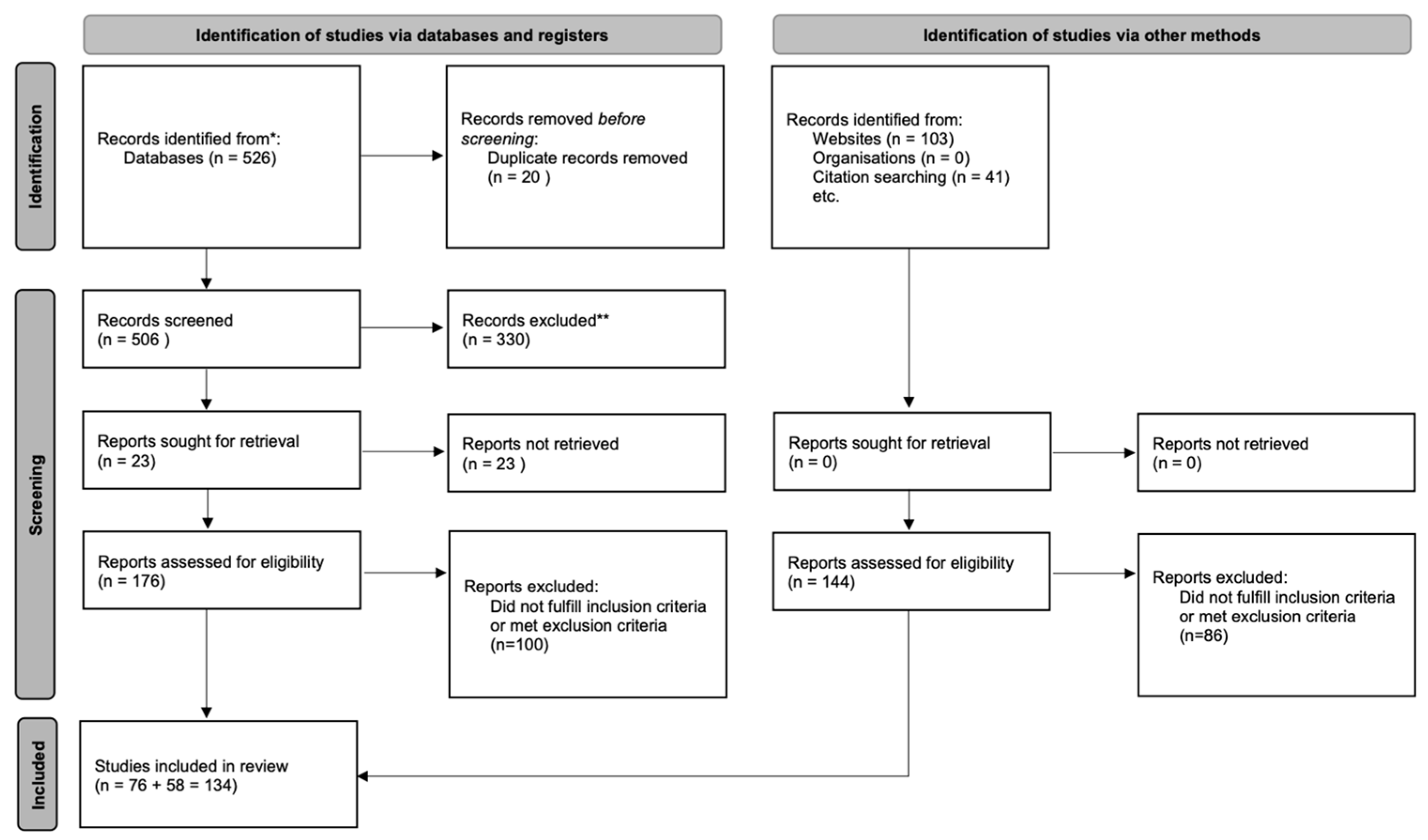
2. Methods
2.1. Inclusion Criteria
2.2. Exclusion Criteria
3. Results and Discussion
3.1. Imaging Biomarkers
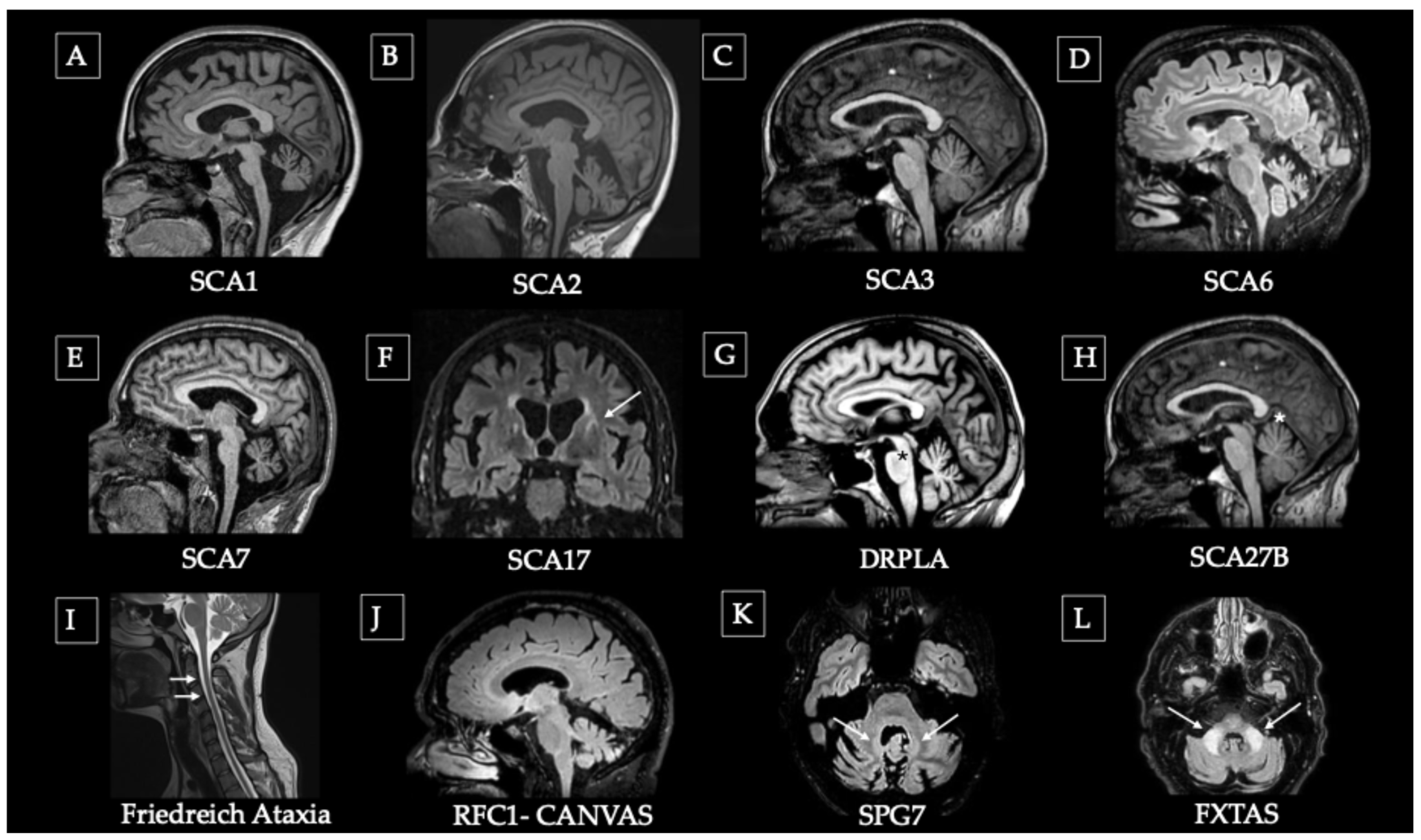
| Disorder | MRI Key Findings (Figure 2) Advanced MRI (DTI/7T/MRS) | Nuclear Medicine PET SPECT |
|---|---|---|
| SCA1 [43,63,64,82,123,124,125] | Olivopontocerebellar atrophy. Spinal involvement. | Hypometabolism in the cerebellum and brainstem. |
| Reduced FA in cerebellar peduncles. Spectrometry revealed reduced neuronal markers, elevated glial and energetic metabolites. | DAT-SPECT altered/normal. | |
| SCA2 [66,82,124,125,126] | Olivopontocerebellar atrophy, later involvement of subcortical structures and cortex. | Hypometabolism in the cerebellar cortex, dentate nucleus, brainstem (particularly the pons), and parahippocampal cortex. |
| Spectrometry revealed reduced neuronal markers, elevated glial and energetic metabolites. | DAT-SPECT may be altered with/without parkinsonian phenotype. | |
| SCA3 (MJD) [68,69,70,71,72,73,74,76,77,82,124] | Olivopontocerebellar atrophy: Progressive pons, cerebellar, superior cerebellar peduncles and spinal atrophy, frequent involvement of substantia nigra and progression to basal ganglia and cortical area. | Hypometabolism in the cerebellar cortex, basal ganglia–putamen. Cortical (motor/premotor) and thalamic hypometabolism in dystonic cases. |
| Early reduction in FA in inferior peduncle and white matter preceding gray matter loss. Spectrometry with reduced neuronal markers, elevated glial and energetic metabolites. | Altered DAT-SPECT parkinsonian phenotype dopa-responsive. | |
| SCA6 [78,79,80,124,127] | Selective cerebellar atrophy. | Cerebellar hypometabolism. Marked reduction in mGluR1 availability in vermis and flocculus. |
| Microstructural alterations in the cerebellar white matter and cerebellar peduncles. | Normal DAT-SPECT. | |
| SCA7 [81,82,128] | Olivopontocerebellar atrophy, medullar and occipital atrophy. | Marked hypometabolism in the cerebellum and brainstem. |
| Spectrometry revealed reduced neuronal markers, elevated glial and energetic metabolites. | Limited data | |
| SCA17 [48,83,84,85,86,129] | Cerebellar atrophy, frontostriatal (caudate and putamen) atrophy, putaminal rim sign. | Hypometabolism in cerebellum, striatum and cortex, atypical presentation with thalamocortical hypometabolism and preserved cerebellar metabolism have been described. |
| Limited data. | DAT-SPECT altered in advances stages. | |
| SCA27B [91,95,96] | Mild cerebellar atrophy predominantly affecting the superior vermis/Normal. | Hypometabolism in premotor cortex and cerebellar central lobule. |
| Microstructural alterations in the superior cerebellar peduncle, with reduced FA and increased MD. | Limited data. | |
| DRPLA [87,88,89,90,130] | Widespread cortical, cerebellar, pontine and midbrain atrophy, white matter hyperintensities, and pontine hypointensity | Striatal hypometabolism. |
| Generalized white matter microstructural alterations correlating with clinical progression. | Limited data. | |
| FRDA [56,97,99,100,101,103,104,105,131] | Mild cerebellar atrophy, predominant in dentate and superior peduncle, reduced cervical spinal cord cross-section. | Cerebellar hypometabolism. |
| Iron deposition in the dentate nucleus with susceptibility-weighted signal changes Superior peduncle and cervical cord degeneration in DTI, fMRI shows compensatory cortical hyperactivation and reorganization. | Limited data. | |
| RFC1 (CANVAS) [106,107,108,109,132,133,134] | Cerebellar vermian atrophy, mainly anterodorsal. | Mild hypoperfusion of cerebellar hemispheres and vermis, preserved striatal metabolism. |
| DTI changes mainly involve dorsal sensory tracts, deep white matter, corpus callosum, and major cerebral tracts. | DAT-SPECT can be altered (even without parkinsonism). | |
| SPG7 [110,111] | Cerebellar atrophy, most prominently cerebellar vermis, hyperintensity of dentate nucleus. | Limited data. |
| DTI: white matter changes characterized by reduced FA and increased MD in the frontal lobes, corticospinal tracts, and brainstem. | Limited data. | |
| FXTAS [112,113,114,115,116,117,135,136] | Hyperintensity of the middle cerebellar peduncle - MCP sign, diffuse WM hyperintensity. | Hypometabolism in the frontotemporal and cerebellar regions. |
| DTI with widespread WM damage; MRS abnormalities suggesting myelin damage (low NAA/Cr and Ch/Cr ratios). | Significant reduction in DAT uptake in the bilateral putamen, with or without parkinsonism. |
3.2. Fluid Biomarkers
| Disease | Biomarker | Key Findings |
|---|---|---|
| SCA1 [140] | NfL | Increased in preataxic and ataxic carriers (plasma ≈ 24.8 ± 11.1 pg/mL vs. 10.2 ± 4.5 pg/mL in controls); correlates with CAG repeat length and clinical progression. |
| SCA2 [140,141] | NfL | Elevated in preataxic and ataxic carriers (plasma ≈ 20.3 ± 6.3 pg/mL, cut-off 19.1 pg/mL); baseline levels predict cerebellar volume loss. Stable longitudinally despite clinical worsening. |
| PolyQ-ATXN2 | Target engagement marker. Requires further validation in patient cohorts | |
| SCA3 [137,140,142,143] | NfL | Significantly elevated in plasma and CSF (plasma ≈ 41.5 ± 14.8 pg/mL vs. 9.1 ± 3.3 pg/mL; CSF ≈ 4262 ± 1762 pg/mL vs. 472 ± 210 pg/mL); correlates with SARA, ICARS, and cerebellar atrophy; strong serum–CSF correlation (r ≈ 0.9). (Higher levels across PolyQ ataxia) |
| PolyQ-ATXN3 | Target engagement marker. Validated in plasma & CSF | |
| Tau | Elevated in preataxic >> ataxic patients >> controls | |
| GFAP | Increased levels no correlation with disease progression | |
| SCA6 [49,144] | NfL | No significant differences vs. controls (plasma ≈ 2.6 log pg/mL vs. 2.5 log pg/mL) |
| SCA7 [140,145] | NfL | Increased levels in plasma (≈21.6 pg/mL vs. 8.2 pg/mL) and CSF (≈2615 pg/mL vs. 415 pg/mL); strongly correlates with CAG repeat length and clinical progression. |
| GFAP | Elevated but less specific than NfL; potential marker of astroglial activation. | |
| SCA17 | Limited data | Further studies are needed |
| DRPLA [88] | NfL | Stepwise increase in plasma: controls (≈9.8 pg/mL) < preataxic (≈13.7 pg/mL) < ataxic (≈38.2 pg/mL); correlates with disease severity and CAG repeat length. |
| SCA27B [146] | NfL | Comparable to age-matched controls (≈22.7 ± 8.2 pg/mL); not significantly elevated; limited sensitivity for progression. |
| FRDA [147,148] | NfL | Elevated and correlated with severity (plasma ≈ 37.4 ± 19.7 pg/mL vs. 15.2 ± 6.4 pg/mL in controls). Levels decline with disease progression |
| Frataxin protein | Target-engagement marker; lower levels correlate with GAA expansion and clinical severity. Stable longitudinally. | |
| Tau | Increased in younger patients | |
| GFAP | Increased in younger patients | |
| Metabolic | Mitochondrial/oxidative alterations in G-CSF and antioxidant trials | |
| RFC1 (CANVAS) [149] | NfL | Increased serum levels (≈24.3 ± 9.2 pg/mL vs. 12.4 ± 4.6 pg/mL); higher in patients with cerebellar involvement (≈28 pg/mL); independent of disease duration. |
| SPG7 | Limited data | Further studies are needed |
| FXTAS | Limited data | Further studies are needed |
Metabolic Markers
3.3. Digital/Neurophysiological Biomarkers
3.3.1. Digital and Gait/Postural Control Biomarkers (Table 5)
3.3.2. Oculomotor & Vestibular Biomarkers (Table 5)
| Disorder | Digital and Gait/Postural Control Biomarkers | Oculomotor & Vestibular Biomarkers |
|---|---|---|
| SCA1 [174,176,177] | Preataxic: progressive stance instability on posturography; increased without visual cue | GEN, abnormal pursuit, dysmetric and slow saccades |
| SCA2 [161,176,177] | Lateral step deviation (wearable sensors during walking) captures 1-year progression in early-stage and preataxic carriers | Marked slow saccade; preserved Quantitative Head Impulse Test (qHIT) gain |
| SCA3 (MJD) [162,176,177] | Stride length variability (CV) and lateral sway detect 1-year progression in early/preataxic | Progressive ophthalmoparesis, hypermetric saccades, GEN, reduced pursuit velocity with saccadic intrusions, moderately vertical saccade velocity & qHIT reduced gain |
| SCA6 [165,176,177] | Lateral Velocity Change (LVC) during turning discriminates carriers from controls and detects 1-year progression also in preataxic group | DBN, GEN almost universal; abnormal pursuit; hypermetric saccade. Frequent saccadic intrusion, qHIT markedly reduced gain. |
| SCA7 [178] | Limited data | Cone–rod retinal degeneration (SCA7-RD) correlated with ataxia severity and CAG repetition |
| SCA17 [176] | Limited data | Abnormal pursuit, slow saccadic movements |
| SCA27B [179] | Limited data | DBN, VOR alteration, GEN |
| DRPLA [180] | Limited data | Nystagmus, dysmetric saccades, supranuclear palsy (advances stages) |
| FRDA [176,181,182,183] | Sway and postural stability measured by posturography correlate strongly with FARS and SARA, particularly under eyes-closed conditions. Gait speed progressively declined and postural instability markedly increased over two years | Oculomotor: frequent saccadic intrusion-SWJ and GEN, horizontal nystagmus, broken pursuit, saccade dysmetria and qHIT strongly reduced gain Optic neuropathy: thinning of pRNFL and macular inner retinal layers on OCT. |
| RFC1 (CANVAS) [176,184] | Limited data | DBN frequent, strongly reduced qHIT gain. Absent VOR on head impulse test (“pivotal sign”), bilateral vestibular loss in caloric and vHIT |
| SPG7 [185] | Limited data | Progressive retinal atrophy measured by OCT |
| FXTAS [173,186,187] | Larger sway area, increased variability, and greater postural sway jerk | Nystagmus, slow saccades, and fixation abnormalities in advanced stages |
3.3.3. Neurophysiology & Other Functional Biomarkers
4. Conclusions
Author Contributions
Funding
Institutional Review Board Statement
Informed Consent Statement
Data Availability Statement
Acknowledgments
Conflicts of Interest
Abbreviations
References
- Rossi, M.; Stephen, C.D.; Damásio, J.; Pedroso, J.L.; Kuo, S.; Lin, C.R.; Ojo, O.; El-Jaafary, S.; Lee, W.; Madoev, H.; et al. Unravelling the Global Tapestry of Genetic Ataxias: Epidemiology and Genetic Testing Approaches. Mov. Disord. 2025, 40, 1805–1820. [Google Scholar] [CrossRef]
- Klockgether, T. Update on degenerative ataxias. Curr. Opin. Neurol. 2011, 24, 339–345. [Google Scholar] [CrossRef]
- Rudaks, L.I.; Yeow, D.; Ng, K.; Deveson, I.W.; Kennerson, M.L.; Kumar, K.R. An Update on the Adult-Onset Hereditary Cerebellar Ataxias: Novel Genetic Causes and New Diagnostic Approaches. Cerebellum 2024, 23, 2152–2168. [Google Scholar] [CrossRef] [PubMed]
- Cui, Z.-T.; Mao, Z.-T.; Yang, R.; Li, J.-J.; Jia, S.-S.; Zhao, J.-L.; Zhong, F.-T.; Yu, P.; Dong, M. Spinocerebellar ataxias: From pathogenesis to recent therapeutic advances. Front. Neurosci. 2024, 18, 1422442. [Google Scholar] [CrossRef]
- Vegezzi, E.; Ishiura, H.; Bragg, D.C.; Pellerin, D.; Magrinelli, F.; Currò, R.; Facchini, S.; Tucci, A.; Hardy, J.; Sharma, N.; et al. Neurological disorders caused by novel non-coding repeat expansions: Clinical features and differential diagnosis. Lancet Neurol. 2024, 23, 725–739. [Google Scholar] [CrossRef]
- Ashizawa, T.; Öz, G.; Paulson, H.L. Spinocerebellar ataxias: Prospects and challenges for therapy development. Nat. Rev. Neurol. 2018, 14, 590–605, Correction in Nat. Rev. Neurol. 2018, 14, 749. https://doi.org/10.1038/s41582-018-0102-z. [Google Scholar] [CrossRef]
- Sasaki, H.; Fukazawa, T.; Yanagihara, T.; Hamada, T.; Shima, K.; Matsumoto, A.; Hashimoto, K.; Ito, N.; Wakisaka, A.; Tashiro, K. Clinical features and natural history of spinocerebellar ataxia type 1. Acta Neurol. Scand. 2009, 93, 64–71. [Google Scholar] [CrossRef] [PubMed]
- Jacobi, H.; Hauser, T.K.; Giunti, P.; Globas, C.; Bauer, P.; Schmitz-Hübsch, T.; Baliko, L.; Filla, A.; Mariotti, C.; Rakowicz, M.; et al. Spinocerebellar ataxia types 1, 2, 3 and 6: The clinical spectrum of ataxia and morphometric brainstem and cerebellar findings. Cerebellum 2012, 11, 155–166. [Google Scholar] [CrossRef]
- Tezenas Du Montcel, S.; Durr, A.; Bauer, P.; Figueroa, K.P.; Ichikawa, Y.; Brussino, A.; Forlani, S.; Rakowicz, M.; Schöls, L.; Mariotti, C.; et al. Modulation of the age at onset in spinocerebellar ataxia by CAG tracts in various genes. Brain 2014, 137, 2444–2455. [Google Scholar] [CrossRef] [PubMed]
- Stahl, F.; Evert, B.O.; Han, X.; Breuer, P.; Wüllner, U. Spinocerebellar Ataxia Type 3 Pathophysiology—Implications for Translational Research and Clinical Studies. Int. J. Mol. Sci. 2024, 25, 3984. [Google Scholar] [CrossRef]
- Nakano, K.K.; Dawson, D.M.; Spence, A. Machado disease: A hereditary ataxia in Portuguese emigrants to Massachusetts. Neurology 1972, 22, 49. [Google Scholar] [CrossRef]
- Solodkin, A.; Gomez, C.M. Spinocerebellar ataxia type 6. In Handbook of Clinical Neurology; Elsevier: Amsterdam, The Netherlands, 2012; Volume 103, pp. 461–473. [Google Scholar] [CrossRef]
- Martin, J.-J. Spinocerebellar ataxia type 7. In Handbook of Clinical Neurology; Elsevier: Amsterdam, The Netherlands, 2012; Volume 103, pp. 475–491. [Google Scholar] [CrossRef]
- Rolfs, A.; Koeppen, A.H.; Bauer, I.; Bauer, P.; Buhlmann, S.; Topka, H.; Schöls, L.; Riess, O. Clinical features and neuropathology of autosomal dominant spinocerebellar ataxia (SCA17). Ann. Neurol. 2003, 54, 367–375. [Google Scholar] [CrossRef]
- Nowak, B.; Kozlowska, E.; Pawlik, W.; Fiszer, A. Atrophin-1 Function and Dysfunction in Dentatorubral–Pallidoluysian Atrophy. Mov. Disord. 2023, 38, 526–536. [Google Scholar] [CrossRef]
- Carroll, L.; Massey, T.; Peall, K.; Wardle, M. Dentatorubral-pallidoluysian Atrophy: An Update. Tremor Other Hyperkinetic Mov. 2018, 8, 577. [Google Scholar] [CrossRef]
- Rafehi, H.; Read, J.; Szmulewicz, D.J.; Davies, K.C.; Snell, P.; Fearnley, L.G.; Scott, L.; Thomsen, M.; Gillies, G.; Pope, K.; et al. An intronic GAA repeat expansion in FGF14 causes the autosomal-dominant adult-onset ataxia SCA27B/ATX-FGF14. Am. J. Human Genet. 2023, 110, 105–119, Erratum in Am. J. Human Genet. 2023, 110, 1018. https://doi.org/10.1016/j.ajhg.2023.05.005. [Google Scholar] [CrossRef] [PubMed]
- Koeppen, A. The pathogenesis of spinocerebellar ataxia. Cerebellum 2005, 4, 62–73. [Google Scholar] [CrossRef] [PubMed]
- McLoughlin, H.S.; Moore, L.R.; Paulson, H.L. Pathogenesis of SCA3 and implications for other polyglutamine diseases. Neurobiol. Dis. 2020, 134, 104635. [Google Scholar] [CrossRef]
- Mariotti, C.; Fancellu, R.; Donato, S. An overview of the patient with ataxia. J. Neurol. 2005, 252, 511–518. [Google Scholar] [CrossRef]
- Zesiewicz, T.A. Ataxia. Continuum 2025, 31, 1093–1119. [Google Scholar] [CrossRef]
- Wirth, T.; Faber, J.; Depienne, C.; Roze, E.; Honnorat, J.; Meissner, W.G.; Giunti, P.; Tranchant, C.; Klockgether, T.; Anheim, M. Progress and challenges in sporadic late-onset cerebellar ataxias. Nat. Rev. Neurol. 2025, 21, 687–705. [Google Scholar] [CrossRef]
- Koeppen, A.H. Nikolaus Friedreich and degenerative atrophy of the dorsal columns of the spinal cord. J. Neurochem. 2013, 126, 4–10, Erratum in J. Neurochem. 2013, 126, 4–10. [Google Scholar] [CrossRef] [PubMed]
- Campuzano, V.; Montermini, L.; Moltò, M.D.; Pianese, L.; Cossée, M.; Cavalcanti, F.; Monros, E.; Rodius, F.; Duclos, F.; Monticelli, A.; et al. Friedreich’s Ataxia: Autosomal Recessive Disease Caused by an Intronic GAA Triplet Repeat Expansion. Science 1996, 271, 1423–1427. [Google Scholar] [CrossRef]
- Delatycki, M.B.; Corben, L.A. Clinical Features of Friedreich Ataxia. J. Child Neurol. 2012, 27, 1133–1137. [Google Scholar] [CrossRef] [PubMed]
- Parkinson, M.H.; Boesch, S.; Nachbauer, W.; Mariotti, C.; Giunti, P. Clinical features of Friedreich’s ataxia: Classical and atypical phenotypes. J. Neurochem. 2013, 126, 103–117. [Google Scholar] [CrossRef]
- Cortese, A.; Simone, R.; Sullivan, R.; Vandrovcova, J.; Tariq, H.; Yau, W.Y.; Humphrey, J.; Jaunmuktane, Z.; Sivakumar, P.; Polke, J.; et al. Biallelic expansion of an intronic repeat in RFC1 is a common cause of late-onset ataxia. Nat. Genet. 2019, 51, 649–658, Erratum in Nat. Genet. 2019, 51, 920. https://doi.org/10.1038/s41588-019-0422-y. [Google Scholar] [CrossRef]
- Cortese, A.; Tozza, S.; Yau, W.Y.; Rossi, S.; Beecroft, S.J.; Jaunmuktane, Z.; Dyer, Z.; Ravenscroft, G.; Lamont, P.J.; Mossman, S.; et al. Cerebellar ataxia, neuropathy, vestibular areflexia syndrome due to RFC1 repeat expansion. Brain 2020, 143, 480–490. [Google Scholar] [CrossRef] [PubMed]
- Traschütz, A.; Cortese, A.; Reich, S.; Dominik, N.; Faber, J.; Jacobi, H.; Hartmann, A.M.; Rujescu, D.; Montaut, S.; Echaniz-Laguna, A.; et al. Natural History, Phenotypic Spectrum, and Discriminative Features of Multisystemic RFC1 Disease. Neurology 2021, 96, e1369–e1382. [Google Scholar] [CrossRef] [PubMed]
- Davies, K.; Szmulewicz, D.J.; Corben, L.A.; Delatycki, M.; Lockhart, P.J. RFC1-Related Disease: Molecular and Clinical Insights. Neurol. Genet. 2022, 8, e200016. [Google Scholar] [CrossRef]
- Van Gassen, K.L.I.; Van Der Heijden, C.D.C.C.; De Bot, S.T.; Den Dunnen, W.F.A.; Van Den Berg, L.H.; Verschuuren-Bemelmans, C.C.; Kremer, H.P.H.; Veldink, J.H.; Kamsteeg, E.-J.; Scheffer, H.; et al. Genotype–phenotype correlations in spastic paraplegia type 7: A study in a large Dutch cohort. Brain 2012, 135, 2994–3004. [Google Scholar] [CrossRef]
- Jacquemont, S.; Hagerman, R.J.; Leehey, M.; Grigsby, J.; Zhang, L.; Brunberg, J.A.; Greco, C.; Des Portes, V.; Jardini, T.; Levine, R.; et al. Fragile X Premutation Tremor/Ataxia Syndrome: Molecular, Clinical, and Neuroimaging Correlates. Am. J. Human Genet. 2003, 72, 869–878. [Google Scholar] [CrossRef]
- Hall, D.; Tassone, F.; Klepitskaya, O.; Leehey, M. Fragile X-associated tremor ataxia syndrome in FMR1 gray zone allele carriers. Mov. Disord. 2012, 27, 296–300. [Google Scholar] [CrossRef] [PubMed]
- Sellier, C.; Freyermuth, F.; Tabet, R.; Tran, T.; He, F.; Ruffenach, F.; Alunni, V.; Moine, H.; Thibault, C.; Page, A.; et al. Sequestration of DROSHA and DGCR8 by Expanded CGG RNA Repeats Alters MicroRNA Processing in Fragile X-Associated Tremor/Ataxia Syndrome. Cell Rep. 2013, 3, 869–880. [Google Scholar] [CrossRef] [PubMed]
- Cabal-Herrera, A.M.; Tassanakijpanich, N.; Salcedo-Arellano, M.J.; Hagerman, R.J. Fragile X-Associated Tremor/Ataxia Syndrome (FXTAS): Pathophysiology and Clinical Implications. Int. J. Mol. Sci. 2020, 21, 4391. [Google Scholar] [CrossRef]
- Finsterer, J. Neuropathy, Ataxia, and Retinitis Pigmentosa Syndrome. J. Clin. Neuromuscul. Dis. 2023, 24, 140–146. [Google Scholar] [CrossRef]
- Zeviani, M.; Moraes, C.T.; DiMauro, S.; Nakase, H.; Bonilla, E.; Schon, E.A.; Rowland, L.P. Deletions of mitochondrial DNA in Kearns-Sayre syndrome. Neurology 1988, 38, 1339. [Google Scholar] [CrossRef] [PubMed]
- Van Goethem, G.; Luoma, P.; Rantamäki, M.; Al Memar, A.; Kaakkola, S.; Hackman, P.; Krahe, R.; Löfgren, A.; Martin, J.J.; De Jonghe, P.; et al. POLG mutations in neurodegenerative disorders with ataxia but no muscle involvement. Neurology 2004, 63, 1251–1257. [Google Scholar] [CrossRef] [PubMed]
- Biomarkers Definitions Working Group. Biomarkers and surrogate endpoints: Preferred definitions and conceptual framework. Clin. Pharmacol. Ther. 2001, 69, 89–95. [Google Scholar] [CrossRef]
- FDA-NIH Biomarker Working Group. BEST (Biomarkers, Endpoints, and Other Tools) Resource; Food and Drug Administration (US): Silver Spring, MD, USA; National Institutes of Health (US): Bethesda, MD, USA, 2016. Available online: https://www.ncbi.nlm.nih.gov/books/NBK326791/ (accessed on 9 September 2025).
- Strimbu, K.; Tavel, J.A. What are biomarkers? Curr. Opin. HIV AIDS 2010, 5, 463–466. [Google Scholar] [CrossRef]
- Cummings, J.; Zhong, K. Biomarker-Driven Therapeutic Management of Alzheimer’s Disease: Establishing the Foundations. Clin. Pharmacol. Ther. 2013, 95, 67–77. [Google Scholar] [CrossRef]
- Martins Junior, C.R.; Martinez, A.R.M.; Vasconcelos, I.F.; De Rezende, T.J.R.; Casseb, R.F.; Pedroso, J.L.; Barsottini, O.G.P.; Lopes-Cendes, Í.; França, M.C., Jr. Structural signature in SCA1: Clinical correlates, determinants and natural history. J. Neurol. 2018, 265, 2949–2959. [Google Scholar] [CrossRef]
- Velázquez-Pérez, L.C.; Rodríguez-Labrada, R.; Fernandez-Ruiz, J. Spinocerebellar Ataxia Type 2: Clinicogenetic Aspects, Mechanistic Insights, and Management Approaches. Front. Neurol. 2017, 8, 472. [Google Scholar] [CrossRef] [PubMed]
- Giunti, P.; Mantuano, E.; Frontali, M.; Veneziano, L. Molecular mechanism of Spinocerebellar Ataxia type 6: Glutamine repeat disorder, channelopathy and transcriptional dysregulation. The multifaceted aspects of a single mutation. Front. Cell. Neurosci. 2015, 9, 36. [Google Scholar] [CrossRef]
- Nambo-Venegas, R.; Valdez-Vargas, C.; Cisneros, B.; Palacios-González, B.; Vela-Amieva, M.; Ibarra-González, I.; Cerecedo-Zapata, C.M.; Martínez-Cruz, E.; Cortés, H.; Reyes-Grajeda, J.P.; et al. Altered Plasma Acylcarnitines and Amino Acids Profile in Spinocerebellar Ataxia Type 7. Biomolecules 2020, 10, 390. [Google Scholar] [CrossRef]
- Goswami, R.; Bello, A.I.; Bean, J.; Costanzo, K.M.; Omer, B.; Cornelio-Parra, D.; Odah, R.; Ahluwalia, A.; Allan, S.K.; Nguyen, N.; et al. The Molecular Basis of Spinocerebellar Ataxia Type 7. Front. Neurosci. 2022, 16, 818757. [Google Scholar] [CrossRef]
- Pérez-Pérez, J.; Olmedo-Saura, G.; Martínez-Horta, S.; Bernal, S.; Pagonabarraga, J.; Kulisevsky, J. Update on Genetic Chorea. Eur. J. Neurol. 2025, 32, e70357. [Google Scholar] [CrossRef]
- Peng, L.; Wang, S.; Chen, Z.; Peng, Y.; Wang, C.; Long, Z.; Peng, H.; Shi, Y.; Hou, X.; Lei, L.; et al. Blood Neurofilament Light Chain in Genetic Ataxia: A Meta-Analysis. Mov. Disord. 2022, 37, 171–181. [Google Scholar] [CrossRef]
- Yamada, M.; Wood, J.D.; Shimohata, T.; Hayashi, S.; Tsuji, S.; Ross, C.A.; Takahashi, H. Widespread occurrence of intranuclear atrophin-1 accumulation in the central nervous system neurons of patients with dentatorubral-pallidoluysian atrophy. Ann. Neurol. 2001, 49, 14–23. [Google Scholar] [CrossRef]
- Reetz, K.; Lischewski, S.A.; Dogan, I.; Didszun, C.; Pishnamaz, M.; Konrad, K.; Marx-Schütt, K.; Farmer, J.; Lynch, D.R.; Corben, L.A.; et al. Friedreich’s ataxia—A rare multisystem disease. Lancet Neurol. 2025, 24, 614–624. [Google Scholar] [CrossRef] [PubMed]
- Wilkinson, P.A. A clinical, genetic and biochemical study of SPG7 mutations in hereditary spastic paraplegia. Brain 2004, 127, 973–980. [Google Scholar] [CrossRef] [PubMed]
- Hagerman, R.J.; Hagerman, P.J. The Spectrum of Fragile X Disorders. N. Engl. J. Med. 2025, 393, 281–288. [Google Scholar] [CrossRef]
- Tricco, A.C.; Lillie, E.; Zarin, W.; O’Brien, K.K.; Colquhoun, H.; Levac, D.; Moher, D.; Peters, M.D.J.; Horsley, T.; Weeks, L.; et al. PRISMA Extension for Scoping Reviews (PRISMA-ScR): Checklist and Explanation. Ann. Intern. Med. 2018, 169, 467–473. [Google Scholar] [CrossRef]
- Scaravilli, A.; Tranfa, M.; Pontillo, G.; Brais, B.; De Michele, G.; La Piana, R.; Saccà, F.; Santorelli, F.M.; Synofzik, M.; Brunetti, A.; et al. CHARON: An Imaging-Based Diagnostic Algorithm to Navigate Through the Sea of Hereditary Degenerative Ataxias. Cerebellum 2024, 23, 2122–2129. [Google Scholar] [CrossRef]
- Cocozza, S.; Pontillo, G.; De Michele, G.; Di Stasi, M.; Guerriero, E.; Perillo, T.; Pane, C.; De Rosa, A.; Ugga, L.; Brunetti, A. Conventional MRI findings in hereditary degenerative ataxias: A pictorial review. Neuroradiology 2021, 63, 983–999. [Google Scholar] [CrossRef]
- Lee, Y.-C.; Liu, C.-S.; Wu, H.-M.; Wang, P.-S.; Chang, M.-H.; Soong, B.-W. The ‘hot cross bun’ sign in the patients with spinocerebellar ataxia. Eur. J. Neurol. 2009, 16, 513–516. [Google Scholar] [CrossRef] [PubMed]
- Sugiyama, A.; Riku, Y.; Koga, S.; Yokota, H.; Mukai, H.; Takao, M.; Hayashi, K.; Takeda, T.; Hirano, S.; Yoshida, M.; et al. Revisiting ‘hot cross bun’ sign: A multicentre MRI study of 97 patients with autopsy-confirmed multiple system atrophy. J. Neurol. Neurosurg. Psychiatry 2025, Epub ahead of print. [Google Scholar] [CrossRef] [PubMed]
- Olmedo-Saura, G.; Iruzubieta, P.; Bernal, S.; Gonzalez-Quereda, L.; Pont-Sunyer, C.; Navalpotro-Gómez, I.; Guisado-Alonso, D.; García-Sanchez, C.; Rodriguez-Santiago, B.; Brais, B.; et al. Expanding the clinical–radiological phenotype of SCA34 and its role in the differential diagnosis of the hot cross bun sign. J. Neurol. 2025, 272, 566. [Google Scholar] [CrossRef]
- Meira, A.T.; Vale, T.C.; Da Costa, S.C.G.; Gauvreau, C.; Piccinin, C.C.; Teive, H.A.G.; França, M.C.; Dupré, N.; Barsottini, O.G.; Pedroso, J.L. Neuroimaging in Cerebellar Ataxias: A Diagnostic Approach. Cerebellum 2025, 24, 154. [Google Scholar] [CrossRef]
- Mascalchi, M.; Vella, A. Magnetic resonance and nuclear medicine imaging in ataxias. In Handbook of Clinical Neurology; Elsevier: Amsterdam, The Netherlands, 2012; Volume 103, pp. 85–110. [Google Scholar] [CrossRef]
- Mascalchi, M.; Vella, A. Neuroimaging Applications in Chronic Ataxias. In International Review of Neurobiology; Elsevier: Amsterdam, The Netherlands, 2018; Volume 143, pp. 109–162. [Google Scholar] [CrossRef]
- Martins, C.R.; Martinez, A.R.M.; De Rezende, T.J.R.; Branco, L.M.T.; Pedroso, J.L.; Barsottini, O.G.P.; Lopes-Cendes, I.; França, M.C. Spinal Cord Damage in Spinocerebellar Ataxia Type 1. Cerebellum 2017, 16, 792–796. [Google Scholar] [CrossRef] [PubMed]
- Yoo, Y.J.; Oh, J. Identification of early neurodegenerative change in presymptomatic spinocerebellar ataxia type 1: A diffusion tensor imaging study. Park. Relat. Disord. 2017, 36, 109–110. [Google Scholar] [CrossRef]
- Robertson, J.W.; Adanyeguh, I.; Bender, B.; Boesch, S.; Brunetti, A.; Cocozza, S.; Coutinho, L.; Deistung, A.; Diciotti, S.; Dogan, I.; et al. The Pattern and Stages of Atrophy in Spinocerebellar Ataxia Type 2: Volumetrics from ENIGMA-Ataxia. Mov. Disord. 2025, 40, 651–661. [Google Scholar] [CrossRef]
- Reetz, K.; Rodríguez-Labrada, R.; Dogan, I.; Mirzazade, S.; Romanzetti, S.; Schulz, J.B.; Cruz-Rivas, E.M.; Alvarez-Cuesta, J.A.; Aguilera Rodríguez, R.; Gonzalez Zaldivar, Y.; et al. Brain atrophy measures in preclinical and manifest spinocerebellar ataxia type 2. Ann. Clin. Transl. Neurol. 2018, 5, 128–137. [Google Scholar] [CrossRef]
- Coarelli, G.; Heinzmann, A.; Ewenczyk, C.; Fischer, C.; Chupin, M.; Monin, M.L.; Hurmic, H.; Calvas, F.; Calvas, P.; Goizet, C.; et al. Safety and efficacy of riluzole in spinocerebellar ataxia type 2 in France (ATRIL): A multicentre, randomised, double-blind, placebo-controlled trial. Lancet Neurol. 2022, 21, 225–233. [Google Scholar] [CrossRef]
- Sobana, S.A.; Huda, F.; Hermawan, R.; Sribudiani, Y.; Koan, T.S.; Dian, S.; Ong, P.A.; Dahlan, N.L.; Utami, N.; Pusparini, I.; et al. Brain MRI Volumetry Analysis in an Indonesian Family of SCA 3 Patients: A Case-Based Study. Front. Neurol. 2022, 13, 912592. [Google Scholar] [CrossRef]
- D’Abreu, A.; França, M.C., Jr.; Yasuda, C.L.; Campos, B.A.G.; Lopes-Cendes, I.; Cendes, F. Neocortical Atrophy in Machado-Joseph Disease: A Longitudinal Neuroimaging Study. J. Neuroimaging 2012, 22, 285–291. [Google Scholar] [CrossRef]
- Kang, J.-S.; Klein, J.C.; Baudrexel, S.; Deichmann, R.; Nolte, D.; Hilker, R. White matter damage is related to ataxia severity in SCA3. J. Neurol. 2014, 261, 291–299. [Google Scholar] [CrossRef] [PubMed]
- Huang, S.-R.; Wu, Y.-T.; Jao, C.-W.; Soong, B.-W.; Lirng, J.-F.; Wu, H.-M.; Wang, P.-S. CAG repeat length does not associate with the rate of cerebellar degeneration in spinocerebellar ataxia type 3. NeuroImage Clin. 2017, 13, 97–105. [Google Scholar] [CrossRef] [PubMed]
- Fahl, C.N.; Branco, L.M.T.; Bergo, F.P.G.; D’Abreu, A.; Lopes-Cendes, I.; França, M.C. Spinal Cord Damage in Machado-Joseph Disease. Cerebellum 2015, 14, 128–132. [Google Scholar] [CrossRef] [PubMed]
- Rezende, T.J.R.; De Paiva, J.L.R.; Martinez, A.R.M.; Lopes-Cendes, I.; Pedroso, J.L.; Barsottini, O.G.P.; Cendes, F.; França, M.C. Structural signature of SCA3: From presymptomatic to late disease stages. Ann. Neurol. 2018, 84, 401–408. [Google Scholar] [CrossRef]
- Ye, Z.-X.; Chen, X.-Y.; Li, M.-C.; Chen, X.-Y.; Qiu, Y.-S.; Yuan, R.-Y.; Chen, Z.-L.; Lin, M.-T.; Hu, J.-P.; Fu, Y.; et al. Associations between CAG repeat size, brain and spinal cord volume loss, and motor symptoms in spinocerebellar ataxia type 3: A cohort study. Orphanet J. Rare Dis. 2025, 20, 35. [Google Scholar] [CrossRef]
- Nunes, M.B.; Martinez, A.R.; Rezende, T.J.; Friedman, J.H.; Lopes-Cendes, I.; D’Abreu, A.; França, M.C., Jr. Dystonia in Machado-Joseph disease: Clinical profile, therapy and anatomical basis. Park. Relat. Disord. 2015, 21, 1441–1447. [Google Scholar] [CrossRef] [PubMed]
- Okamoto, N.; Ikenouchi, A.; Ide, S.; Hashimoto, Y.; Yoshimura, R. A Young Japanese Patient with Spinocerebellar Ataxia Type 3 Presenting Depressive State with Cenesthopathy and Delusion: A Case Report. Cerebellum 2022, 21, 1135–1138. [Google Scholar] [CrossRef]
- Berger, M.; Garcia-Moreno, H.; Ferreira, M.; Hubener-Schmid, J.; Schaprian, T.; Wegner, P.; Elter, T.; Teichmann, K.M.; Santana, M.M.; Grobe-Einsler, M.; et al. Progression of biological markers in spinocerebellar ataxia type 3: Longitudinal analysis of prospective data from the ESMI cohort. Lancet Reg. Health-Eur. 2025, 55, 101339. [Google Scholar] [CrossRef] [PubMed]
- Eichler, L.; Bellenberg, B.; Hahn, H.K.; Köster, O.; Schöls, L.; Lukas, C. Quantitative Assessment of Brain Stem and Cerebellar Atrophy in Spinocerebellar Ataxia Types 3 and 6: Impact on Clinical Status. AJNR Am. J. Neuroradiol. 2011, 32, 890–897. [Google Scholar] [CrossRef]
- Rentiya, Z.; Khan, N.-S.; Ergun, E.; Ying, S.H.; Desmond, J.E. Distinct cerebellar regions related to motor and cognitive performance in SCA6 patients. Neuropsychologia 2017, 107, 25–30. [Google Scholar] [CrossRef] [PubMed]
- Ishibashi, K.; Miura, Y.; Ishikawa, K.; Ishii, K.; Ishiwata, K. Decreased metabotropic glutamate receptor type 1 availability in a patient with spinocerebellar ataxia type 6: A 11C-ITMM PET study. J. Neurol. Sci. 2015, 355, 202–205. [Google Scholar] [CrossRef] [PubMed]
- Coarelli, G.; Dubec-Fleury, C.; Petit, E.; Sayah, S.; Fischer, C.; Nassisi, M.; Gatignol, P.; Dorgham, K.; Daghsen, L.; Daye, P.; et al. Longitudinal Changes of Clinical, Imaging, and Fluid Biomarkers in Preataxic and Early Ataxic Spinocerebellar Ataxia Type 2 and 7 Carriers. Neurology 2024, 103, e209749. [Google Scholar] [CrossRef]
- Adanyeguh, I.M.; Henry, P.; Nguyen, T.M.; Rinaldi, D.; Jauffret, C.; Valabregue, R.; Emir, U.E.; Deelchand, D.K.; Brice, A.; Eberly, L.E.; et al. In vivo neurometabolic profiling in patients with spinocerebellar ataxia types 1, 2, 3, and 7. Mov. Disord. 2015, 30, 662–670. [Google Scholar] [CrossRef] [PubMed]
- Bech, S.; Petersen, T.; Nørremølle, A.; Gjedde, A.; Ehlers, L.; Eiberg, H.; Hjermind, L.E.; Hasholt, L.; Lundorf, E.; Nielsen, J.E. Huntington’s disease-like and ataxia syndromes: Identification of a family with a de novo SCA17/TBP mutation. Park. Relat. Disord. 2010, 16, 12–15. [Google Scholar] [CrossRef]
- Claassen, J.; Gerding, W.M.; Kastrup, O.; Uslar, E.; Goericke, S.; Timmann, D. Excessive brain iron accumulation in spinocerebellar ataxia type 17. Neurology 2015, 84, 212–213. [Google Scholar] [CrossRef]
- Grassini, A.; Cermelli, A.; Roveta, F.; Zotta, M.; Lesca, A.; Marcinnò, A.; Ferrandes, F.; Piella, E.; Boschi, S.; Lombardo, C.; et al. Cognitive dysfunction, social behavior disorder, cerebellar ataxia, and atypical brain FDG-PET presentation in spinocerebellar ataxia 17: A case report. Neurol. Sci. 2024, 45, 2877–2880, Erratum in Neurol. Sci. 2024, 45, 2949. https://doi.org/10.1007/s10072-024-07498-5. [Google Scholar] [CrossRef]
- Brockmann, K.; Reimold, M.; Globas, C.; Hauser, T.K.; Walter, U.; Machulla, H.-J.; Rolfs, A.; Schöls, L. PET and MRI Reveal Early Evidence of Neurodegeneration in Spinocerebellar Ataxia Type 17. J. Nucl. Med. 2012, 53, 1074–1080. [Google Scholar] [CrossRef]
- Li, M.; Chen, X.; Yuan, R.; Fan, S.; Huang, Z.; Liu, Z.; Weng, J.; Zheng, Q.; Gan, S.; Hu, J. MRI-based brain structural changes in adult-onset dentatorubral–pallidoluysian atrophy. Neuroradiology, 2025; Online ahead of print. [Google Scholar] [CrossRef]
- Yuan, R.; Chen, Y.; Lin, W.; Li, M.; Zeng, Y.; Cheng, B.; Yu, X.; Zuo, D.; Sun, H.; Ye, B.; et al. Clinical and Genetic Findings in a Chinese Cohort of Dentatorubral–Pallidoluysian Atrophy Patients. Ann. Neurol. 2025, 98, 750–761. [Google Scholar] [CrossRef]
- Ide, S.; Kakeda, S.; Adachi, H.; Miyata, M.; Iwanaka, Y.; Okada, K.; Korogi, Y. Detection of dentate nuclei abnormality in a patient with dentatorubral-pallidoluysian atrophy using the quantitative susceptibility mapping. J. Neurol. Sci. 2019, 403, 97–98. [Google Scholar] [CrossRef]
- Sunami, Y.; Koide, R.; Arai, N.; Yamada, M.; Mizutani, T.; Oyanagi, K. Radiologic and neuropathologic findings in patients in a family with dentatorubral-pallidoluysian atrophy. AJNR Am. J. Neuroradiol. 2011, 32, 109–114. [Google Scholar] [CrossRef]
- Wong, T.H.; Manuputty, J.; van Seeters, T.; Kamsteeg, E.J.; van de Warrenburg, B. Delayed Progression of Ataxia with a Static Cerebellar Lesion- Consider SCA27B. Cerebellum 2025, 24, 32. [Google Scholar] [CrossRef] [PubMed]
- Wirth, T.; Clément, G.; Delvallée, C.; Bonnet, C.; Bogdan, T.; Iosif, A.; Schalk, A.; Chanson, J.; Pellerin, D.; Brais, B.; et al. Natural History and Phenotypic Spectrum of GAA- FGF14 Sporadic Late-Onset Cerebellar Ataxia (SCA27B). Mov. Disord. 2023, 38, 1950–1956. [Google Scholar] [CrossRef] [PubMed]
- Chukwuocha, I.; Pellerin, D.; Shanmugarajah, P.; Tsironis, T.; Foster, E.; Hoggard, N.; Beauchamp, N.; Turton, L.; McNeill, A.; Brais, B.; et al. Clinical characteristics, cerebellar MR spectroscopy and response to 3,4-diaminopyridine in spinocerebellar ataxia 27B: The Sheffield Ataxia Centre experience. J. Neurol. 2025, 272, 681. [Google Scholar] [CrossRef] [PubMed]
- Petit, E.; López Domínguez, D.; Marelli, C.; Sayah, S.; Pulst, S.M.; Faber, J.; Oz, G.; Paulson, H.L.; Ashizawa, T.; Durr, A.; et al. Prevalence, Severity, and Progression of Cerebellar Cognitive-Affective Syndrome in Patients With Spinocerebellar Ataxias. Neurology 2025, 105, e213980. [Google Scholar] [CrossRef]
- Jiménez-Huete, A.; Patiño-García, A.; Guillén, E.F.; Porta, J.M.; Martin-Bastida, A.; Suárez-Vega, V.; Pérez, N. Brain 18F-FDG PET findings and sequential vestibular testing in SCA27B: A case report. J. Neurol. 2024, 271, 1015–1018. [Google Scholar] [CrossRef]
- Chen, S.; Ashton, C.; Sakalla, R.; Clement, G.; Planel, S.; Bonnet, C.; Lamont, P.J.; Kulanthaivelu, K.; Nalini, A.; Houlden, H.; et al. Involvement of the Superior Cerebellar Peduncles in GAA-FGF14 Ataxia. Neurol. Genet. 2025, 11, e200253. [Google Scholar] [CrossRef]
- Georgiou-Karistianis, N.; Corben, L.A.; Reetz, K.; Adanyeguh, I.M.; Corti, M.; Deelchand, D.K.; Delatycki, M.B.; Dogan, I.; Evans, R.; Farmer, J.; et al. A natural history study to track brain and spinal cord changes in individuals with Friedreich’s ataxia: TRACK-FA study protocol. PLoS ONE 2022, 17, Correction in PLoS ONE 2025, 20, e0324751. https://doi.org/10.1371/journal.pone.0324751. e0269649. [Google Scholar] [CrossRef]
- Cocozza, S.; Bosticardo, S.; Battocchio, M.; Corben, L.; Delatycki, M.; Egan, G.; Georgiou-Karistianis, N.; Monti, S.; Palma, G.; Pane, C.; et al. Gradient of microstructural damage along the dentato-thalamo-cortical tract in Friedreich ataxia. Ann. Clin. Transl. Neurol. 2024, 11, 1691–1702. [Google Scholar] [CrossRef] [PubMed]
- Egger, K.; Clemm von Hohenberg, C.; Schocke, M.F.; Guttmann, C.R.; Wassermann, D.; Wigand, M.C.; Nachbauer, W.; Kremser, C.; Sturm, B.; Scheiber-Mojdehkar, B.; et al. White matter changes in patients with friedreich ataxia after treatment with erythropoietin. J. Neuroimaging 2014, 24, 504–508. [Google Scholar] [CrossRef]
- Pagani, E.; Ginestroni, A.; Della Nave, R.; Agosta, F.; Salvi, F.; De Michele, G.; Piacentini, S.; Filippi, M.; Mascalchi, M. Assessment of Brain White Matter Fiber Bundle Atrophy in Patients with Friedreich Ataxia. Radiology 2010, 255, 882–889. [Google Scholar] [CrossRef] [PubMed]
- Lindig, T.; Bender, B.; Kumar, V.J.; Hauser, T.-K.; Grodd, W.; Brendel, B.; Just, J.; Synofzik, M.; Klose, U.; Scheffler, K.; et al. Pattern of Cerebellar Atrophy in Friedreich’s Ataxia—Using the SUIT Template. Cerebellum 2019, 18, 435–447. [Google Scholar] [CrossRef] [PubMed]
- Stefanescu, M.R.; Dohnalek, M.; Maderwald, S.; Thürling, M.; Minnerop, M.; Beck, A.; Schlamann, M.; Diedrichsen, J.; Ladd, M.E.; Timmann, D. Structural and functional MRI abnormalities of cerebellar cortex and nuclei in SCA3, SCA6 and Friedreich’s ataxia. Brain 2015, 138, 1182–1197. [Google Scholar] [CrossRef] [PubMed]
- Akhlaghi, H.; Corben, L.; Georgiou-Karistianis, N.; Bradshaw, J.; Delatycki, M.B.; Storey, E.; Egan, G.F. A functional MRI study of motor dysfunction in Friedreich’s ataxia. Brain Res. 2012, 1471, 138–154. [Google Scholar] [CrossRef]
- Dogan, I.; Tinnemann, E.; Romanzetti, S.; Mirzazade, S.; Costa, A.S.; Werner, C.J.; Heim, S.; Fedosov, K.; Schulz, S.; Timmann, D.; et al. Cognition in Friedreich’s ataxia: A behavioral and multimodal imaging study. Ann. Clin. Transl. Neurol. 2016, 3, 572–587. [Google Scholar] [CrossRef]
- Vavla, M.; Arrigoni, F.; Nordio, A.; De Luca, A.; Pizzighello, S.; Petacchi, E.; Paparella, G.; D’Angelo, M.G.; Brighina, E.; Russo, E.; et al. Functional and Structural Brain Damage in Friedreich’s Ataxia. Front. Neurol. 2018, 9, 747. [Google Scholar] [CrossRef]
- Szmulewicz, D.J.; Waterston, J.A.; Halmagyi, G.M.; Mossman, S.; Chancellor, A.M.; McLean, C.A.; Storey, E. Sensory neuropathy as part of the cerebellar ataxia neuropathy vestibular areflexia syndrome. Neurology 2011, 76, 1903–1910. [Google Scholar] [CrossRef]
- Szmulewicz, D.J.; Roberts, L.; McLean, C.A.; MacDougall, H.G.; Halmagyi, G.M.; Storey, E. Proposed diagnostic criteria for cerebellar ataxia with neuropathy and vestibular areflexia syndrome (CANVAS). Neur. Clin. Pr. 2016, 6, 61–68. [Google Scholar] [CrossRef]
- Taki, M.; Nakamura, T.; Matsuura, H.; Hasegawa, T.; Sakaguchi, H.; Morita, K.; Ishii, R.; Mizuta, I.; Kasai, T.; Mizuno, T.; et al. Cerebellar ataxia with neuropathy and vestibular areflexia syndrome (CANVAS). Auris Nasus Larynx 2018, 45, 866–870. [Google Scholar] [CrossRef]
- Horowitz, T.; Guedj, E.; Eusebio, A.; Fluchère, F.; Azulay, J.P.; Delmont, E.; Grimaldi, S. Molecular Imaging in CANVAS: A Contribution for Differential Diagnosis? Mov. Disord. Clin. Pr. 2024, 11, 879–885. [Google Scholar] [CrossRef]
- Klebe, S.; Depienne, C.; Gerber, S.; Challe, G.; Anheim, M.; Charles, P.; Fedirko, E.; Lejeune, E.; Cottineau, J.; Brusco, A.; et al. Spastic paraplegia gene 7 in patients with spasticity and/or optic neuropathy. Brain 2012, 135, 2980–2993. [Google Scholar] [CrossRef] [PubMed]
- Montanaro, D.; Vavla, M.; Frijia, F.; Aghakhanyan, G.; Baratto, A.; Coi, A.; Stefan, C.; Girardi, G.; Paparella, G.; De Cori, S.; et al. Multimodal MRI Longitudinal Assessment of White and Gray Matter in Different SPG Types of Hereditary Spastic Paraparesis. Front. Neurosci. 2020, 14, 325. [Google Scholar] [CrossRef] [PubMed]
- Claeys, T.; Boogers, A.; Vanneste, D. MRI findings in fragile X-associated tremor/ataxia syndrome. Acta Neurol. Belg. 2020, 120, 181–183. [Google Scholar] [CrossRef]
- Park, J.H.; Jang, W.; Youn, J.; Ki, C.S.; Kim, B.J.; Kim, H.T.; Louis, E.D.; Cho, J.W. Prevalence of fragile X-associated tremor/ataxia syndrome: A survey of essential tremor patients with cerebellar signs or extrapyramidal signs. Brain Behav. 2019, 9, e01337. [Google Scholar] [CrossRef]
- Famula, J.L.; McKenzie, F.; McLennan, Y.A.; Grigsby, J.; Tassone, F.; Hessl, D.; Rivera, S.M.; Martinez-Cerdeno, V.; Hagerman, R.J. Presence of Middle Cerebellar Peduncle Sign in FMR1 Premutation Carriers Without Tremor and Ataxia. Front. Neurol. 2018, 9, 695. [Google Scholar] [CrossRef] [PubMed]
- Kasuga, K.; Ikeuchi, T.; Arakawa, K.; Yajima, R.; Tokutake, T.; Nishizawa, M. A patient with fragile x-associated tremor/ataxia syndrome presenting with executive cognitive deficits and cerebral white matter lesions. Case Rep. Neurol. 2011, 3, 118–123. [Google Scholar] [CrossRef]
- Filley, C.M.; Brown, M.S.; Onderko, K.; Ray, M.; Bennett, R.E.; Berry-Kravis, E.; Grigsby, J. White matter disease and cognitive impairment in FMR1 premutation carriers. Neurology 2015, 84, 2146–2152. [Google Scholar] [CrossRef]
- Hashimoto, R.; Srivastava, S.; Tassone, F.; Hagerman, R.J.; Rivera, S.M. Diffusion tensor imaging in male premutation carriers of the fragile X mental retardation gene. Mov. Disord. 2011, 26, 1329–1336. [Google Scholar] [CrossRef]
- Salcedo-Arellano, M.J.; Cabal-Herrera, A.M.; Tassanakijpanich, N.; McLennan, Y.A.; Hagerman, R.J. Ataxia as the Major Manifestation of Fragile X-Associated Tremor/Ataxia Syndrome (FXTAS): Case Series. Biomedicines 2020, 8, 136. [Google Scholar] [CrossRef] [PubMed]
- Nitrini, R.; Gonçalves, M.R.R.; Capelli, L.P.; Barbosa, E.R.; Porto, C.S.; Amaro, E.; Otto, P.A.; Vianna-Morgante, A.M. Dementia in Fragile X-associated Tremor/Ataxia Syndrome. Dement. Neuropsychol. 2010, 4, 79–83. [Google Scholar] [CrossRef] [PubMed]
- Salcedo-Arellano, M.J.; Sanchez, D.; Wang, J.Y.; McLennan, Y.A.; Clark, C.J.; Juarez, P.; Schneider, A.; Tassone, F.; Hagerman, R.J.; Martínez-Cerdeño, V. Case Report: Coexistence of Alzheimer-Type Neuropathology in Fragile X-Associated Tremor Ataxia Syndrome. Front. Neurosci. 2021, 15, 720253. [Google Scholar] [CrossRef] [PubMed]
- Madeo, G.; Alemseged, F.; Di Pietro, B.; Schillaci, O.; Pisani, A. Early abnormalities in 123I-ioflupane (DaTSCAN) imaging in the fragile X-associated tremor ataxia syndrome (FXTAS): A case report. Neurol. Sci. 2013, 34, 1475–1477. [Google Scholar] [CrossRef] [PubMed]
- Liani, V.; Torrents, C.; Rolleri, E.; Yusoff, N.A.; Likhitweerawong, N.; Moore, S.; Tassone, F.; Schneider, A.; Santos, E.; Biag, H.M.B.; et al. Premutation Females with preFXTAS. Int. J. Mol. Sci. 2025, 26, 2825. [Google Scholar] [CrossRef]
- Jacobi, H.; du Montcel, S.T.; Bauer, P.; Giunti, P.; Cook, A.; Labrum, R.; Parkinson, M.H.; Durr, A.; Brice, A.; Charles, P.; et al. Long-term disease progression in spinocerebellar ataxia types 1, 2, 3, and 6: A longitudinal cohort study. Lancet Neurol. 2015, 14, 1101–1108. [Google Scholar] [CrossRef]
- Wüllner, U.; Reimold, M.; Abele, M.; Bürk, K.; Minnerop, M.; Dohmen, B.-M.; Machulla, H.-J.; Bares, R.; Klockgether, T. Dopamine Transporter Positron Emission Tomography in Spinocerebellar Ataxias Type 1, 2, 3, and 6. Arch. Neurol. 2005, 62, 1280. [Google Scholar] [CrossRef]
- Oh, M.; Kim, J.S.; Oh, J.S.; Lee, C.S.; Chung, S.J. Different subregional metabolism patterns in patients with cerebellar ataxia by 18F-fluorodeoxyglucose positron emission tomography. PLoS ONE 2017, 12, e0173275. [Google Scholar] [CrossRef]
- Olivito, G.; Lupo, M.; Iacobacci, C.; Clausi, S.; Romano, S.; Masciullo, M.; Molinari, M.; Cercignani, M.; Bozzali, M.; Leggio, M. Structural cerebellar correlates of cognitive functions in spinocerebellar ataxia type 2. J. Neurol. 2018, 265, 597–606. [Google Scholar] [CrossRef]
- De Borba, F.C.; Fernandes, J.M.S.; De Rezende, T.J.R.; González-Salazar, C.; De Melo Teixeira Branco, L.; Wolmer, P.S.; Pedroso, J.L.; Barsottini, O.G.P.; França Junior, M.C. Tract-specific spinal damage in SCA2, SCA3 and SCA6. J. Neurol. 2025, 272, 6. [Google Scholar] [CrossRef]
- Parker, J.A.; Merchant, S.H.; Attaripour-Isfahani, S.; Cho, H.J.; McGurrin, P.; Brooks, B.P.; La Spada, A.R.; Hallett, M.; Huryn, L.A.; Horovitz, S.G. In vivo assessment of neurodegeneration in Spinocerebellar Ataxia type 7. NeuroImage Clin. 2021, 29, 102561. [Google Scholar] [CrossRef]
- Salvatore, E.; Varrone, A.; Sansone, V.; Nolano, M.; Bruni, A.C.; De Rosa, A.; Santoro, L.; Pappatà, S.; Filla, A.; De Michele, G. Characterization of nigrostriatal dysfunction in spinocerebellar ataxia 17. Mov. Disord. 2006, 21, 872–875. [Google Scholar] [CrossRef]
- Sone, D.; Sato, N.; Yokoyama, K.; Sumida, K.; Kanai, M.; Imabayashi, E.; Saito, Y.; Matsuda, H. Striatal glucose hypometabolism in preadolescent-onset dentatorubral–pallidoluysian atrophy. J. Neurol. Sci. 2016, 360, 121–124. [Google Scholar] [CrossRef]
- Georgiou-Karistianis, N.; Corben, L.A.; Lock, E.F.; Bujalka, H.; Adanyeguh, I.; Corti, M.; Deelchand, D.K.; Delatycki, M.B.; Dogan, I.; Farmer, J.; et al. Neuroimaging Biomarkers for Friedreich Ataxia: A Cross-Sectional Analysis of the TRACK-FA Study. Ann. Neurol. 2025, 98, 386–397. [Google Scholar] [CrossRef] [PubMed]
- Matos, P.C.A.A.P.; Rezende, T.J.R.; Schmitt, G.S.; Bonadia, L.C.; Reis, F.; Martinez, A.R.M.; De Lima, F.D.; Bueno, M.G.D.A.; Tomaselli, P.J.; Cendes, F.; et al. Brain Structural Signature of RFC1-Related Disorder. Mov. Disord. 2021, 36, 2634–2641. [Google Scholar] [CrossRef] [PubMed]
- Rezende, T.J.R.; Schmitt, G.S.; De Lima, F.D.; De Brito, M.R.; Matos, P.C.A.A.P.; Bonadia, L.C.; Martinez, A.R.M.; Cendes, F.; Pedroso, J.L.; Barsottini, O.G.P.; et al. RFC1-Related Disorder: In Vivo Evaluation of Spinal Cord Damage. Mov. Disord. 2022, 37, 2122–2128. [Google Scholar] [CrossRef] [PubMed]
- Azevedo, M.P.C.; Lobo, C.C.; Schmitt, G.S.; Matos, P.C.A.A.P.; Barsottini, O.G.P.; Pedroso, J.L.; Marques, W.; Souza, J.P.; Amorim, B.J.; França, M.C. Nigrostriatal dysfunction in RFC1-related disorder/CANVAS. Park. Relat. Disord. 2023, 115, 105854. [Google Scholar] [CrossRef]
- Bacchin, R.; Salgarello, M.; Trentin, M.; Zanette, G.; Tamburin, S. Brain 18F-FDG and 18F-Flumetamol PET Imaging of Fragile X-Associated Tremor Ataxia Syndrome. Clin. Nucl. Med. 2021, 46, e344–e346. [Google Scholar] [CrossRef] [PubMed]
- Hagerman, R.; Hagerman, P. Advances in clinical and molecular understanding of the FMR1 premutation and fragile X-associated tremor/ataxia syndrome. Lancet Neurol. 2013, 12, 786–798. [Google Scholar] [CrossRef]
- Li, Q.-F.; Dong, Y.; Yang, L.; Xie, J.-J.; Ma, Y.; Du, Y.-C.; Cheng, H.-L.; Ni, W.; Wu, Z.-Y. Neurofilament light chain is a promising serum biomarker in spinocerebellar ataxia type 3. Mol. Neurodegener. 2019, 14, 39. [Google Scholar] [CrossRef] [PubMed]
- Yang, L.; Shao, Y.-R.; Li, X.-Y.; Ma, Y.; Dong, Y.; Wu, Z.-Y. Association of the Level of Neurofilament Light With Disease Severity in Patients With Spinocerebellar Ataxia Type 2. Neurology 2021, 97, e2404–e2413. [Google Scholar] [CrossRef] [PubMed]
- Wilke, C.; Haas, E.; Reetz, K.; Faber, J.; Garcia-Moreno, H.; Santana, M.M.; Van De Warrenburg, B.; Hengel, H.; Lima, M.; Filla, A.; et al. Neurofilaments in spinocerebellar ataxia type 3: Blood biomarkers at the preataxic and ataxic stage in humans and mice. EMBO Mol. Med. 2020, 12, e11803. [Google Scholar] [CrossRef]
- Coarelli, G.; Darios, F.; Petit, E.; Dorgham, K.; Adanyeguh, I.; Petit, E.; Brice, A.; Mochel, F.; Durr, A. Plasma neurofilament light chain predicts cerebellar atrophy and clinical progression in spinocerebellar ataxia. Neurobiol. Dis. 2021, 153, 105311. [Google Scholar] [CrossRef] [PubMed]
- Bux, J.; Sen, N.E.; Klink, I.-M.; Hauser, S.; Synofzik, M.; Schöls, L.; Auburger, G.; Riess, O.; Hübener-Schmid, J. TR-FRET-Based Immunoassay to Measure Ataxin-2 as a Target Engagement Marker in Spinocerebellar Ataxia Type 2. Mol. Neurobiol. 2023, 60, 3553–3567. [Google Scholar] [CrossRef]
- Garcia-Moreno, H.; Prudencio, M.; Thomas-Black, G.; Solanky, N.; Jansen-West, K.R.; Hanna AL-Shaikh, R.; Heslegrave, A.; Zetterberg, H.; Santana, M.M.; Pereira De Almeida, L.; et al. Tau and neurofilament light-chain as fluid biomarkers in spinocerebellar ataxia type 3. Eur. J. Neurol. 2022, 29, 2439–2452. [Google Scholar] [CrossRef]
- Prudencio, M.; Garcia-Moreno, H.; Jansen-West, K.R.; AL-Shaikh, R.H.; Gendron, T.F.; Heckman, M.G.; Spiegel, M.R.; Carlomagno, Y.; Daughrity, L.M.; Song, Y.; et al. Toward allele-specific targeting therapy and pharmacodynamic marker for spinocerebellar ataxia type 3. Sci. Transl. Med. 2020, 12, eabb7086. [Google Scholar] [CrossRef]
- Wilke, C.; Bender, F.; Hayer, S.N.; Brockmann, K.; Schöls, L.; Kuhle, J.; Synofzik, M. Serum neurofilament light is increased in multiple system atrophy of cerebellar type and in repeat-expansion spinocerebellar ataxias: A pilot study. J. Neurol. 2018, 265, 1618–1624. [Google Scholar] [CrossRef]
- Hanna Al-Shaikh, R.; Jansen-West, K.; Strongosky, A.; Parrales, Z.; Dunmore, J.A.; Song, Y.; Gendron, T.F.; Guevara, J.C.; Teive, H.A.G.; Dulski, J.; et al. Evaluating Glial Fibrillary Acidic Protein and Neurofilament Light as Potential Biomarkers for Spinocerebellar Ataxia 7. Int. J. Mol. Sci. 2025, 26, 5070. [Google Scholar] [CrossRef] [PubMed]
- Wilke, C.; Pellerin, D.; Mengel, D.; Traschütz, A.; Danzi, M.C.; Dicaire, M.-J.; Neumann, M.; Lerche, H.; Bender, B.; Houlden, H.; et al. GAA-FGF14 ataxia (SCA27B): Phenotypic profile, natural history progression and 4-aminopyridine treatment response. Brain 2023, 146, 4144–4157. [Google Scholar] [CrossRef] [PubMed]
- Rummey, C.; Thomas-Black, G.; Garcia-Moreno, H.; Lynch, D.R.; Abeti, R.; Arisoy, H.; Heslegrave, A.; Zetterberg, H.; Giunti, P.; the European Friedreich’s Ataxia Consortium for Translational Studies (EFACTS). Increase of Plasma Biomarkers in Friedreich’s Ataxia: Potential Insights into Disease Pathology. Mov. Disord. 2025, 40, 1863–1873. [Google Scholar] [CrossRef]
- Lynch, D.R.; Rojsajjakul, T.; Subramony, S.H.; Perlman, S.L.; Keita, M.; Mesaros, C.; Blair, I.A. Frataxin analysis using triple quadrupole mass spectrometry: Application to a large heterogeneous clinical cohort. J. Neurol. 2024, 271, 1844–1849. [Google Scholar] [CrossRef]
- Quartesan, I.; Vegezzi, E.; Currò, R.; Heslegrave, A.; Pisciotta, C.; Iruzubieta, P.; Salvalaggio, A.; Fernández-Eulate, G.; Dominik, N.; Rugginini, B.; et al. Serum Neurofilament Light Chain in Replication Factor Complex Subunit 1 CANVAS and Disease Spectrum. Mov. Disord. 2024, 39, 209–214. [Google Scholar] [CrossRef]
- Velázquez-Pérez, L.; Rodríguez-Chanfrau, J.; García-Rodríguez, J.C.; Sánchez-Cruz, G.; Aguilera-Rodríguez, R.; Rodríguez-Labrada, R.; Rodríguez-Díaz, J.C.; Canales-Ochoa, N.; Gotay, D.A.; Almaguer Mederos, L.E.; et al. Oral zinc sulphate supplementation for six months in SCA2 patients: A randomized, double-blind, placebo-controlled trial. Neurochem. Res. 2011, 36, 1793–1800. [Google Scholar] [CrossRef] [PubMed]
- Napoli, E.; Schneider, A.; Wang, J.Y.; Trivedi, A.; Carrillo, N.R.; Tassone, F.; Rogawski, M.; Hagerman, R.J.; Giulivi, C. Allopregnanolone Treatment Improves Plasma Metabolomic Profile Associated with GABA Metabolism in Fragile X-Associated Tremor/Ataxia Syndrome: A Pilot Study. Mol. Neurobiol. 2019, 56, 3702–3713. [Google Scholar] [CrossRef]
- Zafarullah, M.; Palczewski, G.; Rivera, S.M.; Hessl, D.R.; Tassone, F. Metabolic profiling reveals dysregulated lipid metabolism and potential biomarkers associated with the development and progression of Fragile X-Associated Tremor/Ataxia Syndrome (FXTAS). FASEB J. 2020, 34, 16676–16692. [Google Scholar] [CrossRef] [PubMed]
- Nachbauer, W.; Hering, S.; Seifert, M.; Steinkellner, H.; Sturm, B.; Scheiber-Mojdehkar, B.; Reindl, M.; Strasak, A.; Poewe, W.; Weiss, G.; et al. Effects of erythropoietin on frataxin levels and mitochondrial function in Friedreich ataxia—A dose-response trial. Cerebellum 2011, 10, 763–769. [Google Scholar] [CrossRef] [PubMed]
- Yiu, E.M.; Tai, G.; Peverill, R.E.; Lee, K.J.; Croft, K.D.; Mori, T.A.; Scheiber-Mojdehkar, B.; Sturm, B.; Praschberger, M.; Vogel, A.P.; et al. An open-label trial in Friedreich ataxia suggests clinical benefit with high-dose resveratrol, without effect on frataxin levels. J. Neurol. 2015, 262, 1344–1353. [Google Scholar] [CrossRef]
- Caliandro, P.; Conte, C.; Iacovelli, C.; Tatarelli, A.; Castiglia, S.F.; Reale, G.; Serrao, M. Exploring Risk of Falls and Dynamic Unbalance in Cerebellar Ataxia by Inertial Sensor Assessment. Sensors 2019, 19, 5571. [Google Scholar] [CrossRef]
- Tran, H.; Nguyen, K.D.; Pathirana, P.N.; Horne, M.K.; Power, L.; Szmulewicz, D.J. A comprehensive scheme for the objective upper body assessments of subjects with cerebellar ataxia. J. Neuroeng. Rehabil. 2020, 17, 162. [Google Scholar] [CrossRef]
- Shah, V.V.; Rodriguez-Labrada, R.; Horak, F.B.; McNames, J.; Casey, H.; Hansson Floyd, K.; El-Gohary, M.; Schmahmann, J.D.; Rosenthal, L.S.; Perlman, S.; et al. Gait Variability in Spinocerebellar Ataxia Assessed Using Wearable Inertial Sensors. Mov. Disord. 2021, 36, 2922–2931. [Google Scholar] [CrossRef]
- Manohar, R.; Yang, F.X.; Stephen, C.D.; Schmahmann, J.D.; Eklund, N.M.; Gupta, A.S. At-home wearables and machine learning capture motor impairment and progression in adult ataxias. Brain 2025, 148, 3623–3634. [Google Scholar] [CrossRef] [PubMed]
- Patel, S.; Oubre, B.; Stephen, C.D.; Schmahmann, J.D.; Gupta, A.S. Submovements Derived from Wearable Sensors Capture Ataxia Severity and Differ Across Motor Tasks and Directions of Motion. Cerebellum 2025, 24, 156. [Google Scholar] [CrossRef]
- Jabri, S.; Bushart, D.D.; Kinnaird, C.; Bao, T.; Bu, A.; Shakkottai, V.G.; Sienko, K.H. Preliminary Study of Vibrotactile Feedback during Home-Based Balance and Coordination Training in Individuals with Cerebellar Ataxia. Sensors 2022, 22, 3512. [Google Scholar] [CrossRef]
- Seemann, J.; Daghsen, L.; Cazier, M.; Lamy, J.C.; Welter, M.L.; Giese, M.A.; Synofzik, M.; Durr, A.; Ilg, W.; Coarelli, G. Digital Gait Measures Capture 1-Year Progression in Early-Stage Spinocerebellar Ataxia Type 2. Mov. Disord. 2024, 39, 788–797. [Google Scholar] [CrossRef]
- Ilg, W.; Müller, B.; Faber, J.; van Gaalen, J.; Hengel, H.; Vogt, I.R.; Hennes, G.; van de Warrenburg, B.; Klockgether, T.; Schöls, L.; et al. Digital Gait Biomarkers Allow to Capture 1-Year Longitudinal Change in Spinocerebellar Ataxia Type 3. Mov. Disord. 2022, 37, 2295–2301. [Google Scholar] [CrossRef]
- Cui, M.-L.; Pan, L.-Y.; Lin, W.; Huang, Z.-Y.; Ye, B.-N.; Li, C.-Y.; Ye, Y.-X.; Lin, H.; Lin, M.-T.; Wang, N.; et al. Multidimensional abnormal gait analysis and biomarker identification for patients with spinocerebellar ataxia type 3 using an Azure Kinect-based motion capture system. Gait Posture 2026, 123, 110019. [Google Scholar] [CrossRef]
- Rochester, L.; Galna, B.; Lord, S.; Mhiripiri, D.; Eglon, G.; Chinnery, P.F. Gait impairment precedes clinical symptoms in spinocerebellar ataxia type 6. Mov. Disord. 2014, 29, 252–255. [Google Scholar] [CrossRef]
- Thierfelder, A.; Seemann, J.; John, N.; Harmuth, F.; Giese, M.; Schüle, R.; Schöls, L.; Timmann, D.; Synofzik, M.; Ilg, W. Real-Life Turning Movements Capture Subtle Longitudinal and Preataxic Changes in Cerebellar Ataxia. Mov. Disord. 2022, 37, 1047–1058. [Google Scholar] [CrossRef] [PubMed]
- Suzuki, M.; Hirano, S.; Otte, K.; Schmitz-Hübsch, T.; Izumi, M.; Tamura, M.; Kuroiwa, R.; Sugiyama, A.; Mori, M.; Röhling, H.M.; et al. Digital Motor Biomarkers of Cerebellar Ataxia Using an RGB-Depth Camera-Based Motion Analysis System. Cerebellum 2023, 23, 1031–1041. [Google Scholar] [CrossRef] [PubMed]
- Stephenson, J.; Zesiewicz, T.; Gooch, C.; Wecker, L.; Sullivan, K.; Jahan, I.; Kim, S.H. Gait and balance in adults with Friedreich’s ataxia. Gait Posture 2015, 41, 603–607. [Google Scholar] [CrossRef]
- Roche, B.; Simon, A.-L.; Guilmin-Crépon, S.; Boizeau, P.; Andriss, B.; Alberti, C.; Presedo, A.; Ilharreborde, B.; Husson, I. Test-retest reliability of an instrumented electronic walkway system (GAITRite) for the measurement of spatio-temporal gait parameters in young patients with Friedreich’s ataxia. Gait Posture 2018, 66, 45–50. [Google Scholar] [CrossRef] [PubMed]
- Milne, S.C.; Kim, S.H.; Murphy, A.; Larkindale, J.; Farmer, J.; Malapira, R.; Danoudis, M.; Shaw, J.; Ramakrishnan, T.; Rasouli, F.; et al. The Responsiveness of Gait and Balance Outcomes to Disease Progression in Friedreich Ataxia. Cerebellum 2021, 21, 963–975. [Google Scholar] [CrossRef] [PubMed]
- Kadirvelu, B.; Gavriel, C.; Nageshwaran, S.; Chan, J.P.K.; Nethisinghe, S.; Athanasopoulos, S.; Ricotti, V.; Voit, T.; Giunti, P.; Festenstein, R.; et al. A wearable motion capture suit and machine learning predict disease progression in Friedreich’s ataxia. Nat. Med. 2023, 29, 86–94. [Google Scholar] [CrossRef] [PubMed]
- Faccioli, S.; Cavalagli, A.; Falocci, N.; Mangano, G.; Sanfilippo, I.; Sassi, S. Gait analysis patterns and rehabilitative interventions to improve gait in persons with hereditary spastic paraplegia: A systematic review and meta-analysis. Front. Neurol. 2023, 14, 1256392. [Google Scholar] [CrossRef]
- O’Keefe, J.A.; Robertson-Dick, E.E.; Hall, D.A.; Berry-Kravis, E. Gait and Functional Mobility Deficits in Fragile X-Associated Tremor/Ataxia Syndrome. Cerebellum 2016, 15, 475–482. [Google Scholar] [CrossRef] [PubMed]
- Timm, E.C.; Purcell, N.L.; Ouyang, B.; Berry-Kravis, E.; Hall, D.A.; O’Keefe, J.A. Potential Prodromal Digital Postural Sway Markers for Fragile X-Associated Tremor/Ataxia Syndrome (FXTAS) Detected via Dual-Tasking and Sensory Manipulation. Sensors 2024, 24, 2586. [Google Scholar] [CrossRef]
- Nanetti, L.; Alpini, D.; Mattei, V.; Castaldo, A.; Mongelli, A.; Brenna, G.; Gellera, C.; Mariotti, C. Stance instability in preclinical SCA1 mutation carriers: A 4-year prospective posturography study. Gait Posture 2017, 57, 11–14. [Google Scholar] [CrossRef]
- Leonardi, L.; Aceto, M.G.; Marcotulli, C.; Arcuria, G.; Serrao, M.; Pierelli, F.; Paone, P.; Filla, A.; Roca, A.; Casali, C. A wearable proprioceptive stabilizer for rehabilitation of limb and gait ataxia in hereditary cerebellar ataxias: A pilot open-labeled study. Neurol. Sci. 2017, 38, 459–463. [Google Scholar] [CrossRef]
- Garces, P.; Antoniades, C.A.; Sobanska, A.; Kovacs, N.; Ying, S.H.; Gupta, A.S.; Perlman, S.; Szmulewicz, D.J.; Pane, C.; Németh, A.H.; et al. Quantitative Oculomotor Assessment in Hereditary Ataxia: Discriminatory Power, Correlation with Severity Measures, and Recommended Parameters for Specific Genotypes. Cerebellum 2023, 23, 121–135. [Google Scholar] [CrossRef]
- Moscovich, M.; Okun, M.S.; Favilla, C.; Figueroa, K.P.; Pulst, S.M.; Perlman, S.; Wilmot, G.; Gomez, C.; Schmahmann, J.; Paulson, H.; et al. Clinical evaluation of eye movements in spinocerebellar ataxias: A prospective multicenter study. J. Neuroophthalmol. 2015, 35, 16–21. [Google Scholar] [CrossRef]
- Marianelli, B.F.; Filho, F.M.R.; Salles, M.V.; de Andrade, J.B.C.; Pedroso, J.L.; Sallum, J.M.F.; Barsottini, O.G.P. A Proposal for Classification of Retinal Degeneration in Spinocerebellar Ataxia Type 7. Cerebellum 2021, 20, 384–391. [Google Scholar] [CrossRef] [PubMed]
- Coulette, S.; Lecler, A.; Le Cossec, C.; Philibert, M.; Gaymard, B.; Delmaire, C.; Charbonneau, F.; Torcello, J.; Brandel, J.; Wenta, J.; et al. Video-Oculography as a Key Diagnostic Tool for SCA27B: A Real-Life Experience. Eur. J. Neurol. 2025, 32, e70228. [Google Scholar] [CrossRef] [PubMed]
- Becher, M.W.; Rubinsztein, D.C.; Leggo, J.; Wagster, M.V.; Stine, O.C.; Ranen, N.G.; Franz, M.L.; Abbott, M.H.; Sherr, M.; Macmillan, J.C.; et al. Dentatorubral and pallidoluysian atrophy (DRPLA) Clinical and neuropathological findings in genetically confirmed north american and european pedigrees. Mov. Disord. 1997, 12, 519–530. [Google Scholar] [CrossRef] [PubMed]
- Thomas-Black, G.; Altmann, D.R.; Crook, H.; Solanky, N.; Carrasco, F.P.; Battiston, M.; Grussu, F.; Yiannakas, M.C.; Kanber, B.; Jolly, J.K.; et al. Multimodal Analysis of the Visual Pathways in Friedreich’s Ataxia Reveals Novel Biomarkers. Mov. Disord. 2023, 38, 959–969. [Google Scholar] [CrossRef]
- Casey, H.L.; Shah, V.V.; Muzyka, D.; McNames, J.; El-Gohary, M.; Sowalsky, K.; Safarpour, D.; Carlson-Kuhta, P.; Schmahmann, J.D.; Rosenthal, L.S.; et al. Standing Balance Conditions and Digital Sway Measures for Clinical Trials of Friedreich’s Ataxia. Mov. Disord. 2024, 39, 996–1005. [Google Scholar] [CrossRef]
- Zesiewicz, T.A.; Stephenson, J.B.; Kim, S.H.; Sullivan, K.L.; Jahan, I.; Huang, Y.; Salemi, J.L.; Wecker, L.; Shaw, J.D.; Gooch, C.L. Longitudinal gait and balance decline in Friedreich’s Ataxia: A pilot study. Gait Posture 2017, 55, 25–30. [Google Scholar] [CrossRef]
- Petersen, J.A.; Wichmann, W.W.; Weber, K.P. The pivotal sign of CANVAS. Neurology 2013, 81, 1642–1643. [Google Scholar] [CrossRef]
- Bogdanova-Mihaylova, P.; Chen, H.; Plapp, H.M.; Gorman, C.; Alexander, M.D.; McHugh, J.C.; Moran, S.; Early, A.; Cassidy, L.; Lynch, T.; et al. Neurophysiological and ophthalmological findings of SPG7-related spastic ataxia: A phenotype study in an Irish cohort. J. Neurol. 2021, 268, 3897–3907, Erratum in J. Neurol. 2021, 268, 3908. https://doi.org/10.1007/s00415-021-10593-8. [Google Scholar] [CrossRef]
- O’Keefe, J.A.; Robertson-Dick, E.; Dunn, E.J.; Li, Y.; Deng, Y.; Fiutko, A.N.; Berry-Kravis, E.; Hall, D.A. Characterization and Early Detection of Balance Deficits in Fragile X Premutation Carriers With and Without Fragile X-Associated Tremor/Ataxia Syndrome (FXTAS). Cerebellum 2015, 14, 650–662. [Google Scholar] [CrossRef]
- Hall, D.A.; Leehey, M.A.; Hagerman, R.J.; Pelak, V.S. Eye Movements in Fragile X-Associated Tremor/Ataxia Syndrome. J. Neuro-Ophthalmol. 2021, 41, e661–e664. [Google Scholar] [CrossRef] [PubMed]
- Oubre, B.; Yang, F.; Luddy, A.C.; Manohar, R.; Soja, N.N.; Stephen, C.D.; Schmahmann, J.D.; Kulkarni, D.; White, L.; Patel, S.; et al. A Digital Measure of Eye Movements During Reading Sensitively Captures Oculomotor and Speech Dysfunction, Early Changes, and Disease Progression in Ataxias. Ann. Neurol. 2025, ana.78039. [Google Scholar] [CrossRef] [PubMed]
- Pedroso, J.L.; Bor-Seng-Shu, E.; Braga-Neto, P.; Ribeiro, R.S.; Bezerra, M.L.; do Prado, L.B.; Batista, I.R.; Alessi, H.; Teixeira, M.J.; Manzano, G.M.; et al. Neurophysiological studies and non-motor symptoms prior to ataxia in a patient with machado-joseph disease: Trying to understand the natural history of brain degeneration. Cerebellum 2014, 13, 447–451. [Google Scholar] [CrossRef]
- Herrero, A.J.; Martín, J.; Martín, T.; García-López, D.; Garatachea, N.; Jiménez, B.; Marín, P.J. Whole-body vibration alters blood flow velocity and neuromuscular activity in Friedreich’s ataxia. Clin. Physiol. Funct. Imaging 2011, 31, 139–144. [Google Scholar] [CrossRef]
- Yang, J.C.; Rodriguez, A.; Royston, A.; Niu, Y.Q.; Avar, M.; Brill, R.; Simon, C.; Grigsby, J.; Hagerman, R.J.; Olichney, J.M. Memantine Improves Attentional Processes in Fragile X-Associated Tremor/Ataxia Syndrome: Electrophysiological Evidence from a Randomized Controlled Trial. Sci. Rep. 2016, 6, 21719. [Google Scholar] [CrossRef]
- Romaniello, R.; Zucca, C.; Tonelli, A.; Bonato, S.; Baschirotto, C.; Zanotta, N.; Epifanio, R.; Righini, A.; Bresolin, N.; Bassi, M.T.; et al. A wide spectrum of clinical, neurophysiological and neuroradiological abnormalities in a family with a novel CACNA1A mutation. J. Neurol. Neurosurg. Psychiatry 2010, 81, 840–843. [Google Scholar] [CrossRef] [PubMed]


Disclaimer/Publisher’s Note: The statements, opinions and data contained in all publications are solely those of the individual author(s) and contributor(s) and not of MDPI and/or the editor(s). MDPI and/or the editor(s) disclaim responsibility for any injury to people or property resulting from any ideas, methods, instructions or products referred to in the content. |
© 2026 by the authors. Licensee MDPI, Basel, Switzerland. This article is an open access article distributed under the terms and conditions of the Creative Commons Attribution (CC BY) license.
Share and Cite
Bernardi, E.; López-Lombardía, Ó.; Olmedo-Saura, G.; Pagonabarraga, J.; Kulisevsky, J.; Pérez-Pérez, J. Hereditary Ataxias: From Pathogenesis and Clinical Features to Neuroimaging, Fluid, and Digital Biomarkers—A Scoping Review. Int. J. Mol. Sci. 2026, 27, 881. https://doi.org/10.3390/ijms27020881
Bernardi E, López-Lombardía Ó, Olmedo-Saura G, Pagonabarraga J, Kulisevsky J, Pérez-Pérez J. Hereditary Ataxias: From Pathogenesis and Clinical Features to Neuroimaging, Fluid, and Digital Biomarkers—A Scoping Review. International Journal of Molecular Sciences. 2026; 27(2):881. https://doi.org/10.3390/ijms27020881
Chicago/Turabian StyleBernardi, Eugenio, Óscar López-Lombardía, Gonzalo Olmedo-Saura, Javier Pagonabarraga, Jaime Kulisevsky, and Jesús Pérez-Pérez. 2026. "Hereditary Ataxias: From Pathogenesis and Clinical Features to Neuroimaging, Fluid, and Digital Biomarkers—A Scoping Review" International Journal of Molecular Sciences 27, no. 2: 881. https://doi.org/10.3390/ijms27020881
APA StyleBernardi, E., López-Lombardía, Ó., Olmedo-Saura, G., Pagonabarraga, J., Kulisevsky, J., & Pérez-Pérez, J. (2026). Hereditary Ataxias: From Pathogenesis and Clinical Features to Neuroimaging, Fluid, and Digital Biomarkers—A Scoping Review. International Journal of Molecular Sciences, 27(2), 881. https://doi.org/10.3390/ijms27020881