Review

Metalloprotein-Based Nanomedicines: Design Strategies, Functional Mechanisms, and Biomedical Applications


1
Collaborative Innovation Center of Advanced Microstructures, National Laboratory of Solid State Microstructure, Department of Physics, Nanjing University, Nanjing 210093, China
2
Jinan Microecological Biomedicine Shandong Laboratory, Jinan 250021, China
*
Authors to whom correspondence should be addressed.
Int. J. Mol. Sci. 2026, 27(2), 1076; https://doi.org/10.3390/ijms27021076
Submission received: 26 December 2025 / Revised: 15 January 2026 / Accepted: 18 January 2026 / Published: 21 January 2026
(This article belongs to the Special Issue Nanoparticles in Molecular Pharmaceutics)

Abstract

Metalloprotein-based nanomedicines integrate the multifunctionality of metal centers with the engineerability of proteins to construct advanced nanoplatforms for targeted delivery, diagnostic imaging, and multimodal therapy. In these nanomedicines, metal ions or clusters act as functional cores, enabling imaging contrast enhancement, catalytic reactions, and modulation of pathological microenvironments, while protein frameworks provide structural stability, intrinsic biocompatibility, and programmable bio-interfaces. This review summarizes the design principles of three major metalloprotein-based nanomedicines, including native metalloproteins, engineered metalloproteins, and metal–protein hybrid nanostructures, with a focus on ferritin, transferrin, and heme/cytochrome proteins in the contexts of cancer therapy, imaging diagnostics, antimicrobial, and anti-resistance applications. Through discussion of representative metal- and metalloprotein-based nanomedicine candidates, this review highlights the current challenges and outlines opportunities brought by emerging technologies such as artificial intelligence-guided protein design. Collectively, these advances underscore metal- and metalloprotein-based nanomedicines as multifunctional, tunable, and clinically promising platforms that are poised to become an important pillar of future nanomedicine.

1. Introduction

Metals and their complexes are widely present in living systems and play important roles in many biological processes, from metabolic catalysis and oxygen transport to the regulation of immune responses and signal transduction [1,2,3]. In nanomedicine, metal ions and metal clusters are not merely passive cargo encapsulated within carriers but instead serve as the functional core that governs material behavior [4]. Owing to their distinctive electronic structures and coordination chemistry, transition metals and selected lanthanides play irreplaceable roles in redox catalysis, regulation of reactive oxygen species (ROS), energy conversion, and physical imaging [5,6,7]. Therapeutic and diagnostic modalities such as Fenton and Fenton-like catalytic therapy, photothermal and photoacoustic treatments based on localized surface plasmon resonance or non-radiative relaxation, as well as magnetic resonance and computed tomography (CT) imaging that rely on high atomic number X-ray attenuation, all fundamentally depend on the quantum states, valence transitions and coordination environments of metal centers [8,9,10].
Compared with purely organic small molecules or polymeric carriers, metals can concurrently serve as imaging signal sources, local energy deposition centers, and catalytic hotspots within a single nanoscale construct, thereby providing a unique physicochemical foundation for the development of integrated theranostic platforms.
Within the broader field of metal-based nanomedicine, purely inorganic or polymer–metal nanoplatforms have been intensively studied. Representative inorganic nanomaterials, including gold [11,12], silver [13,14], ferrites [15,16,17,18], and other metal oxides, have shown outstanding performance in photothermal and photodynamic therapy as well as multimodal imaging [19,20]. Porous materials typified by metal–organic frameworks (MOFs) further offer high loading capacity and well-defined channels for stimuli-responsive drug release [21,22]. However, these systems share several intrinsic limitations, including prolonged in vivo retention, limited biodegradability, and restricted programmability of their biointerfaces [23,24,25]. In contrast, metal- and metalloprotein-based nanomedicines, defined here as nanoscale systems in which metal centers are functionally integrated with native or engineered protein scaffolds to actively regulate metal coordination, reactivity, or biological interactions—rather than being passively coated by proteins or incidentally acquiring a protein corona—are expected to retain the imaging and therapeutic functions of metals while exploiting the advantages of proteins in spatial confinement, microenvironment modulation, and biointerface engineering. This approach promises a more favorable balance between therapeutic efficacy and biosafety [26].
Proteins possess highly ordered and programmable three-dimensional architectures, inherent biocompatibility, and rich opportunities for fine-tuning by genetic engineering and chemical modification, making them particularly well suited for dynamic and stimuli-responsive regulation of metal coordination and activity in complex biological environments [27]. In this context, recent studies have increasingly focused on redox- and glutathione (GSH)-responsive metal–protein nanocarriers, which exploit the distinct redox gradients between extracellular environments and intracellular compartments, particularly in tumors and inflamed tissues. By leveraging elevated intracellular GSH levels or aberrant redox states, such systems enable conditional activation of metal-mediated catalysis, controlled metal release, or on-demand imaging signal amplification, thereby mitigating off-target toxicity associated with constitutively active metal species. These advances underscore the growing recognition that precise regulation of metal speciation and reactivity—rather than metal loading alone—is central to the design of safe and effective metal-based nanomedicines [28,29,30]. Such scaffolds can provide stable confined spaces and tunable coordination environments for metal ions or clusters [31,32] and, through receptor-targeting ligands, cell-penetrating peptides and diverse stimuli-responsive units, markedly improve the pharmacokinetics and tissue distribution of nanomedicines [33,34,35]. Building on these features, a variety of representative metal–protein systems have emerged. These include native metalloprotein platforms based on ferritin [36], transferrin [37], hemoglobin and cytochromes [38,39]; engineered metalloproteins constructed via site-directed mutagenesis, metal-ion-induced self-assembly and coordination-driven assembly [40]; and metal–protein hybrid nanostructures prepared by protein coating, protein-templated mineralization or the formation of metal–protein frameworks (MPFs) [41,42]. These systems have demonstrated considerable potential in cancer therapy [43], imaging diagnostics [44], and antimicrobial and anti-resistance treatments [45]. Nevertheless, their complex in vivo behavior, the detailed mechanisms at metal–protein interfaces, and critical issues for clinical translation require systematic clarification.
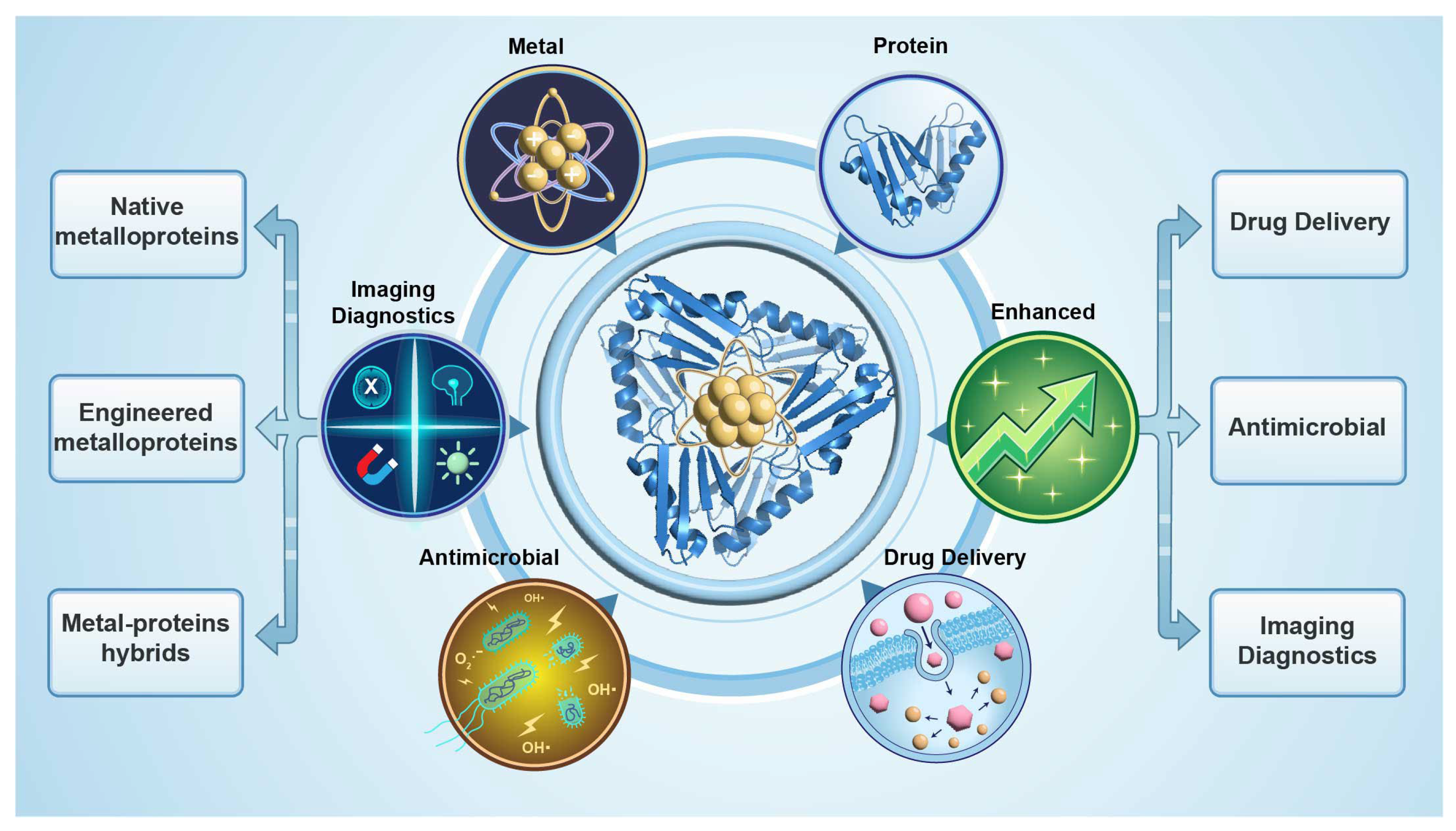
In this review, we first summarize the major types of metal- and metalloprotein-based nanomedicines and their construction strategies and then highlight recent advances in biomedical applications, with an emphasis on tumor treatment, imaging diagnosis, and anti-infective therapy (Figure 1). We further discuss common challenges related to structural characterization, interface mechanisms, process scale-up, and safety evaluation, aiming to distill general design principles for future material development. Finally, we outline emerging opportunities brought by artificial-intelligence-assisted protein design for the fine regulation of metal–protein interfaces and prediction of in vivo behavior, and consider how these advances may collectively accelerate the clinical translation of metal- and metalloprotein-based nanomedicines.

2. Construction Mechanisms of Metalloprotein-Based Nanomedicines

Metal–protein interactions underlie many essential life processes, thus providing metal–protein combinations a natural advantage in the design of nanomedicines. According to how metals associate with proteins, the structural complexity and the degree of functional integration reflect the increasing levels of design sophistication and multifunctionality. The current metalloprotein-based nanomedicines are mainly constructed based on the following mechanisms: (i) native metalloproteins that are directly used in their physiological forms; (ii) engineered metalloproteins obtained by protein engineering and chemical modification; and (iii) metal–protein hybrid nanostructures constructed from metals and proteins as separate building blocks. These three classes represent a continuum ranging from naturally evolved structures, through rationally redesigned proteins, to fully hybrid functional materials. In this section, we summarize their key structural features, metal-binding mechanisms, and typical construction strategies.

2.1. Native Metalloproteins

Native metalloproteins are ubiquitously present in living organisms. Their metal-binding sites have been refined by biological evolution and typically feature specific coordination environments, stable tertiary or quaternary structures, and good physiological compatibility. As such, they represent one commonly foundational module for constructing metal–protein nanomedicines. Typical examples include ferritin, transferrin, and other cofactor-containing proteins such as hemoglobin and cytochromes. Leveraging the intrinsic structural and functional advantages of these proteins, researchers have developed multifunctional designs for drug loading, metal ion delivery, catalytic therapy, and imaging enhancement. From a design perspective, native metalloproteins offer several generalizable principles for constructing metal–protein nanomedicines. These include (i) interface-defined metal coordination environments that constrain metal speciation and reactivity, (ii) a balance between structural stability and functional activation that is often condition-dependent, and (iii) the use of endogenous biological pathways, such as metal transport or receptor-mediated uptake, to couple therapeutic function with physiological compatibility.
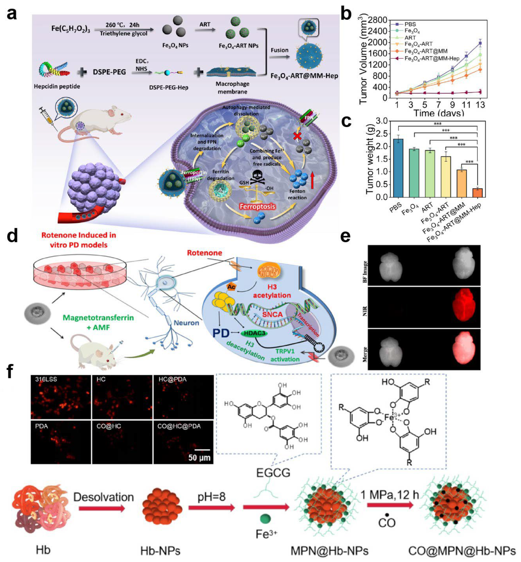
Ferritin has been extensively explored as a nanomedicine platform owing to its hollow cage-like architecture and excellent metal encapsulation capacity under controlled experimental conditions [46]. Human ferritin consists of 24 self-assembled subunits that form a protein shell with an outer diameter of ~12 nm and an internal cavity of ~8 nm. Through controlled mineralization, this cavity can be loaded with iron and a variety of transition metal nanoclusters, giving rise to metal-core–protein-shell nanostructures [36,46]. Ferritin is highly water-soluble and is generally regarded as biocompatible based on extensive in vitro studies and selected in vivo models. Moreover, a wide range of metal species and therapeutic agents have been successfully encapsulated within ferritin nanocages [47,48]. Zhao et al. constructed a ferritin/ferroportin-hijacking nanoplatform (Fe3O4-ART@MM-Hep), featuring an Fe3O4–artemisinin core cloaked with hepcidin-modified macrophage membrane (Figure 2a) [49]. After tumor accumulation, hepcidin binds ferroportin to trigger nanoparticle/FPN internalization and degradation, leading to Fe3O4 disassembly, artemisinin release, ferritin degradation, and thereby massive Fe2+ release. By simultaneously enhancing exogenous iron input, mobilizing endogenous iron stores, and blocking iron efflux, this system amplifies ferroptosis in the reported tumor models, illustrating that ferritin- and transferrin-based systems can outperform non-targeted or purely synthetic nanocarriers primarily under conditions where receptor availability, vascular access, and intracellular processing remain favorable (Figure 2b,c). Collectively, ferritin-based systems illustrate a key design principle for native metalloproteins: robust protein architectures can serve as confinement-defined nanoreactors that stabilize metal species under controlled conditions while enabling functional activation through regulated disassembly, degradation, or metal release in specific biological contexts.
Transferrin is a glycoprotein mainly synthesized by the liver and secreted into the bloodstream, where it is responsible for iron transport. It binds with high affinity to transferrin receptors, which are highly expressed in many types of cancer [50,51,52]. Consequently, transferrin can be used both as a carrier and as a targeting ligand. In certain tumor models, this dual-function strategy has been shown to enhance receptor-mediated uptake [53,54], which may translate into improved tissue distribution of nanomedicines in vivo, depending on receptor expression levels and disease context [55]. Beyond serving as a stabilizing carrier and targeting ligand, the inherent metal-binding capacity of transferrin has been exploited to develop iron-metabolism-responsive therapeutic systems, particularly for ferroptosis regulation and tumor microenvironment remodeling [56]. For example, Gao et al. demonstrated that serum transferrin is indispensable for ferroptosis by delivering iron into cells via transferrin receptor-mediated endocytosis, thereby providing the iron prerequisite for glutaminolysis-driven, cystine starvation-induced ferroptotic cell death [57]. In addition, Sardoiwala et al. constructed a reconstituted magnetotransferrin by nucleating Fe2O3 nanocrystals within the iron-binding clefts of apotransferrin, enabling transferrin receptor-mediated brain targeting and alternating-magnetic-field-induced mild magnetothermal activation of the TRPV1–HDAC3 axis to reduce α-synuclein aggregation and ameliorate Parkinson’s neurobehavioral deficits (Figure 2d,e) [58]. In addition, sequestration of labile iron by transferrin has been used to inhibit the growth of various pathogens, including Gram-negative bacteria such as Pseudomonas aeruginosa [37,59,60]. Despite their frequent designation as “ideal” targeting carriers, ferritin- and transferrin-based systems exhibit context-dependent performance in vivo. Receptor-mediated uptake can be significantly compromised by receptor saturation, competition with abundant endogenous ligands, and heterogeneous receptor expression across tumor types and disease stages. In addition, altered receptor recycling and impaired vascular accessibility in advanced tumors may further limit the effectiveness of these targeting mechanisms [61]. As a result, the in vivo advantage of ferritin- or transferrin-mediated targeting is not universal and may diminish under conditions of high endogenous protein levels or poor tumor perfusion [62]. These observations highlight a broader design principle for receptor-targeted metalloprotein nanomedicines: the effectiveness of biologically inspired targeting strategies is inherently constrained by endogenous ligand competition, receptor dynamics, and disease-stage-dependent microenvironmental accessibility.
Furthermore, heme-containing proteins, such as hemoglobin and cytochromes, are increasingly being incorporated into the design of metal–protein nanomedicines. Hemoglobin can participate in photodynamic therapy by supplying oxygen and exploiting the redox activity of the heme center to promote ROS generation [38,63]. In addition to photodynamic therapy (PDT), hemoglobin-based nanoplatforms have also been engineered as oxygen carriers to overcome tumor hypoxia. For example, Gandhi et al. developed hemoglobin-loaded polycaprolactone nanoparticles (PCL–Hb NPs) that preserve hemoglobin’s oxygen-binding capacity, exhibit acceptable biocompatibility within the tested in vitro and in vivo models, and are efficiently internalized by tumor and stromal cells under both normoxic and hypoxic conditions [64]. By delivering oxygen into hypoxic tumor cells, PCL–Hb NPs downregulate hypoxia-responsive genes and effectively reverse paclitaxel resistance, thereby enhancing chemotherapeutic efficacy in 2D hypoxic cultures and 3D tumor spheroids. In addition, hemoglobin nanocarriers can be used to load and release gaseous transmitters via metal-centered coordination. Wu et al. engineered a hemoglobin/epigallocatechin-3-gallate (EGCG) core–shell nanoparticle coating on stainless steel that stores CO through hemoglobin–CO coordination to modulate implantation-induced inflammation (Figure 2f) [65]. This coating provides sustained CO release and has been shown to lower intracellular ROS levels and attenuate oxidative stress-induced apoptosis in endothelial cells under the tested conditions. In vivo, the hemoglobin/EGCG-based CO-releasing coating suppresses macrophage-mediated inflammatory responses and favorably regulates macrophage polarization at the implant interface. Cytochromes, with their unique electron-transfer and redox properties, can be leveraged to modulate the tumor microenvironment or intervene in metabolic pathways [39]. Heme-containing proteins exemplify a broader stability–activity trade-off in metalloprotein-based nanomedicine design, where highly reactive metal cofactors must be carefully spatially confined or temporally activated to balance therapeutic efficacy with oxidative safety. Although these proteins are not yet as widely used as ferritin and transferrin, their distinctive metal cofactor chemistry enriches the toolbox for building metal–protein nanoplatforms.
Figure 2. Representative native metalloprotein-based nanoplatforms for metal delivery and therapeutic modulation. (a) Schematic illustration of the preparation of Fe3O4-ART@MM-Hep nanoparticles and the ferroportin-hijacking strategy for enhanced ferrotherapy via intracellular Fe2+ elevation. (b) Tumor volume changes during treatment in different groups. (c) Average tumor weights at the end of treatment. Reproduced with permission. *** p < 0.001 [49]. Copyright 2025, American Chemical Society. (d) Schematic illustration of reconstituted magnetotransferrin formed by nucleation of Fe2O3 nanocrystals within the iron-binding clefts of apotransferrin. (e) Ex vivo brain reflectance imaging showing enhanced accumulation of ICG-labeled magnetotransferrin. Reproduced with permission [58]. Copyright 2023, American Chemical Society. (f) Schematic illustration of the preparation of hemoglobin/epigallocatechin-3-gallate (EGCG) core–shell nanoparticles and CO-loaded CO@Hb/EGCG coatings for inflammation modulation. Reproduced with permission [65]. Copyright 2024, Regenerative Biomaterials.
Ijms 27 01076 g002

2.2. Metal-Engineered Proteins

In contrast to the direct use of native metalloproteins, metal-engineered proteins place greater emphasis on redesigning metal-binding sites and their local microenvironments on pre-existing protein scaffolds via site-directed mutagenesis, chemical modification, or the introduction of artificial ligands [66]. By adding or substituting coordinating residues such as His, Cys, Asp, and Glu at key positions, or by attaching small-molecule ligands to side chains, one can selectively tune the affinity, coordination number, and geometry of metal ions [67,68], thereby modulating their redox potentials, substrate specificity, and catalytic efficiency. From a design standpoint, metal-engineered proteins represent a shift from evolutionarily constrained metal utilization to rationally tunable systems, where expanded functional space is gained at the cost of increased sensitivity to folding stability, metal exchange, and immune recognition in vivo. Aires et al. fused an Hsp90-binding tetratricopeptide repeat module with a cysteine-rich metal-nanocluster-stabilizing module to create a chimeric protein that nucleates highly fluorescent Au nanoclusters while preserving high-affinity target recognition. The resulting protein–AuNC hybrid acted as a theranostic biologic in an angiotensin II-induced myocardial fibrosis model. It simultaneously reduced cardiac fibrosis and enabled high-sensitivity tracking of drug biodistribution by fluorescence and synchrotron X-ray fluorescence imaging [69].
Similar strategies have been applied to proteins or minimalist peptide scaffolds lacking natural metal cofactors, enabling the construction of artificial metal centers and converting otherwise ordinary proteins into artificial metalloenzymes with defined catalytic, imaging, or signal-amplification functions [70,71,72]. Aires et al. engineered consensus tetratricopeptide repeat (CTPR) proteins with defined acidic, histidine, or cysteine metal-coordination sites to template iron oxide nanoparticles, thereby controlling core size, Fe(III)/Fe(II) ratio, and magnetic relaxivities [73]. By varying both the nature and the number of metal-binding modules, they were able to tune these protein-stabilized iron oxide nanoparticles between T1- and T2-weighted MRI contrast regimes, highlighting that the design of coordination environments on minimalist protein scaffolds can precisely dictate inorganic core properties and imaging performance. These engineered metalloproteins generally have relatively small molecular weights and well-defined conformations, which facilitates rational optimization based on structure–function relationships, although increased design freedom often comes at the cost of reduced in vivo predictability, including altered folding stability, unexpected immune recognition, and dynamic metal exchange under physiological conditions, due to altered protein stability, unexpected immune recognition, and dynamic metal exchange in physiological environments [74].
Beyond site-specific metal coordination, metal ions are frequently employed at higher structural levels as reversible crosslinkers to induce the self-assembly of engineered proteins [42]. When multiple His residues or short metal-binding peptide motifs are displayed on the protein surface, the addition of related ions under mild conditions can bridge multiple protein units, driving them to assemble from dispersed monomers into size-controlled nanoparticles, cage-like architectures, or physically crosslinked hydrogels [75,76,77]. Liu et al. demonstrated that partially hydrolyzed α-lactalbumin peptides can be directed by various metal ions (Mn2+, Co2+, Ni2+, Zn2+, Cd2+, and Au3+) to self-assemble into hollow nanotubes, which further form supramolecular metallogels with tunable length, stiffness, and gelation kinetics. These results highlight how the identity and coordination strength of metal ions can dictate higher-order protein architectures and their mechanical properties [78]. Owing to the partially reversible nature of metal coordination, these self-assembled systems are often sensitive to mechanical stress, pH, ionic strength, or redox conditions [79] and have been reported to undergo aggregation–disaggregation or sol–gel transitions in response to specific microenvironmental cues, such as pH or ionic strength, under defined conditions, including tumors, inflamed tissues, or intracellular compartments. This behavior has been exploited to design injectable protein hydrogels, environment-responsive carriers, and confined catalytic platforms [40].

2.3. Metal–Protein Hybrid Nanostructures

Unlike the molecular-level engineering of individual proteins, metal–protein hybrid nanostructures focus on building intrinsic hybrid nanomedicines. In such systems, proteins and metal nanostructures are treated as two functionally distinct building blocks. The building blocks of metal–protein hybrid nanostructures should be integrated into a single cooperative entity, thus with specific functionalities and great stability through coating, templating, mineralization, or framework assembly. From a design perspective, metal–protein hybrid nanostructures follow an emergent-function principle, in which therapeutic or imaging performance arises from mesoscale spatial integration rather than discrete coordination sites, but this advantage is inherently accompanied by increased structural complexity, batch-to-batch variability, and challenges in in vivo predictability. Depending on the construction strategy, these materials can be broadly categorized into protein-coated metal nanoparticles [80], protein-templated metal nanoclusters [81], and metal–protein framework materials [82].
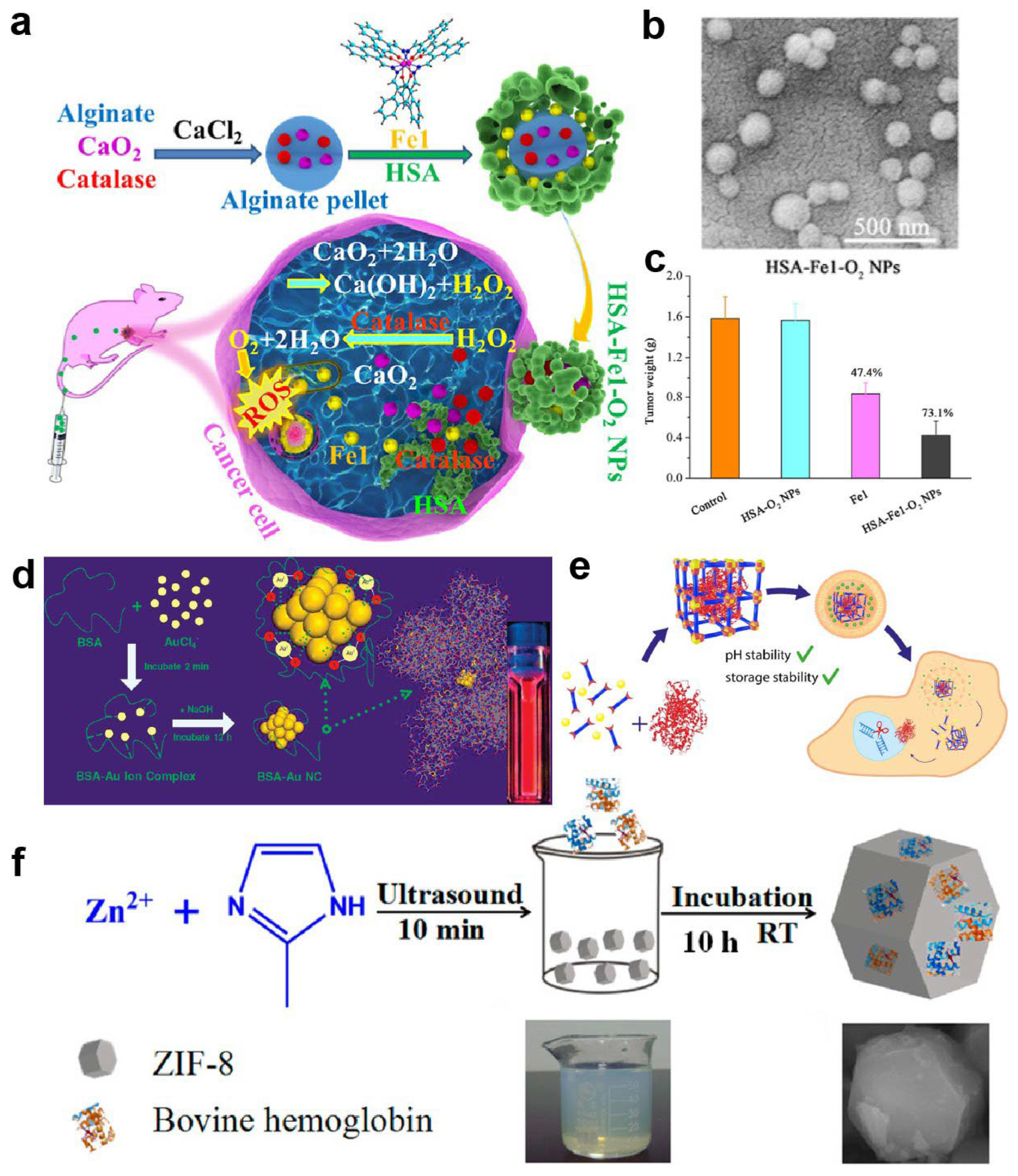
Protein-coated metal nanoparticles represent one of the earliest and most widely used approaches [83]. Metallic nanoparticles such as gold, silver, platinum, and magnetic ferrites can be coated with a shell of serum albumin, ferritin, or other soluble proteins via adsorption or covalent conjugation, forming core–shell structures [41,84]. The protein shell has been shown to improve colloidal stability in aqueous and complex biological media and, in some experimental systems, to reduce nonspecific protein corona formation and reticuloendothelial clearance [85,86,87]; however, these effects are not universally observed across biological contexts [88]. The metal core, in turn, contributes photothermal, photoacoustic, X-ray, or magnetic resonance functionalities [89], so that the resulting core–shell constructs serve as commonly explored theranostic platforms in preclinical research settings with integrated imaging and therapeutic capabilities [90]. For example, Fang et al. encapsulated an oxygen-generating CaO2/catalase alginate core together with a ROS-producing ferric Schiff-base complex inside a human serum albumin shell to construct HSA–Fe1–O2 nanoparticles (Figure 3a,b). This protein-coated metal nanoplatform significantly enhanced intratumoral accumulation of the Fe complex, relieved hypoxia-induced resistance, and was associated with superior tumor suppression and limited systemic toxicity within the specific dosing regimens and tumor models evaluated compared with the free complex (Figure 3c) [91].
Protein-templated metal nanoclusters exploit coordinating residues within or on the surface of proteins to reduce and confine metal ions under mild conditions. A representative example is provided by Xie et al., who used bovine serum albumin (BSA) as a scaffold to synthesize highly stable gold nanoclusters with desirable optical properties (Figure 3d) [92]. In their investigations, Au(III) ions are first bound and sequestered by BSA in aqueous solution; increasing the pH to ~12 then activates the latent reducing ability of BSA, leading to stepwise in situ reduction in the bound ions and formation of Au nanoclusters that are stably embedded in the BSA matrix, where BSA serves simultaneously as reductant and stabilizer. Owing to pronounced quantum size effects, protein-templated metal nanoclusters often exhibit bright, tunable fluorescence and a high specific surface area with strong catalytic activity [93,94,95]. Nanoclusters templated by BSA, metallothionein, and related proteins have been applied in fluorescence imaging, ROS modulation, and antimicrobial therapy, and typically display good water solubility, acceptable biocompatibility, and in vivo stability in selected animal models owing to the protective, confining roles of the protein template [96,97,98].
Metal–protein frameworks (MPFs), a rapidly developing class of hybrid materials, represent another structurally complex and functionally rich family of metal–protein constructs. MPFs are typically formed through multidentate coordination between proteins and multivalent metal ions or metal clusters, combining the designable porosity of MOF-like architectures with the intrinsic biological functions of proteins [99,100]. Their internal pores can accommodate high loadings of diverse guest molecules, including small-molecule drugs, proteins, peptides, and nucleic acids, while the choice and spatial arrangement of metal nodes and protein units allow precise control over release profiles and stimuli responsiveness [101]. For instance, Mirzazadeh Dizaji et al. used a biomimetic mineralization strategy to encapsulate diverse proteins, including BSA, horseradish peroxidase, GFP, and Cas9/sgRNA ribonucleoproteins, into amorphous iron–fumarate nanoparticles, achieving high loading while preserving their catalytic and genome-editing activities after intracellular delivery (Figure 3e) [102]. Notably, these iron–fumarate MPFs protected Cas9 RNPs against acidic pH and long-term storage, underscoring the potential of MPFs as robust carriers for fragile protein therapeutics and gene-editing tools. As another example, Yin and co-workers used bovine hemoglobin (BHb) as the organic component and the zeolitic imidazolate framework ZIF-8 as the inorganic counterpart to construct BHb-embedded MOFs. The resulting ZIF-8@BHb composites, obtained by self-assembly, showed good stability and high catalytic activity and were further developed into a colorimetric platform for the visual detection of hydrogen peroxide (Figure 3f) [82]. Compared with traditional MOFs built from small organic ligands, MPFs based on native proteins usually exhibit improved biodegradability and biocompatibility in comparative preclinical studies, although systematic head-to-head evaluations and clinical validation remain limited [103].
Figure 3. Representative metal–protein hybrid nanostructures. (a) Schematic illustration of the protein-coated metal nanoplatform HSA–Fe1–O2 NPs. (b) Representative morphology characterization of HSA–Fe1–O2 NPs. (c) Tumor weight in mice treated with HSA–O2 NPs, Fe1, or HSA–Fe1–O2 NPs [91]. Copyright 2024, American Chemical Society. (d) Schematic illustration of protein-templated synthesis of gold nanoclusters (Au NCs) within a BSA matrix. Reproduced with permission [92]. Copyright 2009, American Chemical Society. (e) Schematic overview of biomimetic iron–fumarate metal–protein framework nanoparticles encapsulating diverse proteins. Reproduced with permission [102]. Copyright 2022, American Chemical Society. (f) Schematic representation of ZIF-8@BHb metal–protein framework composites formed by self-assembly. Reproduced with permission [82]. Copyright 2016, American Chemical Society.
Ijms 27 01076 g003

2.4. Comparative Assessment and Design Principles

The above construction strategies reveal several general design principles governing metalloprotein-based nanomedicines. First, function is predominantly interface-driven: whether in native metalloproteins, engineered metal-binding sites, or hybrid nanostructures, the coordination environment at the metal–protein interface dictates metal speciation, redox activity, and stimulus responsiveness, thereby determining therapeutic and imaging performance [104,105]. Second, a fundamental stability–activity trade-off is consistently observed. Stronger protein confinement and higher-order assembly enhance colloidal stability, biocompatibility, and in vivo robustness but may simultaneously restrict metal accessibility or catalytic efficiency, necessitating careful balance through protein engineering or responsive coordination [106,107]. Third, controllability of metal speciation and coordination emerges as a central design axis. From evolutionarily optimized metal-binding pockets in native proteins to rationally redesigned coordination motifs and reversible metal-mediated self-assembly, precise regulation of metal valence states, coordination geometry, and release dynamics enables programmable activation and safer in vivo behavior [108,109]. These recurring principles suggest that successful metalloprotein-based nanomedicine design depends less on the nominal material class than on how effectively metal–protein interfaces are engineered to balance stability, activity, and biological compatibility [110,111].
How these general principles manifest in practice depends strongly on the chosen construction paradigm. From a comparative perspective, native metalloproteins, engineered metalloproteins, and metal–protein hybrid nanostructures share the common goal of integrating metal functionality with biological interfaces, yet differ markedly in stability, functional robustness, and translational attributes. Native metalloproteins benefit from evolutionarily optimized metal-binding sites and well-defined tertiary structures, which result in relatively favorable immunological profiles compared with many synthetic nanomaterials and predictable metabolism [112,113]. These features make them particularly attractive for applications requiring long circulation times and minimal immune perturbation. However, their functional scope is constrained by the intrinsic properties of the native metal cofactors, limiting tunability and the range of accessible therapeutic mechanisms [114]. Engineered metalloproteins offer increased design flexibility by allowing site-specific modulation of metal coordination environments, redox properties, and catalytic activity [115]. This enables precise tuning of function–structure relationships and facilitates modular optimization [116]. Nevertheless, such systems often face trade-offs between enhanced functionality and reduced structural robustness, and their immunogenicity and in vivo stability can be highly sensitive to sequence modifications and surface-exposed metal sites [117,118]. Metal–protein hybrid nanostructures provide the highest degree of functional integration, combining the rich physicochemical properties of inorganic nanomaterials with protein-mediated biocompatibility and targeting [69,119]. These systems have demonstrated strong therapeutic and imaging performance in multiple preclinical studies but also present greater challenges in terms of colloidal stability in biofluids, batch-to-batch reproducibility, scalable manufacturing, and regulatory evaluation, owing to their structural complexity and multicomponent nature [75]. Taken together, these three construction paradigms represent a continuum from biological fidelity to engineering freedom. Rational selection among them should be guided by application-specific priorities, balancing functional sophistication against stability, manufacturability, and translational feasibility. Importantly, these attributes are not interchangeable across application contexts; higher functional integration does not necessarily translate into superior in vivo performance when stability, immunological tolerance, or manufacturing constraints dominate design priorities (Table 1).
Overall, metal–protein hybrid nanostructures achieve spatial synergy between metal components and proteins at the structural level, while integrating imaging, therapy, and targeting functionalities at the functional level. With advances in designing protein architectures and engineering metal nanostructures, the application scope of these hybrid materials in nanomedicine is expected to expand further, and they are likely to become key building blocks for next-generation multimodal and intelligent therapeutic systems.

3. Biomedical Applications

Metalloprotein-based nanomedicines combine the multimodal imaging and therapeutic functions of metal centers with the finely tunable biointerfaces of protein scaffolds and therefore exhibit distinct advantages across multiple disease models. In the previous section, we outlined the design concepts of native metalloproteins, engineered metalloproteins, and metal–protein hybrid nanostructures from the perspective of structural types and construction mechanisms and established a basic classification framework based on metal-binding modes and protein scaffold features. Building on this, the present section summarizes representative advances from a function–disease viewpoint. It focuses on cancer therapy, imaging diagnostics, and multimodal theranostics; antimicrobial and anti-resistant therapy; and other disease interventions, with emphasis on the synergy between metal functional cores and protein-mediated fine regulation in specific applications.

3.1. Cancer Therapy

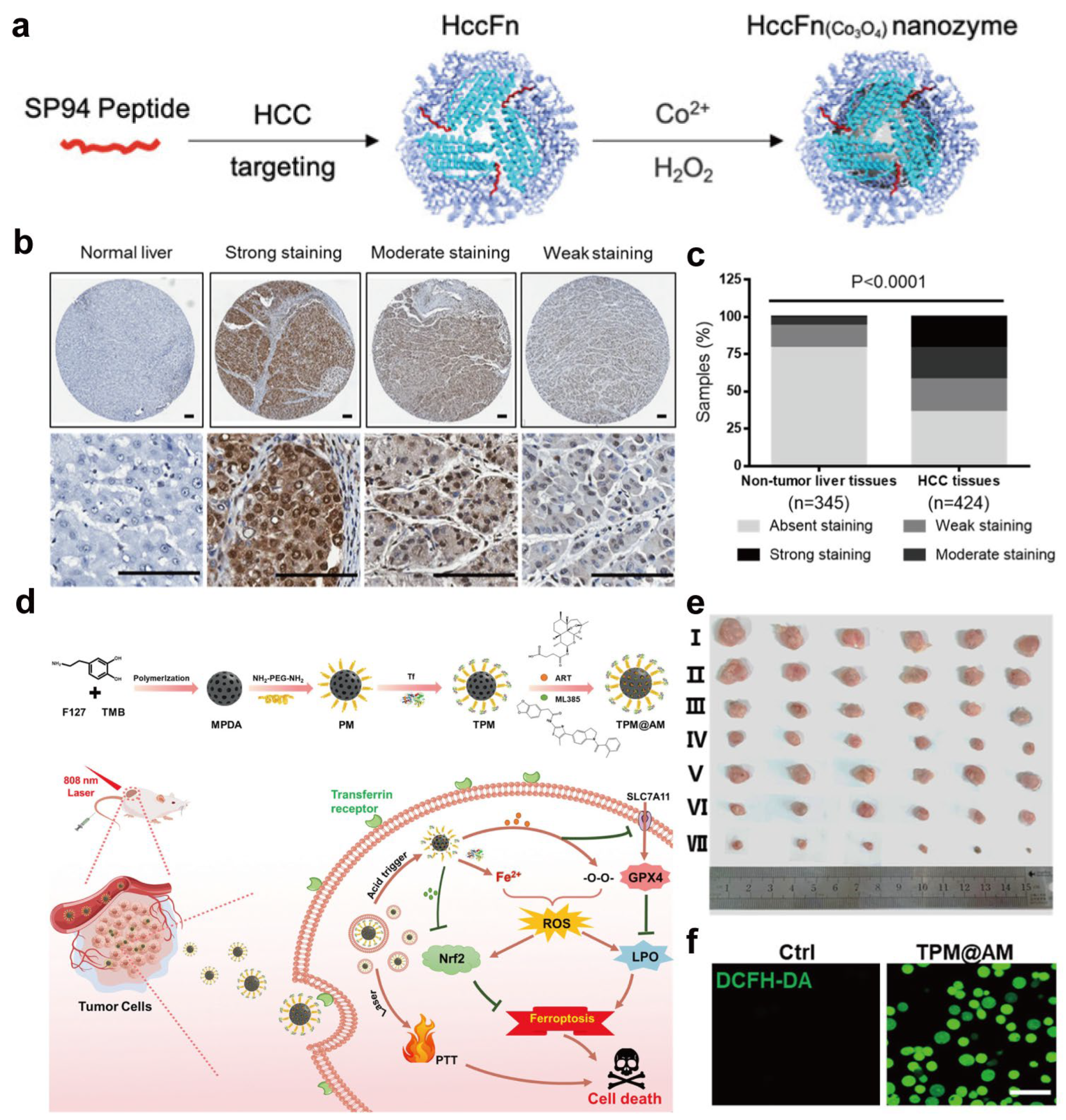
Metal- and metalloprotein-based nanomedicines offer multi-dimensional synergies in cancer treatment. Encapsulating cisplatin, ruthenium complexes, or other metal-based drugs and clusters within protein nanocarriers has been shown to enhance tumor accumulation and mitigate off-target toxicity in selected preclinical models, although such benefits depend strongly on vascular permeability, protein stability, and tumor microenvironmental factors [43,120]. In addition to serving as simple carriers, metal–protein constructs can also function as catalytic nanoprobes for tumor diagnosis. For example, Jiang et al. biomineralized Co3O4 nanozymes inside hepatocellular carcinoma (HCC)-targeted ferritin nanocages (HccFn(Co3O4)) (Figure 4a), in which genetically displayed SP94 peptides on the protein shell confer high affinity for HCC cells and enable one-step immunohistochemical staining of clinical specimens with both diagnostic and prognostic value (Figure 4b,c) [47]. Passive targeting via the enhanced permeability and retention (EPR) effect has been reported to facilitate nanoparticle accumulation in tumors in certain animal models, although its relevance varies across tumor types and clinical settings [121,122,123]. Moreover, many protein scaffolds, such as transferrin and ferritin, can actively target tumor cells through high-affinity interactions with overexpressed receptors, thereby promoting receptor-mediated endocytosis and intracellular delivery in receptor-overexpressing cell models [54,124,125]. As an illustration of such receptor-targeted and microenvironment-responsive design, Chen et al. constructed a transferrin-modified mesoporous polydopamine nanoplatform co-loading the ferroptosis inducer artesunate and the Nrf2 inhibitor ML385 (TPM@AM), which selectively targets TfR-overexpressing tumor cells and releases the payloads in response to the acidic tumor microenvironment and NIR irradiation (Figure 4d) [126]. By increasing intracellular Fe2+ levels, boosting ROS and lipid peroxidation, and simultaneously suppressing Nrf2-mediated antioxidant defenses, TPM@AM markedly amplifies ferroptosis and achieves potent, spatiotemporally controlled tumor suppression in vivo with limited systemic toxicity observed under the tested conditions (Figure 4e,f).
In addition, the protein shell can modulate the local chemical microenvironment and metal coordination state, thereby tuning drug release kinetics and modes of action, while metal centers can participate in the Fenton and Fenton-like reactions to catalytically generate ROS and induce oxidative stress and apoptosis in tumor cells [96,127]. For example, Zhu et al. designed a ferritin-based nanoplatform (Ce6–PEG–HKN15-loaded ferritin) in which chlorin e6 (Ce6) acts as a photosensitizer [128]. Upon laser irradiation, Ce6 confined within the ferritin nanocages produces abundant ROS around the protein shell; the elevated ROS damages the iron-storage protein, triggers ferroptosis by releasing iron, and directly kills tumor cells. The liberated iron further reacts with excess intracellular H2O2 to generate O2, thereby relieving hypoxia, enhancing photodynamic therapy, and amplifying oxidative stress to achieve synergistic chemo-, catalytic, photothermal/photodynamic, and immunotherapy. Beyond such ferroptosis-oriented designs, other metal–protein nanoplatforms can elicit immunogenic cell death, promote tumor-associated antigen exposure, and facilitate immune cell infiltration, enabling combination regimens that integrate chemotherapy or catalytic therapy with immunotherapy and opening new avenues for multimodal cancer treatment [129,130,131].
However, the therapeutic mechanisms highlighted above are intrinsically associated with distinct risk profiles that warrant careful consideration alongside their benefits. Ferroptosis induction and ROS amplification, while effective in promoting tumor cell death, inherently increase the likelihood of off-target oxidative damage and systemic metal ion redistribution following protein degradation [132,133]. Moreover, tumor heterogeneity in redox balance, iron metabolism, and antioxidant capacity can lead to variable therapeutic responses, potentially limiting the robustness of Fenton- and ferroptosis-based strategies across different tumor models [134,135]. More broadly, each mechanism discussed in this section entails specific trade-offs: ROS amplification may cause collateral oxidative injury, hypoxia alleviation can alter vascular permeability, metal ion release may perturb systemic metal homeostasis, receptor-mediated targeting is susceptible to saturation and heterogeneity, and protein degradation may trigger unpredictable immune or metabolic consequences [136]. Recognizing these mechanism-linked risks is essential for realistic evaluation and rational optimization of metal–protein nanomedicines for cancer therapy.

3.2. Imaging Diagnostics and Multimodal Theranostics

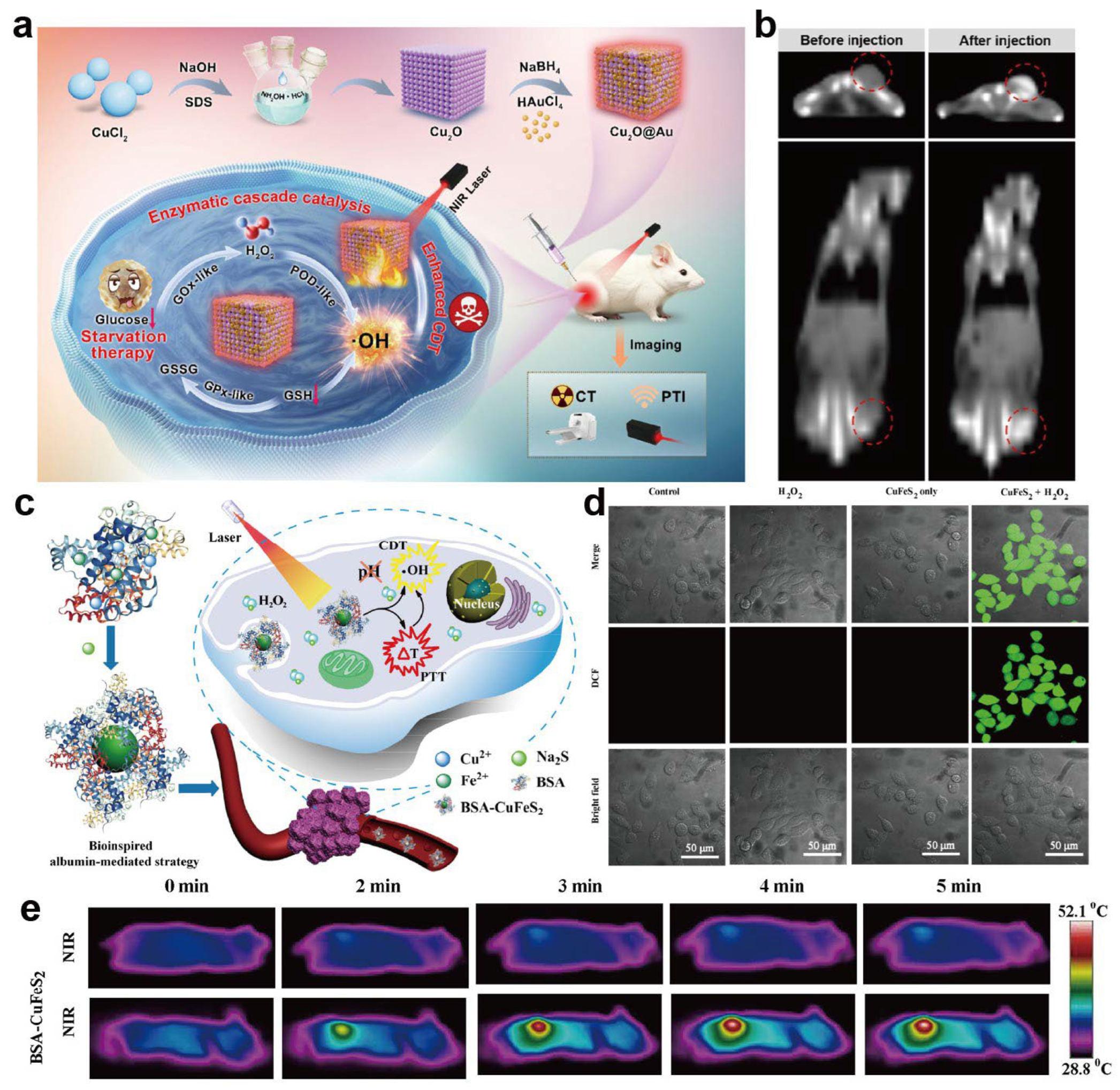
Metal–protein nanomedicines also show unique strengths in imaging diagnostics [137,138]. Metal ions serve as key contrast elements for many imaging modalities: Gd3+ and Mn2+ serve as T1 contrast agents in magnetic resonance imaging (MRI) [139,140], iron-based nanostructures act as T2 agents [141], and gold and other high-Z metals provide strong X-ray attenuation in computed tomography (CT) [142,143,144]. Metal nanoparticles further exhibit excellent photoacoustic signal conversion efficiencies for photoacoustic imaging [145,146,147]. For example, Li et al. designed a Cu2O@Au nanozyme that combines peroxidase (POD)-like catalytic activities with strong photothermal conversion and X-ray attenuation of nanoclusters (Figure 5a), enabling dual photothermal and CT imaging to guide cascade starvation/chemodynamic therapy in non-small-cell lung cancer models (Figure 5b) [148]. This work illustrates how metal-based nanozymes can simultaneously provide high-contrast imaging readouts and potent therapeutic effects within a single platform, enabling real-time imaging-guided and individualized treatment, potentially improving lesion localization and dose control when sufficient contrast-to-noise ratios and stable metal coordination can be maintained in vivo.
Integrating metal centers into protein scaffolds can greatly improve their aqueous solubility and in vivo stability. Moreover, protein-mediated targeting can facilitate preferential accumulation at diseased sites [149]. For instance, Chen and co-workers reported ultrasmall BSA–CuFeS2 nanoparticles as a multifunctional therapeutic platform prepared via a green, biomimetic albumin-mediated strategy (Figure 5c) [150]. These BSA–CuFeS2 NPs exhibit pH-independent Fenton-like catalytic activity for efficient ·OH generation and high photothermal conversion efficiency, thereby enhancing the combined efficacy of chemodynamic therapy (CDT) and photothermal therapy (PTT) (Figure 5d,e). The resulting hybrids display relaxivities comparable to clinical agents, excellent stability under physiological conditions, and negligible cytotoxicity, illustrating how protein engineering can be leveraged to tailor the magnetic and imaging properties of metal–protein nanomaterials. These advances in protein engineering lay the foundation for constructing multifunctional nanoplatforms capable of multimodal imaging and therapy integration.
Figure 5. Metal–protein nanomedicines for imaging-guided theranostics. (a) Schematic illustration of the preparation of the Cu2O@Au nanozyme and its imaging-guided antitumor mechanism. (b) In vivo CT images before and after injection [148]. Copyright 2024, American Chemical Society. (c) Schematic illustration of the biomimetic synthesis of BSA–CuFeS2 nanoparticles and their pH-independent synergistic CDT/PTT. (d) Confocal fluorescence images of intracellular ROS generation in 4T1 cells after different treatments (DCFH-DA staining). Scale bar: 50 μm. (e) Representative infrared thermal images of 4T1 tumor-bearing mice during laser irradiation. Reproduced with permission [150]. Copyright 2019, American Chemical Society.
Ijms 27 01076 g005
When combined with fluorescent, photoacoustic, or nuclear imaging probes, these systems can be developed into multimodal imaging platforms that allow a single nanoconstruct to be used across MRI, CT, photoacoustic, and fluorescence channels in a complementary or synergistic manner [151,152]. By integrating imaging and therapeutic functions (e.g., MRI/PA/CT contrast, PTT, CDT, PDT, or chemotherapy) into a single construct, metal–protein nanomedicines potentially improve lesion localization and dose control when sufficient contrast-to-noise ratios and stable metal coordination can be maintained in vivo [10,149,153].
Nevertheless, the performance of metal–protein nanomedicines in imaging diagnostics and theranostics is also subject to mechanism-specific limitations [75]. Imaging contrast and quantitative accuracy critically depend on the in vivo stability of metal coordination and protein integrity [154]; partial metal dissociation, protein degradation, or dynamic protein corona formation can lead to signal attenuation, background noise, or misleading readouts over time [155,156]. In multimodal systems, differences in sensitivity, penetration depth, and temporal resolution among imaging modalities further complicate quantitative interpretation and cross-modality calibration [138,154]. Moreover, the introduction of therapeutic functions into imaging agents increases system complexity, potentially narrowing the safety margin and challenging dose optimization [157,158]. These considerations highlight the need for careful integration of imaging and therapeutic functions to ensure that enhanced multifunctionality does not compromise diagnostic reliability or translational robustness.

3.3. Antimicrobial and Anti-Resistant Therapy

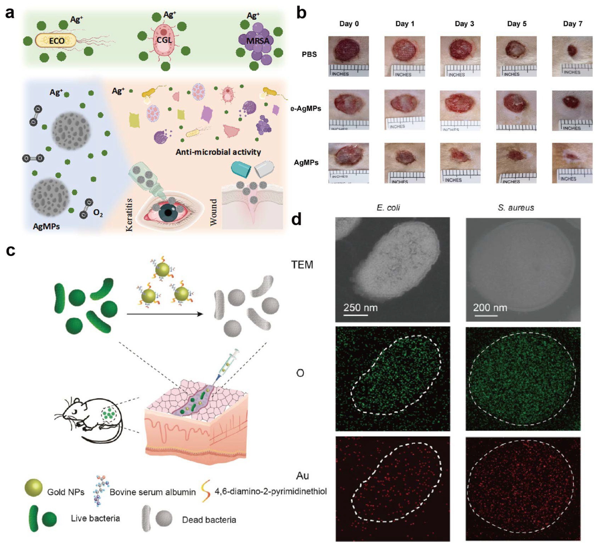
In the field of antimicrobial and anti-resistant therapy, metal–protein nanomedicines exert their effects through multiple mechanisms. Metal nanoparticles such as silver and copper can directly disrupt bacterial membranes, interfere with key metabolic enzymes, and induce excessive intracellular ROS accumulation [159]. Protein shells improve dispersion and biocompatibility and can be engineered to target and accumulate preferentially at infection sites, thereby enhancing local antibacterial efficacy in reported models while reducing systemic toxicity relative to unprotected metal formulations [160]. For example, Shi et al. prepared honeycomb-like silver microspheres (AgMPs) using bovine serum albumin as a template, which enable sustained Ag+ release and exhibit broad-spectrum activity against both bacteria and fungi, including Candida spp. (Figure 6a) [161]. In rat infection models, AgMPs effectively eliminated Candida-infected wounds while maintaining good viability of corneal epithelial cells at therapeutically relevant concentrations, highlighting the potential of protein-assisted silver nanostructures for treating fungal keratitis (Figure 6b).
Iron- or gold-containing metal–protein systems can catalyze the formation of highly reactive species such as hydroxyl radicals in bacteria-enriched regions, selectively killing drug-resistant strains and, to some extent, circumventing classical resistance mechanisms against conventional antibiotics. For example, Sun et al. synthesized a BSA-stabilized gold nanocomposite (Au_DAPT_BSA) that showed superior antibacterial activity against Gram-negative bacteria and improved efficacy against Gram-positive bacteria compared with Au_DAPT alone (Figure 6c) [162]. In mouse models of subcutaneous abscesses caused by clinically isolated multidrug-resistant Escherichia coli or Staphylococcus aureus, Au_DAPT_BSA markedly accelerated healing (Figure 6d).
Beyond the intrinsic antibacterial action of metal–protein hybrid materials, combination therapies have yielded more potent bactericidal effects and helped mitigate antibiotic resistance. For example, Liu et al. constructed a multifunctional antibacterial nanomedicine (BCPP) for targeted photodynamic/photothermal combination therapy (PDT/PTT) [163]. BCPP consists of protoporphyrin IX (PpIX) as the photosensitizer, CuS nanoparticles as the photothermal agent, phenylboronic acid (PBA) as a bacterial-targeting moiety, and BSA as the colloidal stabilizer. Owing to its strong singlet oxygen generation and high photothermal conversion efficiency, BCPP showed excellent targeting and killing efficacy against S. aureus and E. coli. Benefiting from these multi-mechanistic actions, metal–protein nanomedicines can not only improve antibacterial outcomes but also offer a means to delay or reduce the emergence of resistance. These systems therefore represent promising antimicrobial platforms; however, the same ROS-mediated and metal-ion-driven mechanisms that confer potent antimicrobial activity may also disrupt host cell redox balance or commensal microbiota, underscoring the need for spatial and dosage control [164,165].
Across cancer therapy, diagnostic imaging, and antimicrobial applications, several recurring design motifs emerge, including metal-mediated ROS generation, microenvironment-responsive activation, and protein-enabled multimodal functionality [166,167,168]. Rather than representing distinct strategies, these mechanisms often rely on shared physico-chemical principles, such as controlled metal confinement, protein degradation-triggered release, and environmental sensitivity [167,169]. Recognizing these commonalities helps clarify that differences among metalloprotein systems frequently arise from application context rather than fundamentally different design logic. Nevertheless, the same ROS-mediated and metal-ion-driven mechanisms that confer potent antimicrobial activity may also disrupt host cell redox balance or commensal microbiota, underscoring the need for spatial and dosage control [165,170].

3.4. Other Biomedical Applications

Beyond cancer and infection, metal- and metalloprotein-based nanomedicines also hold promise in neurodegenerative diseases, catalysis, and biosensing. In neurological disorders, dysregulated metal ion homeostasis and oxidative stress are recognized as key pathological drivers. Metal–protein nanoplatforms can modulate disease progression by chelating or slowly releasing metal ions and by regulating ROS levels [171,172]. For example, the accumulation of ROS, amyloid-β (Aβ), and neuroinflammation are hallmarks of Alzheimer’s disease (AD). Liu et al. designed ultrasmall copper nanoclusters embedded in human serum albumin (CuNCs@HSA) using an HSA-mediated strategy [173]. CuNCs@HSA exhibited multiple enzyme-like activities, significantly inhibited Aβ fibrillation, and alleviated Aβ-induced oxidative stress, thereby mitigating AD-related pathology in model systems.
The design of nanozymes that can compete with natural enzymes in catalytic efficiency while overcoming their limitations has attracted widespread attention. He et al. developed BSA–Pt nanoparticles in which platinum nanoparticles are supported on a BSA scaffold [174]. BSA–Pt displayed enhanced multi-enzyme-mimicking activities, including peroxidase-, oxidase-, and catalase-like functions. This work not only demonstrated the size-dependent nanozyme activity of Pt nanoparticles but also showcased the practical use of BSA–Pt for catalytic applications such as glucose detection in human serum.
As our understanding of the links between metal homeostasis and disease deepens, the application space of metal–protein nanomedicines in non-oncological indications is expected to expand further. However, the increasing diversity of therapeutic and diagnostic roles also brings new challenges in design, safety, and translational feasibility, which must be carefully addressed to realize their clinical potential.

4. Challenges and Future Perspectives

Despite the encouraging performance of metal- and metalloprotein-based nanomedicines in preclinical studies, their true clinical translation still faces multiple layers of challenges. These issues arise partly from incomplete basic understanding of nanoscale structures, metal–protein interfaces, and dynamic in vivo behavior, and partly from bottlenecks at the engineering and translational levels, including controllable fabrication, batch-to-batch consistency, long-term safety, and regulatory evaluation. Systematically identifying and addressing these challenges is essential for driving this class of nanomedicines from proof-of-concept toward tangible clinical benefit.

4.1. Metabolism and Degradation

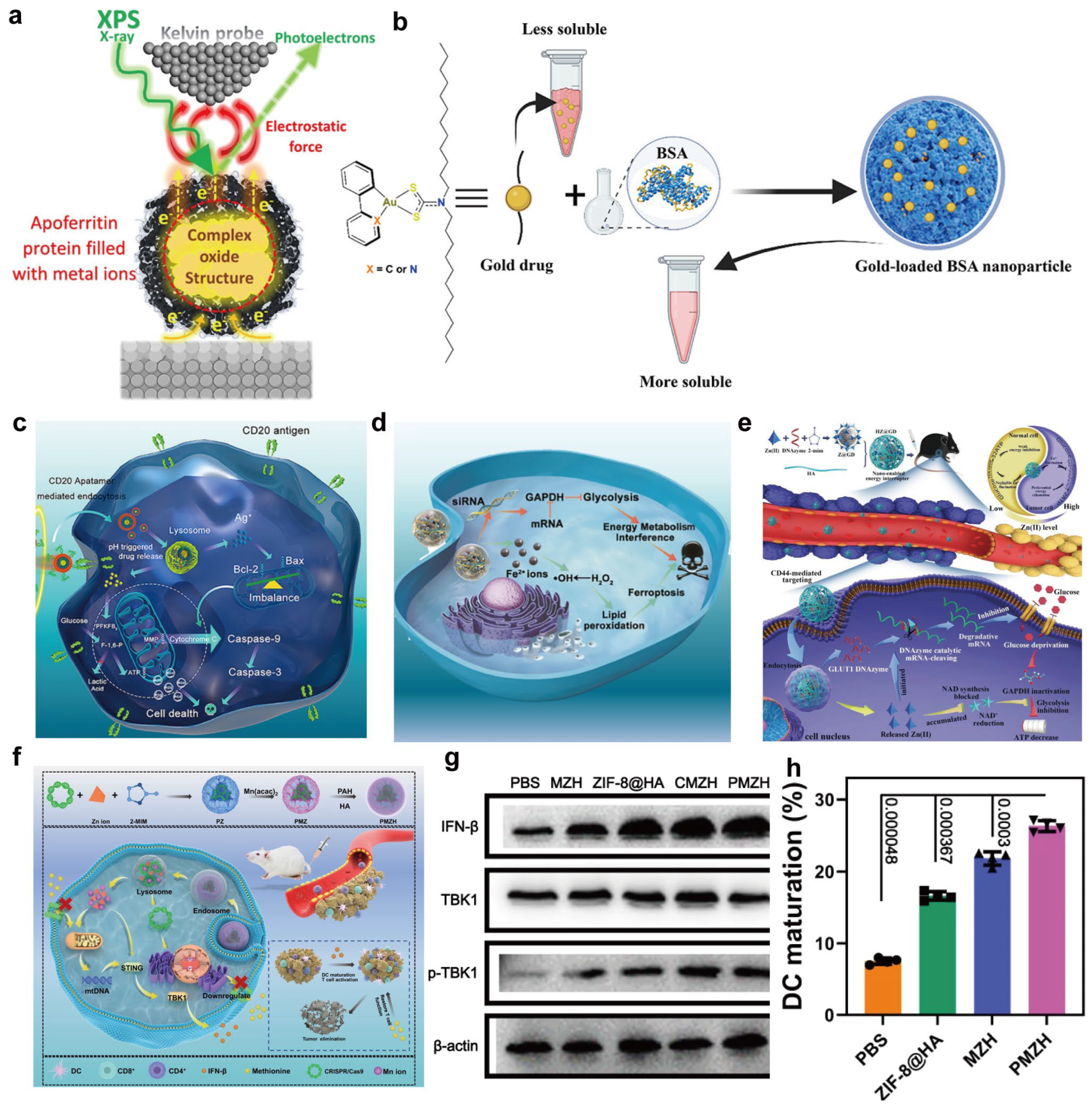
The metabolism and degradation of metal- and metalloprotein-based nanomedicines in vivo are among the key determinants of both efficacy and safety. Metal centers typically exist as ions, nanoclusters, or oxides and may undergo valence changes, ligand exchange, or gradual dissolution in complex biological microenvironments, leading to slow release and redistribution of metal ions [175]. For example, magnetic oxide nanoparticles (MONPs) can interact with apoferritin in the spleen, liver, and inflammatory macrophages, which accelerates nanoparticle degradation and metal ion release and may alter the biological function of the protein partner (Figure 7a) [88]. Similarly, many nanomaterials undergo metabolism through a protein-corona-mediated cascade of transport, transformation, and bioavailability. Some nanomaterials containing essential trace elements may be converted into bioactive species and incorporated into normal metabolic pathways, but the long-term consequences of such transformations (e.g., metal ion accumulation or delayed toxicity) remain poorly understood and require careful investigation [176].
The degradation of the protein scaffold, including enzymatic digestion, denaturation, and partial unfolding, is equally important. Albumin-based nanocarriers, for instance, can be rationally designed with degradable features to improve the solubility, intracellular accumulation, and metabolic stability of metal drugs such as Au(III) complexes (Figure 7b) [177]. Differences in pH, ionic strength and enzyme expression between organs and within the tumor microenvironment (TME) can further lead to spatially heterogeneous metabolic pathways [178,179]. For example, metal–organic nanomaterials (MONs) can interfere with tumor metabolism by modulating glycolysis or oxidative phosphorylation, yet their degradation may be accelerated in acidic TME conditions (Figure 7c,d) [180]. In another study, Wu et al. report a dual gate-controlled ZIF-8/HA “nanoenabled energy interrupter” that disassembles in the acidic, hyaluronidase-rich TME to co-release Zn2+ and a Zn2+-activated DNAzyme. This coordinated Zn2+ overloading and GLUT1 mRNA cleavage synergistically blocks glycolysis, cuts off glucose supply, and drives systemic energy exhaustion in melanoma with minimal damage to normal tissues (Figure 7e) [181]. Moving forward, it will be important to systematically characterize the absorption, distribution, metabolism, and excretion of metal–protein nanomedicines in representative animal models, to distinguish the temporal–spatial distribution of different forms (metal ions, nanoclusters, protein fragments), and to quantitatively correlate metal release and protein structural changes with functional decay. Such knowledge will enable more predictive and tunable control over metabolic and degradation behaviors at the design stage.

4.2. Immunogenicity and Protein Recognition

As nanomedicines that incorporate proteins as major components, metal–protein systems are strongly influenced by immunogenicity and protein recognition in vivo, which directly impact both efficacy and safety. Protein source (autologous, xenogeneic, recombinant or de novo-designed), glycosylation and post-translational modifications, as well as the repertoire of exposed epitopes, all affect how the host immune system recognizes these constructs [182,183]. This recognition in turn shapes the formation of the plasma protein corona, uptake by the reticuloendothelial system (RES), and systemic circulation half-life.
Excessive immunogenicity can cause rapid clearance, neutralizing antibody production, or even hypersensitivity reactions, thereby compromising therapeutic benefit and raising safety concerns. Moreover, controlled immune activation can be harnessed to engage innate and adaptive immunity, offering additional leverage in anticancer and anti-infective therapies. Metal centers themselves may also modulate immune responses by altering cytokine expression, triggering the release of danger-associated molecular patterns, or changing antigen processing pathways, thereby indirectly affecting the type and intensity of immune responses [184]. Huang et al. reported an Mn/Zn-containing nanoplatform that synergistically elevated antitumor immunity by combining metal-ion-induced ROS amplification of tumor cells (Figure 7f), which was associated with enhanced dendritic-cell maturation and partial restoration of CD8+ T-cell function in the reported models (Figure 7g,h) [185]. This highlights the importance of finely tuning metal dosage and immune responses via optimized delivery systems to maximize therapeutic benefit while limiting systemic immune toxicity [186].
Consequently, it is necessary to systematically evaluate both short- and long-term immune effects of different protein scaffolds and metal loading strategies to distinguish undesirable immunogenicity from exploitable immunomodulatory outcomes [183,187]. At the molecular design stage, strategies such as selecting low-immunogenic protein frameworks, humanizing proteins, applying stealth surface modifications, and fine-tuning metal dosing should be employed to strike a rational balance between immune evasion and activation [188,189].
Figure 7. (a) Protein-mediated degradation of magnetic oxide nanoparticles. Interactions with apoferritin in the liver, spleen, and macrophages accelerate nanoparticle disassembly and metal ion release. Reproduced with permission [88]. Copyright 2024, American Chemical Society. (b) Degradation-regulated delivery of albumin-based metal drugs. Rationally engineered albumin nanocarriers improve solubility, intracellular accumulation, and metabolic stability of Au complexes via controlled scaffold degradation. Reproduced with permission [177]. Copyright 2023, American Chemical Society. (c) Schematic synthesis of PDA nanoparticles and (d) their interference with tumor energy metabolism. Reproduced with permission [180]. Copyright 2022 BioMed Central. (e) Dual gate-controlled “nano-enabled energy interrupter” that disassembles in the acidic, hyaluronidase-rich tumor microenvironment to co-release Zn2+ and a Zn2+-activated DNAzyme, synergistically inhibiting glycolysis and inducing systemic energy exhaustion. Reproduced with permission [181]. Copyright 2021, Wiley-VCH. (f) Schematic illustration of the PMZH nanoplatform for nutritional metal ion therapy, methionine metabolism regulation, and immune stimulation. (g) Immunoblot analysis of key STING pathway proteins in 4T1 cells after treatment. (h) Flow cytometric analysis of infiltrating dendritic cells following different treatments. Reproduced with permission [185]. Copyright 2023, Springer Nature.
Ijms 27 01076 g007

4.3. Controllable Preparation and Scale-Up Manufacturing

Controllable preparation and large-scale manufacturing represent significant engineering challenges on the path toward clinical translation of metal- and metalloprotein-based nanomedicines. Synthetic methods commonly used in the laboratory—such as chemical reduction, mineralization, self-assembly, and templated growth—are generally easy to handle at milligram or small-scale production. However, when scaled up to gram or larger quantities, they tend to reveal intrinsic weaknesses, including narrow process windows, limited yields, pronounced batch-to-batch variation, and broad distributions of particle size and metal loading [190]. Moreover, metal–protein systems are typically highly sensitive to temperature, pH, ionic strength, and shear, making it particularly difficult to maintain protein conformation and metal coordination environments consistently during scale-up [191].
Clinical translation demands manufacturing processes that are both reproducible and scalable and that meet strict quality control and traceability requirements. To this end, future process development should incorporate principles of transport and reaction engineering, process intensification, and continuous-flow operation to establish reaction systems and equipment configurations suitable for industrial production. In parallel, online monitoring and process analytical technologies (PAT) should be implemented to track critical quality attributes such as particle size distribution, metal loading, and protein integrity in real time, enabling feedback control and early fault detection [192]. Establishing standard operating procedures and quality evaluation systems in line with good manufacturing practice (GMP) guidelines will be key to achieving a smooth transition from laboratory formulation to engineered product [193,194]. It is worth noting that advances in chemical protein synthesis and semi-synthetic methodologies have already enabled the gram-scale production of artificial metalloproteins, providing a feasible route toward future industrial-scale manufacturing [195,196].

4.4. Clinical Translation

Beyond intrinsic material properties and manufacturing processes, clinical translation is further constrained by regulatory and evaluation-related factors. Metal–protein nanomedicines often comprise multiple components, hierarchical structures, and complex in vivo behaviors, which do not fit neatly into the existing regulatory frameworks developed for either small molecules or conventional biologics [75]. Current safety assessment protocols and in vitro surrogate models are largely designed for single metal ions or relatively simple nanomaterials and are not yet sufficient to fully capture the structural evolution and multi-pathway clearance of metal–protein hybrid systems in vivo, nor the associated potential risks [197,198]. A key unresolved challenge is how to systematically assess structural stability, metal–protein interface mechanisms, in vivo fate, long-term toxicity, and immune effects at the preclinical stage within reasonable cost and timelines [199,200].
In addition to challenges common to nanomedicines in general, metalloprotein-based systems face distinct barriers arising from the tight physicochemical coupling at the metal–protein interface [201]. Unlike polymeric or lipid-based nanocarriers, where drug loading and carrier integrity can often be optimized independently, metal–protein nanomedicines exhibit strong interdependence among protein folding, metal coordination geometry, and metal speciation [202]. Subtle variations in protein conformation, post-translational modification, or local microenvironment during production can propagate into changes in metal-binding affinity, redox state, and release kinetics, leading to amplified batch-to-batch variability that cannot be readily corrected by downstream formulation control [200,203,204].
Building on these interface-driven variabilities, additional challenges emerge from both manufacturing and regulatory perspectives. The dynamic and sometimes reversible nature of metal coordination—particularly for redox-active or labile metal centers—complicates the achievement of consistent metal loading [204,205], as metal ions or clusters often participate directly in structural stabilization or functional activation rather than behaving as passive cargo [206]. Consequently, metalloprotein-based nanomedicines do not fit neatly into existing regulatory frameworks for either biologics or inorganic nanomaterials [207]. The metal center may simultaneously function as a structural element, a catalytic site, and a pharmacologically active species, requiring regulatory evaluation to consider not only compositional consistency but also the stability and transformation of metal–protein interfaces under physiological conditions [208,209].
Moreover, differences in classification, submission pathways, and technical guidelines for nanomedicines across countries and regions further increase the complexity of global development and multi-center clinical trials [199,210]. Future efforts should focus on promoting the development of specific technical guidelines and evaluation standards tailored to the metal- and metalloprotein-based nanomedicines within the broader framework of regulatory science. Early and proactive dialogue with regulatory agencies and clinical stakeholders should be encouraged, and exploratory clinical trials and real-world evidence studies should be leveraged to gradually build robust safety and efficacy data, thereby paving the way for standardized approval pathways.
Taken together, the translational prospects of metal- and metalloprotein-based nanomedicines are governed by a complex interplay among metal chemistry, protein scaffold properties, mechanism of action, and regulatory constraints. To facilitate cross-comparison and to provide a consolidated view of how these factors jointly shape biomedical performance and translational bottlenecks, Table 2 summarizes representative metal–protein systems by linking metal type, protein scaffold, dominant mechanism, biomedical application, and key translational challenges discussed throughout this review.

4.5. Rational Design and AI-Enabled Engineering

Several limitations identified throughout this review—including the difficulty of predicting in vivo metal release, the immunogenicity of engineered protein surfaces, and the long-term biodistribution and fate of metal–protein hybrids—remain poorly addressed by empirical approaches alone [211,212,213]. These challenges repeatedly emerge across native metalloproteins, engineered metal-binding proteins, and metal–protein hybrid nanostructures, reflecting the complex, non-linear coupling between protein structure, metal coordination, and biological context [211]. In this regard, recent advances in AI-assisted protein design and data-driven modeling offer promising tools to complement traditional experimental strategies by integrating structural, biochemical, and in vivo datasets, although they currently remain limited in capturing non-equilibrium, multiscale, and patient-specific dynamics [155,214].
Beyond conceptual appeal, AI-enabled approaches can be explicitly linked to several unresolved problems highlighted in earlier sections of this review. One prominent example is the prediction of in vivo metal release, which remains challenging due to the context-dependent stability of metal–protein coordination under physiological forces, redox gradients, and competitive ligand environments [215]. Data-driven models trained on protein sequence–structure–metal affinity relationships could help identify coordination motifs, thereby narrowing the gap between in vitro metal loading efficiency and in vivo release behavior observed in therapeutic and diagnostic applications [216,217]. Immunogenicity represents another major translational uncertainty, particularly for engineered metalloproteins and metal–protein hybrid nanostructures with non-native surfaces [218]. Machine-learning approaches that integrate sequence features, surface epitope exposure, protein corona composition, and prior immunogenicity datasets may enable early-stage screening of variants with reduced immune recognition [219,220]. Such predictive tools could complement low-throughput and costly empirical immunoassays and are especially relevant for repeat-dosing regimens, where subtle sequence or structural modifications can disproportionately affect immune memory, clearance, and safety profiles [221,222]. Long-term biodistribution and clearance pose additional challenges, especially for ferritin-, albumin-, and protein–inorganic hybrid systems whose degradation pathways and metal fate remain incompletely understood [61,223,224]. AI-assisted pharmacokinetic and biodistribution modeling, informed by protein size, surface charge, metal content, coordination stability, and degradation kinetics, may assist in forecasting organ accumulation patterns and metal redistribution over extended timeframes [225]. Such models could help guide rational design toward safer and more predictable in vivo behavior, addressing concerns raised earlier regarding systemic metal homeostasis and off-target accumulation.
Importantly, the growing availability of high-resolution experimental techniques provides the foundation upon which such AI models can be meaningfully trained and validated. Cryogenic electron microscopy (cryo-EM), for example, has enabled near-atomic visualization of metal–protein interfaces by exploiting the strong scattering signals of transition metals, allowing direct observation of coordination environments and structural evolution [226]. Protein footprinting and related spectroscopic methods further complement cryo-EM by probing metal–protein interactions and conformational dynamics in complex or heterogeneous systems that are otherwise difficult to resolve [227,228]. These approaches are essential for establishing quantitative structure–function relationships that underpin reliable data-driven modeling.
In parallel, AI-assisted tools for protein structure prediction and sequence design have begun to demonstrate practical utility in metalloprotein engineering [229,230]. Approaches such as SPDesign enable backbone-based protein sequence optimization using structural–sequence profiles and ultrafast shape recognition, accelerating the development of functional proteins, including artificial metalloproteins [231]. Machine-learning and deep-learning models have achieved high accuracy in predicting metal-binding sites in proteins and are being extended to model nanoparticle–protein interactions [110,232,233]. Strategies such as MASCoT further illustrate how geometric constraints can be exploited to design artificial metal coordination environments with defined ion specificity [65]. At a higher level, interpretable ML models trained on large datasets of inorganic nanoparticle studies have begun to identify key parameters governing in vivo behavior and toxicity, offering transferable insights for metal–protein nanomedicine optimization [234,235].
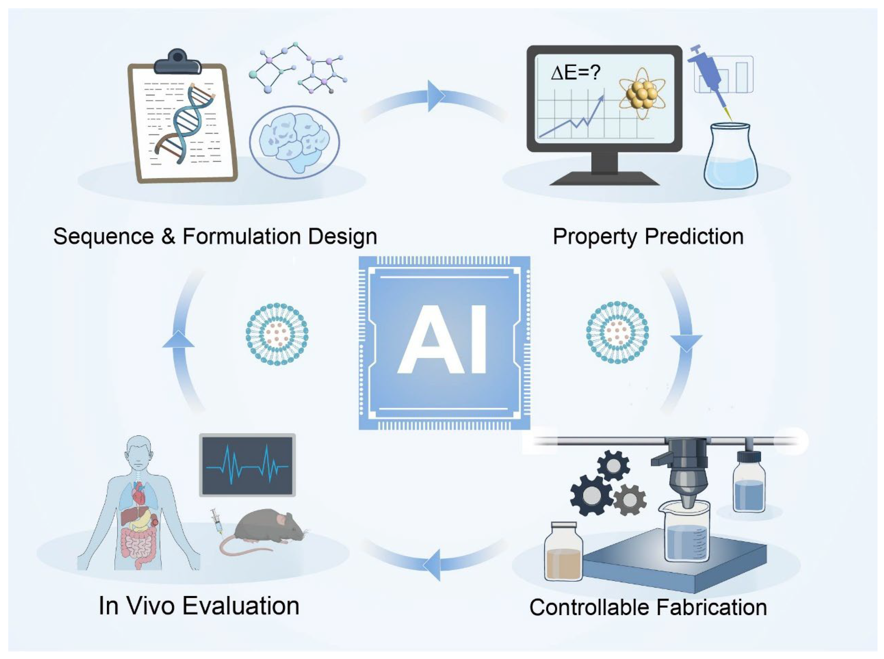
When integrated with automated synthesis platforms, online process analytics, and high-throughput in vitro and in vivo screening [236,237], these AI-enabled tools may enable closed-loop R & D workflows spanning sequence and formulation design, property prediction, controllable fabrication, and biological evaluation [225,238]. Such frameworks have the potential to shift metalloprotein-based nanomedicine development from empirical trial-and-error toward data-driven, model-guided engineering [239]. Nevertheless, AI-assisted approaches should be viewed primarily as hypothesis-generating and decision-support tools rather than definitive solutions, as their predictive power remains constrained by data quality, dataset diversity, and the intrinsic complexity of in vivo biological systems [240,241]. Continued integration of experimental insight, mechanistic understanding, and data-driven modeling will be essential for translating these advances into clinically robust metal- and metalloprotein-based nanomedicines (Figure 8).

5. Conclusions and Outlook

Metal- and metalloprotein-based nanomedicines integrate the multifunctionality of metal centers with the engineerability of protein scaffolds, enabling the construction of advanced nanoplatforms that combine targeting, controlled release, and multimodal functions. These systems have demonstrated potential advantages in pharmacokinetics, targeted accumulation, and functional synergy in preclinical studies, and promising efficacy in a range of pre-clinical studies [75]. In particular, metal nanocluster–protein conjugates, owing to their atomically defined structures and cooperative effects between the metal core and protein shell, exhibit strong and versatile performance in bioimaging, biosensing, and catalysis [155]. In this review, we have provided a systematic overview of current metal–protein nanomedicines from three perspectives: material types and construction mechanisms, biomedical applications, and key challenges, together with future opportunities.
Despite these advances, this field faces three major intertwined challenges: (i) At the level of fundamental mechanisms, interactions at metal–protein interfaces are not yet fully understood. Specifically, binding modes, structural dynamics, and structure–function relationships require deeper elucidation using integrated experimental and computational approaches [155]. (ii) At the level of process engineering, scalable and reproducible manufacturing remains difficult, and more robust and controllable synthetic routes are needed to meet quality requirements for clinical development [242]. (iii) At the translational level, long-term safety, immunogenicity, and complex in vivo biological effects constitute major barriers that must be addressed through toxicity and immunology studies [243]. In addition, the stability, immunogenicity, and manufacturability of protein-based nanoparticles themselves remain important constraints that need careful consideration in material design [75,244].
Future progress will require a paradigm shift from empirical trial-and-error approaches toward rational, data-driven design. Artificial intelligence-assisted protein design, computational chemistry, and multiscale simulations—coupled with high-throughput and automated experimentation—are emerging as powerful tools to drive this transformation [245,246]. Integrating these capabilities with advances in protein engineering, metal nanoscience, and process development is expected to enable closed-loop workflows that link sequence and formulation design, property prediction, controllable fabrication, and in vivo evaluation. We anticipate that, as fundamental understanding deepens and enabling technologies mature, metal- and metalloprotein-based nanomedicines—combining inorganic functionality with biological intelligence—will move closer to clinical translation and have the potential to become important components of future precision medicine strategies, pending further validation.

Author Contributions

Conceptualization, review of the literature, and writing—original draft preparation, T.M. and Z.M.; writing—review and editing, T.M., W.S. and B.X.; validation and supervision, B.X. and Y.C. All authors have read and agreed to the published version of the manuscript.

Funding

This research was funded by the National Key R & D Program of China (2024YFA0919300 (Y.C. and B.X.)), the National Natural Science Foundation of China (Grant No. T2225016 (B.X.), 12574222 (B.X.), 52305188 (W.S.), and T2322010 (Y.C.)), the Research Project of Jinan Microecological Biomedicine Shandong Laboratory (Grant No. JNL-2025010B (Y.C.)), the Fundamental Research Funds for the Central Universities (Grant Nos. 020414380232 (B.X.) and 020514380274 (Y.C.)), the Jiangsu Funding Program for Excellent Postdoctoral Talent (Grant No. 2025ZB632 (W.S.)), the China Postdoctoral Science Foundation (Grant No. 2024M761390 (W.S.)), the AI & AI for Science Project of Nanjing University (Grant No. 020514380343 (Y.C.)), and Nanjing Municipal Science and Technology Bureau (Grant Nos. 202305001 and 202205020 (Y.C)).

Institutional Review Board Statement

Not applicable.

Informed Consent Statement

Not applicable.

Data Availability Statement

No new data were created or analyzed in this study.

Conflicts of Interest

The authors declare no conflicts of interest.

References

  1. Andreini, C.; Bertini, I.; Cavallaro, G.; Holliday, G.L.; Thornton, J.M. Metal ions in biological catalysis: From enzyme databases to general principles. J. Biol. Inorg. Chem. 2008, 13, 1205–1218. [Google Scholar] [CrossRef] [PubMed]
  2. Yang, L.; Han, B.; Zhang, M.; Wang, Y.-H.; Tao, K.; Zhu, M.X.; He, K.; Zhang, Z.-G.; Hou, S. Activation of BK Channels Prevents Hepatic Stellate Cell Activation and Liver Fibrosis Through the Suppression of TGFβ1/SMAD3 and JAK/STAT3 Profibrotic Signaling Pathways. Front. Pharmacol. 2020, 11, 165. [Google Scholar] [CrossRef] [PubMed]
  3. Liu, Y.; Zhang, M.; Bu, W. Bioactive nanomaterials for ion-interference therapy. View 2020, 1, e18. [Google Scholar] [CrossRef]
  4. Yamasaki, S.; Sakata-Sogawa, K.; Hasegawa, A.; Suzuki, T.; Kabu, K.; Sato, E.; Kurosaki, T.; Yamashita, S.; Tokunaga, M.; Nishida, K.; et al. Zinc is a novel intracellular second messenger. J. Cell Biol. 2007, 177, 637–645. [Google Scholar] [CrossRef]
  5. Festa, R.A.; Thiele, D.J. Copper: An essential metal in biology. Curr. Biol. 2011, 21, R877–R883. [Google Scholar] [CrossRef]
  6. Cairo, G.; Bernuzzi, F.; Recalcati, S. A precious metal: Iron, an essential nutrient for all cells. Genes Nutr. 2006, 1, 25–39. [Google Scholar] [CrossRef]
  7. Horning, K.J.; Caito, S.W.; Tipps, K.G.; Bowman, A.B.; Aschner, M. Manganese Is Essential for Neuronal Health. Annu. Rev. Nutr. 2015, 35, 71–108. [Google Scholar] [CrossRef]
  8. Winterbourn, C.C. Toxicity of iron and hydrogen peroxide: The Fenton reaction. Toxicol. Lett. 1995, 82–83, 969–974. [Google Scholar] [CrossRef]
  9. Shen, Z.; Liu, T.; Li, Y.; Lau, J.; Yang, Z.; Fan, W.; Zhou, Z.; Shi, C.; Ke, C.; Bregadze, V.I.; et al. Fenton-Reaction-Acceleratable Magnetic Nanoparticles for Ferroptosis Therapy of Orthotopic Brain Tumors. ACS Nano 2018, 12, 11355–11365. [Google Scholar] [CrossRef]
  10. Chen, L.; Zhou, X.; Nie, W.; Feng, W.; Zhang, Q.; Wang, W.; Zhang, Y.; Chen, Z.; Huang, P.; He, C. Marriage of Albumin–Gadolinium Complexes and MoS2 Nanoflakes as Cancer Theranostics for Dual-Modality Magnetic Resonance/Photoacoustic Imaging and Photothermal Therapy. ACS Appl. Mater. Interfaces 2017, 9, 17786–17798. [Google Scholar] [CrossRef]
  11. Yang, S.; Zhou, L.; Su, Y.; Zhang, R.; Dong, C.-M. One-pot photoreduction to prepare NIR-absorbing plasmonic gold nanoparticles tethered by amphiphilic polypeptide copolymer for synergistic photothermal-chemotherapy. Chin. Chem. Lett. 2019, 30, 187–191. [Google Scholar] [CrossRef]
  12. Liu, T.; Zhu, M.; Chang, X.; Tang, X.; Yuan, P.; Tian, R.; Zhu, Z.; Zhang, Y.; Chen, X. Tumor-Specific Photothermal-Therapy-Assisted Immunomodulation via Multiresponsive Adjuvant Nanoparticles. Adv. Mater. 2023, 35, 2300086. [Google Scholar] [CrossRef] [PubMed]
  13. Singh, P.; Singh, H.; Kim, Y.J.; Mathiyalagan, R.; Wang, C.; Yang, D.C. Extracellular synthesis of silver and gold nanoparticles by Sporosarcina koreensis DC4 and their biological applications. Enzym. Microb. Technol. 2016, 86, 75–83. [Google Scholar] [CrossRef] [PubMed]
  14. Burdușel, A.-C.; Gherasim, O.; Grumezescu, A.M.; Mogoantă, L.; Ficai, A.; Andronescu, E. Biomedical Applications of Silver Nanoparticles: An Up-to-Date Overview. Nanomaterials 2018, 8, 681. [Google Scholar] [CrossRef]
  15. Cano, M.; Núñez-Lozano, R.; Dumont, Y.; Larpent, C.; de la Cueva-Mendez, G. Synthesis and characterization of multifunctional superparamagnetic iron oxide nanoparticles (SPION)/C60 nanocomposites assembled by fullerene–amine click chemistry. RSC Adv. 2016, 6, 70374–70382. [Google Scholar] [CrossRef]
  16. Santhosh, P.B.; Ulrih, N.P. Multifunctional superparamagnetic iron oxide nanoparticles: Promising tools in cancer theranostics. Cancer Lett. 2013, 336, 8–17. [Google Scholar] [CrossRef]
  17. Zhao, Z.; Li, M.; Zeng, J.; Huo, L.; Liu, K.; Wei, R.; Ni, K.; Gao, J. Recent advances in engineering iron oxide nanoparticles for effective magnetic resonance imaging. Bioact. Mater. 2022, 12, 214–245. [Google Scholar] [CrossRef]
  18. Dadfar, S.M.; Roemhild, K.; Drude, N.I.; von Stillfried, S.; Knüchel, R.; Kiessling, F.; Lammers, T. Iron oxide nanoparticles: Diagnostic, therapeutic and theranostic applications. Adv. Drug Deliv. Rev. 2019, 138, 302–325. [Google Scholar] [CrossRef]
  19. Menon, J.U.; Jadeja, P.; Tambe, P.; Vu, K.; Yuan, B.; Nguyen, K.T. Nanomaterials for Photo-Based Diagnostic and Therapeutic Applications. Theranostics 2013, 3, 152–166. [Google Scholar] [CrossRef]
  20. Ali, M.R.K.; Rahman, M.A.; Wu, Y.; Han, T.; Peng, X.; Mackey, M.A.; Wang, D.; Shin, H.J.; Chen, Z.G.; Xiao, H.; et al. Efficacy, long-term toxicity, and mechanistic studies of gold nanorods photothermal therapy of cancer in xenograft mice. Proc. Natl. Acad. Sci. USA 2017, 114, E3110–E3118. [Google Scholar] [CrossRef]
  21. Zheng, Q.; Liu, X.; Zheng, Y.; Yeung, K.W.K.; Cui, Z.; Liang, Y.; Li, Z.; Zhu, S.; Wang, X.; Wu, S. The recent progress on metal–organic frameworks for phototherapy. Chem. Soc. Rev. 2021, 50, 5086–5125. [Google Scholar] [CrossRef]
  22. Yang, J.; Yang, Y.-W. Metal–Organic Frameworks for Biomedical Applications. Small 2020, 16, 1906846. [Google Scholar] [CrossRef] [PubMed]
  23. Singh, P.; Pandit, S.; Mokkapati, V.R.S.S.; Garg, A.; Ravikumar, V.; Mijakovic, I. Gold Nanoparticles in Diagnostics and Therapeutics for Human Cancer. Int. J. Mol. Sci. 2018, 19, 1979. [Google Scholar] [CrossRef] [PubMed]
  24. Shah, A.; Aftab, S.; Nisar, J.; Ashiq, M.N.; Iftikhar, F.J. Nanocarriers for targeted drug delivery. J. Drug Deliv. Sci. Technol. 2021, 62, 102426. [Google Scholar] [CrossRef]
  25. Dykman, L.; Khlebtsov, N. Gold nanoparticles in biomedical applications: Recent advances and perspectives. Chem. Soc. Rev. 2012, 41, 2256–2282. [Google Scholar] [CrossRef]
  26. Li, C.; Wang, J.; Wang, Y.; Gao, H.; Wei, G.; Huang, Y.; Yu, H.; Gan, Y.; Wang, Y.; Mei, L.; et al. Recent progress in drug delivery. Acta Pharm. Sin. B 2019, 9, 1145–1162. [Google Scholar] [CrossRef]
  27. Sun, L.; Liu, H.; Ye, Y.; Lei, Y.; Islam, R.; Tan, S.; Tong, R.; Miao, Y.-B.; Cai, L. Smart nanoparticles for cancer therapy. Signal Transduct. Target. Ther. 2023, 8, 418. [Google Scholar] [CrossRef]
  28. Shubhra, Q.T.H. Iron oxide nanoparticles in magnetic drug targeting and ferroptosis-based cancer therapy. Med. Rev. 2023, 3, 444–447. [Google Scholar] [CrossRef]
  29. Liu, Y.; Ding, M.; Guo, K.; Wang, Z.; Zhang, C.; Shubhra, Q.T.H. Systemic Co-delivery of drugs by a pH- and photosensitive smart nanocarrier to treat cancer by chemo-photothermal-starvation combination therapy. Smart Mater. Med. 2022, 3, 390–403. [Google Scholar] [CrossRef]
  30. Guo, K.; Liu, Y.; Ding, M.; Sun, Q.; Shubhra, Q.T.H. Enhanced drug release from a pH-responsive nanocarrier can augment colon cancer treatment by blocking PD-L1 checkpoint and consuming tumor glucose. Mater. Des. 2022, 219, 110824. [Google Scholar] [CrossRef]
  31. Wang, Z.; Wang, R.; Zhao, Z.; Tang, G.; Che, M.; Zhou, Y.; Jiang, Y.; Zheng, P. Single-Molecule Mechanochemical Mapping of Hierarchical Cu Coordination in High-Capacity Copper Storage Protein Csp1. J. Phys. Chem. Lett. 2025, 16, 10584–10593. [Google Scholar] [CrossRef]
  32. Vita, N.; Platsaki, S.; Baslé, A.; Allen, S.J.; Paterson, N.G.; Crombie, A.T.; Murrell, J.C.; Waldron, K.J.; Dennison, C. A four-helix bundle stores copper for methane oxidation. Nature 2015, 525, 140–143. [Google Scholar] [CrossRef]
  33. Tauro, M.; McGuire, J.; Lynch, C.C. New approaches to selectively target cancer-associated matrix metalloproteinase activity. Cancer Metastasis Rev. 2014, 33, 1043–1057. [Google Scholar] [CrossRef] [PubMed]
  34. Maciewicz, R.A.; Wotton, S.F.; Etherington, D.J.; Duance, V.C. Susceptibility of the cartilage collagens types II, IX and XI to degradation by the cysteine proteinases, cathepsins B and L. FEBS Lett. 1990, 269, 189–193. [Google Scholar] [CrossRef] [PubMed]
  35. Sevenich, L.; Werner, F.; Gajda, M.; Schurigt, U.; Sieber, C.; Müller, S.; Follo, M.; Peters, C.; Reinheckel, T. Transgenic expression of human cathepsin B promotes progression and metastasis of polyoma-middle-T-induced breast cancer in mice. Oncogene 2011, 30, 54–64. [Google Scholar] [CrossRef] [PubMed]
  36. He, D.; Marles-Wright, J. Ferritin family proteins and their use in bionanotechnology. New Biotechnol. 2015, 32, 651–657. [Google Scholar] [CrossRef]
  37. Li, C.; Zhou, L.; Yin, X. Pathophysiological aspects of transferrin-A potential nano-based drug delivery signaling molecule in therapeutic target for varied diseases. Front. Pharmacol. 2024, 15, 1342181. [Google Scholar] [CrossRef]
  38. Zhu, K.; Wang, L.; Xiao, Y.; Zhang, X.; You, G.; Chen, Y.; Wang, Q.; Zhao, L.; Zhou, H.; Chen, G. Nanomaterial-related hemoglobin-based oxygen carriers, with emphasis on liposome and nano-capsules, for biomedical applications: Current status and future perspectives. J. Nanobiotechnol. 2024, 22, 336. [Google Scholar] [CrossRef]
  39. Zhivkov, A.M.; Hristova, S.H.; Popov, T.T. Anticancer Nanoparticle Carriers of the Proapoptotic Protein Cytochrome c. Pharmaceutics 2025, 17, 305. [Google Scholar] [CrossRef]
  40. Cao, Y.; Wei, X.; Lin, Y.; Sun, F. Synthesis of bio-inspired viscoelastic molecular networks by metal-induced protein assembly. Mol. Syst. Des. Eng. 2020, 5, 117–124. [Google Scholar] [CrossRef]
  41. Baksi, A.; Xavier, P.L.; Chaudhari, K.; Goswami, N.; Pal, S.K.; Pradeep, T. Protein-encapsulated gold cluster aggregates: The case of lysozyme. Nanoscale 2013, 5, 2009–2016. [Google Scholar] [CrossRef] [PubMed]
  42. Brodin, J.D.; Ambroggio, X.I.; Tang, C.; Parent, K.N.; Baker, T.S.; Tezcan, F.A. Metal-directed, chemically tunable assembly of one-, two- and three-dimensional crystalline protein arrays. Nat. Chem. 2012, 4, 375–382. [Google Scholar] [CrossRef] [PubMed]
  43. Lee, H.; Park, S.; Kang, J.E.; Lee, H.M.; Kim, S.A.; Rhie, S.J. Efficacy and safety of nanoparticle-albumin-bound paclitaxel compared with solvent-based taxanes for metastatic breast cancer: A meta-analysis. Sci. Rep. 2020, 10, 530. [Google Scholar] [CrossRef]
  44. Terashima, M.; Uchida, M.; Kosuge, H.; Tsao, P.S.; Young, M.J.; Conolly, S.M.; Douglas, T.; McConnell, M.V. Human ferritin cages for imaging vascular macrophages. Biomaterials 2011, 32, 1430–1437. [Google Scholar] [CrossRef] [PubMed]
  45. Eby, D.M.; Schaeublin, N.M.; Farrington, K.E.; Hussain, S.M.; Johnson, G.R. Lysozyme Catalyzes the Formation of Antimicrobial Silver Nanoparticles. ACS Nano 2009, 3, 984–994. [Google Scholar] [CrossRef]
  46. Theil, E.C. Ferritin protein nanocages use ion channels, catalytic sites, and nucleation channels to manage iron/oxygen chemistry. Curr. Opin. Chem. Biol. 2011, 15, 304–311. [Google Scholar] [CrossRef]
  47. Jiang, B.; Yan, L.; Zhang, J.; Zhou, M.; Shi, G.; Tian, X.; Fan, K.; Hao, C.; Yan, X. Biomineralization Synthesis of the Cobalt Nanozyme in SP94-Ferritin Nanocages for Prognostic Diagnosis of Hepatocellular Carcinoma. ACS Appl. Mater. Interfaces 2019, 11, 9747–9755. [Google Scholar] [CrossRef]
  48. Song, N.; Zhang, J.; Zhai, J.; Hong, J.; Yuan, C.; Liang, M. Ferritin: A Multifunctional Nanoplatform for Biological Detection, Imaging Diagnosis, and Drug Delivery. Acc. Chem. Res. 2021, 54, 3313–3325. [Google Scholar] [CrossRef]
  49. Zhao, J.; Yin, Y.; Liu, M.; Lu, Y.; Cao, J.; Qi, X.; Wu, L.; Shen, S. Ferritin/Ferroportin-Regulating Nanoparticles Boosting Intracellular Free Iron for Enhanced Ferrotherapy. ACS Appl. Mater. Interfaces 2025, 17, 26445–26454. [Google Scholar] [CrossRef]
  50. de Jong, G.; van Dijk, J.P.; van Eijk, H.G. The biology of transferrin. Clin. Chim. Acta 1990, 190, 1–46. [Google Scholar] [CrossRef]
  51. Kawabata, H. Transferrin and transferrin receptors update. Free Radic. Biol. Med. 2019, 133, 46–54. [Google Scholar] [CrossRef]
  52. Kawak, P.; Sawaftah, N.M.A.; Pitt, W.G.; Husseini, G.A. Transferrin-Targeted Liposomes in Glioblastoma Therapy: A Review. Int. J. Mol. Sci. 2023, 24, 13262. [Google Scholar] [CrossRef] [PubMed]
  53. Sun, H.; Li, H.; Sadler, P.J. Transferrin as a Metal Ion Mediator. Chem. Rev. 1999, 99, 2817–2842. [Google Scholar] [CrossRef] [PubMed]
  54. Koneru, T.; McCord, E.; Pawar, S.; Tatiparti, K.; Sau, S.; Iyer, A.K. Transferrin: Biology and Use in Receptor-Targeted Nanotherapy of Gliomas. ACS Omega 2021, 6, 8727–8733. [Google Scholar] [CrossRef] [PubMed]
  55. Hou, L.; Shan, X.; Hao, L.; Feng, Q.; Zhang, Z. Copper sulfide nanoparticle-based localized drug delivery system as an effective cancer synergistic treatment and theranostic platform. Acta Biomater. 2017, 54, 307–320. [Google Scholar] [CrossRef]
  56. Feng, H.; Schorpp, K.; Jin, J.; Yozwiak, C.E.; Hoffstrom, B.G.; Decker, A.M.; Rajbhandari, P.; Stokes, M.E.; Bender, H.G.; Csuka, J.M.; et al. Transferrin Receptor Is a Specific Ferroptosis Marker. Cell Rep. 2020, 30, 3411–3423.e3417. [Google Scholar] [CrossRef]
  57. Gao, M.; Monian, P.; Quadri, N.; Ramasamy, R.; Jiang, X. Glutaminolysis and Transferrin Regulate Ferroptosis. Mol. Cell 2015, 59, 298–308. [Google Scholar] [CrossRef]
  58. Sardoiwala, M.N.; Sood, A.; Biswal, L.; Roy Choudhury, S.; Karmakar, S. Reconstituted Super Paramagnetic Protein “Magnetotransferrin” for Brain Targeting to Attenuate Parkinsonism. ACS Appl. Mater. Interfaces 2023, 15, 12708–12718. [Google Scholar] [CrossRef]
  59. Bruhn, K.W.; Spellberg, B. Transferrin-mediated iron sequestration as a novel therapy for bacterial and fungal infections. Curr. Opin. Microbiol. 2015, 27, 57–61. [Google Scholar] [CrossRef]
  60. Lin, L.; Pantapalangkoor, P.; Tan, B.; Bruhn, K.W.; Ho, T.; Nielsen, T.; Skaar, E.P.; Zhang, Y.; Bai, R.; Wang, A.; et al. Transferrin Iron Starvation Therapy for Lethal Bacterial and Fungal Infections. J. Infect. Dis. 2014, 210, 254–264. [Google Scholar] [CrossRef]
  61. Macone, A.; Cappelletti, C.; Incocciati, A.; Piacentini, R.; Botta, S.; Boffi, A.; Bonamore, A. Challenges in Exploiting Human H Ferritin Nanoparticles for Drug Delivery: Navigating Physiological Constraints. WIREs Nanomed. Nanobiotechnol. 2024, 16, e2016. [Google Scholar] [CrossRef]
  62. Chen, Q.; Xu, L.; Chen, J.; Yang, Z.; Liang, C.; Yang, Y.; Liu, Z. Tumor vasculature normalization by orally fed erlotinib to modulate the tumor microenvironment for enhanced cancer nanomedicine and immunotherapy. Biomaterials 2017, 148, 69–80. [Google Scholar] [CrossRef] [PubMed]
  63. Jia, Y.; Duan, L.; Li, J. Hemoglobin-Based Nanoarchitectonic Assemblies as Oxygen Carriers. Adv. Mater. 2016, 28, 1312–1318. [Google Scholar] [CrossRef] [PubMed]
  64. Gandhi, H.A.; Bhattacharya, J. Reversal of hypoxia-induced chemoresistance using hemoglobin-loaded polycaprolactone nanoparticles. Int. J. Biol. Macromol. 2025, 332, 148463. [Google Scholar] [CrossRef]
  65. Wu, S.; Dong, R.; Xie, Y.; Chen, W.; Liu, W.; Weng, Y. CO-loaded hemoglobin/EGCG nanoparticles functional coatings for inflammation modulation of vascular implants. Regen. Biomater. 2024, 12, rbae148. [Google Scholar] [CrossRef] [PubMed]
  66. Lin, Y.-W. Rational design of metalloenzymes: From single to multiple active sites. Coord. Chem. Rev. 2017, 336, 1–27. [Google Scholar] [CrossRef]
  67. Choi, T.S.; Tezcan, F.A. Design of a Flexible, Zn-Selective Protein Scaffold that Displays Anti-Irving–Williams Behavior. J. Am. Chem. Soc. 2022, 144, 18090–18100. [Google Scholar] [CrossRef]
  68. Chalkley, M.J.; Mann, S.I.; DeGrado, W.F. De novo metalloprotein design. Nat. Rev. Chem. 2022, 6, 31–50. [Google Scholar] [CrossRef]
  69. Aires, A.; Maestro, D.; Ruiz del Rio, J.; Palanca, A.R.; Lopez-Martinez, E.; Llarena, I.; Geraki, K.; Sanchez-Cano, C.; Villar, A.V.; Cortajarena, A.L. Engineering multifunctional metal/protein hybrid nanomaterials as tools for therapeutic intervention and high-sensitivity detection. Chem. Sci. 2021, 12, 2480–2487. [Google Scholar] [CrossRef]
  70. Vornholt, T.; Leiss-Maier, F.; Jeong, W.J.; Zeymer, C.; Song, W.J.; Roelfes, G.; Ward, T.R. Artificial metalloenzymes. Nat. Rev. Methods Primers 2024, 4, 78. [Google Scholar] [CrossRef]
  71. Pan, J.; Wang, J.; Wang, W.; Liu, Z.; Huo, S.; Yan, L.; Jiang, W.; Shao, F.; Gu, Y. Renal-clearable and mitochondria-targeted metal-engineered carbon dot nanozymes for regulating mitochondrial oxidative stress in acute kidney injury. Mater. Today Bio 2025, 32, 101717. [Google Scholar] [CrossRef] [PubMed]
  72. La Gatta, S.; Leone, L.; Sgueglia, G.; Šimunić, L.; Liao, Y.-K.; Vaněk, O.; Chino, M.; Nastri, F.; Chiesa, M.; Lombardi, A. Engineering a Functional Histidine Brace Copper-Binding Site into a De Novo-Designed Protein Scaffold. JACS Au 2025, 5, 4799–4810. [Google Scholar] [CrossRef] [PubMed]
  73. Aires, A.; Fernández-Afonso, Y.; Guedes, G.; Guisasola, E.; Gutiérrez, L.; Cortajarena, A.L. Engineered Protein-Driven Synthesis of Tunable Iron Oxide Nanoparticles as T1 and T2 Magnetic Resonance Imaging Contrast Agents. Chem. Mater. 2022, 34, 10832–10841. [Google Scholar] [CrossRef] [PubMed]
  74. Zhang, Q.; Zhang, J.; Song, J.; Liu, Y.; Ren, X.; Zhao, Y. Protein-Based Nanomedicine for Therapeutic Benefits of Cancer. ACS Nano 2021, 15, 8001–8038. [Google Scholar] [CrossRef]
  75. Pan, Y.; Zhao, H.; Huang, W.; Liu, S.; Qi, Y.; Huang, Y. Metal-Protein Hybrid Materials: Unlocking New Frontiers in Biomedical Applications. Adv. Healthc. Mater. 2025, 14, 2404405. [Google Scholar] [CrossRef]
  76. Salgado, E.N.; Radford, R.J.; Tezcan, F.A. Metal-Directed Protein Self-Assembly. Acc. Chem. Res. 2010, 43, 661–672. [Google Scholar] [CrossRef]
  77. Ye, X.; Capezza, A.J.; Xiao, X.; Lendel, C.; Hedenqvist, M.S.; Kessler, V.G.; Olsson, R.T. Protein Nanofibrils and Their Hydrogel Formation with Metal Ions. ACS Nano 2021, 15, 5341–5354. [Google Scholar] [CrossRef]
  78. Liu, B.; Radiom, M.; Zhou, J.; Yan, H.; Zhang, J.; Wu, D.; Sun, Q.; Xuan, Q.; Li, Y.; Mezzenga, R. Cation Triggered Self-Assembly of α-Lactalbumin Nanotubes. Nano Lett. 2024, 24, 4951–4958. [Google Scholar] [CrossRef]
  79. Kakkis, A.; Gagnon, D.; Esselborn, J.; Britt, R.D.; Tezcan, F.A. Metal-Templated Design of Chemically Switchable Protein Assemblies with High-Affinity Coordination Sites. Angew. Chem. Int. Ed. 2020, 59, 21940–21944. [Google Scholar] [CrossRef]
  80. Xavier, P.L.; Chaudhari, K.; Verma, P.K.; Pal, S.K.; Pradeep, T. Luminescent quantum clusters of gold in transferrin family protein, lactoferrin exhibiting FRET. Nanoscale 2010, 2, 2769–2776. [Google Scholar] [CrossRef]
  81. Li, M.; Yang, D.-P.; Wang, X.; Lu, J.; Cui, D. Mixed protein-templated luminescent metal clusters (Au and Pt) for H2O2 sensing. Nanoscale Res. Lett. 2013, 8, 182. [Google Scholar] [CrossRef]
  82. Yin, Y.; Gao, C.; Xiao, Q.; Lin, G.; Lin, Z.; Cai, Z.; Yang, H. Protein-Metal Organic Framework Hybrid Composites with Intrinsic Peroxidase-like Activity as a Colorimetric Biosensing Platform. ACS Appl. Mater. Interfaces 2016, 8, 29052–29061. [Google Scholar] [CrossRef] [PubMed]
  83. Fan, T.-X.; Chow, S.-K.; Zhang, D. Biomorphic mineralization: From biology to materials. Prog. Mater. Sci. 2009, 54, 542–659. [Google Scholar] [CrossRef]
  84. Sun, C.; Yang, H.; Yuan, Y.; Tian, X.; Wang, L.; Guo, Y.; Xu, L.; Lei, J.; Gao, N.; Anderson, G.J.; et al. Controlling Assembly of Paired Gold Clusters within Apoferritin Nanoreactor for in Vivo Kidney Targeting and Biomedical Imaging. J. Am. Chem. Soc. 2011, 133, 8617–8624. [Google Scholar] [CrossRef] [PubMed]
  85. Halder, K.; Dasgupta, S. Temperature dependent human serum albumin Corona formation: A case study on gold nanorods and nanospheres. Int. J. Biol. Macromol. 2025, 290, 138581. [Google Scholar] [CrossRef]
  86. Samal, P.; Satpathy, S.; Panigrahi, L.L.; Jha, S.; Arakha, M. Exploring the intricacies of protein–nanoparticle interaction and its implications in chronic diseases: A comprehensive review. Nanoscale Horiz. 2025, 10, 1615–1641. [Google Scholar] [CrossRef]
  87. Yang, W.; Wang, L.; Fang, M.; Sheth, V.; Zhang, Y.; Holden, A.M.; Donahue, N.D.; Green, D.E.; Frickenstein, A.N.; Mettenbrink, E.M.; et al. Nanoparticle Surface Engineering with Heparosan Polysaccharide Reduces Serum Protein Adsorption and Enhances Cellular Uptake. Nano Lett. 2022, 22, 2103–2111. [Google Scholar] [CrossRef]
  88. Rahimi, E.; Imani, A.; Kim, D.; Rahimi, M.; Fedrizzi, L.; Mol, A.; Asselin, E.; Pané, S.; Lekka, M. Physicochemical Changes of Apoferritin Protein during Biodegradation of Magnetic Metal Oxide Nanoparticles. ACS Appl. Mater. Interfaces 2024, 16, 53299–53310. [Google Scholar] [CrossRef]
  89. Ansari, S.R.; Suárez-López, Y.d.C.; Thersleff, T.; Häggström, L.; Ericsson, T.; Katsaros, I.; Åhlén, M.; Karlgren, M.; Svedlindh, P.; Rinaldi-Ramos, C.M.; et al. Pharmaceutical Quality by Design Approach to Develop High-Performance Nanoparticles for Magnetic Hyperthermia. ACS Nano 2024, 18, 15284–15302. [Google Scholar] [CrossRef]
  90. Chakraborty, I.; Pradeep, T. Atomically Precise Clusters of Noble Metals: Emerging Link between Atoms and Nanoparticles. Chem. Rev. 2017, 117, 8208–8271. [Google Scholar] [CrossRef]
  91. Fang, R.; Li, Y.; Jin, J.; Yang, F.; Chen, J.; Zhang, J. Development of Anticancer Ferric Complex Based on Human Serum Albumin Nanoparticles That Generate Oxygen in Cells to Overcome Hypoxia-Induced Resistance in Metal Chemotherapy. J. Med. Chem. 2024, 67, 1184–1196. [Google Scholar] [CrossRef]
  92. Xie, J.; Zheng, Y.; Ying, J.Y. Protein-Directed Synthesis of Highly Fluorescent Gold Nanoclusters. J. Am. Chem. Soc. 2009, 131, 888–889. [Google Scholar] [CrossRef] [PubMed]
  93. Habeeb Muhammed, M.A.; Verma, P.K.; Pal, S.K.; Retnakumari, A.; Koyakutty, M.; Nair, S.; Pradeep, T. Luminescent Quantum Clusters of Gold in Bulk by Albumin-Induced Core Etching of Nanoparticles: Metal Ion Sensing, Metal-Enhanced Luminescence, and Biolabeling. Chem.–A Eur. J. 2010, 16, 10103–10112. [Google Scholar] [CrossRef] [PubMed]
  94. Bhandari, S.; Pramanik, S.; Khandelia, R.; Chattopadhyay, A. Gold Nanocluster and Quantum Dot Complex in Protein for Biofriendly White-Light-Emitting Material. ACS Appl. Mater. Interfaces 2016, 8, 1600–1605. [Google Scholar] [CrossRef] [PubMed]
  95. Goswami, N.; Giri, A.; Bootharaju, M.S.; Xavier, P.L.; Pradeep, T.; Pal, S.K. Copper Quantum Clusters in Protein Matrix: Potential Sensor of Pb2+ Ion. Anal. Chem. 2011, 83, 9676–9680. [Google Scholar] [CrossRef]
  96. Huang, J.; Lin, L.; Sun, D.; Chen, H.; Yang, D.; Li, Q. Bio-inspired synthesis of metal nanomaterials and applications. Chem. Soc. Rev. 2015, 44, 6330–6374. [Google Scholar] [CrossRef]
  97. Ivleva, E.A.; Obraztsova, E.A.; Pavlova, E.R.; Morozova, O.V.; Ivanov, D.G.; Kononikhin, A.S.; Klinov, D.V. Albumin-stabilized fluorescent metal nanoclusters: Fabrication, physico-chemical properties and cytotoxicity. Mater. Des. 2020, 192, 108771. [Google Scholar] [CrossRef]
  98. Guo, Y.; Amunyela, H.T.N.N.; Cheng, Y.; Xie, Y.; Yu, H.; Yao, W.; Li, H.-W.; Qian, H. Natural protein-templated fluorescent gold nanoclusters: Syntheses and applications. Food Chem. 2021, 335, 127657. [Google Scholar] [CrossRef]
  99. Bailey, J.B.; Zhang, L.; Chiong, J.A.; Ahn, S.; Tezcan, F.A. Synthetic Modularity of Protein–Metal–Organic Frameworks. J. Am. Chem. Soc. 2017, 139, 8160–8166. [Google Scholar] [CrossRef]
  100. Wang, Z.; Wang, R.; Geng, Z.; Luo, X.; Jia, J.; Pang, S.; Fan, X.; Bilal, M.; Cui, J. Enzyme hybrid nanoflowers and enzyme@metal–organic frameworks composites: Fascinating hybrid nanobiocatalysts. Crit. Rev. Biotechnol. 2024, 44, 674–697. [Google Scholar] [CrossRef]
  101. Guo, Y.; Li, Y.; Zhou, S.; Ye, Q.; Zan, X.; He, Y. Metal–Organic Framework-Based Composites for Protein Delivery and Therapeutics. ACS Biomater. Sci. Eng. 2022, 8, 4028–4038. [Google Scholar] [CrossRef] [PubMed]
  102. Mirzazadeh Dizaji, N.; Lin, Y.; Bein, T.; Wagner, E.; Wuttke, S.; Lächelt, U.; Engelke, H. Biomimetic Mineralization of Iron-Fumarate Nanoparticles for Protective Encapsulation and Intracellular Delivery of Proteins. Chem. Mater. 2022, 34, 8684–8693. [Google Scholar] [CrossRef] [PubMed]
  103. Thamer, B.M.; Abdul Hameed, M.M. Bio-metal Organic Frameworks: Classification, Synthesis and Applications. J. Inorg. Organomet. Polym. Mater. 2025, 34, 8684–8693. [Google Scholar] [CrossRef]
  104. Selvan, D.; Prasad, P.; Farquhar, E.R.; Shi, Y.; Crane, S.; Zhang, Y.; Chakraborty, S. Redesign of a Copper Storage Protein into an Artificial Hydrogenase. ACS Catal. 2019, 9, 5847–5859. [Google Scholar] [CrossRef]
  105. Nastri, F.; D’Alonzo, D.; Leone, L.; Zambrano, G.; Pavone, V.; Lombardi, A. Engineering Metalloprotein Functions in Designed and Native Scaffolds. Trends Biochem. Sci. 2019, 44, 1022–1040. [Google Scholar] [CrossRef]
  106. Du, H.; Jiang, D.; Zhang, O.; Wu, Z.; Gao, J.; Zhang, X.; Wang, X.; Deng, Y.; Kang, Y.; Li, D.; et al. A flexible data-free framework for structure-based de novo drug design with reinforcement learning. Chem. Sci. 2023, 14, 12166–12181. [Google Scholar] [CrossRef]
  107. Nakamoto, M.; Zhao, D.; Benice, O.R.; Lee, S.-H.; Shea, K.J. Abiotic Mimic of Endogenous Tissue Inhibitors of Metalloproteinases: Engineering Synthetic Polymer Nanoparticles for Use as a Broad-Spectrum Metalloproteinase Inhibitor. J. Am. Chem. Soc. 2020, 142, 2338–2345. [Google Scholar] [CrossRef]
  108. Rittle, J.; Field, M.J.; Green, M.T.; Tezcan, F.A. An efficient, step-economical strategy for the design of functional metalloproteins. Nat. Chem. 2019, 11, 434–441. [Google Scholar] [CrossRef]
  109. Pinter, T.B.J.; Koebke, K.J.; Pecoraro, V.L. Catalysis and Electron Transfer in De Novo Designed Helical Scaffolds. Angew. Chem. Int. Ed. 2020, 59, 7678–7699. [Google Scholar] [CrossRef]
  110. Noroozi Tiyoula, F.; Vafaee Sharbaf, F.; Rahimian, K.; Gholami, M.; Zabihi, M.R.; Salehi, Z.; Ariaeenejad, S.; Kavousi, K. Artificial intelligence in metalloprotein binding site prediction: A systematic review bridging bioinformatics and biotechnology. Int. J. Biol. Macromol. 2025, 323, 146666. [Google Scholar] [CrossRef]
  111. Dogra, P.; Adolphi, N.L.; Wang, Z.; Lin, Y.-S.; Butler, K.S.; Durfee, P.N.; Croissant, J.G.; Noureddine, A.; Coker, E.N.; Bearer, E.L.; et al. Establishing the effects of mesoporous silica nanoparticle properties on in vivo disposition using imaging-based pharmacokinetics. Nat. Commun. 2018, 9, 4551. [Google Scholar] [CrossRef]
  112. Churchfield, L.A.; George, A.; Tezcan, F.A. Repurposing proteins for new bioinorganic functions. Essays Biochem. 2017, 61, 245–258. [Google Scholar] [CrossRef] [PubMed]
  113. Li, J.; Li, H. New insights into the folding–unfolding mechanism and conformations of cytochrome C. Chem. Sci. 2022, 13, 7498–7508. [Google Scholar] [CrossRef] [PubMed]
  114. Key, H.M.; Dydio, P.; Clark, D.S.; Hartwig, J.F. Abiological catalysis by artificial haem proteins containing noble metals in place of iron. Nature 2016, 534, 534–537. [Google Scholar] [CrossRef] [PubMed]
  115. Kerns, S.A.; Biswas, A.; Minnetian, N.M.; Borovik, A.S. Artificial Metalloproteins: At the Interface between Biology and Chemistry. JACS Au 2022, 2, 1252–1265. [Google Scholar] [CrossRef]
  116. Miller, K.R.; Biswas, S.; Jasniewski, A.; Follmer, A.H.; Biswas, A.; Albert, T.; Sabuncu, S.; Bominaar, E.L.; Hendrich, M.P.; Moënne-Loccoz, P.; et al. Artificial Metalloproteins with Dinuclear Iron–Hydroxido Centers. J. Am. Chem. Soc. 2021, 143, 2384–2393. [Google Scholar] [CrossRef]
  117. Guo, Z.; Chen, Y.; Wang, Y.; Jiang, H.; Wang, X. Advances and challenges in metallic nanomaterial synthesis and antibacterial applications. J. Mater. Chem. B 2020, 8, 4764–4777. [Google Scholar] [CrossRef]
  118. Yan, Z.; Li, X.; Chung, L.W. Multiscale Quantum Refinement Approaches for Metalloproteins. J. Chem. Theory Comput. 2021, 17, 3783–3796. [Google Scholar] [CrossRef]
  119. Uribe, K.B.; Guisasola, E.; Aires, A.; López-Martínez, E.; Guedes, G.; Sasselli, I.R.; Cortajarena, A.L. Engineered Repeat Protein Hybrids: The New Horizon for Biologic Medicines and Diagnostic Tools. Acc. Chem. Res. 2021, 54, 4166–4177. [Google Scholar] [CrossRef]
  120. Elzoghby, A.O.; Samy, W.M.; Elgindy, N.A. Protein-based nanocarriers as promising drug and gene delivery systems. J. Control. Release 2012, 161, 38–49. [Google Scholar] [CrossRef]
  121. Fang, J.; Nakamura, H.; Maeda, H. The EPR effect: Unique features of tumor blood vessels for drug delivery, factors involved, and limitations and augmentation of the effect. Adv. Drug Deliv. Rev. 2011, 63, 136–151. [Google Scholar] [CrossRef]
  122. Maeda, H. The 35th Anniversary of the Discovery of EPR Effect: A New Wave of Nanomedicines for Tumor-Targeted Drug Delivery—Personal Remarks and Future Prospects. J. Pers. Med. 2021, 11, 229. [Google Scholar] [CrossRef] [PubMed]
  123. Zhao, J.; Du, J.; Wang, J.; An, N.; Zhou, K.; Hu, X.; Dong, Z.; Liu, Y. Folic Acid and Poly(ethylene glycol) Decorated Paclitaxel Nanocrystals Exhibit Enhanced Stability and Breast Cancer-Targeting Capability. ACS Appl. Mater. Interfaces 2021, 13, 14577–14586. [Google Scholar] [CrossRef] [PubMed]
  124. Sahay, G.; Alakhova, D.Y.; Kabanov, A.V. Endocytosis of nanomedicines. J. Control. Release 2010, 145, 182–195. [Google Scholar] [CrossRef] [PubMed]
  125. Hong, J.; Li, K.; He, J.; Liang, M. A New Age of Drug Delivery: A Comparative Perspective of Ferritin–Drug Conjugates (FDCs) and Antibody–Drug Conjugates (ADCs). Bioconjugate Chem. 2024, 35, 1142–1147. [Google Scholar] [CrossRef]
  126. Chen, W.; Xie, L.; Lv, C.; Song, E.; Zhu, X.; Song, Y. Transferrin-Targeted Cascade Nanoplatform for Inhibiting Transcription Factor Nuclear Factor Erythroid 2-Related Factor 2 and Enhancing Ferroptosis Anticancer Therapy. ACS Appl. Mater. Interfaces 2023, 15, 28879–28890. [Google Scholar] [CrossRef]
  127. Xue, Q.; Yan, D.; Chen, X.; Li, X.; Kang, R.; Klionsky, D.J.; Kroemer, G.; Chen, X.; Tang, D.; Liu, J. Copper-dependent autophagic degradation of GPX4 drives ferroptosis. Autophagy 2023, 19, 1982–1996. [Google Scholar] [CrossRef]
  128. Zhu, L.; You, Y.; Zhu, M.; Song, Y.; Zhang, J.; Hu, J.; Xu, X.; Xu, X.; Du, Y.; Ji, J. Ferritin-Hijacking Nanoparticles Spatiotemporally Directing Endogenous Ferroptosis for Synergistic Anticancer Therapy. Adv. Mater. 2022, 34, 2207174. [Google Scholar] [CrossRef]
  129. Guo, B.; Yang, F.; Zhang, L.; Zhao, Q.; Wang, W.; Yin, L.; Chen, D.; Wang, M.; Han, S.; Xiao, H.; et al. Cuproptosis Induced by ROS Responsive Nanoparticles with Elesclomol and Copper Combined with αPD-L1 for Enhanced Cancer Immunotherapy. Adv. Mater. 2023, 35, 2212267. [Google Scholar] [CrossRef]
  130. Sun, X.; Liang, X.; Wang, Y.; Ma, P.; Xiong, W.; Qian, S.; Cui, Y.; Zhang, H.; Chen, X.; Tian, F.; et al. A tumor microenvironment-activatable nanoplatform with phycocyanin-assisted in-situ nanoagent generation for synergistic treatment of colorectal cancer. Biomaterials 2023, 301, 122263. [Google Scholar] [CrossRef]
  131. He, R.; Zang, J.; Zhao, Y.; Liu, Y.; Ruan, S.; Zheng, X.; Chong, G.; Xu, D.; Yang, Y.; Yang, Y.; et al. Nanofactory for metabolic and chemodynamic therapy: Pro-tumor lactate trapping and anti-tumor ROS transition. J. Nanobiotechnol. 2021, 19, 426. [Google Scholar] [CrossRef]
  132. Tang, Y.; Zhou, Q.; Fan, Y.; Liu, T.-M.; Zhu, L.; Liu, M. Nanomaterial-mediated ferroptosis as a promising strategy for cancer therapy. Nanoscale 2025, 17, 27066–27084. [Google Scholar] [CrossRef] [PubMed]
  133. Xie, X.; Li, Z.; Tang, H.; Zhang, Y.; Huang, Y.; Zhang, F.; You, Y.; Xu, L.; Wu, C.; Yao, Z.; et al. A homologous membrane-camouflaged self-assembled nanodrug for synergistic antitumor therapy. Acta Biomater. 2024, 183, 292–305. [Google Scholar] [CrossRef] [PubMed]
  134. Xu, W.; Guan, G.; Yue, R.; Dong, Z.; Lei, L.; Kang, H.; Song, G. Chemical Design of Magnetic Nanomaterials for Imaging and Ferroptosis-Based Cancer Therapy. Chem. Rev. 2025, 125, 1897–1961. [Google Scholar] [CrossRef] [PubMed]
  135. Feng, W.; Shi, W.; Wang, Z.; Cui, Y.; Shao, X.; Liu, S.; Rong, L.; Liu, Y.; Zhang, H. Enhancing Tumor Therapy of Fe(III)-Shikonin Supramolecular Nanomedicine via Triple Ferroptosis Amplification. ACS Appl. Mater. Interfaces 2022, 14, 37540–37552. [Google Scholar] [CrossRef]
  136. Liu, S.; Wei, Y.; Liang, Y.; Du, P.; Lei, P.; Yu, D.; Zhang, H. Engineering Nanozymes for Tumor Therapy via Ferroptosis Self-Amplification. Adv. Healthc. Mater. 2024, 13, 2400307. [Google Scholar] [CrossRef]
  137. Ding, H.; Yang, D.; Zhao, C.; Song, Z.; Liu, P.; Wang, Y.; Chen, Z.; Shen, J. Protein–Gold Hybrid Nanocubes for Cell Imaging and Drug Delivery. ACS Appl. Mater. Interfaces 2015, 7, 4713–4719. [Google Scholar] [CrossRef]
  138. Yang, F.; Fang, E.; Luo, M.; Qiu, J.; Huang, Y.; Qiu, J.; Zeng, M. Functional integration and synergistic effects of metal-based nanocomplexes in tumor photothermal therapy and bioimaging. Int. J. Pharm. 2025, 685, 126286. [Google Scholar] [CrossRef]
  139. Huang, C.-C.; Khu, N.-H.; Yeh, C.-S. The characteristics of sub 10 nm manganese oxide T1 contrast agents of different nanostructured morphologies. Biomaterials 2010, 31, 4073–4078. [Google Scholar] [CrossRef]
  140. Baek, M.J.; Park, J.Y.; Xu, W.; Kattel, K.; Kim, H.G.; Lee, E.J.; Patel, A.K.; Lee, J.J.; Chang, Y.; Kim, T.J.; et al. Water-Soluble MnO Nanocolloid for a Molecular T1 MR Imaging: A Facile One-Pot Synthesis, In vivo T1 MR Images, and Account for Relaxivities. ACS Appl. Mater. Interfaces 2010, 2, 2949–2955. [Google Scholar] [CrossRef]
  141. Bomatí-Miguel, O.; Morales, M.P.; Tartaj, P.; Ruiz-Cabello, J.; Bonville, P.; Santos, M.; Zhao, X.; Veintemillas-Verdaguer, S. Fe-based nanoparticulate metallic alloys as contrast agents for magnetic resonance imaging. Biomaterials 2005, 26, 5695–5703. [Google Scholar] [CrossRef] [PubMed]
  142. Dong, Y.C.; Hajfathalian, M.; Maidment, P.S.N.; Hsu, J.C.; Naha, P.C.; Si-Mohamed, S.; Breuilly, M.; Kim, J.; Chhour, P.; Douek, P.; et al. Effect of Gold Nanoparticle Size on Their Properties as Contrast Agents for Computed Tomography. Sci. Rep. 2019, 9, 14912. [Google Scholar] [CrossRef] [PubMed]
  143. Luo, F.; Song, S.; Zhou, G.; Wang, Y.; Fu, Z.; Liu, H. Biotechnology-assisted cancer therapy using metal sulfides based on their optical and thermophysical properties. Nanoscale Adv. 2025, 7, 2773–2795. [Google Scholar] [CrossRef] [PubMed]
  144. Coghi, P.; Chen, Z.; Leong, K.F.; Yu, X.; Hosmane, N.S.; Zhu, Y. Metal coordination-based nanomaterials: Novel drug delivery system for treatment of rheumatoid arthritis. Coord. Chem. Rev. 2026, 546, 217049. [Google Scholar] [CrossRef]
  145. Zhao, Y.; Liu, W.; Tian, Y.; Yang, Z.; Wang, X.; Zhang, Y.; Tang, Y.; Zhao, S.; Wang, C.; Liu, Y.; et al. Anti-EGFR Peptide-Conjugated Triangular Gold Nanoplates for Computed Tomography/Photoacoustic Imaging-Guided Photothermal Therapy of Non-Small Cell Lung Cancer. ACS Appl. Mater. Interfaces 2018, 10, 16992–17003. [Google Scholar] [CrossRef]
  146. Chen, J.; Liu, C.; Hu, D.; Wang, F.; Wu, H.; Gong, X.; Liu, X.; Song, L.; Sheng, Z.; Zheng, H. Single-Layer MoS2 Nanosheets with Amplified Photoacoustic Effect for Highly Sensitive Photoacoustic Imaging of Orthotopic Brain Tumors. Adv. Funct. Mater. 2016, 26, 8715–8725. [Google Scholar] [CrossRef]
  147. Shen, S.; Chao, Y.; Dong, Z.; Wang, G.; Yi, X.; Song, G.; Yang, K.; Liu, Z.; Cheng, L. Bottom-Up Preparation of Uniform Ultrathin Rhenium Disulfide Nanosheets for Image-Guided Photothermal Radiotherapy. Adv. Funct. Mater. 2017, 27, 1700250. [Google Scholar] [CrossRef]
  148. Li, X.; Zhang, X.; Song, L.; Li, Y.; Liu, A.; Li, L.; Nešić, M.D.; Li, D.; Peng, L.; Wang, C.; et al. Nanozyme as Tumor Energy Homeostasis Disruptor to Augment Cascade Catalytic Therapy. ACS Nano 2024, 18, 34656–34670. [Google Scholar] [CrossRef]
  149. Wang, Y.; Yang, T.; Ke, H.; Zhu, A.; Wang, Y.; Wang, J.; Shen, J.; Liu, G.; Chen, C.; Zhao, Y.; et al. Smart Albumin-Biomineralized Nanocomposites for Multimodal Imaging and Photothermal Tumor Ablation. Adv. Mater. 2015, 27, 3874–3882. [Google Scholar] [CrossRef]
  150. Chen, Q.; Luo, Y.; Du, W.; Liu, Z.; Zhang, S.; Yang, J.; Yao, H.; Liu, T.; Ma, M.; Chen, H. Clearable Theranostic Platform with a pH-Independent Chemodynamic Therapy Enhancement Strategy for Synergetic Photothermal Tumor Therapy. ACS Appl. Mater. Interfaces 2019, 11, 18133–18144. [Google Scholar] [CrossRef]
  151. Yu, X.; Li, A.; Zhao, C.; Yang, K.; Chen, X.; Li, W. Ultrasmall Semimetal Nanoparticles of Bismuth for Dual-Modal Computed Tomography/Photoacoustic Imaging and Synergistic Thermoradiotherapy. ACS Nano 2017, 11, 3990–4001. [Google Scholar] [CrossRef] [PubMed]
  152. Zheng, X.; Wu, Y.; Zuo, H.; Chen, W.; Wang, K. Metal Nanoparticles as Novel Agents for Lung Cancer Diagnosis and Therapy. Small 2023, 19, 2206624. [Google Scholar] [CrossRef] [PubMed]
  153. Guo, Z.; Zhu, S.; Yong, Y.; Zhang, X.; Dong, X.; Du, J.; Xie, J.; Wang, Q.; Gu, Z.; Zhao, Y. Synthesis of BSA-Coated BiOI@Bi2S3 Semiconductor Heterojunction Nanoparticles and Their Applications for Radio/Photodynamic/Photothermal Synergistic Therapy of Tumor. Adv. Mater. 2017, 29, 1704136. [Google Scholar] [CrossRef] [PubMed]
  154. Ostruszka, R.; Halili, A.; Pluháček, T.; Rárová, L.; Jirák, D.; Šišková, K. Advanced protein-embedded bimetallic nanocomposite optimized for in vivo fluorescence and magnetic resonance bimodal imaging. J. Colloid Interface Sci. 2024, 663, 467–477. [Google Scholar] [CrossRef]
  155. Zhang, B.; Liu, Z.; Zhang, R.; Yao, Q.; Xie, J. Precision Metal Nanoclusters Meet Proteins: Crafting Next-Gen Hybrid Materials. ACS Nano 2025, 19, 3997–4010. [Google Scholar] [CrossRef]
  156. Malekkhaiat Häffner, S.; Malmsten, M. Membrane interactions and antimicrobial effects of inorganic nanoparticles. Adv. Colloid Interface Sci. 2017, 248, 105–128. [Google Scholar] [CrossRef]
  157. Chen, Q.; Liu, Z. Albumin Carriers for Cancer Theranostics: A Conventional Platform with New Promise. Adv. Mater. 2016, 28, 10557–10566. [Google Scholar] [CrossRef]
  158. Kommineni, N.; Chaudhari, R.; Conde, J.; Tamburaci, S.; Cecen, B.; Chandra, P.; Prasad, R. Engineered Liposomes in Interventional Theranostics of Solid Tumors. ACS Biomater. Sci. Eng. 2023, 9, 4527–4557. [Google Scholar] [CrossRef]
  159. Frei, A.; Verderosa, A.D.; Elliott, A.G.; Zuegg, J.; Blaskovich, M.A.T. Metals to combat antimicrobial resistance. Nat. Rev. Chem. 2023, 7, 202–224. [Google Scholar] [CrossRef]
  160. Łoboda, D.; Kozłowski, H.; Rowińska-Żyrek, M. Antimicrobial peptide–metal ion interactions—A potential way of activity enhancement. New J. Chem. 2018, 42, 7560–7568. [Google Scholar] [CrossRef]
  161. Shi, H.; Ding, J.; Chen, C.; Yao, Q.; Zhang, W.; Fu, Y.; Wang, X.; Ruan, J. Antimicrobial Action of Biocompatible Silver Microspheres and Their Role in the Potential Treatment of Fungal Keratitis. ACS Biomater. Sci. Eng. 2021, 7, 5090–5098. [Google Scholar] [CrossRef]
  162. Sun, Z.; Zheng, W.; Zhu, G.; Lian, J.; Wang, J.; Hui, P.; He, S.; Chen, W.; Jiang, X. Albumin Broadens the Antibacterial Capabilities of Nonantibiotic Small Molecule-Capped Gold Nanoparticles. ACS Appl. Mater. Interfaces 2019, 11, 45381–45389. [Google Scholar] [CrossRef] [PubMed]
  163. Liu, Y.; Lan, Q.; Liu, J.; Shi, Y.; Wu, Q.; Wang, Q.; Yang, S.; Cheng, F. Phenylboronic acid-functionalized BSA@CuS@PpIX nanoparticles for enhanced antibacterial photodynamic/photothermal therapy. J. Drug Deliv. Sci. Technol. 2023, 88, 104965. [Google Scholar] [CrossRef]
  164. Bao, M.; Wang, K.; Li, J.; Li, Y.; Zhu, H.; Lu, M.; Zhang, Y.; Fan, Q.; Han, L.; Wang, K.; et al. ROS Scavenging and inflammation-directed polydopamine nanoparticles regulate gut immunity and flora therapy in inflammatory bowel disease. Acta Biomater. 2023, 161, 250–264. [Google Scholar] [CrossRef] [PubMed]
  165. De Jong, W.H.; Geertsma, R.E.; Borchard, G. Regulatory safety evaluation of nanomedical products: Key issues to refine. Drug Deliv. Transl. Res. 2022, 12, 2042–2047. [Google Scholar] [CrossRef]
  166. Sun, W.; Zhu, C.; Song, J.; Ji, S.-C.; Jiang, B.-P.; Liang, H.; Shen, X.-C. Hydrogen Sulfide Gas Amplified ROS Cascade: FeS@GOx Hybrid Nanozyme Designed for Boosting Tumor Chemodynamic Immunotherapy (Adv. Healthcare Mater. 23/2023). Adv. Healthc. Mater. 2023, 12, 2370140. [Google Scholar] [CrossRef]
  167. Yu, Y.-J.; Yan, J.-H.; Chen, Q.-W.; Qiao, J.-Y.; Peng, S.-Y.; Cheng, H.; Chen, M.; Zhang, X.-Z. Polymeric nano-system for macrophage reprogramming and intracellular MRSA eradication. J. Control. Release 2023, 353, 591–610. [Google Scholar] [CrossRef]
  168. Yang, L.; Zhang, D.; Li, W.; Lin, H.; Ding, C.; Liu, Q.; Wang, L.; Li, Z.; Mei, L.; Chen, H.; et al. Biofilm microenvironment triggered self-enhancing photodynamic immunomodulatory microneedle for diabetic wound therapy. Nat. Commun. 2023, 14, 7658. [Google Scholar] [CrossRef]
  169. Xu, Y.; Wang, Y.; Guan, L.; Zhang, P.; Wu, Z.; Jehan, S.; Peng, C.; Yang, B.; Yao, Y.; Zhuang, P.; et al. Precision Drug Delivery for Multifunctional Treatment of Abdominal Aortic Aneurysm Using Bioactive Tea Polyphenol Nanoparticles. ACS Appl. Mater. Interfaces 2025, 17, 35080–35098. [Google Scholar] [CrossRef]
  170. Yoo, W.; Shealy, N.G.; Zieba, J.K.; Torres, T.P.; Baltagulov, M.; Thomas, J.D.; Shelton, C.D.; McGovern, A.G.; Foegeding, N.J.; Olsan, E.E.; et al. Salmonella Typhimurium expansion in the inflamed murine gut is dependent on aspartate derived from ROS-mediated microbiota lysis. Cell Host Microbe 2024, 32, 887–899.e6. [Google Scholar] [CrossRef]
  171. Navale, G.R.; Ahmed, I.; Lim, M.H.; Ghosh, K. Transition Metal Complexes as Therapeutics: A New Frontier in Combatting Neurodegenerative Disorders through Protein Aggregation Modulation. Adv. Healthc. Mater. 2024, 13, 2401991. [Google Scholar] [CrossRef] [PubMed]
  172. Anirudhan, A.; Prabu, P.; Sanyal, J.; Banerjee, T.K.; Guha, G.; Murugesan, R.; Ahmed, S.S.S.J. Interdependence of metals and its binding proteins in Parkinson’s disease for diagnosis. npj Park. Dis. 2021, 7, 3. [Google Scholar] [CrossRef] [PubMed]
  173. Liu, L.; Liu, W.; Sun, Y.; Dong, X. Serum albumin-embedding copper nanoclusters inhibit Alzheimer’s β-amyloid fibrillogenesis and neuroinflammation. J. Colloid Interface Sci. 2024, 672, 53–62. [Google Scholar] [CrossRef]
  174. He, S.-B.; Chen, R.-T.; Wu, Y.-Y.; Wu, G.-W.; Peng, H.-P.; Liu, A.-L.; Deng, H.-H.; Xia, X.-H.; Chen, W. Improved enzymatic assay for hydrogen peroxide and glucose by exploiting the enzyme-mimicking properties of BSA-coated platinum nanoparticles. Microchim. Acta 2019, 186, 778. [Google Scholar] [CrossRef] [PubMed]
  175. Cao, M.; Cai, R.; Zhao, L.; Guo, M.; Wang, L.; Wang, Y.; Zhang, L.; Wang, X.; Yao, H.; Xie, C.; et al. Molybdenum derived from nanomaterials incorporates into molybdenum enzymes and affects their activities in vivo. Nat. Nanotechnol. 2021, 16, 708–716. [Google Scholar] [CrossRef]
  176. Xiong, P.; Huang, X.; Ye, N.; Lu, Q.; Zhang, G.; Peng, S.; Wang, H.; Liu, Y. Cytotoxicity of Metal-Based Nanoparticles: From Mechanisms and Methods of Evaluation to Pathological Manifestations. Adv. Sci. 2022, 9, 2106049. [Google Scholar] [CrossRef]
  177. Arojojoye, A.S.; Walker, B.; Dawahare, J.C.; Afrifa, M.A.O.; Parkin, S.; Awuah, S.G. Circumventing Physicochemical Barriers of Cyclometalated Gold(III) Dithiocarbamate Complexes with Protein-Based Nanoparticle Delivery to Enhance Anticancer Activity. ACS Appl. Mater. Interfaces 2023, 15, 43607–43620. [Google Scholar] [CrossRef]
  178. Lei, L.; Dong, Z.; Yang, F.; Zhang, X. Metal–Organic Nanomaterials for Tumor Metabolic Blockade and Image to Increase Tumor Therapy. ACS Appl. Mater. Interfaces 2024, 16, 57995–58005. [Google Scholar] [CrossRef]
  179. Yang, G.; Wang, D.; Phua, S.Z.F.; Bindra, A.K.; Qian, C.; Zhang, R.; Cheng, L.; Liu, G.; Wu, H.; Liu, Z.; et al. Albumin-Based Therapeutics Capable of Glutathione Consumption and Hydrogen Peroxide Generation for Synergetic Chemodynamic and Chemotherapy of Cancer. ACS Nano 2022, 16, 2319–2329. [Google Scholar] [CrossRef]
  180. Wang, Y.; Chen, J.; Lu, J.; Xi, J.; Xu, Z.; Fan, L.; Dai, H.; Gao, L. Metal ions/nucleotide coordinated nanoparticles comprehensively suppress tumor by synergizing ferroptosis with energy metabolism interference. J. Nanobiotechnol. 2022, 20, 199. [Google Scholar] [CrossRef]
  181. Wu, S.; Zhang, K.; Liang, Y.; Wei, Y.; An, J.; Wang, Y.; Yang, J.; Zhang, H.; Zhang, Z.; Liu, J.; et al. Nano-enabled Tumor Systematic Energy Exhaustion via Zinc (II) Interference Mediated Glycolysis Inhibition and Specific GLUT1 Depletion. Adv. Sci. 2022, 9, 2103534. [Google Scholar] [CrossRef] [PubMed]
  182. Thacker, S.G.; Her, C.; Kelley-Baker, L.; Ireland, D.D.C.; Manangeeswaran, M.; Pang, E.S.; Verthelyi, D. Detection of innate immune response modulating impurities (IIRMI) in therapeutic peptides and proteins: Impact of excipients. Front. Immunol. 2022, 13, 970499. [Google Scholar] [CrossRef] [PubMed]
  183. Noel, J.C.; Lagassé, D.; Golding, B.; Sauna, Z.E. Emerging approaches to induce immune tolerance to therapeutic proteins. Trends Pharmacol. Sci. 2023, 44, 1028–1042. [Google Scholar] [CrossRef] [PubMed]
  184. Xu, J.; Wang, J.; Ye, J.; Jiao, J.; Liu, Z.; Zhao, C.; Li, B.; Fu, Y. Metal-Coordinated Supramolecular Self-Assemblies for Cancer Theranostics. Adv. Sci. 2021, 8, 2101101. [Google Scholar] [CrossRef]
  185. Huang, Y.; Qin, G.; Cui, T.; Zhao, C.; Ren, J.; Qu, X. A bimetallic nanoplatform for STING activation and CRISPR/Cas mediated depletion of the methionine transporter in cancer cells restores anti-tumor immune responses. Nat. Commun. 2023, 14, 4647. [Google Scholar] [CrossRef]
  186. Hussain, Z.; Rahim, M.A.; Jan, N.; Shah, H.; Rawas-Qalaji, M.; Khan, S.; Sohail, M.; Thu, H.E.; Ramli, N.A.; Sarfraz, R.M.; et al. Cell membrane cloaked nanomedicines for bio-imaging and immunotherapy of cancer: Improved pharmacokinetics, cell internalization and anticancer efficacy. J. Control. Release 2021, 335, 130–157. [Google Scholar] [CrossRef]
  187. Li, S.; Wang, X.; Han, H.; Xiang, S.; Li, M.; Long, G.; Xia, Y.; Zhang, Q.; Li, S. Metallic nanomedicine in cancer immunotherapy. Acta Pharm. Sin. B 2025, 15, 4614–4643. [Google Scholar] [CrossRef]
  188. Ray, M.; Brancolini, G.; Luther, D.C.; Jiang, Z.; Cao-Milán, R.; Cuadros, A.M.; Burden, A.; Clark, V.; Rana, S.; Mout, R.; et al. High affinity protein surface binding through co-engineering of nanoparticles and proteins. Nanoscale 2022, 14, 2411–2418. [Google Scholar] [CrossRef]
  189. Ma, S.; Chen, Y.; Dai, A.; Yin, W.; Guo, J.; Yang, D.; Zhou, F.; Jiang, Y.; Wang, M.-W.; Xu, H.E. Structural mechanism of calcium-mediated hormone recognition and Gβ interaction by the human melanocortin-1 receptor. Cell Res. 2021, 31, 1061–1071. [Google Scholar] [CrossRef]
  190. Verma, Y.; Khan, I.; Dongsar, T.T.; Alsayari, A.; Wahab, S.; Sahebkar, A.; Kesharwani, P. AS1411 aptamer based nanomaterials: A novel approach for breast cancer therapy. Int. J. Biol. Macromol. 2025, 318, 145125. [Google Scholar] [CrossRef]
  191. Aires, A.; Sousaraei, A.; Möller, M.; Cabanillas-Gonzalez, J.; Cortajarena, A.L. Boosting the Photoluminescent Properties of Protein-Stabilized Gold Nanoclusters through Protein Engineering. Nano Lett. 2021, 21, 9347–9353. [Google Scholar] [CrossRef] [PubMed]
  192. Gautam, M.; Kim, J.O.; Yong, C.S. Fabrication of aerosol-based nanoparticles and their applications in biomedical fields. J. Pharm. Investig. 2021, 51, 361–375. [Google Scholar] [CrossRef] [PubMed]
  193. Opdensteinen, P.; Affonso de Oliveira, J.F.; Jung, S.; Steinmetz, N.F. Toward Translation of Cowpea Mosaic Virus Intratumoral Immunotherapy With a Scalable Production Process. Plant Biotechnol. J. 2025, 23, 5013–5031. [Google Scholar] [CrossRef] [PubMed]
  194. Wu, W.; Oliveira, L.T.; Jain, A.; Karpov, Y.; Olsen, K.; Wu, Y.; Gopalakrishna Panicker, R.K. Process development of tangential flow filtration and sterile filtration for manufacturing of mRNA-lipid nanoparticles: A study on membrane performance and filtration modeling. Int. J. Pharm. 2025, 675, 125520. [Google Scholar] [CrossRef]
  195. Reddy, S.M.; Stephen, A.N.; Holden, M.A.; Stockburn, W.J.; Dennison, S.R. Magnetic nanoparticle-facilitated rapid mass production of high affinity polymeric materials (nanoMIPs) for protein recognition and biosensing. Biomater. Sci. 2024, 12, 5845–5855. [Google Scholar] [CrossRef]
  196. Luo, J.; He, C. Chemical protein synthesis enabled engineering of saccharide oxidative cleavage activity in artificial metalloenzymes. Int. J. Biol. Macromol. 2024, 256, 128083. [Google Scholar] [CrossRef]
  197. Liu, X.; Liu, S.; Jin, X.; Liu, H.; Sun, K.; Wang, X.; Li, M.; Wang, P.; Chang, Y.; Wang, T.; et al. An encounter between metal ions and natural products: Natural products-coordinated metal ions for the diagnosis and treatment of tumors. J. Nanobiotechnol. 2024, 22, 726. [Google Scholar] [CrossRef]
  198. Berkner, S.; Schwirn, K.; Voelker, D. Too advanced for assessment? Advanced materials, nanomedicine and the environment. Environ. Sci. Eur. 2022, 34, 71. [Google Scholar] [CrossRef]
  199. Dri, D.A.; Rinaldi, F.; Carafa, M.; Marianecci, C. Nanomedicines and nanocarriers in clinical trials: Surfing through regulatory requirements and physico-chemical critical quality attributes. Drug Deliv. Transl. Res. 2023, 13, 757–769. [Google Scholar] [CrossRef]
  200. Cao, Y.; Qiao, Y.; Cui, S.; Ge, J. Origin of Metal Cluster Tuning Enzyme Activity at the Bio-Nano Interface. JACS Au 2022, 2, 961–971. [Google Scholar] [CrossRef]
  201. Dar, A.I.; Randhawa, S.; Verma, M.; Saini, T.C.; Acharya, A. Debugging the dynamics of protein corona: Formation, composition, challenges, and applications at the nano-bio interface. Adv. Colloid Interface Sci. 2025, 342, 103535. [Google Scholar] [CrossRef]
  202. Fatima, S.; Boggs, D.G.; Ali, N.; Thompson, P.J.; Thielges, M.C.; Bridwell-Rabb, J.; Olshansky, L. Engineering a Conformationally Switchable Artificial Metalloprotein. J. Am. Chem. Soc. 2022, 144, 21606–21616. [Google Scholar] [CrossRef] [PubMed]
  203. Maity, B.; Abe, S.; Ueno, T. Observation of gold sub-nanocluster nucleation within a crystalline protein cage. Nat. Commun. 2017, 8, 14820. [Google Scholar] [CrossRef] [PubMed]
  204. Choi, T.S.; Tezcan, F.A. Overcoming universal restrictions on metal selectivity by protein design. Nature 2022, 603, 522–527. [Google Scholar] [CrossRef] [PubMed]
  205. Stachura, M.; Chakraborty, S.; Gottberg, A.; Ruckthong, L.; Pecoraro, V.L.; Hemmingsen, L. Direct Observation of Nanosecond Water Exchange Dynamics at a Protein Metal Site. J. Am. Chem. Soc. 2017, 139, 79–82. [Google Scholar] [CrossRef]
  206. Jiang, D.; Ye, Z.; Hsieh, C.-Y.; Yang, Z.; Zhang, X.; Kang, Y.; Du, H.; Wu, Z.; Wang, J.; Zeng, Y.; et al. MetalProGNet: A structure-based deep graph model for metalloprotein–ligand interaction predictions. Chem. Sci. 2023, 14, 2054–2069. [Google Scholar] [CrossRef]
  207. Hassan Ibrahim, I. Metalloproteins and metalloproteomics in health and disease. In Advances in Protein Chemistry and Structural Biology; Elsevier: Amsterdam, The Netherlands, 2024; Volume 141, pp. 123–176. [Google Scholar]
  208. Giannakou, C.; Park, M.V.D.Z.; Bosselaers, I.E.M.; de Jong, W.H.; van der Laan, J.W.; van Loveren, H.; Vandebriel, R.J.; Geertsma, R.E. Nonclinical regulatory immunotoxicity testing of nanomedicinal products: Proposed strategy and possible pitfalls. WIREs Nanomed. Nanobiotechnol. 2020, 12, e1633. [Google Scholar] [CrossRef]
  209. Zingg, R.; Fischer, M. The consolidation of nanomedicine. WIREs Nanomed. Nanobiotechnol. 2019, 11, e1569. [Google Scholar] [CrossRef]
  210. Peña, Q.; Wang, A.; Zaremba, O.; Shi, Y.; Scheeren, H.W.; Metselaar, J.M.; Kiessling, F.; Pallares, R.M.; Wuttke, S.; Lammers, T. Metallodrugs in cancer nanomedicine. Chem. Soc. Rev. 2022, 51, 2544–2582. [Google Scholar] [CrossRef]
  211. Li, K.; Yang, J.; Peng, Y.; Zheng, H.; Fu, Z.; Li, M.; Zhou, W.; Yu, G. Advances in metal ion–mediated macromolecular drug delivery. Int. J. Pharm. 2026, 689, 126501. [Google Scholar] [CrossRef]
  212. Cao, Z.; Liu, C.; Wen, J.; Lu, Y. Innovative Formulation Platform: Paving the Way for Superior Protein Therapeutics with Enhanced Efficacy and Broadened Applications. Adv. Mater. 2024, 36, 2403116. [Google Scholar] [CrossRef]
  213. Lv, P.; Li, Y.; Du, G.; Ding, J.; Zhou, J.; Zhang, Y.; Lin, H.; Yang, M.; Zhou, C.; Yang, B. Supramolecular Lipid Nanoparticles Based on Amine β-CD Host–Guest Lipids: Design, Mechanisms, and Biosafety. Pharmaceutics 2025, 17, 1410. [Google Scholar] [CrossRef] [PubMed]
  214. Koebke, K.J.; Pinter, T.B.J.; Pitts, W.C.; Pecoraro, V.L. Catalysis and Electron Transfer in De Novo Designed Metalloproteins. Chem. Rev. 2022, 122, 12046–12109. [Google Scholar] [CrossRef] [PubMed]
  215. Ye, Q.; Li, R.; Liang, B.; Zhu, L.; Xiao, J.; Shi, Z. Predicting the Kinetics of Cu and Cd Release from Diverse Soil Dissolved Organic Matter: A Novel Hybrid Model Integrating Machine Learning with Mechanistic Kinetics Model. Environ. Sci. Technol. 2025, 59, 3713–3722. [Google Scholar] [CrossRef] [PubMed]
  216. Young, T.R.; Martini, M.A.; Foster, A.W.; Glasfeld, A.; Osman, D.; Morton, R.J.; Deery, E.; Warren, M.J.; Robinson, N.J. Calculating metalation in cells reveals CobW acquires CoII for vitamin B12 biosynthesis while related proteins prefer ZnII. Nat. Commun. 2021, 12, 1195. [Google Scholar] [CrossRef]
  217. Zhang, H.; Zhong, J.; Gucwa, M.; Zhang, Y.; Ma, H.; Deng, L.; Mao, L.; Minor, W.; Wang, N.; Zheng, H. PinMyMetal: A hybrid learning system to accurately model transition metal binding sites in macromolecules. Nat. Commun. 2025, 16, 3043. [Google Scholar] [CrossRef]
  218. Jankowski, W.; Kidchob, C.; Bunce, C.; Cloake, E.; Resende, R.; Sauna, Z.E. The MHC Associated Peptide Proteomics assay is a useful tool for the non-clinical assessment of immunogenicity. Front. Immunol. 2023, 14, 1271120. [Google Scholar] [CrossRef]
  219. Ogishi, M.; Yotsuyanagi, H. Quantitative Prediction of the Landscape of T Cell Epitope Immunogenicity in Sequence Space. Front. Immunol. 2019, 10, 827. [Google Scholar] [CrossRef]
  220. Cun, Y.; Ding, H.; Mao, T.; Wang, Y.; Wang, C.; Li, J.; Li, Z.; Hu, M.; Cao, Z.; Qiu, T. SITA: Predicting site-specific immunogenicity for therapeutic antibodies. J. Pharm. Anal. 2025, 15, 101316. [Google Scholar] [CrossRef]
  221. Harris, C.T.; Cohen, S. Reducing Immunogenicity by Design: Approaches to Minimize Immunogenicity of Monoclonal Antibodies. BioDrugs 2024, 38, 205–226. [Google Scholar] [CrossRef]
  222. Hofman, K.; Jangied, P.; Balu-Iyer, S.V. Immunogenicity of Gene and Cell Therapies. BioDrugs 2026, 34, 1–10. [Google Scholar] [CrossRef] [PubMed]
  223. Lamichhane, S.; Lee, S. Albumin nanoscience: Homing nanotechnology enabling targeted drug delivery and therapy. Arch. Pharmacal Res. 2020, 43, 118–133. [Google Scholar] [CrossRef] [PubMed]
  224. Chen, C.; Huang, B.; Zhang, R.; Sun, C.; Chen, L.; Ge, J.; Zhou, D.; Li, Y.; Wu, S.; Qian, Z.; et al. Surface ligand-regulated renal clearance of MRI/SPECT dual-modality nanoprobes for tumor imaging. J. Nanobiotechnol. 2024, 22, 245. [Google Scholar] [CrossRef] [PubMed]
  225. Chou, W.-C.; Chen, Q.; Yuan, L.; Cheng, Y.-H.; He, C.; Monteiro-Riviere, N.A.; Riviere, J.E.; Lin, Z. An artificial intelligence-assisted physiologically-based pharmacokinetic model to predict nanoparticle delivery to tumors in mice. J. Control. Release 2023, 361, 53–63. [Google Scholar] [CrossRef]
  226. Richardson, R.; Hanson, B.; Read, D.; Harlen, O.; Harris, S. Exploring the dynamics of flagellar dynein within the axoneme with Fluctuating Finite Element Analysis. Q. Rev. Biophys. 2020, 53, e9. [Google Scholar] [CrossRef]
  227. Guo, C.; Cheng, M.; Gross, M.L. Protein-Metal-Ion Interactions Studied by Mass Spectrometry-Based Footprinting with Isotope-Encoded Benzhydrazide. Anal. Chem. 2019, 91, 1416–1423. [Google Scholar] [CrossRef]
  228. McKenzie-Coe, A.; Shortt, R.; Jones, L.M. The making of a footprint in protein footprinting: A review in honor of michael L. gross. Mass Spectrom. Rev. 2021, 40, 177–200. [Google Scholar] [CrossRef]
  229. Cheng, M.; Gross, M.L. Mass Spectrometry-Based Protein Footprinting for Protein Structure Characterization. Acc. Chem. Res. 2025, 58, 165–176. [Google Scholar] [CrossRef]
  230. Park, H.; Patel, P.; Haas, R.; Huerta, E.A. APACE: AlphaFold2 and advanced computing as a service for accelerated discovery in biophysics. Proc. Natl. Acad. Sci. USA 2024, 121, e2311888121. [Google Scholar] [CrossRef]
  231. Wang, H.; Liu, D.; Zhao, K.; Wang, Y.; Zhang, G. SPDesign: Protein sequence designer based on structural sequence profile using ultrafast shape recognition. Brief. Bioinform. 2024, 25, bbae146. [Google Scholar] [CrossRef]
  232. Cha, M.; Emre, E.S.T.; Xiao, X.; Kim, J.-Y.; Bogdan, P.; VanEpps, J.S.; Violi, A.; Kotov, N.A. Unifying structural descriptors for biological and bioinspired nanoscale complexes. Nat. Comput. Sci. 2022, 2, 243–252. [Google Scholar] [CrossRef] [PubMed]
  233. Kuznetsova, O.V.; Khlebtsov, N.G.; Jarosz, M.; Timerbaev, A.R. Metal-Specific Response of High-Resolution ICP-MS for Proteins Binding to Gold Nanoparticles in Human Serum. Anal. Chem. 2021, 93, 14918–14922, Correction in Anal. Chem. 2022, 94, 1498. [Google Scholar] [CrossRef] [PubMed]
  234. Mendes, B.B.; Zhang, Z.; Conniot, J.; Sousa, D.P.; Ravasco, J.M.J.M.; Onweller, L.A.; Lorenc, A.; Rodrigues, T.; Reker, D.; Conde, J. A large-scale machine learning analysis of inorganic nanoparticles in preclinical cancer research. Nat. Nanotechnol. 2024, 19, 867–878. [Google Scholar] [CrossRef] [PubMed]
  235. Cosottini, L.; Giachetti, A.; Guerri, A.; Martinez-Castillo, A.; Geri, A.; Zineddu, S.; Abrescia, N.G.A.; Messori, L.; Turano, P.; Rosato, A. Structural Insight Into a Human H Ferritin@Gold-Monocarbene Adduct: Aurophilicity Revealed in a Biological Context. Angew. Chem. Int. Ed. 2025, 64, e202503778. [Google Scholar] [CrossRef]
  236. Lu, J.-M.; Wang, H.-F.; Guo, Q.-H.; Wang, J.-W.; Li, T.-T.; Chen, K.-X.; Zhang, M.-T.; Chen, J.-B.; Shi, Q.-N.; Huang, Y.; et al. Roboticized AI-assisted microfluidic photocatalytic synthesis and screening up to 10,000 reactions per day. Nat. Commun. 2024, 15, 8826. [Google Scholar] [CrossRef]
  237. Egorov, E.; Pieters, C.; Korach-Rechtman, H.; Shklover, J.; Schroeder, A. Robotics, microfluidics, nanotechnology and AI in the synthesis and evaluation of liposomes and polymeric drug delivery systems. Drug Deliv. Transl. Res. 2021, 11, 345–352. [Google Scholar] [CrossRef]
  238. Liu, Y.-T.; Zhang, L.-L.; Jiang, Z.-Y.; Tian, X.-S.; Li, P.-L.; Wu, P.-H.; Du, W.-T.; Yuan, B.-Y.; Xie, C.; Bu, G.-L.; et al. Applications of Artificial Intelligence in Biotech Drug Discovery and Product Development. MedComm 2025, 6, e70317. [Google Scholar] [CrossRef]
  239. Wood, C.H.; Sanchez-Delgado, C.L.; Sternlicht, H.; Soliman, S.S.; Alem, N.; Schaak, R.E. Automated Benchtop Synthesis of a Quadrillion-Plus Member Core@Multishell Nanoparticle Library Using a Massively Generalizable Nanochemical Reaction. ACS Nano 2025, 19, 40568–40578. [Google Scholar] [CrossRef]
  240. Gao, F.; Li, H.; Chen, Z.; Yi, Y.; Nie, S.; Cheng, Z.; Liu, Z.; Guo, Y.; Liu, S.; Qin, Q.; et al. A chemical autonomous robotic platform for end-to-end synthesis of nanoparticles. Nat. Commun. 2025, 16, 7558. [Google Scholar] [CrossRef]
  241. Chan, E.M. Automated Nanocrystal Synthesis: Lessons from 25 Years of Robots, Microfluidics, and Machine Learning. Nano Lett. 2025, 26, 8–17. [Google Scholar] [CrossRef]
  242. Gimondi, S.; Ferreira, H.; Reis, R.L.; Neves, N.M. Microfluidic Devices: A Tool for Nanoparticle Synthesis and Performance Evaluation. ACS Nano 2023, 17, 14205–14228. [Google Scholar] [CrossRef]
  243. Yu, H.-J.; Liu, J.-H.; Liu, W.; Niu, R.; Zhang, B.; Xiong, Y.; Liu, Y.; Wang, Y.-H.; Zhang, H.-J. Metal-based nanomedicines for cancer theranostics. Mil. Med. Res. 2025, 12, 41. [Google Scholar] [CrossRef]
  244. Fan, Z.; Iqbal, H.; Ni, J.; Khan, N.U.; Irshad, S.; Razzaq, A.; Alfaifi, M.Y.; Elbehairi, S.E.I.; Shati, A.A.; Zhou, J.; et al. Rationalized landscape on protein-based cancer nanomedicine: Recent progress and challenges. Int. J. Pharm. X 2024, 7, 100238. [Google Scholar] [CrossRef]
  245. Qi, L.; Li, Z.; Liu, J.; Chen, X. Omics-Enhanced Nanomedicine for Cancer Therapy. Adv. Mater. 2024, 36, 2409102. [Google Scholar] [CrossRef]
  246. Ma, J.; Zhao, X.; Xu, X.; A, L.; Liu, Q.; Qu, P. A Bibliometric Analysis of Strategies for Atherosclerosis Treatment with Organic Nanoparticles. Pharmaceutics 2025, 17, 1131. [Google Scholar] [CrossRef]
Figure 1. Schematic overview of metal–protein nanomedicines, including material design strategies, structural classifications, and representative biomedical applications.
Ijms 27 01076 g001
Figure 4. Metal–protein nanomedicines for targeted cancer diagnosis and therapy. (a) Schematic illustration of HCC-targeted ferritin-encapsulated Co3O4 nanozymes (HccFn(Co3O4)). (b) Immunohistochemical staining of HccFn(Co3O4) in tissue microarrays from HCC patients. Binding affinity was evaluated by staining index. Scale bar: 100 μm. (c) Quantitative comparison of HccFn(Co3O4) staining in HCC and nontumor liver tissues. Reproduced with permission [47]. Copyright 2019, American Chemical Society. (d) Schematic illustration of the transferrin-modified mesoporous polydopamine nanoplatform co-loading artesunate and ML385 (TPM@AM). (e) Representative photographs of excised tumors at day 12 (I: PBS, II: TPM, III: TPM@A, IV: TPM@AM, V: TPM/L, VI: TPM@A/L, VII: TPM@AM/L). Units: cm. (f) Confocal laser scanning microscopy (CLSM) images showing intracellular ROS generation in 4T1 cells. Scale bar: 50 μm. Reproduced with permission [126]. Copyright 2023, American Chemical Society.
Ijms 27 01076 g004
Figure 6. Metal–protein nanomedicines for antimicrobial and anti-resistant therapy. (a) Schematic illustration of protein-templated honeycomb-like silver microspheres (AgMPs) enabling sustained Ag+ release for broad-spectrum antimicrobial activity. (b) Photographs of CGL-infected wounds treated with PBS, pre-AgMPs or AgMPs at different time points (days 0–7). Reproduced with permission [161]. Copyright 2021, American Chemical Society. (c) Schematic illustration of the antibacterial mechanism of the BSA-stabilized gold nanocomposite (Au_DAPT_BSA) in the treatment of subcutaneous abscesses. (d) TEM images and elemental mapping (O, Au) of ultrathin sections of E. coli and S. aureus after treatment with Au_DAPT_BSA [162]. The colored overlays represent elemental mapping of O and Au, and the dashed circles indicate the boundaries of the Au_DAPT_BSA. Copyright 2019, American Chemical Society.
Ijms 27 01076 g006
Figure 8. Overview of current challenges and future perspectives in metal–protein nanomedicine.
Ijms 27 01076 g008
Table 1. Comparative evaluation of metalloprotein-based nanomedicine construction strategies.
StrategyStability in BiofluidsFunctional TunabilityImmunological BehaviorManufacturabilityTranslational Feasibility
Native
metalloproteins
HighLimitedGenerally favorableHighHigh
Engineered
metalloproteins
Moderate–highHighVariable, design-sensitiveModerateModerate
Metal–protein
hybrids
VariableVery highMore complexChallenginglimited
Table 2. Representative metalloprotein-based nanomedicines categorized by metal type, protein scaffold, interface-driven mechanism, biomedical application, and key translational bottlenecks.
Metal TypeProtein ScaffoldInterface
Mechanism
Key
Advantages
Major
Limitations
Translational
Bottleneck
FeFerritinBiomineralizationHigh biocompatibility, targetingLimited loading flexibilityScale-up, batch variability
FeTransferrinNative coordinationReceptor-mediated uptakeReceptor saturationPatient heterogeneity
AuAlbuminProtein templatingStability, fluorescenceLimited metal exposureLong-term fate
Cu/FeEngineered proteinsDesigned coordinationTunable activityStructural complexityManufacturability
Disclaimer/Publisher’s Note: The statements, opinions and data contained in all publications are solely those of the individual author(s) and contributor(s) and not of MDPI and/or the editor(s). MDPI and/or the editor(s) disclaim responsibility for any injury to people or property resulting from any ideas, methods, instructions or products referred to in the content.