Article

Osthole Activates FGF21 Expression by Mediating Activation of ATF4 in Human Hepatocyte HepG2 Cells

1
Division of Complex Biosystem Research, Department of Research and Development, Institute of Natural Medicine, University of Toyama, Toyama 930-0194, Japan
2
Department of Endocrinology and Metabolism, Faculty of Medicine, University of Tsukuba, Ibaraki 305-8575, Japan
3
Transborder Medical Research Center (TMRC), University of Tsukuba, Ibaraki 305-8575, Japan
4
Department of Hematology, Endocrinology and Metabolism, Faculty of Medicine, Niigata University, Niigata 951-8510, Japan
*
Author to whom correspondence should be addressed.
These authors contributed equally to this work.
Int. J. Mol. Sci. 2026, 27(2), 1003; https://doi.org/10.3390/ijms27021003
Submission received: 15 December 2025 / Revised: 16 January 2026 / Accepted: 16 January 2026 / Published: 19 January 2026
(This article belongs to the Special Issue Phytochemicals and Antioxidants in Health and Disease Modulation)

Abstract

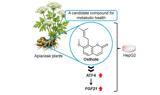
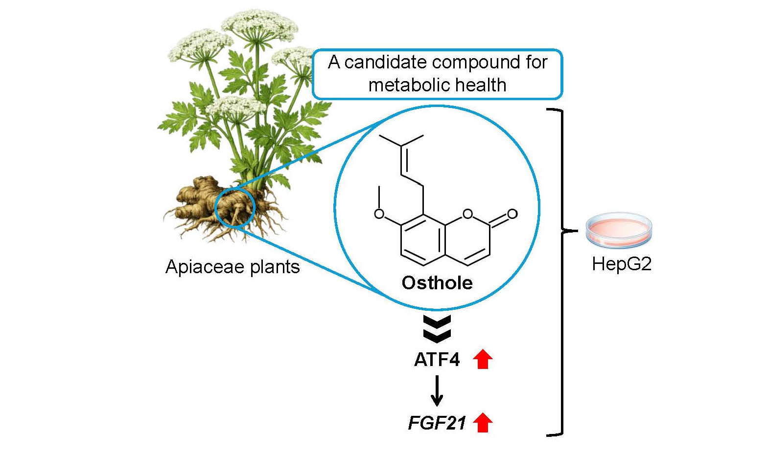
Osthole is a natural coumarin derivative found in several medicinal plants, including Cnidium monnieri and Angelica pubescens. It has been studied for its various biological properties, such as anti-inflammatory, neuroprotective, osteogenic, cardioprotective, antimicrobial, and antiparasitic effects. Osthole was found to induce Fibroblast growth factor 21 (FGF21) expression. Among the known transcription factors that regulate FGF21 induction, activating transcription factor 4 (ATF4) expression was found to be upregulated by osthole. Additionally, as osthole induced ATF4 downstream gene expression, it was concluded that it activates ATF4 signaling. ATF4 knockdown significantly suppressed osthole-mediated induction of FGF21 expression. These findings suggest that osthole activates FGF21 expression via ATF4 activation.

Graphical Abstract

1. Introduction

Metabolically-dysfunction-associated fatty liver disease (MAFLD) affects approximately 20–30% of the global population, making it one of the most prevalent causes of chronic liver disease [1]. Among patients with metabolic dysfunction-associated steatohepatitis (MASH), nearly 50–60% experience comorbidities such as diabetes mellitus, cardiovascular disease, and hyperlipidemia [1]. MAFLD and MASH both show strong correlations with conditions like insulin resistance and obesity [2]. MAFLD is recognized as a “multi-hit” condition [3], resulting from a complex interplay of mechanisms including insulin resistance, oxidative stress, mitochondrial dysfunction, endoplasmic reticulum stress, and apoptosis. Gaining a deeper understanding of these interconnected processes may offer valuable insights into MAFLD progression.
The fruits of Cnidium monnieri and Angelica pubescens have long been utilized in traditional Chinese medicine. Osthol, a key bioactive compound extracted from Cnidium monnieri and Angelica pubescens, exhibits both anti-inflammatory and hepatic fat-oxidizing properties. Osthole not only reduced fasting blood glucose levels and hepatic fat accumulation but also improved insulin resistance in rats with diet-induced obesity [4]. Osthole reduced the hepatic fat level by activating the hepatic gene expression of peroxisome proliferator-activated receptor α (PPARα) and PPARγ in rats [5]. In an alcoholic fatty liver disease mouse model, osthole activated superoxide dismutase, resulting in reduced oxidative stress [6].
FGF21 is primarily produced in the liver and released into the bloodstream, where it influences various peripheral tissues to regulate systemic nutrient metabolism. It reduces plasma glucose levels by increasing glucose uptake in adipose tissue [7]. It also plays a crucial role in regulating lipid metabolism and reducing lipid accumulation in adipose tissue through several mechanisms. It promotes fatty acid oxidation in adipose tissue, shifting metabolism toward energy expenditure rather than lipid storage [8]. It activates thermogenesis by inducing thermogenic gene expression and white adipose tissue browning, thereby increasing the levels of peroxisome proliferator-activated receptor gamma coactivator 1α (PGC-1α) [8]. FGF21 also plays a significant role in regulating food intake and macronutrient preference. FGF21 reduces the consumption of sweet foods and alcohol while boosting protein intake [9]. FGF21 is a key metabolic hormone that has been shown to ameliorate various lifestyle-related diseases, including obesity, type 2 diabetes, dyslipidemia, and fatty liver disease [10].
FGF21 improves the physiological characteristics of some mouse models of type 2 diabetes [7,11]; therefore, it could be a potential therapeutic target for metabolic diseases. FGF21 gene expression is regulated by certain transcription factors. It is upregulated by PPARα [12] and cyclic adenosine monophosphate-responsive element-binding protein H (CREBH) [13] in the fasted state and by carbohydrate-responsive element-binding protein (ChREBP) [14] in the fed state. In addition, nuclear factor erythroid 2-related factor 2 (NRF2) [15] and endoplasmic reticulum (ER) stress transcription factors, including activating transcription factor 4 (ATF4) [16], ATF6 [17], and X-box binding protein 1s (XBP1s) [18], increase Fgf21 expression.
ATF4, a key regulator of the Integrated Stress Response (ISR), plays a crucial role in ER stress adaptation [19]. ISR is triggered by various stressors, including amino acid deprivation, oxidative stress, and ER stress [19]. ATF4 influences lipid metabolism, amino acid transport, and mitochondrial function [19]. phosphorylated protein kinase RNA-like ER kinase (PERK) phosphorylates eukaryotic initiation factor 2α (eIF2α), leading to ATF4 induction [20]. ATF4 expression is upregulated by NRF2 [21], transcription factor E3 (TFE3), and transcription factor EB (TFEB) [22], ATF4 expression is downregulated by CCAAT/enhancer-binding protein β (C/EBPβ) [23,24].
This study aimed to identify an activator of FGF21 in herbal medicines and their constituent components. We have identified osthole as a potential activator of FGF21 expression in HepG2 cells.

2. Results

2.1. Osthole Induces FGF21 Expression in HepG2 Cells

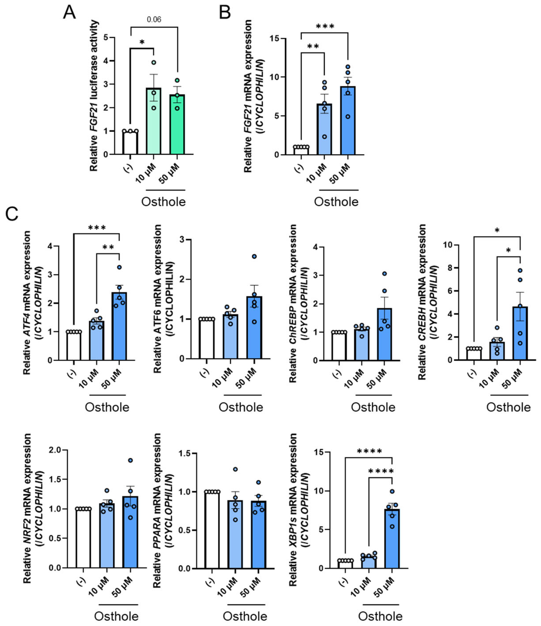
Since osthole has been reported to activate PPARα, a potential activator of FGF21, we hypothesized that it enhances FGF21 expression by activating PPARα. To investigate the effects of osthole on FGF21 expression, we conducted a luciferase assay using pGL3-FGF21-transfected HepG2 cells, which belong to a human hepatoma cell line. As expected, osthole activated FGF21 luciferase activity at concentrations of 10 µM and 50 µM after 24 h compared to the control group (Figure 1A). Additionally, qPCR analyses confirmed that osthole significantly increased FGF21 mRNA levels in HepG2 cells at concentrations of 10 µM and 50 µM after 24 h compared to the control group (Figure 1B). Osthole increased FGF21 expression more efficiently than fenofibrate, a positive control for FGF21 activation (Figure S1). These findings suggest that osthole acts as a potential activator of FGF21 expression.

2.2. ATF4 Increased FGF21 Expression in Response to Osthole

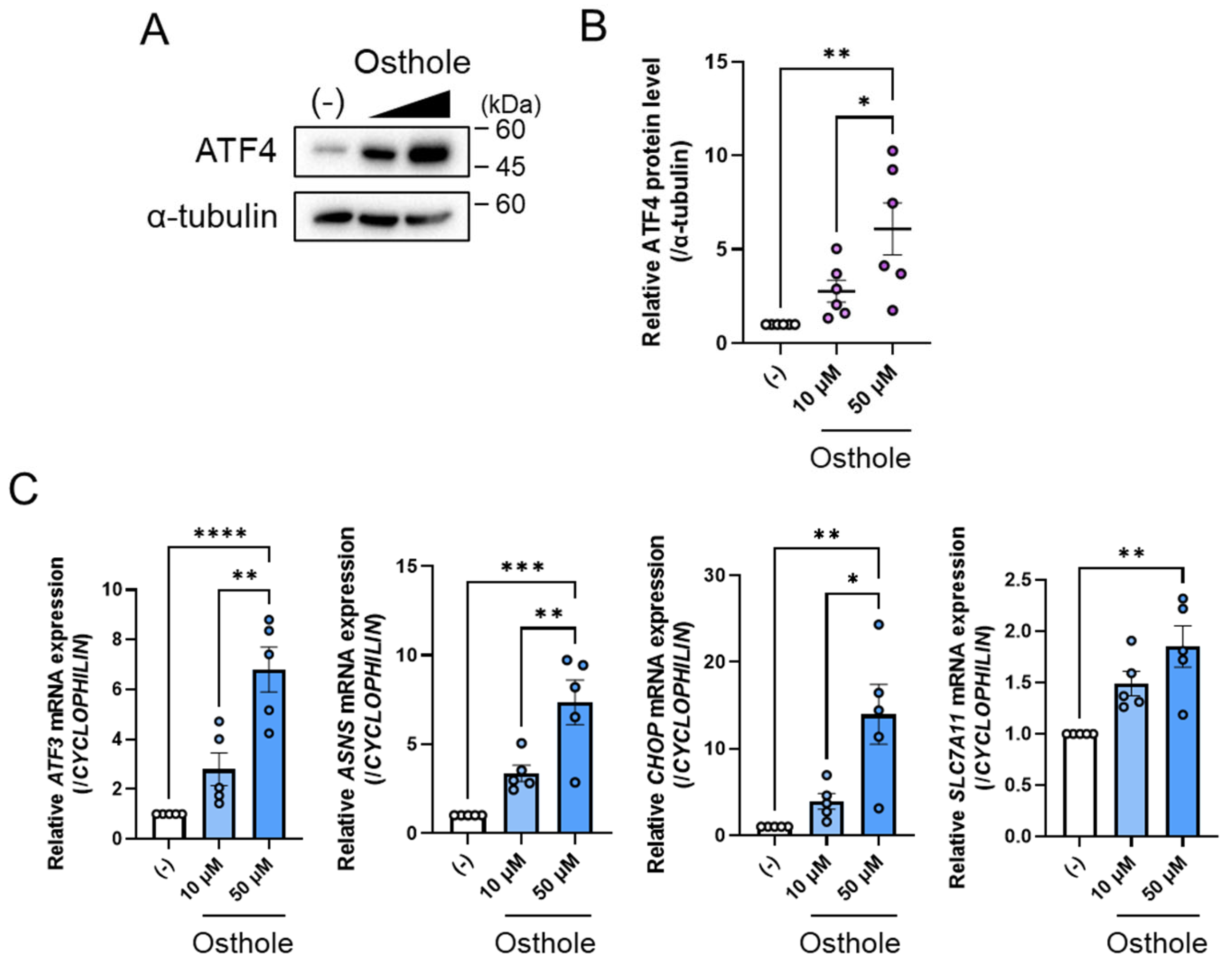
The expression of Fgf21 is upregulated by certain transcription factors, including ATF4, ATF6, ChREBP, CREBH, NRF2, PPARα, and XBP1s. To identify a transcription factor that controls osthole-mediated FGF21 expression, we examined the gene expression of these factors. Fifty micromolar osthole significantly increased ATF4 expression compared to the control, and this increase tended to be dose-dependent (Figure 1C). Fifty micromolar osthole significantly increased the expression of ATF4, CREBH, and XBP1s compared to the control group (Figure 1C). However, the expression of ATF6 and ChREBP tended to increase with osthole administration, but this increase was not statistically significant (Figure 1C). NRF2 and PPARA were unchanged (Figure 1C). In this study, we focused on ATF4. We also confirmed that osthole increased ATF4 protein levels (Figure 2A). Consistent with this, 50 μM osthole increased the expression of some of ATF4’s typical target genes, such as ATF3, asparagine synthetase (ASNS), CCAAT/enhancer-binding protein homologous protein (CHOP), and solute carrier family 7 member 11 (SLC7A11) [25], and these increments tended to be dose-dependent (Figure 2B). Taken together, these results suggest that ATF4 is an effective activator of osthole-mediated FGF21 expression in HepG2 cells.

2.3. Osthole Does Not Change the Molecules to Activate ATF4 Expression

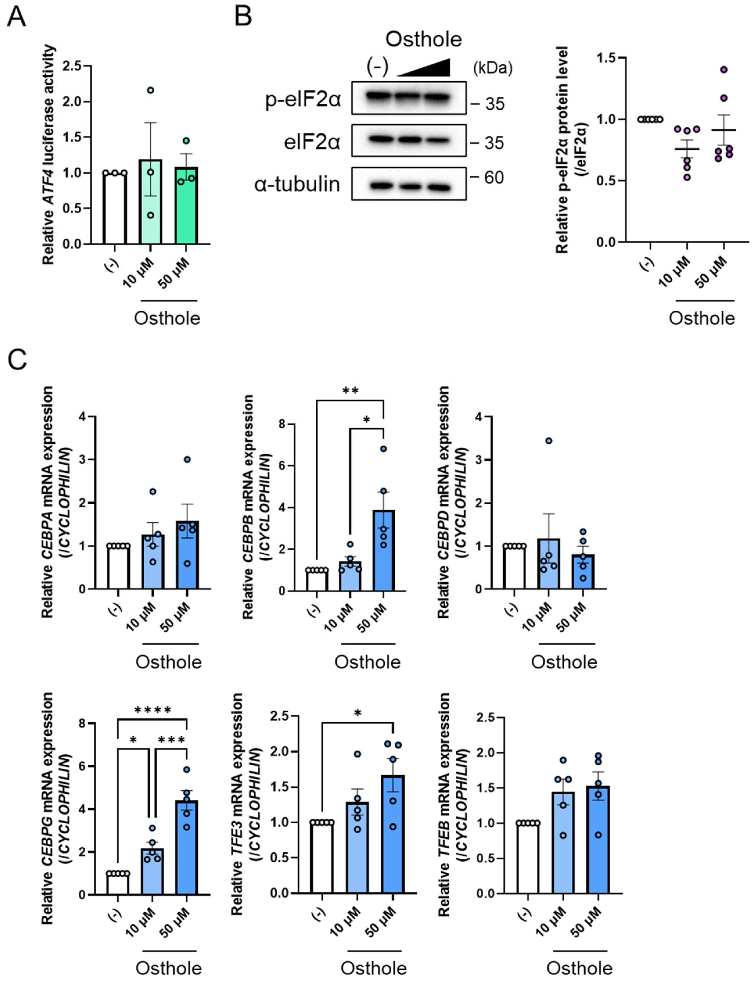
As osthole was found to increase the gene expression and protein levels of ATF4 (Figure 1C and Figure 2A), it was hypothesized that it might enhance ATF4 promoter activity. To test this theory, we performed a luciferase assay using a pGL3-ATF4 vector containing the ATF4 promoter region from −0.5 kbp to −100 bp. However, osthole was found not to affect ATF4 promoter activity (Figure 3A). Consequently, we investigated the factors that might regulate ATF4 expression in response to osthole. The PERK-eIF2α signaling pathway increases ATF4 protein levels [26]. ER stress induces PERK, which activates eIF2α by phosphorylating it and eventually inducing ATF4 translation. Therefore, we determined the levels of phospho-eIF2α (p-eIF2α) and total eIF2α by Western blotting. We found that osthole did not alter the levels of p-eIF2α (Figure 3B), indicating that osthole does not affect PERK-eIF2α signaling pathway. Next, we examined transcription factors controlling ATF4 expression, such as the C/EBP family, NRF2, TFE3, and TFEB. The expression of CEBPA and CEBPD was not apparently regulated by osthole (Figure 3C). However, CEBPB expression increased significantly with 50 μM osthole compared to the control group (Figure 3C). Moreover, osthole increased CEBPG expression in a dose-dependent manner (Figure 3C). Treatment with 50 μM osthole significantly upregulated TFE3 expression compared with the control group, whereas NRF2 and TFEB expression remained unchanged (Figure 3C). Taken together, these results indicate that TFE3 may function as a potential mediator of ATF4-FGF21 signaling. Nevertheless, the induction of ATF4 by osthole cannot be accounted for by any of the currently known ATF4-regulating transcription factors.

2.4. ATF4 Knockdown Suppresses Osthole-Induced Fgf21 Expression

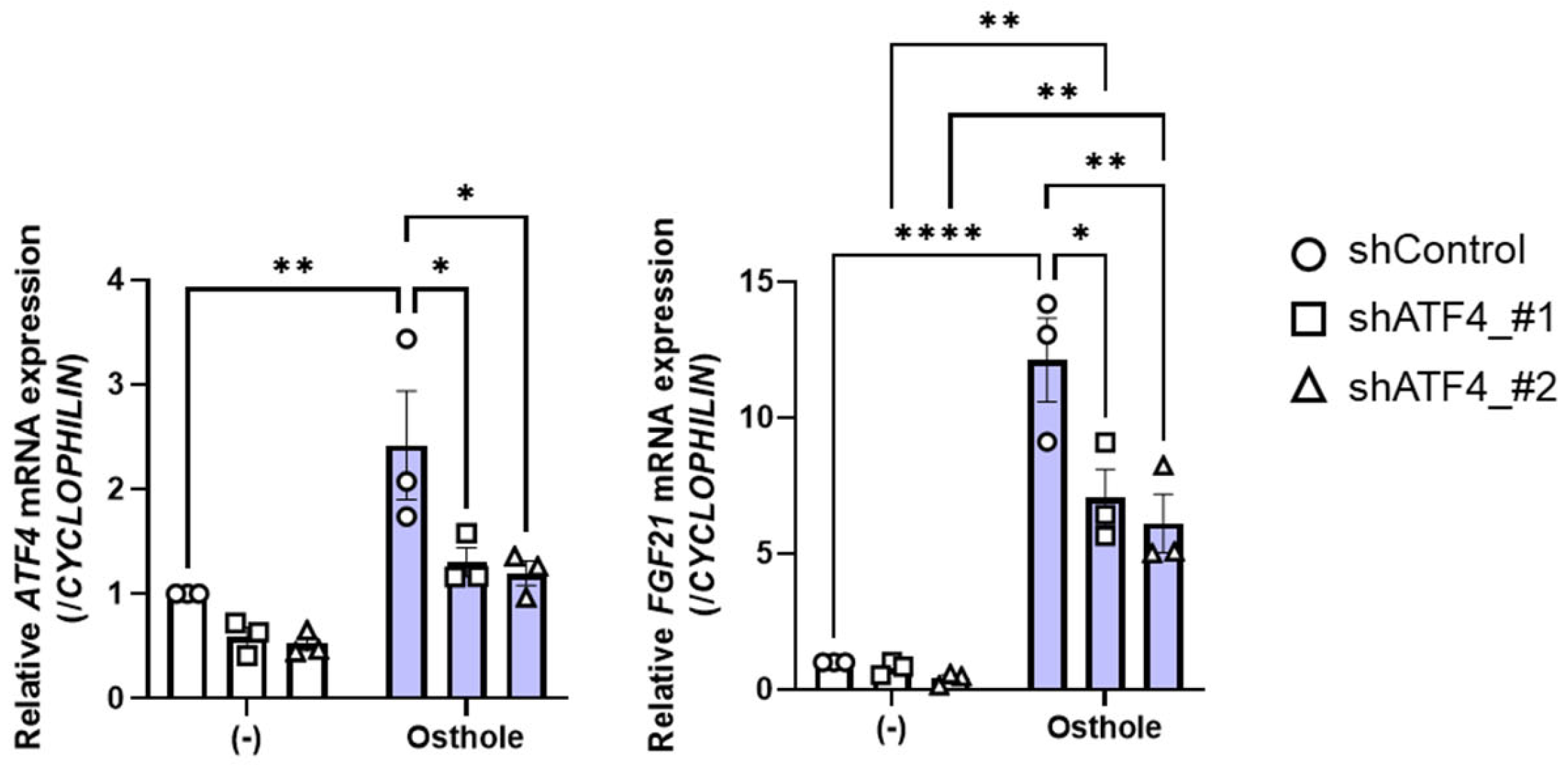
To confirm the necessity of ATF4 for osthole-induced FGF21 expression, we performed a loss-of-function analysis in HepG2 cells by knocking down ATF4 using shRNA (shATF4). The cells were transfected with two types of shRNA vector against ATF4 and then incubated for 24 h before being treated with 50 μM osthole for a further 24 h. We confirmed that both types of shATF4 significantly suppressed ATF4 expression with and without osthole treatment (Figure 4). ATF4 knockdown efficiently suppressed osthole-induced FGF21 expression (Figure 4), demonstrating that ATF4 plays a critical role in osthole’s effects on FGF21 expression.

3. Discussion

FGF21 has been a major focus in metabolic research due to its potential therapeutic applications. Scientists are actively investigating compounds that can enhance FGF21 expression to improve metabolic health, particularly in conditions like obesity, diabetes, and muscle metabolism. Our study revealed that osthole, which is included in Cnidium monnieri, would be a novel compound to increase FGF21 expression in HepG2 cells, which belong to a human hepatoma cell line, by activating ATF4.
In this study, we isolated osthole from the WAKANYAKU library as a candidate to increase FGF21 expression. Osthole reduces triglyceride (TG) and free fatty acid levels in serum and hepatic tissues in high-fat-induced fatty liver rat and quail models or alcoholic-induced fatty liver rat and mouse models [27,28,29]. These effects are due to increments in the levels of mRNA and protein of PPARα and PPARγ in the liver, which are caused by osthole [30,31]. In cultured hepatocytes, osthole suppresses oleic acid/lipopolysaccharide-induced lipid accumulation and the inflammatory response by activating PPARα [32]. Taken together, osthole is thought to be a dual PPARα/γ agonist. However, in our study, osthole did not increase PPARα expression; therefore, we could not confirm that osthole activates PPARα.
PPARα [12], along with CREBH [13], is an important regulator of FGF21 mRNA, which is elevated in the liver during fasting. PPARα was not upregulated by osthole whereas CREBH was upregulated. ChREBP, which is also regulated during feeding [14], was unchanged. FGF21 expression is induced by ATF4 under conditions of protein restriction and amino acid deficiency. Although ATF4 belongs to this same transcription factor family, other ER stress–responsive transcription factors, including CREBH [33], ATF6 and XBP1s [18], can also induce FGF21 expression. In this study, osthole was found to upregulate the expression of CREBH, ATF4, and XBP1s, suggesting that osthole may activate ER stress signaling, thereby contributing to increased FGF21 expression. Osthole was found to increase the gene expression of ATF3 and ASNS, which are target genes of ATF4. Therefore, it is important to note that osthole activates ATF4 signaling. Osthole-induced FGF21 expression was suppressed by ATF4 knockdown, confirming ATF4’s essential role in osthole’s mechanism of action. Treatment with 50 μM osthole increased the expression of ATF4, CREBH, and XBP1s. These results imply that osthole-induced FGF21 expression requires the activation of several transcription factors. However, ATF4 knockdown experiments revealed that the reduction in ATF4 expression was comparable to the decrease in osthole-induced FGF21 expression. If so, a complete knockdown of ATF4 would be expected to almost entirely abolish osthole-induced FGF21 expression. This outcome would suggest that ATF4 is essential for this process, while CREBH and XBP1s are dispensable, which is contradictory to their observed induction. Therefore, we propose that the osthole–FGF21 signaling pathway is initiated by ATF4 and subsequently amplified through a positive feedback loop involving the activation of CREBH and XBP1s.
ATF4 translation is known to be activated by PERK–eIF2α signaling [15]. However, osthole did not alter the levels of phosphorylated eIF2α, indicating that osthole-induced ATF4 activation is not mediated through this pathway. At the transcriptional level, TFE3, TFEB, and NRF2 are reported activators of ATF4 expression [21,22], whereas the C/EBP family functions as repressors [23,24]. The observed changes in the expression of these genes alone cannot fully explain the mechanism. Although osthole increased TFE3 expression, no studies have reported that TFE3 directly regulates FGF21 expression. Thus, the precise mechanism by which osthole upregulates ATF4 remains unclear. TFE3 is a master regulator of autophagy [34], and given that ATF4 controls amino acid metabolism, we speculate that there may be a functional relationship between them. Further studies will be necessary to clarify whether TFE3 serves as a key mediator of ATF4 upregulation. Importantly, TFE3-mediated activation of autophagy contributes to the improvement of lifestyle-related diseases. In addition, TFE3 enhances insulin signaling and suppresses lipid synthesis [35], thereby ameliorating lifestyle-related diseases. These effects of TFE3 may underlie, at least in part, the therapeutic potential of osthole in lifestyle-related diseases.
Previous studies have reported that several herbal-derived compounds can induce FGF21 expression through distinct signaling pathways. For instance, berberine, a bioactive constituent of Coptis chinensis and Berberis spp. [36], as well as Kursi Wufarikun Ziyabit, a traditional herbal formulation containing Geranium collinum and Hypericum scabrum [37], enhance FGF21 expression via activation of AMP-activated protein kinase. In addition, wogonin has been shown to induce FGF21 through ATF4-dependent signaling [38]. In this context, our study identifies osthole, a bioactive component of herbal medicines, as a novel activator of FGF21, thereby expanding the repertoire of phytochemicals capable of modulating FGF21 and providing new insight into the diversity of upstream regulatory mechanisms.
In conclusion, although osthole has been reported to activate PPARα in the liver of mice and in rat hepatocytes [31,32], its role in regulating FGF21 expression had remained unclear. Our data demonstrates that osthole does not induce PPARA expression in HepG2 cells but instead stimulates FGF21 expression through an ATF4-dependent pathway. Using HepG2 cells as a model of hepatic FGF21 regulation, we identify a previously unrecognized mechanism by which osthole activates FGF21. Given the established metabolic benefits of FGF21, these findings highlight osthole as a promising candidate for the prevention and treatment of lifestyle-related metabolic diseases.

4. Materials and Methods

4.1. Cell Culture

HepG2 cells (RGB1648) were provided by RIKEN BioResource Research Center, Ibaraki, Japan. HepG2 cells were cultured at 37 °C in a 5% CO2 environment in DMEM medium (Nacalai Tesque, 08458-45, Kyoto, Japan) supplemented with 10% fetal bovine serum (CORNING, 35-079-CV, Corning, NY, USA), 100 U/mL penicillin, and 100 μg/mL streptomycin (Nacalai Tesque, 09367-34, Kyoto, Japan). Cells were treated with 10 µM or 50 µM osthole (Tokyo Chemical Industry Co., Ltd., O0426, Tokyo, Japan).

4.2. Plasmids

pGL3-FGF21 contained −2 kbp to −40 bp of the mouse Fgf21 promoter [13]. pGL3-ATF4 contained −0.5 kbp to −100 bp of the mouse Atf4 promoter [38]. Control shRNA: shControl (pDNA(VB010000-0023jze): 5′-CCTAAGGTTAAGTCGCCCTCG-3′) and human ATF4 shRNA vectors: shATF4 1 (pDNA(VB900147-1829hks) 5′-CATGATCCCTCAGTGCATAAA-3′ and shATF4 2 pDNA(VB900147-1830wwp) 5′-CCTAGGTCTCTTAGATGATTA-3′) were purchased from Vector Builder (Chicago, IL, USA). These plasmids were transfected into HepG2 cells with FuGENE6 (Promega, E2691, Madison, WI, USA).

4.3. Luciferase Analysis

HepG2 cells were transfected with the indicated luciferase vector and pRL-CMV (Promega, E2261, Madison, WI, USA), as a reference, using FuGENE6 (Promega, E2691, Madison, WI, USA). After 24 h of incubation, cells were treated with 10 µM or 50 µM osthole for 24 h. Firefly and Renilla luciferase activity was measured using the Dual-Luciferase® Reporter Assay System (Promega, E1910, Madison, WI, USA). Firefly luciferase activities were normalized to Renilla luciferase activities.

4.4. Quantitative Polymerase Chain Reaction (qPCR)

Total RNA was isolated from collected cells using Sepasol®-RNA I Super G (Nacalai Tesque, 09379-55, Kyoto, Japan) as per the manufacturer’s protocol. All samples passed the RNA quality control as assessed on NanoDrop Lite Spectrophotometer (Thermo Fisher Scientific, Waltham, MA, USA). Total RNA (1 µg) was reverse-transcribed using ReverTra Ace® qPCR RT Master Mix (Toyobo, FSQ-201, Osaka, Japan). qPCR was performed on a CFX Connect Real-Time PCR Detection System (Bio-Rad, Hercules, CA, USA) using THUNDERBIRD® Next SYBR® qPCR Mix (Toyobo, QPX-201, Osaka, Japan). Samples were quantified via the 2−ΔΔCt method and normalized to Cyclophilin levels to quantify the relative mRNA expression. The qPCR primer sequences are outlined in Table 1.

4.5. Western Blotting

Cells were lysed in the lysis buffer containing 10 mM Tris-HCl, 100 mM NaCl, 1 mM EDTA, 1 mM EGTA, 1 mM NaF, 20 mM Sodium pyrophosphate, 2 mM Sodium orthovanadate, 0.1% SDS, 0.5% Sodium deoxycholate, 1% Triton X-100, 10% Glycerol, and Complete Protease Inhibitor (Roche, 4693116001, Basel, Switzerland). The total cell lysates were subjected to 10% sodium dodecyl sulfate-polyacrylamide gel electrophoresis (SDS-PAGE) and transferred to Immobilon-P PVDF membranes (Millipore, IPVH00010, Burlington, MA, USA). The membranes were blocked with 5% skim milk in TBS-T. The membranes were incubated with anti-ATF4 (Santa Cruz, sc-390063, 1:000, Dallas, TX, USA), anti-phosho-eIF2α (Cell Signaling Technologies, 3298, 1:1000, Danvers, MA, USA), anti-eIF2α (Cell Signaling Technologies, 5324, 1:1000, Danvers, MA, USA), and anti-α-tubulin (Santa Cruz Biotechnology, sc-32293, 1:1000, Dallas, TX, USA) antibodies. Alpha-tubulin was used as an internal control. After washing, the membranes were incubated with horseradish peroxidase-conjugated mouse IgG (Cell Signaling Technologies, 7076, 1:5000, Danvers, MA, USA) and rabbit IgG (Cell Signaling Technologies, 7074, 1:5000, Danvers, MA, USA). The immunoreactive bands were detected by ChemiDoc XRS+ (BioRad, Hercules, CA, USA) using ImmunoStar LD (WAKO, 290-69904, Osaka, Japan). The immunoreactive band intensity was quantified using Image Lab software 6.1 (BioRad, Hercules, CA, USA).

4.6. Statistical Analysis

Continuous data were expressed as the mean ± standard error (SE), while categorical data were expressed as frequencies with percentages. Comparisons of continuous data between two groups were performed using the unpaired Student’s t-test. Comparisons of continuous data among multiple groups were performed using the one-way ANOVA, followed by Tukey’s post hoc test. All statistical analyses were performed using GraphPad Prism 10 (GraphPad Prism Software, San Diego, CA, USA). p-values < 0.05 were considered statistically significant.

5. Conclusions

In this study, we identified osthole as a phytochemical that induces FGF21 expression, providing a basis for exploring its potential therapeutic relevance to lifestyle-related metabolic diseases. Although our results demonstrate that osthole primarily activates FGF21 expression through an ATF4-dependent pathway, potential interactions with other transcription factors, including CREBH and XBP1s, were suggested but not mechanistically resolved. In particular, the molecular basis by which osthole, together with ATF4, activates CREBH and XBP1s remains unclear. Elucidating the cooperative or hierarchical regulatory mechanisms among these transcription factors will be an important focus of future studies. In addition, the ATF4–FGF21 axis was mainly examined using HepG2 cells, and it remains to be determined whether a similar regulatory mechanism operates in other hepatic cell models or in extrahepatic tissues. Finally, this study was largely based on in vitro analyses. Further investigation is required to determine whether osthole induces hepatic FGF21 expression and improves metabolic phenotypes in lifestyle disease models in vivo, as well as its effects on other tissues and potential adverse effects.

Supplementary Materials

The following supporting information can be downloaded at https://www.mdpi.com/article/10.3390/ijms27021003/s1.

Author Contributions

Conceptualization, M.A. and Y.N.; methodology, M.A. and Y.N.; investigation, A.T., M.A., T.Y., R.K., I.T., K.S., Y.T. and Y.N.; writing—original draft preparation, M.A. and Y.N.; writing—review and editing, M.A. and Y.N.; visualization, M.A.; supervision, T.M., H.S. (Hirohito Sone) and H.S. (Hitoshi Shimano); project administration, Y.N.; funding acquisition, Y.N. All authors have read and agreed to the published version of the manuscript.

Funding

This work was supported by JSPS KAKENHI Grant Number 21H04861 (to Y.N.), 2025 Director Leadership Expenses, Institute of Natural Medicine, University of Toyama (to A.T.).

Institutional Review Board Statement

Not applicable.

Informed Consent Statement

Not applicable.

Data Availability Statement

The original contributions presented in this study are included in the article and Supplementary Materials. Further inquiries can be directed to the corresponding author.

Acknowledgments

This research was performed by utilizing the INM deposited WAKANYAKU library, Institute of Natural Medicine, University of Toyama in 2020.

Conflicts of Interest

The authors declare no conflicts of interest.

Abbreviations

The following abbreviations are used in this manuscript:
FGF21Fibroblast growth factor 21
ATF4activating transcription factor 4
MAFLDmetabolic dysfunction-associated fatty liver disease
MASHmetabolic dysfunction-associated steatohepatitis
PPARperoxisome proliferator-activated receptor
PGC-1αperoxisome proliferator-activated receptor gamma coactivator 1-alpha
CREBHcyclic adenosine monophosphate-responsive element-binding protein H
ChREBPcarbohydrate-responsive element-binding protein
NRF2nuclear factor erythroid 2-related factor 2
ERendoplasmic reticulum
XBP1sX-box binding protein 1s
ISRIntegrated Stress Response
PERKPKR-like ER kinase
TFE3transcription factor E3
CEBPBCCAAT/enhancer-binding protein β
ASNSasparagine synthetase
CHOPCCAAT/enhancer-binding protein homologous protein
SLC7A11solute carrier family 7 member 11

References

  1. Lazo, M.; Hernaez, R.; Bonekamp, S.; Kamel, I.R.; Brancati, F.L.; Guallar, E.; Clark, J.M. Non-alcoholic fatty liver disease and mortality among US adults: Prospective cohort study. BMJ 2011, 343, d6891. [Google Scholar] [CrossRef]
  2. Targher, G.; Byrne, C.D. Clinical Review: Nonalcoholic fatty liver disease: A novel cardiometabolic risk factor for type 2 diabetes and its complications. J. Clin. Endocrinol. Metab. 2013, 98, 483–495. [Google Scholar] [CrossRef] [PubMed]
  3. Tilg, H.; Moschen, A.R. Evolution of inflammation in nonalcoholic fatty liver disease: The multiple parallel hits hypothesis. Hepatology 2010, 52, 1836–1846. [Google Scholar] [CrossRef] [PubMed]
  4. Qi, Z.; Xue, J.; Zhang, Y.; Wang, H.; Xie, M. Osthole ameliorates insulin resistance by increment of adiponectin release in high-fat and high-sucrose-induced fatty liver rats. Planta Med. 2011, 77, 231–235. [Google Scholar] [CrossRef]
  5. Zhang, Y.; Xie, M.L.; Xue, J.; Gu, Z.L. Osthole regulates enzyme protein expression of CYP7A1 and DGAT2 via activation of PPARalpha/gamma in fat milk-induced fatty liver rats. J. Asian Nat. Prod. Res. 2008, 10, 807–812. [Google Scholar] [CrossRef] [PubMed]
  6. Zhang, J.; Xue, J.; Wang, H.; Zhang, Y.; Xie, M. Osthole improves alcohol-induced fatty liver in mice by reduction of hepatic oxidative stress. Phytother. Res. 2011, 25, 638–643. [Google Scholar] [CrossRef]
  7. Kharitonenkov, A.; Shiyanova, T.L.; Koester, A.; Ford, A.M.; Micanovic, R.; Galbreath, E.J.; Sandusky, G.E.; Hammond, L.J.; Moyers, J.S.; Owens, R.A.; et al. FGF-21 as a novel metabolic regulator. J. Clin. Investig. 2005, 115, 1627–1635. [Google Scholar] [CrossRef]
  8. Fisher, F.M.; Kleiner, S.; Douris, N.; Fox, E.C.; Mepani, R.J.; Verdeguer, F.; Wu, J.; Kharitonenkov, A.; Flier, J.S.; Maratos-Flier, E.; et al. FGF21 regulates PGC-1alpha and browning of white adipose tissues in adaptive thermogenesis. Genes Dev. 2012, 26, 271–281. [Google Scholar] [CrossRef]
  9. Talukdar, S.; Owen, B.M.; Song, P.; Hernandez, G.; Zhang, Y.; Zhou, Y.; Scott, W.T.; Paratala, B.; Turner, T.; Smith, A.; et al. FGF21 Regulates Sweet and Alcohol Preference. Cell Metab. 2016, 23, 344–349. [Google Scholar] [CrossRef]
  10. Yang, M.; Liu, C.; Jiang, N.; Liu, Y.; Luo, S.; Li, C.; Zhao, H.; Han, Y.; Chen, W.; Li, L.; et al. Fibroblast growth factor 21 in metabolic syndrome. Front. Endocrinol. 2023, 14, 1220426. [Google Scholar] [CrossRef]
  11. Xu, J.; Lloyd, D.J.; Hale, C.; Stanislaus, S.; Chen, M.; Sivits, G.; Vonderfecht, S.; Hecht, R.; Li, Y.S.; Lindberg, R.A.; et al. Fibroblast growth factor 21 reverses hepatic steatosis, increases energy expenditure, and improves insulin sensitivity in diet-induced obese mice. Diabetes 2009, 58, 250–259. [Google Scholar] [CrossRef]
  12. Inagaki, T.; Dutchak, P.; Zhao, G.; Ding, X.; Gautron, L.; Parameswara, V.; Li, Y.; Goetz, R.; Mohammadi, M.; Esser, V.; et al. Endocrine regulation of the fasting response by PPARalpha-mediated induction of fibroblast growth factor 21. Cell Metab. 2007, 5, 415–425. [Google Scholar] [CrossRef]
  13. Nakagawa, Y.; Satoh, A.; Yabe, S.; Furusawa, M.; Tokushige, N.; Tezuka, H.; Mikami, M.; Iwata, W.; Shingyouchi, A.; Matsuzaka, T.; et al. Hepatic CREB3L3 controls whole-body energy homeostasis and improves obesity and diabetes. Endocrinology 2014, 155, 4706–4719. [Google Scholar] [CrossRef]
  14. Iizuka, K.; Takeda, J.; Horikawa, Y. Glucose induces FGF21 mRNA expression through ChREBP activation in rat hepatocytes. FEBS Lett. 2009, 583, 2882–2886. [Google Scholar] [CrossRef]
  15. Yu, Y.; He, J.; Li, S.; Song, L.; Guo, X.; Yao, W.; Zou, D.; Gao, X.; Liu, Y.; Bai, F.; et al. Fibroblast growth factor 21 (FGF21) inhibits macrophage-mediated inflammation by activating Nrf2 and suppressing the NF-kappaB signaling pathway. Int. Immunopharmacol. 2016, 38, 144–152. [Google Scholar] [CrossRef]
  16. De Sousa-Coelho, A.L.; Marrero, P.F.; Haro, D. Activating transcription factor 4-dependent induction of FGF21 during amino acid deprivation. Biochem. J. 2012, 443, 165–171. [Google Scholar] [CrossRef] [PubMed]
  17. Chen, X.; Zhang, F.; Gong, Q.; Cui, A.; Zhuo, S.; Hu, Z.; Han, Y.; Gao, J.; Sun, Y.; Liu, Z.; et al. Hepatic ATF6 Increases Fatty Acid Oxidation to Attenuate Hepatic Steatosis in Mice Through Peroxisome Proliferator-Activated Receptor alpha. Diabetes 2016, 65, 1904–1915. [Google Scholar] [CrossRef]
  18. Jiang, S.; Yan, C.; Fang, Q.C.; Shao, M.L.; Zhang, Y.L.; Liu, Y.; Deng, Y.P.; Shan, B.; Liu, J.Q.; Li, H.T.; et al. Fibroblast growth factor 21 is regulated by the IRE1alpha-XBP1 branch of the unfolded protein response and counteracts endoplasmic reticulum stress-induced hepatic steatosis. J. Biol. Chem. 2014, 289, 29751–29765. [Google Scholar] [CrossRef] [PubMed]
  19. Ryoo, H.D. The integrated stress response in metabolic adaptation. J. Biol. Chem. 2024, 300, 107151. [Google Scholar] [CrossRef] [PubMed]
  20. Pakos-Zebrucka, K.; Koryga, I.; Mnich, K.; Ljujic, M.; Samali, A.; Gorman, A.M. The integrated stress response. EMBO Rep. 2016, 17, 1374–1395. [Google Scholar] [CrossRef]
  21. Afonyushkin, T.; Oskolkova, O.V.; Philippova, M.; Resink, T.J.; Erne, P.; Binder, B.R.; Bochkov, V.N. Oxidized phospholipids regulate expression of ATF4 and VEGF in endothelial cells via NRF2-dependent mechanism: Novel point of convergence between electrophilic and unfolded protein stress pathways. Arterioscler. Thromb. Vasc. Biol. 2010, 30, 1007–1013. [Google Scholar] [CrossRef]
  22. Martina, J.A.; Diab, H.I.; Brady, O.A.; Puertollano, R. TFEB and TFE3 are novel components of the integrated stress response. EMBO J. 2016, 35, 479–495. [Google Scholar] [CrossRef]
  23. Dey, S.; Savant, S.; Teske, B.F.; Hatzoglou, M.; Calkhoven, C.F.; Wek, R.C. Transcriptional repression of ATF4 gene by CCAAT/enhancer-binding protein beta (C/EBPbeta) differentially regulates integrated stress response. J. Biol. Chem. 2012, 287, 21936–21949. [Google Scholar] [CrossRef]
  24. Torrence, M.E.; MacArthur, M.R.; Hosios, A.M.; Valvezan, A.J.; Asara, J.M.; Mitchell, J.R.; Manning, B.D. The mTORC1-mediated activation of ATF4 promotes protein and glutathione synthesis downstream of growth signals. eLife 2021, 10, e63326. [Google Scholar] [CrossRef] [PubMed]
  25. Tanaka, S.; Mochizuki, Y.; Komeiji, Y.; Okiyama, Y.; Fukuzawa, K. Electron-correlated fragment-molecular-orbital calculations for biomolecular and nano systems. Phys. Chem. Chem. Phys. 2014, 16, 10310–10344. [Google Scholar] [CrossRef] [PubMed]
  26. Harding, H.P.; Novoa, I.; Zhang, Y.; Zeng, H.; Wek, R.; Schapira, M.; Ron, D. Regulated translation initiation controls stress-induced gene expression in mammalian cells. Mol. Cell 2000, 6, 1099–1108. [Google Scholar] [CrossRef] [PubMed]
  27. Song, F.; Xie, M.L.; Zhu, L.J.; Zhang, K.P.; Xue, J.; Gu, Z.L. Experimental study of osthole on treatment of hyperlipidemic and alcoholic fatty liver in animals. World J. Gastroenterol. 2006, 12, 4359–4363. [Google Scholar] [CrossRef]
  28. Zhang, Y.; Xie, M.L.; Zhu, L.J.; Gu, Z.L. Therapeutic effect of osthole on hyperlipidemic fatty liver in rats. Acta Pharmacol. Sin. 2007, 28, 398–403. [Google Scholar] [CrossRef]
  29. Sun, F.; Xie, M.L.; Zhu, L.J.; Xue, J.; Gu, Z.L. Inhibitory effect of osthole on alcohol-induced fatty liver in mice. Dig. Liver Dis. 2009, 41, 127–133. [Google Scholar] [CrossRef]
  30. Zhang, Y.; Xie, M.; Xue, J.; Gu, Z. Osthole improves fat milk-induced fatty liver in rats: Modulation of hepatic PPAR-alpha/gamma-mediated lipogenic gene expression. Planta Med. 2007, 73, 718–724. [Google Scholar] [CrossRef]
  31. Sun, F.; Xie, M.L.; Xue, J.; Wang, H.B. Osthol regulates hepatic PPAR alpha-mediated lipogenic gene expression in alcoholic fatty liver murine. Phytomedicine 2010, 17, 669–673. [Google Scholar] [CrossRef] [PubMed]
  32. Zhao, X.; Xue, J.; Xie, M. Osthole inhibits oleic acid/lipopolysaccharide-induced lipid accumulation and inflammatory response through activating PPARalpha signaling pathway in cultured hepatocytes. Exp. Gerontol. 2019, 119, 7–13. [Google Scholar] [CrossRef] [PubMed]
  33. Zhang, K.; Shen, X.; Wu, J.; Sakaki, K.; Saunders, T.; Rutkowski, D.T.; Back, S.H.; Kaufman, R.J. Endoplasmic reticulum stress activates cleavage of CREBH to induce a systemic inflammatory response. Cell 2006, 124, 587–599. [Google Scholar] [CrossRef]
  34. Martina, J.A.; Diab, H.I.; Lishu, L.; Jeong, A.L.; Patange, S.; Raben, N.; Puertollano, R. The nutrient-responsive transcription factor TFE3 promotes autophagy, lysosomal biogenesis, and clearance of cellular debris. Sci. Signal 2014, 7, ra9. [Google Scholar] [CrossRef]
  35. Nakagawa, Y.; Shimano, H.; Yoshikawa, T.; Ide, T.; Tamura, M.; Furusawa, M.; Yamamoto, T.; Inoue, N.; Matsuzaka, T.; Takahashi, A.; et al. TFE3 transcriptionally activates hepatic IRS-2, participates in insulin signaling and ameliorates diabetes. Nat. Med. 2006, 12, 107–113. [Google Scholar] [CrossRef]
  36. Sun, Y.; Xia, M.; Yan, H.; Han, Y.; Zhang, F.; Hu, Z.; Cui, A.; Ma, F.; Liu, Z.; Gong, Q.; et al. Berberine attenuates hepatic steatosis and enhances energy expenditure in mice by inducing autophagy and fibroblast growth factor 21. Br. J. Pharmacol. 2018, 175, 374–387. [Google Scholar] [CrossRef] [PubMed]
  37. Yasen, M.; Jiang, L.; Sharofova, M.; Xin, X.; Nuraliev, Y.; Ye, J.; Aisa, H.A. Herbal medicine of Kursi Wufarikun Ziyabit inhibits mitochondrial ATP production to activate AMPK in hepatocytes in therapy of type 2 diabetes. bioRxiv 2020. bioRxiv:2020.2009.2028.316349. [Google Scholar] [CrossRef]
  38. Yamada, Y.; Saito, H.; Araki, M.; Tsuchimoto, Y.; Muroi, S.I.; Suzuki, K.; Toume, K.; Kim, J.D.; Matsuzaka, T.; Sone, H.; et al. Wogonin, a Compound in Scutellaria baicalensis, Activates ATF4-FGF21 Signaling in Mouse Hepatocyte AML12 Cells. Nutrients 2022, 14, 3920. [Google Scholar] [CrossRef]
Figure 1. Osthole is identified as an activator of FGF21 expression in HepG2 cells. (A) The luciferase reporter assay for FGF21 promoter activity. HepG2 cells were co-transfected with pGL3-FGF21 and pRL-SV40 vectors. Twenty-four hours after transfection, cells were treated with 10 µM or 50 µM osthole or dimethyl sulfoxide (DMSO), which is indicated as (−), for an additional 24 h. n = 3 per group. (B) FGF21 gene expression levels in HepG2 cells treated with 10 µM or 50 µM osthole or DMSO, which is indicated as (−), for 24 h. n = 5 per group. (C) Expression of transcription factors involved in the regulation of FGF21 expression in HepG2 cells treated with 10 µM or 50 µM osthole or DMSO, which is indicated as (−), for 24 h. n = 5 per group. Data are presented as the mean ± SE. * p < 0.05; ** p < 0.01; *** p < 0.001; **** p < 0.0001. Statistical comparisons were performed using the one-way ANOVA, followed by Tukey’s post hoc test.
Ijms 27 01003 g001
Figure 2. Osthole effects on ATF4 signaling in HepG2 cells (A,B) ATF4 protein levels in HepG2 cells treated with 10 µM or 50 µM osthole or DMSO, which is indicated as (−), for 24 h (A). Band intensities were quantified. n = 6 per group (B). (C) Expression levels of ATF4-regulated genes in HepG2 cells treated with 10 µM or 50 µM osthole or DMSO, which is indicated as (−), for 24 h. n = 5 per group. Data are presented as the mean ± SE. * p < 0.05; ** p < 0.01; *** p < 0.001; **** p < 0.0001. Statistical comparisons were performed using the one-way ANOVA, followed by Tukey’s post hoc test.
Ijms 27 01003 g002
Figure 3. Osthole does not directly affect the promoter activity of ATF4 in HepG2 cells. (A) The luciferase reporter assay for ATF4 promoter activity. HepG2 cells were co-transfected with pGL3-ATF4 and pRL-SV40 vectors. Twenty-four hours after transfection, cells were treated with 10 µM or 50 µM osthole or DMSO, which is indicated as (−), for an additional 24 h. n = 3 per group. (B) Phosphorylated and total eIF2α protein levels in HepG2 cells treated with 10 µM or 50 µM osthole or DMSO, which is indicated as (−), for 24 h. Band intensities were quantified. n = 6 per group. (C) Expression levels of ATF4-regulated genes in HepG2 cells treated with 10 µM or 50 µM osthole or DMSO, which is indicated as (−), for 24 h. n = 5 per group. Data are presented as the mean ± SE. * p < 0.05; ** p < 0.01; *** p < 0.001; **** p < 0.0001. Statistical comparisons were performed using the one-way ANOVA, followed by Tukey’s post hoc test.
Ijms 27 01003 g003
Figure 4. Knockdown of ATF4 suppresses osthole-induced FGF21 expression. Expression levels of ATF4 and FGF21 in HepG2 cells transfected with shATF4 plasmids and treated with 50 µM osthole or DMSO, which is indicated as (−), for 24 h. n = 3 per group. Data are presented as the mean ± SE. * p < 0.05; ** p < 0.01; **** p < 0.0001. Statistical comparisons were performed using the one-way ANOVA, followed by Tukey’s post hoc test.
Ijms 27 01003 g004
Table 1. Primers used for real-time PCR analysis.
Gene NameFwdRv
ASNSCTGTGAAGAACAACCTCAGGATCAACAGAGTGGCAGCAACCAAGC
ATF3CGCTGGAATCAGTCACTGTCAGCTTGTTTCGGCACTTTGCAGCTG
ATF4CCAACAACAGCAAGGAGGATAGGTTCATCTGGCATGGTTTC
ATF6CAGGCAGTACCAACGCTTATGCCGCAGAACTCCAGGTGCTTGAAG
CEBPAAGGAGGATGAAGCCAAGCAGCTAGTGCGCGATCTGGAACTGCAG
CEBPBAGAAGACCGTGGACAAGCACAGCTCCAGGACCTTGTGCTGCGT
CEBPDTCCGGCAGTTCTTCAAGCAGCTGAGGTATGGGTCGTTGCTGAGT
CEBPGGCTTACAGCAGGTTCCTCAGCTCGTTGCCGATACTCGTCACTGT
CHOPGGTATGAGGACCTGCAAGAGGTCTTGTGACCTCTGCTGGTTCTG
ChREBPGCGTTTTGACCAGATGCGAGACCGTTGAAGGACTCAAACAGAGGC
CREBHCCTCTGTGACCATAGAACGGTGAGATTGCATC
FGF21ACTCCAGTCCTCTCCTAGTGGAGCGATCCATA
NRF2CACATCCAGTCAGAAACCAGTGGGGAATGTCTGCGCCAAAAGCTG
SLC7A11TCCTGCTTTGGCTCCATGAACGAGAGGAGTGTGCTTGCGGACAT
TFE3GATCATCAGCCTGGAGTCCAGTAGCAGATTCCCTGACACAGGCA
TFEBCCTGGAGATGACCAACAAGCAGTAGGCAGCTCCTGCTTCACCAC
XBP1sAGCTTTTACGAGAGAAAACTCAGCCTGCACCTGCTGCG
CYCLOPHILINGGCAAATGCTGGACCCAACACATGCTGGTCTTGCCATTCCTGGA
Disclaimer/Publisher’s Note: The statements, opinions and data contained in all publications are solely those of the individual author(s) and contributor(s) and not of MDPI and/or the editor(s). MDPI and/or the editor(s) disclaim responsibility for any injury to people or property resulting from any ideas, methods, instructions or products referred to in the content.