Beyond the Kidney: A Systematic Review of Extrarenal Manifestations in MAPKBP1-Related Nephronophthisis

 , , , , ,
  , , and
Abstract
1. Introduction
1.1. Identification of MAPKBP1 as a Nephronophthisis-Associated Gene
1.2. MAPKBP1 as a Scaffold Protein of the JNK Signaling Pathway
1.3. From a Cilia-Independent Model to a Cilia–JNK Signaling Defect
1.4. Rationale: The Emerging Clinical Spectrum of NPHP20
2. Materials and Methods
2.1. Reporting Guideline and Protocol
2.2. Search Strategy
2.3. Eligibility Criteria
- •
- the title or abstract clearly focuses on non-human models (e.g., animals, plants).
- •
- the title or abstract is written in languages other than English.
2.4. Selection Process
2.5. Data Collection Process and Data Items
2.6. Effect Measures, Synthesis and Statistical Analysis
2.7. Quality Assessment and Risk of Bias
- •
- The inability to determine whether a clinical manifestation was absent in a patient, or simply not reported or not investigated using a specific diagnostic exam. This limitation was taken into account when calculating the frequencies.
- •
- Application of a targeted panel instead of exome sequencing. The genetic testing strategy was defined for each report to assess whether other possible genetic diagnoses were excluded in relation to extrarenal manifestations.
- •
- Conflicting nomenclature and classification of genetic variants. All variants were converted to the most up-to-date nomenclature using the transcript NM_014994.3 (MANE Select) according to GRCh38. They were revised by two independent reviewers applying the ACMG criteria, to assess bias related to patients with uncertain or likely benign variants.
2.8. Inclusion of New Patient Data
3. Results
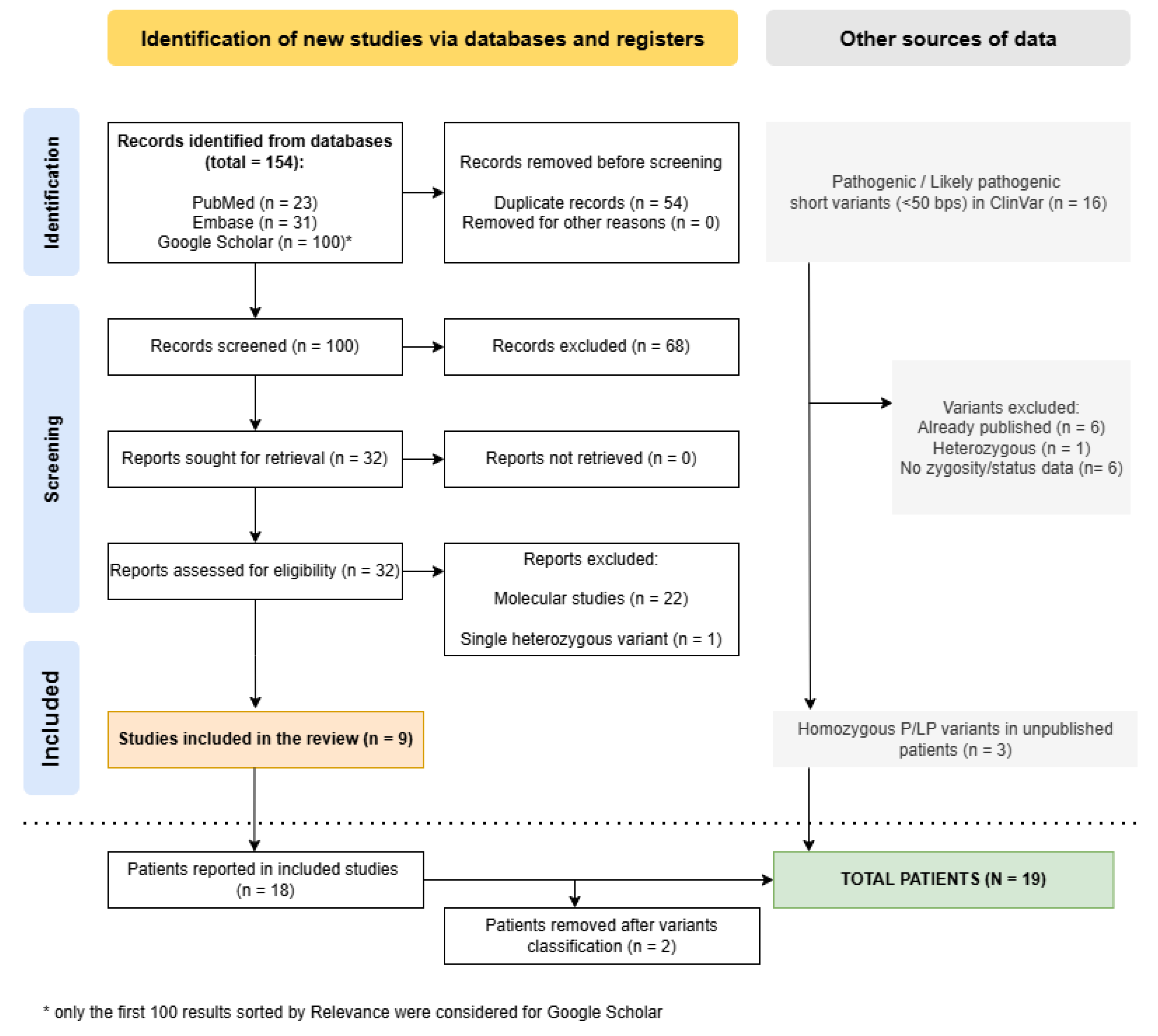
3.1. Study Selection
3.2. Results of Quality and Risk of Bias Assessment
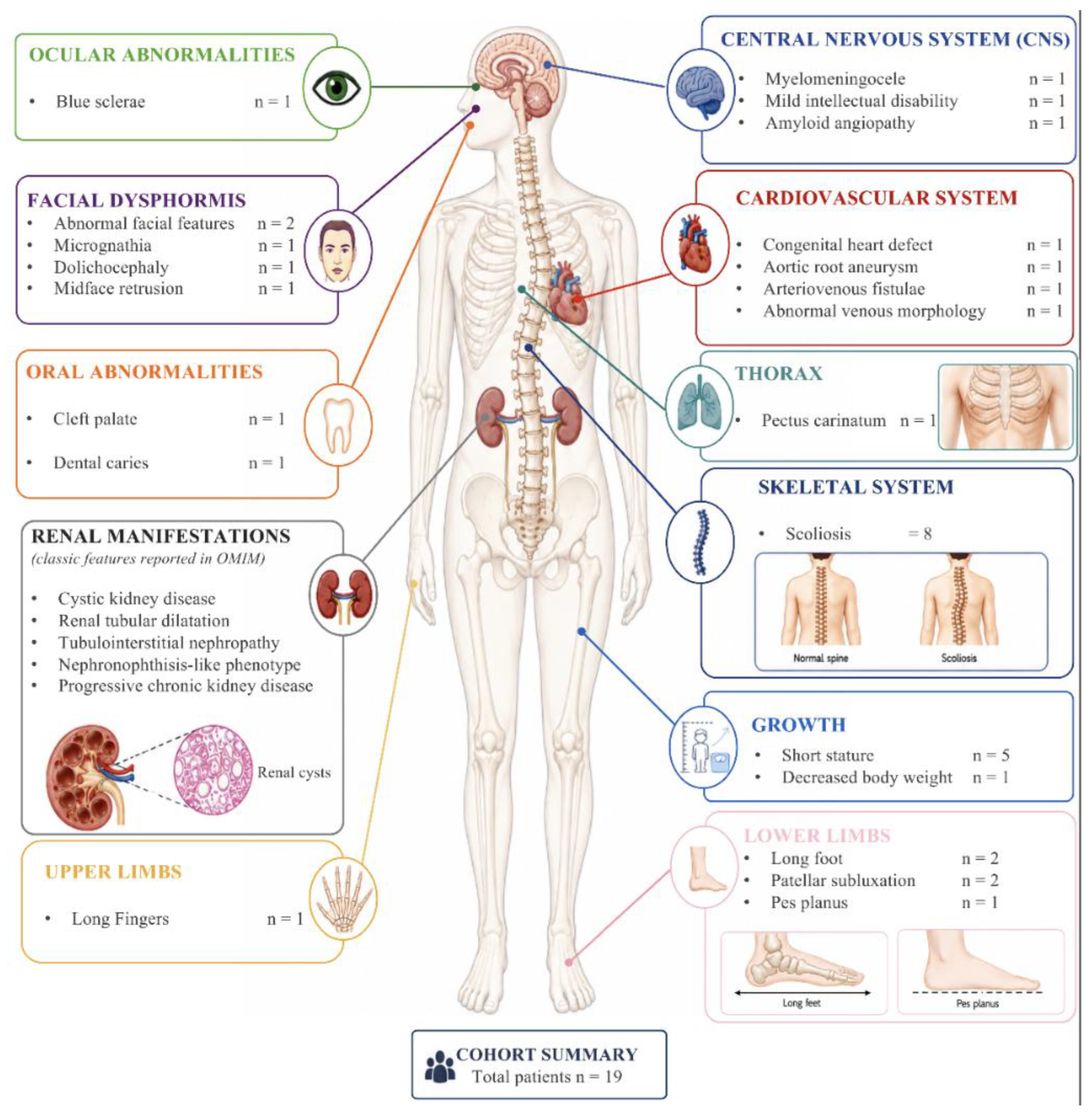
3.3. Clinical Characterization of Included Patients
3.4. Testing Strategies and Genotype Description
4. Discussion
5. Conclusions
Supplementary Materials
Author Contributions
Funding
Institutional Review Board Statement
Informed Consent Statement
Data Availability Statement
Conflicts of Interest
References
- Gupta, S.; Ozimek-Kulik, J.E.; Phillips, J.K. Nephronophthisis-Pathobiology and Molecular Pathogenesis of a Rare Kidney Genetic Disease. Genes 2021, 12, 1762. [Google Scholar] [CrossRef] [Scilit] [PubMed]
- Stokman, M.; Lilien, M.; Knoers, N. Nephronophthisis-Related Ciliopathies Synonym: NPHP-RC. In GeneReviews® [Internet]; University of Washington: Seattle, WA, USA, 1993. [Google Scholar]
- Sakakibara, N.; Nozu, K.; Yamamura, T.; Horinouchi, T.; Nagano, C.; Ye, M.J.; Ishiko, S.; Aoto, Y.; Rossanti, R.; Hamada, R.; et al. Comprehensive Genetic Analysis Using Next-Generation Sequencing for the Diagnosis of Nephronophthisis-Related Ciliopathies in the Japanese Population. J. Hum. Genet. 2022, 67, 427–440. [Google Scholar] [CrossRef] [Scilit] [PubMed]
- Macia, M.S.; Halbritter, J.; Delous, M.; Bredrup, C.; Gutter, A.; Filhol, E.; Mellgren, A.E.C.; Leh, S.; Bizet, A.; Braun, D.A.; et al. Mutations in MAPKBP1 Cause Juvenile or Late-Onset Cilia-Independent Nephronophthisis. Am. J. Hum. Genet. 2017, 100, 323–333, Erratum in Am. J. Hum. Genet. 2017, 100, 372. [Google Scholar] [CrossRef] [Scilit] [PubMed]
- Al-Hamed, M.H.; Alzaidan, H.; Hussein, M.; Albaik, L.; Qari, A.; Sayer, J.A.; Imtiaz, F. Novel Pathogenic MAPKBP1 Variant in a Family with Nephronophthisis. Clin. Kidney J. 2021, 14, 728–730. [Google Scholar] [CrossRef] [Scilit] [PubMed]
- Schönauer, R.; Jin, W.; Ertel, A.; Nemitz-Kliemchen, M.; Panitz, N.; Hantmann, E.; Seidel, A.; Braun, D.A.; Shril, S.; Hansen, M.; et al. Novel Nephronophthisis-Associated Variants Reveal Functional Importance of MAPKBP1 Dimerization for Centriolar Recruitment. Kidney Int. 2020, 98, 958–969. [Google Scholar] [CrossRef] [Scilit] [PubMed]
- Nigro, E.; Amicone, M.; D’Arco, D.; Sellitti, G.; De Marco, O.; Guarino, M.; Riccio, E.; Pisani, A.; Daniele, A. Molecular Diagnosis and Identification of Novel Pathogenic Variants in a Large Cohort of Italian Patients Affected by Polycystic Kidney Diseases. Genes 2023, 14, 1236. [Google Scholar] [CrossRef] [Scilit] [PubMed]
- Findeisen, C.; Papazian, M.; Pöschla, L.; Ertel, A.; Jin, W.; Panitz, N.; Hantmann, E.; Coucke, P.; Abdulwahab, F.; AlAbdi, L.; et al. Structure-Activity Analysis Reveals Perturbed Cilia-Jun N-Terminal Kinase Signaling in MAPKBP1-Associated Kidney Disease. Kidney Int. Rep. 2025, 10, 2836–2851. [Google Scholar] [CrossRef] [Scilit] [PubMed]
- Koyano, S.; Ito, M.; Takamatsu, N.; Shiba, T.; Yamamoto, K.-I.; Yoshioka, K. A Novel Jun N-Terminal Kinase (JNK)-Binding Protein That Enhances the Activation of JNK by MEK Kinase 1 and TGF-L-Activated Kinase 1. FEBS Lett. 1999, 457, 385–388. [Google Scholar] [CrossRef] [Scilit] [PubMed]
- Cohen-Katsenelson, K.; Wasserman, T.; Darlyuk-Saadon, I.; Rabner, A.; Glaser, F.; Aronheim, A. Identification and Analysis of a Novel Dimerization Domain Shared by Various Members of C-Jun n-Terminal Kinase (Jnk) Scaffold Proteins. J. Biol. Chem. 2013, 288, 7294–7304. [Google Scholar] [CrossRef] [Scilit] [PubMed]
- Yamaguchi, T.; Miyashita, C.; Koyano, S.; Kanda, H.; Yoshioka, K.; Shiba, T.; Takamatsu, N.; Ito, M. JNK-Binding Protein 1 Regulates NF-ΚB Activation Through TRAF2 and TAK1. Cell Biol. Int. 2009, 33, 364–368. [Google Scholar] [CrossRef] [Scilit] [PubMed]
- Yamashita, M.; Fatyol, K.; Jin, C.; Wang, X.; Liu, Z.; Zhang, Y.E. TRAF6 Mediates Smad-Independent Activation of JNK and P38 by TGF-β. Mol. Cell 2008, 31, 918–924. [Google Scholar] [CrossRef] [Scilit] [PubMed]
- Salehi, O.; Mack, H.; Colville, D.; Lewis, D.; Savige, J. Ocular Manifestations of Renal Ciliopathies. Pediatr. Nephrol. 2024, 39, 1327–1346. [Google Scholar] [CrossRef] [Scilit] [PubMed]
- Shamseldin, H.E.; Shaheen, R.; Ewida, N.; Bubshait, D.K.; Alkuraya, H.; Almardawi, E.; Howaidi, A.; Sabr, Y.; Abdalla, E.M.; Alfaifi, A.Y.; et al. The Morbid Genome of Ciliopathies: An Update. Genet. Med. 2020, 22, 1051–1060, Erratum in Genet. Med. 2022, 24, 966. [Google Scholar] [CrossRef] [Scilit] [PubMed]
- Page, M.J.; McKenzie, J.E.; Bossuyt, P.M.; Boutron, I.; Hoffmann, T.C.; Mulrow, C.D.; Shamseer, L.; Tetzlaff, J.M.; Akl, E.A.; Brennan, S.E.; et al. The PRISMA 2020 Statement: An Updated Guideline for Reporting Systematic Reviews. BMJ 2021, 372, n71. [Google Scholar] [CrossRef] [Scilit] [PubMed]
- Landrum, M.J.; Lee, J.M.; Benson, M.; Brown, G.R.; Chao, C.; Chitipiralla, S.; Gu, B.; Hart, J.; Hoffman, D.; Jang, W.; et al. ClinVar: Improving Access to Variant Interpretations and Supporting Evidence. Nucleic Acids Res. 2018, 46, D1062–D1067. [Google Scholar] [CrossRef] [Scilit] [PubMed]
- Munn, Z.; Hugh Barker, T.; Moola, S.; Tufanaru, C.; Stern, C.; McArthur, A.; Stephenson, M.; Aromataris, E. Methodological Quality of Case Series Studies: An Introduction to the Jbi Critical Appraisal Tool. JBI Evid. Synth. 2020, 18, 2127–2133. [Google Scholar] [CrossRef] [Scilit] [PubMed]
- Haddaway, N.R.; Page, M.J.; Pritchard, C.C.; McGuinness, L.A. PRISMA2020: An R Package and Shiny App for Producing PRISMA 2020-Compliant Flow Diagrams, with Interactivity for Optimised Digital Transparency and Open Synthesis. Campbell Syst. Rev. 2022, 18, e1230. [Google Scholar] [CrossRef] [Scilit] [PubMed]
- Aghjeh, M.E.; Suer, I.; Kalayci, T.; Dirim, A.B.; Kaya, M.; Ozturk, S. Deciphering the Genetic Basis of Focal Segmental Glomerulosclerosis Using Next-Generation Sequencing; Research Square: Durham, NC, USA, 2026. [Google Scholar]
- Dzedzik, S.; Dube, G.K.; Batal, I. The Case|Nephrotic Syndrome in Kidney Allograft Recipient. Kidney Int. 2026, 109, 233–234. [Google Scholar] [CrossRef] [Scilit] [PubMed]
- Broad Institute. GnomAD, version 4.0; Broad Institute: Cambridge, MA, USA, 2023. [Google Scholar]
- Matsuguchi, T.; Chiba, N.; Bandow, K.; Kakimoto, K.; Masuda, A.; Ohnishi, T. JNK Activity Is Essential for Atf4 Expression and Late-Stage Osteoblast Differentiation. J. Bone Miner. Res. 2009, 24, 398–410. [Google Scholar] [CrossRef] [Scilit] [PubMed]
- Xu, R.; Zhang, C.; Shin, D.Y.; Kim, J.M.; Lalani, S.; Li, N.; Yang, Y.S.; Liu, Y.; Eiseman, M.; Davis, R.J.; et al. C-Jun N-Terminal Kinases (JNKs) Are Critical Mediators of Osteoblast Activity In Vivo. J. Bone Miner. Res. 2017, 32, 1811–1815. [Google Scholar] [CrossRef] [Scilit] [PubMed]
- Huang, Y.F.; Lin, J.J.; Lin, C.H.; Su, Y.; Hung, S.C. C-Jun N-Terminal Kinase 1 Negatively Regulates Osteoblastic Differentiation Induced by BMP2 via Phosphorylation of Runx2 at Ser104. J. Bone Miner. Res. 2012, 27, 1093–1105. [Google Scholar] [CrossRef] [Scilit] [PubMed]
- Greenblatt, M.B.; Shim, J.H.; Zou, W.; Sitara, D.; Schweitzer, M.; Hu, D.; Lotinun, S.; Sano, Y.; Baron, R.; Park, J.M.; et al. The P38 MAPK Pathway Is Essential for Skeletogenesis and Bone Homeostasis in Mice. J. Clin. Investig. 2010, 120, 2457–2473. [Google Scholar] [CrossRef] [Scilit] [PubMed]
- Moola, S.; Munn, Z.; Tufanaru, C.; Aromataris, E.; Sears, K.; Sfetcu, R.; Currie, M.; Qureshi, R.; Mattis, P.; Lisy, K.; et al. Chapter 7: Systematic reviews of etiology and risk. In JBI Manual for Evidence Synthesis; Aromataris, E., Munn, Z., Eds.; JBI: Philadelphia, PA, USA, 2020. [Google Scholar]



| Patient | Age | Sex | Test | Variants | Skeletal Features | Craniofacial Dysmorphisms | Cardiovascular Features | Other | Ref |
|---|---|---|---|---|---|---|---|---|---|
| P01-I | 25 | F | Exome | p.Arg198* p.Arg1459* | Patellar subluxation; Long fingers; Long foot | - | - | - | [4] |
| P01-II | 27 | M | Sanger | p.Arg198* p.Arg1459* | Short stature | - | Congenital heart defect | Myelomeningocele; (PDE6A-related retinitis) | [4] |
| P02 | 15 | M | Exome | p.Arg434* p.Arg434* | - | Abnormality of the face | - | - | [4] |
| P03 | 23 | M | Exome | p.Arg538Gln p.Arg538Gln | - | - | - | - | [4] |
| P04 | 15 | F | Exome | c.2426-1G>A c.2426-1G>A | Short stature | - | - | - | [4] |
| P05-I | 39 | F | Exome | p.Gln937* p.Gln937* | Scoliosis | - | - | - | [4] |
| P05-II | 38 | M | Sanger | p.Gln937* p.Gln937* | Scoliosis | - | Amyloid angiopathy | Cholesteatoma; Vesicoureteral reflux | [4] |
| P05-III | 21 | F | Sanger | p.Gln937* p.Gln937* | Scoliosis; Short stature | Cleft palate | - | Mild intellectual disability | [4] |
| P06 | 37 | F | Exome | c.4299+5G>C c.4299+5G>C | Scoliosis; Long fingers | Abnormality of the face | - | [6] | |
| P07 | 14 | F | Panel | c.3315-2A>G c.3315-2A>G | Short stature | - | Abnormal venous morphology | - | [6] |
| P08-I | 31 | M | Exome | p.Arg312* p.Arg312* | - | - | - | - | [5] |
| P08-II | 12 | M | Sanger | p.Arg312* p.Arg312* | - | - | - | - | [5] |
| P09 | 12 | F | Panel | c.3314+1G>A p.Gln868* | - | - | - | (Hypokalemia) | [3] |
| P10 | 20 | M | Panel | p.Gly752fs p.Gly752fs | - | - | - | - | [7] |
| P11 | 20 | F | Exome | p.Ser826fs p.Ser826fs | Scoliosis | - | - | Decreased body weight; Cholelithiasis | [8] |
| P12 | 47 | M | Exome | c.270-2A>G c.270-2A>G | Scoliosis; Long fingers; Long foot; Pes planus | Micrognathia | Aortic root aneurysm; Arterio- venous fistula | - | [16] SCV007606 611.1 |
| P13 | 0–9 | na | na | c.2885+2C>T c.2885+2C>T | Scoliosis | - | - | - | [16] SCV006333 410.1 |
| P14 | na | na | na | p.Ser424fs p.Ser424fs | Scoliosis; Short stature; Patellar subluxation; Pectus carinatum; | Dolichocephaly; Midface retrusion | - | Blue sclerae; Carious teeth; Joint laxity | [16] SCV002318 839.1 |
| P15 | 47 | F | na | p.Val1193fs p.Val1193fs | - | - | - | Polyarthritis | [20] |
| Excluded patients with variants reclassified as class 3 or class 2 according to ACMG criteria | |||||||||
| E01 | 3 | na | Exome | p.Arg1102Cys p.Arg1102Cys | - | - | Congenital heart defect | (Pulmonary hypoplasia) | [14] |
| E02 | 55 | na | Exome | p.Pro879Ser p.Pro879Ser | - | - | - | - | [19] |
| N | Variant (c.) | Protein (p.) | Type | ACMG Class | Allele Count | Allele Number | Allele Frequency |
|---|---|---|---|---|---|---|---|
| 1 | c.592C>T | p.Arg198* | Stop-gain | 5 | 1 | 1,613,552 | 6.20 × 10⁻⁷ |
| 2 | c.1270_1271del | p.Ser424Leufs*16 | Frameshift | 5 | / | / | / |
| 3 | c.934C>T | p.Arg312* | Stop-gain | 4 | 1 | 1,612,238 | 6.20 × 10⁻⁷ |
| 4 | c.1300C>T | p.Arg434* | Stop-gain | 5 | 2 | 1,614,208 | 1.24 × 10⁻⁶ |
| 5 | c.1613G>A | p.Arg538Gln | Missense | 4 | 2 | 1,611,462 | 1.24 × 10⁻⁶ |
| 6 | c.1904+1G>A | p.? | Splicing | 5 | 1 | 1,612,330 | 6.20 × 10⁻⁷ |
| 7 | c.2092+1G>A | p.? | Splicing | 5 | / | / | / |
| 8 | c.2156+1G>A | p.? | Splicing | 4 | / | / | / |
| 9 | c.2227C>T | p.Gln743* | Stop-gain | 5 | 1 | 1,614,226 | 6.19 × 10⁻⁷ |
| 10 | c.2253del | p.Gly752Aspfs*74 | Frameshift | 4 | / | / | / |
| 11 | c.2426-1G>A | p.? | Splicing | 5 | / | / | / |
| 12 | c.2476_2480del | p.Ser826Glyfs*34 | Frameshift | 4 | / | / | / |
| 13 | c.2602C>T | p.Gln868* | Stop-gain | 4 | / | / | / |
| 14 | c.270-2A>G | p.? | Splicing | 4 | / | / | / |
| 15 | c.2809C>T | p.Gln937* | Stop-gain | 5 | / | / | / |
| 16 | c.2885+2C>T | p.? | Splicing | 5 | / | / | / |
| 17 | c.3314+1G>A | p.? | Splicing | 4 | / | / | / |
| 18 | c.3315-2A>G | p.? | Splicing | 4 | / | / | / |
| 19 | c.3428del | p.Pro1143Argfs*186 | Frameshift | 5 | / | / | / |
| 20 | c.3578_3579del | p.Val1193Alafs*10 | Frameshift | 4 | / | / | / |
| 21 | c.4299+1G>A | p.? | Splicing | 4 | / | / | / |
| 22 | c.4299+5G>C | p.? | Splicing | 4 | 1 | 1,588,072 | 6.30 × 10⁻⁷ |
| 23 | c.4375C>T | p.Arg1459* | Stop-gain | 4 | 10 | 1,612,074 | 6.20319 × 10⁻⁶ |
| Reclassified variants (not considered for frequency counts) | |||||||
| 1 | c.2635C>T | p.Pro879Ser | Missense | 3 | 1 | 1,614,156 | 6.19519 × 10⁻⁷ |
| 2 | c.3304C>T | p.Arg1102Cys | Missense | 2 | 135 | 1,613,918 | 8.36458 × 10⁻⁵ |
Disclaimer/Publisher’s Note: The statements, opinions and data contained in all publications are solely those of the individual author(s) and contributor(s) and not of MDPI and/or the editor(s). MDPI and/or the editor(s) disclaim responsibility for any injury to people or property resulting from any ideas, methods, instructions or products referred to in the content. |
© 2026 by the authors. Licensee MDPI, Basel, Switzerland. This article is an open access article distributed under the terms and conditions of the Creative Commons Attribution (CC BY) license.
Share and Cite
Ambrosini, E.; Luberto, A.; Busciglio, S.; Baroni, M.C.; Taiani, A.; Cannizzaro, I.R.; Gandolfini, I.; Maggiore, U.; Martorana, D.; Uliana, V.; et al. Beyond the Kidney: A Systematic Review of Extrarenal Manifestations in MAPKBP1-Related Nephronophthisis. Int. J. Mol. Sci. 2026, 27, 8370. https://doi.org/10.3390/ijms27188370
Ambrosini E, Luberto A, Busciglio S, Baroni MC, Taiani A, Cannizzaro IR, Gandolfini I, Maggiore U, Martorana D, Uliana V, et al. Beyond the Kidney: A Systematic Review of Extrarenal Manifestations in MAPKBP1-Related Nephronophthisis. International Journal of Molecular Sciences. 2026; 27(18):8370. https://doi.org/10.3390/ijms27188370
Chicago/Turabian StyleAmbrosini, Enrico, Anita Luberto, Sabrina Busciglio, Maria Chiara Baroni, Antonietta Taiani, Ilenia Rita Cannizzaro, Ilaria Gandolfini, Umberto Maggiore, Davide Martorana, Vera Uliana, and et al. 2026. "Beyond the Kidney: A Systematic Review of Extrarenal Manifestations in MAPKBP1-Related Nephronophthisis" International Journal of Molecular Sciences 27, no. 18: 8370. https://doi.org/10.3390/ijms27188370
APA StyleAmbrosini, E., Luberto, A., Busciglio, S., Baroni, M. C., Taiani, A., Cannizzaro, I. R., Gandolfini, I., Maggiore, U., Martorana, D., Uliana, V., Barili, V., & Percesepe, A. (2026). Beyond the Kidney: A Systematic Review of Extrarenal Manifestations in MAPKBP1-Related Nephronophthisis. International Journal of Molecular Sciences, 27(18), 8370. https://doi.org/10.3390/ijms27188370