Article

An Unbiased Drug Screen in a Drosophila Model of LMNA-Muscular Dystrophy Identifies Calcium Channel Blockers as Potential Treatments

1
Interdisciplinary Graduate Program in Genetics, University of Iowa, Iowa City, IA 52242, USA
2
Department of Biochemistry and Molecular Biology, Carver College of Medicine, University of Iowa, Iowa City, IA 52242, USA
3
Department of Biomedical Engineering, University of Iowa, Iowa City, IA 52242, USA
*
Author to whom correspondence should be addressed.
Int. J. Mol. Sci. 2026, 27(18), 8367; https://doi.org/10.3390/ijms27188367 (registering DOI)
Submission received: 10 June 2026 / Revised: 4 September 2026 / Accepted: 15 September 2026 / Published: 19 September 2026
(This article belongs to the Special Issue Molecular Mechanisms and Therapies in Skeletal Muscle Diseases)

Abstract

Mutations in the LMNA gene encoding A-type lamins cause multiple muscular dystrophies (LMNA-MD). Lamins are intermediate filaments with three conserved domains: an N-terminal head, a coiled-coil rod, and a C-terminal domain possessing an Ig-like fold. Missense mutations in all three domains can cause LMNA-MD by mechanisms that are not well understood. Currently, there are limited treatments for LMNA-MD beyond symptom management. In this study, our goal was to identify candidate treatments through an in vivo drug repurposing screen using Drosophila models of LMNA-MD that recapitulate aspects of the disease phenotype. Expression of LamC R264Q (Drosophila orthologue of human LMNA R249Q) in fly muscles causes muscle defects and premature death. We fed 1520, mostly FDA/EMA-approved, drugs to larvae and screened for rescue of lethality. This screen resulted in 68 positive hit drugs that partially restored viability. Molecular fingerprinting clustered a subset of these compounds into six structurally related groups, including L-type voltage-gated calcium channel inhibitors. We tested these inhibitors using multiple fly models expressing different mutant lamins and discovered that only flies expressing amino acid substitutions in the rod domain benefited, supporting a need for personalized treatments. Collectively, our findings support L-type voltage-gated calcium channel blockers as candidate treatments that warrant testing in pre-clinical models.

1. Introduction

Lamins are intermediate filaments that form a meshwork underlying the inner nuclear membrane (INM) known as the nuclear lamina [1]. Lamins are subdivided into the B-type and A-type, with the B-type lamins B1 and B2 encoded by the LMNB1 and LMNB2 genes, respectively, and the A-type lamins A and C encoded by the LMNA gene via alternative splicing [1,2]. All lamins have three conserved structural regions: a globular N-terminal head, an alpha-helical central rod, and a C-terminal tail, which includes a nuclear localization sequence and an Ig-like fold domain. Lamins homopolymerize to form filaments. This process begins with the formation of lamin dimers, which result from the formation of coiled-coils via interactions between the alpha-helices of the central rod. These dimers combine to form head-to-tail protofilaments, which then interact in an anti-parallel lateral manner through the coil 1b and coil 2 regions of the rod to form the higher-order structure of the lamina. Super-resolution microscopy revealed that A- and B-type lamins form independent networks. Prior to cellular differentiation, B-type lamins form a meshwork underlying the inner nuclear membrane. As cells differentiate, A-type lamins are expressed and form a second meshwork between the B-type lamins and the nucleoplasm [3].
Mutations in lamin-encoding genes cause a collection of human diseases known as laminopathies. While there are diseases associated with LMNB1 and LMNB2 mutations, the vast majority of laminopathies are caused by mutations in the LMNA gene [4,5,6,7]. There are at least 16 diseases associated with approximately 500 pathogenic mutations across the full length of the LMNA gene, with the majority of these inherited in an autosomal dominant fashion [6,7,8,9,10,11,12]. Phenotypes associated with LMNA mutations can be broadly divided into accelerated aging syndromes, diseases affecting adipose tissue, peripheral neuropathies, and striated muscle diseases, which are the most common and account for 55–79% of laminopathy cases [13,14]. In addition, there is poor genotype–phenotype correlation, making disease symptoms difficult to predict [6,7,8,9,10,11,12,13,14].
The striated muscle laminopathies are Emery–Dreifuss muscular dystrophy (EDMD), limb girdle muscular dystrophy type 1B (LGMD1B), lamin-associated congenital muscular dystrophy (L-CMD), and dilated cardiomyopathy with conduction defects (DCM-CD) [15,16,17,18]. Muscular dystrophies caused by LMNA mutations are collectively referred to as LMNA-MD, with phenotypes existing on a continuous spectrum where CMD is the most severe phenotype [15,19]. DCM-CD can either present without skeletal muscle involvement or co-occur with LMNA-MD [18,19]. The overwhelming majority of striated muscle laminopathies are caused by dominantly inherited missense mutations in LMNA that affect all domains of the lamin A/C protein [7,20,21]. This includes the rod domain, where commonly occurring substitutions include H222P and R249Q, often associated with EDMD, and R249W, often associated with L-CMD [20,21,22,23].
There are currently limited treatments for LMNA-MD other than symptom management [1,24]. However, there is an ongoing clinical trial for LMNA-associated DCM testing NVC-001 (Nuevocor Pte. Ltd., Singapore), an adeno-associated virus encoding a truncated version of the nuclear envelope protein SUN1 [25]. Treatments used for other muscular dystrophies often have limited relevance to LMNA-MD. The standard of care pharmacological treatment for Duchenne muscular dystrophy (DMD) is corticosteroids [26,27]. Reports on the effectiveness of corticosteroids for LMNA-MD are variable and largely restricted to L-CMD [28,29,30]. Gene therapies, which include splice-modifying antisense oligonucleotides (ASOs) and viral vector gene replacement, are cutting-edge treatments with significant success for other types of muscular dystrophies [31,32,33,34,35]. Limited development of splice-modifying ASOs for LMNA-MD has occurred, with efficient skipping demonstrated only for LMNA exon 5 in human cells [36]. Viral vector gene replacement in its current form is unlikely to be successful for LMNA-MD, as it is currently only used to treat diseases caused by loss-of-function mutations [31,33,34,35]. Mutations that cause LMNA-MD are typically dominantly inherited and encode a protein with a single amino acid substitution that acts as a dominant negative, minimizing the therapeutic benefit of overexpressing a wild-type protein [7,23,29,37,38]. Additionally, there are significant safety and economic concerns surrounding viral vector gene therapies [39,40,41]. Thus, while gene therapy development remains a long-term goal, pharmacological approaches offer a faster route to translation for dominantly inherited diseases caused by pathogenic missense mutations.
Muscular dystrophy has been extensively modeled in the fruit fly Drosophila melanogaster [42]. DMD, LGMD, and EDMD have been successfully modeled in Drosophila, with phenotypes resembling the progressive loss of muscle function, locomotor ability, and cardiac function as seen in human patients [20,43,44,45,46,47,48,49,50,51,52,53,54,55,56,57,58,59,60,61]. Drosophila models of human diseases have been used in drug screens with the goal of identifying human disease mechanisms and potential treatments [62,63]. Suppression of lethality is often used as a readout in these screens, as feeding drugs to Drosophila larvae can result in easily scorable rescue of pupal or adult viability, leading to relatively high-throughput screening [62,63]. There are multiple examples of this approach in the oncology space [62,63,64,65,66,67], as well as in Drosophila models of congenital glycosylation disorders [68,69]. However, to our knowledge, a drug repurposing screen has yet to be completed in a Drosophila model of muscular dystrophy.
To address the limited treatments currently available for LMNA-MD, we completed an unbiased drug screen using the Prestwick Chemical Library to rescue lethality caused by expression of mutant A-type lamins. Drosophila Lamin C (LamC) R264Q is orthologous to human lamin A/C R249Q, a substitution which most commonly causes EDMD [20]. Expression of a LamC R264Q transgene exclusively in larval body wall muscles causes lethality at the pupal stage [20]. This screen identified 68 drugs that rescued adult viability. Clustering of these 68 hit compounds by molecular structure identified six classes with related structures. We followed up on L-type voltage-gated calcium channel blockers given the physiological relevance of calcium signaling to muscle function, including mouse models of Lmna-associated cardiac disease [70,71,72,73]. We found that multiple classes of L-type calcium channel blockers rescue the lethality caused by muscle-specific expression of specific mutant lamins. Collectively, our findings demonstrate the utility of Drosophila models as powerful tools for in vivo screening, support L-type calcium channel antagonists as potential drug repurposing candidates and suggest the need for personalized treatments for LMNA-MD.

2. Results

2.1. A Drug Screen Was Performed for Rescue of Lethality Caused by LamC R264Q

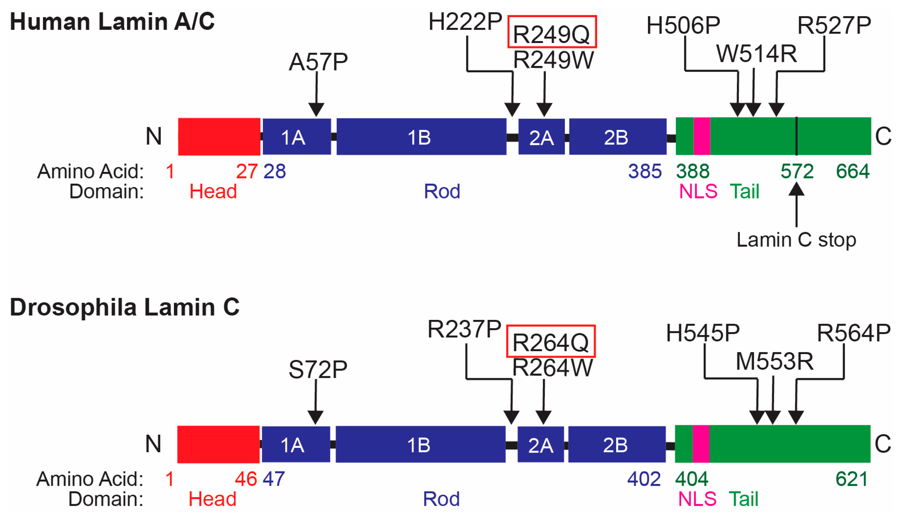
There are few treatments available for LMNA-MD, with standard care consisting of symptom management without treating the cause of the disease [30]. We have previously developed and characterized a Drosophila model transgenic for LamC R264Q [20]. The R264Q substitution in Drosophila LamC is orthologous to the human lamin A/C R249Q substitution (Figure 1). This amino acid substitution affects the rod domain of the A-type lamin protein and is primarily associated with EDMD in humans [23]. Expression of LamC R264Q in the Drosophila larval body wall muscles results in completely penetrant lethality at the pupal stage [20]. Lethality is accompanied by reduced A- and B-type lamins at the nuclear periphery, perinuclear aggregation of A-type lamins, and nucleoplasmic aggregation of B-type lamins and nuclear pores, resulting in nuclear lobulations [20]. Taken together, these findings demonstrate that this single amino acid substitution in the rod domain of A-type lamins can alter nuclear architecture in intact muscle within a developing organism [20].
The binary nature of the complete loss of adult viability caused by LamC R264Q expression represents an optimal phenotype for drug screening. To identify novel disease mechanisms and treatments for LMNA-MD, we completed an unbiased screen of the Prestwick Chemical Library (version 20, Prestwick Chemical, Illkirch, France) for rescue of lethality caused by expression of LamC R264Q in the larval body wall muscles of Drosophila. The Prestwick Chemical Library contains 1520 off-patent drugs with over 600 known biological targets. Ninety-eight percent of drugs in the library are either FDA- or EMA-approved and are known to be safe in humans, allowing the library to be used in disease models to identify drug repurposing candidates. The library has previously been used for lethality suppression screens in Drosophila to identify candidate treatments for other genetic diseases, making it ideal for our application [68,69].
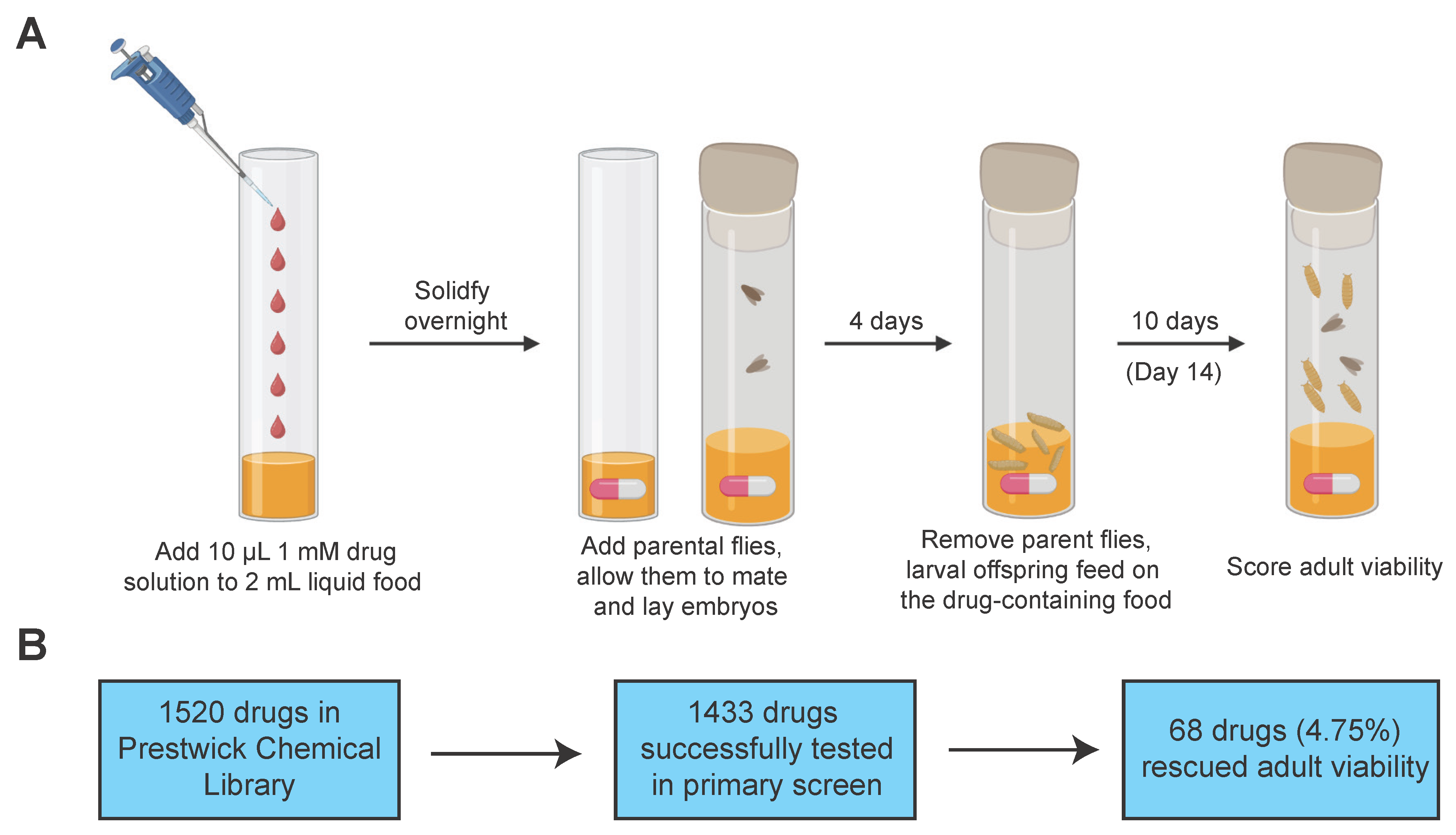
The Prestwick Chemical Library was purchased with each drug pre-dissolved at 10 mM in DMSO. For our screen, these drugs were diluted to 1 mM and 10% DMSO in PBS. Each drug was mixed with 2 mL of liquid fly food per vial, which resulted in a final concentration of 5 µM drug and 0.05% DMSO. For the primary screen, each drug was tested in a single vial at a single concentration of 5 µM. Thus, we recognize that our results could be an underestimation of the number of positive compounds. Parental flies possessing the C57-Gal4 driver were mated to flies possessing the LamC R264Q transgene in each vial. After mating, 100% of the offspring produced by these parents express the LamC R264Q transgene exclusively in larval body wall muscles [74,75]. The parental generation was removed from the vials, and larval progeny consumed the food containing either DMSO alone or the drug throughout the larval stages, until they pupated on the walls of the vials. After 14 days, adult viability was scored (Figure 2A). Given the complete penetrance of lethality in the absence of drug treatment (food alone or food plus DMSO only) [20], the emergence of even a single adult fly out of ~30 pupae per vial following drug treatment was considered a positive hit in the primary screen.
To validate this approach, we first tested whether DMSO alone altered viability. We treated 902 total progeny expressing a wild-type LamC transgene across 30 vials with 0.05% DMSO. Of these, 861 developed into viable adults (95.45% viability), which is consistent with the adult viability observed for this genotype in the absence of DMSO [20,54,56]. To confirm that DMSO did not have a therapeutic effect, 1133 total progeny expressing LamC R264Q across 35 independent vials were treated with 0.05% DMSO. These progeny died at the pupal stage as expected, with zero adult survivors. This is consistent with our previous data in the absence of drug, where LamC R264Q expression resulted in completely penetrant pupal lethality [20]. These experiments confirm that DMSO is neither toxic nor therapeutically beneficial in this context, supporting the conclusion that the emergence of adult flies following drug treatment indicates a positive effect of the drug and not the vehicle used in the screen.
Using this approach, we successfully screened 1433 drugs (87 vials had unsuccessful matings) from the Prestwick Chemical Library for rescue of lethality caused by muscle-specific expression of LamC R264Q (Supplemental Table S1). Treatment with 68 drugs resulted in at least one viable adult for a hit rate of 4.75% (Figure 2B). The number of viable adults per vial containing each hit drug ranged from one to eight, with one to three being most common. The number of dead pupae per vial averaged 32. A list of the 68 positive hit drugs, their molecular target or the biological process they target according to the compound key provided by Prestwick Chemical, and their action against that target is provided in Table 1. Note that many of the drugs with a defined molecular target have an orthologue in Drosophila that is expressed at the larval and/or pupal stage (Table 1) (FlyBase, FB2026_02, released 18 June 2026).

2.2. Structural Clustering of Positive Hit Compounds Identified Dihydropyridine L-Type Voltage-Gated Calcium Channel Antagonists Among the Drugs That Rescued Lethality

To prioritize the 68 hit drugs for follow-up testing, we hypothesized that drugs with similar structures likely have the same or similar molecular targets, making assessment of structural similarity a way to identify enriched biological targets of the drugs in our hit compound list. We compared the structures of the hit drugs using the open-source cheminformatics tool RDKit (https://www.rdkit.org (accessed on 15 September 2025)) [76,77,78]. RDKit can be used to generate molecular fingerprints, or binary code representations of a compound, which can be compared to each other using similarity indices such as the Tanimoto similarity index to generate numerical measures of the similarity between every pairwise comparison of compounds in a data set [78,79,80]. The Tanimoto similarity index ranges from 0 to 1, where a value of 0 indicates completely different molecules and a value of 1 indicates identical molecules [78]. All similarity indices for a given data set can then be clustered using algorithms such as the Butina clustering algorithm to identify larger clusters of compounds that all reach a certain threshold of similarity to each other while being different from all other molecules in the data set [81].
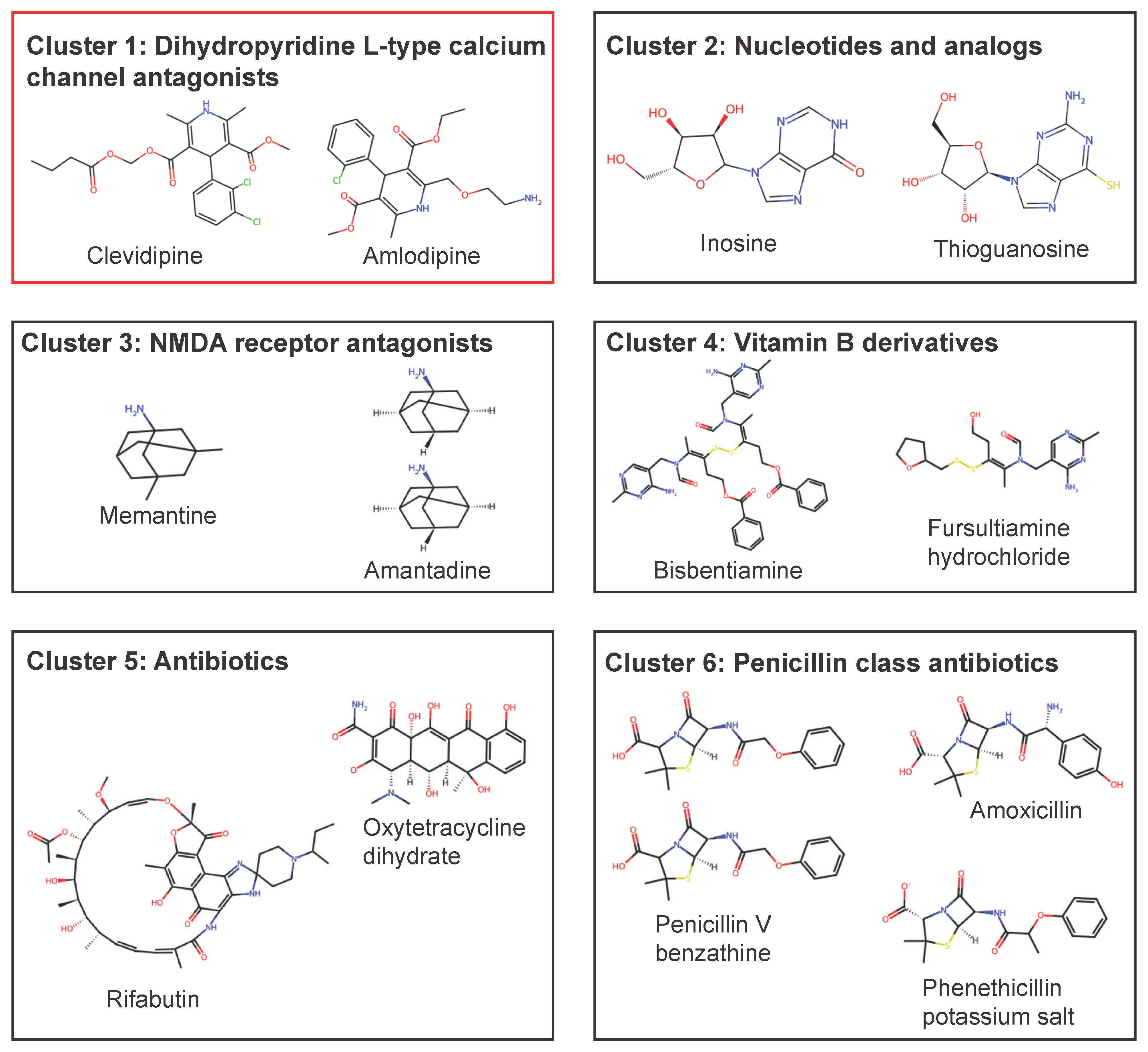
When this workflow was applied to our 68 hit compounds, it identified six clusters of at least two compounds that had a Tanimoto similarity of 0.75 or more, meaning all compounds in each cluster are at least 75% structurally similar to each other (Figure 3). In total, 13 compounds were included in the clusters, with five clusters containing two compounds and one cluster containing three. Compounds in each cluster were indeed functionally related, with the six clusters including two classes of antibiotics, nucleotides, NMDA receptor antagonists, vitamin B derivatives, and dihydropyridine (DHP) L-type calcium channel (LTCC) antagonists (Figure 3).
Of the six clusters, the one with the clearest connection to muscle function comprised the DHP LTCC antagonists. LTCC antagonists are readily available drugs that are used to treat various cardiac conditions, most notably hypertension [82,83]. These drugs are structurally classified into DHPs and non-DHPs, which block LTCCs through different binding sites, variable affinities, and with slightly different physiological effects [83,84]. The DHPs include clevidipine and amlodipine identified in our screen (Table 1), as well as many others [84]. The two most notable non-DHPs are verapamil, which is classified as a phenylalkylamine, and diltiazem, which is a benzothiazepine [84]. Importantly, our screen also identified ethaverine (Table 1), which is a non-DHP LTCC antagonist that is structurally similar to verapamil [85]. This reveals a limitation of structural clustering as a means of hit prioritization, as not all compounds with the same molecular target have similar structures.

2.3. Secondary Testing Verified DHP Calcium Channel Blockers as Positive Hits in the LamC R264Q Model

Given the physiological differences in the actions of DHP and non-DHP calcium channel blockers [84,86] and the fact that we identified two structurally similar DHPs compared to one non-DHP in our screen (Table 1), we prioritized the DHPs for secondary testing. To confirm clevidipine and amlodipine as hits in our screen, we purchased both drugs from a new source outside of Prestwick Chemical to control for batch effects and impurities or altered drug conformations that potentially could be present in the drug solution used in our screen (Table S2). Like in the primary screen, these newly purchased drug aliquots were dissolved at 10 mM in DMSO and diluted to 1 mM and 10% DMSO in PBS. Newly purchased clevidipine and amlodipine were again fed to larvae expressing LamC R264Q in the larval body wall muscle through a method largely identical to the primary screen at concentrations ranging from 0.5 to 50.0 µM (0.5, 1.25, 2.5, 3.75, 5.0, 12.5, 25.0, 37.5, 50.0 µM). In total, 58 vials were treated with clevidipine, and eight vials produced at least one live adult at concentrations ranging from 0.5 to 37.5 µM (0.5, 1.25, 2.5, 5.0, 12.5, 25.0, 37.5 µM). A total of 67 vials were treated with amlodipine, and six produced at least one adult at concentrations ranging from 2.5 to 37.5 µM (2.5, 3.75, 5.0, 10.0, 37.5 µM). In parallel, 52 additional vials were treated with vehicle 0.05% DMSO as a control, and none yielded any live adults, consistent with the controls in the primary screen (Figure S1). These results confirm that DHP L-type calcium channel blockers partially rescue pupal lethality caused by LamC R264Q expression.

2.4. DHP Calcium Channel Blockers Selectively Rescue Lethality Caused by LamC R264Q, R264W, and R237P

It is well established that different lamin amino acid substitutions result in different molecular defects despite converging on a similar muscular dystrophy phenotype [52,54,58]. Given this, a treatment that rescues muscle defects caused by one lamin substitution may not be universally effective against muscle defects caused by other lamin substitutions. To determine the broad applicability of the rescue of lethality we observed in the LamC R264Q model following DHP treatment, we tested the DHPs in six additional Drosophila models expressing different mutant lamins (Figure 1). These include the LamC S72P, R237P, R264W, H545P, M553R, and R564P substitutions, which are spread across the full length of the lamin protein and are all orthologous to pathogenic mutations in humans [20,52,54,56,87,88]. The Drosophila models expressing LamC R237P, R264W, M553R, and R564P transgenes have been previously characterized, and independent expression of these mutant lamins in larval body wall muscles results in completely penetrant pupal lethality akin to what is caused by LamC R264Q [20,52,54,56]. To characterize the LamC S72P and H545P models, we expressed the mutant lamin transgenes specifically in the larval body wall muscles using the C57-Gal4 driver [74,75]. Expression of these mutant LamC transgenes also resulted in complete death at the pupal stage, with expression of the previously characterized LamC M553R used as a positive control (Table S3) [52]. Given that expression of these six mutant lamins results in complete loss of adult viability, as did the expression of LamC R264Q, the calcium channel blockers were tested in these models.
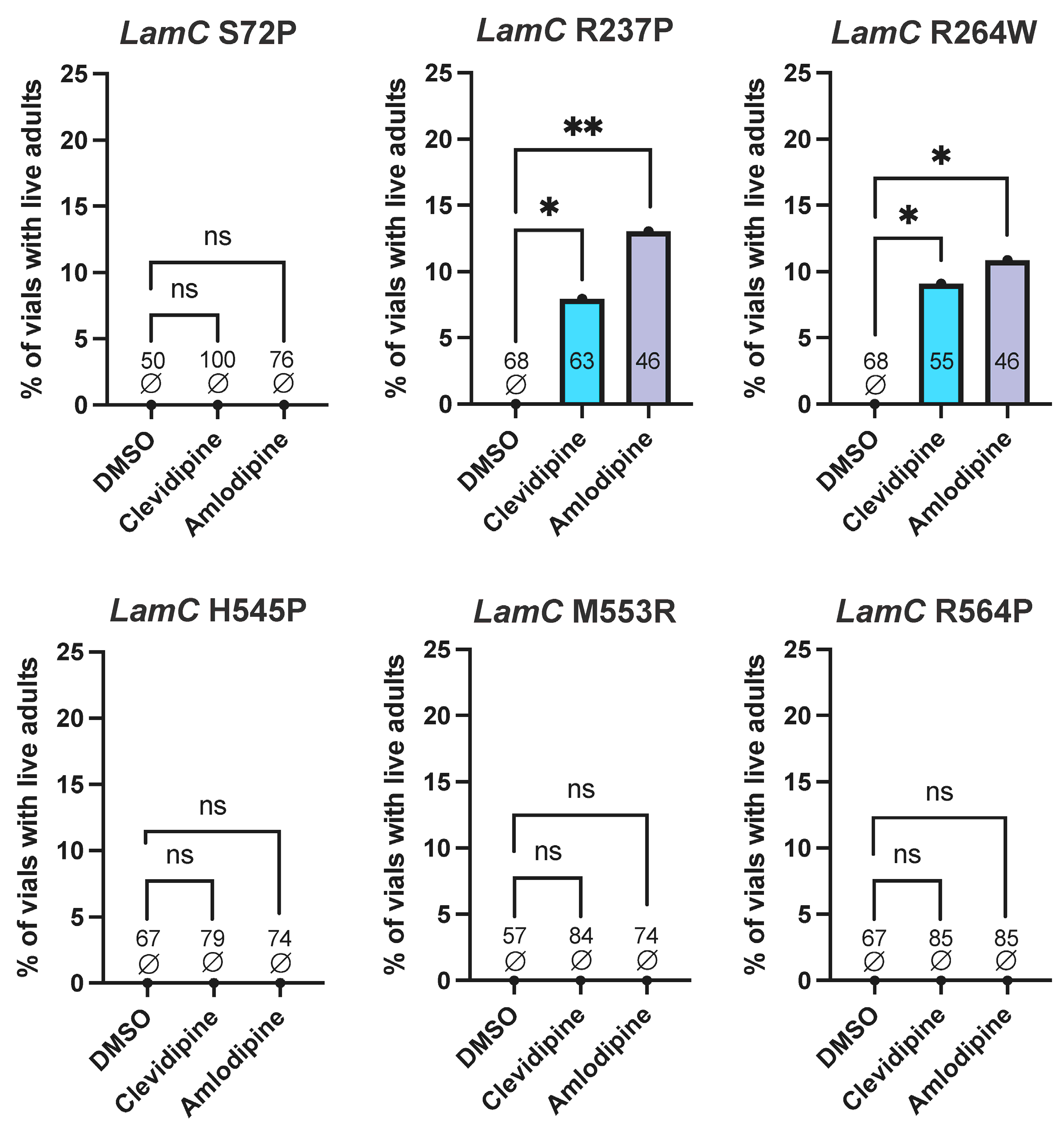
Drosophila larvae expressing these additional mutant lamins in the larval body wall muscles were independently treated with clevidipine, amlodipine, or DMSO only as a control in an identical fashion to the secondary screen. Clevidipine and amlodipine treatment resulted in vials producing live adults only following expression of either the LamC R237P or R264W transgene. Of the 63 vials of larvae expressing LamC R237P treated with clevidipine, five vials produced live adults at drug concentrations ranging from 1.25 µM to 5 µM (1.25, 2.5, 3.75, 5.0 µM). Similarly, 46 vials of larvae expressing LamC R237P were treated with amlodipine, and six vials yielded live adults at concentrations ranging from 1.25 µM to 25 µM (1.25, 3.75, 12.5, 25.0 µM). Of 55 vials of larvae expressing LamC R264W treated with clevidipine, five produced live adults at concentrations ranging from 0.5 to 5 µM (0.5, 1.25, 2.5, 3.75, 5.0 µM). In addition, 46 vials of larvae expressing LamC R264W were treated with amlodipine, and five produced live adults at concentrations ranging from 2.5 to 37.5 µM (2.5, 3.75, 5.0, 25.0, 37.5 µM). Similar numbers of vials of larvae expressing the S72P, H545P, M553R, and R564P mutant lamins were treated with each drug, and none produced live adults (Figure 4). Thus, the ability of DHP L-type calcium channel blockers to restore adult viability following mutant lamin expression extends beyond the R264Q mutant lamin used in our initial screen to include LamC R264W and R237P. However, the rescue is not universal, as the drugs provided no apparent benefit to larvae expressing LamC S72P, H545P, M553R, and R564P, suggesting a need for personalized treatments for individuals with LMNA-MD [52,54,58].

2.5. Non-DHP Calcium Channel Blockers Rescue Lethality Caused by LamC R264Q, R264W, and R237P

LTCC blockers are broadly divided into DHPs, such as clevidipine and amlodipine, and non-DHPs [83,84,86]. Given that our primary screen identified both types as positive hit drugs, we next sought to determine whether our previous findings on the DHP calcium channel blockers extended to non-DHPs [85]. While both drug classes inhibit L-type calcium channels, they have different physiological effects, binding affinities with various LTCCs, and primary binding sites in humans [83], necessitating follow-up testing. While the non-DHP ethaverine was identified in our primary screen, it is structurally similar to verapamil [85]. Verapamil and diltiazem are the most widely prescribed non-DHPs and were thus prioritized for testing [84,86].
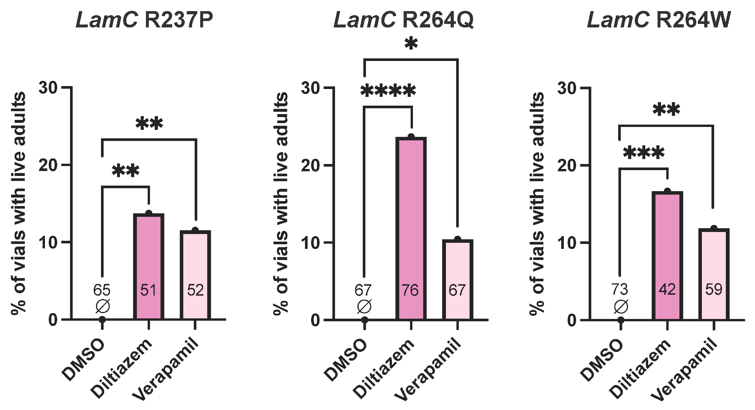
Given that the DHP calcium channel blockers rescued lethality caused by expression of the LamC R237P, R264Q, and R264W transgenes, verapamil and diltiazem were tested in these three models to determine whether non-DHPs could also restore adult viability. All three models were treated with verapamil and diltiazem at concentrations ranging from 0.5 to 50.0 µM (0.5, 1.25, 2.5, 3.75, 5.0, 12.5, 25.0, 37.5, 50.0 µM) through the same methods as DHP treatment. In total, 51 vials containing larvae expressing LamC R237P, 76 vials containing larvae expressing LamC R264Q, and 42 vials containing larvae expressing LamC R264W were treated with diltiazem, while 52, 67, and 59 vials of larvae expressing each mutant lamin, respectively, were treated with verapamil. Treatment with diltiazem and verapamil was effective in all three models (Figure 5). Diltiazem treatment resulted in seven vials yielding adults following expression of LamC R237P, 18 vials yielding adults following expression of LamC R264Q, and seven vials yielding adults following expression of LamC R264W. Verapamil treatment resulted in six vials yielding adults following expression of LamC R237P, seven vials yielding adults following expression of LamC R264Q, and seven vials yielding adults following expression of LamC R264W (Figure 5). The efficacy of diltiazem and verapamil in the same Drosophila models in which the DHPs were effective suggests that L-type calcium channel inhibition rescues lethality caused by mutant lamins regardless of the mechanism by which the channel is targeted. These findings bolster the case for the L-type calcium channel as a potential therapeutic target in LMNA-MD.

2.6. Calcium Channel Blocker Treatment Reduces Intracellular Calcium in Muscles of Larvae Expressing LamC R264Q

Given that we obtained rescue of lethality upon treatment with the calcium channel blockers, we wanted to determine if the drug treatment improved muscle physiology. Of the flies expressing mutant lamins that responded to clevidipine, those expressing LamC R237P showed thin and fragile muscles compared to controls upon dissection (Figure S2). Therefore, we performed a morphometric analysis on larval body wall muscle from larvae expressing wild-type LamC and LamC R237P with and without clevidipine treatment. Flies expressing LamC R564P were used as a positive control since our prior studies demonstrated that this mutant lamin caused decreased muscle size [56]. Larval body wall muscles were fixed and stained with phalloidin (Figure S2). Larval body wall muscles have a characteristic arrangement of muscles, allowing the same muscle to be identified and measured across larvae [89,90]. Quantification of the muscle width showed that larvae expressing LamC R564P had reduced muscle width relative to the control, consistent with our prior findings [56]. The muscles of larvae expressing LamC R237P showed reduced muscle width relative to the control, and this did not change upon treatment with clevidipine (Figure S2). Thus, the benefits of clevidipine are not through enhancing muscle physiology.
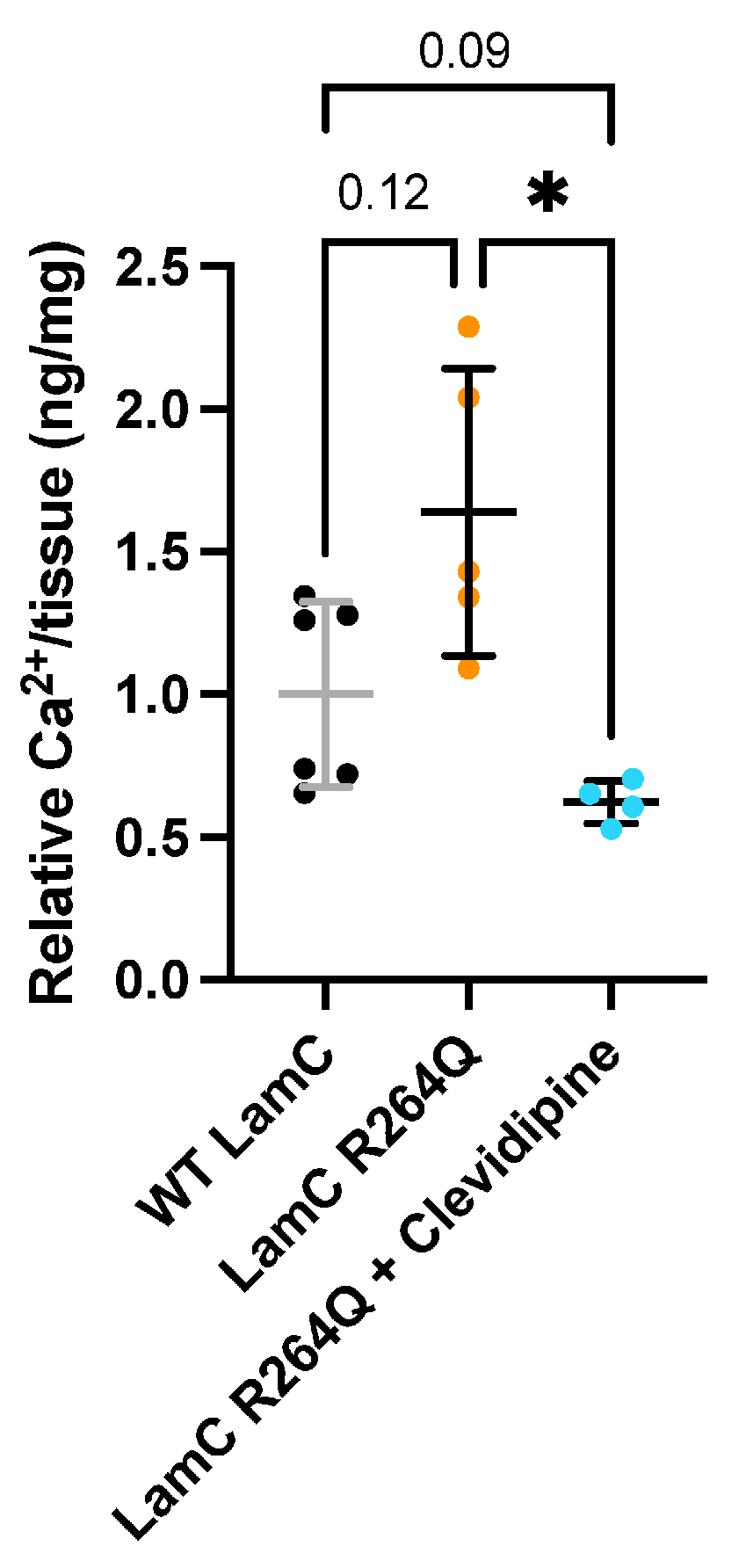
Since we obtained rescue of viability upon treatment with calcium channel blockers, we aimed to determine their mechanism of action in Drosophila muscle. We dissected muscles from larvae expressing wild-type LamC and LamC R264Q with and without treatment with 1.0 and 5.0 μM clevidipine and measured intracellular calcium. Muscle fillets were prepared from wandering third instar Drosophila larvae, treated with detergent, homogenized, centrifuged, and the supernatant used in a colorimetric assay (Abcam, Cambridge, UK). The results showed that larvae expressing LamC R264Q had increased calcium compared to the control, albeit variable and trending towards significance (Figure 6). Extracts from clevidipine-treated larvae expressing LamC R264Q showed lower calcium than those from untreated larvae expressing LamC R264Q, trending below the levels in larvae expressing wild-type LamC (Figure 6). Thus, LamC R264Q appears to generally increase calcium levels in muscle and clevidipine reduces those levels, consistent with clevidipine functioning on the Drosophila calcium channel Ca-α1D expressed in muscle [91,92].

3. Discussion

We completed an unbiased drug screen to identify compounds that rescue lethality caused by muscle-specific expression of mutant lamins in Drosophila. Of the 1433 drugs screened, 68 rescued adult viability. Prioritization of these hit compounds by structural similarity clustering revealed six groups of two to three compounds that were at least 75% structurally similar, suggesting engagement of the same biological target. We selected the dihydropyridine (DHP) L-type calcium channel antagonists clevidipine and amlodipine for further analysis given the role of calcium in muscle function [93,94,95]. Calcium is the key driver of muscle excitation–contraction coupling (ECC), and increased intracellular calcium is a common mechanism known to underlie many types of muscular dystrophy [82,83]. Elevated calcium influx can lead to abnormal ECC, calcium-dependent protease (calpain) activation, mitochondrial dysfunction, and increased production of reactive oxygen species, all of which are factors that can lead to muscle dysfunction and, ultimately, myofiber necrosis [82,96,97].
Support for this calcium hypothesis as a common pathogenic mechanism of muscular dystrophy has been derived from manipulations of calcium flux in several animal models and analysis of patient samples. There is ample evidence that myofibers in muscular dystrophy patients and several mouse models of muscular dystrophy have elevated levels of intracellular cytoplasmic calcium [82,96]. Reducing calcium release from the sarcoplasmic reticulum (SR) or increasing calcium reuptake into the SR rescues muscle phenotypes and improves dystrophic pathology in Sgcd-null mice, a model of LGMD, and mdx mice, a model of DMD [98,99,100,101]. Direct calcium influx across the sarcolemma independent of the SR involves transient receptor potential channels (TRPCs), voltage-independent channels that open to allow calcium influx in response to stretch or decreased SR calcium content [82,102]. An increased amount of calcium enters the cytoplasm of muscle cells through these channels in dystrophic versus non-dystrophic muscle [102]. Overexpression of dominant-negative TRPCs that are not permeable to calcium rescues dystrophic pathology in mouse models of muscular dystrophy, while overexpression of wild-type TRPCs induces dystrophic pathology in wild-type mice [103]. These studies robustly demonstrate that increased intracellular calcium contributes to the pathogenesis of muscular dystrophy, and correcting dysregulation of calcium homeostasis has therapeutic potential [82].
An attractive therapeutic target for addressing this dysregulated calcium homeostasis is LTCCs, as they are readily targetable by DHP and non-DHP calcium channel blockers [104]. In mammals, the LTCCs are Cav1.1, 1.2, 1.3, and 1.4 encoded by CACNA1S, C, D, and F, respectively [105,106]. Cav1.1 is the skeletal muscle-specific LTCC [93], Cav1.2 is the primary LTCC in cardiomyocytes and vascular smooth muscle [104], Cav1.3 is the primary LTCC in cells of the sinoatrial and atrioventricular nodes of the heart [104], and Cav1.4 is largely restricted to the retina and plays no role in striated muscle [104]. Biochemically, DHPs have a higher affinity for inactivated LTCCs and bind the external, lipid-facing surface of the channel pore to stabilize the inactive channel state [104,107,108]. Conversely, the non-DHPs verapamil and diltiazem have higher affinity for the open channel state and bind the central cavity of the channel pore on the intracellular side, directly blocking the pore [104,107,108]. Physiologically, DHPs primarily target Cav1.2 in vascular smooth muscle and cause vasodilation, lowering blood pressure [84,104,109]. Cav1.3 is five- to ten-fold less sensitive to DHPs than Cav1.2, while the non-DHPs target Cav1.2 and Cav1.3 equivalently [84,104,109]. Inhibition of Cav1.3 in the sinoatrial and atrioventricular nodes of the heart by the non-DHPs reduces cardiac conduction and contractility, making them useful for treating cardiac arrhythmias in addition to hypertension [84,104,109].
Despite these primary actions on Cav1.2 and Cav1.3 in vascular smooth muscle and the heart, both DHPs and non-DHPs have been extensively tested for therapeutic benefit in animal models of muscular dystrophy based on their ability to target Cav1.1 with nanomolar affinity and the ready availability of the drugs [104,110,111]. However, the LTCC appears to be a poorer target in skeletal muscle, as the skeletal muscle LTCC is mechanically coupled to RyR1 and external calcium does not need to pass through the LTCC to induce RyR1 opening [82]. Despite this, the non-DHP verapamil and diltiazem and the DHP nifedipine improved dystrophic phenotypes, diaphragm muscle pathology, and muscle strength in mdx mice while decreasing intracellular calcium content [112,113,114]. In Dysf-null mice, a model of LGMD, increased calcium leak through RyR1 is a known mechanism of pathogenesis [115]. DHPs, specifically nifedipine, and the non-DHP verapamil can reduce this leak by modifying the interaction between Cav1.1 and RyR1 [116,117,118]. In myotonic dystrophy, increased skipping of CACNA1S exon 29 results in a Cav1.1 isoform highly deleterious to muscle function, altering the gating of RyR1 and leading to significantly increased calcium influx [119,120]. The reduced function of muscles expressing this Cav1.1 isoform is attenuated by the non-DHP verapamil [120].
While mammals have four LTCCs, Ca-α1D is the single LTCC in Drosophila [121]. Mutations in the Ca-α1D gene cause several phenotypes indicative of muscle contraction defects in embryos, larvae, and adult flies [91]. Calcium currents through this channel in larval muscle are sensitive to DHPs, diltiazem, and verapamil [92]. Ca-α1D is also the primary calcium channel expressed in the fly myocardium, and knockdown of Ca-α1D specifically in the fly heart abolishes cardiac contraction [122]. Calcium currents through Ca-α1D in the Drosophila heart are also DHP-sensitive [122]. Thus, Drosophila Ca-α1D shares physiological similarities with mammalian Cav1.1, 1.2, and 1.3 and has conserved roles in cardiac and muscle function [123]. Based on our findings of intracellular calcium in muscle in these models (Figure 6), we infer that clevidipine is likely functioning through the Drosophila Ca-α1D channel; however, this requires further investigation.
Consistent with the conservation of the LTCC in Drosophila and the success of LTCC blockers in rescuing muscle phenotypes in other animal models of muscular dystrophy, follow-up testing of clevidipine and amlodipine from an alternative source confirmed the ability of these DHPs to restore adult viability following expression of the R264Q mutant lamin in our studies. Clevidipine and amlodipine treatment rescued lethality caused by three mutant lamins but not four others tested, demonstrating that the positive effect of these compounds is not confined to a single amino acid substitution. However, the positive effect is also not universal across the mutational spectrum of laminopathies. The fact that the non-DHPs verapamil and diltiazem were also effective at restoring viability in the three mutants rescued by the DHPs demonstrates that the therapeutic benefit of blocking the L-type calcium channels is not confined to an individual drug class. Collectively, these results support L-type calcium channel antagonists as potential drug repurposing candidates in at least some cases of LMNA-MD.
While our study and those of others support the efficacy of LTCC blockers in animal models of muscular dystrophy, clinical trials assessing their benefits for individuals with DMD showed mixed results [82,112]. Verapamil is effective in improving muscle strength but results in significant cardiac side effects [124]. Diltiazem has been assessed in multiple trials with opposing results [125,126]. Nifedipine, a DHP, was deemed to have no significant benefit in DMD [127]. The lack of a clear effect in patients as opposed to animal models could be due to several factors, including differences in drug metabolism, variations in dosing, and heterogeneity in clinical trial cohorts [112]. While further studies are necessary to clarify the mechanisms underlying the variability in outcomes in humans, the success of these compounds in animals supports the view that pathological calcium influx is a targetable mechanism in muscular dystrophy.
Muscular dystrophies in which altered calcium homeostasis has been implicated as a mechanism of pathogenesis are primarily caused by mutations in genes encoding cytoskeletal proteins or proteins that connect the cytoskeleton and extracellular matrix and contribute to intracellular force transmission [82,96,128,129]. All the genes encoding nuclear envelope proteins that are implicated in muscular dystrophy are either part of the Linker of Nucleoskeleton and Cytoskeleton (LINC) complex or directly interact with LINC complex components [128,130]. This raises the possibility that muscular dystrophy-associated alterations to nuclear envelope proteins could dysregulate calcium homeostasis through a loss of muscle membrane integrity driven by cytoskeletal defects akin to those caused by mutations in genes encoding cytoskeletal components directly [128]. It is known that pathogenic mutations in lamins can cause mislocalization of other nuclear envelope proteins, including the LINC complex, leading to impaired nucleo-cytoskeletal coupling, mislocalization of cytoskeletal proteins in the perinuclear region, and impaired intracellular force transmission [1,20,54,58,131,132]. While further studies are needed to define the connection between calcium regulation and lamins, our drug screen has provided new insights on disease mechanisms and potential treatments for LMNA-MD.

4. Materials and Methods

4.1. Drosophila Culture

Drosophila stocks were maintained on a sucrose and cornmeal-based medium at 25 °C unless otherwise stated. The generation of LamC transgenic stocks was previously described, except for LamC H545P and S72P [20,52,54,56]. Muscle-specific expression of the mutant LamC transgenes was achieved by crossing the transgenic lines to the C57-Gal4 driver, which expresses the yeast Gal4 specifically in larval body wall muscles [74,75]. The resulting progeny express the LamC transgene exclusively in the larval body wall muscles.

4.2. Primary Drug Screen

The Prestwick Chemical Library (version 20, Illkirch, France) was purchased commercially. The library contained 1520 compounds dissolved at 10 mM in DMSO. Drugs were diluted to 1 mM in 10% DMSO with phosphate-buffered saline in 96-well plates (USA Scientific, cat# 5665-1101, Ocala, FL, USA). In total, 10 µL of each drug was added to a 15 mL vial (Sigma Aldrich, cat# Z617865-1600EA, St. Louis, MO, USA) containing 2 mL liquid Drosophila culture medium, generating a final drug concentration of 5 µM and 0.05% DMSO. Next, 10 µL of 10% DMSO was added to separate vials with 2 mL of Drosophila medium as a control. The food was allowed to solidify at 17 °C overnight. Compounds were screened in batches of 40. Two C57-Gal4 female virgins and two LamC R264Q transgenic males were placed in each vial, allowed to mate, and removed after four days. Offspring expressing LamC R264Q in larval body wall muscles consumed the drug-containing food throughout development at 25 °C, pupated, and adult viability was assessed 14 days post mating. Drugs were deemed “hits” if they resulted in any restoration of adult viability, given that expression of LamC R264Q is well established to result in completely penetrant pupal lethality [20]. The number of live adults per hit vial ranged from one to eight, and the number of dead pupae remaining per vial averaged approximately 30.

4.3. Molecular Fingerprinting and Structural Clustering

The molecular similarity of the hit drugs was computed using the RDKit package followed by Butina clustering according to standard methods [76,78,81]. RDKit is an open-source cheminformatics tool (https://www.rdkit.org (accessed on 15 September 2025), https://doi.org/10.5281/zenodo.6961488) that was run using Python 3.1.3. SMILES strings for all screened molecules were provided as part of the purchase of the Prestwick Chemical Library. SMILES strings of the 68 hit drugs identified in the primary screen were imported into RDKit for processing. Molecular fingerprints were generated as bit vectors and, subsequently, a bulk Tanimoto similarity calculation was employed to determine the molecular similarity for all pairwise comparisons of compounds. The dissimilarity (distance) matrix was then calculated as 1 minus the Tanimoto similarity for each pairwise comparison. The dissimilarity coefficients for each pairwise comparison were further assessed using the Butina clustering approach, using a cutoff threshold of 0.25, to generate clusters of multiple structurally similar drugs.

4.4. Secondary Screen and the Assessment of Broad Efficacy

A list of compounds used for the secondary screen and the assessment of broad efficacy is provided in Table S1. Compounds were dissolved at 10 mM in DMSO and diluted to 1 mM in 10% DMSO in phosphate-buffered saline immediately prior to use. Compounds were added to 15 mL vials (Sigma Aldrich, cat# Z617865-1600EA, St. Louis, MO, USA) at appropriate volumes to generate final concentrations between 0.5 and 50.0 µM (0.5, 1.25, 2.5, 3.75, 5.0, 12.5, 25.0, 37.5, 50.0 µM). 10 µL of 10% DMSO in phosphate-buffered saline alone was added to additional vials as a vehicle-only control. Adult viability 14 days post mating was assessed identically to the primary screen. The number of live adults per vial ranged from one to five. The number of vials producing or not producing live adults was compared between drug-treated flies and those treated with DMSO alone as a control in a contingency table. Statistical significance was assessed using Fisher’s Exact Test in GraphPad Prism (v10.6.1, GraphPad Software, San Diego, CA, USA).

4.5. Quantification of Adult Viability

Flies possessing wild-type LamC and either LamC H545P or S72P transgenes were crossed with flies possessing the muscle-specific C57-Gal4 driver for quantitative assessment of adult viability. After four days, the adults were removed from the vials, and progeny were allowed to develop. The resulting dead pupae and live adults were counted after 14 days. The percent adult viability was calculated as [# living adults/(# living adults + # dead pupae)] × 100. The total number of progeny ranged from 300 to 695 per genotype.

4.6. Measurements of Intramuscular Calcium

Muscle tissue was dissected from 15 third instar larvae per sample, with four to six independent samples analyzed per genotype. The muscles were weighed, washed, treated with detergent, homogenized, centrifuged, and the supernatant mixed with a chromogenic reagent that detects calcium (#ab102505, Abcam, Cambridge, UK). Optical density was detected using a 96-well plate reader (Synergy NEO, Agilent BioTek, Santa Clara, CA, USA) with the absorbance read at 575 nm. A dilution of calcium standards at known concentrations provided by the manufacturer was used to generate a standard curve, allowing for calcium concentrations/mg tissue to be calculated. For each independent trial, values were normalized to those of larvae expressing wild-type LamC, set to 1, so that independent trials could be compared (Figure 6). The average and standard deviations were plotted using GraphPad Prism (v10.6.1, GraphPad Software, San Diego, CA, USA) and a one-way ANOVA with Dunnett’s correction was used to determine statistical significance.

4.7. Morphometric Analysis of Muscle Fibers

Larval body wall muscles were dissected, fixed with formaldehyde, and stained with phalloidin as previously described [20,52,56]. Muscles were imaged using a Leica Thunder microscope (Leica, Wetzlar, Germany). Muscles six and seven were identified in each larval muscle fillet and width measurements were made using the straight-line selection tool in Fiji software (https://imagej.net/software/fiji/ (accessed on 16 August 2026), ImageJ2, v2.16.0/1.54p) [133]. Muscles from three to six larvae were measured per genotype. Averages with standard deviation were plotted, and a one-way ANOVA was performed with Dunnett’s correction (GraphPad Prism v10.6.1, GraphPad Software, San Diego, CA, USA).

Supplementary Materials

The following supporting information can be downloaded at https://www.mdpi.com/article/10.3390/ijms27188367/s1.

Author Contributions

Conceptualization, L.L.W.; Methodology, N.P.M. and L.L.W.; Software, H.M.-M.; Formal analysis, N.P.M., B.A.P., B.E.H., Z.D., H.M.-M. and A.S.; Investigation, N.P.M., B.A.P., B.E.H., Z.D., H.M.-M., M.W.S., A.T.W., A.R.B. and A.S.; Resources, N.P.M. and L.L.W.; Data Curation, N.P.M., B.A.P., B.E.H., A.R.B. and L.L.W., Writing—Original Draft Preparation, N.P.M. and L.L.W., Writing—Review and Editing, N.P.M., B.A.P., B.E.H., Z.D., H.M.-M., M.W.S., A.T.W., A.R.B., A.S. and L.L.W., Supervision, N.P.M. and L.L.W., Project Administration, L.L.W., Funding Acquisition, N.P.M. and L.L.W. All authors have read and agreed to the published version of the manuscript.

Funding

National Institutes of Health, National Institute of General Medical Sciences T32GM144636 to N.P.M.; National Institutes of Health, National Institute of Arthritis and Musculoskeletal and Skin Diseases R21AR075193 to L.L.W.; Muscular Dystrophy Association Research Grant 22-1061420 to L.L.W.; American Heart Association Predoctoral Fellowship 25PRE1373209 to N.P.M.

Institutional Review Board Statement

Not applicable.

Informed Consent Statement

Not applicable.

Data Availability Statement

The original contributions presented in this study are included in the article/Supplementary Materials. Further inquiries can be directed to the corresponding author.

Acknowledgments

The authors would like to acknowledge Michael J. Schnieders for assistance with molecular fingerprinting, Clement Y. Chow and Kevin A. Hope for technical assistance, and the University of Iowa Office of Undergraduate Research for material support.

Conflicts of Interest

The authors declare no conflicts of interest.

References

  1. Sobo, J.M.; Alagna, N.S.; Sun, S.X.; Wilson, K.L.; Reddy, K.L. Lamins: The backbone of the nucleocytoskeleton interface. Curr. Opin. Cell Biol. 2024, 86, 102313. [Google Scholar] [CrossRef] [Scilit] [PubMed]
  2. Hetzer, M.W. The nuclear envelope. Cold Spring Harb. Perspect. Biol. 2010, 2, a000539. [Google Scholar] [CrossRef] [Scilit] [PubMed]
  3. Nmezi, B.; Xu, J.; Fu, R.; Armiger, T.J.; Rodriguez-Bey, G.; Powell, J.S.; Ma, H.; Sullivan, M.; Tu, Y.; Chen, N.Y.; et al. Concentric organization of A- and B-type lamins predicts their distinct roles in the spatial organization and stability of the nuclear lamina. Proc. Natl. Acad. Sci. USA 2019, 116, 4307–4315. [Google Scholar] [CrossRef] [Scilit] [PubMed]
  4. Padiath, Q.S.; Saigoh, K.; Schiffmann, R.; Asahara, H.; Yamada, T.; Koeppen, A.; Hogan, K.; Ptacek, L.J.; Fu, Y.H. Lamin B1 duplications cause autosomal dominant leukodystrophy. Nat. Genet. 2006, 38, 1114–1123, Correction in Nat. Genet. 2007, 39, 276. [Google Scholar] [CrossRef] [Scilit] [PubMed]
  5. Hegele, R.A.; Cao, H.; Liu, D.M.; Costain, G.A.; Charlton-Menys, V.; Rodger, N.W.; Durrington, P.N. Sequencing of the reannotated LMNB2 gene reveals novel mutations in patients with acquired partial lipodystrophy. Am. J. Hum. Genet. 2006, 79, 383–389. [Google Scholar] [CrossRef] [Scilit] [PubMed]
  6. Worman, H.J.; Ostlund, C.; Wang, Y. Diseases of the nuclear envelope. Cold Spring Harb. Perspect. Biol. 2010, 2, a000760. [Google Scholar] [CrossRef] [Scilit] [PubMed]
  7. Worman, H.J.; Bonne, G. “Laminopathies”: A wide spectrum of human diseases. Exp. Cell Res. 2007, 313, 2121–2133. [Google Scholar] [CrossRef] [Scilit] [PubMed]
  8. Worman, H.J.; Courvalin, J.C. How do mutations in lamins A and C cause disease? J. Clin. Investig. 2004, 113, 349–351. [Google Scholar] [CrossRef] [PubMed]
  9. Worman, H.J. Nuclear lamins and laminopathies. J. Pathol. 2012, 226, 316–325. [Google Scholar] [CrossRef] [Scilit] [PubMed]
  10. Anderson, C.L.; Langer, E.R.; Routes, T.C.; McWilliams, S.F.; Bereslavskyy, I.; Kamp, T.J.; Eckhardt, L.L. Most myopathic lamin variants aggregate: A functional genomics approach for assessing variants of uncertain significance. npj Genom. Med. 2021, 6, 103. [Google Scholar] [CrossRef] [Scilit] [PubMed]
  11. Storey, E.C.; Fuller, H.R. Genotype-Phenotype Correlations in Human Diseases Caused by Mutations of LINC Complex-Associated Genes: A Systematic Review and Meta-Summary. Cells 2022, 11, 4065. [Google Scholar] [CrossRef] [Scilit] [PubMed]
  12. Crasto, S.; My, I.; Di Pasquale, E. The Broad Spectrum of LMNA Cardiac Diseases: From Molecular Mechanisms to Clinical Phenotype. Front. Physiol. 2020, 11, 761. [Google Scholar] [CrossRef] [Scilit] [PubMed]
  13. Bertrand, A.T.; Ben Yaou, R.; Bonne, G. Chapter 72—Diseases of the Nucleoskeleton. In Muscle; Hill, J.A., Olson, E.N., Eds.; Academic Press: Boston, MA, USA; Waltham, MA, USA, 2012; pp. 1003–1012. [Google Scholar]
  14. Nicolas, H.A.; Akimenko, M.A.; Tesson, F. Cellular and Animal Models of Striated Muscle Laminopathies. Cells 2019, 8, 291. [Google Scholar] [CrossRef] [Scilit] [PubMed]
  15. Quijano-Roy, S.; Mbieleu, B.; Bonnemann, C.G.; Jeannet, P.Y.; Colomer, J.; Clarke, N.F.; Cuisset, J.M.; Roper, H.; De Meirleir, L.; D’Amico, A.; et al. De novo LMNA mutations cause a new form of congenital muscular dystrophy. Ann. Neurol. 2008, 64, 177–186. [Google Scholar] [CrossRef] [Scilit] [PubMed]
  16. Bonne, G.; Di Barletta, M.R.; Varnous, S.; Becane, H.M.; Hammouda, E.H.; Merlini, L.; Muntoni, F.; Greenberg, C.R.; Gary, F.; Urtizberea, J.A.; et al. Mutations in the gene encoding lamin A/C cause autosomal dominant Emery-Dreifuss muscular dystrophy. Nat. Genet. 1999, 21, 285–288. [Google Scholar] [CrossRef] [Scilit] [PubMed]
  17. Muchir, A.; Bonne, G.; van der Kooi, A.J.; van Meegen, M.; Baas, F.; Bolhuis, P.A.; de Visser, M.; Schwartz, K. Identification of mutations in the gene encoding lamins A/C in autosomal dominant limb girdle muscular dystrophy with atrioventricular conduction disturbances (LGMD1B). Hum. Mol. Genet. 2000, 9, 1453–1459. [Google Scholar] [CrossRef] [Scilit] [PubMed]
  18. Fatkin, D.; MacRae, C.; Sasaki, T.; Wolff, M.R.; Porcu, M.; Frenneaux, M.; Atherton, J.; Vidaillet, H.J., Jr.; Spudich, S.; De Girolami, U.; et al. Missense mutations in the rod domain of the lamin A/C gene as causes of dilated cardiomyopathy and conduction-system disease. N. Engl. J. Med. 1999, 341, 1715–1724. [Google Scholar] [CrossRef] [Scilit] [PubMed]
  19. Worman, H.J. Human Diseases Related to Nuclear Envelope Proteins. In The Functional Nucleus; Bazett-Jones, D.P., Dellaire, G., Eds.; Springer International Publishing: Cham, Switzerland, 2016; pp. 3–25. [Google Scholar]
  20. Hinz, B.E.; Walker, S.G.; Xiong, A.; Gogal, R.A.; Schnieders, M.J.; Wallrath, L.L. In Silico and In Vivo Analysis of Amino Acid Substitutions That Cause Laminopathies. Int. J. Mol. Sci. 2021, 22, 11226. [Google Scholar] [CrossRef] [Scilit] [PubMed]
  21. Ben Yaou, R.; Yun, P.; Dabaj, I.; Norato, G.; Donkervoort, S.; Xiong, H.; Nascimento, A.; Maggi, L.; Sarkozy, A.; Monges, S.; et al. International retrospective natural history study of LMNA-related congenital muscular dystrophy. Brain Commun. 2021, 3, fcab075. [Google Scholar] [CrossRef] [Scilit] [PubMed]
  22. Arimura, T.; Helbling-Leclerc, A.; Massart, C.; Varnous, S.; Niel, F.; Lacene, E.; Fromes, Y.; Toussaint, M.; Mura, A.M.; Keller, D.I.; et al. Mouse model carrying H222P-Lmna mutation develops muscular dystrophy and dilated cardiomyopathy similar to human striated muscle laminopathies. Hum. Mol. Genet. 2005, 14, 155–169. [Google Scholar] [CrossRef] [Scilit] [PubMed]
  23. Bonne, G.; Mercuri, E.; Muchir, A.; Urtizberea, A.; Becane, H.M.; Recan, D.; Merlini, L.; Wehnert, M.; Boor, R.; Reuner, U.; et al. Clinical and molecular genetic spectrum of autosomal dominant Emery-Dreifuss muscular dystrophy due to mutations of the lamin A/C gene. Ann. Neurol. 2000, 48, 170–180. [Google Scholar] [CrossRef] [Scilit]
  24. Cesar, S.; Coll, M.; Fiol, V.; Fernandez-Falgueras, A.; Cruzalegui, J.; Iglesias, A.; Moll, I.; Perez-Serra, A.; Martinez-Barrios, E.; Ferrer-Costa, C.; et al. LMNA-related muscular dystrophy: Identification of variants in alternative genes and personalized clinical translation. Front. Genet. 2023, 14, 1135438. [Google Scholar] [CrossRef] [Scilit] [PubMed]
  25. Ng, S.R.; Wang, A.; Tukijan, F.; Murugiah, A.; Tan, L.T.; Teo, A.; Lee, Q.Y.; Tan, H.; Choy, A.; Ong, M.; et al. Abstract: 4359043: NVC-001-An AAV gene therapy functional cure for LMNA dilated cardiomyopathy. Circulation 2025, 152, A4359043. [Google Scholar] [CrossRef] [Scilit]
  26. Czifrus, E.; Berlau, D.J. Corticosteroids for the treatment of Duchenne muscular dystrophy: A safety review. Expert Opin. Drug Saf. 2024, 23, 1237–1247. [Google Scholar] [CrossRef] [Scilit] [PubMed]
  27. Zulfiqar, E.; Hurjkaliani, S.; Umar, S.A.; Shahzad, M.; Ahsan, M.; Tahir, Q.; Nizami, U.; Rizvi, B.; Khan, A.A.; Baloch, A.; et al. Comparing intermittent and daily prednisone in duchenne muscular dystrophy: A systematic review and meta-analysis. Ann. Med. Surg. 2025, 87, 1637–1645. [Google Scholar] [CrossRef] [Scilit] [PubMed]
  28. Komaki, H.; Hayashi, Y.K.; Tsuburaya, R.; Sugie, K.; Kato, M.; Nagai, T.; Imataka, G.; Suzuki, S.; Saitoh, S.; Asahina, N.; et al. Inflammatory changes in infantile-onset LMNA-associated myopathy. Neuromuscul. Disord. 2011, 21, 563–568. [Google Scholar] [CrossRef] [Scilit] [PubMed]
  29. Fan, Y.; Tan, D.; Song, D.; Zhang, X.; Chang, X.; Wang, Z.; Zhang, C.; Chan, S.H.; Wu, Q.; Wu, L.; et al. Clinical spectrum and genetic variations of LMNA-related muscular dystrophies in a large cohort of Chinese patients. J. Med. Genet. 2021, 58, 326–333. [Google Scholar] [CrossRef] [PubMed]
  30. Atalaia, A.; Ben Yaou, R.; Wahbi, K.; De Sandre-Giovannoli, A.; Vigouroux, C.; Bonne, G. Laminopathies’ Treatments Systematic Review: A Contribution Towards a ‘Treatabolome’. J. Neuromuscul. Dis. 2021, 8, 419–439. [Google Scholar] [CrossRef] [Scilit] [PubMed]
  31. Sheikh, O.; Yokota, T. Restoring Protein Expression in Neuromuscular Conditions: A Review Assessing the Current State of Exon Skipping/Inclusion and Gene Therapies for Duchenne Muscular Dystrophy and Spinal Muscular Atrophy. BioDrugs 2021, 35, 389–399. [Google Scholar] [CrossRef] [Scilit] [PubMed]
  32. Leckie, J.; Zia, A.; Yokota, T. An Updated Analysis of Exon-Skipping Applicability for Duchenne Muscular Dystrophy Using the UMD-DMD Database. Genes 2024, 15, 1489. [Google Scholar] [CrossRef] [Scilit] [PubMed]
  33. Chwalenia, K.; Feng, V.Y.; Hemmer, N.; Friedrichsen, H.J.; Vorobieva, I.; Wood, M.J.A.; Roberts, T.C. AAV microdystrophin gene replacement therapy for Duchenne muscular dystrophy: Progress and prospects. Gene Ther. 2025, 32, 447–461. [Google Scholar] [CrossRef] [Scilit] [PubMed]
  34. Querin, G.; Colella, M. Gene therapy for primary myopathies: Literature review and prospects. Arch. Pediatr. 2023, 30, 8S18–18S23. [Google Scholar] [CrossRef] [Scilit] [PubMed]
  35. Mendell, J.R.; Pozsgai, E.R.; Lewis, S.; Griffin, D.A.; Lowes, L.P.; Alfano, L.N.; Lehman, K.J.; Church, K.; Reash, N.F.; Iammarino, M.A.; et al. Gene therapy with bidridistrogene xeboparvovec for limb-girdle muscular dystrophy type 2E/R4: Phase 1/2 trial results. Nat. Med. 2024, 30, 199–206. [Google Scholar] [CrossRef] [Scilit] [PubMed]
  36. Scharner, J.; Figeac, N.; Ellis, J.A.; Zammit, P.S. Ameliorating pathogenesis by removing an exon containing a missense mutation: A potential exon-skipping therapy for laminopathies. Gene Ther. 2015, 22, 503–515. [Google Scholar] [CrossRef] [Scilit] [PubMed]
  37. Mercuri, E.; Poppe, M.; Quinlivan, R.; Messina, S.; Kinali, M.; Demay, L.; Bourke, J.; Richard, P.; Sewry, C.; Pike, M.; et al. Extreme variability of phenotype in patients with an identical missense mutation in the lamin A/C gene: From congenital onset with severe phenotype to milder classic Emery-Dreifuss variant. Arch. Neurol. 2004, 61, 690–694. [Google Scholar] [CrossRef] [Scilit] [PubMed]
  38. Bertrand, A.T.; Chikhaoui, K.; Yaou, R.B.; Bonne, G. Clinical and genetic heterogeneity in laminopathies. Biochem. Soc. Trans. 2011, 39, 1687–1692. [Google Scholar] [CrossRef] [Scilit] [PubMed]
  39. Byrne, B.J.; Corti, M.; Muntoni, F. Considerations for Systemic Use of Gene Therapy. Mol. Ther. 2021, 29, 422–423. [Google Scholar] [CrossRef] [Scilit] [PubMed]
  40. McNally, E.M. Improving the safety of systemic viral gene therapy. J. Clin. Investig. 2024, 134, e177078. [Google Scholar] [CrossRef] [Scilit] [PubMed]
  41. Phares, S.; Trusheim, M.; Emond, S.K.; Pearson, S.D. Managing the challenges of paying for gene therapy: Strategies for market action and policy reform in the United States. J. Comp. Eff. Res. 2024, 13, e240118. [Google Scholar] [CrossRef] [Scilit] [PubMed]
  42. Zhao, Y.; Wang, Y.; Tulehalede, A.; Meng, Z.; Xu, L.; Bai, H.; Sha, J.; Xie, W.; Geng, J. Drosophila melanogaster: A Model Organism in Muscular Dystrophy Studies. Int. J. Mol. Sci. 2025, 26, 1459. [Google Scholar] [CrossRef] [Scilit] [PubMed]
  43. Potikanond, S.; Nimlamool, W.; Noordermeer, J.; Fradkin, L.G. Muscular Dystrophy Model. Adv. Exp. Med. Biol. 2018, 1076, 147–172. [Google Scholar] [CrossRef] [Scilit] [PubMed]
  44. van der Plas, M.C.; Pilgram, G.S.; de Jong, A.W.; Bansraj, M.R.; Fradkin, L.G.; Noordermeer, J.N. Drosophila Dystrophin is required for integrity of the musculature. Mech. Dev. 2007, 124, 617–630. [Google Scholar] [CrossRef] [Scilit] [PubMed]
  45. Taghli-Lamallem, O.; Akasaka, T.; Hogg, G.; Nudel, U.; Yaffe, D.; Chamberlain, J.S.; Ocorr, K.; Bodmer, R. Dystrophin deficiency in Drosophila reduces lifespan and causes a dilated cardiomyopathy phenotype. Aging Cell 2008, 7, 237–249. [Google Scholar] [CrossRef] [Scilit] [PubMed]
  46. Allikian, M.J.; Bhabha, G.; Dospoy, P.; Heydemann, A.; Ryder, P.; Earley, J.U.; Wolf, M.J.; Rockman, H.A.; McNally, E.M. Reduced life span with heart and muscle dysfunction in Drosophila sarcoglycan mutants. Hum. Mol. Genet. 2007, 16, 2933–2943. [Google Scholar] [CrossRef] [Scilit] [PubMed]
  47. Nakamura, N.; Stalnaker, S.H.; Lyalin, D.; Lavrova, O.; Wells, L.; Panin, V.M. Drosophila Dystroglycan is a target of O-mannosyltransferase activity of two protein O-mannosyltransferases, Rotated Abdomen and Twisted. Glycobiology 2010, 20, 381–394. [Google Scholar] [CrossRef] [Scilit] [PubMed]
  48. Haines, N.; Seabrooke, S.; Stewart, B.A. Dystroglycan and protein O-mannosyltransferases 1 and 2 are required to maintain integrity of Drosophila larval muscles. Mol. Biol. Cell 2007, 18, 4721–4730. [Google Scholar] [CrossRef] [Scilit] [PubMed]
  49. Ueyama, M.; Akimoto, Y.; Ichimiya, T.; Ueda, R.; Kawakami, H.; Aigaki, T.; Nishihara, S. Increased apoptosis of myoblasts in Drosophila model for the Walker-Warburg syndrome. PLoS ONE 2010, 5, e11557. [Google Scholar] [CrossRef] [Scilit] [PubMed]
  50. Bhide, S.; Trujillo, A.S.; O’Connor, M.T.; Young, G.H.; Cryderman, D.E.; Chandran, S.; Nikravesh, M.; Wallrath, L.L.; Melkani, G.C. Increasing autophagy and blocking Nrf2 suppress laminopathy-induced age-dependent cardiac dysfunction and shortened lifespan. Aging Cell 2018, 17, e12747. [Google Scholar] [CrossRef] [Scilit] [PubMed]
  51. Coombs, G.S.; Rios-Monterrosa, J.L.; Lai, S.; Dai, Q.; Goll, A.C.; Ketterer, M.R.; Valdes, M.F.; Uche, N.; Benjamin, I.J.; Wallrath, L.L. Modulation of muscle redox and protein aggregation rescues lethality caused by mutant lamins. Redox Biol. 2021, 48, 102196. [Google Scholar] [CrossRef] [Scilit] [PubMed]
  52. Dialynas, G.; Flannery, K.M.; Zirbel, L.N.; Nagy, P.L.; Mathews, K.D.; Moore, S.A.; Wallrath, L.L. LMNA variants cause cytoplasmic distribution of nuclear pore proteins in Drosophila and human muscle. Hum. Mol. Genet. 2012, 21, 1544–1556. [Google Scholar] [CrossRef] [Scilit] [PubMed]
  53. Dialynas, G.; Shrestha, O.K.; Ponce, J.M.; Zwerger, M.; Thiemann, D.A.; Young, G.H.; Moore, S.A.; Yu, L.; Lammerding, J.; Wallrath, L.L. Myopathic lamin mutations cause reductive stress and activate the nrf2/keap-1 pathway. PLoS Genet. 2015, 11, e1005231. [Google Scholar] [CrossRef] [Scilit] [PubMed]
  54. Shaw, N.M.; Rios-Monterrosa, J.L.; Fedorchak, G.R.; Ketterer, M.R.; Coombs, G.S.; Lammerding, J.; Wallrath, L.L. Effects of mutant lamins on nucleo-cytoskeletal coupling in Drosophila models of LMNA muscular dystrophy. Front. Cell Dev. Biol. 2022, 10, 934586. [Google Scholar] [CrossRef] [Scilit] [PubMed]
  55. Earle, A.J.; Kirby, T.J.; Fedorchak, G.R.; Isermann, P.; Patel, J.; Iruvanti, S.; Moore, S.A.; Bonne, G.; Wallrath, L.L.; Lammerding, J. Mutant lamins cause nuclear envelope rupture and DNA damage in skeletal muscle cells. Nat. Mater. 2020, 19, 464–473. [Google Scholar] [CrossRef] [Scilit] [PubMed]
  56. Walker, S.G.; Langland, C.J.; Viles, J.; Hecker, L.A.; Wallrath, L.L. Drosophila Models Reveal Properties of Mutant Lamins That Give Rise to Distinct Diseases. Cells 2023, 12, 1142. [Google Scholar] [CrossRef] [Scilit] [PubMed]
  57. Chandran, S.; Suggs, J.A.; Wang, B.J.; Han, A.; Bhide, S.; Cryderman, D.E.; Moore, S.A.; Bernstein, S.I.; Wallrath, L.L.; Melkani, G.C. Suppression of myopathic lamin mutations by muscle-specific activation of AMPK and modulation of downstream signaling. Hum. Mol. Genet. 2019, 28, 351–371. [Google Scholar] [CrossRef] [Scilit] [PubMed]
  58. Zwerger, M.; Jaalouk, D.E.; Lombardi, M.L.; Isermann, P.; Mauermann, M.; Dialynas, G.; Herrmann, H.; Wallrath, L.L.; Lammerding, J. Myopathic lamin mutations impair nuclear stability in cells and tissue and disrupt nucleo-cytoskeletal coupling. Hum. Mol. Genet. 2013, 22, 2335–2349. [Google Scholar] [CrossRef] [Scilit] [PubMed]
  59. Morel, V.; Lepicard, S.; Rey, A.N.; Parmentier, M.L.; Schaeffer, L. Drosophila Nesprin-1 controls glutamate receptor density at neuromuscular junctions. Cell. Mol. Life Sci. 2014, 71, 3363–3379. [Google Scholar] [CrossRef] [Scilit] [PubMed]
  60. Collins, M.A.; Mandigo, T.R.; Camuglia, J.M.; Vazquez, G.A.; Anderson, A.J.; Hudson, C.H.; Hanron, J.L.; Folker, E.S. Emery-Dreifuss muscular dystrophy-linked genes and centronuclear myopathy-linked genes regulate myonuclear movement by distinct mechanisms. Mol. Biol. Cell 2017, 28, 2303–2317. [Google Scholar] [CrossRef] [Scilit] [PubMed]
  61. Mandigo, T.R.; Turcich, B.D.; Anderson, A.J.; Hussey, M.R.; Folker, E.S. Drosophila emerins control LINC complex localization and transcription to regulate myonuclear position. J. Cell Sci. 2019, 132, jcs235580. [Google Scholar] [CrossRef] [Scilit] [PubMed]
  62. Dar, A.C.; Das, T.K.; Shokat, K.M.; Cagan, R.L. Chemical genetic discovery of targets and anti-targets for cancer polypharmacology. Nature 2012, 486, 80–84. [Google Scholar] [CrossRef] [Scilit] [PubMed]
  63. Levine, B.D.; Cagan, R.L. Drosophila Lung Cancer Models Identify Trametinib plus Statin as Candidate Therapeutic. Cell Rep. 2016, 14, 1477–1487. [Google Scholar] [CrossRef] [Scilit] [PubMed]
  64. Bangi, E.; Murgia, C.; Teague, A.G.; Sansom, O.J.; Cagan, R.L. Functional exploration of colorectal cancer genomes using Drosophila. Nat. Commun. 2016, 7, 13615. [Google Scholar] [CrossRef] [Scilit] [PubMed]
  65. Bangi, E.; Ang, C.; Smibert, P.; Uzilov, A.V.; Teague, A.G.; Antipin, Y.; Chen, R.; Hecht, C.; Gruszczynski, N.; Yon, W.J.; et al. A personalized platform identifies trametinib plus zoledronate for a patient with KRAS-mutant metastatic colorectal cancer. Sci. Adv. 2019, 5, eaav6528. [Google Scholar] [CrossRef] [Scilit] [PubMed]
  66. Bangi, E.; Smibert, P.; Uzilov, A.V.; Teague, A.G.; Gopinath, S.; Antipin, Y.; Chen, R.; Hecht, C.; Gruszczynski, N.; Yon, W.J.; et al. A Drosophila platform identifies a novel, personalized therapy for a patient with adenoid cystic carcinoma. iScience 2021, 24, 102212. [Google Scholar] [CrossRef] [Scilit] [PubMed]
  67. Sonoshita, M.; Scopton, A.P.; Ung, P.M.U.; Murray, M.A.; Silber, L.; Maldonado, A.Y.; Real, A.; Schlessinger, A.; Cagan, R.L.; Dar, A.C. A whole-animal platform to advance a clinical kinase inhibitor into new disease space. Nat. Chem. Biol. 2018, 14, 291–298. [Google Scholar] [CrossRef] [Scilit] [PubMed]
  68. Hope, K.A.; Berman, A.R.; Peterson, R.T.; Chow, C.Y. An in vivo drug repurposing screen and transcriptional analyses reveals the serotonin pathway and GSK3 as major therapeutic targets for NGLY1 deficiency. PLoS Genet. 2022, 18, e1010228. [Google Scholar] [CrossRef] [Scilit] [PubMed]
  69. Dalton, H.M.; Young, N.J.; Berman, A.R.; Evans, H.D.; Peterson, S.J.; Patterson, K.A.; Chow, C.Y. A drug repurposing screen reveals dopamine signaling as a critical pathway underlying potential therapeutics for the rare disease DPAGT1-CDG. PLoS Genet. 2024, 20, e1011458. [Google Scholar] [CrossRef] [Scilit] [PubMed]
  70. Dridi, H.; Wu, W.; Reiken, S.R.; Ofer, R.M.; Liu, Y.; Yuan, Q.; Sittenfeld, L.; Kushner, J.; Muchir, A.; Worman, H.J.; et al. Ryanodine receptor remodeling in cardiomyopathy and muscular dystrophy caused by lamin A/C gene mutation. Hum. Mol. Genet. 2021, 29, 3919–3934, Correction in Hum. Mol. Genet. 2024, 33, 1273–1276. [Google Scholar] [CrossRef] [Scilit] [PubMed]
  71. Gaglianone, R.B.; Lamboley, C.R.; Dick, T.; Meizoso-Huesca, A.; Singh, D.P.; Launikonis, B.S. Ryanodine Receptor Ca2+ Leak-Induced Redistribution of Ca2+ in Dystrophic mdx Mouse Muscle. Acta Physiol. 2026, 242, e70251. [Google Scholar] [CrossRef] [Scilit] [PubMed]
  72. Sauer, J.; Marksteiner, J.; Lilliu, E.; Dostal, C.; Antunes Goncalves, A.I.; Kiss, A.; Podesser, B.K.; Kubista, H.; Todt, H.; Koenig, X.; et al. Calcium handling properties and arrhythmia vulnerability of cardiomyocytes from dystrophin-deficient mdx mice. Cell Calcium 2026, 136, 103162. [Google Scholar] [CrossRef] [Scilit] [PubMed]
  73. Seguret, M.; Jouve, C.; Ruiz-Velasco, A.; Deshayes, L.; Guesmia, Z.; Pereira, C.; Ragot, V.; Castelli, C.; Wahbi, K.; Fauconnier, J.; et al. LMNA p.H222P mutation causes contractile dysfunction via impaired mitochondrial calcium uptake in human cardiac laminopathy. Cardiovasc. Res. 2026, cvag163. [Google Scholar] [CrossRef] [Scilit] [PubMed]
  74. Dialynas, G.; Speese, S.; Budnik, V.; Geyer, P.K.; Wallrath, L.L. The role of Drosophila Lamin C in muscle function and gene expression. Development 2010, 137, 3067–3077. [Google Scholar] [CrossRef] [Scilit] [PubMed]
  75. Koh, Y.H.; Popova, E.; Thomas, U.; Griffith, L.C.; Budnik, V. Regulation of DLG localization at synapses by CaMKII-dependent phosphorylation. Cell 1999, 98, 353–363. [Google Scholar] [CrossRef] [Scilit] [PubMed]
  76. Ambure, P.; Aher, R.B.; Roy, K. Recent Advances in the Open Access Cheminformatics Toolkits, Software Tools, Workflow Environments, and Databases. In Computer-Aided Drug Discovery; Zhang, W., Ed.; Springer: New York, NY, USA, 2016; pp. 257–296. [Google Scholar]
  77. Landrum, G. RDKit: Cheminformatics and Machine Learning Software. RDKit.org. 2013. Available online: https://www.rdkit.org (accessed on 15 September 2025).
  78. Ryzhkov, F.V.; Ryzhkova, Y.E.; Elinson, M.N. Python tools for structural tasks in chemistry. Mol. Divers. 2025, 29, 3733–3752. [Google Scholar] [CrossRef] [Scilit] [PubMed]
  79. Kristensen, T.G.; Nielsen, J.; Pedersen, C.N. A tree-based method for the rapid screening of chemical fingerprints. Algorithms Mol. Biol. 2010, 5, 9. [Google Scholar] [CrossRef] [Scilit] [PubMed]
  80. Huang, L.; Luo, H.; Li, S.; Wu, F.X.; Wang, J. Drug-drug similarity measure and its applications. Brief. Bioinform. 2021, 22, bbaa265. [Google Scholar] [CrossRef] [Scilit] [PubMed]
  81. Butina, D. Unsupervised Data Base Clustering Based on Daylight’s Fingerprint and Tanimoto Similarity: A Fast and Automated Way To Cluster Small and Large Data Sets. J. Chem. Inf. Comput. Sci. 1999, 39, 747–750. [Google Scholar] [CrossRef] [Scilit]
  82. Burr, A.R.; Molkentin, J.D. Genetic evidence in the mouse solidifies the calcium hypothesis of myofiber death in muscular dystrophy. Cell Death Differ. 2015, 22, 1402–1412. [Google Scholar] [CrossRef] [Scilit] [PubMed]
  83. Jones, K.E.; Hayden, S.L.; Meyer, H.R.; Sandoz, J.L.; Arata, W.H.; Dufrene, K.; Ballaera, C.; Lopez Torres, Y.; Griffin, P.; Kaye, A.M.; et al. The Evolving Role of Calcium Channel Blockers in Hypertension Management: Pharmacological and Clinical Considerations. Curr. Issues Mol. Biol. 2024, 46, 6315–6327. [Google Scholar] [CrossRef] [Scilit] [PubMed]
  84. McKeever, R.G.; Patel, P.; Hamilton, R.J. Calcium Channel Blockers. In StatPearls [Internet]; StatPearls: Treasure Island, FL, USA, 2024. [Google Scholar]
  85. Wang, Y.; Rosenberg, R.L. Ethaverine, a derivative of papaverine, inhibits cardiac L-type calcium channels. Mol. Pharmacol. 1991, 40, 750–755. [Google Scholar] [CrossRef] [Scilit]
  86. Frishman, W.H. Calcium channel blockers: Differences between subclasses. Am. J. Cardiovasc. Drugs 2007, 7, 17–23. [Google Scholar] [CrossRef] [Scilit] [PubMed]
  87. Guillin-Amarelle, C.; Fernandez-Pombo, A.; Sanchez-Iglesias, S.; Araujo-Vilar, D. Lipodystrophic laminopathies: Diagnostic clues. Nucleus 2018, 9, 249–260. [Google Scholar] [CrossRef] [Scilit] [PubMed]
  88. Angori, S.; Capanni, C.; Faulkner, G.; Bean, C.; Boriani, G.; Lattanzi, G.; Cenni, V. Emery-Dreifuss Muscular Dystrophy-Associated Mutant Forms of Lamin A Recruit the Stress Responsive Protein Ankrd2 into the Nucleus, Affecting the Cellular Response to Oxidative Stress. Cell Physiol. Biochem. 2017, 42, 169–184. [Google Scholar] [CrossRef] [Scilit] [PubMed]
  89. Balakrishnan, M.; Sisso, W.J.; Baylies, M.K. Analyzing muscle structure and function throughout the larval instars in live Drosophila. STAR Protoc. 2021, 2, 100291. [Google Scholar] [CrossRef] [Scilit] [PubMed]
  90. Bothe, I.; Baylies, M.K. Drosophila myogenesis. Curr. Biol. 2016, 26, R786–R791. [Google Scholar] [CrossRef] [Scilit] [PubMed]
  91. Eberl, D.F.; Ren, D.; Feng, G.; Lorenz, L.J.; Van Vactor, D.; Hall, L.M. Genetic and developmental characterization of Dmca1D, a calcium channel alpha1 subunit gene in Drosophila melanogaster. Genetics 1998, 148, 1159–1169. [Google Scholar] [CrossRef] [Scilit] [PubMed]
  92. Ren, D.; Xu, H.; Eberl, D.F.; Chopra, M.; Hall, L.M. A mutation affecting dihydropyridine-sensitive current levels and activation kinetics in Drosophila muscle and mammalian heart calcium channels. J. Neurosci. 1998, 18, 2335–2341. [Google Scholar] [CrossRef] [Scilit] [PubMed]
  93. Napolitano, C.; Antzelevitch, C. Phenotypical manifestations of mutations in the genes encoding subunits of the cardiac voltage-dependent L-type calcium channel. Circ. Res. 2011, 108, 607–618. [Google Scholar] [CrossRef] [Scilit] [PubMed]
  94. Calderon, J.C.; Bolanos, P.; Caputo, C. The excitation-contraction coupling mechanism in skeletal muscle. Biophys. Rev. 2014, 6, 133–160. [Google Scholar] [CrossRef] [Scilit] [PubMed]
  95. Eisner, D.A.; Caldwell, J.L.; Kistamas, K.; Trafford, A.W. Calcium and Excitation-Contraction Coupling in the Heart. Circ. Res. 2017, 121, 181–195. [Google Scholar] [CrossRef] [Scilit] [PubMed]
  96. Whitehead, N.P.; Yeung, E.W.; Allen, D.G. Muscle damage in mdx (dystrophic) mice: Role of calcium and reactive oxygen species. Clin. Exp. Pharmacol. Physiol. 2006, 33, 657–662. [Google Scholar] [CrossRef] [Scilit] [PubMed]
  97. Spencer, M.J.; Croall, D.E.; Tidball, J.G. Calpains are activated in necrotic fibers from mdx dystrophic mice. J. Biol. Chem. 1995, 270, 10909–10914. [Google Scholar] [CrossRef] [Scilit] [PubMed]
  98. Goonasekera, S.A.; Lam, C.K.; Millay, D.P.; Sargent, M.A.; Hajjar, R.J.; Kranias, E.G.; Molkentin, J.D. Mitigation of muscular dystrophy in mice by SERCA overexpression in skeletal muscle. J. Clin. Investig. 2011, 121, 1044–1052. [Google Scholar] [CrossRef] [Scilit] [PubMed]
  99. Wang, X.; Weisleder, N.; Collet, C.; Zhou, J.; Chu, Y.; Hirata, Y.; Zhao, X.; Pan, Z.; Brotto, M.; Cheng, H.; et al. Uncontrolled calcium sparks act as a dystrophic signal for mammalian skeletal muscle. Nat. Cell Biol. 2005, 7, 525–530. [Google Scholar] [CrossRef] [Scilit] [PubMed]
  100. Bellinger, A.M.; Reiken, S.; Carlson, C.; Mongillo, M.; Liu, X.; Rothman, L.; Matecki, S.; Lacampagne, A.; Marks, A.R. Hypernitrosylated ryanodine receptor calcium release channels are leaky in dystrophic muscle. Nat. Med. 2009, 15, 325–330. [Google Scholar] [CrossRef] [Scilit] [PubMed]
  101. Andersson, D.C.; Meli, A.C.; Reiken, S.; Betzenhauser, M.J.; Umanskaya, A.; Shiomi, T.; D’Armiento, J.; Marks, A.R. Leaky ryanodine receptors in beta-sarcoglycan deficient mice: A potential common defect in muscular dystrophy. Skelet. Muscle 2012, 2, 9. [Google Scholar] [CrossRef] [Scilit] [PubMed]
  102. Vandebrouck, C.; Martin, D.; Colson-Van Schoor, M.; Debaix, H.; Gailly, P. Involvement of TRPC in the abnormal calcium influx observed in dystrophic (mdx) mouse skeletal muscle fibers. J. Cell Biol. 2002, 158, 1089–1096. [Google Scholar] [CrossRef] [Scilit] [PubMed]
  103. Millay, D.P.; Goonasekera, S.A.; Sargent, M.A.; Maillet, M.; Aronow, B.J.; Molkentin, J.D. Calcium influx is sufficient to induce muscular dystrophy through a TRPC-dependent mechanism. Proc. Natl. Acad. Sci. USA 2009, 106, 19023–19028. [Google Scholar] [CrossRef] [Scilit] [PubMed]
  104. Zamponi, G.W.; Striessnig, J.; Koschak, A.; Dolphin, A.C. The Physiology, Pathology, and Pharmacology of Voltage-Gated Calcium Channels and Their Future Therapeutic Potential. Pharmacol. Rev. 2015, 67, 821–870. [Google Scholar] [CrossRef] [Scilit] [PubMed]
  105. Dolphin, A.C. Voltage-gated calcium channels and their auxiliary subunits: Physiology and pathophysiology and pharmacology. J. Physiol. 2016, 594, 5369–5390. [Google Scholar] [CrossRef] [Scilit] [PubMed]
  106. Dolphin, A.C. Calcium channel diversity: Multiple roles of calcium channel subunits. Curr. Opin. Neurobiol. 2009, 19, 237–244. [Google Scholar] [CrossRef] [Scilit] [PubMed]
  107. Tang, L.; Gamal El-Din, T.M.; Lenaeus, M.J.; Zheng, N.; Catterall, W.A. Structural Basis for Diltiazem Block of a Voltage-Gated Ca2+ Channel. Mol. Pharmacol. 2019, 96, 485–492. [Google Scholar] [CrossRef] [Scilit] [PubMed]
  108. Tang, L.; Gamal El-Din, T.M.; Swanson, T.M.; Pryde, D.C.; Scheuer, T.; Zheng, N.; Catterall, W.A. Structural basis for inhibition of a voltage-gated Ca2+ channel by Ca2+ antagonist drugs. Nature 2016, 537, 117–121. [Google Scholar] [CrossRef] [Scilit] [PubMed]
  109. Taira, N. Differences in cardiovascular profile among calcium antagonists. Am. J. Cardiol. 1987, 59, 24B–29B. [Google Scholar] [CrossRef] [Scilit] [PubMed]
  110. Takahashi, M.; Seagar, M.J.; Jones, J.F.; Reber, B.F.; Catterall, W.A. Subunit structure of dihydropyridine-sensitive calcium channels from skeletal muscle. Proc. Natl. Acad. Sci. USA 1987, 84, 5478–5482. [Google Scholar] [CrossRef] [Scilit] [PubMed]
  111. Glossmann, H.; Striessnig, J. Molecular properties of calcium channels. In Reviews of Physiology, Biochemistry and Pharmacology; Springer: Berlin/Heidelberg, Germany, 1990; Volume 114, pp. 1–105. [Google Scholar] [CrossRef] [Scilit] [PubMed]
  112. Mareedu, S.; Million, E.D.; Duan, D.; Babu, G.J. Abnormal Calcium Handling in Duchenne Muscular Dystrophy: Mechanisms and Potential Therapies. Front. Physiol. 2021, 12, 647010. [Google Scholar] [CrossRef] [Scilit] [PubMed]
  113. Matsumura, C.Y.; Pertille, A.; Albuquerque, T.C.; Santo Neto, H.; Marques, M.J. Diltiazem and verapamil protect dystrophin-deficient muscle fibers of MDX mice from degeneration: A potential role in calcium buffering and sarcolemmal stability. Muscle Nerve 2009, 39, 167–176. [Google Scholar] [CrossRef] [Scilit] [PubMed]
  114. Altamirano, F.; Valladares, D.; Henriquez-Olguin, C.; Casas, M.; Lopez, J.R.; Allen, P.D.; Jaimovich, E. Nifedipine treatment reduces resting calcium concentration, oxidative and apoptotic gene expression, and improves muscle function in dystrophic mdx mice. PLoS ONE 2013, 8, e81222. [Google Scholar] [CrossRef] [Scilit] [PubMed]
  115. Lukyanenko, V.; Muriel, J.M.; Bloch, R.J. Coupling of excitation to Ca2+ release is modulated by dysferlin. J. Physiol. 2017, 595, 5191–5207. [Google Scholar] [CrossRef] [Scilit] [PubMed]
  116. Meizoso-Huesca, A.; Lamboley, C.R.; Krycer, J.R.; Hodson, M.P.; Hudson, J.E.; Launikonis, B.S. Muscle-specific Ryanodine receptor 1 properties underlie limb-girdle muscular dystrophy 2B/R2 progression. Nat. Commun. 2025, 16, 3056. [Google Scholar] [CrossRef] [Scilit] [PubMed]
  117. Bloch, R.J.; Muriel, J.; Lukyanenko, V. Dysferlin and the Regulation of Ca2+ Release in Skeletal Muscle. Cells 2025, 14, 1724. [Google Scholar] [CrossRef] [Scilit] [PubMed]
  118. Kerr, J.P.; Ziman, A.P.; Mueller, A.L.; Muriel, J.M.; Kleinhans-Welte, E.; Gumerson, J.D.; Vogel, S.S.; Ward, C.W.; Roche, J.A.; Bloch, R.J. Dysferlin stabilizes stress-induced Ca2+ signaling in the transverse tubule membrane. Proc. Natl. Acad. Sci. USA 2013, 110, 20831–20836. [Google Scholar] [CrossRef] [Scilit] [PubMed]
  119. Tang, Z.Z.; Yarotskyy, V.; Wei, L.; Sobczak, K.; Nakamori, M.; Eichinger, K.; Moxley, R.T.; Dirksen, R.T.; Thornton, C.A. Muscle weakness in myotonic dystrophy associated with misregulated splicing and altered gating of CaV1.1 calcium channel. Hum. Mol. Genet. 2012, 21, 1312–1324. [Google Scholar] [CrossRef] [Scilit] [PubMed]
  120. Cisco, L.A.; Sipple, M.T.; Edwards, K.M.; Thornton, C.A.; Lueck, J.D. Verapamil mitigates chloride and calcium bi-channelopathy in a myotonic dystrophy mouse model. J. Clin. Investig. 2024, 134, e173576. [Google Scholar] [CrossRef] [Scilit] [PubMed]
  121. Zheng, W.; Feng, G.; Ren, D.; Eberl, D.F.; Hannan, F.; Dubald, M.; Hall, L.M. Cloning and characterization of a calcium channel alpha 1 subunit from Drosophila melanogaster with similarity to the rat brain type D isoform. J. Neurosci. 1995, 15, 1132–1143. [Google Scholar] [CrossRef] [Scilit] [PubMed]
  122. Limpitikul, W.B.; Viswanathan, M.C.; O’Rourke, B.; Yue, D.T.; Cammarato, A. Conservation of cardiac L-type Ca2+ channels and their regulation in Drosophila: A novel genetically-pliable channelopathic model. J. Mol. Cell Cardiol. 2018, 119, 64–74. [Google Scholar] [CrossRef] [Scilit] [PubMed]
  123. Chorna, T.; Hasan, G. The genetics of calcium signaling in Drosophila melanogaster. Biochim. Biophys. Acta 2012, 1820, 1269–1282. [Google Scholar] [CrossRef] [Scilit] [PubMed]
  124. Emery, A.E.; Skinner, R.; Howden, L.C.; Matthews, M.B. Verapamil in Duchenne muscular dystrophy. Lancet 1982, 1, 559. [Google Scholar] [CrossRef] [Scilit] [PubMed]
  125. Bertorini, T.E.; Palmieri, G.M.; Griffin, J.W.; Igarashi, M.; McGee, J.; Brown, R.; Nutting, D.F.; Hinton, A.B.; Karas, J.G. Effect of chronic treatment with the calcium antagonist diltiazem in Duchenne muscular dystrophy. Neurology 1988, 38, 609–613. [Google Scholar] [CrossRef] [Scilit] [PubMed]
  126. Pernice, W.; Beckmann, R.; Ketelsen, U.P.; Frey, M.; Schmidt-Redemann, B.; Haap, K.P.; Roehren, R.; Sauer, M. A double-blind placebo controlled trial of diltiazem in Duchenne dystrophy. Klin. Wochenschr. 1988, 66, 565–570. [Google Scholar] [CrossRef] [Scilit] [PubMed]
  127. Moxley, R.T., 3rd; Brooke, M.H.; Fenichel, G.M.; Mendell, J.R.; Griggs, R.C.; Miller, J.P.; Province, M.A.; Patterson, V. Clinical investigation in Duchenne dystrophy. VI. Double-blind controlled trial of nifedipine. Muscle Nerve 1987, 10, 22–33. [Google Scholar] [CrossRef] [Scilit] [PubMed]
  128. Lombardi, M.L.; Lammerding, J. Keeping the LINC: The importance of nucleocytoskeletal coupling in intracellular force transmission and cellular function. Biochem. Soc. Trans. 2011, 39, 1729–1734. [Google Scholar] [CrossRef] [Scilit] [PubMed]
  129. Durbeej, M.; Campbell, K.P. Muscular dystrophies involving the dystrophin-glycoprotein complex: An overview of current mouse models. Curr. Opin. Genet. Dev. 2002, 12, 349–361. [Google Scholar] [CrossRef] [Scilit] [PubMed]
  130. Ben Yaou, R.; Leturcq, F.; Bonne, G. Emery-Dreifuss Muscular Dystrophy. In GeneReviews ((R)); Adam, M.P., Bick, S., Mirzaa, G.M., Pagon, R.A., Wallace, S.E., Amemiya, A., Eds.; University of Washington: Seattle, WA, USA, 1993. [Google Scholar]
  131. Barton, L.J.; Soshnev, A.A.; Geyer, P.K. Networking in the nucleus: A spotlight on LEM-domain proteins. Curr. Opin. Cell Biol. 2015, 34, 1–8. [Google Scholar] [CrossRef] [Scilit] [PubMed]
  132. Wilson, K.L.; Foisner, R. Lamin-binding Proteins. Cold Spring Harb. Perspect. Biol. 2010, 2, a000554. [Google Scholar] [CrossRef] [Scilit] [PubMed]
  133. Schindelin, J.; Arganda-Carreras, I.; Frise, E.; Kaynig, V.; Longair, M.; Pietzsch, T.; Preibisch, S.; Rueden, C.; Saalfeld, S.; Schmid, B.; et al. Fiji: An open-source platform for biological-image analysis. Nat. Methods 2012, 9, 676–682. [Google Scholar] [CrossRef] [Scilit]
Figure 1. Schematic of the human and Drosophila A-type lamin structure showing the amino acid substitutions studied. The primary drug screen was completed using a Drosophila model of LamC R264Q that corresponds to human lamin A/C R249Q. Drosophila models of the additional amino acid substitutions were used for subsequent drug tests. Each substitution in Drosophila LamC is orthologous to a known pathogenic substitution in human lamin A/C (positions indicated by arrows).
Ijms 27 08367 g001
Figure 2. Methods and results of the primary screen. (A) Schematic representation of the primary screening procedure. (B) A flowchart of the results of the primary screen indicating that 68 drugs rescued adult viability following expression of LamC R264Q.
Ijms 27 08367 g002
Figure 3. Molecular fingerprinting revealed six clusters of structurally similar compounds among the positive hit drugs. Six clusters of compounds with at least 75% structural similarity to each other were identified from the primary screen hit drug list through comparisons of molecular fingerprints generated in RDKit using Tanimoto similarity and Butina clustering.
Ijms 27 08367 g003
Figure 4. Mutant LamC-specific rescue of lethality by clevidipine and amlodipine. Larvae with muscle-specific expression of different mutant lamins (indicated at the top of each graph) were tested for rescue of lethality upon treatment with either clevidipine or amlodipine. The total number of vials scored per genotype is indicated. The percentage of live adults was plotted, and statistical significance was determined using Fisher’s Exact Test (GraphPad Prism). *, p < 0.05; **, p < 0.01.
Ijms 27 08367 g004
Figure 5. Non-DHP calcium channel inhibitors rescue lethality caused by mutant lamins. Larvae with muscle-specific expression of different mutant lamins (indicated at the top of each graph) were tested for rescue by treatment with either diltiazem or verapamil. The total number of vials scored per genotype is indicated. The percentage of live adults was plotted, and statistical significance was determined using Fisher’s Exact Test (GraphPad Prism). *, p < 0.05; **, p < 0.01; ***, p < 0.001; ****, p < 0.0001.
Ijms 27 08367 g005
Figure 6. Clevidipine reduces levels of calcium in larval muscles expressing LamC R264Q. Four to six independent groups of 15 larvae per genotype were used for calcium assays. Muscles were dissected, treated with detergent, homogenized, centrifuged, and the supernatant collected for calcium measurements using a colorimetric assay (Abcam, Cambridge, UK). Calcium was elevated in larvae expressing LamC R264Q relative to larvae expressing wild-type LamC, with values trending towards significance. Clevidipine-treated larvae expressing LamC R264Q showed a reduction in intracellular calcium compared to those untreated. Error bars indicate standard deviation, and the p values were calculated by a one-way ANOVA with Dunnett’s correction (GraphPad Prism). *, p < 0.05.
Ijms 27 08367 g006
Table 1. The primary screen identified 68 positive hits.
Chemical NameCAS NumberHuman Therapeutic EffectMolecular Target or ProcessTarget MechanismOrtholog of Target Expressed in
Larvae/Pupae
Xylazine7361-61-7AnalgesicAdrenergic Alpha 2 receptoragonistYes
Ufenamate67330-25-0AnalgesicCyclooxygenase-1, 2inhibitorYes
Mefenamic acid61-68-7AnalgesicCyclooxygenase-2inhibitorYes
Niridazole61-57-4AnthelminticDNA biosynthetic processinhibitor-
Hycanthone3105-97-3AnthelminticDNA-(apurinic or apyrimidinic site) lyaseinhibitorYes
Memantine hydrochloride41100-52-1Anti-AlzheimerGlutamate receptorantagonistYes
Inosine58-63-9Anti-Alzheimerreactive oxygen species biosynthetic processinhibitorYes
Fursultiamine hydrochloride2105-43-3Anti-AlzheimerVitamin B1N/ANo
Nalmefene hydrochloride58895-64-0Anti-opioidOpioid receptors mu/kappa/deltaantagonistYes
N-Acetylaspartylglutamic acid3106-85-2AntiallergicMetabotropic glutamate receptor 3agonistYes
Bisbentiamine2667-89-2AntianemicNot knownN/A-
Acebutolol hydrochloride34381-68-5AntianginalAdrenergic beta-1 receptorantagonistYes
Benzbromarone3562-84-3AntianginalUric acid uptakeinhibitorYes
Terbutaline hemisulfate23031-32-5AntiasthmaticAdrenergic beta-2 receptoragonistYes
Cromolyn disodium salt15826-37-6AntiasthmaticG-protein coupled receptor 35agonistNo
Repirinast73080-51-0AntiasthmaticHistamine releaseinhibitor-
Seratrodast112665-43-7AntiasthmaticThromboxane A2 receptor antagonistNo
Oxytetracycline dihydrate6153-64-6Antibacterial30S unitinhibitorNo
Sulfaquinoxaline sodium salt967-80-6AntibacterialBacterial dihydrofolate synthetaseinhibitorNo
Amoxicillin26787-78-0AntibacterialBacterial transpeptidaseinhibitorNo
Dicloxacillin sodium salt hydrate13412-64-1AntibacterialBacterial transpeptidaseinhibitorNo
Cefuroxime sodium salt56238-63-2AntibacterialBacterial transpeptidaseinhibitorNo
Phenethicillin potassium salt132-93-4AntibacterialBacterial transpeptidaseinhibitorNo
Rifabutin72559-06-9AntibacterialDNA-directed RNA polymerase subunit betainhibitorYes
Colistin sulfate1264-72-8AntibacterialNot known --
Penicillin V benzathine5928-84-7AntibacterialPeptidoglycan synthesisinhibitorNo
Oxolinic acid14698-29-4AntibacterialTopoisomerase IIinhibitorYes
Ticlopidine hydrochloride53885-35-1AnticoagulantPurinergic P2Y12 receptorantagonistYes
Levetiracetam102767-28-2AnticonvulsantSynaptic vesicle glycoprotein 2AagonistYes
Isocarboxazid59-63-2AntidepressantMonoamine oxidaseinhibitorYes
Venlafaxine93413-69-5AntidepressantSerotonin transporter, NE transporterinhibitorYes
Milnacipran hydrochloride101152-94-7AntidepressantSerotonin transporter, NE transporterinhibitorYes
Dosulepin hydrochloride897-15-4AntidepressantTumor protein p53agonistYes
Palonosetron hydrochloride135729-62-3AntiemeticSerotoninergic 5-HT3 receptorantagonistYes
Butoconazole nitrate64872-77-1AntifungalCytochrome P450 51inhibitorYes
Posaconazole171228-49-2AntifungalCytochrome P450 51inhibitorYes
Butylparaben94-26-8AntifungalNot known--
Tolnaftate2398-96-1AntifungalSqualene monooxygenaseinhibitorNo
Chlormidazole3689-76-7Antifungalsynthesis of ergosterolinhibitor-
Pheniramine maleate132-20-7AntihistaminicHistaminergic H1 receptor (Histamine?)antagonistYes
Astemizole68844-77-9AntihistaminicHistaminergic H1 receptor (Histamine?)antagonistYes
Clevidipine167221-71-8Antihypertensivedihydropyridine calcium channelantagonistYes
Amlodipine88150-42-9AntihypertensiveVoltage-gated L-type Ca2+ channelantagonistYes
Carbimazole22232-54-8AntihyperthyroidThyroid peroxidaseinhibitorYes
Propylthiouracil51-52-5AntihyperthyroidThyroid peroxidaseinhibitorYes
Cholecalciferol67-97-0AntihypoparathyroidVitamin D3N/ANo
Halofantrine hydrochloride36167-63-2AntimalarialFerriprotoporphyrin IXantagonistYes
Anastrozole120511-73-1AntineoplasticCytochrome P450 19A1inhibitorYes
Procarbazine hydrochloride366-70-1AntineoplasticDNA alkylatingactivator-
Etoposide33419-42-0AntineoplasticDNA topoisomerase IIinhibitorYes
Thioguanosine85-31-4AntineoplasticHypoxanthine-guanine phosphoribosyl transferaseinhibitorNo
Ropinirole hydrochloride91374-20-8AntiparkinsonianDopamine D2 receptoragonistYes
Prasugrel150322-43-3AntiplateletPurinergic receptor P2Y12antagonistYes
Piperidolate hydrochloride129-77-1AntispasticCholinergic receptorsantagonistYes
Ethaverine hydrochloride985-13-7AntispasticVoltage-gated L-type calcium channelblockerYes
Fenspiride hydrochloride5053-08-7AntitussiveBradykinin receptorantagonistNo
Famotidine76824-35-6AntiulcerHistamine H2 receptorantagonistYes
Amantadine768-94-5AntiviralGlutamate [NMDA] receptorantagonistYes
Aminophylline317-34-0BronchodilatorPhosphodiesterase 3, 4inhibitorYes
Zardaverine101975-10-4BronchodilatorPhosphodiesterase 3, 4DinhibitorYes
Dexrazoxane24584-09-6ChemoprotectantDNA topoisomerase IIinhibitorYes
Iodipamide606-17-7ContrastantCholesteryl ester transfer proteininhibitorNo
Tripamide73803-48-2DiureticSodium-(potassium)-chloride cotransporterinhibitorYes
Ciprofibrate52214-84-3HypocholesterolemicPeroxisome proliferator-activated alpha receptoragonistYes
Teriflunomide163451-81-8ImmunomodulatorDihydroorotate dehydrogenaseinhibitorYes
Bethanechol chloride590-63-6Muscle relaxantMuscarinic acetylcholine receptor M1, M2, M3, M4agonistYes
Ifenprodil tartrate23210-58-4VasodilatorAdrenergic receptor Alpha-1antagonistYes
Betahistine mesylate54856-23-4VasodilatorHistamine H3 receptoragonistYes
-, unknown target.
Disclaimer/Publisher’s Note: The statements, opinions and data contained in all publications are solely those of the individual author(s) and contributor(s) and not of MDPI and/or the editor(s). MDPI and/or the editor(s) disclaim responsibility for any injury to people or property resulting from any ideas, methods, instructions or products referred to in the content.