Article

Left Ventricular Molecular Signature in Chronic Aortic Regurgitation
by
Bachar El Oumeiri
1,2,3,*,
Laurence Dewachter
2,3,
Philippe Van de Borne
4,
Géraldine Hubesch
2,3,
Pascale Jespers
2,3,
Constantin Stefanidis
1,
Kathleen Mc Entee
2,3 and
Frédéric Vanden Eynden
1
1
Department of Cardiac Surgery, H.U.B.—Erasme University Hospital, Route de Lennik 808, 1070 Brussels, Belgium
2
Laboratory of Physiology and Pharmacology, Faculty of Medicine, Université Libre de Bruxelles, 1070 Brussels, Belgium
3
Experimental Laboratory of Intensive Care, Université Libre de Bruxelles, 1050 Brussels, Belgium
4
Department of Cardiology, H.U.B.—Erasme University Hospital, Route de Lennik 808, 1070 Brussels, Belgium
*
Author to whom correspondence should be addressed.
Int. J. Mol. Sci. 2026, 27(17), 7950; https://doi.org/10.3390/ijms27177950
Submission received: 31 July 2026 / Revised: 2 September 2026 / Accepted: 4 September 2026 / Published: 7 September 2026
(This article belongs to the Special Issue Multifactorial Aspects of Hypertension: Advances and Challenges)

Abstract

Molecular mechanisms underlying the progression from compensated eccentric hypertrophy to heart failure in chronic aortic regurgitation (AR) remain poorly understood. We investigated associated transcriptional changes induced by chronic volume overload in an experimental model of severe AR. AR was induced in male Wistar rats (n = 10) by retrograde aortic valve perforation and compared with age-matched control rats (n = 8). Sixty days after surgery, cardiac remodeling was evaluated by echocardiography, invasive hemodynamics and myocardial gene-expression profiling using RT-qPCR. Chronic AR induced marked left ventricular dilatation and systolic dysfunction, consistent with decompensated eccentric hypertrophy. These functional alterations were accompanied by coordinated transcriptional changes involving pathways related to apoptosis, oxidative balance, metabolic regulation and calcium handling. AR hearts exhibited an increased BAX/BCL2 ratio, together with reduced SOD2 and increased GPX1 expression, suggesting a transcriptional profile consistent with activation of pro-apoptotic and antioxidant responses. Metabolic remodeling was characterized by decreased AMPKα1, PPARγ and GLUT4 expression, together with increased OLR1 and 15-LOX. KLK10 expression was increased. Reduced SERCA2A expression was observed, consistent with transcriptional alterations involving calcium-handling pathways. Chronic AR is associated with marked structural and functional cardiac remodeling accompanied by coordinated transcriptional alterations across multiple pathways implicated in myocardial dysfunction. This provides potential molecular pathways for further investigation.

1. Introduction

Aortic regurgitation is among the more prevalent forms of valvular heart disease, with increasing prevalence in ageing populations [1,2]. Severe chronic aortic regurgitation (AR) imposes a sustained volume overload on the left ventricle (LV), leading to progressive chamber dilatation and eccentric hypertrophy. Initially adaptive, this remodeling preserves cardiac output through preload reserve and LV enlargement, allowing patients to remain asymptomatic for prolonged periods despite ongoing hemodynamic stress [3,4,5,6]. Over time, however, persistent overload promotes adverse remodeling, contractile dysfunction and ultimately heart failure [7].
Although surgical intervention remains the definitive treatment for severe AR, no medical therapy has been shown to consistently prevent LV dilatation, reverse remodeling or improve clinical outcomes. Consequently, current clinical management is largely limited to blood pressure control and clinical surveillance, with heart failure–directed therapy guided by standard guidelines once overt decompensation occurs [8,9]. Even after surgical correction, LV reverse remodeling remains incomplete in a substantial proportion of patients, underscoring the persistence of maladaptive myocardial changes beyond the resolution of volume overload [10]. A better understanding of the mechanisms driving the transition from compensated hypertrophy to LV dysfunction is therefore needed.
Previous experimental studies have identified transcriptional changes associated with early compensated remodeling and advanced volume-overload cardiomyopathy in AR [11,12]. However, the critical intermediate phase during which adaptive remodeling evolves toward decompensation remains insufficiently characterized. Defining the molecular alterations occurring during this transition may reveal novel therapeutic targets capable of delaying or preventing irreversible LV dysfunction.
Accordingly, the present study aimed to characterize structural, functional and molecular remodeling during the transition toward LV decompensation in a rat model of chronic AR. We hypothesized that the transition toward LV decompensation is characterized by coordinated molecular remodeling involving oxidative stress, apoptosis, metabolic reprogramming, inflammatory activation and calcium-handling abnormalities, with a potential contribution of the kallikrein–kinin system.

2. Results

As illustrated in Table 1a, AR was confirmed by the presence of a regurgitant jet quantified as severe, with a pressure half-time (PHT) < 200 ms.

2.1. LV Decompensation and Eccentric Remodeling

Sixty days after AR induction, animals developed marked LV remodeling characterized by chamber dilatation, eccentric hypertrophy and systolic dysfunction (Table 1a). Fractional shortening (FS) was significantly reduced, and ejection fraction (EF) showed a non-significant trend toward reduction (p = 0.08), whereas LV end-systolic diameter (LVESD) and end-diastolic diameter (LVEDD) were markedly increased. Relative wall thickness (RWT) decreased despite preserved septal and posterior wall thickness measurements (Table 1a), indicating progressive eccentric remodeling.
Consistent with chronic volume overload, LV outflow tract (LVOT) diameter, stroke volume (SV) and cardiac output (CO) were significantly increased. LV mass and wall stress indices, including Laplace-derived systolic wall stress, maximal wall stress (σmax) and end-systolic wall stress (σEs), were also elevated, supporting the presence of sustained mechanical overload and maladaptive remodeling. Heart rate and Doppler-derived systolic and diastolic time intervals, including pre-ejection period (PEP), LV ejection time (LVET), systolic time (ST), diastolic time (DT) and RR interval (RR), remained unchanged. Accordingly, PEP/LVET and ST/RR ratios were preserved between baseline and day 60 (Table 1a). Together, these findings demonstrate a transition toward LV decompensation despite the maintenance of forward flow.
To determine whether these changes exceeded those associated with normal growth and aging over the same period, corresponding measurements were obtained in control rats (Table 1b). Over 60 days, control animals showed the expected increase in body weight and a modest increase in LV mass but no significant changes in LV dimensions, systolic function, wall thickness, hemodynamic parameters, stroke volume, or cardiac output. Thus, the marked changes in LV dimensions, FS, RWT, stroke volume, cardiac output and wall stress observed in AR rats were not observed in control animals and are consistent with pathological remodeling associated with chronic AR rather than with time-related physiological growth.

2.2. Transcriptional Changes Related to Oxidative Balance and Apoptotic Signaling

AR was associated with transcriptional changes in genes related to apoptotic signaling and antioxidant defense. The BAX/BCL2 ratio was significantly increased (Figure 1A), reflecting a transcriptional profile consistent with a shift toward pro-apoptotic signaling. However, confirmation of apoptosis at the protein and cellular levels would require complementary approaches, such as assessment of cleaved caspase-3 or TUNEL staining. This was accompanied by reduced expression of the mitochondrial antioxidant enzyme SOD2 and compensatory upregulation of GPX1, whereas SOD1 remained unchanged (Figure 1B). The concomitant reduction in SOD2 and increase in GPX1 may reflect an altered transcriptional response to redox imbalance, with GPX1 potentially representing a compensatory response to increased hydrogen peroxide availability. Overall, these findings identify transcriptional alterations in pathways related to redox homeostasis and apoptotic signaling in the LV myocardium of rats with chronic AR but do not by themselves establish functional oxidative stress or apoptosis.

2.3. Transcriptional Metabolic Remodeling and Altered Lipid-Related Signaling

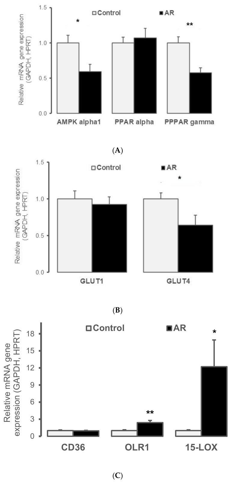
Chronic AR was associated with coordinated changes in the expression of genes involved in myocardial metabolic regulation. Expression of AMPKα1 and PPARγ was reduced (Figure 2A), together with a significant decrease in the insulin-responsive glucose transporter GLUT4 (Figure 2B). In parallel, expression of 15-LOX, encoding 15-lipoxygenase involved in polyunsaturated fatty acid metabolism and lipid peroxidation, and oxidized low-density lipoprotein receptor 1 (OLR1; also known as 1-LOX), a scavenger receptor mediating uptake of oxidized lipoproteins, was increased (Figure 2C). In contrast, CD36 and PPARα, which are involved in fatty acid uptake and metabolic regulation, respectively, remained unchanged (Figure 2A,C). These findings indicate transcriptional metabolic remodeling and alterations in lipid-related pathways in the LV myocardium during chronic AR. However, in the absence of protein-level and metabolite measurements, these transcriptional changes cannot be interpreted as evidence of a functional shift in myocardial substrate utilization.

2.4. Transcriptional Changes in the Kallikrein–Kinin System

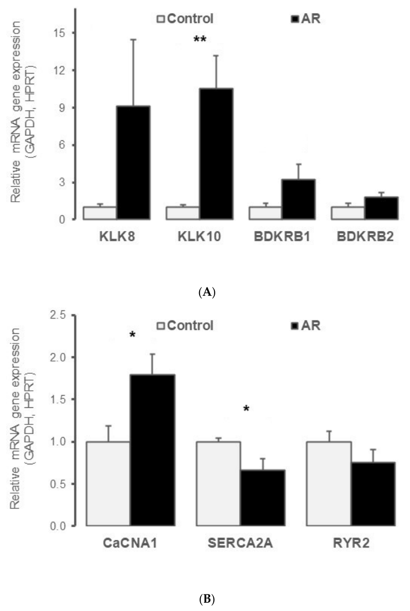
Chronic AR was associated with transcriptional changes involving components of the kallikrein–kinin system (Figure 3A). KLK10 expression was significantly increased, whereas KLK8 and bradykinin receptor BDKRB1 showed non-significant trends toward increased expression (p = 0.09 and p = 0.06, respectively). Expression of the constitutive bradykinin receptor BDKRB2 remained unchanged. These findings provide a transcriptional signal suggesting possible involvement of the kallikrein–kinin signaling pathway in the LV myocardium during chronic AR, driven primarily by the significant change in KLK10. The non-significant changes in KLK8 and BDKRB1 should be interpreted cautiously and require confirmation at the protein and functional levels.

2.5. Transcriptional Changes in Calcium-Handling Pathways

As illustrated in Figure 3B, chronic AR was associated with changes in the expression of genes involved in myocardial calcium handling. SERCA2A, a key regulator of sarcoplasmic reticulum calcium reuptake, was significantly reduced, whereas expression of the L-type calcium channel subunit (CaCNA1) was increased. In contrast, RYR2 expression remained unchanged. This transcriptional profile suggests remodeling of calcium-handling pathways in the LV myocardium during chronic AR and may contribute to the impaired systolic function observed in this model. However, confirmation at the protein and functional levels would be required to establish alterations in calcium handling.

3. Discussion

The present study provides an integrated characterization of LV remodeling during the transition from compensated volume overload to ventricular decompensation in experimental chronic AR. Sixty days after AR induction, rats developed marked LV dilatation, eccentric hypertrophy, increased wall stress and impaired systolic function. These functional alterations were accompanied by coordinated changes in LV gene expression involving pathways related to redox homeostasis, apoptotic signaling, metabolic regulation, kallikrein–kinin signaling and calcium handling. Collectively, these findings suggest that ventricular decompensation in chronic AR is associated with a multifaceted myocardial transcriptional remodeling response extending beyond the hemodynamic consequences of volume overload. However, because the molecular analyses were limited to mRNA expression, these findings should be considered hypothesis-generating and require confirmation at the protein and functional levels.
The structural and functional alterations observed in our model are consistent with the natural history of chronic AR. Chronic regurgitant volume overload promotes progressive LV enlargement and eccentric hypertrophy through activation of preload reserve mechanisms that initially preserve forward cardiac output and wall stress distribution [3,4,5]. However, persistent overload may eventually exceed these adaptive mechanisms, resulting in increased wall stress, contractile dysfunction and heart failure [3,5,7]. In the present study, the reduction in FS, together with marked increases in LV end-diastolic and end-systolic diameters and decreased relative wall thickness, was consistent with progression toward decompensated eccentric hypertrophy. EF showed a non-significant trend toward reduction (p = 0.08), whereas SV and CO remained elevated, suggesting persistence of compensatory hemodynamic mechanisms despite impaired systolic performance [3,5]. These observations support the concept that sustained mechanical stress contributes to the transition from adaptive remodeling to ventricular decompensation.
One of the main findings of this study is the identification of a myocardial transcriptional profile involving genes related to redox homeostasis and apoptosis signaling. AR hearts exhibited decreased expression of the mitochondrial antioxidant enzyme SOD2 together with compensatory upregulation of GPX1. Reduced SOD2 expression may be consistent with altered mitochondrial antioxidant capacity, whereas the concomitant increase in GPX1 may represent a compensatory response within the glutathione-dependent antioxidant system. Given that SOD2 catalyzes the dismutation of mitochondrial superoxide to hydrogen peroxide, changes in SOD2 expression may alter the balance between different reactive oxygen species and antioxidant pathways. Increased GPX1 expression could, therefore, reflect an adaptive response aimed at maintaining hydrogen peroxide detoxification through the glutathione peroxidase pathway. Similar dissociation between mitochondrial and compensatory antioxidant pathways has been described in experimental models of chronic cardiac stress [13,14]. However, because biochemical markers of oxidative stress and protein-level measurements were not available in the present study, these findings should be interpreted as a transcriptional signal consistent with altered redox homeostasis rather than as direct evidence of oxidative stress.
The increased BAX/BCL2 ratio provides a further transcriptional signal related to pro-apoptotic signaling. Oxidative imbalance and mitochondrial dysfunction have been implicated in cardiomyocyte apoptosis and adverse ventricular remodeling in several experimental settings [13,14,15]. Comparable changes in apoptosis-related genes have also been reported in other forms of LV remodeling, such as the post-myocardial infarction myocardium [16], suggesting that alterations in apoptotic signaling may represent a common feature of adverse ventricular remodeling rather than a mechanism specific to chronic AR. Nevertheless, the present findings do not establish the occurrence of cardiomyocyte apoptosis, as no histological or protein-level markers such as TUNEL or cleaved caspase-3 were available. Further studies will therefore be required to determine whether the observed transcriptional changes translate into increased apoptosis and whether this process contributes causally to LV decompensation.
Metabolic remodeling emerged as another prominent feature of the decompensated phenotype. We observed coordinated downregulation of AMPKα1 and PPARγ, two critical regulators of cellular energy homeostasis, together with significantly decreased expression of GLUT4, the principal insulin-responsive glucose transporter in cardiomyocytes. AMPK plays a central role in maintaining myocardial energy balance during conditions of increased workload, whereas altered PPARγ signaling contributes to impaired metabolic flexibility [17,18]. Reduced GLUT4 expression may indicate altered regulation of insulin-responsive glucose transport in the myocardium [19]. In parallel, increased expression of OLR1 and 15-LOX suggests changes in pathways involved in oxidized lipid signaling and lipid peroxidation. Both pathways have been implicated in cardiac inflammation, oxidative stress and ventricular dysfunction [20,21]. The absence of significant changes in PPARα and CD36 further suggests that these transcriptional alterations were selective rather than indicative of a generalized change in all major metabolic pathways. Taken together, these findings support the presence of transcriptional metabolic remodeling in chronic AR. However, they do not establish a functional switch in myocardial substrate utilization, as protein abundance, metabolic fluxes and tissue metabolite levels were not assessed. Further studies will be required to determine whether these transcriptional changes translate into altered myocardial energy metabolism.
An additional finding was a transcriptional signal involving components of the kallikrein–kinin system. KLK10 expression was significantly increased, whereas KLK8 and BDKRB1 showed non-significant trends toward increased expression. Previous studies have implicated the cardiac kallikrein–kinin system in experimental volume-overload states, including pathways related to inflammatory signaling and extracellular matrix remodeling [22]. The inducible receptor BDKRB1 is of particular interest because its expression may increase in response to tissue injury and inflammatory stimuli. However, the present data do not establish activation of the kallikrein–kinin system. Rather, the significant increase in KLK10, together with non-significant trends in KLK8 and BDKRB1, provides a transcriptional signal suggesting possible involvement of this pathway in chronic AR. The functional significance of these changes, as well as their relationship to ventricular remodeling, will require confirmation at the protein and functional levels.
Alterations in calcium-handling gene expression provide another potential link between myocardial transcriptional remodeling and impaired ventricular performance. The reduced expression of SERCA2A, a major regulator of sarcoplasmic reticulum Ca2+ reuptake, may be consistent with altered regulation of intracellular Ca2+ cycling and could contribute to impaired contractile performance [23]. In parallel, increased expression of CaCNA1 may reflect a compensatory or maladaptive transcriptional response to chronic overload. In contrast, RYR2 expression remained unchanged. Taken together, the reduced SERCA2A and increased CACNA1C expression suggest transcriptional remodeling of selected calcium-handling pathways in the LV myocardium during chronic AR. In the context of reduced calcium sequestration efficiency, enhanced calcium influx may promote calcium dysregulation and further impair excitation-contraction coupling [23,24]. However, because our analyses were limited to mRNA expression, these findings should not be interpreted as direct evidence of altered calcium handling. Protein-level measurements and functional assessment of Ca2+ transients, channel activity and sarcoplasmic reticulum Ca2+ cycling will be required to determine whether these transcriptional alterations translate into functional abnormalities.
Several limitations should be acknowledged. First, the study was conducted exclusively in male rats, precluding assessment of potential sex-dependent differences in myocardial responses to chronic volume overload [25]. Additionally, the approximately 30% mortality observed in the AR group raises the possibility of survivor bias in the 60-day molecular analyses, since only animals surviving the most severe hemodynamic insult were available for gene-expression analysis. Second, molecular analyses were restricted to mRNA expression and therefore do not necessarily reflect protein abundance, enzymatic activity or biological function. This limitation is particularly relevant to the interpretation of the findings related to oxidative stress, apoptosis, metabolism, kallikrein–kinin signaling and calcium handling. Third, biochemical and histological analyses that could have provided direct functional confirmation of oxidative stress and apoptosis were not available in this cohort. Fourth, the relatively small sample size may have limited statistical power to detect more subtle changes in some signaling pathways. In addition, the molecular analyses were exploratory and no formal correction for multiple comparisons was applied, which increases the possibility of false-positive findings. Additionally, although significant baseline-to-day-60 changes were observed in the AR group but not in controls, a formal statistical comparison of the temporal trajectories between groups (e.g., a group-by-time interaction analysis) was not performed. This limitation should be considered when interpreting the extent to which the observed changes exceed physiological, growth-related remodeling. Finally, the observational nature of the study precludes establishing causal relationships between the identified molecular alterations and ventricular dysfunction. Future studies integrating transcriptional, protein, biochemical and functional measurements, together with longitudinal assessment of ventricular remodeling, will be necessary to determine which of these pathways contribute directly to progression from compensated hypertrophy to ventricular decompensation. Advanced functional imaging approaches, including global longitudinal strain, may further help establish links between myocardial remodeling and clinically relevant trajectories toward decompensation [26].

4. Materials and Methods

4.1. Experimental Model of Chronic Aortic Regurgitation

Animals and study design. All experimental procedures were approved by the Institutional Animal Care and Use Committee of the Faculty of Medicine of the Université Libre de Bruxelles (Brussels, Belgium; protocol approval number 644N) and conducted in accordance with the National Institutes of Health Guide for the Care and Use of Laboratory Animals. Twenty-two adult male Wistar rats (502 ± 27 g; Janvier, Le Genest-Saint-Isle, France) were housed under controlled environmental conditions (12 h light/dark cycle) with free access to standard chow and water. As previously described [27], animals were randomly assigned to either a control group (n = 8) or an aortic regurgitation (AR) group (n = 14). Under 1.5% isoflurane anesthesia, severe AR was induced by retrograde perforation of an aortic valve leaflet. The severity of the regurgitant lesion was confirmed intraoperatively by Doppler echocardiography. Echocardiographic measurements were performed by two investigators blinded to group allocation; molecular (RT-qPCR) analyses were performed using automated instrumentation and standardized protocols, and blinding was therefore not applicable, as these analyses did not involve subjective interpretation by an operator. Animals presenting insufficiently severe aortic regurgitation (PHT > 200 msec) were excluded from the study. The AR group showed an overall mortality of approximately 30% (4 animals), related either to aortic tearing during the valve-leaflet puncture procedure (peri-operative) or to pulmonary edema/hemodynamic decompensation occurring predominantly within the first 3 post-operative days; some animals were euthanized for humane endpoints when they showed persistent overt heart failure despite diuretic treatment. No mortality occurred in the control group. After these exclusions, the final analyzed cohorts comprised n = 8 control and n = 10 AR rats.
Experimental timeline. AR induction or control surgery was performed on day 0. Echocardiographic and invasive hemodynamic measurements were obtained at baseline and repeated 60 days after surgery. At the end of the follow-up period, animals underwent terminal tissue collection for molecular analyses.

4.2. Functional and Hemodynamic Assessment

LV structure and function were assessed by transthoracic echocardiography, followed by invasive hemodynamic measurements.
Echocardiography. Transthoracic 2D, M-mode and Doppler echocardiography were performed using a Vivid E90 ultrasound system (GE Healthcare, Wauwatosa, WI, USA) equipped with a 12 MHz phased-array transducer (GE 12S-D). Animals were maintained under 1.5% isoflurane anesthesia throughout the procedure. Continuous electrocardiogram (ECG) monitoring was performed using limb leads. All measurements were obtained by a single experienced operator according to current echocardiographic recommendations [28]. Standard parasternal long-axis, short-axis and apical views were acquired to measure LV dimensions, wall thickness, fractional shortening (FS) and ejection fraction (LVEF). FS was calculated from M-mode tracings using the formula FS (%) = [(LVEDD − LVESD)/LVEDD] × 100, and LVEF was estimated using the Teichholz method. Aortic Doppler recordings were used to determine forward stroke volume (SV) and cardiac output (CO). The severity of AR was quantified by pressure half-time (PHT) of the regurgitant jet, with values < 200 msec indicating severe regurgitation. Relative wall thickness (RWT) was calculated as: RWT = 2 × PWTd/LVEDD, where PWTd represents posterior wall thickness at end-diastole (in mm).
Pre-ejection period (PEP; in msec), LV ejection time (LVET; in msec), RR inter-beat interval (in msec), systolic time (in msec) and diastolic time (in msec) were also derived from simultaneous Doppler and electrocardiographic recordings.
Invasive hemodynamic measurements. Invasive pressure measurements were obtained using a 1.6F micro-tip pressure catheter (Transonic Systems Inc., Ithaca, NY, USA). During the initial surgical procedure, the catheter was introduced through the right carotid artery into the LV to assist AR induction and confirm the severity of the regurgitant lesion, as previously described [27]. Control animals underwent the same procedure without valve perforation. At day 60, invasive hemodynamic assessment was performed in all animals using arterial catheterization before sacrifice. Pressure signals were recorded and analyzed using the ADV500PV acquisition system (Transonic Systems Inc.).

4.3. Tissue Collection and Quantitative Gene Expression Analysis

LV sampling. Following completion of the 60-day protocol, animals were euthanized by exsanguination under deep 5% isoflurane anesthesia. Hearts were rapidly excised and the LV was carefully dissected, snap-frozen in liquid nitrogen and stored at –80 °C until further molecular analysis.
RNA extraction and reverse transcription. Total RNA was extracted from snap-frozen LV myocardium using TRIzol reagent (Invitrogen, Merelbeke, Belgium) and purified using the RNeasy® Mini Kit (QIAGEN, Hilden, Germany), according to the manufacturer’s instructions. RNA concentration and purity were assessed spectrophotometrically using a Nanodrop ND-1000® (Isogen Life Sciences, De Meern, The Netherlands), and RNA integrity was verified by agarose gel electrophoresis with GelRed® (Biotium, Hayward, CA, USA) staining. cDNA was synthesized from 1 µg of total RNA using random hexamers and Superscript II Reverse Transcriptase (Invitrogen, Merelbeke, Belgium), following the manufacturer’s protocol.
Real-time quantitative polymerase chain reaction (RT-qPCR). Gene expression was quantified by RT-qPCR using SYBR Green chemistry and gene-specific primers (Table 2). Candidate genes were selected to evaluate pathways involved in apoptosis (BCL2 and BAX), oxidative stress (GPX1, GSR, SOD1 and SOD2), metabolic regulation (AMPK, PPARα and PPARγ), glucose (GLUT1 and GLUT4) and lipid transport (CD36), oxidative injury (OLR1 and 15-LOX), kallikrein–kinin signaling (KLK8, KLK10, BDKRB1 and BDKRB2) and excitation-contraction coupling (ATP2A2/SERCA2, RYR2 and CaCNA1). Whenever possible, intron-spanning primers were used. Relative gene expression levels were normalized to the housekeeping genes GAPDH and HPRT1 and quantified using the Pfaffl method [29]. All reactions were performed in triplicate, using an iCycler detection system (BioRad Laboratories, Hercules, CA, USA).

4.4. Statistical Analysis

Statistical analyses were performed using StatView 5.0 (SAS Institute, Cary, NC, USA). Sample size was based on previous studies from our group using the same experimental model [27] rather than a formal a priori power calculation, which is acknowledged as a limitation.
Data from the tables are presented as mean ± standard deviation (SD), whereas data shown in the figures are presented as mean ± standard error of the mean (SEM). The number of animals analyzed is indicated by n and represents independent biological observations. For the figures, data are presented as mean ± standard error of the mean (SEM). For longitudinal echocardiographic and hemodynamic measurements, baseline and 60-day values were compared using paired Student’s t-tests. Normality of the data was assessed using the Shapiro–Wilk test.
For gene expression analyses, comparisons between control and AR groups were performed using an unpaired Student’s t-test when the normality assumption was met and the Mann–Whitney test when it was not. Because these molecular analyses were exploratory and hypothesis-generating, no formal correction for multiple comparisons was applied. This approach is acknowledged as a limitation, given the number of genes investigated.
All statistical tests were two-sided, and a p value < 0.05 was considered statistically significant. Exact p values are reported where appropriate; for graphical presentation, statistical significance is indicated as follows: * 0.01 < p < 0.05, ** 0.001 < p < 0.01 and *** p < 0.001.

5. Conclusions

In conclusion, chronic AR is associated with a multifaceted myocardial transcriptional signature involving oxidative imbalance, pro-apoptotic signaling, metabolic remodeling, kallikrein–kinin pathway signaling and altered calcium-handling gene expression. These changes accompany LV structural and functional decompensation and identify molecular pathways potentially involved in myocardial remodeling during chronic volume overload. Further protein-level, biochemical and functional studies are required to establish their mechanistic and therapeutic relevance.

Author Contributions

B.E.O. Conceptualization, methodology, writing—original draft, writing—review and editing, and investigation. L.D. validation and writing—review and editing. P.V.d.B. funding acquisition, writing—review and editing, supervision, and validation. G.H. data curation and resources. P.J. data curation and investigation. C.S. formal analysis, software, and visualization. K.M.E. Conceptualization, methodology, project administration, and supervision. F.V.E. funding acquisition, resources, supervision, and validation. All authors have read and agreed to the published version of the manuscript.

Funding

This research was funded by the Fonds pour la Chirurgie Cardiaque, Brussels, Belgium, grant number 489639.

Institutional Review Board Statement

The animal study protocol was approved by the Institutional Animal Care and Use Committee of the Université Libre de Bruxelles (Brussels, Belgium; protocol code 644N, 28 June 2017). Studies were conducted in accordance with the Guide for the Care and Use of Laboratory Animals published by the National Institutes of Health (NIH Publication No. 85-23; revised 1996).

Informed Consent Statement

Not applicable.

Data Availability Statement

The original contributions presented in this study are included in the article. Further inquiries can be directed to the corresponding author.

Conflicts of Interest

The authors declare no conflicts of interest. The sponsors had no role in the design, execution, interpretation, or writing of the study.

References

  1. Coffey, S.; Cairns, B.J.; Iung, B. The modern epidemiology of heart valve disease. Heart 2016, 102, 75–85. [Google Scholar] [CrossRef] [Scilit] [PubMed]
  2. Kodali, S.K.; Velagapudi, P.; Hahn, R.T.; Abbott, D.; Leon, M.B. Valvular Heart Disease in Patients ≥80 Years of Age. J. Am. Coll. Cardiol. 2018, 71, 2058–2072. [Google Scholar] [CrossRef] [Scilit] [PubMed]
  3. Bekeredjian, R.; Grayburn, P.A. Valvular heart disease: Aortic regurgitation. Circulation 2005, 112, 125–134. [Google Scholar] [PubMed]
  4. Carabello, B.A. Aortic regurgitation. A lesion with similarities to both aortic stenosis and mitral regurgitation. Circulation 1990, 82, 1051–1053. [Google Scholar] [CrossRef] [Scilit] [PubMed]
  5. Ricci, D.R. Afterload mismatch and preload reserve in chronic aortic regurgitation. Circulation 1982, 66, 826–834. [Google Scholar] [CrossRef] [Scilit] [PubMed]
  6. Marigliano, A.-N.; Ortiz, J.-T.; Casas, J.; Evangelista, A. Aortic Regurgitation: From Valvular to Myocardial Dysfunction. J. Clin. Med. 2024, 13, 2929. [Google Scholar] [CrossRef] [Scilit] [PubMed]
  7. Bonow, R.O. Chronic mitral regurgitation and aortic regurgitation: Have indications for surgery changed? J. Am. Coll. Cardiol. 2013, 61, 693–701. [Google Scholar] [PubMed]
  8. Writing Committee Members; Otto, C.M.; Nishimura, R.A.; Bonow, R.O.; Carabello, B.A.; Erwin, J.P., 3rd; Gentile, F.; Jneid, H.; Krieger, E.V.; Mack, M.; et al. 2020 ACC/AHA guideline for the management of patients with valvular heart disease: A report of the American College of Cardiology/American Heart Association Joint Committee on Clinical Practice Guidelines. J. Am. Coll. Cardiol. 2021, 77, e25–e197, Erratum in J. Am. Coll. Cardiol. 2024, 84, 1772. [Google Scholar] [PubMed]
  9. Ponikowski, P.; Voors, A.A.; Anker, S.D.; Bueno, H.; Cleland, J.G.F.; Coats, A.J.S.; Falk, V.; González-Juanatey, J.R.; Harjola, V.-P.; Jankowska, E.A.; et al. 2016 ESC Guidelines for the diagnosis and treatment of acute and chronic heart failure. Eur. Heart J. 2016, 37, 2129–2200, Erratum in Eur. Heart J. 2018, 39, 860. [Google Scholar] [CrossRef] [Scilit] [PubMed]
  10. Arnold, Z.; Elnekheli, A.; Geisler, D.; Aschacher, T.; Lenz, V.; Winkler, B.; Moidl, R.; Grabenwöger, M. Left Ventricular Reverse Remodeling after Surgical Aortic Valve Replacement for Aortic Regurgitation—An Explorative Study. Diseases 2024, 12, 191. [Google Scholar] [CrossRef] [Scilit] [PubMed]
  11. Champetier, S.; Bojmehrani, A.; Beaudoin, J.; Lachance, D.; Plante, E.; Roussel, E.; Couet, J.; Arsenault, M. Gene profiling of left ventricle eccentric hypertrophy in aortic regurgitation in rats: Rationale for targeting the beta-adrenergic and renin-angiotensin systems. Am. J. Physiol. Heart Circ. Physiol. 2009, 296, H669–H677. [Google Scholar] [CrossRef] [Scilit] [PubMed]
  12. Roussel, E.; Drolet, M.C.; Walsh-Wilkinson, E.; Dhahri, W.; Lachance, D.; Gascon, S.; Sarrhini, O.; Rousseau, J.A.; Lecomte, R.; Couet, J.; et al. Transcriptional Changes Associated with Long-Term Left Ventricle Volume Overload in Rats: Impact on Enzymes Related to Myocardial Energy Metabolism. BioMed Res. Int. 2015, 2015, 949624. [Google Scholar] [CrossRef] [Scilit] [PubMed]
  13. Dhalla, A.K.; Singal, P.K. Antioxidant changes in hypertrophied and failing guinea pig hearts. Am. J. Physiol. 1994, 266, H1280–H1285. [Google Scholar] [CrossRef] [Scilit] [PubMed]
  14. Pias, E.K.; Ekshyyan, O.Y.; Rhoads, C.A.; Fuseler, J.; Harrison, L.; Aw, T.Y. Differential effects of superoxide dismutase isoform expression on hydroperoxide-induced apoptosis in PC-12 cells. J. Biol. Chem. 2003, 278, 13294–13301. [Google Scholar] [CrossRef] [Scilit] [PubMed]
  15. Letai, A.; Bassik, M.C.; Walensky, L.D.; Sorcinelli, M.D.; Weiler, S.; Korsmeyer, S.J. Distinct BH3 domains either sensitize or activate mitochondrial apoptosis, serving as prototype cancer therapeutics. Cancer Cell 2002, 2, 183–192. [Google Scholar] [CrossRef] [Scilit] [PubMed]
  16. Kuveljic, J.; Djordjevic, A.; Zivotic, I.; Dekleva, M.; Kolakovic, A.; Zivkovic, M.; Stankovic, A.; Djuric, T. Expression of HMGB1, TGF-β1, BIRC3, ADAM17, CDKN1A, and FTO in Relation to Left Ventricular Remodeling in Patients Six Months after the First Myocardial Infarction: A Prospective Study. Genes 2024, 15, 1296. [Google Scholar] [CrossRef] [Scilit] [PubMed]
  17. Lee, S.Y.; Ku, H.C.; Kuo, Y.H.; Chiu, H.L.; Su, M.J. Pyrrolidinyl caffeamide against ischemia/reperfusion injury in cardiomyocytes through AMPK/AKT pathways. J. Biomed. Sci. 2015, 22, 18. [Google Scholar] [CrossRef] [Scilit] [PubMed]
  18. Dong, H.W.; Zhang, L.F.; Bao, S.L. AMPK regulates energy metabolism through the SIRT1 signaling pathway to improve myocardial hypertrophy. Eur. Rev. Med. Pharmacol. Sci. 2018, 22, 2757–2766. [Google Scholar] [PubMed]
  19. Katz, E.B.; Stenbit, A.E.; Hatton, K.; Depinho, R.; Charron, M.J. Cardiac and adipose tissue abnormalities but not diabetes in mice deficient in GLUT4. Nature 1995, 377, 151–155. [Google Scholar] [CrossRef] [Scilit] [PubMed]
  20. Sawamura, T.; Kume, N.; Aoyama, T.; Moriwaki, H.; Hoshikawa, H.; Aiba, Y.; Tanaka, T.; Miwa, S.; Katsura, Y.; Kita, T.; et al. An endothelial receptor for oxidized low-density lipoprotein. Nature 1997, 386, 73–77. [Google Scholar] [CrossRef] [Scilit] [PubMed]
  21. Kayama, Y.; Minamino, T.; Toko, H.; Sakamoto, M.; Shimizu, I.; Takahashi, H.; Okada, S.; Tateno, K.; Moriya, J.; Yokoyama, M.; et al. Cardiac 12/15 lipoxygenase–induced inflammation is involved in heart failure. J. Exp. Med. 2009, 206, 1565–1574. [Google Scholar] [CrossRef] [Scilit] [PubMed]
  22. Wei, C.C.; Chen, Y.; Powell, L.C.; Zheng, J.; Shi, K.; Bradley, W.E.; Powell, P.C.; Ahmad, S.; Ferrario, C.M.; Dell’Italia, L.J. Cardiac kallikrein-kinin system is upregulated in chronic volume overload and mediates an inflammatory induced collagen loss. PLoS ONE 2012, 7, e40110. [Google Scholar] [CrossRef] [Scilit] [PubMed]
  23. Nusier, M.; Shah, A.K.; Dhalla, N.S. Structure-function relationships and modifications of cardiac sarcoplasmic reticulum Ca2+-transport. Physiol. Res. 2021, 70, S443–S470. [Google Scholar] [CrossRef] [Scilit] [PubMed]
  24. Nediani, C.; Formigli, L.; Perna, A.M.; Ibba-Manneschi, L.; Zecchi-Orlandini, S.; Fiorillo, C.; Ponziani, V.; Cecchi, C.; Liguori, P.; Fratini, G.; et al. Early changes induced in the left ventricle by pressure overload. An experimental study on swine heart. J. Mol. Cell. Cardiol. 2000, 32, 131–142. [Google Scholar] [CrossRef] [Scilit] [PubMed]
  25. Beaumont, C.; Walsh-Wilkinson, É.; Drolet, M.C.; Roussel, É.; Melançon, N.; Fortier, É.; Harpin, G.; Beaudoin, J.; Arsenault, M.; Couet, J. Testosterone deficiency reduces cardiac hypertrophy in a rat model of severe volume overload. Physiol. Rep. 2019, 7, e14088. [Google Scholar] [CrossRef] [Scilit] [PubMed]
  26. Kim, M.-R.; Shaikh, T.; Taylor, S.; Wang, S.; Nguyen, D.; Khetarpal, B.K.; Namazi, A.; Goel, V.S.; Sagaribay, R.; Batra, K. Global Longitudinal Strain as a Prognostic Biomarker for Asymptomatic Moderate to Severe Aortic Regurgitation with Preserved Ejection Fraction: A Systematic Review and Meta-Analysis. J. Clin. Med. 2025, 14, 6534. [Google Scholar] [CrossRef] [Scilit] [PubMed]
  27. El-Oumeiri, B.; Mc Entee, K.; Annoni, F.; Herpain, A.; Vanden Eynden, F.; Jespers, P.; Van Nooten, G.; van de Borne, P. Effects of the cardiac myosin activator Omecamtiv-mecarbil on severe chronic aortic regurgitation in Wistar rats. BMC Cardiovasc. Disord. 2018, 18, 99. [Google Scholar] [CrossRef] [Scilit] [PubMed]
  28. Nagueh, S.F.; Smiseth, O.A.; Appleton, C.P.; Byrd, B.F., 3rd; Dokainish, H.; Edvardsen, T.; Flachskampf, F.A.; Gillebert, T.C.; Klein, A.L.; Lancellotti, P.; et al. Recommendations for the Evaluation of Left Ventricular Diastolic Function by Echocardiography: An Update from the American Society of Echocardiography and the European Association of Cardiovascular Imaging. J. Am. Soc. Echocardiogr. 2016, 29, 277–314. [Google Scholar] [CrossRef] [Scilit] [PubMed]
  29. Pfaffl, M.W. A new mathematical model for relative quantification in real-time RT–PCR. Nucleic Acids Res. 2001, 29, e45. [Google Scholar] [CrossRef] [Scilit] [PubMed]
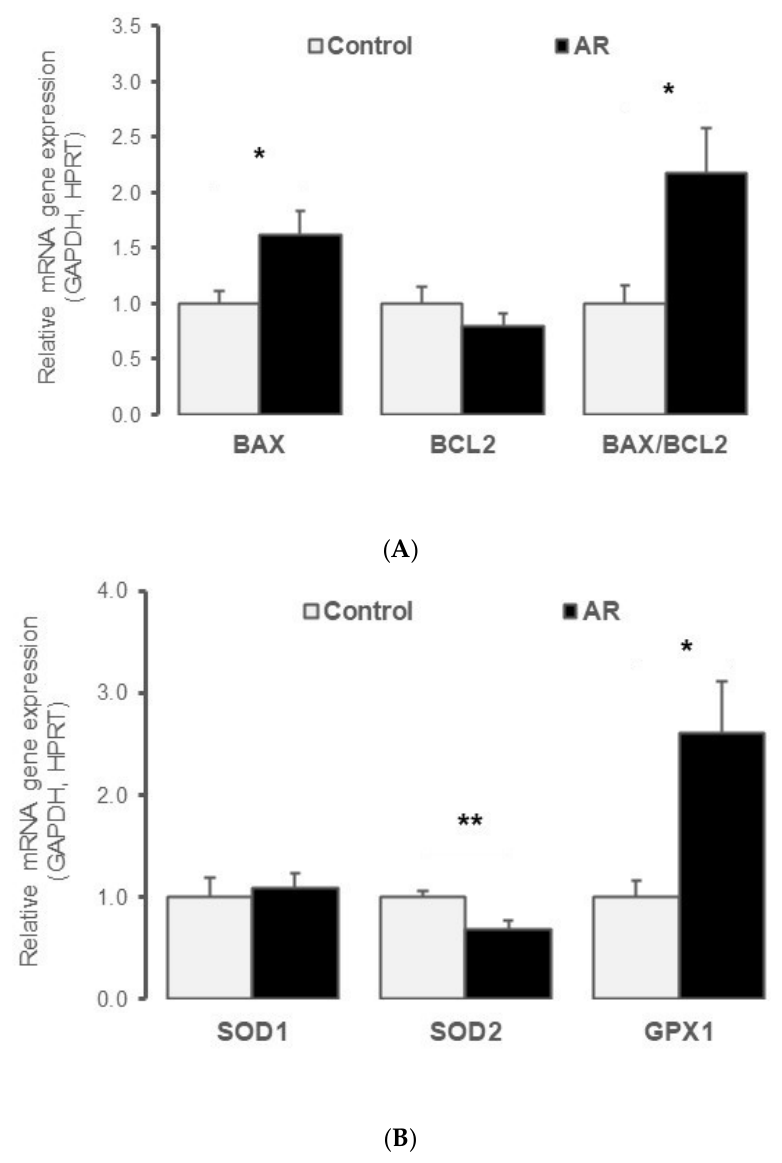
Figure 1. Chronic aortic regurgitation promotes apoptotic signaling and oxidative stress in the left ventricular myocardium. (A) Relative mRNA expression of apoptosis-related genes (BAX and BCL2) and the corresponding BAX/BCL2 ratio in left ventricular tissue from control (n = 8; white bars) and aortic regurgitation (AR; n = 10; black bars) rats. (B) Relative mRNA expression of antioxidant enzymes involved in reactive oxygen species detoxification (SOD1, SOD2, GPX1) in left ventricular tissue from control (n = 8) and AR (n = 10) rats. Data are mean ± SEM; * 0.01 < p < 0.05, ** 0.001 < p < 0.01 versus controls.
Ijms 27 07950 g001
Figure 2. Chronic aortic regurgitation is associated with transcriptional metabolic remodeling and altered lipid-related signaling in the left ventricle. (A) Relative mRNA expression of key metabolic regulators (AMPKα1, PPARα and PPARγ) in left ventricular tissue from control (n = 8) and AR (n = 10) rats. (B) Relative mRNA expression of glucose transporters (GLUT1 and GLUT4) in left ventricular tissue from control (n = 8) and AR (n = 10) rats. (C) Relative mRNA expression of genes involved in oxidized lipid uptake (CD36) and lipid peroxidation (OLR1/1-LOX-1, 15-LOX) in left ventricular tissue from control (n = 8) and AR (n = 10) rats. Data are mean ± SEM; * 0.01 < p < 0.05, ** 0.001 < p < 0.01 versus control rats.
Ijms 27 07950 g002
Figure 3. Chronic aortic regurgitation is associated with transcriptional changes in kallikrein–kinin and calcium-handling pathways. (A) Relative mRNA expression of kallikrein–kinin system components (KLK8, KLK10, BDKRB1 and BDKRB2) in left ventricular tissue from control (n = 8) and AR (n = 10) rats. (B) Relative mRNA expression of calcium-handling genes (SERCA2A, RYR2 and CaCNA1) in left ventricular tissue from control (n = 8) and AR (n = 10) rats. Data are mean ± SEM; * 0.01 < p < 0.05, ** 0.001 < p < 0.01 versus control rats.
Ijms 27 07950 g003
Table 1. (a) Echocardiographic and hemodynamic evidence of left ventricular decompensation 60 days after induction of chronic aortic regurgitation (AR group). (b) Echocardiographic and hemodynamic parameters measured at baseline and 60 days in control rats.
(a)
ParameterBaselineAfter 60 Daysp
Body weight, g502 ± 27577 ± 24***
LV Systolic Function
FS, %42 ± 233 ± 1***
EF, %76 ± 365 ± 4NS
LV Structure
LVESD, mm4.6 ± 0.27.2 ± 0.3***
LVEDD, mm7.9 ± 0.210.7 ± 0.3***
LV Mass, mg1094 ± 1401750 ± 209**
Wall Thickness
SWTs, mm3.03 ± 0.182.85 ± 0.22NS
SWTd, mm2.20 ± 0.182.38 ± 0.24NS
PWTs, mm3.00 ± 0.142.89 ± 0.24NS
PWTd, mm1.88 ± 0.171.76 ± 0.15NS
RWT0.49 ± 0.050.33 ± 0.03*
LVOT/Aortic Outflow
LVOT diameter, mm2.41 ± 0.062.82 ± 0.06***
LVOT VTI, mm66 ± 481 ± 3**
Hemodynamics
HR, beats/min315 ± 19301 ± 15NS
Systolic BP Pre, mmHg119 ± 2115 ± 2NS
Diastolic BP Pre, mmHg82 ± 159 ± 3***
Systolic BP Post, mmHg106 ± 3/
Diastolic BP Post, mmHg63 ± 3/
Time Intervals
PEP, msec28 ± 231 ± 3NS
LVET, msec81 ± 187 ± 3**
ST, msec109 ± 3119 ± 4**
DT, msec88 ± 985 ± 8NS
RR, msec197 ± 11202 ± 9NS
PEP/LVET0.34 ± 0.020.36 ± 0.03NS
ST/RR0.58 ± 0.040.59 ± 0.02NS
PHT, msec/92 ± 7
Output Parameters
SV, mL0.30 ± 0.020.51 ± 0.04***
CO, mL/min96 ± 8153 ± 13**
LV Wall Stress
Laplace-s46 ± 476 ± 7**
Laplace-d77 ± 673 ± 9NS
σmax224 ± 18282 ± 29*
σEs, dyn/cm260 ± 5104 ± 11**
(b)
ParameterBaselineAfter 60 daysp
Body weight, g477 ± 31548 ± 37**
LV Systolic Function
FS, %36.5 ± 6.735.75 ± 6.5NS
EF, %70.38 ± 9.4671.38 ± 9.38NS
LV Structure
LVESD, mm5.26 ± 0.525.69 ± 0.65NS
LVEDD, mm8.65 ± 0.78.91 ± 0.48NS
LV Mass, mg745 ± 258992 ± 148*
Wall Thickness
SWTs, mm2.42 ± 0.342.43 ± 0.45NS
SWTd, mm1.61 ± 0.131.66 ± 0.22NS
PWTs, mm2.52 ± 0.122.7 ± 0.2NS
PWTd, mm1.71 ± 0.111.74 ± 0.21NS
RWT0.40 ± 0.040.39 ± 0.06NS
Hemodynamics
HR, beats/min269 ± 49261 ± 62NS
Systolic BP, mmHg125 ± 15128 ± 8NS
Diastolic BP, mmHg86 ± 1488 ± 9NS
Time Intervals
PEP, msec20 ± 722 ± 6NS
LVET, msec87 ± 1585 ± 12NS
ST, msec109 ± 17107 ± 16NS
DT, msec121 ± 28130 ± 36NS
RR, msec230 ± 40236 ± 51NS
PEP/LVET0.23 ± 0.080.26 ± 0.06NS
ST/RR0.47 ± 0.040.45 ± 0.05NS
PHT, msec//
Output Parameters
SV, mL0.29 ± 0.060.29 ± 0.09NS
CO, mL/min79 ± 2178 ± 30NS
FS, fractional shortening; EF, ejection fraction; LVESD, left ventricular end-systolic diameter; LVEDD, left ventricular end-diastolic diameter; SWTs/SWTd, septal wall thickness at systole/diastole; PWTs/PWTd, posterior wall thickness at systole/diastole; RWT, relative wall thickness; LVOT, left ventricular outflow tract; VTI, velocity-time integral; HR, heart rate; BP, blood pressure; PEP, pre-ejection period; LVET, LV ejection time; ST, systolic time; DT, diastolic time; RR, RR interval; PHT, pressure half-time; SV, stroke volume; CO, cardiac output; σmax, maximal wall stress; σEs, end-systolic wall stress. Data are mean ± SD. * 0.01 < p < 0.05; ** 0.001 < p < 0.01; *** p < 0.001, 60-day AR versus baseline. NS, not significant.
Table 2. Primer sequences used for RT-qPCR analysis.
GenePrimer Sequence (5′–3′)
Glycerol-3-phosphate dehydrogenase (GAPDH)Sense: 5′-AAGATGGTGAAGGTCGGTGT-3′
Antisense: 5′-ATGAAGGGGTCGTTGATGG-3′
Hypoxanthine guanine phosphoribosyltransferase (HPRT)Sense: 5′-ACAGGCCAGACTTTGTTGGA-3′
Antisense: 5′-ATCCACTTTCGCTGATGACAC-3′
AMP-activated protein kinase (AMPK)Sense: 5′-TTCGGGAAAGTGAAGGTGGG-3′
Antisense: 5′-TCTCTGCGGATTTTCCCGAC-3′
Arachidonate 15-lipoxygenase (15-LOX)Sense: 5′-GCACTCTTCCGTCCATCTTG-3′
Antisense: 5′-GCTTCTCCATTGTTGCTTCCT-3′
ATPase sarcoplasmic/endoplasmic reticulum calcium transporting 2 (Atp2a2/SERCA2)Sense: 5′-GCAGGTCAAGAAGCTCAAGG-3′
Antisense: 5′-TCTCTGCGGATTTTCCCGAC-3′
Bcl2-associated X apoptosis regulator (BAX)Sense: 5′-CGTGGTTGCCCTCTTCTACT-3′
Antisense: 5′-TCACGGAGGAAGTCCAGTGT-3′
B-cell lymphoma 2 (BCL2)Sense: 5′-TTTCTCCTGGCTGTCTCTGAA-3′
Antisense: 5′-CATATTTGTTTGGGGCAGGT-3′
Bradykinin receptor B1 (BDKRB1)Sense: 5′-AAGCTACGTGCCTGCTCATC-3′
Antisense: 5′-CGGGGACGACTTTAACAGAG-3′
Bradykinin receptor B2 (BDKRB2)Sense: 5′-GCTGTCGTGGAAGTGGCTAT-3′
Antisense: 5′-AAGGTCCCGTTATGAGCAGA-3′
Calcium voltage-gated channel subunit alpha1C (CacCNA1)Sense: 5′-CCTATTTCCGTGACCTGTGG-3′
Antisense: 5′-GGAGGGACTTGATGGTGTTG-3′
CD36 fatty acid transporter (CD36)Sense: 5′-TTTCTGCTTTCTCATCGCCG-3′
Antisense: 5′-GGATGTGGAACCCATAACTGG-3′
Glutathione peroxidase (GPX1)Sense: 5′-CCGACCCCAAGTACATCATT-3′
Antisense: 5′-AACACCGTCTGGACCTACCA-3′
Kallikrein-related peptidase 8 (KLK8)Sense: 5′-CGGAGACAGATGGGTCCTAA-3′
Antisense: 5′-ATCTCTTGCTCGGGCTCAT-3′
Kallikrein-related peptidase 10 (KLK10)Sense: 5′-GCAGGTCTCCCTCTTCCATA-3′
Antisense: 5′-CAGTGGCTTATTTCTCCAGCA-3′
Oxidized low-density lipoprotein receptor 1 (Olr1/1-LOX)Sense: 5′-CATTCACCTCCCCATTTT-3′
Antisense: 5′-GTAAAGAAACGCCCCTGGT-3′
Peroxisome proliferator-activated receptor alpha (PPARα)Sense: 5′-TTAGAGGCGAGCCAAGACTG-3′
Antisense: 5′-CAGAGCACCAATCTGTGATGA-3′
Peroxisome proliferator-activated receptor gamma (PPARγ)Sense: 5′-GCGCTAAATTCATCTTAACTC-3′
Antisense: 5′-CTGTGTCAACCATGGTAATTT-3′
Ryanodine receptor 2 (RYR2)Sense: 5′-GGAACTGACGGAGGAAAGTG-3′
Antisense: 5′-GAGACCAGCATTTGGGTTGT-3′
Solute carrier family 2 member 1 (Slc2a1/GLUT1)Sense: 5′-TCTTCGAGAAGGCAGGTGTG-3′
Antisense: 5′-TCCACGACGAACAGCGAC-3′
Solute carrier family 2 member 4 (Slc2a4/GLUT4)Sense: 5′-AGGCCGGGACACTATACCC-3′
Antisense: 5′-TCCCCATCTTCAGAGCCGAT-3′
Superoxide dismutase 1 (SOD1)Sense: 5′-GGTCCACGAGAAACAAGATGA-3′
Antisense: 5′-CAATCACACCACAAGCCAAG-3′
Superoxide dismutase 2 (SOD2)Sense: 5′-AAGGAGCAAGGTCGCTTACA-3′
Antisense: 5′-ACACATCAATCCCCAGCAGT-3′
Disclaimer/Publisher’s Note: The statements, opinions and data contained in all publications are solely those of the individual author(s) and contributor(s) and not of MDPI and/or the editor(s). MDPI and/or the editor(s) disclaim responsibility for any injury to people or property resulting from any ideas, methods, instructions or products referred to in the content.