Effects of Exercise Training on Experimental Chronic Kidney Disease: A Systematic Review and Meta-Analysis



Abstract
1. Introduction
2. Methods
2.1. Protocol and Registration
2.2. Information Sources
2.3. Eligibility Criteria
2.3.1. Animal Model Type
2.3.2. Types of Intervention
2.3.3. Comparator
2.3.4. Study Outcomes
2.3.5. Study Design
2.4. Search Strategy
2.5. Study Selection
2.6. Data Extraction
2.7. Data Management
2.8. Data Items
2.9. Risk of Bias Assessment
2.10. Data Collection Process
2.11. Data Synthesis
2.12. Effect Measures
2.13. Statistical Analysis
3. Results
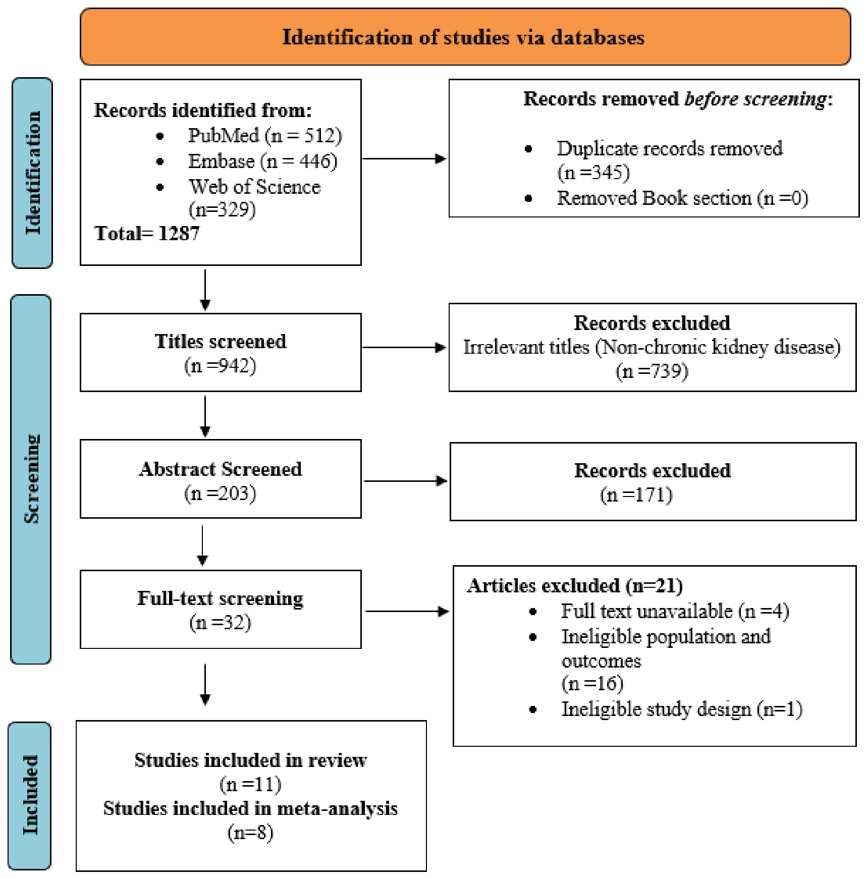
3.1. Search Results
3.2. Biological Specimens Used for Biomarker Assessment
3.2.1. Serum and Plasma Biomarkers
3.2.2. Whole-Blood Biomarkers
3.2.3. Urinary Biomarkers
3.2.4. Kidney Tissue and Renal Homogenates
3.2.5. Renal Inflammatory and Molecular Signaling Biomarkers
3.2.6. Renal Mitochondrial and Apoptotic Biomarkers
3.2.7. Skeletal Muscle Tissue Biomarkers
3.2.8. Renal Histopathology and Structural Tissue Outcomes
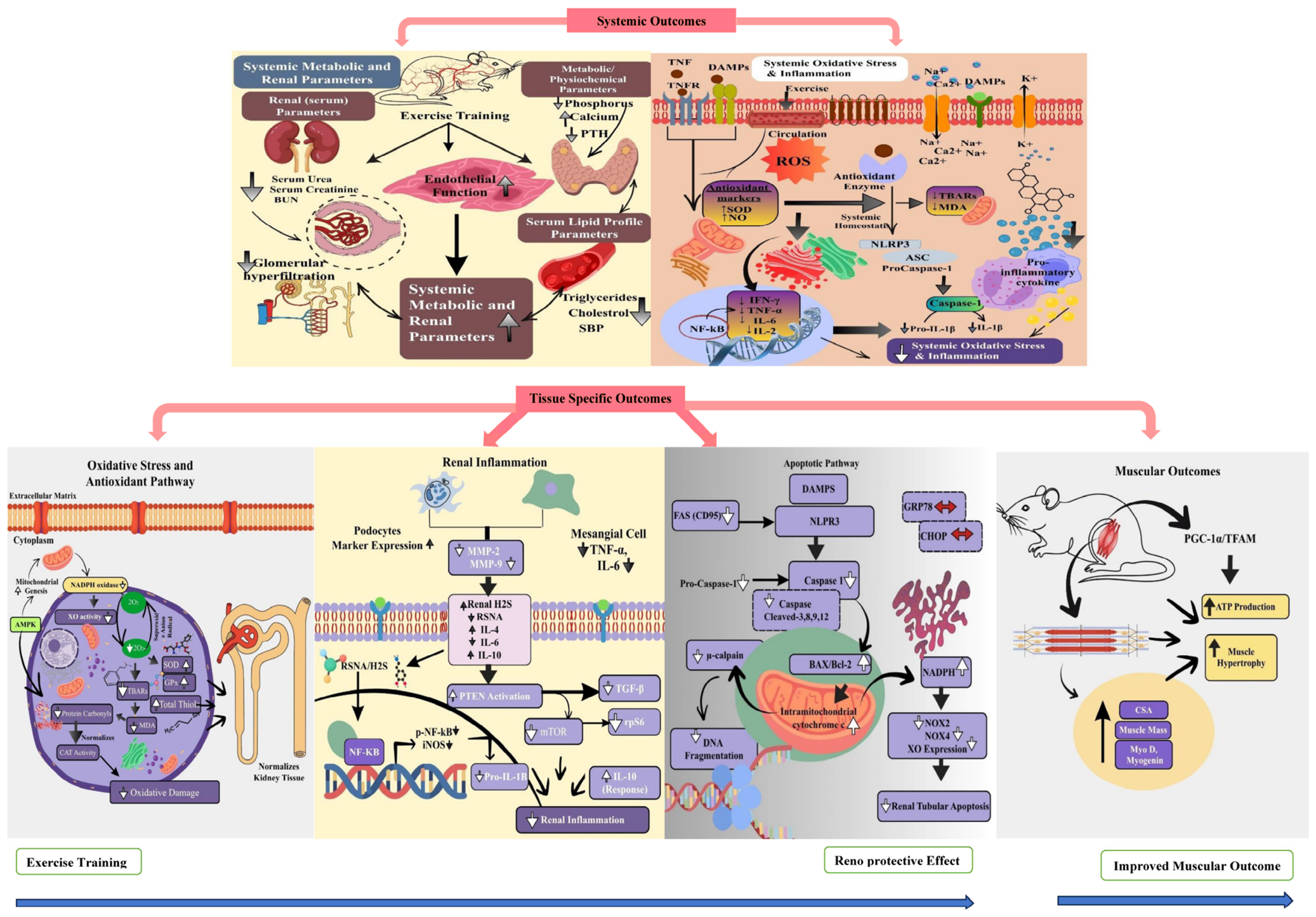
3.3. Effect of Exercise Training on Systemic Outcomes in Experimental Chronic Kidney Disease Model
3.3.1. Renal Parameters (Serum/Plasma/Urine)
3.3.2. Kidney Functional Parameters
3.3.3. Physiological and Physicochemical Outcomes
3.3.4. Systemic Oxidative Stress and Antioxidant Markers
3.3.5. Systemic Inflammatory and Molecular Signaling Markers
3.4. Effect of Exercise on Tissue-Specific Outcomes in Experimental Chronic Kidney Disease Model
3.4.1. Tissue-Specific Oxidative Stress and Antioxidant Markers
3.4.2. Tissue-Specific Inflammatory and Molecular Signaling Markers
3.4.3. Apoptosis and Cell Death Pathways
3.4.4. Histopathology and Structural Kidney Changes
3.4.5. Muscular Outcomes
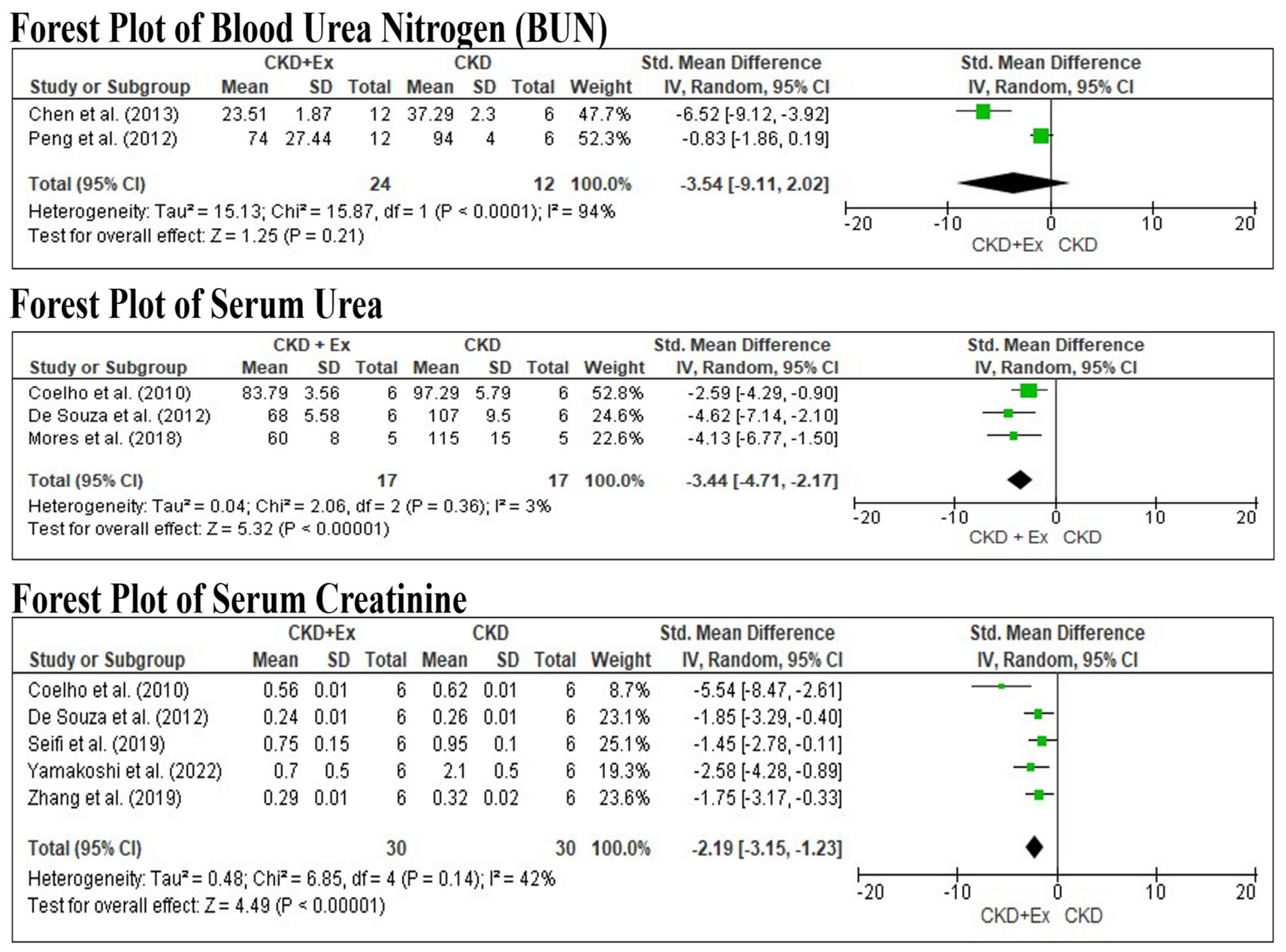
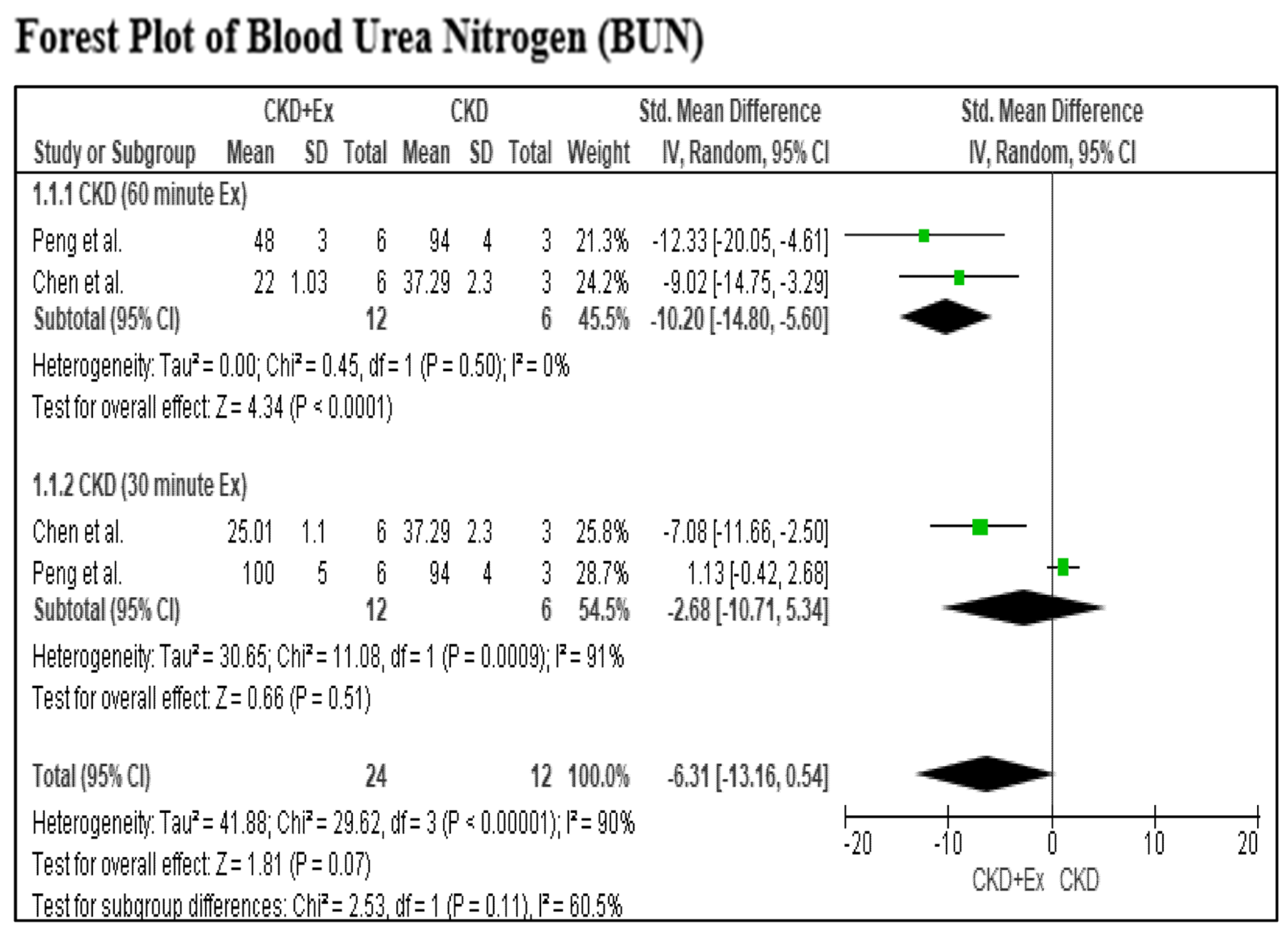
3.5. Pooled Effects of Exercise on Renal Parameters in Chronic Kidney Disease
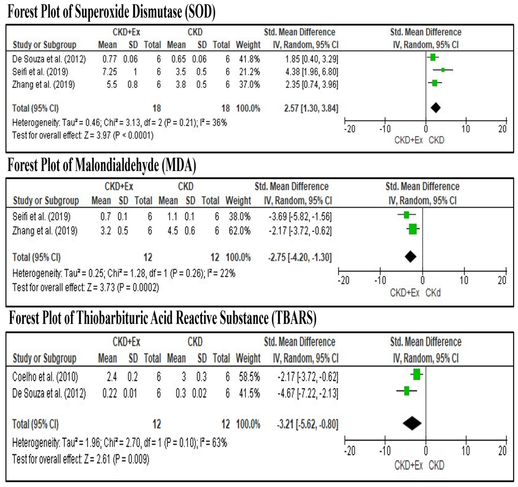
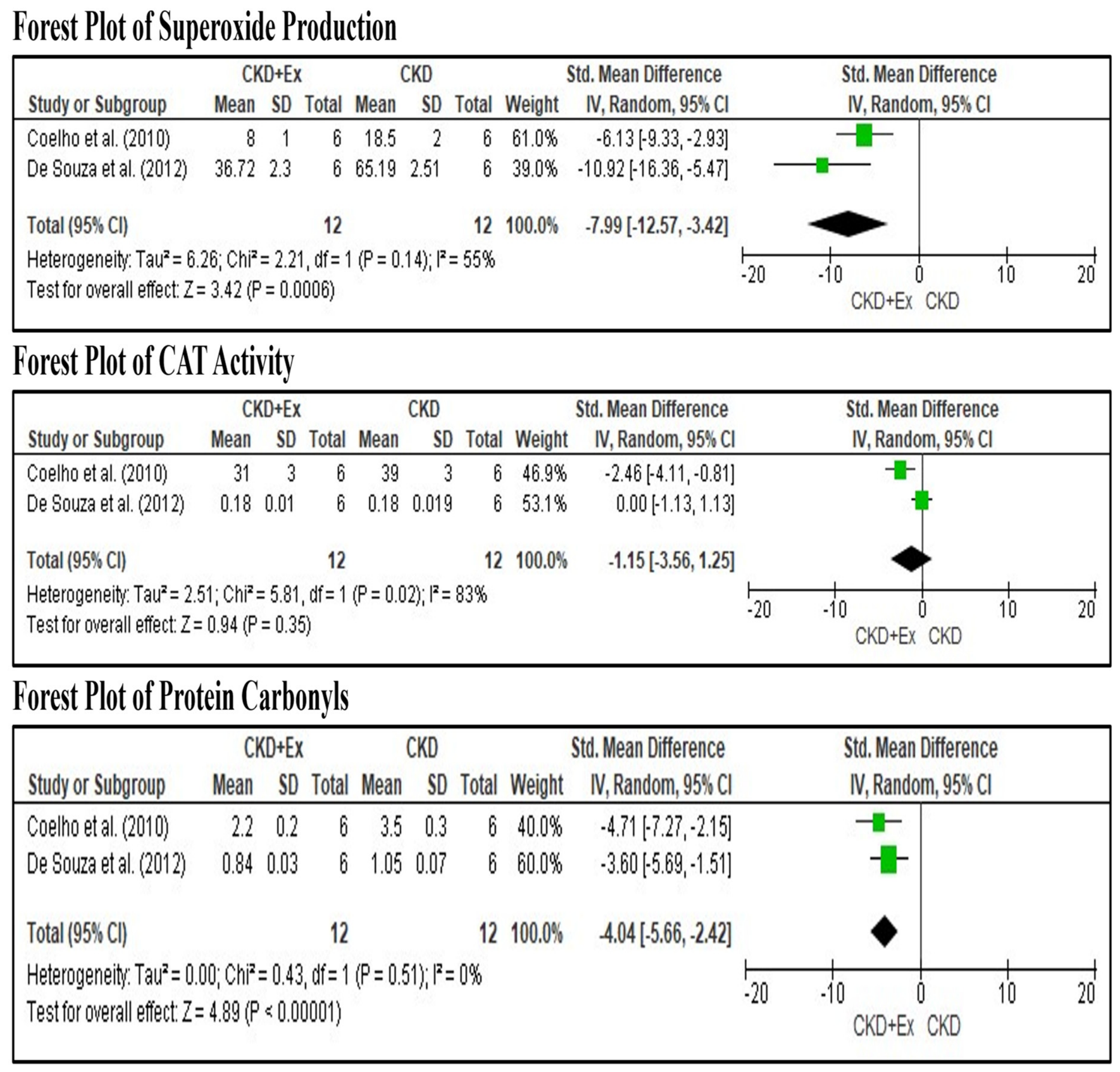
3.6. Pooled Effects of Exercise on Oxidative Stress Markers
4. Discussion
4.1. Clinical Implication
4.2. Limitations and Future Recommendations
5. Conclusions
6. Patient-Friendly Summary
Supplementary Materials
Author Contributions
Funding
Data Availability Statement
Acknowledgments
Conflicts of Interest
References
- Levey, A.S.; Coresh, J. Chronic kidney disease. Lancet 2012, 379, 165–180. [Google Scholar] [CrossRef] [Scilit] [PubMed]
- Egdorf, N. Chronic Kidney Disease: A Systemic Overview. Available online: https://dr.lib.iastate.edu/server/api/core/bitstreams/0e319a18-76bb-4fe1-82c7-092650088a81/content (accessed on 22 August 2026).
- Lim, Y.J.; Sidor, N.A.; Tonial, N.C.; Che, A.; Urquhart, B.L. Uremic toxins in the progression of chronic kidney disease and cardiovascular disease: Mechanisms and therapeutic targets. Toxins 2021, 13, 142. [Google Scholar] [CrossRef] [Scilit] [PubMed]
- Yue, N.C.; Ho, S.C.; Mak, J.K.; Cheng, F.W.; Tam, E.Y.; Lau, W.C.; Hoo, R.L.; Tan, K.C.; Yap, D.Y.; Cheung, C.L. Prevalence and incidence of chronic kidney diseases in Hong Kong: A 12-year territory-wide retrospective cohort study. Lancet Reg. Health–West. Pac. 2025, 64, 101726. [Google Scholar] [CrossRef] [Scilit] [PubMed]
- Zoccali, C.; Vanholder, R.; Massy, Z.A.; Ortiz, A.; Sarafidis, P.; Dekker, F.W.; Fliser, D.; Fouque, D.; Heine, G.H.; Jager, K.J.; et al. The systemic nature of CKD. Nat. Rev. Nephrol. 2017, 13, 344–358. [Google Scholar] [CrossRef] [Scilit] [PubMed]
- Stenvinkel, P.; Ketteler, M.; Johnson, R.J.; Lindholm, B.; Pecoits-Filho, R.; Riella, M.; Heimbürger, O.; Cederholm, T.; Girndt, M. IL-10, IL-6, and TNF-α: Central factors in the altered cytokine network of uremia—The good, the bad, and the ugly. Kidney Int. 2005, 67, 1216–1233. [Google Scholar] [CrossRef] [Scilit] [PubMed]
- Booth, F.W.; Roberts, C.K.; Laye, M.J. Lack of exercise is a major cause of chronic diseases. Compr. Physiol. 2011, 2, 1143–1211. [Google Scholar] [CrossRef] [Scilit]
- Bello, A.K.; Alrukhaimi, M.; Ashuntantang, G.E.; Basnet, S.; Rotter, R.C.; Douthat, W.G.; Kazancioglu, R.; Köttgen, A.; Nangaku, M.; Powe, N.R.; et al. Complications of chronic kidney disease: Current state, knowledge gaps, and strategy for action. Kidney Int. Suppl. 2017, 7, 122–129. [Google Scholar] [CrossRef] [Scilit] [PubMed]
- Duni, A.; Liakopoulos, V.; Roumeliotis, S.; Peschos, D.; Dounousi, E. Oxidative Stress in the Pathogenesis and Evolution of Chronic Kidney Disease: Untangling Ariadne’s Thread. Int. J. Mol. Sci. 2019, 20, 3711. [Google Scholar] [CrossRef] [Scilit] [PubMed]
- Sedeek, M.; Nasrallah, R.; Touyz, R.M.; Hébert, R.L. NADPH oxidases, reactive oxygen species, and the kidney: Friend and foe. J. Am. Soc. Nephrol. 2013, 24, 1512–1518. [Google Scholar] [CrossRef] [Scilit] [PubMed]
- Juan, C.A.; Pérez de la Lastra, J.M.; Plou, F.J.; Pérez-Lebeña, E. The chemistry of reactive oxygen species (ROS) revisited: Outlining their role in biological macromolecules (DNA, lipids and proteins) and induced pathologies. Int. J. Mol. Sci. 2021, 22, 4642. [Google Scholar] [CrossRef] [Scilit] [PubMed]
- Malek, M.; Nematbakhsh, M. Renal ischemia/reperfusion injury; from pathophysiology to treatment. J. Ren. Inj. Prev. 2015, 4, 20–27. [Google Scholar] [CrossRef] [Scilit] [PubMed]
- Zhao, H.; Wu, L.; Yan, G.; Chen, Y.; Zhou, M.; Wu, Y.; Li, Y. Inflammation and tumor progression: Signaling pathways and targeted intervention. Signal Transduct. Target. Ther. 2021, 6, 263. [Google Scholar] [CrossRef] [Scilit] [PubMed]
- Giacco, F.; Brownlee, M. Oxidative stress and diabetic complications. Circ. Res. 2010, 107, 1058–1070. [Google Scholar] [CrossRef] [Scilit] [PubMed]
- Ebert, T.; Neytchev, O.; Witasp, A.; Kublickiene, K.; Stenvinkel, P.; Shiels, P.G. Inflammation and Oxidative Stress in Chronic Kidney Disease and Dialysis Patients. Antioxid. Redox Signal. 2021, 35, 1426–1448. [Google Scholar] [CrossRef] [Scilit] [PubMed]
- Navarro-González, J.F.; Mora-Fernández, C. The Role of Inflammatory Cytokines in Diabetic Nephropathy. J. Am. Soc. Nephrol. 2008, 19, 433–442. [Google Scholar] [CrossRef] [Scilit] [PubMed]
- Pedersen, B.K.; Hoffman-Goetz, L. Exercise and the Immune System: Regulation, Integration, and Adaptation. Physiol. Rev. 2000, 80, 1055–1081. [Google Scholar] [CrossRef] [Scilit] [PubMed]
- Ren, N.; Wang, W.-F.; Zou, L.; Zhao, Y.-L.; Miao, H.; Zhao, Y.-Y. The nuclear factor kappa B signaling pathway is a master regulator of renal fibrosis. Front. Pharmacol. 2024, 14, 1335094. [Google Scholar] [CrossRef] [Scilit] [PubMed]
- Wang, X.H.; Mitch, W.E.; Price, S.R. Pathophysiological mechanisms leading to muscle loss in chronic kidney disease. Nat. Rev. Nephrol. 2021, 18, 138–152. [Google Scholar] [CrossRef] [Scilit] [PubMed]
- Powers, S.K.; Hogan, M.C. Exercise and oxidative stress. J. Physiol. 2016, 594, 5079. [Google Scholar] [CrossRef] [Scilit] [PubMed]
- Gomez-Cabrera, M.-C.; Domenech, E.; Viña, J. Moderate exercise is an antioxidant: Upregulation of antioxidant genes by training. Free. Radic. Biol. Med. 2008, 44, 126–131. [Google Scholar] [CrossRef] [Scilit] [PubMed]
- Gleeson, M.; Bishop, N.C.; Stensel, D.J.; Lindley, M.R.; Mastana, S.S.; Nimmo, M.A. The anti-inflammatory effects of exercise: Mechanisms and implications for the prevention and treatment of disease. Nat. Rev. Immunol. 2011, 11, 607–615. [Google Scholar] [CrossRef] [Scilit] [PubMed]
- Petersen, A.M.W.; Pedersen, B.K. The anti-inflammatory effect of exercise. J. Appl. Physiol. 2005, 98, 1154–1162. [Google Scholar] [CrossRef] [Scilit] [PubMed]
- Allen, J.; Sun, Y.; Woods, J.A. Exercise and the regulation of inflammatory responses. Prog. Mol. Biol. Transla-Tional Sci. 2015, 135, 337–354. [Google Scholar] [CrossRef] [Scilit] [PubMed]
- Bishop, N.C.; Burton, J.O.; Graham-Brown, M.P.M.; Stensel, D.J.; Viana, J.L.; Watson, E.L. Exercise and chronic kidney disease: Potential mechanisms underlying the physiological benefits. Nat. Rev. Nephrol. 2023, 19, 244–256. [Google Scholar] [CrossRef] [Scilit] [PubMed]
- Small, D.M.; Beetham, K.S.; Howden, E.J.; Briskey, D.R.; Johnson, D.W.; Isbel, N.M.; Gobe, G.C.; Coombes, J.S. Effects of exercise and lifestyle intervention on oxidative stress in chronic kidney disease. Redox Rep. 2017, 22, 127–136. [Google Scholar] [CrossRef] [Scilit] [PubMed]
- Baião, V.M.; Cunha, V.A.; Duarte, M.P.; Andrade, F.P.; Ferreira, A.P.; Nóbrega, O.T.; Viana, J.L.; Ribeiro, H.S. Effects of Exercise on Inflammatory Markers in Individuals with Chronic Kidney Disease: A Systematic Review and Meta-Analysis. Metabolites 2023, 13, 795. [Google Scholar] [CrossRef] [Scilit] [PubMed]
- Feng, W.; Wang, Y.; Gu, X.; Yu, D.; Liu, Z. Exercise as a modulator of systemic inflammation and oxidative stress biomarkers across clinical and healthy populations: An umbrella meta-analysis. BMC Sports Sci. Med. Rehabil. 2025, 17, 360. [Google Scholar] [CrossRef] [Scilit] [PubMed]
- Chen, K.-C.; Peng, C.-C.; Hsieh, C.-L.; Peng, R.Y. Exercise Ameliorates Renal Cell Apoptosis in Chronic Kidney Disease by Intervening in the Intrinsic and the Extrinsic Apoptotic Pathways in a Rat Model. Evid.-Based Complement. Altern. Med. 2013, 2013, 368450. [Google Scholar] [CrossRef] [Scilit] [PubMed]
- Coelho, B.L.; Rocha, L.G.; Scarabelot, K.S.; Scheffer, D.L.; Ronsani, M.M.; Silveira, P.C.; Silva, L.A.; Souza, C.T.; Pinho, R.A. Physical Exercise Prevents the Exacerbation of Oxidative Stress Parameters in Chronic Kidney Disease. J. Ren. Nutr. 2010, 20, 169–175. [Google Scholar] [CrossRef] [Scilit] [PubMed]
- de Souza, P.S.; da Rocha, L.G.C.; Tromm, C.B.; Scheffer, D.L.; Victor, E.G.; da Silveira, P.C.L.; de Souza, C.T.; Silva, L.A.; Pinho, R.A. Therapeutic action of physical exercise on markers of oxidative stress induced by chronic kidney disease. Life Sci. 2012, 91, 132–136. [Google Scholar] [CrossRef] [Scilit] [PubMed]
- Moraes, M.R.; Rosa, T.S.; Souza, M.K.; Neves, R.V.P.; Bacurau, R.F.P.; Passos, C.S.; Cenedeze, M.A.; Passos, M.T.; Machado, F.G.; Filho, A.P.-S.; et al. Resistance training downregulates macrophages infiltration in the kidney of 5/6 nephrectomized rats. Life Sci. 2018, 213, 190–197. [Google Scholar] [CrossRef] [Scilit] [PubMed]
- Peng, C.-C.; Chen, K.-C.; Hsieh, C.-L.; Peng, R.Y. Swimming Exercise Prevents Fibrogenesis in Chronic Kidney Disease by Inhibiting the Myofibroblast Transdifferentiation. PLoS ONE 2012, 7, e37388. [Google Scholar] [CrossRef] [Scilit] [PubMed]
- Yamakoshi, S.; Nakamura, T.; Xu, L.; Kohzuki, M.; Ito, O. Exercise Training Ameliorates Renal Oxidative Stress in Rats with Chronic Renal Failure. Metabolites 2022, 12, 836. [Google Scholar] [CrossRef] [Scilit] [PubMed]
- Zhang, Y.; Liu, Y.; Bi, X.; Hu, C.; Ding, F.; Ding, W. Therapeutic Approaches in Mitochondrial Dysfunction, Inflammation, and Autophagy in Uremic Cachexia: Role of Aerobic Exercise. Mediat. Inflamm. 2019, 2019, 2789014. [Google Scholar] [CrossRef] [Scilit] [PubMed]
- Organ, J.; Allen, M.; Myers-White, A.; Elkhatib, W.; O’NEill, K.; Chen, N.; Moe, S.; Avin, K. Effects of treadmill running in a rat model of chronic kidney disease. Biochem. Biophys. Rep. 2018, 16, 19–23. [Google Scholar] [CrossRef] [Scilit] [PubMed]
- Saud, A.; Luiz, R.; Leite, A.; Muller, C.; Visona, I.; Reinecke, N.; Silva, W.; Gloria, M.; Razvickas, C.; Casarini, D.; et al. Resistance exercise training ameliorates chronic kidney disease outcomes in a 5/6 nephrectomy model. Life Sci. 2021, 275, 119362. [Google Scholar] [CrossRef] [Scilit] [PubMed]
- Seifi, B.; Sajedizadeh, A.; Kadkhodaee, M.; Ranjbaran, M. Long-term exercise restores hydrogen sulfide in the kidney and contributes to exercise benefits in 5/6 nephrectomized rats. Clin. Exp. Hypertens. 2018, 41, 87–91. [Google Scholar] [CrossRef] [Scilit] [PubMed]
- Souza, M.K.; Neves, R.V.P.; Rosa, T.S.; Cenedeze, M.A.; Arias, S.C.A.; Fujihara, C.K.; Bacurau, R.F.P.; Câmara, N.O.S.; Moraes, M.R.; e Silva Filho, A.P. Resistance training attenuates inflammation and the progression of renal fibrosis in chronic renal disease. Life Sci. 2018, 206, 93–97. [Google Scholar] [CrossRef] [Scilit] [PubMed]
- Heifets, M.; Davis, T.A.; Tegtmeyer, E.; Klahr, S. Exercise training ameliorates progressive renal disease in rats with subtotal nephrectomy. Kidney Int. 1987, 32, 815–820. [Google Scholar] [CrossRef] [Scilit] [PubMed]
- Powers, S.K.; Lennon, S.L. Analysis of cellular responses to free radicals: Focus on exercise and skeletal muscle. Proc. Nutr. Soc. 1999, 58, 1025–1033. [Google Scholar] [CrossRef] [Scilit] [PubMed]
- Viana, J.L.; Kosmadakis, G.C.; Watson, E.L.; Bevington, A.; Feehally, J.; Bishop, N.C.; Smith, A.C. Evidence for Anti-Inflammatory Effects of Exercise in CKD. J. Am. Soc. Nephrol. 2014, 25, 2121–2130. [Google Scholar] [CrossRef] [Scilit] [PubMed]
- Lima, W.V.; Visona, I.; Schor, N.; Almeida, W.S. Preconditioning by aerobic exercise reduces acute ischemic renal injury in rats. Physiol. Rep. 2019, 7, e14176. [Google Scholar] [CrossRef] [Scilit] [PubMed]
- Huang, C.-C.; Lin, Y.-Y.; Yang, A.-L.; Kuo, T.-W.; Kuo, C.-H.; Lee, S.-D. Anti-Renal Fibrotic Effect of Exercise Training in Hypertension. Int. J. Mol. Sci. 2018, 19, 613. [Google Scholar] [CrossRef] [Scilit] [PubMed]
- Zhou, Z.; Ying, C.; Zhou, X.; Shi, Y.; Xu, J.; Zhu, Y.; Wang, M.; Li, Y.; Li, X.; Xiang, J. Aerobic exercise training alleviates renal injury in db/db mice through inhibiting Nox4-mediated NLRP3 inflammasome activation. Exp. Gerontol. 2022, 168, 111934. [Google Scholar] [CrossRef] [Scilit] [PubMed]
- Lourenço, Í.; Neto, W.K.; Amorim, L.d.S.P.; Ortiz, V.M.M.; Geraldo, V.L.; Ferreira, G.H.d.S.; Caperuto, É.C.; Gama, E.F. Muscle hypertrophy and ladder-based resistance training for rodents: A systematic review and meta-analysis. Physiol. Rep. 2020, 8, e14502. [Google Scholar] [CrossRef] [Scilit] [PubMed]



| References | 1 | 2 | 3 | 4 | 5 | 6 | 7 | 8 | 9 | 10 | Total |
|---|---|---|---|---|---|---|---|---|---|---|---|
| Chen et al. [29] | ✓ | ✓ | ✓ | ✓ | ✓ | ✓ | ✓ | 7/10 | |||
| Coelho et al. [30] | ✓ | ✓ | ✓ | ✓ | ✓ | ✓ | 6/10 | ||||
| De Souza et al. [31] | ✓ | ✓ | ✓ | ✓ | ✓ | ✓ | ✓ | 7/10 | |||
| Moraes et al. [32] | ✓ | ✓ | ✓ | ✓ | ✓ | ✓ | ✓ | 7/10 | |||
| Peng et al. [33] | ✓ | ✓ | ✓ | ✓ | ✓ | ✓ | 6/10 | ||||
| Yamakoshi et al. [34] | ✓ | ✓ | ✓ | ✓ | ✓ | ✓ | ✓ | 7/10 | |||
| Zhang et al. [35] | ✓ | ✓ | ✓ | ✓ | ✓ | ✓ | 6/10 | ||||
| Organ et al. [36] | ✓ | ✓ | ✓ | ✓ | ✓ | 5/10 | |||||
| Saud et al. [37] | ✓ | ✓ | ✓ | ✓ | ✓ | ✓ | ✓ | 7/10 | |||
| Seifi et al. [38] | ✓ | ✓ | ✓ | ✓ | ✓ | ✓ | ✓ | 7/10 | |||
| Souza et al. [39] | ✓ | ✓ | ✓ | ✓ | ✓ | ✓ | ✓ | 7/10 |
| Study Characteristics | Outcomes Based on the Systemic Level | |||||||||
|---|---|---|---|---|---|---|---|---|---|---|
| References | Sample | Exercise Type | Exercise Parameters | Sample Size | Renal Parameters (Serum/Plasma/Urine) | Kidney Functional (Systemic) | Physiological and Physicochemical (Systemic) | Oxidative Stress & Antioxidant Markers (Systemic) | Inflammatory & Molecular Signaling Markers (Systemic) | Histopathology & Structural Kidney Changes (Systemic) |
| Chen et al. [29] | S: Sprague-Dawley rats Sex: Male A: 4 weeks old W: 225–250 g Model: DRCKD | Treadmill training | D: 30–60 min/session (pooled exercise group) F: 3 days/week P: 11 weeks | CKD: N = 6 rats CKD+EX: N = 6 rats | CKD: ↑ BUN CKD+EX: ↓ BUN | CKD: ↑ KW/BW CKD+EX: ↓ KW/BW | CKD: ↓ RBC ↑ platelets ↑ WBC ↑ cholesterol ↑ triglycerides CKD+EX: ↑ RBC ↓platelets, WBC partially normalized, ↑ cholesterol ↑ triglycerides | CKD: ↓ SOD, ↑ MDA CKD+EX: ↑ SOD, ↓ MDA | Not assessed | Not assessed |
| Coelho et al. [30] | S: Wistar rats, Sex: Male A: 3 months old W: 250–300 g Model: 5/6 nephrectomy | Treadmill running | Speed: up to 1 km/h D: 50 min/day F: 5 days/week P: 8 weeks | CKD: N = 6 rats CKD+EX: N = 6 rats | CKD: ↑ Urea, ↑ Creatinine CKD+EX: ↔ Urea, ↔Creatinine | Not assessed | Not assessed | Not assessed | Not assessed | Not assessed |
| De Souza et al. [31] | S: Wistar rats Sex: Male A: 3 months old W: 250–300 g Model: 5/6 nephrectomy | Treadmill running | Speed: 13–17 m/min D: 50 min/day F: 5 days/week P: 8 weeks | CKD: N = 6 rats CKD+EX: N = 6 rats | CKD: ↑ Urea, ↑ Creatinine, CKD+EX: ↓ Urea, ↔ Creatinine, | Not assessed | Not assessed | Not assessed | Not assessed | Not assessed |
| Moraes et al. [32] | S: Wistar rats Sex: Male A: 8 weeks old W: 230–250 g Model: 5/6 nephrectomy | Resistance Exercise: Vertical ladder climbing | D: 8 weeks, progressive load based on CLmax F: 3 days/week, non-consecutive days. | CKD: N = 5 rats CKD+EX: N = 5 rats | CKD: ↑ Urea, ↑ Creatinine, ↑ Uprot, CKD+EX: ↓ Urea, ↓ Creatinine, ↓ Uprot, | CKD: ↓ GFR CKD+EX: ↑ GFR, | CKD: ↑ SBP, ↑ Lipids, ↑ Lactate, ↔ Glucose, ↑ Na+/K+ ↓ BW CKD+EX: ↓ SBP, ↓ Lipids, ↓ Lactate, ↔ Glucose, ↓ Na+/K+, ↑ BW | CKD: ↓ NO, ↔ SOD CKD+EX: ↑ NO, ↑ SOD | CKD: ↑ IFN-γ, ↑ TNF-α, ↑ IL-2, ↑ IL-6 CKD+EX: ↓ IFN-γ, ↓ TNF-α, ↓ IL-2, ↓ IL-6 | Not assessed |
| Peng et al. [33] | S: Sprague-Dawley rats Sex: Male A: 4 weeks old W: 220–250 g Model: DRCKD | Swimming Exercise | D: 30 or 60 min F: 3 days/week P = 11 weeks | CKD: N = 6 rats CKD+EX: N = 6 rats | CKD: ↑ BUN, ↑ Creatinine, ↑ Proteinuria CKD+EX: ↓ BUN, ↔ Creatinine, ↓ proteinuria | CKD: ↑ KW/BW CKD+EX: ↓ KW/BW | CKD: ↓ BW CKD+EX: ↑ BW | CKD: ↓ SOD, ↑ TBARS CKD+EX: ↑ SOD, ↓TBARS | CKD: ↑ IL-6, ↑ MMP-2, ↑ MMP-9 CKD+EX: ↓ IL-6, ↓ MMP-2, ↓ MMP-9 | Not assessed |
| Yamakoshi et al. [34] | S: Sprague–Dawley rats, Sex: Male A: 6 weeks old Model: 5/6 nephrectomy | Treadmill running | Speed: 20 m/min D: 60 min/day F: 5 days/week 12 weeks | CKD: N = 6 rats CKD+EX: N = 6 rats | CKD: ↑ creatinine ↑ Uprot CKD+EX: ↓ creatinine, ↓ Uprot | Not assessed | CKD: ↓ BW, ↑ SBP CKD+EX: ↔ BW, ↑ SBP | CKD: ↑ MDA CKD+EX: ↓ MDA # | Not assessed | Not assessed |
| Zhang et al. [35] | S: C57BL/6J mice Sex: Male A: 8–12 weeks old Model: 5/6 nephrectomy | Aerobic exercise (wheel running) | Speed: 6.5 m/min D: 1 h/day F: 5 days/week P: 8 weeks | CKD: N = 6 mice CKD+EX: N = 6 mice | CKD: ↑ Creatinine, ↑ BUN, CKD+EX: ↓ Creatinine #, ↓ BUN # | Not assessed | CKD: ↓ BW CKD+EX: ↑ BW #. | Not assessed | Not assessed | Not assessed |
| Organ et al. [36] | S: Cy/+ rats Sex: Not specified A: 25 weeks Model: Progressive CKD (Cy/+ rat) | Treadmill running | Speed: 8 m/min → 18 m/min D: 60 min/session F: 5 days/week P: 10 weeks | CKD: N = 8 rats CKD+EX: N = 8 rats | CKD: ↑ BUN CKD+EX: ↔ BUN | Not assessed | CKD: ↑ Phosphorus; ↑ PTH; ↓ Ca CKD+EX: ↓ Phosphorus; ↔ PTH; ↑Ca | Not assessed | Not assessed | Not assessed |
| Saud et al. [37] | S: Wistar rats; Sex: Male; W: 230–250 g; Model: 5/6 nephrectomy CKD | Resistance Exercise Training | D: 8–12 climbs/session with progressive load F: 5 days/week P: 8 weeks | CKD: N = 6 rats CKD+EX: N = 7 rats | CKD: ↑ BUN, ↑ Uprot, ↑ creatinine CKD+EX: ↓ BUN, ↓ Uprot, ↓ creatinine | Not assessed | CKD: ↑ SBP ↓ BW CKD+EX: ↑ BW | Not assessed | Not assessed | Not assessed |
| Seifi et al. [38] | S: Wistar rats Sex: Male W: 250–300 g Model: 5/6 nephrectomy CKD | Treadmill exercise | Speed: 18 m/min P: 8 weeks | CKD: N = 6 rats CKD+EX: N = 6 rats | CKD: ↑ creatinine; ↑ BUN CKD+EX: ↓ creatinine; ↓ BUN | Not assessed | CKD: ↑ SBP CKD+EX: ↓ SBP | Not assessed | Not assessed | Not assessed |
| Souza et al. [39] | S: Munich-Wistar rats, Sex: Male, A: 8 weeks old, W: 240 ± 20 g, Model: 5/6 nephrectomy CKD | Resistance training | D: 12 min/session 3 days/week F: 8–12 dynamic ladder climbs per session P: 10 weeks | CKD: N = 5 rats CKD+EX: N = 5 rats | CKD: ↓ CK levels, CKD+EX: ↑ CK levels | Not assessed | CKD: ↔ BW CKD+EX: ↔ BW | Not assessed | Not assessed | Not assessed |
| Tissue-Specific Outcomes | ||||||
|---|---|---|---|---|---|---|
| References | Sample Size | Oxidative Stress & Antioxidant Markers (Tissue) | Inflammatory & Molecular Signaling Markers (Tissue) | Apoptosis & Cell Death Pathways (Tissue) | Histopathology & Structural Kidney Changes (Tissue) | Muscular Outcomes (Tissue) |
| Chen et al. [29] | CKD: N = 6 rats CKD+EX: N = 6 rats | Not assessed | CKD (Kidney tissue): ↑ Fas (CD95); ↔ GRP78; ↔ CHOP CKD+EX (Kidney tissue): ↓ Fas (CD95) ↔ GRP78; ↔ CHOP | CKD (Kidney tissue): ↑ Bax; ↓ Bcl-2; ↓ intramitochondrial cytochrome-c; ↑ caspase-9 cleaved, 3,8,12; ↑ μ-calpain; ↑ DNA fragmentation CKD+EX (Kidney tissue): ↓ Bax; ↑ Bcl-2; ↑intramitochondrial cytochrome c; ↓ caspase-9, cleaved, 3,8,12; ↓ μ-calpain; ↓ DNA fragmentation | CKD (Kidney tissue): ↑ TUNEL-positive cells, Marked renal cortical damage CKD+EX (Kidney tissue): ↓ TUNEL-positive cells; preservation of renal cortical architecture | Not assessed |
| Coelho et al. [30] | CKD: N = 6 rats CKD+EX: N = 6 rats | CKD (Kidney tissue): ↑ Superoxide, ↑ TBARS, ↑ Protein carbonyls, ↓ Total thiols, ↑ SOD, ↑ CAT CKD+EX (Kidney tissue): ↓ Superoxide, ↓ TBARS, ↓ Protein carbonyls, ↑ Total thiols, ↓ SOD, ↓ CAT | Not assessed | Not assessed | Not assessed | Not assessed |
| De Souza et al. [31] | CKD: N = 6 rats CKD+EX: N = 6 rats | CKD (Kidney tissue): ↑ Superoxide, ↔ SOD, ↔ CAT, ↔ GPx, ↑TBARS/Protein carbonyl CKD+EX (Kidney tissue): ↓ Superoxide, ↑ SOD, ↔ CAT, ↑ GPx, ↓ TBARS/Protein carbonyl | Not assessed | Not assessed | Not assessed | Not assessed |
| Moraes et al. [32] | CKD: N = 5 rats CKD+EX: N = 5 rats | Not assessed | Not assessed | Not assessed | CKD (Kidney tissue): ↑ Fibrosis, ↑ Macrophage, ↓ HO-1, ↓ iNOS CKD+EX (Kidney tissue): ↓ Fibrosis, ↓ Macrophage, ↑ HO-1, ↑ iNOS | CKD (Muscle tissue): ↓ Muscle mass (soleus/plantaris/EDL) CKD+EX (Muscle tissue): ↑ Muscle mass (soleus/plantaris/EDL) |
| Peng et al. [33] | CKD: N = 6 rats CKD+EX: N = 6 rats | Not assessed | CKD (Kidney tissue): ↓ TNF-α, ↑ PDGFR, ↑ p-PDGFR, ↑ α-SMA, ↑ CD34 CKD+EX (Kidney tissue): ↑ TNF-α, ↓ PDGFR, ↓ p-PDGFR, ↓ α-SMA, ↓ CD34 | Not assessed | CKD: Kidney tissue (cortex & cortex–medulla junction): ↑ Collagen deposition, ↑ Glomerular volume, ↑ Edema CKD+EX: Kidney tissue (cortex & cortex–medulla junction): ↓ Collagen deposition, ↓ Glomerular volume, ↓ Edema | Not assessed |
| Yamakoshi et al. [34] | CKD: N = 6 rats CKD+EX: N = 6 rats | CKD (Kidney tissue): ↑ Renal NADPH oxidase activity, ↑ XO activity, ↑ Nox2, ↑ Nox4, ↑ XO expression CKD+EX (Kidney tissue): ↓ Renal NADPH oxidase activity #, ↓ XO activity #, ↓ Nox2 #, ↓ Nox4 #, ↓ XO expression # | Not assessed | Not assessed | CKD (Kidney tissue): ↑ Cortical damage, renal dysfunction CKD+EX (Kidney tissue): Preservation of renal cortical structure # | Not assessed |
| Zhang et al. [35] | CKD: N = 6 mice CKD+EX: N = 6 mice | CKD (Muscle tissue): ↓ SOD2, ↑ MDA CKD+EX (Muscle tissue): ↑ SOD2#, ↓ MDA # | CKD (Muscle tissue): ↑ IL-6, ↑ TNF-α, ↑ NLRP3, ↑ ASC, ↑ Caspase-1, ↑ IL-1β, ↑ IL-18 CKD+EX (Muscle tissue): ↓ IL-6 #, ↓ TNF-α #, ↓ NLRP3 #, ↓ ASC #, ↓ caspase-1 #, ↓ IL-1β #, ↓ IL-18 # | CKD (Muscle tissue): ↑ Bax, ↑ Cleaved caspase-3 CKD+EX (Muscle tissue): ↓ Bax#, ↓ Cleaved caspase-3 # | CKD (Kidney tissue): ↑ Glomerular injury, ↑ Tubulointerstitial fibrosis CKD+EX (Kidney Tissue): ↓ Glomerular injury #, ↓ Tubulointerstitial fibrosis # | CKD (Muscle tissue): ↓ Grip strength, ↓ CSA gastrocnemius, ↓ MyHC, ↓ MyoD, ↓ Myogenin, ↓ Pax-7, ↑ Atrogin-1, ↑ MuRF-1, ↑ Myostatin, ↓ mtDNA, ↓ ATP, ↓ PGC-1α, ↓ TFAM, ↓ CoxIV, ↓ Gastrocnemius weight, ↓ Tibialis anterior weight CKD+EX (Muscle tissue): ↑ grip strength #, ↑ CSA gastrocnemius #, ↑ MyHC #, ↑ MyoD #, ↑ Myogenin #, ↑ Pax-7 #, ↓ Atrogin-1 #, ↓ MuRF-1 #, ↓ Myostatin #, ↑ mtDNA #, ↑ ATP #, ↑ PGC-1α #, ↑ TFAM #, ↑ CoxIV #, ↑Gastrocnemius weight, ↑ Tibialis muscle weights # |
| Organ et al. [36] | CKD: N = 8 rats CKD+EX: N = 8 rats | Not assessed | Not assessed | Not assessed | Not assessed | CKD (Muscle tissue): Normal EDL muscle protein turnover marker/torque and cross-sectional area CKD+EX (Muscle tissue): ↔ Maximum EDL muscle torque ↔ Half-relaxation time ↔ EDL Muscle fiber cross-sectional area ↑ EDL muscle catabolism, ↑ Ubiquitin protein expression (~50%) ↑ Atrogin-1 gene expression, ↔ IGF-1 expression ↔ p70 signaling pathway ↓ Pax-7 expression ↔ MyoD, ↔ Myogenin |
| Saud et al. [37] | CKD: N = 6 rats CKD+EX: N = 7 rats | Not assessed | CKD (Kidney tissue): ↓ IL-10, ↑ TNF-α, ↑ TGF-β, ↑ mTOR, ↑ rpS6, ↓ PTEN. CKD+EX (Kidney tissue): ↑ IL-10, ↓ TNF-α, ↓ TGF-β, ↓ mTOR, ↓ rpS6, ↑ PTEN | Not assessed | CKD (Kidney tissue): ↑ glomerulosclerosis, ↑ capsular adhesions, ↑ interstitial fibrosis, ↑ inflammation, ↑ tubular atrophy, ↑ tubular cell loss, ↑ thickened tubular basement membrane. CKD+EX (kidney tissue): ↓ renal injury score, ↓ glomerulosclerosis, ↓ interstitial fibrosis, ↓ inflammation, ↓ renal architecture | CKD (Muscle tissue): ↓ muscle mass (EDL/tibialis anterior/plantaris/soleus), ↓ maximal weight carried. CKD+EX (Muscle tissue): ↑ muscle mass (EDL/tibialis anterior/plantar/soleus), ↑ maximal load capacity |
| Seifi et al. [38] | CKD: N = 6 rats CKD+EX: N = 6 rats | CKD (Kidney tissue): ↑ MDA; ↓ SOD CKD+EX (Kidney tissue): ↓ MDA; ↑ SOD | CKD (Kidney tissue): ↓ renal H2S; ↑ RSNA CKD+EX (Kidney tissue): ↑ renal H2S; ↓ RSNA | Not assessed | CKD (Kidney tissue): ↑ tubular injury; ↓ renal histology; preservation of renal structure CKD+EX (Kidney tissue): ↓ tubular injury; ↑ renal histology; preservation of renal structure | Not assessed |
| Souza et al. [39] | CKD: N = 5 rats CKD+EX: N = 5 rats | Not assessed | CKD (Kidney tissue): ↑ IL-6, ↓ IL-4, ↓ IL-10 CKD+EX (Kidney tissue): ↓ IL-6, ↑ IL-4, ↑ IL-10 | Not assessed | CKD (Kidney tissue): ↑ renal fibrosis, ↑ collagen accumulation/fibrosis CKD+EX (Kidney tissue): ↓ renal fibrosis, renal architecture preserved | CKD (Muscle tissue): ↓ soleus/gastrocnemius/quadriceps muscle strength ↔ muscle mass CKD+EX (Muscle tissue): ↑ soleus/gastrocnemius/quadriceps muscle strength, ↔ muscle mass |
Disclaimer/Publisher’s Note: The statements, opinions and data contained in all publications are solely those of the individual author(s) and contributor(s) and not of MDPI and/or the editor(s). MDPI and/or the editor(s) disclaim responsibility for any injury to people or property resulting from any ideas, methods, instructions or products referred to in the content. |
© 2026 by the authors. Licensee MDPI, Basel, Switzerland. This article is an open access article distributed under the terms and conditions of the Creative Commons Attribution (CC BY) license.
Share and Cite
Rehman, M.; Shah, I.A.; Tsai, K.-L.; Lee, S.-D.; Wu, B.-T. Effects of Exercise Training on Experimental Chronic Kidney Disease: A Systematic Review and Meta-Analysis. Int. J. Mol. Sci. 2026, 27, 7830. https://doi.org/10.3390/ijms27177830
Rehman M, Shah IA, Tsai K-L, Lee S-D, Wu B-T. Effects of Exercise Training on Experimental Chronic Kidney Disease: A Systematic Review and Meta-Analysis. International Journal of Molecular Sciences. 2026; 27(17):7830. https://doi.org/10.3390/ijms27177830
Chicago/Turabian StyleRehman, Muheebur, Iqbal Ali Shah, Kun-Ling Tsai, Shin-Da Lee, and Bor-Tsang Wu. 2026. "Effects of Exercise Training on Experimental Chronic Kidney Disease: A Systematic Review and Meta-Analysis" International Journal of Molecular Sciences 27, no. 17: 7830. https://doi.org/10.3390/ijms27177830
APA StyleRehman, M., Shah, I. A., Tsai, K.-L., Lee, S.-D., & Wu, B.-T. (2026). Effects of Exercise Training on Experimental Chronic Kidney Disease: A Systematic Review and Meta-Analysis. International Journal of Molecular Sciences, 27(17), 7830. https://doi.org/10.3390/ijms27177830