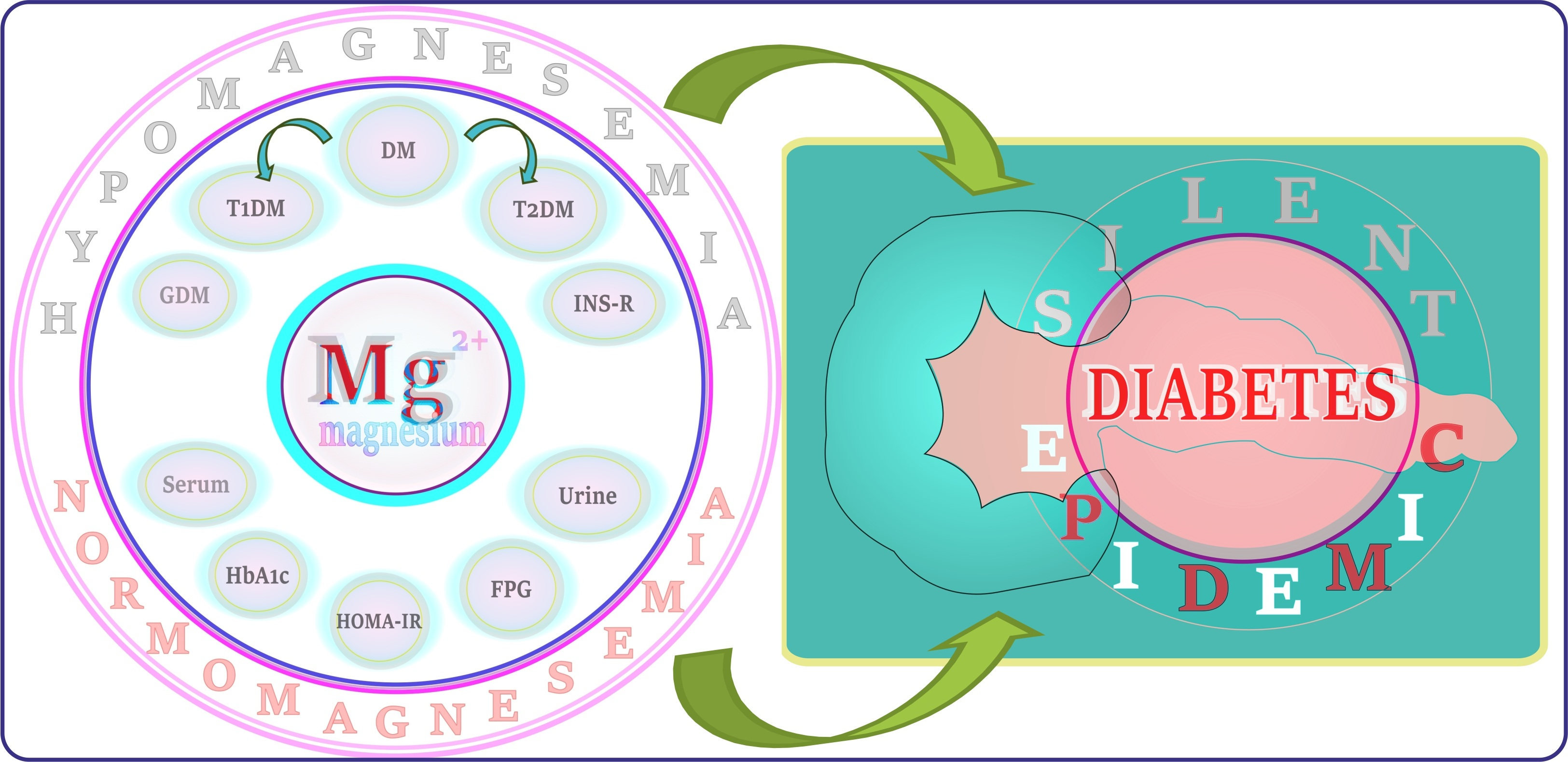
Magnesium and Diabetes: A Review of Recent Studies on the Efficacy of Supplementation with Mg in Humans Suffering from This Chronic Metabolic Disease

 ,
,  ,
,  ,
,  and
and
Abstract
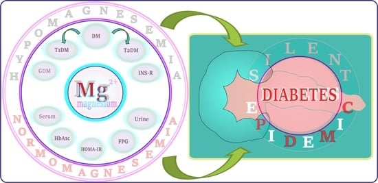
1. Introduction
1.1. Diabetes: Selected Issues in a Nutshell
1.1.1. Diabetes: Background
1.1.2. Diabetes Worldwide
1.1.3. Type 1 and Type 2 Diabetes Mellitus
1.1.4. Gestational Diabetes Mellitus
1.1.5. Insulin Resistance
1.1.6. Risk Factors for Diabetes
1.1.7. Complications of Diabetes
1.2. Magnesium: Selected Issues in a Nutshell
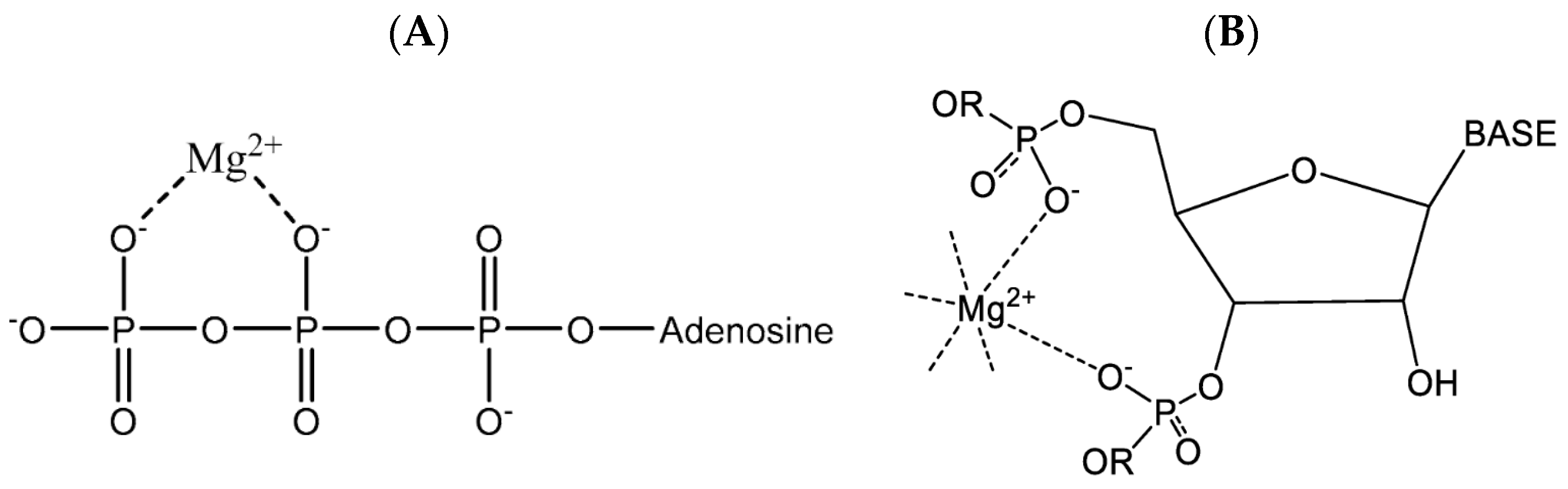
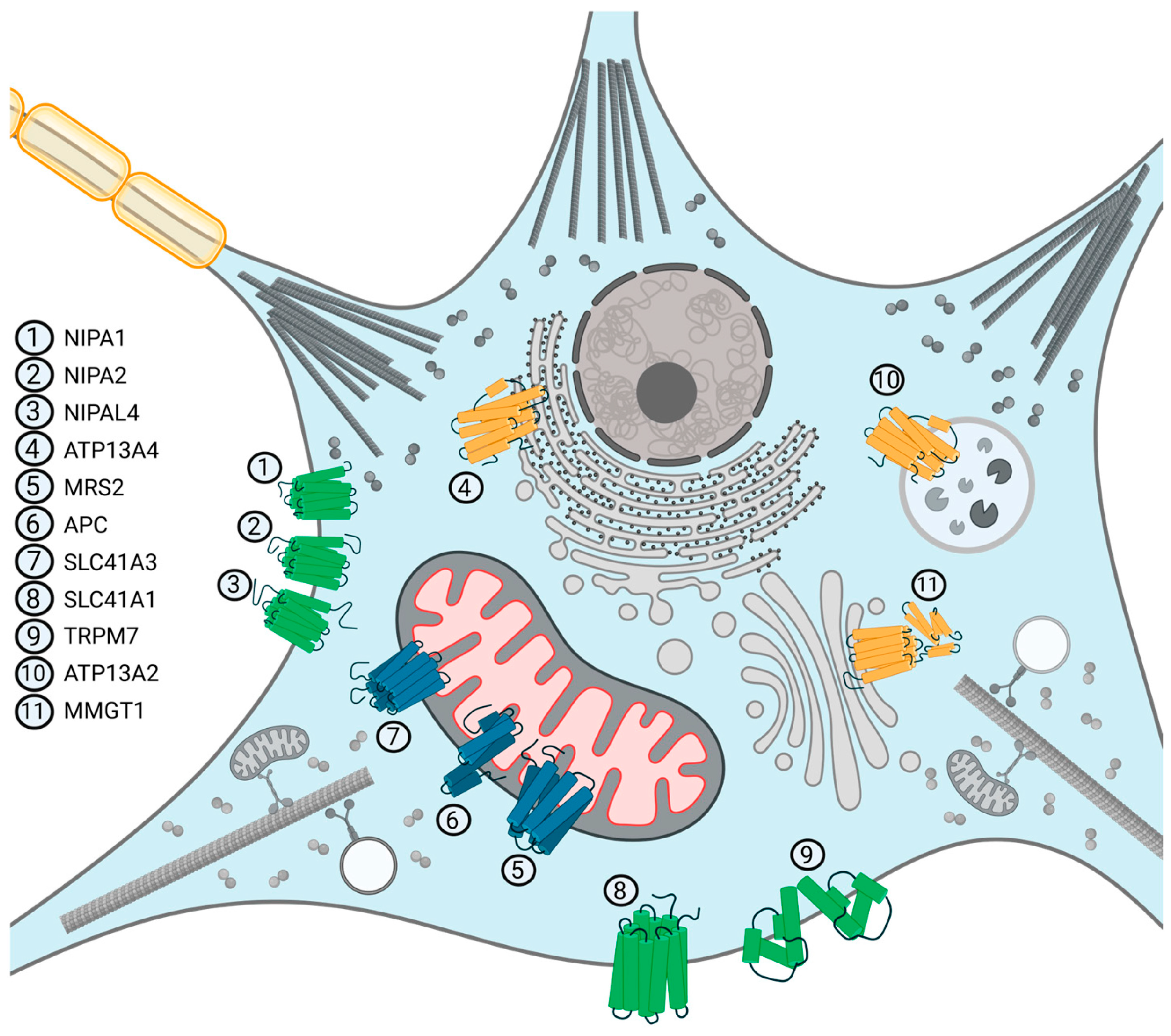
1.2.1. Role of Mg in Biological Systems
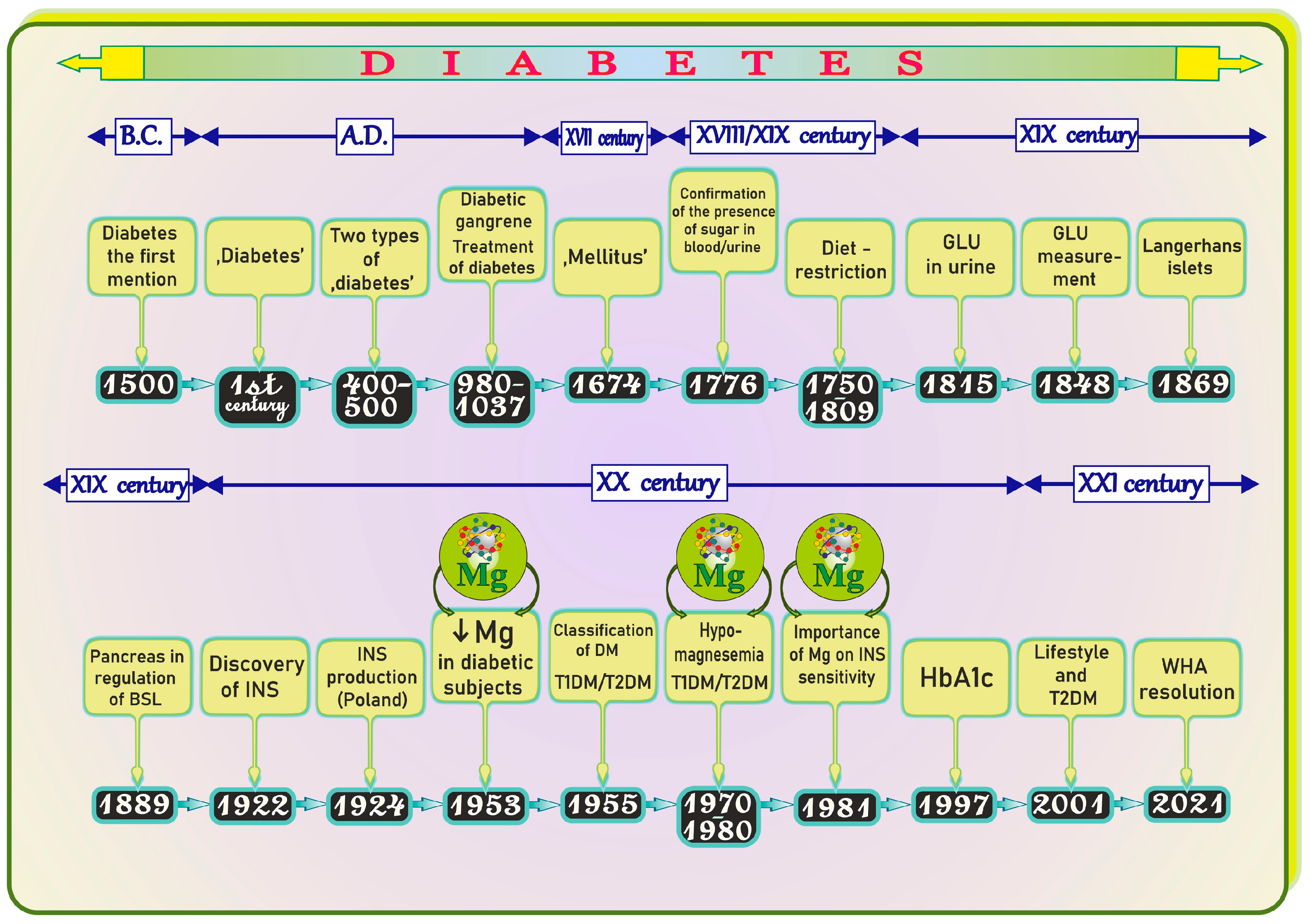
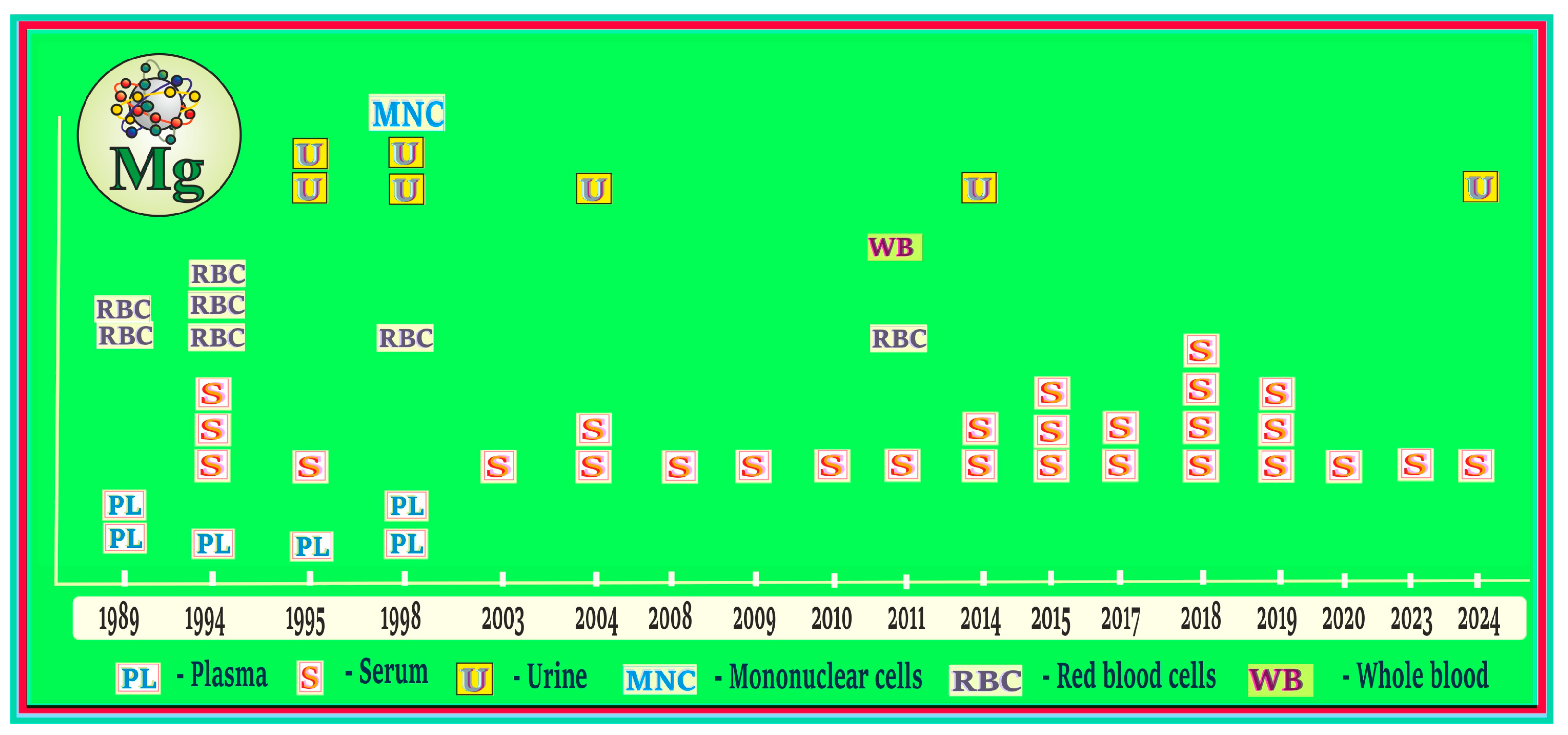
1.2.2. A Brief Historical Framework Related to Diabetes and Mg on the Timeline
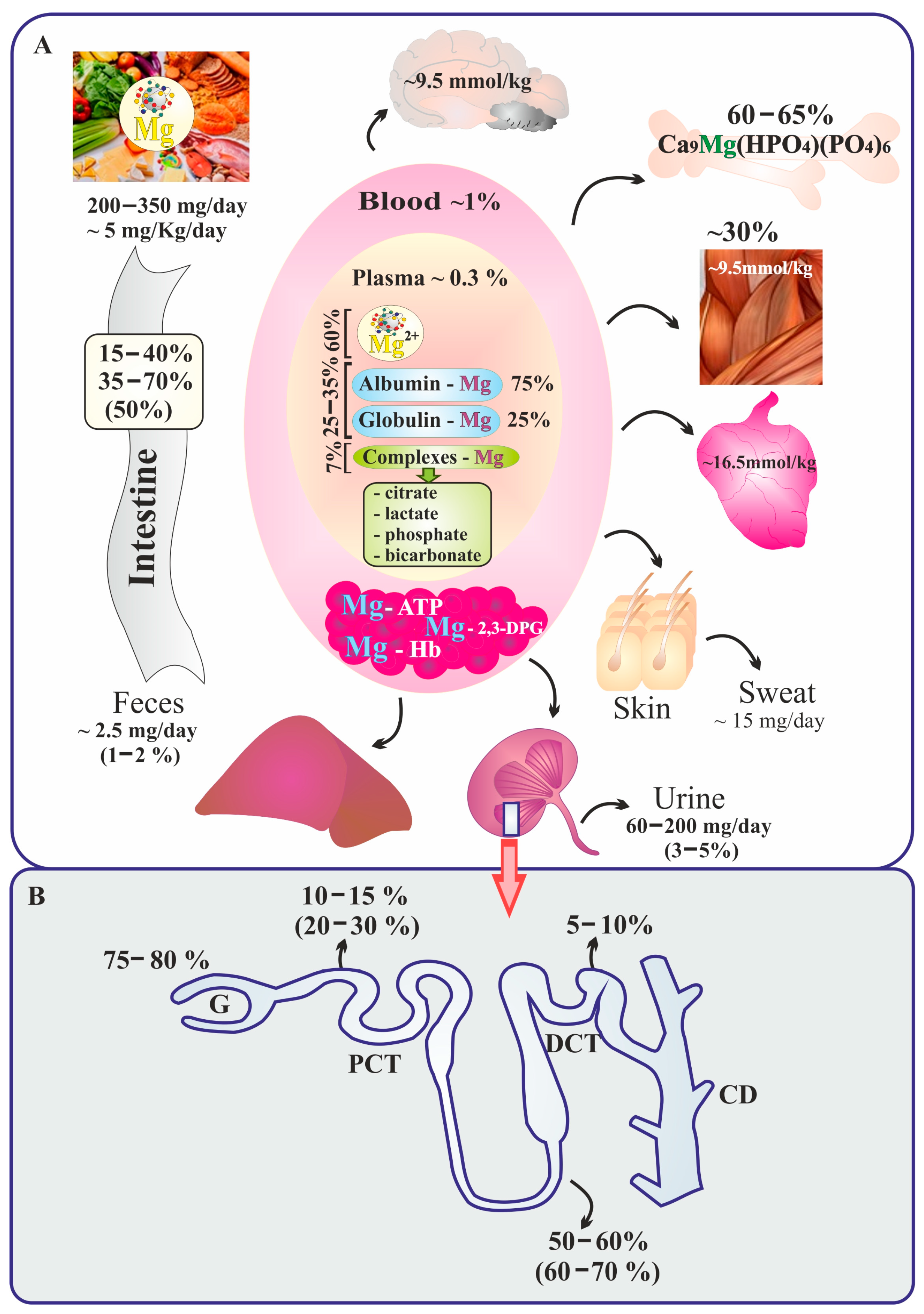
2. Absorption, Distribution, and Renal Handling of Mg
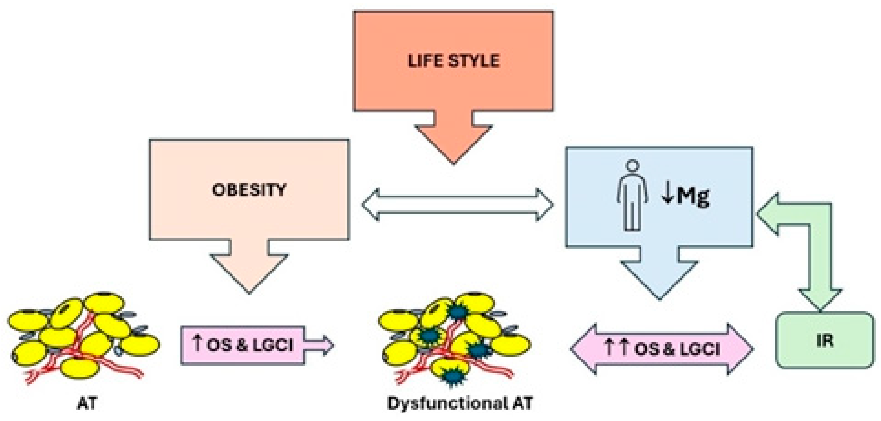
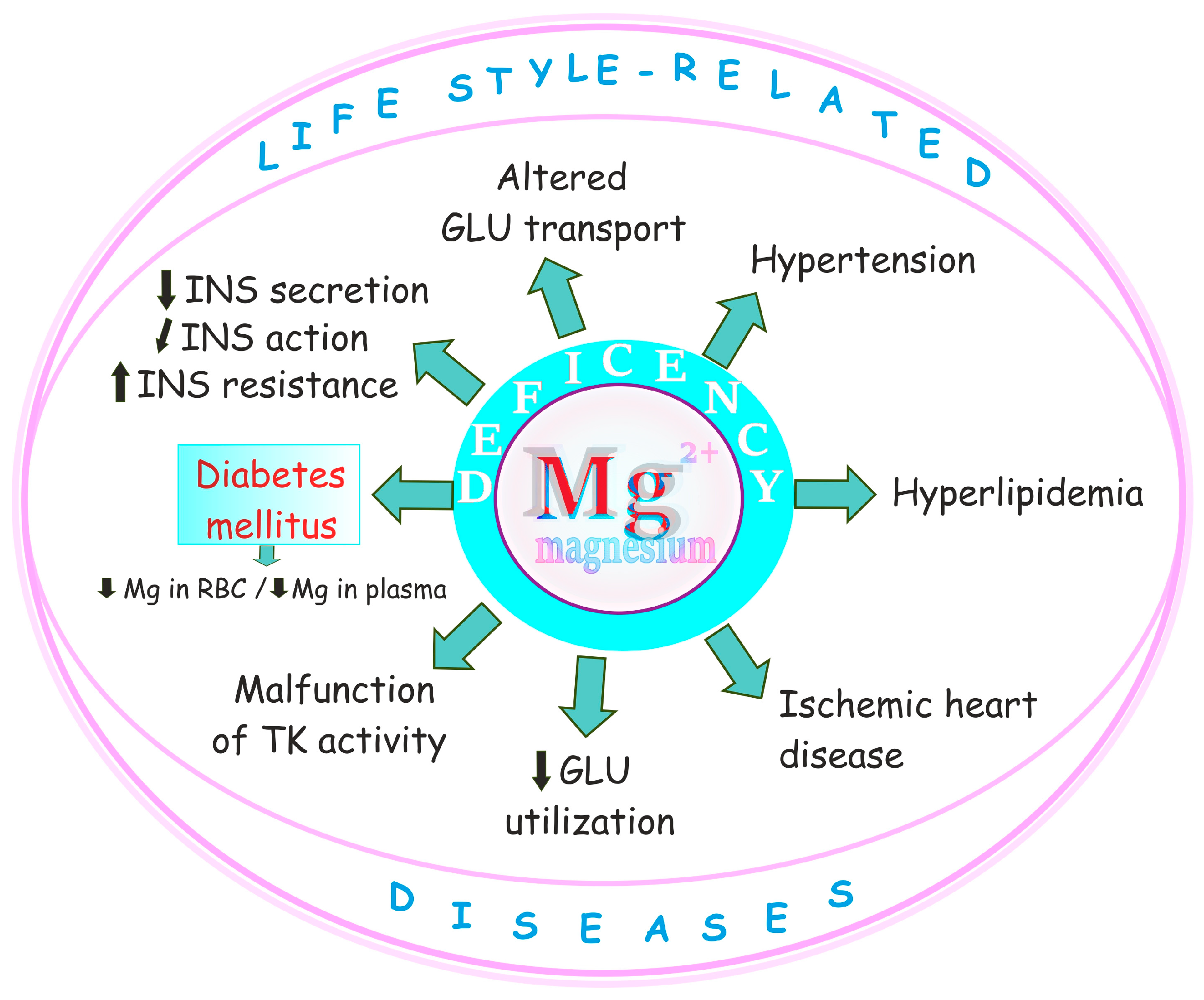
3. Hypomagnesemia and Lifestyle-Related Diseases Including Diabetes and Obesity—A Brief Outline
4. Mechanisms of Hypomagnesemia in Diabetes—A Brief Overview
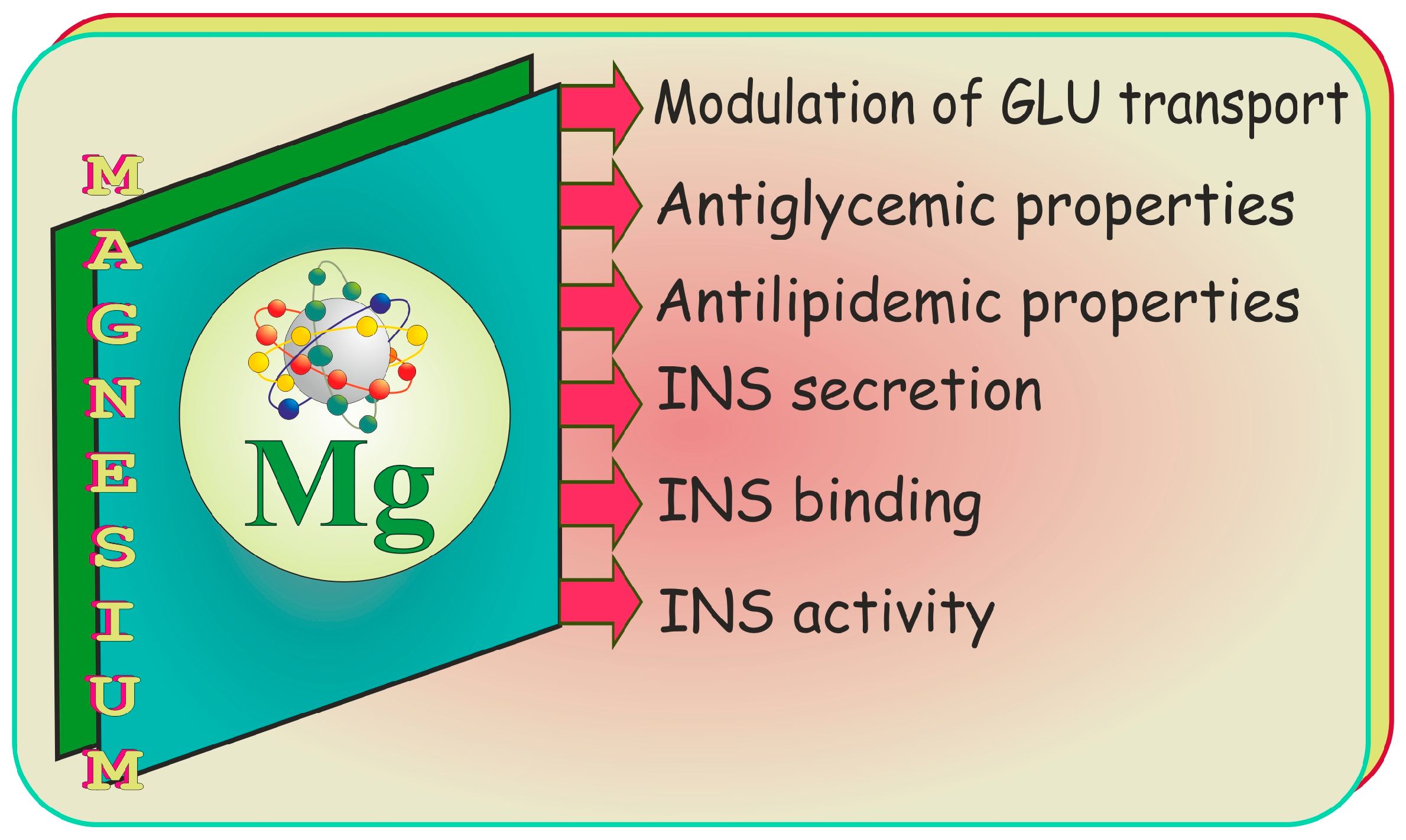
5. Mechanisms of Action of Mg in Diabetes—A Brief Outline
5.1. Effect on Glucose Transporter Protein Type-4 (GLUT4)
5.2. Effect on Insulin Receptor
5.3. Role in the Secretion and Action of Insulin
5.4. Oxidative Stress Reduction
5.5. Anti-Inflammatory Effects
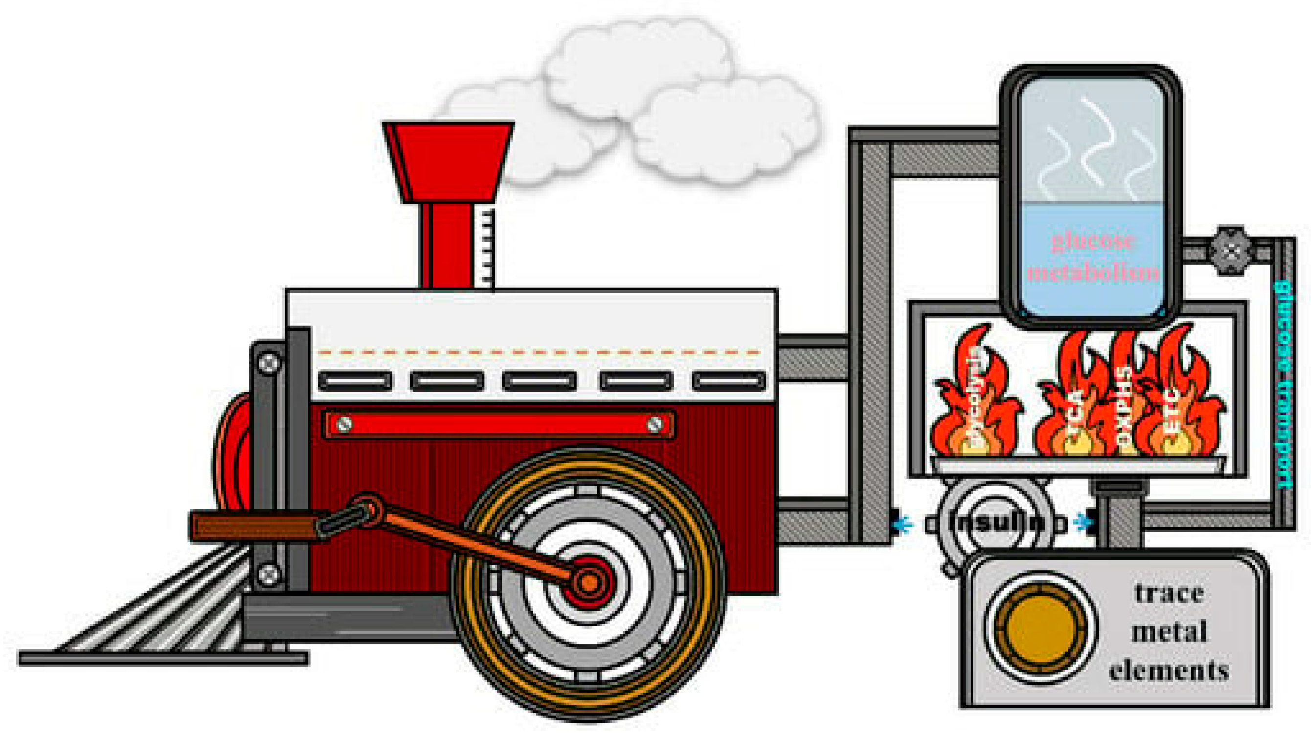
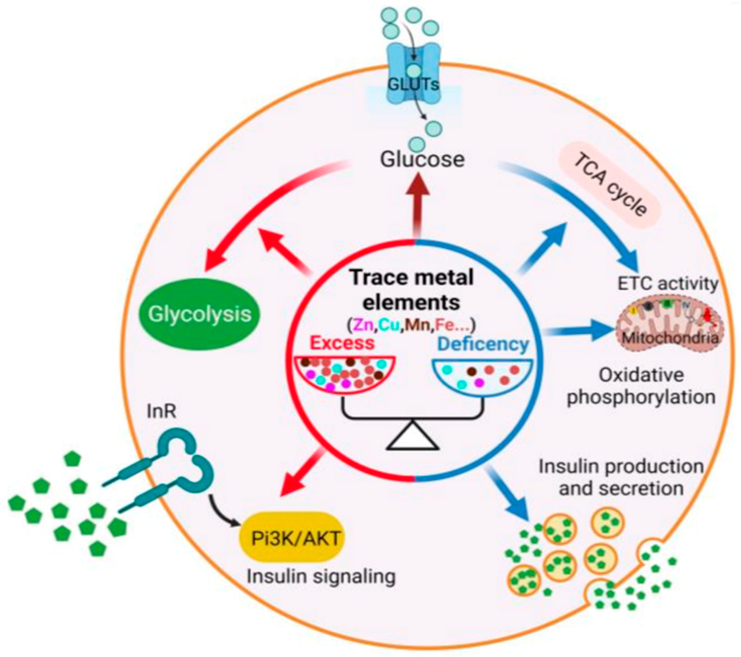
5.6. Metal Ion Homeostasis
5.7. Mitochondrial Function
6. Mitochondria, Mg2+, Oxidative Stress, and Diabetes
7. Association of Mg with Metabolism of Other Essential Metals (Cu, Zn, Fe, Mn, Se, and V) in Diabetes
8. Methodology—Literature Search Strategy on Mg and Metabolic Profile in Diabetes in Humans
8.1. Databases: General Outline
8.2. Query Terms Used for the Literature Search on Mg and Metabolic Profile in Diabetes in Humans
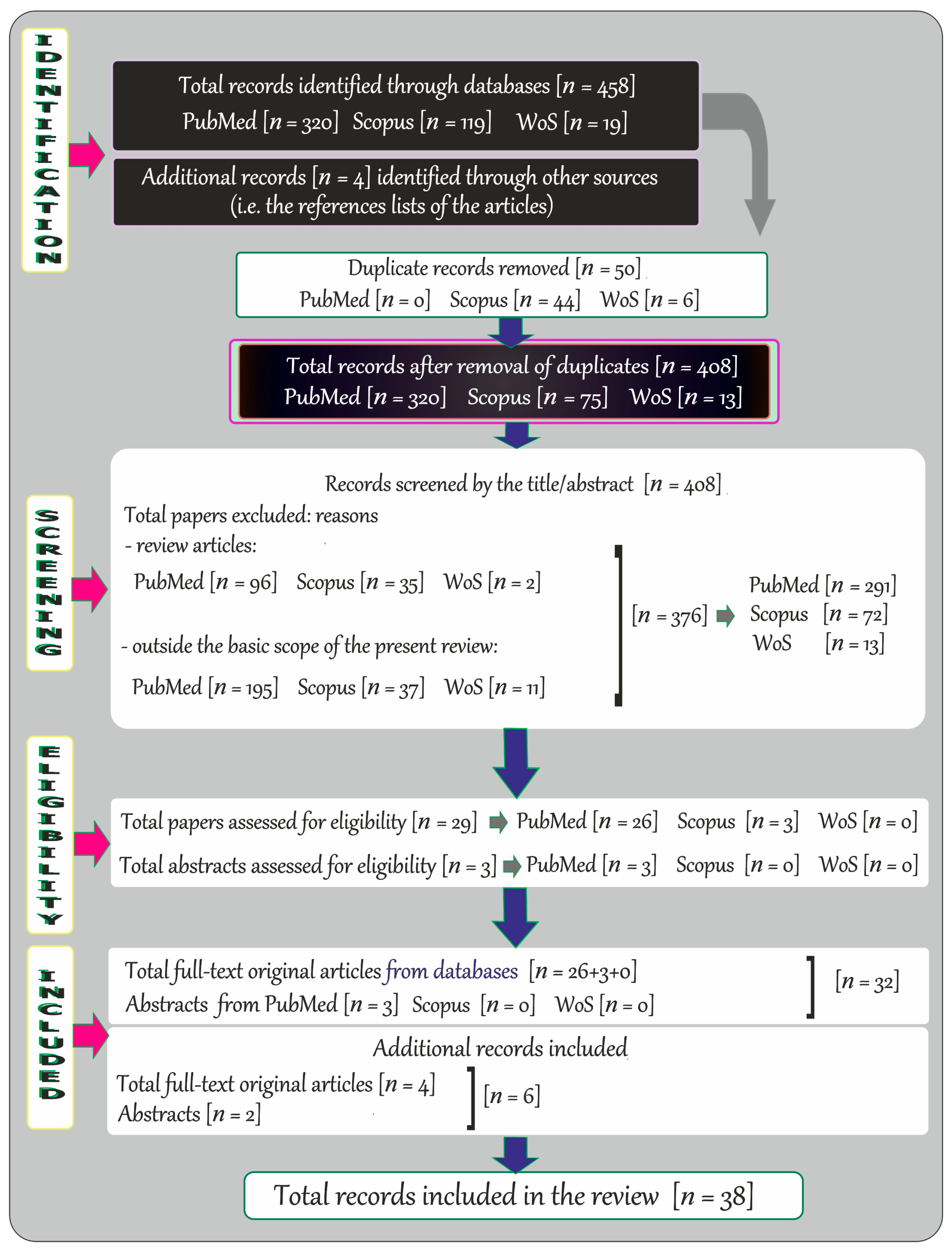
8.3. Search Results and Literature Review Flowchart on Mg in Diabetes in Humans
9. Effects of Oral Mg Supplementation on Diabetes: Humans—A Summarizing Note
10. Summary and Conclusions
Author Contributions
Funding
Data Availability Statement
Conflicts of Interest
References
- Harding, J.L.; Pavkov, M.E.; Magliano, D.J.; Shaw, J.E.; Gregg, E.W. Global trends in diabetes complications: A review of current evidence. Diabetologia 2019, 62, 3–16. [Google Scholar] [CrossRef] [Scilit]
- Lim, A.K.H. Diabetic nephropathy—Complications and treatment. Int. J. Nephrol. Renov. Dis. 2014, 7, 361–381. [Google Scholar] [CrossRef] [Scilit]
- Mather, H.M.; Nisbet, J.A.; Burton, G.H.; Pasten, G.J.; Bland, J.M.; Bailey, P.A.; Pilkington, T.R.E. Hypomagnesemia in diabetes. Clin. Chim. Acta 1979, 95, 235–242. [Google Scholar] [CrossRef] [Scilit]
- Gürlek, A.; Bayraktar, M.; Özaltin, N. Intracellular magnesium depletion relates to increased urinary magnesium loss in type 1 diabetes. Horm. Metab. Res. 1998, 30, 99–102. [Google Scholar] [CrossRef] [Scilit]
- Sjögren, A.; Florén, C.H.; Nilsson, A. Magnesium deficiency in IDDM related to level of glycosylated hemoglobin. Diabetes 1986, 35, 459–463. [Google Scholar] [CrossRef] [Scilit]
- Pun, K.K.; Ho, P.W.M. Subclinical hyponatremia, hyperkalemia and hypomagnesemia in patients with poorly controlled diabetes mellitus. Diabetes Res. Clin. Pract. 1989, 7, 163–167. [Google Scholar] [CrossRef] [Scilit]
- Schnack, C.; Bauer, I.; Pregant, P.; Hopmeier, P.; Schernthaner, G. Hypomagnesemia in type 2 (non-insulin dependent) diabetes mellitus is not corrected by improvement of long-term metabolic control. Diabetologia 1992, 35, 77–79. [Google Scholar] [CrossRef] [Scilit]
- Oyewole, O.O.; Ale, A.O.; Ogunlana, M.O.; Gurayah, T. Burden of disability in type 2 diabetes mellitus and the moderating effects of physical activity. World J. Clin. Cases 2023, 16, 3128–3139. [Google Scholar] [CrossRef] [Scilit]
- American Diabetes Association (ADA). Diagnosis and classification of diabetes mellitus. Diabetes Care 2011, 34, S62–S69. [Google Scholar] [CrossRef]
- Liu, D.D.; Zhang, C.Y.; Zhang, J.T.; Gu, L.M.; Xu, G.T.; Zhang, J.F. Epigenetic modifications and metabolic memory in diabetic retinopathy: Beyond the surface. Neural Regen. Res. 2023, 18, 1441–1449. [Google Scholar] [CrossRef] [Scilit]
- Mittal, R.; Prasad, K.; Lemos, J.R.N.; Arevalo, G.; Hirani, K. Unveiling gestational diabetes: An overview of pathophysiology and management. Int. J. Mol. Sci. 2025, 26, 2320. [Google Scholar] [CrossRef] [Scilit]
- Ogle, G.D.; Wang, F.; Haynes, A.; Gregory, G.A.; King, T.W.; Deng, K.; Dabelea, D.; James, S.; Jenkins, A.J.; Li, X.; et al. Global type 1 diabetes prevalence, incidence, and mortality estimates 2025: Results from the International diabetes Federation Atlas, 11th Edition, and the T1D Index Version 3.0. Diabetes Res. Clin. Pract. 2025, 225, 112277. [Google Scholar] [CrossRef] [Scilit]
- Gregory, G.; Robinson, T.; Linklater, S.; Wang, F.; Colagiuri, S.; de Beaufort, C.; Donaghue, K.C.; International Diabetes Federation Diabetes Atlas Type 1 Diabetes in Adults Special Interest Group; Magliano, D.J.; Maniam, J.; et al. Global incidence, prevalence, and mortality of type 1 diabetes in 2021 with projection to 2040: A modelling study. Lancet Diabetes Endocrinol. 2022, 10, 741–760, Erratum in Lancet Diabetes Endocrinol. 2022, 10, e11. [Google Scholar] [CrossRef] [Scilit] [PubMed]
- International Diabetes Federation (IDF). IDF Diabetes Atlas, 11th ed.; International Diabetes Federation (IDF): Brussels, Belgium, 2025. [Google Scholar]
- Harding, J.L.; Wander, P.L.; Zhang, X.; Li, X.; Karuranga, S.; Chen, H.; Sun, H.; Xie, Y.; Oram, R.A.; Magliano, D.J.; et al. The incidence of adult-onset type 1 diabetes: A systematic review from 32 countries and regions. Diabetes Care 2022, 45, 994–1006, Erratum in Diabetes Care 2023, 46, 2084. https://doi.org/10.2337/dc23-er11. [Google Scholar] [CrossRef] [Scilit]
- Rutkowski, M.; Wojciechowska, A.; Śmigielski, W.; Drygas, W.; Piwońska, A.; Pająk, A.; Tykarski, A.; Kozakiewicz, K.; Waśniewska, M.K.; Zdrojewski, T. Prevalence of diabetes and impaired fasting glucose in Poland in 2005–2014: Results of the WOBASZ surveys. Diabet. Med. 2020, 37, 1528–1535. [Google Scholar] [CrossRef] [Scilit]
- Niechciał, E.; Michalak, M.; Skowrońska, B.; Fichna, P. Increasing trend of childhood type 1 diabetes incidence: 20-year observation from Greater Poland Province, Poland. Acta Diabetol. 2024, 61, 1609–1617. [Google Scholar] [CrossRef] [Scilit]
- Jarosz-Chobot, P.; Polanska, J.; Szadkowska, A.; Kretowski, A.; Bandurska-Stankiewicz, E.; Ciechanowska, M.; Deja, G.; Mysliwiec, M.; Peczynska, J.; Rutkowska, J.; et al. Rapid increase in the incidence of type 1 diabetes in Polish children from 1989 to 2004, and predictions for 2010 to 2025. Diabetologia 2011, 54, 508–515. [Google Scholar] [CrossRef] [Scilit]
- Diabetes: Factos e Números, Sociedade Portuguesa de Diabetologia (SPD), Os anos de 2022, 2023 e 2024, Relatório Anual do Observatório Nacional da Diabetes, Edição de 2024. Available online: https://relatorioanualdiabetes.com/wp-content/uploads/2025/11/251103-SPD-RelatorioAnual-1.pdf (accessed on 20 August 2026).
- Sherry, N.A.; Tsai, E.B.; Herold, K.C. Natural history of β-cell function in type 1 diabetes. Diabetes 2005, 54, S32–S39. [Google Scholar] [CrossRef] [Scilit]
- Barrett, J.; Clayton, D.; Concannon, P.; Akolkar, B.; Cooper, J.D.; Erlich, H.A.; Hulier, C.; Morahan, G.; Nerup, J.; Nierras, C.; et al. Genome-wide association study and meta-analysis find that over 40 loci affect risk of type 1 diabetes. Nat. Genet. 2009, 41, 703–707. [Google Scholar] [CrossRef] [Scilit]
- Erlich, H.; Valdes, A.M.; Noble, J.; Carlson, J.A.; Varney, M.; Concannon, P.; Mychaleckyj, J.C.; Todd, J.A.; Bonella, P.; Fear, A.L.; et al. for the Type 1 Diabetes Genetics Consortium. HLA DR-DQ haplotypes and genotypes and type 1 diabetes risk: Analysis of the type 1 diabetes genetics consortium families. Diabetes 2008, 57, 1084–1092. [Google Scholar] [CrossRef] [Scilit]
- Aamodt, K.I.; Powers, A.C. The pathophysiology, presentation and classification of Type 1 diabetes. Diabetes Obes. Metab. 2025, 27, 15–27. [Google Scholar] [CrossRef] [Scilit]
- Bjornstad, P.; Dart, A.; Donaghue, K.C.; Dost, A.; Feldman, E.L.; Tan, G.S.; Wadwa, R.P.; Zabeen, B.; Marcovecchio, M.L. ISPAD Clinical Practice Consensus Guidelines 2022: Microvascular and macrovascular complications in children and adolescents with diabetes. Pediatr. Diabetes. 2022, 23, 1432–1450. [Google Scholar] [CrossRef] [Scilit]
- Roden, M.; Shulman, G.I. The integrative biology of type 2 diabetes. Nature 2019, 576, 51–60. [Google Scholar] [CrossRef] [Scilit] [PubMed]
- Galicia-Garcia, U.; Benito-Vicente, A.; Jebari, S.; Larrea-Sebal, A.; Siddiqi, H.; Uribe, K.B.; Ostolaza, H.; Martín, C. Pathophysiology of type 2 diabetes mellitus. Int. J. Mol. Sci. 2020, 21, 6275. [Google Scholar] [CrossRef] [Scilit]
- Bellou, V.; Belbasis, L.; Tzoulaki, I.; Evangelou, E. Risk factors for type 2 diabetes mellitus: An exposure-wide umbrella review of meta-analyses. PLoS ONE 2018, 13, e0194127. [Google Scholar] [CrossRef] [Scilit]
- McCarthy, M.I. Genomics, type 2 diabetes, and obesity. N. Engl. J. Med. 2010, 363, 2339–2350. [Google Scholar] [CrossRef] [Scilit]
- Chan, J.C.; Cheung, C.K.; Swaminathan, R.; Nicholls, M.G.; Cockram, C.S. Obesity, albuminuria and hypertension among Hong Kong Chinese with non-insulin-dependent diabetes mellitus (NIDDM). Postgrad. Med. J. 1993, 69, 204–210. [Google Scholar] [CrossRef] [Scilit]
- Dabelea, D.; DeGroat, J.; Sorrelman, C.; Glass, M.; Percy, C.A.; Avery, C.; Hu, D.; D’Agostino, R.B.; Beyer, J.; Imperatore, G.; et al. for the SEARCH for Diabetes in Youth Study Group; Diabetes in Navajo Youth: Prevalence, incidence, and clinical characteristics: The SEARCH for Diabetes in Youth Study. Diabetes Care 2009, 32, S141–S147. [Google Scholar] [CrossRef] [Scilit]
- Liu, L.L.; Yi, J.P.; Beyer, J.; Mayer-Davis, E.J.; Dolan, L.M.; Dabelea, D.M.; Lawrence, J.M.; Rodriguez, B.L.; Marcovina, S.M.; Waitzfelder, B.E.; et al. Type 1 and Type 2 diabetes in Asian and Pacific Islander U.S. youth: The SEARCH for Diabetes in Youth Study. Diabetes Care 2009, 32, S133–S140. [Google Scholar] [CrossRef] [Scilit]
- Antar, S.A.; Ashour, N.A.; Sharaky, M.; Khattab, M.; Ashour, N.A.; Zaid, R.T.; Roh, E.J.; Elkamhawy, A.; Al-Karmalawy, A.A. Diabetes mellitus: Classification, mediators, and complications; A gate to identify potential targets for the development of new effective treatments. Biomed. Pharmacother. 2023, 168, 115734. [Google Scholar] [CrossRef] [Scilit]
- Dutta, S.; Shah, R.B.; Singhal, S.; Dutta, S.B.; Bansal, S.; Sinha, S.; Haque, M. Metformin: A review of potential mechanism and therapeutic utility beyond diabetes. Drug Des. Devel. Ther. 2023, 17, 1907–1932. [Google Scholar] [CrossRef] [Scilit]
- Aristizábal-Colorado, D.; Corredor-Rengifo, D.; Sierra-Castillo, S.; López-Corredor, C.; Vernaza-Trujillo, D.A.; Weir-Restrepo, D.; Izquierdo-Condoy, J.S.; Ortiz-Prado, E.; Rico-Fontalvo, J.; Gómez-Mesa, J.E.; et al. A decade of progress in type 2 diabetes and cardiovascular disease: Advances in SGLT2 inhibitors and GLP-1 receptor agonists—A comprehensive review. Front. Endocrinol. 2025, 16, 1605746, Correction in Front. Endocrinol. 2025, 16, 1663787. https://doi.org/10.3389/fendo.2025.1663787. [Google Scholar] [CrossRef] [Scilit]
- Galdón Sanz-Pastor, A.; Justel Enríquez, A.; Sánchez Bao, A.; Ampudia-Blasco, F.J. Current barriers to initiating insulin therapy in individuals with type 2 diabetes. Front. Endocrinol. 2024, 15, 1366368. [Google Scholar] [CrossRef] [Scilit]
- Kunarathnam, V.; Vadakekut, E.S.; Mahdy, H. Gestational Diabetes. In StatPearls; StatPearls Publishing LLC: Treasure Island, FL, USA, 2026. [Google Scholar]
- Wedekind, L.; Belkacemi, L. Altered cytokine network in gestational diabetes mellitus affects maternal insulin and placental–fetal development. J. Diabetes Complicat. 2016, 30, 1393–1400. [Google Scholar] [CrossRef] [Scilit]
- McIntyre, H.D.; Chang, A.M.; Callaway, L.K.; Cowley, D.M.; Dyer, A.R.; Radaelli, T.; Farrell, K.A.; Huston-Presley, L.; Amini, S.B.; Kirwan, J.P.; et al. Hyperglycemia and adverse pregnancy outcome (HAPO) study cooperative research group. Hormonal and metabolic factors associated with variations in insulin sensitivity in human pregnancy. Diabetes Care 2010, 33, 356–360. [Google Scholar] [CrossRef] [Scilit]
- Lean, S.C.; Derricott, H.; Jones, R.L.; Heazell, A.E.P. Advanced maternal age and adverse pregnancy outcomes: A systematic review and meta-analysis. PLoS ONE 2017, 12, e0186287. [Google Scholar] [CrossRef] [Scilit]
- Hincapie, M.A.; Badeghiesh, A.; Baghlaf, H.; Dahan, M.H. Association between pre-gestational diabetes in women with polycystic ovary syndrome and adverse obstetric outcomes. Eur. J. Obstet. Gynecol. Reprod. Biol. 2025, 304, 109–114. [Google Scholar] [CrossRef] [Scilit]
- Duncan, J.M. Case of pregnancy and labour complicated with diabetes mellitus. Edinb. Med. J. 1873, 18, 696–698. [Google Scholar]
- Hufnagel, A.; Dearden, L.; Fernandez-Twinn, D.S.; Ozanne, S.E. Programming of cardiometabolic health: The role of maternal and fetal hyperinsulinaemia. J. Endocrinol. 2022, 253, R47–R63. [Google Scholar] [CrossRef] [Scilit]
- Parikh, N.I.; Gonzalez, J.M.; Anderson, C.A.M.; Judd, S.E.; Rexrode, K.M.; Hlatky, M.A.; Gunderson, E.P.; Stuart, J.J.; Vaidya, D. American Heart Association Council on Epidemiology and Prevention; Council on Arteriosclerosis, Thrombosis and Vascular Biology; Council on Cardiovascular and Stroke Nursing; and the Stroke Council. Adverse pregnancy outcomes and cardiovascular disease risk: Unique opportunities for cardiovascular disease prevention in women: A scientific statement from the American heart association. Circulation 2021, 143, e902–e916. [Google Scholar] [CrossRef] [Scilit]
- Kampmann, U.; Madsen, L.R.; Skajaa, G.O.; Iversen, D.S.; Moeller, N.; Ovesen, P. Gestational diabetes: A clinical update. World J. Diabetes 2015, 6, 1065–1072. [Google Scholar] [CrossRef] [Scilit]
- Nielsen, J.H.; Olesen, C.R.; Kristiansen, T.M.; Bak, C.K.; Overgaard, C. Reasons for women’s non-participation in follow-up screening after gestational diabetes. Women Birth 2015, 28, e157–e163. [Google Scholar] [CrossRef] [Scilit]
- Quaresima, P.; Saccone, G.; Pellegrino, R.; Vaccarisi, S.; Taranto, L.; Mazzulla, R.; Bernardo, S.; Venturella, R.; Di Carlo, C.; Morelli, M. Incidental diagnosis of a pancreatic adenocarcinoma in a woman affected by gestational diabetes mellitus: Case report and literature review. Am. J. Obstet. Gynecol. MFM 2021, 3, 100471. [Google Scholar] [CrossRef] [Scilit]
- Dudley, D.J. Diabetic-associated stillbirth: Incidence, pathophysiology, and prevention. Obstet. Gynecol. Clin. North Am. 2007, 34, 293–307. [Google Scholar] [CrossRef] [Scilit]
- Moore, T.R. A comparison of amniotic fluid fetal pulmonary phospholipids in normal and diabetic pregnancy. Am. J. Obstet. Gynecol. 2002, 186, 641–650. [Google Scholar] [CrossRef] [Scilit]
- Metzger, B.E.; Persson, B.; Lowe, L.P.; Dyer, A.R.; Cruickshank, J.K.; Deerochanawong, C.; Halliday, H.L.; Hennis, A.J.; Liley, H.; Ng, P.C.; et al. Hyperglycemia and adverse pregnancy outcome study: Neonatal glycemia. Pediatrics 2010, 126, e1545–1552. [Google Scholar] [CrossRef] [Scilit]
- Lee, S.H.; Park, S.Y.; Choi, C.S. Insulin resistance: From mechanisms to therapeutic strategies. Diabetes Metab. J. 2022, 46, 15–37. [Google Scholar] [CrossRef] [Scilit]
- Zhao, X.; An, X.; Yang, C.; Sun, W.; Ji, H.; Lian, F. The crucial role and mechanism of insulin resistance in metabolic disease. Front. Endocrinol. 2023, 14, 1149239. [Google Scholar] [CrossRef] [Scilit]
- Nekoua, M.P.; Alidjinou, E.K.; Hober, D. Persistent coxsackievirus B infection and pathogenesis of type 1 diabetes mellitus. Nat. Rev. Endocrinol. 2022, 18, 503–516. [Google Scholar] [CrossRef] [Scilit]
- Stene, L.C.; Norris, J.M.; Rewers, M.J. Risk factors for type 1 diabetes. In Diabetes in America; Lawrence, J.M., Casagrande, S.S., Herman, W.H., Wexler, D.J., Cefalu, W.T., Eds.; National Institute of Diabetes and Digestive and Kidney Diseases (NIDDK): Bethesda, MD, USA, 2023. [Google Scholar]
- Ismail, L.; Materwala, H.; Al Kaabi, J. Association of risk factors with type 2 diabetes: A systematic review. Comput. Struct. Biotechnol. J. 2021, 10, 1759–1785. [Google Scholar] [CrossRef] [Scilit]
- Tang, S.S.; Zhao, X.F.; An, X.D.; Sun, W.J.; Kang, X.M.; Sun, Y.T.; Jiang, L.L.; Gao, Q.; Li, Z.H.; Ji, H.Y.; et al. Classification and identification of risk factors for type 2 diabetes. World J. Diabetes 2025, 16, 100371. [Google Scholar] [CrossRef] [Scilit]
- Zhao, L.; Yuan, J.; Yang, Q.; Ma, J.; Yang, F.; Zou, Y.; Liu, K.; Liu, F. Diabetes and its complications: Molecular mechanisms, prevention and treatment. Signal Transduct. Target Ther. 2026, 11, 22. [Google Scholar] [CrossRef] [Scilit]
- Reddy, M.A.; Das, S.; Zhuo, C.; Jin, W.; Wang, M.; Lanting, L.; Natarajan, R. Regulation of vascular smooth muscle cell dysfunction under diabetic conditions by miR-504. Arterioscler. Thromb. Vasc. Biol. 2016, 36, 864–873. [Google Scholar] [CrossRef] [Scilit]
- Mlinar, B.; Marc, J.; Janez, A.; Pfeifer, M. Molecular mechanisms of insulin resistance and associated diseases. Clin. Chim. Acta 2007, 375, 20–35. [Google Scholar] [CrossRef] [Scilit]
- Witzel, I.I.; Jelinek, H.F.; Khalaf, K.; Lee, S.; Khandoker, A.H.; Alsafar, H. Identifying common genetic risk factors of diabetic neuropathies. Front. Endocrinol. 2015, 6, 88. [Google Scholar] [CrossRef] [Scilit]
- Rolo, A.P.; Palmeira, C.M. Diabetes and mitochondrial function: Role of hyperglycemia and oxidative stress. Toxicol. Appl. Pharmacol. 2006, 212, 167–178. [Google Scholar] [CrossRef] [Scilit]
- Zhang, X.; Zhang, J.; Ren, Y.; Sun, R.; Zhai, X. Unveiling the pathogenesis and therapeutic approaches for diabetic nephropathy: Insights from panvascular diseases. Front. Endocrinol. 2024, 15, 1368481. [Google Scholar] [CrossRef] [Scilit]
- Egbuche, O.; Biggs, M.L.; Ix, J.H.; Kizer, J.R.; Lyles, M.F.; Siscovick, D.S.; Djoussé, L.; Mukamal, K.J. Fatty acid binding protein-4 and risk of cardiovascular disease: The cardiovascular health study. J. Am. Heart Assoc. 2020, 9, e014070. [Google Scholar] [CrossRef] [Scilit]
- Nawrot, M.; Peschard, S.; Lestavel, S.; Staels, B. Intestine-liver crosstalk in type 2 diabetes and non-alcoholic fatty liver disease. Metabolism 2021, 123, 154844. [Google Scholar] [CrossRef] [Scilit]
- Balakrishnan, R.; Thurmond, D.C. Mechanisms by which skeletal muscle myokines ameliorate insulin resistance. Int. J. Mol. Sci. 2022, 23, 4636. [Google Scholar] [CrossRef] [Scilit]
- Pafili, K.; Kahl, S.; Mastrototaro, L.; Strassburger, K.; Pesta, D.; Herder, C.; Pützer, J.; Dewidar, B.; Hendlinger, M.; Granata, C.; et al. Mitochondrial respiration is decreased in visceral but not subcutaneous adipose tissue in obese individuals with fatty liver disease. J. Hepatol. 2022, 77, 1504–1514. [Google Scholar] [CrossRef] [Scilit]
- Wu, M.; Tan, J.; Cao, Z.; Cai, Y.; Huang, Z.; Chen, Z.; He, W.; Liu, X.; Jiang, Y.; Gao, Q.; et al. Sirt5 improves cardiomyocytes fatty acid metabolism and ameliorates cardiac lipotoxicity in diabetic cardiomyopathy via CPT2 de-succinylation. Redox Biol. 2024, 73, 103184. [Google Scholar] [CrossRef] [Scilit]
- Rusciano, D.; Bagnoli, P. Oxygen, the paradox of life and the eye. Front. Biosci. 2024, 29, 319. [Google Scholar] [CrossRef] [Scilit]
- Kulkarni, A.; Thool, A.R.; Daigavane, S. Understanding the clinical relationship between diabetic retinopathy, nephropathy, and neuropathy: A comprehensive review. Cureus 2024, 16, e56674. [Google Scholar] [CrossRef] [Scilit]
- Yang, D.R.; Wang, M.Y.; Zhang, C.L.; Wang, Y. Endothelial dysfunction in vascular complications of diabetes: A comprehensive review of mechanisms and implications. Front. Endocrinol. 2024, 15, 1359255. [Google Scholar] [CrossRef] [Scilit]
- Yeh, M.L.; Huang, J.F.; Dai, C.Y.; Huang, C.F.; Yu, M.L.; Chuang, W.L. Metabolic dysfunction-associated steatotic liver disease and diabetes: The cross-talk between hepatologist and diabetologist. Expert Rev. Gastroenterol. Hepatol. 2024, 18, 431–439. [Google Scholar] [CrossRef] [Scilit]
- Kopf, S.; Groener, J.B.; Kender, Z.; Fleming, T.; Brune, M.; Riedinger, C.; Volk, N.; Herpel, E.; Pesta, D.; Szendrödi, J.; et al. Breathlessness and restrictive lung disease: An important diabetes-related feature in patients with type 2 diabetes. Respiration 2018, 96, 29–40. [Google Scholar] [CrossRef] [Scilit]
- Berbudi, A.; Rahmadika, N.; Tjahjadi, A.I.; Ruslami, R. Type 2 diabetes and its Impact on the immune system. Curr. Diabetes Rev. 2020, 16, 442–449. [Google Scholar] [CrossRef] [Scilit] [PubMed]
- Bottini, N.; Vang, T.; Cucca, F.; Mustelin, T. Role of PTPN22 in type 1 diabetes and other autoimmune diseases. Semin. Immunol. 2006, 18, 207–213. [Google Scholar] [CrossRef] [Scilit]
- Kakleas, K.; Soldatou, A.; Karachaliou, F.; Karavanaki, K. Associated autoimmune diseases in children and adolescents with type 1 diabetes mellitus (T1DM). Autoimmun. Rev. 2015, 14, 781–797. [Google Scholar] [CrossRef] [Scilit]
- Goodarzi, M.O.; Rotter, J.I. Genetics insights in the relationship between type 2 diabetes and coronary heart disease. Circ. Res. 2020, 126, 1526–1548. [Google Scholar] [CrossRef] [Scilit]
- Li, X.; Song, D.; Leng, S.X. Link between type 2 diabetes and Alzheimer’s disease: From epidemiology to mechanism and treatment. Clin. Interv. Aging 2015, 10, 549–560. [Google Scholar] [CrossRef] [Scilit]
- Hossain, M.A.; Al Amin, M.; Hasan, M.I.; Sohel, M.; Ahammed, M.A.; Mahmud, S.M.H.; Rahman, M.R.; Rahman, M.H. Bioinformatics and system biology approaches to identify molecular pathogenesis of polycystic ovarian syndrome, type 2 diabetes, obesity, and cardiovascular disease that are linked to the progression of female infertility. Inform. Med. Unlocked 2022, 30, 100960. [Google Scholar] [CrossRef] [Scilit]
- Catargi, B.; Rigalleau, V.; Poussin, A.; Ronci-Chaix, N.; Bex, V.; Vergnot, V.; Gin, H.; Roger, P.; Tabarin, A. Occult Cushing’s syndrome in type-2 diabetes. J. Clin. Endocrinol. Metab. 2003, 88, 5808–5813. [Google Scholar] [CrossRef] [Scilit]
- Wu, J.; Tang, L.; Zheng, F.; Chen, X.; Li, L. A review of the last decade: Pancreatic cancer and type 2 diabetes. Arch. Physiol. Biochem. 2024, 130, 660–668. [Google Scholar] [CrossRef] [Scilit]
- Fontecilla-Camps, J.C. The complex roles of adenosine triphosphate in bioenergetics. Chembiochem 2022, 23, e202200064. [Google Scholar] [CrossRef] [Scilit]
- Mudryk, K.; Lee, C.; Tomaník, L.; Malerz, S.; Trinter, F.; Hergenhahn, U.; Neumark, D.M.; Slavíček, P.; Bradforth, S.; Winter, B. How does Mg2+(aq) interact with ATP(aq)? Biomolecular structure through the lens of liquid-jet photoemission spectroscopy. J. Am. Chem. Soc. 2024, 146, 16062–16075. [Google Scholar] [CrossRef] [Scilit]
- Nam, K.; Thodika, A.R.A.; Tischlik, S.; Phoeurk, C.; Nagy, T.M.; Schierholz, L.; Ådén, J.; Rogne, P.; Drescher, M.; Sauer-Eriksson, A.E.; et al. Magnesium induced structural reorganization in the active site of adenylate kinase. Sci. Adv. 2024, 10, eado5504. [Google Scholar] [CrossRef] [Scilit]
- Case, D.R.; Zubieta, J.; Doyle, R.P. The coordination chemistry of bio-relevant ligands and their magnesium complexes. Molecules 2020, 25, 3172. [Google Scholar] [CrossRef] [Scilit]
- Cowan, J.A. Understanding the thermodynamics of magnesium binding to RNA structural motifs. Life 2024, 14, 765. [Google Scholar] [CrossRef] [Scilit]
- Küpper, H.; Küpper, F.; Spiller, M. Environmental relevance of heavy metal-substituted chlorophylls using the example of water plants. J. Exp. Bot. 1996, 47, 259–266. [Google Scholar] [CrossRef] [Scilit]
- Barbagallo, M.; Veronese, N.; Dominguez, L.J. Magnesium in aging, health and diseases. Nutrients 2021, 13, 463. [Google Scholar] [CrossRef] [Scilit]
- Romani, A.M.P. Cellular magnesium homeostasis. Arch. Biochem. Biophys. 2011, 512, 1–23. [Google Scholar] [CrossRef] [Scilit]
- Jin, F.; Huang, Y.; Hattori, M. Recent advances in the structural biology of Mg2+ channels and transporters. J. Mol. Biol. 2022, 434, 167729. [Google Scholar] [CrossRef] [Scilit]
- Jiang, H. Metal transporters in neurodegeneration. In Biometals in Neurodegenerative Diseases; White, A.R., Aschner, M., Costa, L.G., Bush, A.I., Eds.; Academic Press: Cambridge, MA, USA, 2017; Chapter 16; pp. 313–347. [Google Scholar] [CrossRef] [Scilit]
- Cibulka, M.; Brodnanova, M.; Halasova, E.; Kurca, E.; Kolisek, M.; Grofik, M. The Role of magnesium in Parkinson’s disease: Status quo and implications for future research. Int. J. Mol. Sci. 2024, 25, 8425. [Google Scholar] [CrossRef] [Scilit]
- Chetan, P.H.; Sialy, R.; Banzal, D. Magnesium deficiency and diabetes mellitus. Curr. Sci. 2002, 83, 1456–1463. [Google Scholar]
- Găman, M.A.; Dobrică, E.C.; Cozma, M.A.; Antonie, N.I.; Stănescu, A.M.A.; Găman, A.M.; Diaconu, C.C. Crosstalk of magnesium and serum lipids in dyslipidemia and associated disorders: A systematic review. Nutrients 2021, 13, 1411. [Google Scholar] [CrossRef] [Scilit]
- Barbagallo, M.; Dominguez, L.J.; Brucato, V.; Galioto, A.; Pineo, A.; Ferlisi, A.; Tranchina, E.; Belvedere, M.; Putignano, E.; Costanza, G. Magnesium Metabolism in Insulin Resistance, Metabolic Syndrome, and Type 2 Diabetes Mellitus In New Perspectives in Magnesium Research: Nutrition and Health, 1st ed.; Nishizawa, Y., Morii, H., Durlach, J., Eds.; Springer: London, UK, 2007; Chapter 17; pp. 213–223. [Google Scholar]
- Lakhtakia, R. The history of diabetes mellitus. Sultan Qaboos Univ. Med. J. 2013, 13, 368–370. [Google Scholar] [CrossRef] [Scilit]
- Langwiński, W.; Trzeciak, M.; Skrzypski, M. Cukrzyca typu 2—Przegląd aktualnego stanu wiedzy. Kosmos 2018, 67, 517–527. [Google Scholar] [CrossRef] [Scilit]
- Rostène, W.; De Meyts, P. Insulin: A 100-year-old discovery with a fascinating history. Endocr. Rev. 2021, 42, 503–527. [Google Scholar] [CrossRef] [Scilit]
- Słodkiego, Miłego Zycia… Historia Rozumienia i Leczenia Cukrzycy. 2022. Available online: https://www.mp.pl/cukrzyca/aktualnosci/301040,slodkiego-milego-zycia-historia-rozumienia-i-leczenia-cukrzycy (accessed on 3 June 2026).
- Stutzman, F.L.; Amatuzio, D.S. Blood serum magnesium in portal cirrhosis and diabetes mellitus. J. Lab. Clin. Med. 1953, 41, 215–219. [Google Scholar]
- Bergman, R.N.; Phillips, L.S.; Cobelli, C. Physiologic evaluation of factors controlling glucose tolerance in man: Measurement of insulin sensitivity and beta-cell glucose sensitivity from the response to intravenous glucose. J. Clin. Investig. 1981, 68, 1456–1467. [Google Scholar] [CrossRef] [Scilit]
- Mather, H.M.; Levin, G.E. Magnesium status in diabetes. Lancet 1979, 1, 924. [Google Scholar] [CrossRef] [Scilit]
- McNair, P.; Christiansen, C.; Madsbad, S.; Lauritzen, E.; Faber, O.; Binder, C.; Transbøl, I. Hypomagnesemia, a risk factor in diabetic retinopathy. Diabetes 1978, 27, 1075–1077. [Google Scholar] [CrossRef]
- Levin, G.E.; Mather, H.M.; Pilkington, T.R. Tissue magnesium status in diabetes mellitus. Diabetologia 1981, 21, 131–134. [Google Scholar] [CrossRef] [Scilit]
- Fujii, S.; Takemura, T.; Wada, M.; Akai, T.; Okuda, K. Magnesium levels in plasma, erythrocyte and urine in patients with diabetes mellitus. Horm. Metabol. Res. 1982, 14, 161–162. [Google Scholar] [CrossRef] [Scilit]
- Vanroelen, W.F.; Van Gaal, L.F.; Van Rooy, P.E.; De Leeuw, I.H. Serum and erythrocyte magnesium levels in type 1 and type 2 diabetics. Acta Diabetol. 1985, 22, 185–190. [Google Scholar] [CrossRef] [Scilit]
- Cukrzyca Polska, Cukrzyca—Rodzaje, Przyczyny, Objawy i Leczenie, Historia Cukrzycy. Available online: https://cukrzycapolska.pl/cukrzyca/historia-cukrzycy (accessed on 3 June 2026).
- Kröse, J.L.; de Baaij, J.H.F. Magnesium biology. Nephrol. Dial. Transpl. 2024, 39, 1965–1975, Erratum in Nephrol. Dial. Transpl. 2025, 40, 1261. https://doi.org/10.1093/ndt/gfae219. [Google Scholar] [CrossRef] [Scilit]
- Rude, R.K. Magnesium homeostasis. In Principles of Bone Biology, 3rd ed.; Bilezikian, J.P., Raisz, L.G., Martin, T.J., Eds.; Academic Press: Cambridge, MA, USA, 2008; Volume I, Chapter 24; pp. 487–513. [Google Scholar] [CrossRef] [Scilit]
- Adomako, E.A.; Yu, A.S.L. Magnesium disorders: Core Curriculum 2024. Am. J. Kidney Dis. 2024, 83, 803–815. [Google Scholar] [CrossRef] [Scilit]
- Voramann, J. Magnesium: Nutrition and metabolism. Mol. Asp. Med. 2003, 24, 27–37. [Google Scholar] [CrossRef] [Scilit]
- Laurencin, D.; Almora-Barrios, N.; de Leeuw, N.H.; Gervais, C.; Bonhomme, C.; Mauri, F.; Chrzanowski, W.; Knowles, J.; Newport, R.J.; Wong, A.; et al. Magnesium incorporation into hydroxyapatite. Biomaterials 2011, 37, 1826–1837. [Google Scholar] [CrossRef] [Scilit]
- Swaminathan, R. Magnesium metabolism and its disorders. Clin. Biochem. Rev. 2003, 24, 47–66. [Google Scholar] [CrossRef] [Scilit]
- Saris, N.E.L.; Mervaala, E.; Karppanen, H.; Khawaja, J.A.; Lewenstam, A. Magnesium. An update on phisiological, clinical and analytical aspects. Clin. Chim. Acta 2000, 294, 1–26. [Google Scholar] [CrossRef] [Scilit]
- Satoh, J.; Romero, M.F. Mg2+ transport in the kidney. BioMetals 2002, 15, 285–295. [Google Scholar] [CrossRef] [Scilit]
- Schlingmann, K.P.; Konrad, M.; Seyberth, H.W. Genetics of hereditary disorders of magnesium homeostasis. Pediatr. Nephrol. 2004, 19, 13–25. [Google Scholar] [CrossRef] [Scilit]
- Jahnen-Dechent, W.; Ketteler, M. Magnesium basics. Clin. Kidney J. 2012, 5, i3–i14. [Google Scholar] [CrossRef] [Scilit]
- Touyz, R.M.; de Baaij, J.H.F.; Hoenderop, J.G.J. Magnesium disorders. N. Engl. J. Med. 2024, 390, 1998–2009. [Google Scholar] [CrossRef] [Scilit]
- Barbagallo, M.; Veronese, N.; Dominguez, L.J. Magnesium—An ion with multiple invaluable actions, often insufficiently supplied: From in vitro to clinical research. Nutrients 2023, 15, 3135. [Google Scholar] [CrossRef] [Scilit]
- Fiorentini, D.; Cappadone, C.; Farruggia, G.; Prata, C. Magnesium: Biochemistry, nutrition, detection, and social impact of diseases linked to its deficiency. Nutrients 2021, 13, 1136. [Google Scholar] [CrossRef] [Scilit]
- Yamanaka, R.; Tabata, S.; Shindo, Y.; Hotta, K.; Suzuki, K.; Soga, T.; Oka, K. Mitochondrial Mg(2+) homeostasis decides cellular energy metabolism and vulnerability to stress. Sci. Rep. 2016, 6, 30027. [Google Scholar] [CrossRef] [Scilit]
- De Baaij, J.H.; Hoenderop, J.G.; Bindels, R.J. Magnesium in man: Implications for health and disease. Physiol. Rev. 2015, 95, 1–46. [Google Scholar] [CrossRef] [Scilit]
- Iheagwam, F.N.; Joseph, A.J.; Adedoyin, E.D.; Iheagwam, O.T.; Ejoh, S.A. Mitochondrial dysfunction in diabetes: Shedding light on a widespread oversight. Pathophysiology 2025, 32, 9. [Google Scholar] [CrossRef] [Scilit] [PubMed]
- Liu, M.; Jeong, E.M.; Liu, H.; Xie, A.; So, E.Y.; Shi, G.; Jeong, G.E.; Zhou, A.; Dudley, S.C., Jr. Magnesium supplementation improves diabetic mitochondrial and cardiac diastolic function. JCI Insight 2019, 4, e123182. [Google Scholar] [CrossRef] [Scilit]
- Xu, X.; Hou, S.; Sun, W.; Zhu, J.; Yuan, J.; Cui, Z.; Wu, D.; Tang, J. Rare hypomagnesemia, seizures, and mental retardation in a 4-month-old patient caused by novel CNNM2 mutation Tyr189Cys: Genetic analysis and review. Mol. Genet. Genom. Med. 2022, 10, e1898. [Google Scholar] [CrossRef] [Scilit]
- Aal-Hamad, A.H.; Al-Alawi, A.M.; Kashoub, M.S.; Falhammar, H. Hypermagnesemia in clinical practice. Medicina 2023, 59, 1190. [Google Scholar] [CrossRef] [Scilit]
- White, J.R.; Campbell, R.K. Magnesium and diabetes: A review. Ann. Pharmacother. 1993, 27, 775–780. [Google Scholar] [CrossRef] [Scilit]
- Ma, J.; Folsom, A.R.; Melnick, S.L.; Eckfelt, J.H.; Sharrett, A.R.; Nabulsi, A.A.; Hutchinson, R.G.; Metcalf, P.A. Associations of serum and dietary magnesium with cardiovascular disease, hypertension, diabetes, insulin, and carotid arterial wall thickness: The ARIC study. Atherosclerosis risk in communities study. J. Clin. Epidemiol. 1995, 48, 927–940. [Google Scholar] [CrossRef] [Scilit]
- Nadler, J.L.; Buchanan, T.; Natarajan, R.; Antonipillai, I.; Bergman, R.; Rude, R. Magnesium deficiency produces insulin resistance and increased thromboxane synthesis. Hypertension 1993, 21, 1024–1029. [Google Scholar] [CrossRef] [Scilit]
- Takaya, J.; Higashino, H.; Kobayashi, Y. Intracellular magnesium and insulin resistance. Magnes. Res. 2004, 17, 126–136. [Google Scholar]
- Huerta, M.G.; Roemmich, J.N.; Kington, M.L.; Bovbjerg, V.E.; Holmes, V.F.; Patrie, J.T.; Rogol, A.D.; Nadler, J.L. Magnesium deficiency is associated with insulin resistance in obese children. Diabetes Care 2005, 28, 1175–1181. [Google Scholar] [CrossRef] [Scilit]
- Kostov, K. Effects of magnesium deficiency on mechanisns of insulin resistance in type 2 diabetes: Focusing on the processes of insulin secretion and signaling. Int. J. Mol. Sci. 2019, 20, 1351. [Google Scholar] [CrossRef] [Scilit]
- Barbagallo, M.; Dominguez, L.J.; Galioto, A.; Ferlisi, A.; Cani, C.; Malfa, L.; Pineo, A.; Busardo, A.; Paolisso, G. Role of magnesium in insulin action, diabetes and cardio-metabolic syndrome X. Mol. Asp. Med. 2003, 24, 39–52. [Google Scholar] [CrossRef] [Scilit]
- Wang, Y.; Yang, P.; Yan, Z.; Ma, Q.; Zhang, Z.; Wang, Y.; Su, Y. The relationship between erythrocyte and diabetes mellitus. J. Diabetes Res. 2021, 2021, 6656062. [Google Scholar] [CrossRef] [Scilit]
- Paolisso, G.; Sgambato, S.; Giugliano, D.; Torella, R.; Varricchio, M.; Scheen, A.J.; D’Onfrio, F.; Lefèbvre, P.J. Impaired insulin-induced erythrocyte magnesium accumulation is correlated to impaired insulin-mediated glucose disposal in type 2 (non-insulin-dependent) diabetic patients. Diabetologia 1988, 31, 910–915. [Google Scholar] [CrossRef] [Scilit]
- McNair, P.; Christensen, M.S.; Christiansen, C.; Madsbad, S.; Transbøl, I. Renal hypomagnesaemia in human diabetes mellitus: Its relation to glucose homeostasis. Eur. J. Clin. Investig. 1982, 12, 81–85. [Google Scholar] [CrossRef] [Scilit]
- Menzel, R.; Pusch, H.; Ratzmann, G.W.; Besch, W.; Förster, F.; Albrecht, G.; Ruhnke, R. Serum magnesium in insulin-dependent diabetics and healthy subjects in relations to insulin secretion and glycemia during glucose-glucagon test. Exp. Clin. Endocrinol. 1985, 85, 81–88. [Google Scholar] [CrossRef] [Scilit]
- Sjögren, A.; Floren, C.; Nilson, A. Magnesium, potassium and zinc deficiency in subjects with type I and type II diabetes. Acta Med. Scand. 1988, 224, 461–465. [Google Scholar] [CrossRef] [Scilit]
- Gommers, L.M.M.; Hoenderop, J.G.J.; Bindels, R.J.M.; de Baaij, J.H.F. Hypomagnesemia in type 2 diabetes: A vicious circle? Diabetes 2016, 65, 3–13. [Google Scholar] [CrossRef] [Scilit]
- Resnick, L.M.; Gupta, R.K.; Gruenspan, H.; Alderman, M.H.; Laragh, J.H. Hypertension and peripheral insulin resistance: Possible mediating role of intracellular free magnesium. Am. J. Hypertens. 1990, 3, 373–379. [Google Scholar] [CrossRef] [Scilit]
- Piuri, G.; Zocchi, M.; Della Porta, M.; Ficara, V.; Manoni, M.; Zuccotti, G.V.; Pinotti, L.; Maier, J.A.; Cazzola, R. Magnesium in obesity, metabolic syndrome, and type 2 diabetes. Nutrients 2021, 13, 320. [Google Scholar] [CrossRef] [Scilit]
- Ruze, R.; Liu, T.; Zou, X.; Song, J.; Chen, Y.; Xu, R.; Yin, X.; Xu, Q. Obesity and type 2 diabetes mellitus: Connections in epidemiology, pathogenesis, and treatments. Front. Endocrinol. 2023, 14, 1161521. [Google Scholar] [CrossRef] [Scilit]
- Cazzola, R.; Della Porta, M.; Piuri, G.; Maier, J.A. Magnesium: A defense line to mitigate inflammation and oxidative stress in adipose tissue. Antioxidants 2024, 13, 893. [Google Scholar] [CrossRef] [Scilit]
- De Pavia Sousa, M.; Cruz, K.J.C.; de Sousa Melo, S.R.; de Araujo, D.S.C.; da Cunha Soares, T.; do Nascimento Marreiro, D. Influence of magnesium and calcium on oxidative stress in obesity. Res. Soc. Dev. 2020, 9, e124911776. [Google Scholar] [CrossRef] [Scilit]
- Morais, J.B.; Severo, J.S.; Santos, L.R.; de Sousa Melo, S.R.; de Oliveira Santos, R.; de Oliveira, A.R.; Cruz, K.J.; do Nascimento Marreiro, D. Role of magnesium in oxidative stress in individuals with obesity. Biol. Trace Elem. Res. 2017, 176, 20–26. [Google Scholar] [CrossRef] [Scilit]
- Palmer, B.F.; Clegg, D.J. Electrolyte and acid-base disturbances in patients with diabetes mellitus. N. Engl. J. Med. 2015, 373, 548–559, Erratum in N. Engl. J. Med. 2019, 381, 1598. https://doi.org/10.1056/NEJMx190026. [Google Scholar] [CrossRef] [Scilit]
- Feng, J.; Wang, H.; Jing, Z.; Wang, Y.; Cheng, Y.; Wang, W.; Sun, W. Role of magnesium in type 2 diabetes mellitus. Biol. Trace Elem. Res. 2020, 196, 74–85. [Google Scholar] [CrossRef] [Scilit]
- Villegas, R.; Gao, Y.T.; Dai, Q.; Yang, G.; Cai, H.; Li, H.; Zheng, W.; Shu, X.O. Dietary calcium and magnesium intakes and the risk of type 2 diabetes: The Shanghai Women’s Health Study. Am. J. Clin. Nutr. 2009, 89, 1059–1067. [Google Scholar] [CrossRef] [Scilit]
- Pham, P.C.; Pham, P.M.; Pham, S.V.; Miller, J.M.; Pham, P.T. Hypomagnesemia in patients with type 2 diabetes. Clin. J. Am. Soc. Nephrol. 2007, 2, 366–373. [Google Scholar] [CrossRef] [Scilit]
- Peters, K.E.; Chubb, S.A.; Davis, W.A.; Davis, T.M. The relationship between hypomagnesemia, metformin therapy and cardiovascular disease complicating type 2 diabetes: The Fremantle Diabetes Study. PLoS ONE 2013, 8, e74355. [Google Scholar] [CrossRef] [Scilit]
- Lennon, E.J.; Lemann, J., Jr.; Piering, W.F.; Larson, L.S. The effect of glucose on urinary cation excretion during chronic extracellular volume expansion in normal man. J. Clin. Investig. 1974, 53, 1424–1433. [Google Scholar] [CrossRef] [Scilit]
- Roy, D.R.; Seely, J.F. Effect of glucose on renal excretion of electrolytes in the rat. Am. J. Physiol. 1981, 240, F17–F24. [Google Scholar] [CrossRef] [Scilit]
- Ng, H.Y.; Kuo, W.H.; Tain, Y.L.; Leung, F.F.; Lee, W.C.; Lee, C.T. Effect of dapagliflozin and magnesium supplementation on renal magnesium handling and magnesium homeostasis in metabolic syndrome. Nutrients 2021, 13, 4088, Erratum in Nutrients 2021, 16, 3413. https://doi.org/10.3390/nu16193413. [Google Scholar] [CrossRef] [Scilit]
- Premaratne, E.; Verma, S.; Ekinci, E.I.; Theverkalam, G.; Jerums, G.; MacIsaac, R.J. The impact of hyperfiltration on the diabetic kidney. Diabetes Metab. 2015, 41, 5–17. [Google Scholar] [CrossRef] [Scilit]
- Le Grimellec, C. Micropuncture study along the proximal convoluted tubule. Electrolyte reabsorption in first convolutions. Pflug. Arch. 1975, 354, 133–150. [Google Scholar] [CrossRef] [Scilit]
- De Baaij, J.H.F. Magnesium reabsorption in the kidney. Am. J. Physiol. Ren. Physiol. 2023, 324, F227–F244. [Google Scholar] [CrossRef] [Scilit]
- Barbagallo, M.; Dominguez, L.J. Magnesium and type 2 diabetes: An update. Int. J. Diabetes Clin. Res. 2015, 2, 019. [Google Scholar] [CrossRef] [Scilit]
- Song, Y.; Hsu, Y.H.; Niu, T.; Manson, J.E.; Buring, J.E.; Liu, S. Common genetic variants of the ion channel transient receptor potential membrane melastatin 6 and 7 (TRPM6 and TRPM7), magnesium intake, and risk of type 2 diabetes in women. BMC Med. Genet. 2009, 10, 4. [Google Scholar] [CrossRef] [Scilit]
- Oost, L.J.; Tack, C.J.; de Baaij, J.H.F. Hypomagnesemia and cardiovascular risk in type 2 diabetes. Endocr. Rev. 2023, 44, 357–378. [Google Scholar] [CrossRef] [Scilit]
- Akimbekov, N.S.; Coban, S.O.; Atfi, A.; Razzaque, M.S. The role of magnesium in pancreatic beta-cell function and homeostasis. Front. Nutr. 2024, 11, 1458700. [Google Scholar] [CrossRef] [Scilit]
- Pelczyńska, M.; Moszak, M.; Bogdański, P. The role of magnesium in the pathogenesis of metabolic disorders. Nutrients 2022, 14, 1714. [Google Scholar] [CrossRef] [Scilit]
- Lefebvre, P.; Letois, F.; Sultan, A.; Nocca, D.; Mura, T.; Galtier, F. Nutrient deficiencies in patients with obesity considering bariatric surgery: A cross-sectional study. Surg. Obes. Relat. Dis. 2014, 10, 540–546. [Google Scholar] [CrossRef] [Scilit]
- Babapour, M.; Mohammadi, H.; Kazemi, M.; Hadi, A.; Rezazadegan, M.; Askari, G. Associations between serum magnesium concentrations and polycystic ovary syndrome status: A systematic review and meta-analysis. Biol. Trace Elem. Res. 2021, 199, 1297–1305. [Google Scholar] [CrossRef] [Scilit]
- Han, H.; Fang, X.; Wei, X.; Liu, Y.; Jin, Z.; Chen, Q.; Fan, Z.; Aaseth, J.; Hiyoshi, A.; He, J.; et al. Dose-response relationship between dietary magnesium intake, serum magnesium concentration and risk of hypertension: A systematic review and meta-analysis of prospective cohort studies. Nutr. J. 2017, 16, 26. [Google Scholar] [CrossRef] [Scilit]
- Kurstjens, S.; de Baaij, J.H.; Bouras, H.; Bindels, R.J.; Tack, C.J.; Hoenderop, J.G. Determinants of hypomagnesemia in patients with type 2 diabetes mellitus. Eur. J. Endocrinol. 2017, 176, 11–19. [Google Scholar] [CrossRef] [Scilit]
- Kim, D.J.; Xun, P.; Liu, K.; Loria, C.; Yokota, K.; Jacobs, D.R., Jr.; He, K. Magnesium intake in relation to systemic inflammation, insulin resistance, and the incidence of diabetes. Diabetes Care 2010, 33, 2604–2610. [Google Scholar] [CrossRef] [Scilit]
- Morakinyo, A.O.; Samuel, T.A.; Adekunbi, D.A. Magnesium upregulates insulin receptor and glucose transporter-4 in streptozotocin-nicotinamide-induced type-2 diabetic rats. Endocr. Regul. 2018, 52, 6–16. [Google Scholar] [CrossRef] [Scilit]
- Huang, S.; Czech, M.P. The GLUT4 glucose transporter. Cell Metab. 2007, 5, 237–252. [Google Scholar] [CrossRef] [Scilit]
- Lee, J.; Pilch, P.F. The insulin receptor: Structure, function, and signaling. Am. J. Physiol. 1994, 266, C319–C334. [Google Scholar] [CrossRef] [Scilit]
- De Sousa Melo, S.R.; Dos Santos, L.R.; da Cunha Soares, T.; Cardoso, B.E.P.; da Silva Dias, T.M.; Morais, J.B.S.; de Paiva Sousa, M.; de Sousa, T.G.V.; da Silva, N.C.; da Silva, L.D.; et al. Participation of magnesium in the secretion and signaling pathways of insulin: An updated review. Biol. Trace Elem. Res. 2022, 200, 3545–3553. [Google Scholar] [CrossRef] [Scilit]
- Liu, H.; Li, N.; Jin, M.; Miao, X.; Zhang, X.; Zhong, W. Magnesium supplementation enhances insulin sensitivity and decreases insulin resistance in diabetic rats. Iran. J. Basic Med. Sci. 2020, 23, 990–998. [Google Scholar] [CrossRef] [Scilit]
- Pizzino, G.; Irrera, N.; Cucinotta, M.; Pallio, G.; Mannino, F.; Arcoraci, V.; Squadrito, F.; Altavilla, D.; Bitto, A. Oxidative Stress: Harms and benefits for human health. Oxid. Med. Cell. Longev. 2017, 2017, 8416763. [Google Scholar] [CrossRef] [Scilit] [PubMed]
- Zhang, P.; Li, T.; Wu, X.; Nice, E.C.; Huang, C.; Zhang, Y. Oxidative stress and diabetes: Antioxidative strategies. Front. Med. 2020, 14, 583–600. [Google Scholar] [CrossRef] [Scilit]
- Fatima, F.; Zakaria, M. Oxidative stress and insulin resistance. In Fundamental Principles of Oxidative Stress in Metabolism and Reproduction, Metabolism and Reproduction; Alam, F., Rehman, R., Eds.; Academic Press: Cambridge, MA, USA, 2024; Chapter 8; pp. 123–137. [Google Scholar] [CrossRef] [Scilit]
- Tsalamandris, S.; Antonopoulos, A.S.; Oikonomou, E.; Papamikroulis, G.A.; Vogiatzi, G.; Papaioannou, S.; Deftereos, S.; Tousoulis, D. The Role of inflammation in diabetes: Current concepts and future perspectives. Eur. Cardiol. 2019, 14, 50–59. [Google Scholar] [CrossRef] [Scilit]
- Shirur, V.; Dhone, S.K.; Shah, S.; Sinha, A.; Sachdev, S.S. Role of copper in type 2 diabetes mellitus: A mini-review. J. Stress Physiol. Biochem. 2024, 20, 140–145. [Google Scholar]
- Swaminathan, S.; Fonseca, V.A.; Alam, M.G.; Shah, S.V. The Role of iron in diabetes and its complications. Dabetes Care 2007, 30, 1926–1933. [Google Scholar] [CrossRef] [Scilit]
- Liamis, G.; Liberopoulos, E.; Barkas, F.; Elisaf, M. Diabetes mellitus and electrolyte disorders. World J. Clin. Cases 2014, 2, 488–496. [Google Scholar] [CrossRef] [Scilit]
- Ścibior, A. Vanadium (V) and magnesium (Mg)—In vivo interactions: A review. Chem. Biol. Interact. 2016, 258, 214–233. [Google Scholar] [CrossRef] [Scilit]
- Amaral, L.M.P.F.; Moniz, T.; Dilva, A.M.N.; Rangel, M. Vanadium compounds with antidiabetic potential. Int. J. Mol. Sci. 2023, 24, 15675. [Google Scholar] [CrossRef] [Scilit]
- Aureliano, M.; Ohlin, C.A. Decavanadate in vitro and in vivo effects: Facts and opinions. J. Inorg. Biochem. 2014, 137, 123–130. [Google Scholar] [CrossRef] [Scilit]
- Sivita, W.I.; Yorek, M.A. Mitochondrial dysfunction in diabetes: From molecular mechanisms to functional significance and therapeutic opportunities. Antioxid. Redox Signal. 2010, 12, 537–577. [Google Scholar] [CrossRef] [Scilit]
- Pilchova, I.; Klacanova, K.; Tatarkova, Z.; Kaplan, P.; Racay, P. The Involvement of Mg2+ in regulation of cellular and mitochondrial functions. Oxid. Med. Cell. Longev. 2017, 2017, 6797460. [Google Scholar] [CrossRef] [Scilit]
- Kühlbrandt, W. Structure and function of mitochondrial membrane protein complexes. BMC Biol. 2015, 13, 89. [Google Scholar] [CrossRef] [Scilit]
- Gout, E.; Rébeillé, F.; Douce, R.; Bligny, R. Interplay of Mg2+, ADP, and ATP in the cytosol and mitochondria: Unravelling the role of Mg2+ in cell respiration. Proc. Natl. Acad. Sci. USA 2014, 111, E4560–E4567. [Google Scholar] [CrossRef] [Scilit]
- Xiao Liang, K. Interplay of mitochondria and diabetes: Unraveiling novel therapeutic strategies. Mitochondrion 2024, 75, 101850. [Google Scholar] [CrossRef] [Scilit]
- Rovira-Llopis, S.; Bañuls, C.; Diaz-Morales, N.; Hernandez-Mijares, A.; Rocha, M.; Victor, V.M. Mitochondrial dynamics in type 2 diabetes: Pathophysiological implications. Redox Biol. 2017, 11, 637–645. [Google Scholar] [CrossRef] [Scilit]
- Kim, D.; Roy, S. Effects of diabetes on mitochondrial morphology and its implications in diabetic retinopathy. Investig. Ophthalmol. Vis. Sci. 2020, 61, 10. [Google Scholar] [CrossRef] [Scilit]
- Killilea, D.W.; Killilea, A.N. Mineral requirements for mitochondrial function: A connection to redox balance and cellular differentiation. Free Radic. Biol. Med. 2022, 182, 182–191. [Google Scholar] [CrossRef] [Scilit]
- Fujita, K.; Shindo, Y.; Katsuta, Y.; Goto, M.; Hotta, K.; Oka, K. Intracellular Mg2+ protects mitochondria from oxidative stress in human keratinocytes. Commun. Biol. 2023, 6, 868. [Google Scholar] [CrossRef] [Scilit]
- Mohammadi, H.; Shamshirian, A.; Eslami, S.; Shamshirian, D.; Ebrahimzadeh, M.A. Magnesium sulfate attenuates lethality and oxidative damage induced by different models of hypoxia in mice. Biomed. Res. Int. 2020, 2020, 2624734. [Google Scholar] [CrossRef] [Scilit]
- López-Baltanás, R.; Rodríguez-Ortiz, M.E.; Díaz-Tocados, J.M.; Martinez-Moreno, J.M.; Membrives, C.; Rodelo-Haad, C.; Pendón Ruiz de Mier, M.V.; Rodríguez, M.; Canalejo, A.; Almadén, Y.; et al. Dietary Mg supplementation decreases oxidative stress, inflammation, and vascular dysfunction in an experimental model of metabolic syndrome with renal failure. Antioxidants 2023, 12, 283. [Google Scholar] [CrossRef] [Scilit]
- Ponnusamy, T.; Velusamy, P.; Shanmughapriya, S. Mrs2-mediated mitochondrial magnesium uptake is essential for the regulation of MCU-mediated mitochondrial Ca2+ uptake and viability. Mitochondrion 2024, 76, 101877. [Google Scholar] [CrossRef] [Scilit]
- Rugolo, M.; Zoccarato, F. Magnesium transport by brain mitochondria: Energy requirement and dependence on Ca2+ fluxes. J. Neurochem. 1984, 42, 1127–1130. [Google Scholar] [CrossRef] [Scilit]
- Liu, S.; Chen, X.; Qi, X.; Bai, J.; Tong, B.; Zhang, D.; Yin, X.; Yu, P. The role of metal ion metabolism in the pathogenesis of diabetes and associated complications. Front. Endocrinol. 2025, 16, 1541809. [Google Scholar] [CrossRef] [Scilit]
- Sun, Z.; Shao, Y.; Yan, K.; Yao, T.; Liu, L.; Sun, F.; Wu, J.; Huang, Y. The link between trace metal elements and glucose metabolism: Evidence from zinc, copper, iron, and manganese-mediated metabolic regulation. Metabolites 2023, 13, 1048. [Google Scholar] [CrossRef] [Scilit]
- Sobczak, A.I.S.; Stefanowicz, F.; Pitt, S.J.; Ajjan, R.A.; Stewart, A.J. Total plasma magnesium, zinc, copper and selenium concentrations in type-I and type-II diabetes. Biometals 2019, 32, 123–138. [Google Scholar] [CrossRef] [Scilit]
- Rolić, T.; Yazdani, M.; Mandić, S.; Distante, S. Iron metabolism, calcium, magnesium and trace elements: A review. Biol. Trace Elem. Res. 2025, 203, 2216–2225. [Google Scholar] [CrossRef] [Scilit]
- Veronese, N.; Watutantrige-Fernando, S.; Luchini, C.; Solmi, M.; Sartore, G.; Sergi, G.; Manzato, E.; Barbagallo, M.; Maggi, S.; Stubbs, B. Effect of magnesium supplementation on glucose metabolism in people with or at risk of diabetes: A systematic review and meta-analysis of double-blind randomized controlled trials. Eur. J. Clin. Nutr. 2016, 70, 1354–1359, Erratum in Eur. J. Clin. Nutr. 2016, 70, 1463. [Google Scholar] [CrossRef] [Scilit]
- Fillebeen, C.; Lam, N.H.; Chow, S.; Botta, A.; Sweeney, G.; Pantopoulos, K. Regulatory connections between iron and glucose metabolism. Int. J. Mol. Sci. 2020, 21, 7773. [Google Scholar] [CrossRef] [Scilit]
- Chen, G.; Wu, L.; Ji, C.; Xia, J.; Liu, G. Association between blood essential metal elements in early pregnancy and gestational diabetes mellitus. Front. Nutr. 2025, 12, 1554840. [Google Scholar] [CrossRef] [Scilit]
- Shi, Z.M.; Hu, X.S.; Yuan, B.J.; Gibson, R.; Dai, Y.; Garg, M. Association between magnesium: Iron intake ratio and diabetes in Chinese adults in Jiangsu Province. Diabet. Med. 2008, 25, 1164–1170. [Google Scholar] [CrossRef] [Scilit]
- Xu, H.; Dong, X.; Wang, J.; Cheng, X.; Qu, S.; Jia, T.; Liu, J.; Li, Z.; Yao, Y. Association of calcium, magnesium, zinc, and copper intakes with diabetic retinopathy in diabetics: National health and nutrition examination survey, 2007–2018. Curr. Eye Res. 2023, 48, 485–491. [Google Scholar] [CrossRef] [Scilit]
- Hosseini Dastgerdi, A.; Ghanbari Rad, M.; Soltani, N. The therapeutic effects of magnesium in insulin secretion and insulin resistance. Adv. Biomed. Res. 2022, 11, 54. [Google Scholar] [CrossRef] [Scilit]
- Kubbara, E.A.; Hamdan, S.Z.; Hajali, T.A.; Rezk, M.Y.; Hamdan, H.Z. Association between levels of magnesium and diabetic retinopathy in diabetic patients with type 2 diabetes mellitus: An updated systematic review and meta-analysis. Nutrients 2026, 18, 1162. [Google Scholar] [CrossRef] [Scilit] [PubMed] [PubMed Central]
- Aureliano, M.; De Sousa-Coelho, A.L.; Dolan, C.C.; Roess, D.A.; Crans, D.C. Biological consequences of vanadium effects on formation of reactive oxygen species and lipid peroxidation. Int. J. Mol. Sci. 2023, 24, 5382. [Google Scholar] [CrossRef] [Scilit]
- Assadi, F. Hypomagnesemia: An evidence-based approach to clinical cases. Iran. J. Kindey Dis. 2010, 4, 12–19. [Google Scholar]
- Kazmi, S.K.S.; Farooq, M.; Iftikhar, I.; Fatima, N.; Shahzad, M.; Ijaz, A.U.; Khalid, H. Association of hypomagnesemia with diabetic complications. Cureus 2024, 16, e56605. [Google Scholar] [CrossRef] [Scilit]
- De Lourdes Lima, M.; Cruz, T.; Pousada, J.C.; Rodrigues, L.E.; Barbosa, K.; Cangucu, V. The effect of magnesium supplementation in increasing doses on the control of type 2 diabetes. Diabetes Care 1998, 21, 682–686. [Google Scholar] [CrossRef] [Scilit]
- Guerrero-Romero, F.; Rodríguez-Morán, M. The effect of lowering blood pressure by magnesium supplementation in diabetic hypertensive adults with low serum magnesium levels: A randomized, double-blind, placebo-controlled clinical trial. J. Hum. Hypertens. 2009, 23, 245–251. [Google Scholar] [CrossRef] [Scilit]
- Barragán-Rodríguez, L.; Rodríguez-Morán, M.; Guerrero-Romero, F. Efficacy and safety of oral magnesium supplementation in the treatment of depression in the elderly with type 2 diabetes: A randomized, equivalent trial. Magnes. Res. 2008, 21, 218–223. [Google Scholar]
- Eibl, N.L.; Kopp, H.P.; Nowak, H.R.; Schnack, C.J.; Hopmeier, P.G.; Schernthaner, G. Hypomagnesemia in type II diabetes: Effect of a 3-month replacement therapy. Diabetes Care 1995, 18, 188–192. [Google Scholar] [CrossRef] [Scilit]
- Drenthen, L.C.A.; de Baaij, J.H.F.; Rodwell, L.; van Herwaarden, A.E.; Tack, C.J.; de Galan, B.E. Oral magnesium supplementation does not affect insulin sensitivity in people with insulin-treated type 2 diabetes and a low serum magnesium: A randomised controlled trial. Diabetologia 2024, 67, 52–61. [Google Scholar] [CrossRef] [Scilit]
- Halawa, N.; Elsaid, T.W.; El Wakeel, L.M.; Shawki, M.A. Impact of magnesium supplementation on clinical outcome and disease progression of patients with diabetic nephropathy: A prospective randomized trial. Ther. Adv. Chronic Dis. 2023, 14, 1–14. [Google Scholar] [CrossRef] [Scilit]
- Lal, J.; Vasudev, K.; Kela, A.K.; Jain, S.K. Effect of oral magnesium supplementation on the lipid profile and blood glucose of patients with type 2 diabetes mellitus. J. Assoc. Physicians India 2003, 51, 37–42. [Google Scholar]
- Guerrero-Romero, F.; Tamez-Perez, H.E.; González-González, G.; Salinas-Martínez, A.M.; Montes-Villarreal, J.; Treviño-Ortiz, J.H.; Rodríguez-Morán, M. Oral magnesium supplementation improves insulin sensitivity in non-diabetic subjects with insulin resistance. A double-blind placebo-controlled randomized trial. Diabetes Metab. 2004, 30, 253–258. [Google Scholar] [CrossRef] [Scilit]
- Guerrero-Romero, F.; Simental-Medía, L.E.; Hernández-Ronquillo, G.; Rodriguez-Morán, M. Oral magnesium supplementation improves glycaemic status in subjects with prediabetes and hypomagnesemia: A double-blind placebo-controlled randomized trial. Diabetes Metab. 2015, 41, 202–207. [Google Scholar] [CrossRef] [Scilit]
- Rodríguez-Morán, M.; Guerrero-Romero, F. Oral magnesium supplementation improves insulin sensitivity and metabolic control in type 2 diabetic subjects. Diabetes Care 2003, 26, 1147–1152. [Google Scholar] [CrossRef] [Scilit]
- Afazi, H.; Kashi, A.H.J.; Momen-Heravi, M.; Razzaghi, R.; Amirani, E.; Bahmani, F.; Gilasi, H.R.; Asemi, Z. The effects of magnesium and vitamin E co-supplementation on wound healing and metabolic status in patients with diabetic foot elcer: A randomized, double-blind, placebo-controlled trial. Wound Repair Regen. 2019, 27, 277–284. [Google Scholar] [CrossRef] [Scilit]
- Shahbah, D.; Hassan, T.; Morsy, S.; El Saadany, H.; Fathy, M.; Al.-Ghobashy, A.; Elsamad, N.; Emam, A.; Elhewala, A.; Ibrahim, B.; et al. Oral magnesium supplementation improves glycemic control and lipid profile in children with type 1 diabetes and hypomagnesemia. Medicine 2017, 96, e6352. [Google Scholar] [CrossRef] [Scilit]
- Singh, Y.R.; Verma, S.; Agrawal, D.; Singh, B.; Bhardwaj, A.; Agrawal, G.A. A study of magnesium supplementation on glycemic control in patients of type-2 diabetes mellitus. Indian J. Clin. Anat. Physiol. 2015, 2, 26–30. [Google Scholar]
- De Valk, H.W.; Verkaaik, R.; van Rijn, H.J.M.; Geerdink, R.A.; Struyvenberg, A. Oral magnesium supplementation in insulin-requiring type 2 diabetic patients. Diabet. Med. 1998, 15, 503–507. [Google Scholar] [CrossRef]
- Eriksson, J.; Kohvakka, A. Magnesium and ascorbic acid supplementation in diabetes mellitus. Ann. Nutr. Metab. 1995, 39, 217–223. [Google Scholar] [CrossRef] [Scilit]
- Corica, F.; Allegra, A.; Di Benedetto, A.; Giacobbe, M.S.; Romano, G.; Cucinotta, D.; Buemi, M.; Ceruso, D. Effects of oral magnesium supplementation on plasma lipid concentrations in patients with non-insulin-dependent diabetes mellitus. Magnes. Res. 1994, 7, 43–47. [Google Scholar]
- Gullestad, L.; Jacobsen, T.; Dolva, L.Q. Effect of magnesium treatment on glycemic control and metabolic parameters in NIDDM patients. Diabetes Care 1994, 17, 460–461. [Google Scholar] [CrossRef] [Scilit]
- Navarrete-Cortes, A.; Ble-Castillo, J.L.; Guerrero-Romero, F.; Cordova-Uscanga, R.; Juárez-Rojop, I.E.; Aguilar-Mariscal, H.; Tovilla-Zarate, C.A.; Lopez-Guevara, M.D.R. No effect of magnesium supplementation on metabolic control and insulin sensitivity in type 2 diabetic patients with normomagnesemia. Magnes. Res. 2014, 27, 48–56. [Google Scholar] [CrossRef] [Scilit]
- Paolisso, G.; Scheen, A.; Cozzolino, D.; Di Maro, G.; Varricchio, M.; D’onofrio, F.; Lefebvre, P.J. Changes in glucose turnover parameters and improvement of glucose oxidation after 4-week magnesium administration in elderly noninsulin-dependent (type II) diabetic patients. J. Clin. Endocrinol. Metab. 1994, 78, 1510–1514. [Google Scholar] [CrossRef] [Scilit]
- Barbagallo, M.; Dominguez, L.J.; Galioto, A.; Pineo, A.; Belvedere, M. Oral magnesium supplementation improves vascular function in elderly diabetic patients. Magnes. Res. 2010, 23, 131–137. [Google Scholar] [CrossRef] [Scilit]
- Purvis, J.R.; Cummings, D.M.; Landsman, P.; Caroll, R.; Barakat, H.; Bray, J.; Whitley, C.; Horner, R.D. Effect of oral magnesium supplementation on selected cardiovascular risk factor in non-insulin-dependent diabetes. Arch. Farm. Med. 1994, 3, 503–508. [Google Scholar] [CrossRef] [Scilit]
- Solati, M.; Ouspid, E.; Hosseini, S.; Soltani, N.; Keshavarz, M.; Dehghani, M. Oral magnesium supplementation in type II diabetic patients. Med. J. Islam. Repub. Iran. 2014, 28, 67. [Google Scholar]
- Yokota, K.; Kato, M.; Lister, F.; Ii, H.; Hayakawa, T.; Kikuta, T.; Kageyama, S.; Tajima, N. Clinical efficacy of magnesium supplementation in patients with type 2 diabetes. J. Am. Coll. Nutr. 2004, 23, 506S–509S. [Google Scholar] [CrossRef] [Scilit]
- Albaker, W.I.; Al-Hariri, M.T.; Al Elq, A.H.; Alomair, N.A.; Alamoudi, A.S.; Voutchkov, N.; Ihm, A.; Namazi, M.A.; Alsayyah, A.A.; AlRubaish, F.A.; et al. Beneficial effects of adding magnesium to desalinated drinking water on metabolic and insulin resistance parameters among patients with type 2 diabetes mellitus: A randomized controlled clinical trial. npj Clean Water 2022, 5, 63, Correction in NPJ Clean Water 2023, 6, 31. https://doi.org/10.1038/s41545-022-00219-5. [Google Scholar] [CrossRef] [Scilit]
- Sadeghian, M.; Azadbakht, L.; Khalili, N.; Mortazavi, M.; Esmaillzadeh, A. Oral magnesium supplementation improved lipid profile but increased insulin resistance in patients with diabetic nephropathy: A double-blind randomized controlled clinical trial. Biol. Trace Elem. Res. 2019, 193, 23–35. [Google Scholar] [CrossRef] [Scilit]
- ELDerawi, W.A.; Naser, I.A.; Taleb, M.H.; Abutair, A.S. The effects of oral magnesium supplementation on glycemic response among type 2 diabetes patients. Nutrients 2018, 11, 44. [Google Scholar] [CrossRef] [Scilit]
- Razzaghi, R.; Pidar, F.; Momen-Heravi, M.; Bahmani, F.; Akbari, H.; Asemi, Z. Magnesium supplementation and the effects on wound healing and metabolic status in patients with diabetic foot ulcer: A randomized, double-blind, placebo-controlled trial. Biol. Trace Elem. Res. 2018, 181, 207–215. [Google Scholar] [CrossRef] [Scilit]
- Talari, H.R.; Zakizade, M.; Soleimani, A.; Bahmani, F.; Ghaderi, A.; Mirhosseini, N.; Eslahi, M.; Babadi, M.; Mansournia, M.A.; Asemi, Z. Effects of magnesium supplementation on carotid intima-media thickness and metabolic profiles in diabetic haemodialysis patients: A randomised, double-blind, placebo-controlled trial. Br. J. Nutr. 2019, 121, 809–817. [Google Scholar] [CrossRef] [Scilit]
- Maktabi, M.; Jamilian, M.; Amirani, E.; Chamani, M.; Asemi, Z. The effects of magnesium and vitamin E co-supplementation on parameters of glucose homeostasis and lipid profiles in patients with gestational diabetes. Lipids Health Dis. 2018, 17, 163. [Google Scholar] [CrossRef] [Scilit]
- Hamedifard, Z.; Farrokhian, A.; Reiner, Ž.; Bhmani, F.; Asemi, Z.; Ghotbi, M.; Taghizadeh, M. The effects of combined magnesium and zinc supplementation on metabolic status in patients with typer 2 diabetes mellitus and coronary heart disease. Lipids Health Dis. 2020, 19, 112. [Google Scholar] [CrossRef] [Scilit]
- Paolisso, G.; Sgambato, S.; Pizza, G.; Passariello, N.; Varricchio, M.; D’Onofrio, F. Improved insulin response and action by chronic magnesium administration in aged NIDDM subjects. Diabetes Care 1989, 12, 265–269. [Google Scholar] [CrossRef] [Scilit]
- Jamilian, M.; Samimi, M.; Faraneh, A.E.; Aghadavod, E.; Shahrzad, H.D.; Chamani, M.; Mafi, A.; Asemi, Z. Magnesium supplementation affects gene expression related to insulin and lipid in patients with gestational diabetes. Magnes. Res. 2017, 30, 71–79. [Google Scholar] [CrossRef] [Scilit]
- Hosseini, S.; Rahimi, M.; Farmoudeh, A.; Amoli, A.F.; Monfared, N. Effects of magnesium-vitamin E co-supplementation on glucose metabolism, oxidative stress, and pregnancy outcomes among women with gestational diabetes: A double-blind clinical trial study. J. Adv. BioMed Sci. 2022, 12, 281–291. [Google Scholar] [CrossRef] [Scilit]
- Mooren, F.C.; Krüger, K.; Völker, K.; Wadepuhl, M.; Kraus, A. Oral magnesium supplementation reduces insulin resistance in non-diabetic subjects—A double-blind placebo-controlled, randomized trial. Diabetes Obes. Metab. 2011, 13, 281–284. [Google Scholar] [CrossRef] [Scilit]
- Hägg, E.; Carlberg, B.C.; Hillörn, V.S.; Villumsen, J. Magnesium therapy in type 1 diabetes. A double blind study concerning the effects on kidney function and serum lipid levels. Magnes. Res. 1999, 12, 123–130. [Google Scholar]
- Paolisso, G.; Passariello, N.; Pizza, G.; Marrazzo, G.; Giunta, R.; Sgambato, S.; Varricchio, M.; D’Onofrio, F. Dietary magnesium supplements improve B-cell response to glucose and argininę in elderly non-insulin dependent diabetic subjects. Acta Endocrinol. 1989, 121, 16–20. [Google Scholar] [CrossRef] [Scilit]
- Ortiz-Martínez, M.; González-González, M.; Martagón, A.J.; Hlavinka, V.; Willson, R.C.; Rito-Palomares, M. Recent developments in biomarkers for diagnosis and screening of type 2 diabetes mellitus. Curr. Diab. Rep. 2022, 22, 95–115. [Google Scholar] [CrossRef] [Scilit]
- Ceriello, A.; Prattichizzo, F.; Phillip, M.; Hirsch, I.B.; Mathieu, C.; Battelino, T. Glycaemic management in diabetes: Old and new approaches. Lancet Diabetes Endocrinol. 2022, 10, 75–84. [Google Scholar] [CrossRef] [Scilit]
- Koroshi, A. Microalbuminuria, is it so important? Hippokratia 2007, 11, 105–107. [Google Scholar] [CrossRef] [Scilit]
- Park, S.Y.; Gautier, J.F.; Chon, S. Assessment of insulin secretion and insulin resistance in human. Diabetes Metab. J. 2021, 45, 641–654. [Google Scholar] [CrossRef] [Scilit]
- Cascella, M.; Vaqar, S. Hypermagnesemia; StatPearls Publishing: Treasure Island, FL, USA, 2024. [Google Scholar]
- Pardo, M.R.; Garicano Vilar, E.; San Mauro Martin, I.; Camina Martin, M.A. Bioavailability of magnesium food supplements: A systematic review. Nutrition 2021, 89, 111294. [Google Scholar] [CrossRef] [Scilit] [PubMed]
- Walker, A.F.; Marakis, G.; Christie, S.; Byng, M. Mg citrate found more bioavailable than other Mg preparations in a randomised, double-blind study. Magnes. Res. 2003, 16, 183–191. [Google Scholar]
- Costello, R.B.; Rosanoff, A.; Dai, Q.; Saldanha, L.G.; Potischman, N.A. Perspective: Characterization of dietary supplements containing calcium and magnesium and their respective ratio—Is a rising ratio a cause for concern? Adv. Nutr. 2021, 12, 291–297. [Google Scholar] [CrossRef] [Scilit]
- Institute of Medicine (US) Standing Committee on the Scientific Evaluation of Dietary Reference Intakes. Dietary Reference Intakes for Calcium, Phosphorus, Magnesium, Vitamin D, and Fluoride; National Academies Press (US): Washington, DC, USA, 1997. [Google Scholar]
- Kothari, M.; Wanjari, A.; Shaikh, S.M.; Tantia, P.; Waghmare, B.V.; Parepalli, A.; Hamdulay, K.F.; Nelakuditi, M. A Comprehensive review on understanding magnesium disorders: Pathophysiology, clinical manifestations, and management strategies. Cureus 2024, 16, e68385. [Google Scholar] [CrossRef] [Scilit]
- National Institutes of Health (NIH). Strengthening Knowledge and Understanding of Dietary Supplements, Magnesium. 6 January 2026. Available online: https://ods.od.nih.gov/factsheets/Magnesium-HealthProfessional/ (accessed on 20 August 2026).
- Malinowska, J. Magnez–Czy Trzeba go Suplementować? 28 Września 2022. Available online: https://ncez.pzh.gov.pl/abc-zywienia/fakty-i-mity/magnez-czy-trzeba-go-suplementowac/?utm_source (accessed on 20 August 2026).
- Onishi, S.; Yoshino, S. Cathartic-induced fatal hypermagnesemia in the elderly. Intern. Med. 2006, 45, 207–210. [Google Scholar] [CrossRef] [Scilit]
- Xing, J.H.; Soffer, E.E. Adverse effects of laxatives. Dis. Colon Rectum 2001, 44, 1201–1209. [Google Scholar] [CrossRef] [Scilit]
- Aikawa, J.K. Magnesium: Its Biological Significance; CRC Press: Boca Raton, FL, USA, 1981. [Google Scholar]
- Hashizume, N.; Mori, M. An analysis of hypermagnesemia and hypomagnesemia. Jpn. J. Med. 1990, 29, 368–372. [Google Scholar] [CrossRef] [Scilit]
- Van Laecke, S. Hypomagnesemia and hypermagnesemia. Acta Clin. Belg. 2019, 74, 41–47. [Google Scholar] [CrossRef] [Scilit] [PubMed]
- Radandt, J.M.; Marchbanks, C.R.; Dudley, M.N. Interactions of fluoroquinolones with other drugs: Mechanisms, variability, clinical significance, and management. Clin. Infect. Dis. 1992, 14, 272–284. [Google Scholar] [CrossRef] [Scilit]
- Ogawa, R.; Echizen, H. Clinically significant drug interactions with antacids: An update. Drugs 2011, 71, 1839–1864. [Google Scholar] [CrossRef] [Scilit]
- Arora, P.; Hindman, J.T.; West, S.; Ling, J.; Palaparthy, R.; Marathe, D.D. The effect of antacid and mineral supplements on bictegravir pharmacokinetics: Results from a Phase 1, open-label, drug-drug interaction study. Antimicrob. Agents Chemother. 2026, 70, e0078125. [Google Scholar] [CrossRef] [Scilit]
- Neuvonen, P.J.; Kivistö, K.T. Enhancement of drug absorption by antacids. An unrecognised drug interaction. Clin. Pharmacokinet. 1994, 27, 120–128. [Google Scholar] [CrossRef] [Scilit]
- Gröber, U. Magnesium and drugs. Int. J. Mol. Sci. 2019, 20, 2094. [Google Scholar] [CrossRef] [Scilit]



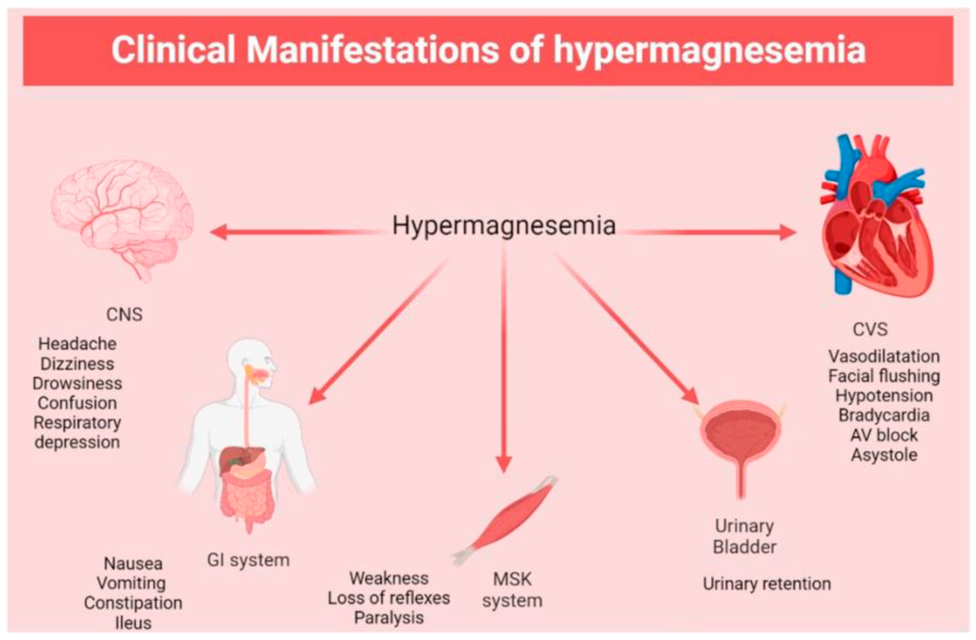




| Type of Study | Condition | Number of Treated Subjects | Age (Years, Mean ± SD) | Mg Compound/Dosage | Time of Treatment | Results | Ref. |
|---|---|---|---|---|---|---|---|
| Randomized, double-blinded, placebo-controlled | T2DM Hypo-Mg | n = 35 | 55.4 ± 10.2 | Mg oxide 20.7 mmoL/d | 30 d | FPG → HbA1c → | [207] |
| Randomized, double-blinded, placebo-controlled | T2DM Hypo-Mg | n = 39 | 51.2 ± 11.0 | Mg oxide 41.4 mmoL/d | 30 d | FPG → HbA1c → | [207] |
| Randomized, double-blinded, placebo-controlled | T2DM and HBP Hypo-Mg | n = 40 | 58.9 ± 8.5 | Mg chloride (450 mg/d of elemental Mg) | 4 mo | FPG → HbA1c → | [208] |
| Randomized controlled clinical trial | T2DM and depression Hypo-Mg | n = 12 | 69 ± 5.9 | Mg chloride (450 mg/d of elemental Mg) | 12 wk | FPG → HbA1c → | [209] |
| Randomized, double-blinded, placebo-controlled | T2DM Hypo-Mg | n = 18 | 63 ± 8.0 | Mg citrate 30 mmoL/d (730 mg/d of elemental Mg) | 3 mo | HbA1c → | [210] |
| Randomized, double-blinded, placebo-controlled, cross-over | T2DM Hypo-Mg | n = 14 | 67 ± 6.0 | Mg gluconate 15 mmoL/d (360 mg/d of elemental Mg) | 6 wk | HbA1c → | [211] |
| Prospective, randomized, controlled, open-label | Type 2 DN Hypo-Mg Normo-Mg | n = 26 n = 14 n = 12 | 61.4 ± 7.5 | Mg citrate 15 mmoL (360 mg/d of elemental Mg) | 12 wk | HbA1c → HbA1c → | [212] |
| N/A | DM Hypo-Mg | n = 40 | N/A | Mg oxide 600 mg | 12 wk | FPG → | [213] |
| Randomized, double-blinded, placebo-controlled | IR Hypo-Mg | n = 32 | 43 ± 7.9 | Mg chloride 2.5 g (300 mg/d of elemental Mg) | 3 mo | ↓ HOMA-IR ↓ FPG | [214] |
| Randomized, double-blinded, placebo-controlled | Pre-D Hypo-Mg | n = 59 | 42.5 ± 9.5 | Mg chloride (382 mg/d of elemental Mg) | 4 mo | ↓ HOMA-IR ↓ FPG | [215] |
| Randomized, double-blinded, placebo-controlled | T2DM Hypo-Mg | n = 32 | 59.7 ± 8.3 | Mg chloride 2.5 g/d (12.8 mmoL/d) | 16 wk | ↓ HOMA-IR ↓ FPG ↓ HbA1c | [216] |
| Randomized, double-blinded, placebo-controlled | Grade 3 DFU Hypo-Mg | n = 29 | 57.2 ± 11.0 | Mg oxide + Vit. E 250 mg/d + 400 IU/d | 12 wk | ↓ HOMA-IR ↓ FPG ↓ HbA1c | [217] |
| Observational | T1DM Hypo-Mg | n = 20 | 11.2 ± 3.41 | Mg oxide 300 mg | 3 mo | ↓ HbA1c | [218] |
| Controlled | T2DM Hypo-Mg | n = 60 | N/A | Mg chloride tablet 300 mg/d | 16 wk | ↓ FPG | [219] |
| Randomized, double-blinded, placebo-controlled | T2DM | n = 25 | 63 ± 8.2 | Mg aspartate 15 mmoL/d | 3 mo | FPG → HbA1c → | [220] |
| Randomized, double-blinded, cross-over | T2DM | n = 27 | 61 ± 2.0 | Mg (unspecified compound) 600 mg/d (24.7 mmoL/d) | 90 d | FPG → HbA1c → | [221] |
| Randomized, double-blinded, cross-over | T1DM | n = 29 | 43 ± 2.0 | Mg (unspecified compound) 600 mg/d (24.7 mmoL/d) | 90 d | FPG → HbA1c → | [221] |
| Randomized, placebo-controlled | T2DM | n = 26 | 63 ± 5.0 | Mg pidolate 4.5 g/d (16.2 mmoL/d) | 1 mo | FPG → HbA1c → | [222] |
| Randomized, double-blinded, placebo-controlled | T2DM | n = 25 | 64 ± 8.0 | Mg lactate-citrate 15 mmoL/d (184.5 mg/d of elemental Mg) | 4 mo | FPG → HbA1c → | [223] |
| Randomized, double-blinded, placebo-controlled, cross-over | T2DM | n = 56 | 52.84 ± 8.42 | Mg lactate 1.5 g/d (360 mg/d of elemental Mg) | 3 mo | FPG → HbA1c→ HOMA-IR → | [224] |
| Randomized, double-blinded, cross-over | T2DM | n = 4 | 73 ± 2.5 | Mg pidolate 4.5 g/d (15.8 mmoL/d) | 4 wk | FPG → Improvement of INS sensitivity and GLU oxidation | [225] |
| Controlled | T2DM | n = 30 | 71.1 ± 6.1 | Mg pidolate 368 mg/d | 1 mo | FPG → | [226] |
| Randomized, double-blinded, placebo-controlled, cross-over | T2DM | n = 28 | 28–84 | Mg chloride 384 mg/d (1.9 mmoL/d) | 6 wk | GLU → | [227] |
| Randomized, double-blinded, placebo-controlled | T2DM | n = 25 | 46.76 ± 6.93 | Mg sulfate 300 mg/d of elemental Mg | 3 mo | HOMA-IR →, ↓ FPG HbA1c → | [228] |
| N/A | T2DM | n = 9 | 51.6 ± 2.6 | MAG21 solution (Mg chloride), 300 mg/d | 30 d | ↓ HOMA-IR, FPG → HbA1c → | [229] |
| Randomized, cross-sectional, controlled clinical trial | T2DM | n = 32 | 55.9 ± 8.9 | Mg chloride 50 mg/L daily | 3 mo | ↓ HOMA-IR, FPG → ↓ HbA1c | [230] |
| Randomized, double-blinded, placebo-controlled | Type 2 DN | n = 40 | 41.2 ± 8.8 | Mg oxide 250 mg (150 mg of elemental Mg) | 12 wk | ↑ HOMA-IR, FPG → HbA1c → | [231] |
| Randomized controlled clinical trial | T2DM | n = 20 | 35–60 | Mg tablets (oxide, gluconate, lactate) 250 mg/d of elemental Mg | 3 mo | ↓ HOMA-IR, ↙ FBS ↓ HbA1c | [232] |
| Randomized, double-blinded, placebo-controlled | T2DM with grade 3 DFU | n = 35 | 60.1 ± 11.1 | Mg oxide 250 mg/d | 12 wk | ↙ HOMA-IR, ↓ FPG ↓ HbA1c | [233] |
| Randomized, double-blinded, placebo-controlled | DHD | n = 27 | 58.8 ± 10.1 | Mg oxide 250 mg/d | 24 wk | ↓ HOMA-IR ↙ FPG ↓ HbA1c | [234] |
| Randomized, double-blinded, placebo-controlled | GDM | n = 30 | 30.1 ± 5.9 | Mg oxide + Vit. E 250 mg/d + 400 IU/d | 6 wk | ↓ HOMA-IR, ↓ FPG | [235] |
| Randomized, double-blinded, placebo-controlled | T2DM and CHD | n = 27 | 61.7 ± 9.4 | Mg oxide + Zn sulfate 250 mg/d + 150 mg/d | 12 wk | ↓ FPG | [236] |
| Randomized, cross-over | T2DM | n = 8 | 72.2 ± 2.0 | Mg pidolate 2 g/d (7.0 mmoL/d) | 4 wk | ↓ FPG Improvement of INS response and action | [237] |
| Randomized, double-blinded, placebo-controlled | GDM | n = 20 | 27.8 ± 3.4 | Mg oxide 250 mg/d | 6 wk | ↓ FPG | [238] |
| Double-blinded, placebo-controlled | GDM | n = 30 | N/A | Mg + Vit. E 250 mg/d + 400 mg/d | 6 wk | ↓ FPG Improvement of INS sensitivity | [239] |
| Randomized, double-blinded, placebo-controlled | IR | n = 25 | 30–70 | Mg aspartate hydrochloride 15 mmoL (365 mg/d of elemental Mg) | 6 mo | ↓ FPG ↓ ISI-HOMA | [240] |
| Randomized, double-blinded, placebo-controlled | T1DM | n = 3 | N/A | Mg hydroxide 20–30 mmoL/d | 12 mo | ↑ HbA1c | [241] |
| Type of Study | Condition | Number of Treated Subjects | Age (Years, Mean ± SD) | Mg concentration After Treatment | Biological Specimens | Units | Ref. |
|---|---|---|---|---|---|---|---|
| Randomized, double-blinded, placebo-controlled | T2DM Hypo-Mg | n = 35 | 55.4 ± 10.2 | 0.76 → 1.59 → 121 ↑ | Plasma MonoNC Urine | mmol/L μg/mg TP mg/24 h | [207] |
| Randomized, double-blinded, placebo-controlled | T2DM Hypo-Mg | n = 39 | 51.2 ± 11.0 | 0.80 ↗ 1.62 ↗ 113 ↑ | Plasma MonoNC Urine | mmol/L μg/mg TP mg/24 h | [207] |
| Randomized, double-blinded, placebo-controlled | T2DM and HBP Hypo-Mg | n = 40 | 58.9 ± 8.5 | 0.81 ↑ | Serum | mmol/L | [208] |
| Randomized controlled clinical trial | T2DM and depression Hypo-Mg | n = 12 | 69 ± 5.9 | 0.86 ↑ | Serum | mmol/L | [209] |
| Randomized, double-blinded, placebo-controlled | T2DM Hypo-Mg | n = 18 | 63 ± 8.0 | 0.81 ↑ 2.8 ↑ | Serum Urine | mmol/L mmol/24 h | [210] |
| Randomized, double-blinded, placebo-controlled, cross-over | T2DM Hypo-Mg | n = 14 | 67 ± 6.0 | 0.75 ↑ 0.23 ↑ | Serum Urine | mmol/L Mg/Cre ratio | [211] |
| Prospective, randomized, controlled, open-label | Type 2 DN Hypo-Mg | n = 26 n = 14 | 61.4 ± 7.5 | 0.70 ↑ | Serum | mmol/L | [212] |
| Prospective, randomized, controlled, open-label | Type 2 DN Normo-Mg | n = 26 n = 12 | 61.4 ± 7.5 | 0.82 ↑ | Serum | mmol/L | [212] |
| Randomized, double-blinded, placebo-controlled | IR Hypo-Mg | n = 32 | 43 ± 7.9 | 0.81 ↑ | Serum | mmol/L | [214] |
| Randomized, double-blinded, placebo-controlled | Pre-D Hypo-Mg | n = 59 | 42.5 ± 9.5 | 0.81 ↑ | Serum | mmol/L | [215] |
| Randomized, double-blinded, placebo-controlled | T2DM Hypo-Mg | n = 32 | 59.7 ± 8.3 | 0.74 ↑ | Serum | mmol/L | [216] |
| Randomized, double-blinded, placebo-controlled | Grade 3 DFU Hypo-Mg | n = 29 | 57.2 ± 11.0 | 0.75 ↑ | Serum | mmol/L | [217] |
| Observational | T1DM Hypo-Mg | n = 20 | 11.2 ± 3.41 | 0.80 ↑ | Serum | mmol/L | [218] |
| Controlled | T2DM Hypo-Mg | n = 60 | N/A | 0.71 ↑ | Serum | mmol/L | [219] |
| Randomized, double-blinded, placebo-controlled | T2DM | n = 25 | 63 ± 8.2 | 0.82 ↑ 2.47 → 5.5 ↑ | Plasma RBC Urine | mmol/L mmol/L mmol/24 h | [220] |
| Randomized, double-blinded, cross-over | T1DM | n = 29 | 43 ± 2.0 | 0.80 ↗ 3.69 ↑ | Plasma Urine | mmol/L mmol/24 h | [221] |
| Randomized, double-blinded, cross-over | T2DM | n = 27 | 61 ± 2.0 | 0.79 ↑ 3.35 ↑ | Plasma Urine | mmol/L mmol/24 h | [221] |
| Randomized, placebo-controlled | T2DM | n = 26 | 63 ± 5.0 | 0.82 → 1.85 → | Serum RBC | mmol/L mmol/L | [222] |
| Randomized, double-blinded, placebo-controlled | T2DM | n = 25 | 64 ± 8.0 | N/A → | Serum | mmol/L | [223] |
| Randomized, double-blinded, placebo-controlled, cross-over | T2DM | n = 56 | 52.84 ± 8.42 | 0.95 → | Serum | mmol/L | [224] |
| Randomized, double-blinded, cross-over | T2DM | n = 4 | 73 ± 2.5 | 0.89 ↑ 2.27 ↑ | Plasma RBC | mmol/L mmol/L | [225] |
| Controlled | T2DM | n = 30 | 71.1 ± 6.1 | 0.49 ↑ * 0.93 → ** | Serum Serum | mmol/L mmol/L | [226] |
| Randomized, double-blinded, placebo-controlled, cross-over | T2DM | n = 28 | 28–84 | N/A → N/A → | Serum RBC | mmol/L mmol/L | [227] |
| Randomized, double-blinded, placebo-controlled | T2DM | n = 25 | 46.76 ± 6.93 | 0.89 → 5.99 ↑ | Serum Urine | mmol/L mg/dL | [228] |
| N/A | T2DM | n = 9 | 51.6 ± 2.6 | 0.95 ↑ 9.2 ↑ | Serum Urine | mmol/L mg/dL | [229] |
| Randomized, double-blinded, placebo-controlled | Type 2 DN | n = 40 | 41.2 ± 8.8 | 0.99 → | Serum | mmol/L | [231] |
| Randomized controlled clinical trial | T2DM | n = 20 | 35–60 | 0.85 ↑ | Serum | mmol/L | [232] |
| Randomized, double-blinded, placebo-controlled | T2DM with grade 3 DFU | n = 35 | 61.1 ± 11.1 | 0.95 ↑ | Serum | mmol/L | [233] |
| Randomized, double-blinded, placebo-controlled | DHD | n = 27 | 58.8 ± 10.1 | 0.99 ↑ | Serum | mmol/L | [234] |
| Randomized, double-blinded, placebo-controlled | GDM | n = 30 | 30.1 ± 5.9 | 0.90 ↑ | Serum | mmol/L | [235] |
| Randomized, double-blinded, placebo-controlled | T2DM and CHD | n = 27 | 61.7 ± 9.4 | 0.86 ↑ | Serum | mmol/L | [236] |
| Randomized, cross-over | T2DM | n = 8 | 72.2 ± 2.0 | 0.86 ↑ 2.03 ↑ | Plasma RBC | mmol/L mmol/L | [237] |
| Randomized, double-blinded, placebo-controlled | GDM | n = 20 | 27.8 ± 3.4 | 0.78 ↑ | Serum | mmol/L | [238] |
| Randomized, double-blinded, placebo-controlled | IR | n = 25 | 30–70 | 0.922 ↗ 1.920 → 0.608 ↑ | Serum RBC WB | mmol/L mmol/L mmol/L | [240] |
| Randomized, double-blinded, cross-over | T2DM | n = 8 | 59–72 | 0.88 ↑ 2.08 ↑ | Plasma RBC | mmol/L mmol/L | [242] |
Disclaimer/Publisher’s Note: The statements, opinions and data contained in all publications are solely those of the individual author(s) and contributor(s) and not of MDPI and/or the editor(s). MDPI and/or the editor(s) disclaim responsibility for any injury to people or property resulting from any ideas, methods, instructions or products referred to in the content. |
© 2026 by the authors. Licensee MDPI, Basel, Switzerland. This article is an open access article distributed under the terms and conditions of the Creative Commons Attribution (CC BY) license.
Share and Cite
Ścibior, A.; Aureliano, M.; Romanowska, Z.; Radko, L.; Męcik-Kronenberg, T. Magnesium and Diabetes: A Review of Recent Studies on the Efficacy of Supplementation with Mg in Humans Suffering from This Chronic Metabolic Disease. Int. J. Mol. Sci. 2026, 27, 7620. https://doi.org/10.3390/ijms27177620
Ścibior A, Aureliano M, Romanowska Z, Radko L, Męcik-Kronenberg T. Magnesium and Diabetes: A Review of Recent Studies on the Efficacy of Supplementation with Mg in Humans Suffering from This Chronic Metabolic Disease. International Journal of Molecular Sciences. 2026; 27(17):7620. https://doi.org/10.3390/ijms27177620
Chicago/Turabian StyleŚcibior, Agnieszka, Manuel Aureliano, Zuzanna Romanowska, Lidia Radko, and Tomasz Męcik-Kronenberg. 2026. "Magnesium and Diabetes: A Review of Recent Studies on the Efficacy of Supplementation with Mg in Humans Suffering from This Chronic Metabolic Disease" International Journal of Molecular Sciences 27, no. 17: 7620. https://doi.org/10.3390/ijms27177620
APA StyleŚcibior, A., Aureliano, M., Romanowska, Z., Radko, L., & Męcik-Kronenberg, T. (2026). Magnesium and Diabetes: A Review of Recent Studies on the Efficacy of Supplementation with Mg in Humans Suffering from This Chronic Metabolic Disease. International Journal of Molecular Sciences, 27(17), 7620. https://doi.org/10.3390/ijms27177620