Placental Growth Factor in Diabetic Retinopathy: Disease-Selective Signaling and Mechanistic Rationale for Targeted Therapy




Abstract
1. Introduction
2. Context-Dependent Roles of PlGF in the Retina
3. Distinct and Overlapping Features of VEGF-A and PlGF Signaling
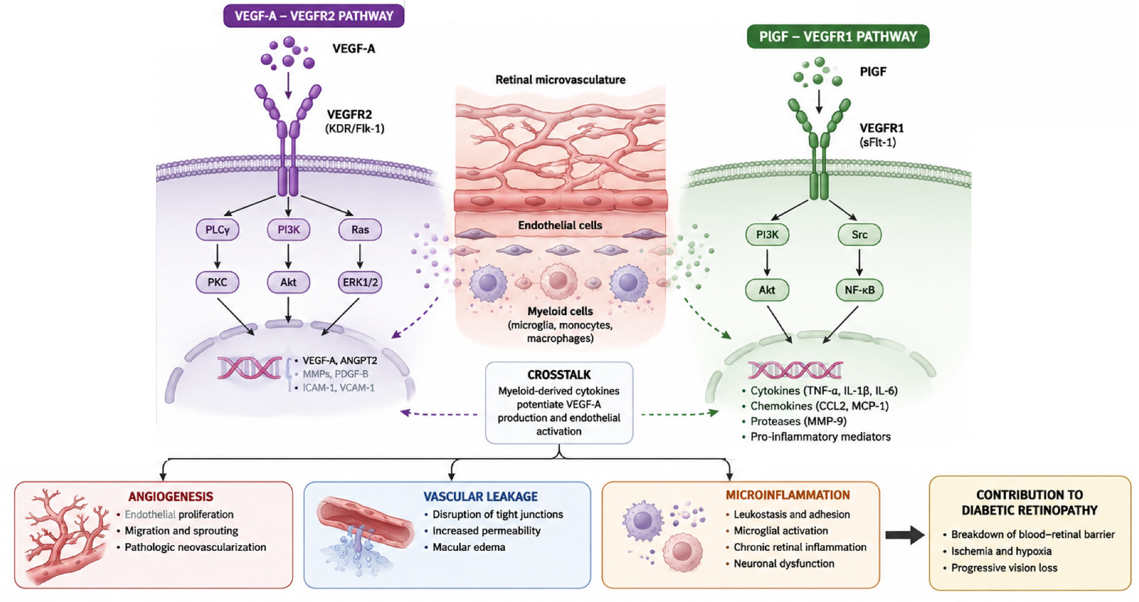
4. VEGF-A/VEGFR2 and PlGF/VEGFR1 Pathways in DR
4.1. Structural and Signaling Basis for PlGF Selectivity
4.2. PlGF in Retinal Vascular Pathology
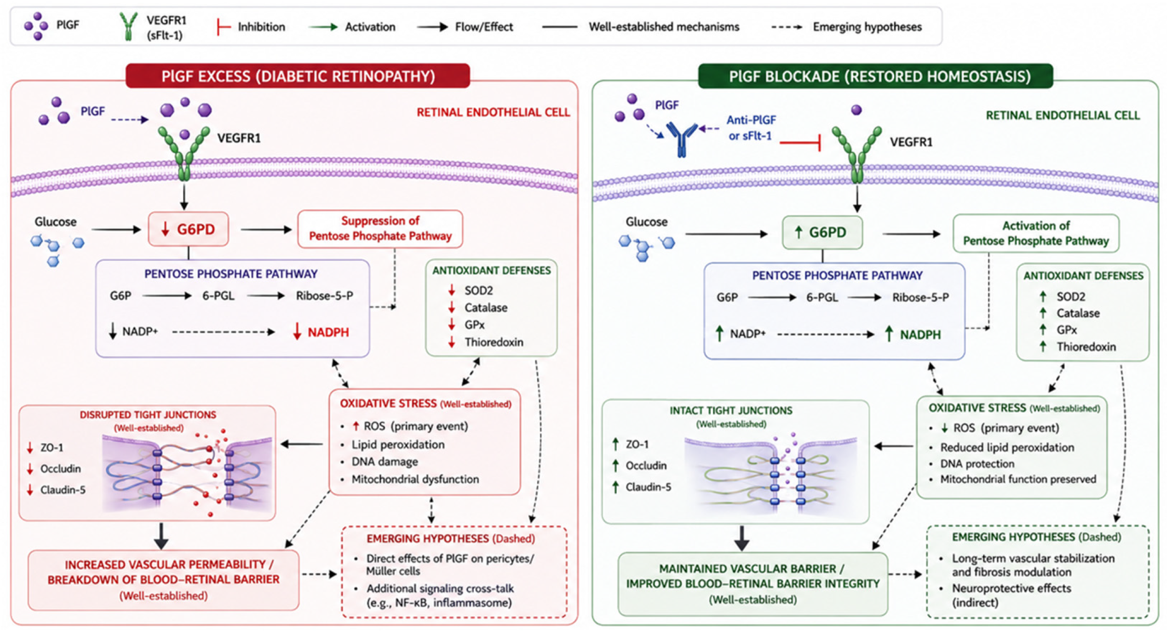
4.3. Barrier Failure, Inflammation, and Metabolic and Redox Stress
4.4. Neurodegeneration
4.5. Therapeutic Implications and Drug Positioning
4.6. Relevance to Clinical Heterogeneity in DR
5. Clinical Evidence and Therapeutic Implications
5.1. Ocular Fluid Biomarkers
5.2. Growth Factor and Fibrosis Profiles Under Anti-VEGF Therapy
5.3. Preclinical Performance of PlGF Neutralization
5.4. Potential Therapeutic Positioning
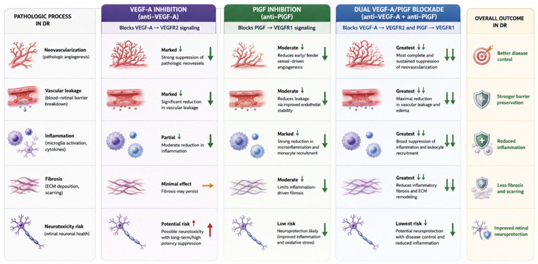
5.5. Comparative Analysis: PlGF vs. VEGF Inhibitors in DR Treatment
5.6. Biomarker Interpretation and Disease Stratification
5.7. Evidence from Dual-Ligand Inhibition
5.8. Limitations and Future Development
6. Conclusions
Author Contributions
Funding
Data Availability Statement
Conflicts of Interest
References
- Aiello, L.P.; Avery, R.L.; Arrigg, P.G.; Keyt, B.A.; Jampel, H.D.; Shah, S.T.; Pasquale, L.R.; Thieme, H.; Iwamoto, M.A.; Park, J.E. Vascular endothelial growth factor in ocular fluid of patients with diabetic retinopathy and other retinal disorders. N. Engl. J. Med. 1994, 331, 1480–1487. [Google Scholar] [CrossRef] [Scilit] [PubMed]
- Wang, W.; Lo, A.C. Diabetic retinopathy: Pathophysiology and treatments. Int. J. Mol. Sci. 2018, 19, 1816. [Google Scholar] [CrossRef] [Scilit] [PubMed]
- Van Bergen, T.; Etienne, I.; Cunningham, F.; Moons, L.; Schlingemann, R.O.; Feyen, J.H.; Stitt, A.W. The role of placental growth factor (PlGF) and its receptor system in retinal vascular diseases. Prog. Retin. Eye Res. 2019, 69, 116–136. [Google Scholar] [CrossRef] [Scilit] [PubMed]
- Van Bergen, T.; Hu, T.-T.; Etienne, I.; Reyns, G.E.; Moons, L.; Feyen, J.H. Neutralization of placental growth factor as a novel treatment option in diabetic retinopathy. Exp. Eye Res. 2017, 165, 136–150. [Google Scholar] [CrossRef] [Scilit] [PubMed]
- Funatsu, H.; Yamashita, H.; Noma, H.; Mimura, T.; Yamashita, T.; Hori, S. Increased levels of vascular endothelial growth factor and interleukin-6 in the aqueous humor of diabetics with macular edema. Am. J. Ophthalmol. 2002, 133, 70–77. [Google Scholar] [CrossRef] [Scilit] [PubMed]
- Ishida, S.; Usui, T.; Yamashiro, K.; Kaji, Y.; Ahmed, E.; Carrasquillo, K.G.; Amano, S.; Hida, T.; Oguchi, Y.; Adamis, A.P. VEGF164 is proinflammatory in the diabetic retina. Investig. Ophthalmol. Vis. Sci. 2003, 44, 2155–2162. [Google Scholar] [CrossRef] [Scilit] [PubMed]
- Al Kahtani, E.; Xu, Z.; Al Rashaed, S.; Wu, L.; Mahale, A.; Tian, J.; Abboud, E.; Ghazi, N.; Kozak, I.; Gupta, V. Vitreous levels of placental growth factor correlate with activity of proliferative diabetic retinopathy and are not influenced by bevacizumab treatment. Eye 2017, 31, 529–536. [Google Scholar] [CrossRef] [Scilit] [PubMed]
- Nguyen, Q.D.; De Falco, S.; Behar-Cohen, F.; Lam, W.C.; Li, X.; Reichhart, N.; Ricci, F.; Pluim, J.; Li, W.W. Placental growth factor and its potential role in diabetic retinopathy and other ocular neovascular diseases. Acta Ophthalmol. 2018, 96, e1–e9. [Google Scholar] [CrossRef] [Scilit] [PubMed]
- Huang, H.; Lennikov, A.; Saddala, M.S.; Gozal, D.; Grab, D.J.; Khalyfa, A.; Fan, L. Placental growth factor negatively regulates retinal endothelial cell barrier function through suppression of glucose-6-phosphate dehydrogenase and antioxidant defense systems. FASEB J. 2019, 33, 13695. [Google Scholar] [CrossRef] [Scilit] [PubMed]
- Jiao, W.; Ji, J.-F.; Xu, W.; Bu, W.; Zheng, Y.; Ma, A.; Zhao, B.; Fan, Q. Distinct downstream signaling and the roles of VEGF and PlGF in high glucose-mediated injuries of human retinal endothelial cells in culture. Sci. Rep. 2019, 9, 15339. [Google Scholar] [CrossRef] [Scilit] [PubMed]
- Saddala, M.S.; Lennikov, A.; Huang, H. Placental growth factor regulates the pentose phosphate pathway and antioxidant defense systems in human retinal endothelial cells. J. Proteom. 2020, 217, 103682. [Google Scholar] [CrossRef] [Scilit] [PubMed]
- Uemura, A.; Fruttiger, M.; d’Amore, P.A.; De Falco, S.; Joussen, A.M.; Sennlaub, F.; Brunck, L.R.; Johnson, K.T.; Lambrou, G.N.; Rittenhouse, K.D. VEGFR1 signaling in retinal angiogenesis and microinflammation. Prog. Retin. Eye Res. 2021, 84, 100954. [Google Scholar] [CrossRef] [Scilit] [PubMed]
- Huang, H. Pericyte-endothelial interactions in the retinal microvasculature. Int. J. Mol. Sci. 2020, 21, 7413. [Google Scholar] [CrossRef] [Scilit] [PubMed]
- Kowalczuk, L.; Touchard, E.; Omri, S.; Jonet, L.; Klein, C.; Valamanes, F.; Berdugo, M.; Bigey, P.; Massin, P.; Jeanny, J.-C. Placental growth factor contributes to micro-vascular abnormalization and blood-retinal barrier breakdown in diabetic retinopathy. PLoS ONE 2011, 6, e17462. [Google Scholar] [CrossRef] [Scilit] [PubMed]
- Lazzara, F.; Fidilio, A.; Platania, C.B.M.; Giurdanella, G.; Salomone, S.; Leggio, G.M.; Tarallo, V.; Cicatiello, V.; De Falco, S.; Eandi, C.M. Aflibercept regulates retinal inflammation elicited by high glucose via the PlGF/ERK pathway. Biochem. Pharmacol. 2019, 168, 341–351. [Google Scholar] [CrossRef] [Scilit] [PubMed]
- Lazzara, F.; Trotta, M.C.; Platania, C.B.M.; D’Amico, M.; Petrillo, F.; Galdiero, M.; Gesualdo, C.; Rossi, S.; Drago, F.; Bucolo, C. Stabilization of HIF-1α in human retinal endothelial cells modulates expression of miRNAs and proangiogenic growth factors. Front. Pharmacol. 2020, 11, 1063. [Google Scholar] [CrossRef] [Scilit] [PubMed]
- Deissler, H.L.; Deissler, H.; Lang, G.K.; Lang, G.E. VEGF but not PlGF disturbs the barrier of retinal endothelial cells. Exp. Eye Res. 2013, 115, 162–171. [Google Scholar] [CrossRef] [Scilit] [PubMed]
- Carmeliet, P.; Moons, L.; Luttun, A.; Vincenti, V.; Compernolle, V.; De Mol, M.; Wu, Y.; Bono, F.; Devy, L.; Beck, H. Synergism between vascular endothelial growth factor and placental growth factor contributes to angiogenesis and plasma extravasation in pathological conditions. Nat. Med. 2001, 7, 575–583. [Google Scholar] [CrossRef] [Scilit] [PubMed]
- Mesquita, J.; Sousa, J.P.C.; Tavares-Ratado, P.; Vaz-Pereira, S.; Neves, A.; Rocha, A.S.; Santos, F.; Passarinha, L.; Tomaz, C. Comparison of serum and vitreous PIGF in diabetic retinopathy patients and non-diabetic patients. Investig. Ophthalmol. Vis. Sci. 2015, 56, 5179. [Google Scholar]
- Du, S.; Ju, L.; Zheng, W. Aqueous humour concentrations of TGF-β, PLGF and FGF-1 and total retinal blood flow in patients with early non-proliferative diabetic retinopathy. Acta Ophthalmol. 2019, 97, e482. [Google Scholar] [CrossRef] [Scilit] [PubMed]
- Mesquita, J.; Santos, F.M.; Sousa, J.P.; Vaz-Pereira, S.; Tavares-Ratado, P.; Neves, A.; Mesquita, R.; Tomaz, C.T. Serum and vitreous levels of Placenta Growth factor in Diabetic Retinopathy patients: Correlation with Disease Severity and Optical Coherence Tomographic parameters. Cureus 2024, 16, e54862. [Google Scholar] [CrossRef] [Scilit] [PubMed]
- Ozer, F.; Tokuc, E.O.; Albayrak, M.G.B.; Akpinar, G.; Kasap, M.; Karabas, V.L. Comparison of before versus after intravitreal bevacizumab injection, growth factor levels and fibrotic markers in vitreous samples from patients with proliferative diabetic retinopathy. Graefe’s Arch. Clin. Exp. Ophthalmol. 2022, 260, 1899–1906. [Google Scholar] [CrossRef] [Scilit] [PubMed]
- Huang, H.; He, J.; Johnson, D.K.; Wei, Y.; Liu, Y.; Wang, S.; Lutty, G.A.; Duh, E.J.; Carmeliet, P.; Semba, R.D. Deletion of placental growth factor prevents diabetic retinopathy and is associated with Akt activation and HIF1α-VEGF pathway inhibition. Diabetes 2015, 64, 200–212, Erratum in Diabetes 2015, 64, 1067. [Google Scholar] [CrossRef] [Scilit] [PubMed]
- Kang, Q.; Yang, C. Oxidative stress and diabetic retinopathy: Molecular mechanisms, pathogenetic role and therapeutic implications. Redox Biol. 2020, 37, 101799. [Google Scholar] [CrossRef] [Scilit] [PubMed]
- Saddala, M.S.; Lennikov, A.; Grab, D.J.; Liu, G.-S.; Tang, S.; Huang, H. Proteomics reveals ablation of PlGF increases antioxidant and neuroprotective proteins in the diabetic mouse retina. Sci. Rep. 2018, 8, 16728. [Google Scholar] [CrossRef] [Scilit] [PubMed]
- Sivaprasad, S.; Prevost, A.T.; Bainbridge, J.; Edwards, R.T.; Hopkins, D.; Kelly, J.; Luthert, P.; Murphy, C.; Ramu, J.; Sarafraz-Shekary, N. Clinical efficacy and mechanistic evaluation of aflibercept for proliferative diabetic retinopathy (acronym CLARITY): A multicentre phase IIb randomised active-controlled clinical trial. BMJ Open 2015, 5, e008405. [Google Scholar] [CrossRef] [Scilit] [PubMed]
- Alsaedi, N.G.; Alselaimy, R.M.; Alshamrani, A.A.; AlAjmi, M.; Khandekar, R.; Al-Dhibi, H.; Al-Abdullah, A.A. Aflibercept versus ranibizumab as a second line therapy after bevacizumab for diabetic macular edema. Clin. Ophthalmol. 2021, 15, 2975–2980. [Google Scholar] [CrossRef] [Scilit] [PubMed]
- Tan, G.S.; Cheung, N.; Simo, R.; Cheung, G.C.; Wong, T.Y. Diabetic macular oedema. Lancet Diabetes Endocrinol. 2017, 5, 143–155. [Google Scholar] [CrossRef] [Scilit] [PubMed]
- Autiero, M.; Waltenberger, J.; Communi, D.; Kranz, A.; Moons, L.; Lambrechts, D.; Kroll, J.; Plaisance, S.; De Mol, M.; Bono, F. Role of PlGF in the intra-and intermolecular cross talk between the VEGF receptors Flt1 and Flk1. Nat. Med. 2003, 9, 936–943. [Google Scholar] [CrossRef] [Scilit] [PubMed]
- Witmer, A.; Vrensen, G.; Van Noorden, C.; Schlingemann, R. Vascular endothelial growth factors and angiogenesis in eye disease. Prog. Retin. Eye Res. 2003, 22, 1–29. [Google Scholar] [CrossRef] [Scilit] [PubMed]
- Pitale, P.M.; Gorbatyuk, M.S. Diabetic retinopathy: From animal models to cellular signaling. Int. J. Mol. Sci. 2022, 23, 1487. [Google Scholar] [CrossRef] [Scilit] [PubMed]
- Krick, T.W.; Bressler, N.M. Recent clinically relevant highlights from the diabetic retinopathy clinical research network. Curr. Opin. Ophthalmol. 2018, 29, 199–205. [Google Scholar] [CrossRef] [Scilit] [PubMed]



| Feature | VEGF-A | PlGF |
|---|---|---|
| Major receptor in the retina | VEGFR2 (KDR/Flk-1); also binds VEGFR1 | VEGFR1 (FLT1) only |
| Retinal cell-specific expression | RPE cells, Müller glia, endothelial cells, ganglion cells, pericytes | Müller glia, RPE cells, endothelial cells, infiltrating macrophages/microglia (markedly upregulated under diabetic and ischemic conditions) |
| Role in normal vasculature | Essential for developmental and maintenance angiogenesis | Limited in physiological angiogenesis |
| Dominant pathological effects | Angiogenesis, vascular leakage, endothelial proliferation, increased permeability | Vascular leakage, chronic inflammation, leukocyte recruitment, fibrosis, metabolic dysfunction |
| Major downstream signaling pathways | VEGFR2 → PI3K/Akt, ERK1/2, p38 MAPK, PLCγ/PKC, STAT3, eNOS | VEGFR1 → PKC–ERK1/2–eNOS, NF-κB, JNK/p38 MAPK, ROS generation, pentose phosphate pathway suppression, inflammatory cytokine production |
| Effect on the blood–retinal barrier | Disrupts tight junctions (ZO-1, occludin, claudin-5) and increases vascular permeability | Potently disrupts barrier integrity through inflammatory signaling; inhibition restores tight junction proteins and vascular stability |
| Representative therapeutic agents | Ranibizumab, Bevacizumab, Aflibercept, Faricimab (indirect VEGF-A inhibition via VEGF-A binding) | Aflibercept (PlGF trap), Conbercept, OPT-302 (investigational), anti-PlGF monoclonal antibodies (preclinical) |
| Potential adverse effects of inhibition | Long-term inhibition may impair physiological vascular homeostasis, neuroprotection, wound healing, and choriocapillaris maintenance | Preclinical studies suggest minimal effects on normal retinal vasculature; long-term clinical safety remains under investigation |
| Level of supporting evidence | Extensive clinical evidence (multiple Phase III trials and real-world studies) | Strong preclinical evidence with emerging clinical evidence; limited Phase II/III validation |
Disclaimer/Publisher’s Note: The statements, opinions and data contained in all publications are solely those of the individual author(s) and contributor(s) and not of MDPI and/or the editor(s). MDPI and/or the editor(s) disclaim responsibility for any injury to people or property resulting from any ideas, methods, instructions or products referred to in the content. |
© 2026 by the authors. Licensee MDPI, Basel, Switzerland. This article is an open access article distributed under the terms and conditions of the Creative Commons Attribution (CC BY) license.
Share and Cite
Kim, J.; Ju, W.-K.; Won, J.Y. Placental Growth Factor in Diabetic Retinopathy: Disease-Selective Signaling and Mechanistic Rationale for Targeted Therapy. Int. J. Mol. Sci. 2026, 27, 7354. https://doi.org/10.3390/ijms27167354
Kim J, Ju W-K, Won JY. Placental Growth Factor in Diabetic Retinopathy: Disease-Selective Signaling and Mechanistic Rationale for Targeted Therapy. International Journal of Molecular Sciences. 2026; 27(16):7354. https://doi.org/10.3390/ijms27167354
Chicago/Turabian StyleKim, Jongmin, Won-Kyu Ju, and Jae Yon Won. 2026. "Placental Growth Factor in Diabetic Retinopathy: Disease-Selective Signaling and Mechanistic Rationale for Targeted Therapy" International Journal of Molecular Sciences 27, no. 16: 7354. https://doi.org/10.3390/ijms27167354
APA StyleKim, J., Ju, W.-K., & Won, J. Y. (2026). Placental Growth Factor in Diabetic Retinopathy: Disease-Selective Signaling and Mechanistic Rationale for Targeted Therapy. International Journal of Molecular Sciences, 27(16), 7354. https://doi.org/10.3390/ijms27167354