Article

Structural Dynamics and Susceptibility of Aminonaphthoquinone–Chalcone Hybrids Against Human Tyrosinase

by
Sahachai Sabuakham
1,†,
Napat Nuramrum
1,†,
Thanyada Rungrotmongkol
2,3,
Ratchanok Pingaew
4 and
Panupong Mahalapbutr
1,*
1
Department of Biochemistry, Center for Translational Medicine, Faculty of Medicine, Khon Kaen University, Khon Kaen 40002, Thailand
2
Program in Bioinformatics and Computational Biology, College of Interdisciplinary and Integrative Studies, Chulalongkorn University, Bangkok 10330, Thailand
3
Center of Excellence in Structural and Computational Biology, Department of Biochemistry, Faculty of Science, Chulalongkorn University, Bangkok 10330, Thailand
4
Department of Chemistry, Faculty of Science, Srinakharinwirot University, Bangkok 10110, Thailand
*
Author to whom correspondence should be addressed.
These authors contributed equally to this work.
Int. J. Mol. Sci. 2026, 27(16), 7328; https://doi.org/10.3390/ijms27167328
Submission received: 8 April 2026 / Revised: 4 August 2026 / Accepted: 10 August 2026 / Published: 17 August 2026

Abstract

Tyrosinase is a key enzyme that catalyzes the rate-limiting step in melanogenesis. Although numerous mushroom-derived tyrosinase inhibitors have been identified, many exhibit limited efficacy against human tyrosinase. In this study, in silico approaches were employed to evaluate the inhibitory potential of aminonaphthoquinone–chalcone hybrids against human tyrosinase. Molecular docking analysis revealed that all 10 hybrids showed higher binding affinity than kojic acid. Among them, six hybrids (3, 4, 5, 7, 9, and 10) displayed higher binding affinity than the others and interacted with amino acid residues within the active site of tyrosinase. Molecular dynamics simulations revealed that the six selected hybrids maintained stable binding to human tyrosinase throughout the simulations. Among them, compound 9 exhibited the highest structural stability, consistent with its most favorable MM/PBSA-predicted binding free energy and the greatest number of key residue interactions (K306, K334, S358, S360, N364, H367, I368, S375, Q376, and V377). Van der Waals interactions were identified as the predominant driving force governing ligand–protein binding in all complexes. These findings provide atomistic insights into the interactions between aminonaphthoquinone–chalcone hybrids and human tyrosinase and may facilitate the future development of novel human tyrosinase inhibitors.

1. Introduction

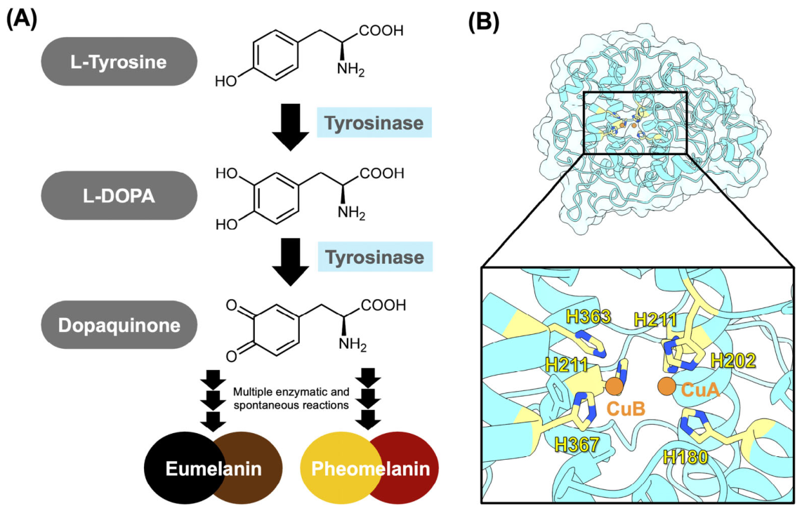
Tyrosinase, the rate-limiting enzyme in melanogenesis, catalyzes the conversion of L-tyrosine to L-DOPA and subsequently to dopaquinone, which is further processed through several enzymatic and spontaneous reactions to produce melanin (Figure 1A) [1,2]. In addition, tyrosinase-related protein 1 (TRP1) catalyzes the oxidation of 5,6-dihydroxyindole-2-carboxylic acid (DHICA), which is generated from dopachrome by tyrosinase-related protein 2 (TRP2), thereby contributing to eumelanin synthesis [3,4]. The active site of tyrosinase contains a coupled binuclear copper center (Figure 1B) that binds molecular oxygen to form an oxy-tyrosinase intermediate, which initiates the monooxygenation reaction [5,6]. In contrast, TRP1 and TRP2 require zinc ions for melanin biosynthesis [4,7,8].
Excessive melanin production and accumulation contribute to various hyperpigmentation disorders, including solar lentigines, melasma, freckles, and post-inflammatory hyperpigmentation [2,9]. Therefore, tyrosinase has emerged as a primary target for the development of depigmenting agents [10]. Although several tyrosinase inhibitors have been developed, many are associated with significant adverse effects [2]. For example, hydroquinone and arbutin may cause skin irritation and contact dermatitis [11,12], whereas kojic acid is chemically unstable, particularly during storage [13]. Furthermore, the development of most tyrosinase inhibitors has relied on mushroom tyrosinase, limiting their efficacy and translational potential against human tyrosinase because of structural differences between the two enzymes [14]. Therefore, there remains a critical need to discover novel, potent inhibitors that specifically target human tyrosinase.
Figure 1. (A) Melanogenesis pathway [13]. (B) Homology model of human tyrosinase. The catalytic histidine residues and copper atoms are shown in yellow and orange, respectively.
Ijms 27 07328 g001
Recent studies have identified several promising inhibitors of human tyrosinase. Mann et al. screened a library of 50,000 compounds and compared the active hits with established depigmenting agents. Among the tested compounds, Thiamidol™ (isobutylamido thiazolyl resorcinol) emerged as the most potent human tyrosinase inhibitor, exhibiting an IC50 value of 1.1 μmol/L [15]. Hassan et al. identified vanillin and coumarin derivatives (V7 and C9) as potent human tyrosinase inhibitors, showing higher binding affinity than kojic acid and arbutin [16]. Furthermore, anthocyanin-rich extracts from the seed coats of black soybean were shown to inhibit human tyrosinase activity [17].
Several naphthoquinone- and chalcone-based compounds have been reported to possess anti-tyrosinase activity. Among naphthoquinone derivatives, plumbagin and lawsone inhibited tyrosinase activity in B16F10 mouse melanoma cells [18,19]. β-Lapachone not only suppressed tyrosinase activity but also reduced melanin synthesis by downregulating the expression of microphthalmia-associated transcription factor (MITF), tyrosinase, and tyrosinase-related proteins [20]. Chalcone derivatives have likewise demonstrated potent anti-tyrosinase activity. For example, 6-hydroxy-2-(3,4,5-trimethoxybenzylidene)-3,4-dihydronaphthalen-1(2H)-one (C2), inhibited tyrosinase with an IC50 value of 8.8 μM, slightly outperforming kojic acid (IC50 = 9.7 μM) [21]. In addition, chalcone–hydroxypyridinone hybrids (1a and 1d) potently inhibited both the monophenolase activity (IC50 = 3.07 μM and 2.25 μM, respectively) and diphenolase activity (IC50 = 17.05 μM and 11.70 μM, respectively) of tyrosinase [22]. Similarly, butein and homobutein exhibited inhibitory activity against tyrosinase, with IC50 values of 10.88 and 14.78 μM for monophenolase activity and 15.20 and 12.36 μM for diphenolase activity, respectively [23].
Based on these findings, the molecular hybridization of naphthoquinone and chalcone scaffolds represents a rational strategy for developing novel tyrosinase inhibitors by integrating the complementary pharmacological features of both scaffolds into a single molecular framework. In particular, aminonaphthoquinone–chalcone hybrids incorporate structural features associated with potent tyrosinase inhibition, including extended aromatic π-systems, multiple hydrogen-bond donor and acceptor functionalities [24,25], and trimethoxy substitution patterns [21]. These characteristics are consistent with the structural requirements for effective tyrosinase inhibition reported in previous studies. Nevertheless, despite the well-documented anti-tyrosinase activities of naphthoquinone and chalcone derivatives, the inhibitory potential of aminonaphthoquinone–chalcone hybrids against human tyrosinase has not yet been investigated.
In this study, molecular docking, molecular dynamics (MD) simulations, and molecular mechanics/Poisson–Boltzmann surface area (MM/PBSA) binding free energy (ΔGbind) calculations were employed to investigate the inhibitory potential of aminonaphthoquinone–chalcone hybrids (Figure 2) against human tyrosinase. The findings provide molecular insights into the binding mechanisms of these hybrids and may facilitate the rational design and development of novel human tyrosinase inhibitors based on the aminonaphthoquinone–chalcone scaffold.

2. Results and Discussion

2.1. Molecular Docking

To investigate the binding affinity and binding pattern of the studied compounds against human tyrosinase, molecular docking was performed using the CB-Dock2 web server. Kojic acid, a well-established tyrosinase inhibitor, was used as a positive control and exhibited a predicted binding energy of −5.0 kcal/mol (Supplementary Figure S1). All 10 aminonaphthoquinone–chalcone hybrids demonstrated higher predicted binding affinity than kojic acid. As shown in Table 1, the binding energy of the hybrids in complex with human tyrosinase was in a range of −9.2 to −7.5 kcal/mol. Among them, compound 4 showed the lowest binding energy (−9.2 kcal/mol), followed by compound 10 (−8.5 kcal/mol) and compounds 3, 5, 7, and 9 (−8.4 kcal/mol), all of which exhibited lower binding energies than the remaining derivatives. These results suggest that compounds 3, 4, 5, 7, 9, and 10 possess higher predicted binding affinity toward human tyrosinase than the other hybrids.
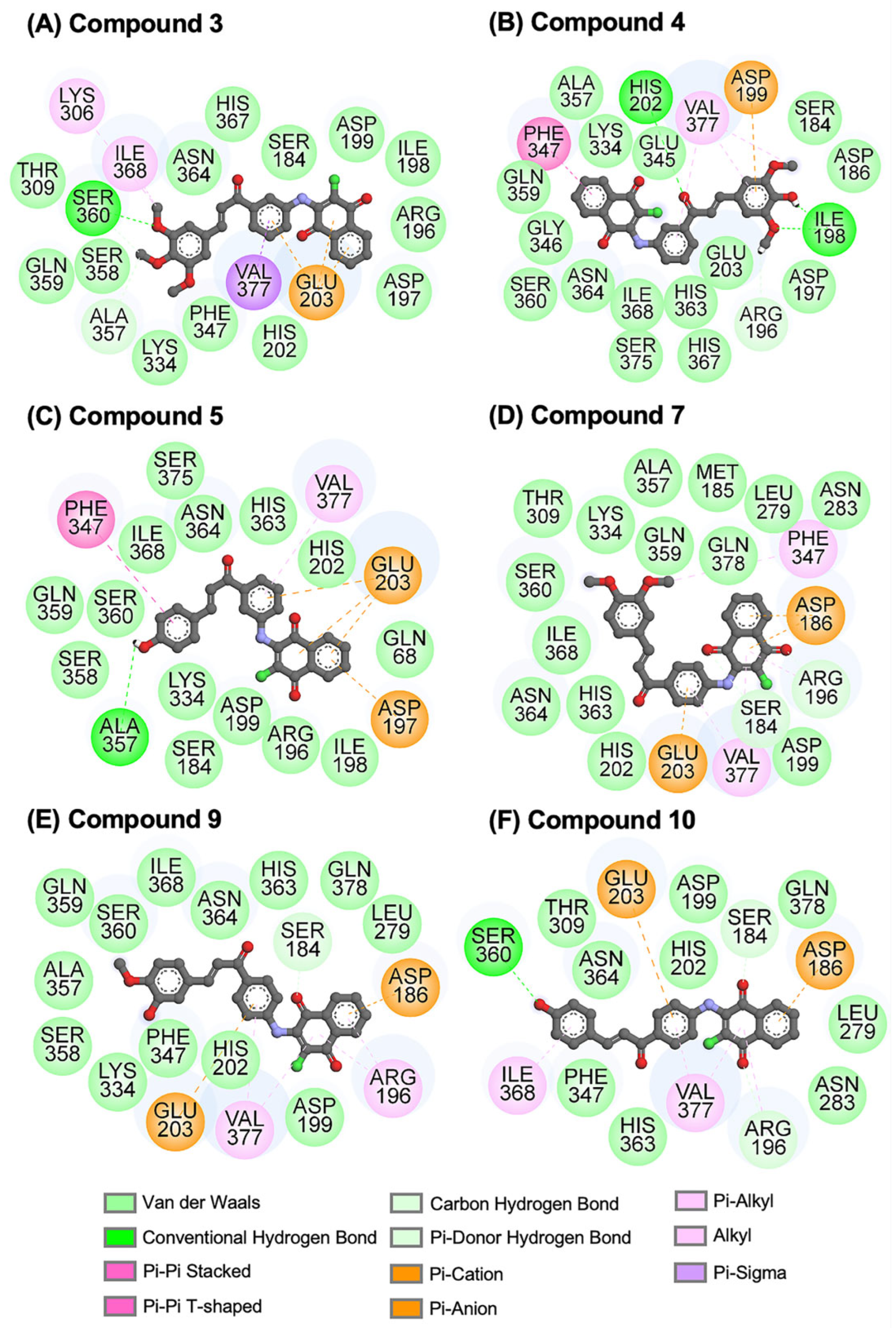
The two-dimensional (2D) interactions between the ligands and human tyrosinase were further investigated. As displayed in Figure 3A, the methoxy group of compound 3 formed a hydrogen bond (H-bond) with residue S360. Compound 4 (Figure 3B) formed H-bonds with residues I198 and H202, while its aromatic rings established Pi interactions with residues D199, F347, and V377. The hydroxyl (−OH) group of compound 5 formed an H-bond with residue A357 (Fig-ure 3C). Similarly, the −OH group of compound 10 (Figure 3F) formed an H-bond with S360, while its aromatic rings established Pi interactions with residues D186, E203, I368, and V377.
Although no H-bonds were detected for compounds 7 and 9 in complex with human tyrosinase (Figure 3D,E), both compounds established Pi interactions with several residues. Compound 7 interacted with D186, E203, F347, and V377, whereas compound 9 interacted with D186, R196, E203, and V377. Notably, residue V377 was identified as a key binding residue in all ligand–human tyrosinase complexes. This observation is consistent with previous studies showing that V377 is one of the key amino acid residues involved in the binding of thujaplicins and Thiamidol™ to human tyrosinase [27,28].
The presence of methoxy and −OH groups in the aromatic moieties of the potent compounds (3, 4, 5, 7, 9, and 10) potentially formed hydrogen bonds, van der Waals (vdW) forces, and Pi interactions with the human tyrosinase. These findings align well with previous reports stating that the presence of −OH groups enhanced binding affinity and increased their anti-tyrosinase activity [24,25].
It should be noted that all six studied hybrids interacted with amino acid residues located within the active site of human tyrosinase, consistent with the previously reported binding of amphotericin B [29], kojic acid, arbutin [16], coumarin (compound C9) [16], and vanillin (compound V7) [16] to human tyrosinase. Based on these findings, the six aminonaphthoquinone–chalcone hybrids were selected for further MD simulations, identification of key binding residues, ΔGbind calculations, and drug-likeness prediction.

2.2. Structural Stability and Compactness of Protein–Ligand Complex

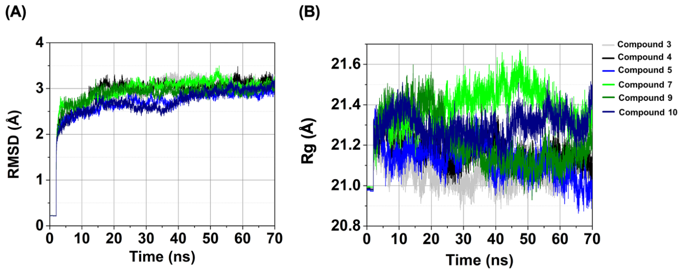
The root-mean-square deviation (RMSD) was calculated to assess structural stability of the complexes throughout the simulation period [30]. As shown in Figure 4A, the RMSD values of all complexes gradually increased during the initial stage of the simulation and reached equilibrium after approximately 60 ns. Thereafter, all complexes maintained relatively stable fluctuations, with average RMSD values ranging from ~2.91 to 3.14 Å, as calculated over the final 10 ns of the simulation. The average RMSD values were 3.14 ± 0.06, 3.14 ± 0.07, 2.91 ± 0.06, and 3.07 ± 0.07 Å for compounds 3, 4, 5, and 7, respectively, in complex with human tyrosinase. Notably, the complex with compound 5, followed by those with compounds 9 and 10, exhibited lower RMSD values than the other systems, suggesting greater structural stability in the aqueous environment.
We further investigated the compactness of the ligand–protein complexes using the radius of gyration (Rg) as an indicator of structural compactness during complexation [31]. As shown in Figure 4B, the Rg values of all complexes remained relatively stable during the final 10 ns of the simulation, ranging from approximately 20.9 to 21.5 Å. Notably, the complexes of compounds 3, 4, 5, and 9 with human tyrosinase exhibited lower average Rg values than the other complexes. In particular, the compound 5–human tyrosinase complex showed the lowest average Rg value (21.02 ± 0.06 Å), suggesting the highest degree of structural compactness among the studied complexes.
These findings regarding the structural stability and compactness suggest that five compounds (3, 4, 5, 9, and 10), particularly compound 9, formed more compact and structurally stable complexes with human tyrosinase than the other complexes. The corresponding rolling-average RMSD and Rg profiles are provided in the Supporting Information (Figures S2–S5). Overall, the MD simulation results demonstrated that compounds 4, 7, and particularly 9 maintained stable conformations and favorable interactions within the active site (Figure S6).

2.3. Atomic Contacts and H-Bonds

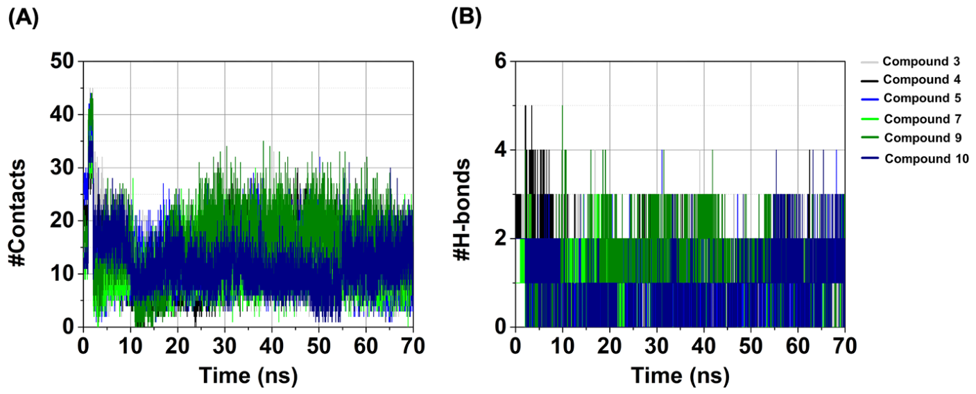
Atomic contacts contribute to the binding affinity of protein–ligand complex [32]. The number of atomic contacts (#Contacts) between the ligands and human tyrosinase was calculated by counting the residues within 3.5 Å of each ligand. As depicted in Figure 5A, the average #Contacts values were 11 ± 4, 12 ± 4, 9 ± 4, 11 ± 4, 16 ± 4, and 13 ± 4 for compounds 3, 4, 5, 7, 9, and 10 in complex with human tyrosinase, respectively. Among these complexes, compound 9–human tyrosinase complex exhibited the highest #Contacts, followed by the complexes with compounds 10 and 4.
H-bonds are essential non-covalent interactions that play a pivotal role in ligand–protein binding [33]. In this study, H-bonds were identified based on a donor–acceptor distance of ≤3.5 Å and a donor–hydrogen–acceptor (HBD–H···HBA) angle of ≥120° [34] during the last 10 ns of the simulation. The results are presented in Figure 5B. Among the investigated complexes, the compound 9–human tyrosinase complex exhibited the highest average number of H-bonds (#H-bonds; 1.6 ± 0.6), followed by the compound 10 (1.4 ± 0.8) and compound 4 (1.0 ± 0.4) complexes. The rolling-average analyses of #Contacts and #H-bonds are provided in the Supporting Information (Figures S7–S10).
The H-bond occupancy analysis (Supplementary Table S1) revealed that compound 4 formed the most persistent H-bond with human tyrosinase (97.85% occupancy at residue S360), followed by compound 9 (86.70% occupancy at residue S375). Compound 10 also established two stable H-bonds with S375 (62.85% occupancy) and A357 (54.65% occupancy). In contrast, no H-bonds were detected for the compound 7–human tyrosinase complex.
Three independent MD replicas of the human tyrosinase complexes with compounds 4, 7, and 9 were performed to validate the reproducibility of the ligand–protein interactions. The #Contacts and #H-bonds analyses revealed highly consistent interaction patterns across the independent trajectories (Figure S11). Among the three compounds, compound 9 exhibited the most favorable interaction profile, characterized by the highest numbers of protein–ligand contacts and H-bonds. These findings are further supported by the key binding residue analyses (Figure 6 and Figure 7) and ΔGbind calculations (Table 2), as discussed in the following sections.

2.4. Key Binding Residues

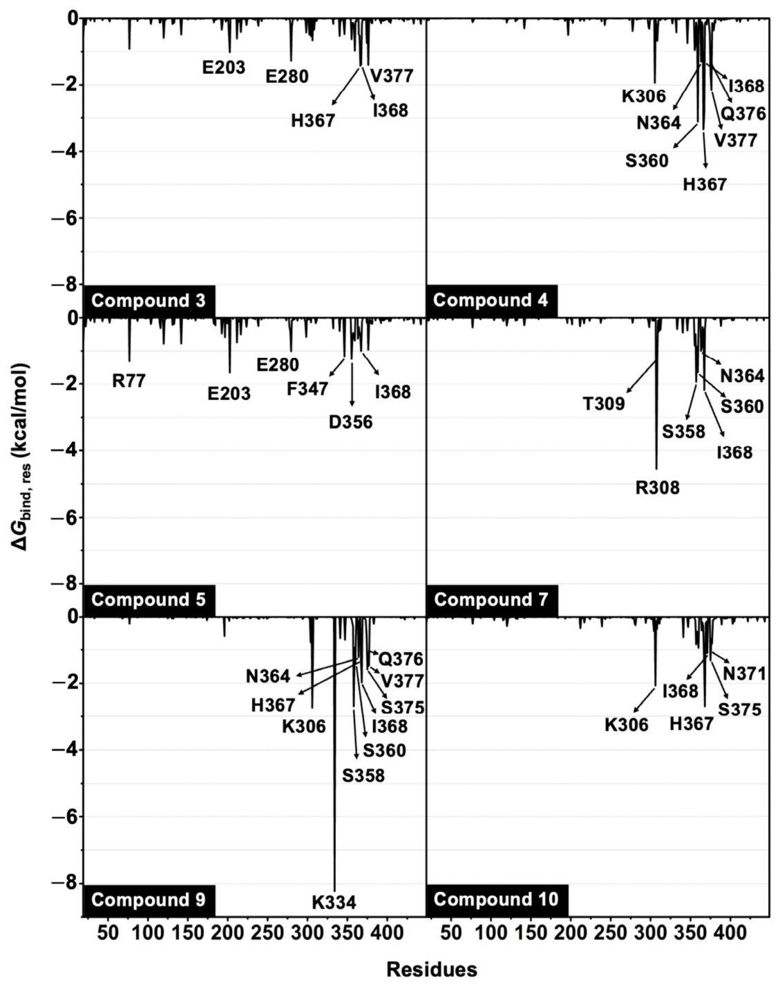
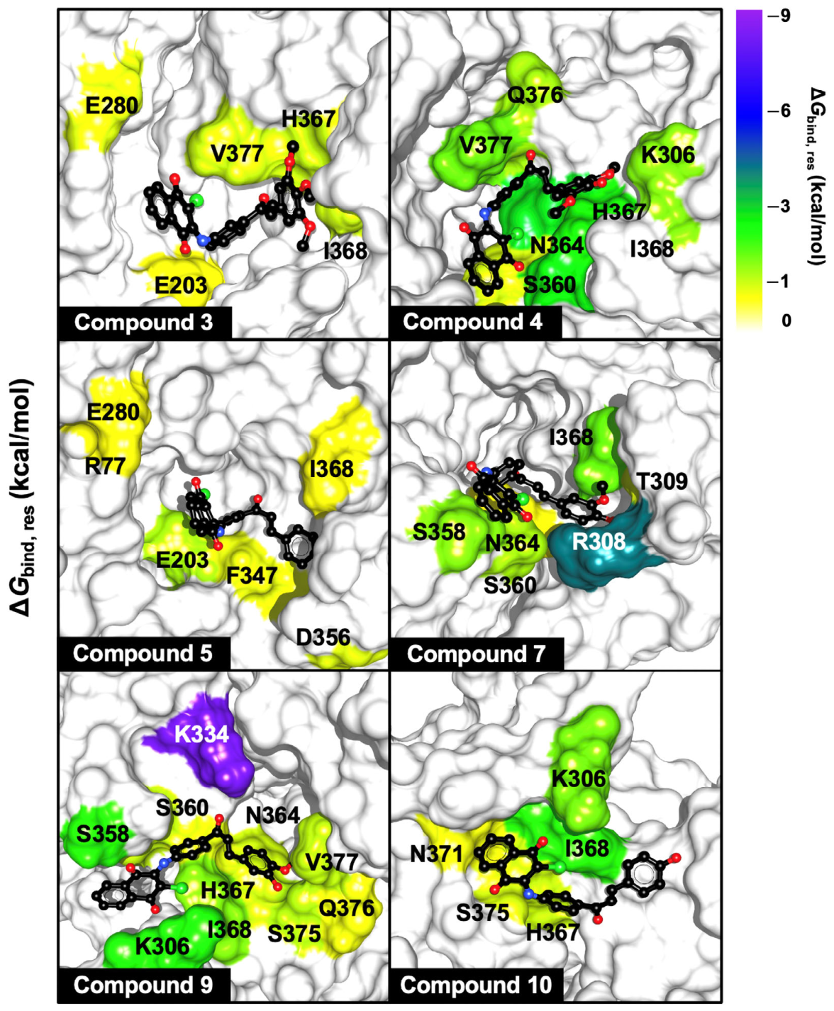
To identify the amino acid residues involved in the binding of the six aminonaphthoquinone–chalcone hybrids to human tyrosinase, per-residue binding free energy decomposition (ΔGbind,res) was performed using the MM/PBSA method on 100 snapshots extracted from the final 10 ns of the MD simulations. The results are presented in Figure 6. Key binding residues with ΔGbind,res values of ≤ −1.0 kcal/mol are shown in Figure 7.
The analysis revealed that all six compounds interacted with residues located within the active site of human tyrosinase. Compound 4 formed favorable interactions with seven residues (K306, S360, N364, H367, I368, Q376, and V377), whereas compound 5 interacted with six residues (R77, E203, E208, F347, D356, and I368), and compound 7 also engaged six residues (R308, T309, S358, S360, N364, and I368). Notably, the compound 9–human tyrosinase complex exhibited the largest network of stabilizing interactions, involving 10 key residues (K306, K334, S358, S360, N364, H367, I368, S375, Q376, and V377). Among these, K334 made the strongest energetic contribution, with a ΔGbind, res value of −8.25 kcal/mol (Figure 6). In addition, compounds 3, 4, 9, and 10 interacted with the catalytic residue H367. Among all investigated compounds, compound 9 displayed the greatest number of hotspot residues, consistent with its most favorable ΔGbind (Table 2).
Overall, these findings demonstrate that all six aminonaphthoquinone–chalcone hybrids are capable of binding within the active site of human tyrosinase. The observed binding mode is consistent with those reported for known human tyrosinase inhibitors, including Thiamidol™ [28], amphotericin B [29], epicatechin gallate, epigallocatechin gallate [35], kojic acid, arbutin [16], coumarin derivative C9, and vanillin derivative V7 [16].

2.5. Predicted Binding Free Energy

To evaluate the binding affinity of the six aminonaphthoquinone–chalcone hybrids toward human tyrosinase, the ΔGbind and its energy components were calculated using the MM/PBSA method [36] based on 100 snapshots extracted from the final 10 ns of the MD simulations. As summarized in Table 2, the molecular mechanics energy (ΔEMM) in the gas phase revealed that vdW interactions (ΔEvdW) were the primary driving force for ligand binding, whereas electrostatic interactions (ΔEele) made a comparatively smaller contribution. This finding agreed well with the previous studies showing that vdW interactions dominate the binding of Thiamidol™, catechin, epicatechin gallate, and epigallocatechin gallate to the catalytic site of human tyrosinase [28,35].
The calculated ΔGbind values of the six aminonaphthoquinone–chalcone hybrids ranged from −20.32 to −38.38 kcal/mol (Table 2). Among the investigated compounds, compound 9 exhibited the most favorable ΔGbind (−38.38 ± 0.67 kcal/mol), followed by compound 4 (−35.00 ± 0.62 kcal/mol), compound 7 (−34.40 ± 0.34 kcal/mol), compound 10 (−29.99 ± 0.39 kcal/mol), compound 3 (−29.55 ± 0.32 kcal/mol), and compound 5 (−20.32 ± 0.42 kcal/mol).
To further validate the reliability of the ΔGbind estimates, MM/PBSA calculations were performed for the human tyrosinase complexes with compounds 4, 7, and 9 using three independent MD simulations. As summarized in Table S2, all three compounds consistently exhibited favorable ΔGbind values across independent simulations, with average ΔGbind values of −33.59, −32.80, and −38.56 kcal/mol for compounds 4, 7, and 9, respectively. Notably, compound 9 demonstrated a higher binding affinity than the other compounds, which agrees well with the structural stability (Figure 4A), compactness (Figure 4B), #Contacts (Figure 5A), #H-bonds (Figure 5B), and key binding residue (Figure 6 and Figure 7) results as mentioned above.

2.6. Drug Likeness

Prediction of drug-likeness is an important step in early-stage drug discovery, as it helps evaluate the likelihood that candidate compounds possess favorable physicochemical properties for further development [37]. In this study, the drug-likeness of the six selected aminonaphthoquinone–chalcone hybrids was evaluated using the SwissADME online platform [38]. Key molecular descriptors, including molecular weight (MW), the number of hydrogen bond donors and acceptors (HBD and HBA, respectively), the number of rotatable bonds (RBs), topological polar surface area (TPSA), and lipophilicity (log P), were assessed. As summarized in Table 3, all six compounds satisfied Lipinski’s rule of five, including (i) MW < 500 Da, (ii) HBD ≤ 5 and HBA ≤ 10, (iii) RB ≤ 10, (iv) TPSA ≤ 140 Ų, and (v) log P ≤ 5 [39,40]. These results suggest that the six aminonaphthoquinone–chalcone hybrids possess favorable drug-like properties and therefore represent promising candidates for further development as human tyrosinase inhibitors.

3. Materials and Methods

3.1. System Preparation and Molecular Docking

The FASTA sequence of human tyrosinase was obtained from the National Center for Biotechnology Information (GenBank: AAB60319.1). A homology model of human tyrosinase was constructed using the SWISS-MODEL server with the crystal structure of human tyrosinase-related protein 1 in complex with kojic acid (PDB ID: 5M8Q) [4] as the template. Following model construction, the two Zn2+ ions in the template were replaced with Cu2+ ions (CuA and CuB), while preserving the experimentally resolved coordination geometry with the six conserved catalytic histidine residues (His180, His202, and His211 coordinating CuA, and His363, His367, and His390 coordinating CuB).
The protonation state of human tyrosinase at physiological pH (7.4) was predicted using the PDB2PQR web server [41]. The three-dimensional (3D) structures of the aminonaphthoquinone–chalcone hybrids were generated according to the previous report [26] using GaussView 6 (Semichem Inc., Shawnee Mission, KS, USA) [42]. Each ligand was subsequently subjected to full geometry optimization at the 6-31G(d) level of theory using Gaussian 09 [43] to obtain its lowest-energy conformation. The protonation state of each ligand was determined at pH 7.4 using Marvinsketch version 22.6.5 (ChemAxon Ltd., Budapest, Hungary). Following established procedures [44,45,46,47,48], electrostatic potential (ESP) charges were then calculated for the optimized ligand structures at the HF/6-31G(d) level of theory using Gaussian 09 [43]. Molecular docking was performed using the CB-Dock2 web server [49], and the resulting binding poses were analyzed with BIOVIA Discovery Studio Visualizer 2024 (v24.1.0.23298; Dassault Systèmes BIOVIA Corp., San Diego, CA, USA) [50].

3.2. MD Simulation

All-atom MD simulation of each protein–ligand complex was performed using the AMBER20 software package (University of California, San Francisco, CA, USA [51]. The bonded and non-bonded parameters for all ligands were assigned using the General Amber Force Field (GAFF) [52], whereas the protein was parameterized with the AMBER ff14SB force field [53]. Missing hydrogen atoms were added using the LEaP module. The catalytic copper ions were treated as non-bonded Cu2+ ions using the copper ion parameters available in the AMBER LEaP ion library. Each system was solvated in a periodic TIP3P water box [54] and Na+ ions were added to neutralize the system. Simulations were carried out under isothermal–isobaric (NPT) conditions at 310 K and 1 atm. The SHAKE algorithm [55] was applied to constrain all bonds involving hydrogen atoms. A residue-based cutoff of 12 Å was used for non-bonded interactions, and long-range electrostatic interactions were treated using the particle mesh Ewald (PME) method [56].
Each system was first subjected to energy minimization using the steepest descent (SD) and conjugate gradient (CG) methods for 1,000 and 2,000 steps, respectively, to remove unfavorable contacts. The systems were then gradually heated to 310 K over 100 ps, followed by 5 ns of restrained MD simulations with progressively reduced positional restraints (100, 50, 20, 10, 5, and 1 kcal/mol·Å²). After that the unrestrained MD simulations were conducted in the NPT ensemble (1 atm and 310 K) until reaching 70 ns. Snapshots collected during the final 10 ns (60–70 ns) of each trajectory were used for subsequent analyses.

3.3. Structural Analysis and Binding Free Energy Calculation of Ligand–Protein Complexes

The CPPTRAJ [57] in the AMBER20 package was used to analyze the structural and dynamic properties of the protein–ligand complexes, including RMSD, Rg, #Contacts, and H-bond formation. Moreover, the MM/PBSA calculations [36] were employed to estimate the binding affinity and identify key stabilizing amino acid residues involved in the ligand binding. The MM/PBSA calculations were performed using an interior dielectric constant of 5.0 without normal-mode analysis, based on 100 snapshots extracted from the final 10 ns of the MD trajectories.

3.4. Drug Likeness Prediction

The drug-likeness of aminonaphthoquinone–chalcone hybrids was evaluated using the SwissADME web server (www.swissadme.ch/) (accessed on 11 October 2025) [38].

4. Conclusions

In this study, we investigated the binding characteristics of 10 aminonaphthoquinone–chalcone hybrids toward human tyrosinase using molecular docking, MD simulations, and MM/PBSA ΔGbind calculations. Molecular docking identified compounds 3, 4, 5, 7, 9, and 10 as the most promising candidates, exhibiting higher predicted binding affinity than the remaining hybrids. Subsequent MD simulations demonstrated that, among the investigated complexes, the compound 9–human tyrosinase complex exhibited the highest numbers of protein–ligand contacts and H-bonds, suggesting the most stable interaction profile. MM/PBSA calculations further revealed that vdW interactions were the dominant driving force for ligand binding. Notably, compound 9 displayed the most favorable ΔGbind (−38.38 ± 0.67 kcal/mol) and interacted with the greatest number of active-site residues (K306, K334, S358, S360, N364, H367, I368, S375, Q376, and V377). The reproducibility of these findings was confirmed by three independent MD simulations, which consistently supported the structural stability and favorable binding of compounds 4, 7, and particularly 9. In addition, all six selected compounds satisfied Lipinski’s rule of five, indicating favorable drug-like properties. Collectively, these findings identify compound 9 as the most promising aminonaphthoquinone–chalcone hybrid for further experimental evaluation as a potential human tyrosinase inhibitor. Nevertheless, experimental studies are required to validate its inhibitory activity and therapeutic potential.

Supplementary Materials

The following supporting information can be downloaded at: https://www.mdpi.com/article/10.3390/ijms27167328/s1.

Author Contributions

P.M.: Conceptualization. N.N., S.S. and P.M.: Investigation, Formal Analysis, Methodology. N.N., S.S., P.M. and R.P.: Data Curation, Validation, Visualization. P.M., N.N. and S.S.: Writing—Original Draft, Writing—Review and Editing. P.M.: Funding Acquisition, Project Administration. P.M.: Supervision. P.M., R.P. and T.R.: Resources. T.R.: Software. All authors have read and agreed to the published version of the manuscript.

Funding

This project is funded by National Research Council of Thailand (NRCT) (grant number N42A680148).

Institutional Review Board Statement

Not applicable, as this study did not involve human participants or animal experiments.

Informed Consent Statement

Not applicable.

Data Availability Statement

The data presented in this study are available within the article and Supplementary Materials.

Acknowledgments

Generative AI (ChatGPT-3.5) was employed for language refinement and verification in this manuscript. The authors entirely generated all contents, ideas, and findings without AI assistance. Following language editing, the authors thoroughly reviewed and validated the final version to ensure its accuracy and integrity.

Conflicts of Interest

The authors declare no conflicts of interest.

References

  1. Raposo, G.; Marks, M.S. Melanosomes-dark organelles enlighten endosomal membrane transport. Nat. Rev. Mol. Cell Biol. 2007, 8, 786–797. [Google Scholar] [CrossRef] [Scilit] [PubMed]
  2. Kumari, S.; Tien Guan Thng, S.; Kumar Verma, N.; Gautam, H.K. Melanogenesis Inhibitors. Acta Derm. Venereol. 2018, 98, 924–931. [Google Scholar] [CrossRef] [Scilit] [PubMed]
  3. Li, C.; Kuai, L.; Cui, R.; Miao, X. Melanogenesis and the Targeted Therapy of Melanoma. Biomolecules 2022, 12, 1874. [Google Scholar] [CrossRef] [Scilit] [PubMed]
  4. Lai, X.; Wichers, H.J.; Soler-Lopez, M.; Dijkstra, B.W. Structure of Human Tyrosinase Related Protein 1 Reveals a Binuclear Zinc Active Site Important for Melanogenesis. Angew. Chem. Int. Ed. 2017, 56, 9812–9815. [Google Scholar] [CrossRef] [Scilit] [PubMed]
  5. Kipouros, I.; Stanczak, A.; Ginsbach, J.W.; Andrikopoulos, P.C.; Rulisek, L.; Solomon, E.I. Elucidation of the tyrosinase/O2/monophenol ternary intermediate that dictates the monooxygenation mechanism in melanin biosynthesis. Proc. Natl. Acad. Sci. USA 2022, 119, e2205619119. [Google Scholar] [CrossRef] [Scilit] [PubMed]
  6. Lai, X.; Wichers, H.J.; Soler-Lopez, M.; Dijkstra, B.W. Structure and Function of Human Tyrosinase and Tyrosinase-Related Proteins. Chemistry 2018, 24, 47–55. [Google Scholar] [CrossRef] [Scilit] [PubMed]
  7. Solano, F. On the Metal Cofactor in the Tyrosinase Family. Int. J. Mol. Sci. 2018, 19, 633. [Google Scholar] [CrossRef] [Scilit] [PubMed]
  8. Wagatsuma, T.; Suzuki, E.; Shiotsu, M.; Sogo, A.; Nishito, Y.; Ando, H.; Hashimoto, H.; Petris, M.J.; Kinoshita, M.; Kambe, T. Pigmentation and TYRP1 expression are mediated by zinc through the early secretory pathway-resident ZNT proteins. Commun. Biol. 2023, 6, 403. [Google Scholar] [CrossRef] [Scilit] [PubMed]
  9. Cestari, T.F.; Dantas, L.P.; Boza, J.C. Acquired hyperpigmentations. An. Bras. Dermatol. 2014, 89, 11–25. [Google Scholar] [CrossRef] [Scilit] [PubMed]
  10. Zolghadri, S.; Bahrami, A.; Hassan Khan, M.T.; Munoz-Munoz, J.; Garcia-Molina, F.; Garcia-Canovas, F.; Saboury, A.A. A comprehensive review on tyrosinase inhibitors. J. Enzym. Inhib. Med. Chem. 2019, 34, 279–309. [Google Scholar] [CrossRef] [Scilit] [PubMed]
  11. Hu, Z.M.; Zhou, Q.; Lei, T.C.; Ding, S.F.; Xu, S.Z. Effects of hydroquinone and its glucoside derivatives on melanogenesis and antioxidation: Biosafety as skin whitening agents. J. Dermatol. Sci. 2009, 55, 179–184. [Google Scholar] [CrossRef] [Scilit] [PubMed]
  12. Numata, T.; Tobita, R.; Tsuboi, R.; Okubo, Y. Contact dermatitis caused by arbutin contained in skin-whitening cosmetics. Contact Dermat. 2016, 75, 187–188. [Google Scholar] [CrossRef] [Scilit] [PubMed]
  13. Pillaiyar, T.; Manickam, M.; Namasivayam, V. Skin whitening agents: Medicinal chemistry perspective of tyrosinase inhibitors. J. Enzym. Inhib. Med. Chem. 2017, 32, 403–425. [Google Scholar] [CrossRef] [Scilit] [PubMed]
  14. Chang, T.S. An updated review of tyrosinase inhibitors. Int. J. Mol. Sci. 2009, 10, 2440–2475. [Google Scholar] [CrossRef] [Scilit] [PubMed]
  15. Mann, T.; Gerwat, W.; Batzer, J.; Eggers, K.; Scherner, C.; Wenck, H.; Stab, F.; Hearing, V.J.; Rohm, K.H.; Kolbe, L. Inhibition of Human Tyrosinase Requires Molecular Motifs Distinctively Different from Mushroom Tyrosinase. J. Invest. Dermatol. 2018, 138, 1601–1608. [Google Scholar] [CrossRef] [Scilit] [PubMed]
  16. Hassan, M.; Ashraf, Z.; Abbas, Q.; Raza, H.; Seo, S.Y. Exploration of Novel Human Tyrosinase Inhibitors by Molecular Modeling, Docking and Simulation Studies. Interdiscip. Sci. 2018, 10, 68–80. [Google Scholar] [CrossRef] [Scilit] [PubMed]
  17. Jhan, J.K.; Chung, Y.C.; Chen, G.H.; Chang, C.H.; Lu, Y.C.; Hsu, C.K. Anthocyanin contents in the seed coat of black soya bean and their anti-human tyrosinase activity and antioxidative activity. Int. J. Cosmet. Sci. 2016, 38, 319–324. [Google Scholar] [CrossRef] [Scilit] [PubMed]
  18. Oh, T.I.; Yun, J.M.; Park, E.J.; Kim, Y.S.; Lee, Y.M.; Lim, J.H. Plumbagin Suppresses alpha-MSH-Induced Melanogenesis in B16F10 Mouse Melanoma Cells by Inhibiting Tyrosinase Activity. Int. J. Mol. Sci. 2017, 18, 320. [Google Scholar] [CrossRef] [Scilit] [PubMed]
  19. Lee, S.M.; Chen, Y.S.; Lin, C.C.; Chen, K.H. Hair dyes resorcinol and lawsone reduce production of melanin in melanoma cells by tyrosinase activity inhibition and decreasing tyrosinase and microphthalmia-associated transcription factor (MITF) expression. Int. J. Mol. Sci. 2015, 16, 1495–1508. [Google Scholar] [CrossRef] [Scilit] [PubMed]
  20. Kim, J.H.; Lee, S.M.; Myung, C.H.; Lee, K.R.; Hyun, S.M.; Lee, J.E.; Park, Y.S.; Jeon, S.R.; Park, J.I.; Chang, S.E.; et al. Melanogenesis inhibition of β-lapachone, a natural product from Tabebuia avellanedae, with effective in vivo lightening potency. Arch. Dermatol. Res. 2015, 307, 229–238. [Google Scholar] [CrossRef] [Scilit] [PubMed]
  21. Ranjbar, S.; Kamarei, M.M.; Khoshneviszadeh, M.; Hosseinpoor, H.; Edraki, N.; Khoshneviszadeh, M. Benzylidene-6-hydroxy-3,4-dihydronaphthalenone chalconoids as potent tyrosinase inhibitors. Res. Pharm. Sci. 2021, 16, 425–435. [Google Scholar] [CrossRef] [Scilit] [PubMed]
  22. Singh, L.R.; Chen, Y.L.; Xie, Y.Y.; Xia, W.; Gong, X.W.; Hider, R.C.; Zhou, T. Functionality study of chalcone-hydroxypyridinone hybrids as tyrosinase inhibitors and influence on anti-tyrosinase activity. J. Enzym. Inhib. Med. Chem. 2020, 35, 1562–1567. [Google Scholar] [CrossRef] [Scilit] [PubMed]
  23. Pan, W.; Giovanardi, I.; Sagynova, T.; Cariola, A.; Bresciani, V.; Masetti, M.; Valgimigli, L. Potent Antioxidant and Anti-Tyrosinase Activity of Butein and Homobutein Probed by Molecular Kinetic and Mechanistic Studies. Antioxidants 2023, 12, 1763. [Google Scholar] [CrossRef] [Scilit] [PubMed]
  24. Chen, W.C.; Tseng, T.S.; Hsiao, N.W.; Lin, Y.L.; Wen, Z.H.; Tsai, C.C.; Lee, Y.C.; Lin, H.H.; Tsai, K.C. Discovery of highly potent tyrosinase inhibitor, T1, with significant anti-melanogenesis ability by zebrafish in vivo assay and computational molecular modeling. Sci. Rep. 2015, 5, 7995. [Google Scholar] [CrossRef] [Scilit] [PubMed]
  25. Hwang, Y.; Lee, J.; Jung, H.J.; Ullah, S.; Ko, J.; Jeong, Y.; Park, Y.J.; Kang, M.K.; Yun, H.; Kim, M.S.; et al. A Novel Class of Potent Anti-Tyrosinase Compounds with Antioxidant Activity, 2-(Substituted phenyl)-5-(trifluoromethyl)benzo[d]thiazoles: In Vitro and In Silico Insights. Antioxidants 2022, 11, 1375. [Google Scholar] [CrossRef] [Scilit] [PubMed]
  26. Leechaisit, R.; Mahalapbutr, P.; Boonsri, P.; Karnchanapandh, K.; Rungrotmongkol, T.; Prachayasittikul, V.; Prachayasittikul, S.; Ruchirawat, S.; Prachayasittikul, V.; Pingaew, R. Discovery of Novel Naphthoquinone–Chalcone Hybrids as Potent FGFR1 Tyrosine Kinase Inhibitors: Synthesis, Biological Evaluation, and Molecular Modeling. ACS Omega 2023, 8, 32593–32605. [Google Scholar] [CrossRef] [Scilit] [PubMed]
  27. Yoshimori, A.; Oyama, T.; Takahashi, S.; Abe, H.; Kamiya, T.; Abe, T.; Tanuma, S. Structure-activity relationships of the thujaplicins for inhibition of human tyrosinase. Bioorg. Med. Chem. 2014, 22, 6193–6200. [Google Scholar] [CrossRef] [Scilit] [PubMed]
  28. Mahalapbutr, P.; Nuramrum, N.; Rungrotmongkol, T.; Kongtaworn, N.; Sabuakham, S. Structural dynamics and susceptibility of isobutylamido thiazolyl resorcinol (Thiamidol™) against human and mushroom tyrosinases. J. Biomol. Struct. Dyn. 2023, 41, 11810–11817. [Google Scholar] [CrossRef] [Scilit] [PubMed]
  29. Mahalapbutr, P.; Sabuakham, S.; Nasoontorn, S.; Rungrotmongkol, T.; Silsirivanit, A.; Suriya, U. Discovery of amphotericin B, an antifungal drug as tyrosinase inhibitor with potent anti-melanogenic activity. Int. J. Biol. Macromol. 2023, 246, 125587. [Google Scholar] [CrossRef] [Scilit] [PubMed]
  30. Sargsyan, K.; Grauffel, C.; Lim, C. How Molecular Size Impacts RMSD Applications in Molecular Dynamics Simulations. J. Chem. Theory Comput. 2017, 13, 1518–1524. [Google Scholar] [CrossRef] [Scilit] [PubMed]
  31. Ghahremanian, S.; Rashidi, M.M.; Raeisi, K.; Toghraie, D. Molecular dynamics simulation approach for discovering potential inhibitors against SARS-CoV-2: A structural review. J. Mol. Liq. 2022, 354, 118901. [Google Scholar] [CrossRef] [Scilit] [PubMed]
  32. Wang, D.D.; Ou-Yang, L.; Xie, H.; Zhu, M.; Yan, H. Predicting the impacts of mutations on protein-ligand binding affinity based on molecular dynamics simulations and machine learning methods. Comput. Struct. Biotechnol. J. 2020, 18, 439–454. [Google Scholar] [CrossRef] [Scilit] [PubMed]
  33. Madushanka, A.; Moura, R.T., Jr.; Verma, N.; Kraka, E. Quantum Mechanical Assessment of Protein-Ligand Hydrogen Bond Strength Patterns: Insights from Semiempirical Tight-Binding and Local Vibrational Mode Theory. Int. J. Mol. Sci. 2023, 24, 6311. [Google Scholar] [CrossRef] [Scilit] [PubMed]
  34. Tan, K.P.; Singh, K.; Hazra, A.; Madhusudhan, M.S. Peptide bond planarity constrains hydrogen bond geometry and influences secondary structure conformations. Curr. Res. Struct. Biol. 2021, 3, 1–8. [Google Scholar] [CrossRef] [Scilit] [PubMed]
  35. Tang, H.; Cui, F.; Li, H.; Huang, Q.; Li, Y. Understanding the inhibitory mechanism of tea polyphenols against tyrosinase using fluorescence spectroscopy, cyclic voltammetry, oximetry, and molecular simulations. RSC Adv. 2018, 8, 8310–8318. [Google Scholar] [CrossRef] [Scilit] [PubMed]
  36. Miller, B.R., III; McGee, T.D., Jr.; Swails, J.M.; Homeyer, N.; Gohlke, H.; Roitberg, A.E. MMPBSA.py: An Efficient Program for End-State Free Energy Calculations. J. Chem. Theory Comput. 2012, 8, 3314–3321. [Google Scholar] [CrossRef] [Scilit] [PubMed]
  37. Hu, Q.; Feng, M.; Lai, L.; Pei, J. Prediction of Drug-Likeness Using Deep Autoencoder Neural Networks. Front. Genet. 2018, 9, 585. [Google Scholar] [CrossRef] [Scilit] [PubMed]
  38. Daina, A.; Michielin, O.; Zoete, V. SwissADME: A free web tool to evaluate pharmacokinetics, drug-likeness and medicinal chemistry friendliness of small molecules. Sci. Rep. 2017, 7, 42717. [Google Scholar] [CrossRef] [Scilit] [PubMed]
  39. Lipinski, C.A.; Lombardo, F.; Dominy, B.W.; Feeney, P.J. Experimental and computational approaches to estimate solubility and permeability in drug discovery and development settings. Adv. Drug Deliv. Rev. 1997, 23, 3–25, Reprint in Adv. Drug Deliv. Rev. 2001, 46, 3–26. https://doi.org/10.1016/S0169-409X(00)00129-0. [Google Scholar] [CrossRef] [Scilit]
  40. Benet, L.Z.; Hosey, C.M.; Ursu, O.; Oprea, T.I. BDDCS, the Rule of 5 and drugability. Adv. Drug Deliv. Rev. 2016, 101, 89–98. [Google Scholar] [CrossRef] [Scilit] [PubMed]
  41. Jurrus, E.; Engel, D.; Star, K.; Monson, K.; Brandi, J.; Felberg, L.E.; Brookes, D.H.; Wilson, L.; Chen, J.; Liles, K.; et al. Improvements to the APBS biomolecular solvation software suite. Protein Sci. 2018, 27, 112–128. [Google Scholar] [CrossRef] [Scilit] [PubMed]
  42. Dennington, R.; Keith, T.A.; Millam, J.M. GaussView 6 User’s Manual; Semichem Inc.: Shawnee Mission, KS, USA, 2016. [Google Scholar]
  43. Frisch, M.J.; Trucks, G.; Schlegel, H.B.; Scuseria, G.E.; Robb, M.A.; Cheeseman, J.; Scalmani, G.; Barone, V.; Mennucci, B.; Petersson, G.A.; et al. Gaussian 09, revision A.1; Gaussian Inc.: Wallingford, CT, USA, 2009. [Google Scholar]
  44. Mahalapbutr, P.; Charoenwongpaiboon, T.; Phongern, C.; Kongtaworn, N.; Hannongbua, S.; Rungrotmongkol, T. Molecular encapsulation of a key odor-active 2-acetyl-1-pyrroline in aromatic rice with β-cyclodextrin derivatives. J. Mol. Liq. 2021, 337, 116394. [Google Scholar] [CrossRef] [Scilit]
  45. Mahalapbutr, P.; Nutho, B.; Wolschann, P.; Chavasiri, W.; Kungwan, N.; Rungrotmongkol, T. Molecular insights into inclusion complexes of mansonone E and H enantiomers with various β-cyclodextrins. J. Mol. Graph. Model. 2018, 79, 72–80. [Google Scholar] [CrossRef] [Scilit] [PubMed]
  46. Mahalapbutr, P.; Leechaisit, R.; Thongnum, A.; Todsaporn, D.; Prachayasittikul, V.; Rungrotmongkol, T.; Prachayasittikul, S.; Ruchirawat, S.; Prachayasittikul, V.; Pingaew, R. Discovery of Anilino-1,4-naphthoquinones as Potent EGFR Tyrosine Kinase Inhibitors: Synthesis, Biological Evaluation, and Comprehensive Molecular Modeling. ACS Omega 2022, 7, 17881–17893. [Google Scholar] [CrossRef] [Scilit] [PubMed]
  47. Sanachai, K.; Mahalapbutr, P.; Sanghiran Lee, V.; Rungrotmongkol, T.; Hannongbua, S. In Silico Elucidation of Potent Inhibitors and Rational Drug Design against SARS-CoV-2 Papain-like Protease. J. Phys. Chem. B 2021, 125, 13644–13656. [Google Scholar] [CrossRef] [Scilit] [PubMed]
  48. Oo, A.; Kerdpol, K.; Mahalapbutr, P.; Rungrotmongkol, T. Molecular encapsulation of emodin with various β-cyclodextrin derivatives: A computational study. J. Mol. Liq. 2022, 347, 118002. [Google Scholar] [CrossRef] [Scilit]
  49. Liu, Y.; Yang, X.; Gan, J.; Chen, S.; Xiao, Z.-X.; Cao, Y. CB-Dock2: Improved protein–ligand blind docking by integrating cavity detection, docking and homologous template fitting. Nucleic Acids Res. 2022, 50, W159–W164. [Google Scholar] [CrossRef] [Scilit] [PubMed]
  50. Baroroh, U.; Biotek, M.; Muscifa, Z.S.; Destiarani, W.; Rohmatullah, F.G.; Yusuf, M. Molecular interaction analysis and visualization of protein-ligand docking using Biovia Discovery Studio Visualizer. Indones. J. Comput. Biol. 2023, 2, 22–30. [Google Scholar] [CrossRef] [Scilit]
  51. Salomon-Ferrer, R.; Case, D.A.; Walker, R.C. An overview of the Amber biomolecular simulation package. WIREs Comput. Mol. Sci. 2012, 3, 198–210. [Google Scholar] [CrossRef] [Scilit]
  52. Wang, J.; Wolf, R.M.; Caldwell, J.W.; Kollman, P.A.; Case, D.A. Development and testing of a general amber force field. J. Comput. Chem. 2004, 25, 1157–1174. [Google Scholar] [CrossRef] [Scilit] [PubMed]
  53. Maier, J.A.; Martinez, C.; Kasavajhala, K.; Wickstrom, L.; Hauser, K.E.; Simmerling, C. ff14SB: Improving the Accuracy of Protein Side Chain and Backbone Parameters from ff99SB. J. Chem. Theory Comput. 2015, 11, 3696–3713. [Google Scholar] [CrossRef] [Scilit] [PubMed]
  54. Jorgensen, W.L.; Chandrasekhar, J.; Madura, J.D.; Impey, R.W.; Klein, M.L. Comparison of simple potential functions for simulating liquid water. J. Chem. Phys. 1983, 79, 926–935. [Google Scholar] [CrossRef] [Scilit]
  55. Ryckaert, J.-P.; Ciccotti, G.; Berendsen, H.J.C. Numerical integration of the cartesian equations of motion of a system with constraints: Molecular dynamics of n-alkanes. J. Comput. Phys. 1977, 23, 327–341. [Google Scholar] [CrossRef] [Scilit]
  56. Darden, T.; York, D.; Pedersen, L. Particle mesh Ewald: An N⋅log(N) method for Ewald sums in large systems. J. Chem. Phys. 1993, 98, 10089–10092. [Google Scholar] [CrossRef] [Scilit]
  57. Roe, D.R.; Cheatham, T.E., III. PTRAJ and CPPTRAJ: Software for Processing and Analysis of Molecular Dynamics Trajectory Data. J. Chem. Theory Comput. 2013, 9, 3084–3095. [Google Scholar] [CrossRef] [Scilit] [PubMed]
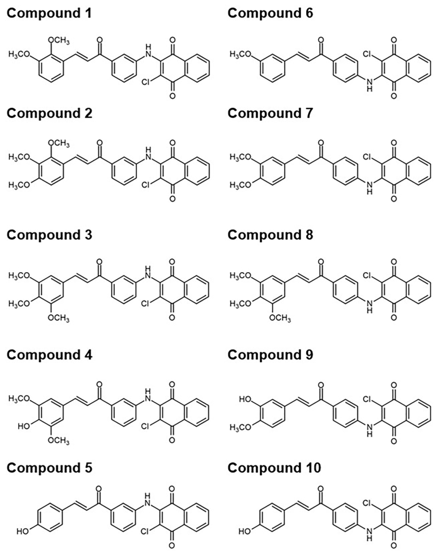
Figure 2. Chemical structures of the 10 aminonaphthoquinone–chalcone hybrids reported in a previous study [26].
Ijms 27 07328 g002
Figure 3. 2D interaction profile of six aminonaphthoquinone–chalcone hybrids in complex with human tyrosinase predicted by molecular docking.
Ijms 27 07328 g003
Figure 4. Time evolution of (A) RMSD and (B) Rg for six aminonaphthoquinone–chalcone hybrid/human tyrosinase complexes obtained from MD simulations.
Ijms 27 07328 g004
Figure 5. Time evolution of (A) #Contacts and (B) #H-bonds for six aminonaphthoquinone–chalcone hybrids in complex with human tyrosinase obtained from MD simulations.
Ijms 27 07328 g005
Figure 6. MM/PBSA-based ΔGbind,res of six aminononaphthoquinone–chalcone hybrids in complex with human tyrosinase.
Ijms 27 07328 g006
Figure 7. Binding pattern of six aminononaphthoquinone–chalcone hybrids within the active site of human tyrosinase captured from the final MD snapshot. The catalytic coppers are hidden. The key binding residues are colored according to their ΔGbind,res values with the color gradient ranging from white (highest ΔGbind,res) to purple (lowest ΔGbind,res).
Ijms 27 07328 g007
Table 1. CB-Dock2 binding energies of complexes between the 10 aminonaphthoquinone–chalcone hybrids and human tyrosinase.
CompoundCB-Dock2 Binding Energy (kcal/mol)
1−8.1
2−8.1
3−8.4
4−9.2
5−8.4
6−8.3
7−8.4
8−7.5
9−8.4
10−8.5
Table 2. ΔGbind and their energy components calculated using the MM/PBSA method for six aminonaphthoquinone–chalcone hybrids in complex with human tyrosinase. Data are shown as the mean ± standard error of the mean (SEM) from 100 snapshots extracted from the last 10 ns of the simulation.
Energy ComponentCompound 3Compound 4Compound 5Compound 7Compound 9Compound 10
ΔEvdW−39.11 ± 0.37−36.10 ± 0.62−33.58 ± 0.48−35.92 ± 0.35−36.57 ± 0.69−32.12 ± 0.44
ΔEele0.99 ± 0.15−4.74 ± 0.221.38 ± 0.200.21 ± 0.15−4.19 ± 0.181.26 ± 0.20
ΔEMM−38.12 ± 0.43−40.84 ± 0.67−32.20 ± 0.48−35.70 ± 0.38−40.76 ± 0.65−30.86 ± 0.44
ΔGsolv,nonpolar−6.51 ± 0.05−5.94 ± 0.05−5.55 ± 0.07−5.61 ± 0.04−6.48 ± 0.06−5.56 ± 0.05
ΔGsolv,polar15.07 ± 0.3011.78 ± 0.2417.43 ± 0.366.92 ± 0.198.86 ± 0.136.43 ± 0.24
ΔGsolv8.57 ± 0.275.85 ± 0.2211.88 ± 0.331.31 ± 0.152.38 ± 0.140.87 ± 0.22
ΔGbind−29.55 ± 0.32−35.00 ± 0.62−20.32 ± 0.42−34.40 ± 0.34−38.38 ± 0.67−29.99 ± 0.39
Table 3. Predicted drug-likeness of compounds 3, 4, 5, 7, 9, and 10 according to Lipinski’s rule of five.
CompoundLipinski’s Rule of Five
MW
(≤500 Da)
HBD (≤5)HBA
(≤10)
RB
(≤10)
TPSA
(≤140 Å2)
Log P
(≤5)
Drug–Likeness
3503.9316890.931.65Yes
4489.90267101.931.46Yes
5429.8524583.472.13Yes
7473.9015781.701.99Yes
9459.8825692.701.79Yes
10429.8524583.472.13Yes
MW, molecular weight; HBD, number of hydrogen bond donors; HBA, number of hydrogen bond acceptors; RB, number of rotatable bonds; TPSA, topological polar surface area; log P, lipophilicity.
Disclaimer/Publisher’s Note: The statements, opinions and data contained in all publications are solely those of the individual author(s) and contributor(s) and not of MDPI and/or the editor(s). MDPI and/or the editor(s) disclaim responsibility for any injury to people or property resulting from any ideas, methods, instructions or products referred to in the content.