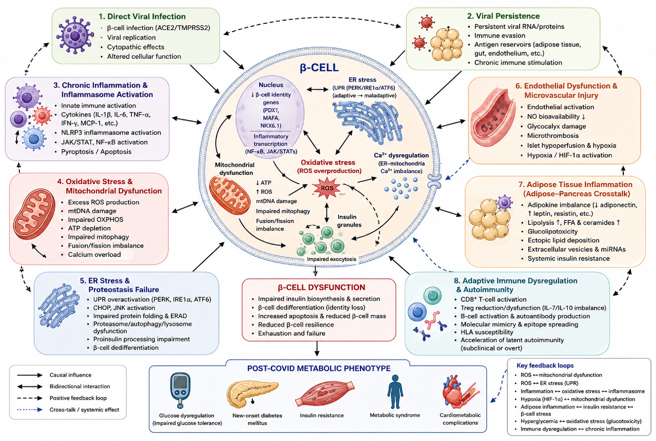
β-Cell Dysfunction in COVID-19 and Post-COVID Syndrome: Molecular Mechanisms Linking Inflammation, Oxidative Stress, and Insulin Secretion


Highlights
- Persistent inflammation, oxidative stress, mitochondrial dysfunction, and hypox-ia-related signalling contribute to impaired insulin secretion and pancreatic β-cell dysfunction in post-COVID metabolic disturbances.
- Post-COVID metabolic dysfunction represents a heterogeneous clinical phenotype characterized by insulin resistance, dysglycaemia, and increased risk of new-onset diabetes mellitus.
- Early identification of metabolic abnormalities and β-cell dysfunction may im-prove risk stratification and support timely preventive interventions after SARS-CoV-2 infection.
- Understanding the molecular mechanisms linking cellular stress pathways to im-paired insulin secretion may facilitate development of targeted therapeutic strat-egies aimed at preserving β-cell function and metabolic health.
Abstract
1. Introduction
2. Literature Search Strategy and Review Methodology
3. Physiological Basis of β-Cell Function
3.1. Glucose Sensing and Stimulus–Secretion Coupling
3.2. Mitochondrial Homeostasis in β-Cell Function
3.3. Calcium Signalling and Exocytotic Machinery in Pancreatic β-Cells
3.4. Endoplasmic Reticulum Homeostasis and the Unfolded Protein Response in Pancreatic β-Cells
3.5. β-Cell Plasticity, Functional Heterogeneity, and Adaptive Responses
3.6. Integration of Physiological Mechanisms Underlying β-Cell Function
4. Mechanisms of β-Cell Dysfunction in COVID-19 and Post-COVID Conditions
4.1. Direct Viral Infection of Pancreatic β-Cells: Receptor Expression and Mechanisms of Viral Entry
4.2. Viral Persistence and Chronic Cellular Stress
4.3. Chronic Inflammation, Cytokine Signalling, and Inflammasome Activation in β-Cell Dysfunction
4.4. Oxidative Stress, Mitochondrial Dysfunction, and Redox Imbalance in SARS-CoV-2-Induced β-Cell Injury
4.5. SARS-CoV-2-Induced Endoplasmic Reticulum Stress and Proteostasis Failure
4.6. Endothelial Dysfunction and Microvascular Injury in SARS-CoV-2-Induced β-Cell Dysfunction
4.7. Adipose Tissue–Pancreas Crosstalk in Post-COVID Metabolic Dysfunction
4.8. Adaptive Immune Dysregulation, Autoimmunity, and β-Cell Injury
4.9. Integrated Mechanistic Model of SARS-CoV-2-Induced β-Cell Dysfunction

5. Clinical Evidence and Phenotypic Spectrum of Post-COVID Metabolic Dysfunction
5.1. Defining the Post-COVID Metabolic Phenotype
5.2. Clinical Cohort Studies Supporting the Post-COVID Metabolic Phenotype
5.3. New-Onset Diabetes After COVID-19: Clinical Heterogeneity and Pathophysiological Interpretation
5.4. Interaction Between Oxidative Stress and Insulin Secretion
5.5. Risk Modifiers and Individual Susceptibility
5.6. Clinical Implications for Risk-Adapted Screening and Long-Term Follow-Up
6. Biomarkers for Early Detection and Risk Stratification of Post-COVID Metabolic Dysfunction
6.1. Biomarkers of β-Cell Function, Stress, and Injury
6.2. Inflammatory and Immunological Biomarkers
6.3. Biomarkers of Oxidative Stress and Mitochondrial Dysfunction
6.4. Endothelial and Vascular Biomarkers
6.5. Emerging Multi-Omics Biomarkers
6.6. Integrated Biomarker Panels and AI-Based Risk Prediction
7. Therapeutic Perspectives: From Conventional Management to Precision Medicine
7.1. Current Therapeutic Approaches
7.2. Emerging Mechanism-Based Therapeutic Strategies
7.3. Precision Medicine and Future Therapeutic Directions
8. Conclusions and Future Perspectives
Author Contributions
Funding
Data Availability Statement
Acknowledgments
Conflicts of Interest
Abbreviations
| ACE2 | Angiotensin-Converting Enzyme 2 |
| ATP | Adenosine Triphosphate |
| COVID-19 | Coronavirus Disease 2019 |
| ER | Endoplasmic Reticulum |
| GIP | Glucose-Dependent Insulinotropic Polypeptide |
| GLP-1 | Glucagon-Like Peptide-1 |
| GLUT | Glucose Transporter |
| HbA1c | Glycated Hemoglobin |
| HIF-1α | Hypoxia-Inducible Factor-1 Alpha |
| HOMA-IR | Homeostatic Model Assessment of Insulin Resistance |
| IFN-γ | Interferon Gamma |
| IL-6 | Interleukin-6 |
| KATP | ATP-Sensitive Potassium Channel |
| NRP1 | Neuropilin-1 |
| OGTT | Oral Glucose Tolerance Test |
| ROS | Reactive Oxygen Species |
| SARS-CoV-2 | Severe Acute Respiratory Syndrome Coronavirus 2 |
| SNARE | Soluble N-Ethylmaleimide-Sensitive Factor Attachment Protein Receptor |
| SUR1 | Sulfonylurea Receptor 1 |
| TMPRSS2 | Transmembrane Serine Protease 2 |
| TNF-α | Tumor Necrosis Factor Alpha |
| TyG | Triglyceride–Glucose Index |
| UPR | Unfolded Protein Response |
| VAMP2 | Vesicle-Associated Membrane Protein 2 |
References
- Gupta, A.; Madhavan, M.V.; Sehgal, K.; Nair, N.; Mahajan, S.; Sehrawat, T.S.; Bikdeli, B.; Ahluwalia, N.; Ausiello, J.C.; Wan, E.Y.; et al. Extrapulmonary manifestations of COVID-19. Nat. Med. 2020, 26, 1017–1032. [Google Scholar] [CrossRef] [PubMed]
- Nalbandian, A.; Sehgal, K.; Gupta, A.; Madhavan, M.V.; McGroder, C.; Stevens, J.S.; Cook, J.R.; Nordvig, A.S.; Shalev, D.; Sehrawat, T.S.; et al. Post-acute COVID-19 syndrome. Nat. Med. 2021, 27, 601–615. [Google Scholar] [CrossRef] [PubMed]
- Bornstein, S.R.; Rubino, F.; Khunti, K.; Mingrone, G.; Hopkins, D.; Birkenfeld, A.L.; Boehm, B.; Amiel, S.; Holt, R.I.G.; Skyler, J.S.; et al. Practical recommendations for the management of diabetes in patients with COVID-19. Lancet Diabetes Endocrinol. 2020, 8, 546–550. [Google Scholar] [CrossRef] [PubMed]
- Soriano, J.B.; Murthy, S.; Marshall, J.C.; Relan, P.; Diaz, J.V.; WHO Clinical Case Definition Working Group on Post-COVID-19 Condition. A Clinical Case Definition of Post-COVID-19 Condition by a Delphi Consensus. Lancet Infect. Dis. 2022, 22, e102–e107. [Google Scholar] [CrossRef] [PubMed]
- Davis, H.E.; McCorkell, L.; Vogel, J.M.; Topol, E.J. Long COVID: Major Findings, Mechanisms and Recommendations. Nat. Rev. Microbiol. 2023, 21, 133–146, Erratum in Nat. Rev. Microbiol. 2023, 21, 408. [Google Scholar] [CrossRef] [PubMed]
- Stefan, N.; Birkenfeld, A.L.; Schulze, M.B. Global pandemics interconnected—Obesity, impaired metabolic health and COVID-19. Nat. Rev. Endocrinol. 2021, 17, 135–149. [Google Scholar] [CrossRef] [PubMed]
- Huang, I.; Lim, M.A.; Pranata, R. Diabetes mellitus is associated with increased mortality and severity of disease in COVID-19 pneumonia: A systematic review, meta-analysis, and meta-regression. Diabetes Metab. Syndr. 2020, 14, 395–403. [Google Scholar] [CrossRef] [PubMed]
- Barron, E.; Bakhai, C.; Kar, P.; Weaver, A.; Bradley, D.; Ismail, H.; Knighton, P.; Holman, N.; Khunti, K.; Sattar, N.; et al. Associations of type 1 and type 2 diabetes with COVID-19-related mortality in England: A whole-population study. Lancet Diabetes Endocrinol. 2020, 8, 813–822. [Google Scholar] [CrossRef] [PubMed]
- Rubino, F.; Amiel, S.A.; Zimmet, P.; Alberti, G.; Bornstein, S.; Eckel, R.H.; Mingrone, G.; Boehm, B.; Cooper, M.E.; Chai, Z.; et al. New-onset diabetes in COVID-19. N. Engl. J. Med. 2020, 383, 789–790. [Google Scholar] [CrossRef] [PubMed]
- Sathish, T.; Kapoor, N.; Cao, Y.; Tapp, R.J.; Zimmet, P. Proportion of newly diagnosed diabetes in COVID-19 patients: A systematic review and meta-analysis. Diabetes Obes. Metab. 2021, 23, 870–874. [Google Scholar] [PubMed]
- Tsvetkova, V.; Todorova, M.; Atanasova, M.; Gencheva, I.; Todorova, K. Comparative characteristics of the immunometabolic profile of individuals with newly developed metabolic disorders and classic metabolic syndrome. COVID 2026, 6, 4. [Google Scholar] [CrossRef]
- Ceriello, A.; Stoian, A.P.; Rizzo, M. COVID-19 and diabetes management: What should be considered? Diabetes Res. Clin. Pract. 2020, 163, 108151. [Google Scholar] [CrossRef] [PubMed]
- Montefusco, L.; Ben Nasr, M.; D’Addio, F.; Loretelli, C.; Rossi, A.; Pastore, I.; Daniele, G.; Abdelsalam, A.; Maestroni, A.; Dell’Acqua, M.; et al. Long-term disruption of glycometabolic control after SARS-CoV-2 infection. Nat. Metab. 2021, 3, 774–785. [Google Scholar] [CrossRef] [PubMed]
- Evans, J.L.; Goldfine, I.D.; Maddux, B.A.; Grodsky, G.M. Oxidative stress and stress-activated signaling pathways: A unifying hypothesis of type 2 diabetes. Endocr. Rev. 2002, 23, 599–622. [Google Scholar] [CrossRef] [PubMed]
- Robertson, R.P. Chronic oxidative stress as a central mechanism for glucose toxicity in pancreatic islet beta cells in diabetes. J. Biol. Chem. 2004, 279, 42351–42354. [Google Scholar] [CrossRef] [PubMed]
- Tsvetkova, V.; Todorova, M.; Atanasova, M.; Gencheva, I.; Todorova, K. Diabetes-related β-cell dysfunction across COVID-19 and metabolic syndrome is more closely associated with chronic oxidative stress than with transient hypoxia. Diabetology 2026, 7, 71. [Google Scholar] [CrossRef]
- Xie, Y.; Al-Aly, Z. Risks and burdens of incident diabetes in long COVID. Lancet Diabetes Endocrinol. 2022, 10, 311–321. [Google Scholar] [CrossRef] [PubMed]
- Khunti, K.; Del Prato, S.; Mathieu, C.; Kahn, S.E.; Gabbay, R.A.; Buse, J.B. COVID-19, Hyperglycemia, and New-Onset Diabetes. Diabetes Care 2021, 44, 2645–2655. [Google Scholar] [CrossRef] [PubMed]
- Rorsman, P.; Ashcroft, F.M. Pancreatic β-Cell Electrical Activity and Insulin Secretion: Of Mice and Men. Physiol. Rev. 2018, 98, 117–214. [Google Scholar] [CrossRef] [PubMed]
- Ashcroft, F.M.; Rorsman, P. Diabetes Mellitus and the β Cell: The Last Ten Years. Cell 2012, 148, 1160–1171. [Google Scholar] [CrossRef] [PubMed]
- Tseng, W.-W.; Chu, C.-H.; Lee, Y.-J.; Zhao, S.; Chang, C.; Ho, Y.-P.; Wei, A.-C. Metabolic Regulation of Mitochondrial Morphologies in Pancreatic Beta Cells: Coupling of Bioenergetics and Mitochondrial Dynamics. Commun. Biol. 2024, 7, 1267. [Google Scholar] [CrossRef] [PubMed]
- Zhang, Z.; Wakabayashi, N.; Wakabayashi, J.; Tamura, Y.; Song, W.-J.; Sereda, S.; Clerc, P.; Polster, B.M.; Aja, S.M.; Pletnikov, M.V.; et al. The Dynamin-Related GTPase Opa1 Is Required for Glucose-Stimulated ATP Production in Pancreatic Beta Cells. Mol. Biol. Cell 2011, 22, 2235–2245. [Google Scholar] [CrossRef] [PubMed]
- Hennings, T.G.; Chopra, D.G.; DeLeon, E.R.; VanDeusen, H.R.; Sesaki, H.; Merrins, M.J.; Ku, G.M. In Vivo Deletion of β-Cell Drp1 Impairs Insulin Secretion without Affecting Islet Oxygen Consumption. Endocrinology 2018, 159, 3245–3256. [Google Scholar] [CrossRef] [PubMed]
- Oropeza, D.; Jouvet, N.; Bouyakdan, K.; Perron, G.; Ringuette, L.-J.; Philipson, L.H.; Kiss, R.S.; Poitout, V.; Alquier, T.; Estall, J.L. PGC-1 Coactivators in β-Cells Regulate Lipid Metabolism and Are Essential for Insulin Secretion Coupled to Fatty Acids. Mol. Metab. 2015, 4, 811–822. [Google Scholar] [CrossRef] [PubMed]
- Yoon, J.C.; Xu, G.; Deeney, J.T.; Yang, S.-N.; Rhee, J.; Puigserver, P.; Levens, A.R.; Yang, R.; Zhang, C.-Y.; Lowell, B.B.; et al. Suppression of β Cell Energy Metabolism and Insulin Release by PGC-1α. Dev. Cell 2003, 5, 73–83. [Google Scholar] [CrossRef] [PubMed]
- Deas, E.; Piipari, K.; Machhada, A.; Li, A.; Gutierrez-del-Arroyo, A.; Withers, D.J.; Wood, N.W.; Abramov, A.Y. PINK1 Deficiency in β-Cells Increases Basal Insulin Secretion and Improves Glucose Tolerance in Mice. Open Biol. 2014, 4, 140051. [Google Scholar] [CrossRef] [PubMed]
- Park, K.; Lim, H.; Kim, J.; Hwang, Y.; Lee, Y.S.; Bae, S.H.; Kim, H.; Kim, H.; Kang, S.-W.; Kim, J.Y.; et al. Lysosomal Ca2+-Mediated TFEB Activation Modulates Mitophagy and Functional Adaptation of Pancreatic β-Cells to Metabolic Stress. Nat. Commun. 2022, 13, 1300. [Google Scholar] [CrossRef] [PubMed]
- Ravier, M.A.; Rutter, G.A. Glucose or insulin, but not zinc ions, inhibit glucagon secretion from mouse pancreatic α-cells. Diabetes 2005, 54, 1789–1797. [Google Scholar] [CrossRef] [PubMed]
- Rizzuto, R.; De Stefani, D.; Raffaello, A.; Mammucari, C. Mitochondria as sensors and regulators of calcium signalling. Nat. Rev. Mol. Cell Biol. 2012, 13, 566–578. [Google Scholar] [CrossRef] [PubMed]
- Rieusset, J. The Role of Endoplasmic Reticulum–Mitochondria Contact Sites in the Control of Glucose Homeostasis. Cell Death Dis. 2018, 9, 388. [Google Scholar] [CrossRef] [PubMed]
- Giorgi, C.; Marchi, S.; Pinton, P. The Close Interplay between Mitochondria and Endoplasmic Reticulum through MAMs and Its Importance for Cell Physiology. Cell Death Differ. 2015, 22, 1792–1804. [Google Scholar]
- Gandasi, N.R.; Barg, S. Contact-induced clustering of syntaxin and munc18 docks secretory granules at the exocytosis site. Cell Rep. 2014, 5, 3914. [Google Scholar] [CrossRef] [PubMed]
- Gandasi, N.R.; Yin, P.; Omar-Hmeadi, M.; Ottosson-Laakso, E.; Vikman, P.; Barg, S. Glucose-dependent granule docking limits insulin secretion and is decreased in human type 2 diabetes. Cell Metab. 2018, 27, 470–478. [Google Scholar] [CrossRef] [PubMed]
- Arunagiri, A.; Fonseca, S.G.; Chen, R.; Haataja, L.; Tsai, B.; Oyadomari, S.; Kaufman, R.J.; Liu, M.; Arvan, P. Endoplasmic Reticulum Stress and the Protein Folding Problem in Diabetes. Nat. Rev. Endocrinol. 2018, 14, 570–581. [Google Scholar]
- Eizirik, D.L.; Cardozo, A.K.; Cnop, M. The Role for Endoplasmic Reticulum Stress in Diabetes Mellitus. Endocr. Rev. 2008, 29, 42–61. [Google Scholar] [PubMed]
- Hetz, C.; Papa, F.R. The Unfolded Protein Response and Cell Fate Control. Mol. Cell 2018, 69, 169–181. [Google Scholar] [CrossRef] [PubMed]
- Walter, P.; Ron, D. The Unfolded Protein Response: From Stress Pathway to Homeostatic Regulation. Science 2011, 334, 1081–1086. [Google Scholar] [CrossRef] [PubMed]
- Cnop, M.; Foufelle, F.; Velloso, L.A. Endoplasmic Reticulum Stress, Obesity and Diabetes. Trends Mol. Med. 2012, 18, 59–68. [Google Scholar] [CrossRef] [PubMed]
- Camunas-Soler, J.; Dai, X.Q.; Hang, Y.; Bautista, A.; Lyon, J.; Suzuki, K.; Kim, S.K.; Quake, S.R.; MacDonald, P.E. Patch-Seq Links Single-Cell Transcriptomes to Human Islet Dysfunction in Diabetes. Cell Metab. 2020, 31, 1017–1031. [Google Scholar] [CrossRef] [PubMed]
- Benninger, R.K.P.; Hodson, D.J. New Understanding of β-Cell Heterogeneity and In Situ Islet Function. Diabetes 2018, 67, 537–547. [Google Scholar] [CrossRef] [PubMed]
- Pipeleers, D. Heterogeneity in Pancreatic β-Cell Population. Diabetes 1992, 41, 777–781. [Google Scholar] [CrossRef]
- Dominguez-Gutierrez, G.; Xin, Y.; Gromada, J. Heterogeneity of Human Pancreatic β-Cells. Mol. Metab. 2019, 27S, S7–S14. [Google Scholar] [CrossRef] [PubMed]
- Mawla, A.M.; Huising, M.O. Navigating the Depths and Avoiding the Shallows of Pancreatic Islet Cell Transcriptomes. Diabetes 2019, 68, 1380–1393. [Google Scholar] [CrossRef] [PubMed]
- Johnston, N.R.; Mitchell, R.K.; Haythorne, E.; Pessoa, M.P.; Semplici, F.; Ferrer, J.; Piemonti, L.; Marchetti, P.; Bugliani, M.; Bosco, D.; et al. Beta Cell Hubs Dictate Pancreatic Islet Responses to Glucose. Cell Metab. 2016, 24, 389–401. [Google Scholar] [CrossRef] [PubMed]
- Talchai, C.; Xuan, S.; Lin, H.V.; Sussel, L.; Accili, D. Pancreatic β Cell Dedifferentiation as a Mechanism of Diabetic β Cell Failure. Cell 2012, 150, 1223–1234. [Google Scholar] [CrossRef] [PubMed]
- Weir, G.C.; Bonner-Weir, S. Five Stages of Evolving β-Cell Dysfunction During Progression to Diabetes. Diabetes 2004, 53, S16–S21. [Google Scholar] [CrossRef] [PubMed]
- Müller, J.A.; Groß, R.; Conzelmann, C.; Krüger, J.; Merle, U.; Steinhart, J.; Weil, T.; Koepke, L.; Bozzo, C.P.; Read, C.; et al. SARS-CoV-2 Infects and Replicates in Cells of the Human Endocrine and Exocrine Pancreas. Nat. Metab. 2021, 3, 149–165. [Google Scholar] [CrossRef] [PubMed]
- Coate, K.C.; Cha, J.; Shrestha, S.; Wang, W.; Gonçalves, L.M.; Almaça, J.; Kapp, M.E.; Fasolino, M.; Morgan, A.; Dai, C.; et al. SARS-CoV-2 cell entry factors ACE2 and TMPRSS2 are expressed in the microvasculature and ducts of human pancreas but are not enriched in β-cells. Cell Metab. 2020, 32, 1028–1040.e4. [Google Scholar] [CrossRef] [PubMed]
- Fignani, D.; Licata, G.; Brusco, N.; Nigi, L.; Grieco, G.E.; Marselli, L.; Overbergh, L.; Gysemans, C.; Colli, M.L.; Marchetti, P.; et al. SARS-CoV-2 receptor angiotensin I-converting enzyme type 2 (ACE2) is expressed in human pancreatic β-cells and in the human pancreas microvasculature. Front. Endocrinol. 2020, 11, 596898. [Google Scholar] [CrossRef] [PubMed]
- Hoffmann, M.; Kleine-Weber, H.; Schroeder, S.; Krüger, N.; Herrler, T.; Erichsen, S.; Schiergens, T.S.; Herrler, G.; Wu, N.H.; Nitsche, A.; et al. SARS-CoV-2 cell entry depends on ACE2 and TMPRSS2 and is blocked by a clinically proven protease inhibitor. Cell 2020, 181, 271–280.e8. [Google Scholar] [CrossRef] [PubMed]
- Bestle, D.; Heindl, M.R.; Limburg, H.; Van Lam van, T.; Pilgram, O.; Moulton, H.; Stein, D.A.; Hardes, K.; Eickmann, M.; Dolnik, O.; et al. TMPRSS2 and Furin Are Both Essential for Proteolytic Activation of SARS-CoV-2 in Human Airway Cells. Life Sci. Alliance 2020, 3, e202000786. [Google Scholar] [CrossRef] [PubMed]
- Cantuti-Castelvetri, L.; Ojha, R.; Pedro, L.D.; Djannatian, M.; Franz, J.; Kuivanen, S.; van der Meer, F.; Kallio, K.; Kaya, T.; Anastasina, M.; et al. Neuropilin-1 facilitates SARS-CoV-2 cell entry and infectivity. Science 2020, 370, 856–860. [Google Scholar] [CrossRef] [PubMed]
- Ou, X.; Liu, Y.; Lei, X.; Li, P.; Mi, D.; Ren, L.; Guo, L.; Guo, R.; Chen, T.; Hu, J.; et al. Characterization of Spike Glycoprotein of SARS-CoV-2 on Virus Entry and Its Immune Cross-Reactivity with SARS-CoV. Nat. Commun. 2020, 11, 1620, Erratum in Nat. Commun. 2021, 12, 2144. [Google Scholar] [CrossRef] [PubMed]
- Jackson, C.B.; Farzan, M.; Chen, B.; Choe, H. Mechanisms of SARS-CoV-2 Entry into Cells. Nat. Rev. Mol. Cell Biol. 2022, 23, 3–20. [Google Scholar]
- Li, M.Y.; Li, L.; Zhang, Y.; Wang, X.S. Expression of the SARS-CoV-2 Cell Receptor Gene ACE2 in a Wide Variety of Human Tissues. Signal Transduct. Target. Ther. 2020, 5, 148. [Google Scholar]
- Kusmartseva, I.; Wu, W.; Syed, F.; Van Der Heide, V.; Jorgensen, M.; Joseph, P.; Tang, X.; Candelario-Jalil, E.; Yang, C.; Nick, H.; et al. Expression of SARS-CoV-2 Entry Factors in the Pancreas of Normal Organ Donors and Individuals with COVID-19. Cell Metab. 2020, 32, 1041–1051.e6. [Google Scholar] [CrossRef] [PubMed]
- Stein, S.R.; Ramelli, S.C.; Grazioli, A.; Chung, J.Y.; Singh, M.; Yinda, C.K.; Winkler, C.W.; Sun, J.; Dickey, J.M.; Ylaya, K.; et al. SARS-CoV-2 Infection and Persistence throughout the Human Body and Brain. Nature 2022, 612, 758–763. [Google Scholar] [CrossRef] [PubMed]
- Altmann, D.M.; Boyton, R.J. Decoding the Unknowns in Long COVID. Nat. Rev. Immunol. 2023, 23, 635–648. [Google Scholar]
- Peluso, M.J.; Deeks, S.G. Early Clues Regarding the Pathogenesis of Long COVID. Trends Immunol. 2022, 43, 268–270. [Google Scholar] [CrossRef] [PubMed]
- Su, Y.; Yuan, D.; Chen, D.G.; Ng, R.H.; Wang, K.; Choi, J.; Li, S.; Hong, S.; Zhang, R.; Xie, J.; et al. Multiple Early Factors Anticipate Post-Acute COVID-19 Sequelae. Cell 2022, 185, 881–895.e20. [Google Scholar] [CrossRef] [PubMed]
- Swank, Z.; Senussi, Y.; Manickas-Hill, Z.; Yu, X.G.; Li, J.Z.; Alter, G.; Walt, D.R. Persistent Circulating SARS-CoV-2 Spike Is Associated with Post-Acute COVID-19 Sequelae. Clin. Infect. Dis. 2023, 76, e487–e490. [Google Scholar] [PubMed]
- Mehandru, S.; Merad, M. Pathological Sequelae of Long-Haul COVID. Nat. Immunol. 2022, 23, 194–202. [Google Scholar] [CrossRef] [PubMed]
- Proal, A.D.; VanElzakker, M.B. Long COVID or Post-Acute Sequelae of COVID-19 (PASC): An Overview of Biological Factors. Front. Microbiol. 2021, 12, 698169. [Google Scholar] [CrossRef] [PubMed]
- Peluso, M.J.; Deeks, S.G. Mechanisms of long COVID and the path toward therapeutics. Cell 2024, 187, 5500–5529. [Google Scholar] [CrossRef] [PubMed]
- Eizirik, D.L.; Colli, M.L.; Ortis, F. The Role of Inflammation in Insulitis and β-Cell Loss in Type 1 Diabetes. Nat. Rev. Endocrinol. 2009, 5, 219–226. [Google Scholar] [CrossRef] [PubMed]
- Donath, M.Y.; Shoelson, S.E. Type 2 Diabetes as an Inflammatory Disease. Nat. Rev. Immunol. 2011, 11, 98–107. [Google Scholar] [CrossRef] [PubMed]
- Swanson, K.V.; Deng, M.; Ting, J.P.-Y. The NLRP3 Inflammasome: Molecular Activation and Regulation to Therapeutics. Nat. Rev. Immunol. 2019, 19, 477–489. [Google Scholar] [CrossRef] [PubMed]
- Heneka, M.T.; Golenbock, D.; Latz, E.; Morgan, D.; Brown, R. Immediate and Long-Term Consequences of COVID-19 Infections for the Development of Neurological Disease. Alzheimers Res. Ther. 2020, 12, 69. [Google Scholar] [CrossRef] [PubMed]
- O’Neill, L.A.J.; Kishton, R.J.; Rathmell, J. A Guide to Immunometabolism for Immunologists. Nat. Rev. Immunol. 2016, 16, 553–565. [Google Scholar] [CrossRef] [PubMed]
- Buck, M.D.; Sowell, R.T.; Kaech, S.M.; Pearce, E.L. Metabolic Instruction of Immunity. Cell 2017, 169, 570–586. [Google Scholar] [CrossRef] [PubMed]
- Maechler, P.; Wollheim, C.B. Mitochondrial Function in Normal and Diabetic β-Cells. Nature 2001, 414, 807–812. [Google Scholar] [CrossRef] [PubMed]
- Newsholme, P.; Cruzat, V.F.; Keane, K.N.; Carlessi, R.; de Bittencourt, P.I.H., Jr. Molecular Mechanisms of ROS Production and Oxidative Stress in Diabetes. Biochem. J. 2016, 473, 4527–4550. [Google Scholar] [CrossRef] [PubMed]
- Singh, K.K.; Chaubey, G.; Chen, J.Y.; Suravajhala, P. Decoding SARS-CoV-2 Hijacking of Host Mitochondria in COVID-19 Pathogenesis. Am. J. Physiol. Cell Physiol. 2020, 319, C258–C267. [Google Scholar] [CrossRef] [PubMed]
- Palmer, C.S. Innate Metabolic Responses against Viral Infections. Nat. Metab. 2022, 4, 1245–1259. [Google Scholar] [CrossRef] [PubMed]
- Lowell, B.B.; Shulman, G.I. Mitochondrial Dysfunction and Type 2 Diabetes. Science 2005, 307, 384–387. [Google Scholar] [CrossRef] [PubMed]
- Picard, M.; Shirihai, O.S. Mitochondrial Signal Transduction. Cell Metab. 2022, 34, 1620–1653. [Google Scholar] [CrossRef] [PubMed]
- V’kovski, P.; Kratzel, A.; Steiner, S.; Stalder, H.; Thiel, V. Coronavirus biology and replication: Implications for SARS-CoV-2. Nat. Rev. Microbiol. 2021, 19, 155–170. [Google Scholar] [CrossRef] [PubMed]
- Fung, T.S.; Liu, D.X. Coronavirus infection, ER stress, apoptosis and innate immunity. Front. Microbiol. 2014, 5, 296. [Google Scholar] [CrossRef] [PubMed]
- Knoops, K.; Kikkert, M.; Worm, S.H.E.; Zevenhoven-Dobbe, J.C.; van der Meer, Y.; Koster, A.J.; Mommaas, A.M.; Snijder, E.J. SARS-coronavirus replication is supported by a reticulovesicular network of modified endoplasmic reticulum. PLoS Biol. 2008, 6, e226. [Google Scholar] [CrossRef] [PubMed]
- Tanneti, N.S.; Stillwell, H.A.; Weiss, S.R. Human coronaviruses: Activation and antagonism of innate immune responses. Microbiol. Mol. Biol. Rev. 2025, 89, e0001623. [Google Scholar] [CrossRef] [PubMed]
- Hetz, C.; Zhang, K.; Kaufman, R.J. Mechanisms, regulation and functions of the unfolded protein response. Nat. Rev. Mol. Cell Biol. 2020, 21, 421–438. [Google Scholar] [CrossRef] [PubMed]
- Libby, P.; Lüscher, T. COVID-19 is, in the end, an endothelial disease. Eur. Heart J. 2020, 41, 3038–3044. [Google Scholar] [CrossRef] [PubMed]
- Teuwen, L.-A.; Geldhof, V.; Pasut, A.; Carmeliet, P. COVID-19: The vasculature unleashed. Nat. Rev. Immunol. 2020, 20, 389–391, Erratum in Nat. Rev. Immunol. 2020, 20, 448. [Google Scholar] [CrossRef] [PubMed]
- Varga, Z.; Flammer, A.J.; Steiger, P.; Haberecker, M.; Andermatt, R.; Zinkernagel, A.S.; Mehra, M.R.; Schuepbach, R.A.; Ruschitzka, F.; Moch, H. Endothelial cell infection and endotheliitis in COVID-19. Lancet 2020, 395, 1417–1418. [Google Scholar] [CrossRef] [PubMed]
- Brissova, M.; Shostak, A.; Fligner, C.L.; Revetta, F.L.; Washington, M.K.; Powers, A.C.; Hull, R.L. Human islets have fewer blood vessels than mouse islets but maintain a highly specialized microvasculature. Diabetes 2015, 64, 2729–2736. [Google Scholar] [CrossRef] [PubMed]
- Almaça, J.; Weitz, J.; Rodriguez-Diaz, R.; Pereira, E.; Caicedo, A. The pericyte of the pancreatic islet regulates capillary diameter and local blood flow. Cell Metab. 2018, 27, 630–644.e4. [Google Scholar] [CrossRef] [PubMed]
- Brissova, M.; Powers, A.C. Pancreatic islet vascularization: Development, remodeling, and role in β-cell function. Diabetes 2018, 67, 1019–1027. [Google Scholar]
- Yamaoka-Tojo, M. Endothelial glycocalyx damage as a systemic inflammatory microvascular endotheliopathy in COVID-19. Biomedicines 2020, 8, 399. [Google Scholar] [CrossRef] [PubMed]
- Ackermann, M.; Verleden, S.E.; Kuehnel, M.; Haverich, A.; Welte, T.; Laenger, F.; Vanstapel, A.; Werlein, C.; Stark, H.; Tzankov, A.; et al. Pulmonary vascular endothelialitis, thrombosis, and angiogenesis in COVID-19. N. Engl. J. Med. 2020, 383, 120–128. [Google Scholar] [CrossRef] [PubMed]
- Semenza, G.L. Hypoxia-inducible factors in physiology and medicine. Cell 2012, 148, 399–408. [Google Scholar] [CrossRef] [PubMed]
- Taylor, C.T.; Colgan, S.P. Regulation of immunity and inflammation by hypoxia in immunological niches. Nat. Rev. Immunol. 2017, 17, 774–785. [Google Scholar] [CrossRef] [PubMed]
- Pugh, C.W.; Ratcliffe, P.J. New horizons in hypoxia signaling pathways. Cell 2017, 170, 1260–1280. [Google Scholar] [CrossRef] [PubMed]
- Scherer, P.E. The many secret lives of adipocytes: Implications for diabetes. Diabetologia 2019, 62, 223–232. [Google Scholar] [CrossRef] [PubMed]
- Kahn, C.R.; Wang, G.; Lee, K.Y. Altered adipose tissue and adipocyte function in the pathogenesis of metabolic syndrome. J. Clin. Investig. 2019, 129, 3990–4000. [Google Scholar] [CrossRef]
- Ouchi, N.; Parker, J.L.; Lugus, J.J.; Walsh, K. Adipokines in inflammation and metabolic disease. Nat. Rev. Immunol. 2011, 11, 85–97. [Google Scholar] [CrossRef] [PubMed]
- Hotamisligil, G.S. Foundations of immunometabolism and implications for metabolic health and disease. Nature 2017, 542, 177–185. [Google Scholar] [CrossRef] [PubMed]
- Martínez-Colón, G.J.; Ratnasiri, K.; Chen, H.; Jiang, S.; Zanley, E.; Rustagi, A.; Verma, R.; Chen, H.; Andrews, J.R.; Mertz, K.D.; et al. SARS-CoV-2 infection drives an inflammatory response in human adipose tissue through infection of adipocytes and macrophages. Sci. Transl. Med. 2022, 14, eabm9151. [Google Scholar] [CrossRef] [PubMed]
- Reiterer, M.; Rajan, M.; Gómez-Banoy, N.; Lau, J.D.; Gomez-Escobar, L.G.; Ma, L.; Gilani, A.; Alvarez-Mulett, S.; Sholle, E.T.; Chandar, V.; et al. Hyperglycemia and SARS-CoV-2 infection in adipose tissue drive disease severity. Cell Metab. 2021, 33, 2174–2188. [Google Scholar] [CrossRef] [PubMed]
- Unger, R.H.; Scherer, P.E. Gluttony, sloth and the metabolic syndrome: A roadmap to lipotoxicity. Trends Endocrinol. Metab. 2010, 21, 345–352. [Google Scholar] [CrossRef] [PubMed]
- Petersen, M.C.; Shulman, G.I. Mechanisms of insulin action and insulin resistance. Physiol. Rev. 2018, 98, 2133–2223. [Google Scholar] [CrossRef] [PubMed]
- Cnop, M.; Welsh, N.; Jonas, J.-C.; Jörns, A.; Lenzen, S.; Eizirik, D.L. Mechanisms of pancreatic β-cell death in type 1 and type 2 diabetes. Diabetes 2005, 54, S97–S107. [Google Scholar] [CrossRef] [PubMed]
- Thomou, T.; Mori, M.A.; Dreyfuss, J.M.; Konishi, M.; Sakaguchi, M.; Wolfrum, C.; Rao, T.N.; Winnay, J.N.; Garcia-Martin, R.; Grinspoon, S.K.; et al. Adipose-derived circulating microRNAs regulate gene expression in distant tissues. Nature 2017, 542, 450–455, Erratum in Nature 2017, 545, 252. [Google Scholar] [CrossRef] [PubMed]
- Crewe, C.; An, Y.A.; Scherer, P.E. The ominous triad of adipose tissue dysfunction: Inflammation, fibrosis and impaired angiogenesis. J. Clin. Investig. 2017, 127, 74–82. [Google Scholar] [CrossRef] [PubMed]
- Phetsouphanh, C.; Darley, D.R.; Wilson, D.B.; Howe, A.; Munier, C.M.L.; Patel, S.K.; Juno, J.A.; Burrell, L.M.; Kent, S.J.; Dore, G.J.; et al. Immunological dysfunction persists for 8 months following initial mild-to-moderate SARS-CoV-2 infection. Nat. Immunol. 2022, 23, 210–216. [Google Scholar] [CrossRef] [PubMed]
- Sette, A.; Crotty, S. Adaptive immunity to SARS-CoV-2 and COVID-19. Cell 2021, 184, 861–880. [Google Scholar] [CrossRef] [PubMed]
- Rydyznski Moderbacher, C.; Ramirez, S.I.; Dan, J.M.; Grifoni, A.; Hastie, K.M.; Weiskopf, D.; Belanger, S.; Abbott, R.K.; Kim, C.; Choi, J.; et al. Antigen-specific adaptive immunity to SARS-CoV-2 in acute COVID-19 and associations with age and disease severity. Cell 2020, 183, 996–1012.e19. [Google Scholar] [CrossRef] [PubMed]
- Wang, E.Y.; Mao, T.; Klein, J.; Dai, Y.; Huck, J.D.; Liu, F.; Zhou, T.; Israelow, B.; Wong, P.; Lucas, C.; et al. Diverse functional autoantibodies in patients with COVID-19. Nature 2021, 595, 283–288. [Google Scholar] [CrossRef] [PubMed]
- Woodruff, M.C.; Haddad, N.S.; Xu, M.; Wu, J.; Bortnick, A.; Alsabban, J.; Lee, J.; Sanz, I. Dysregulated naive B cells and de novo autoreactivity in severe COVID-19. Nature 2022, 611, 139–147. [Google Scholar] [CrossRef] [PubMed]
- Muri, J.; Cecchinato, V.; Cavalli, A.; Shanbhag, A.A.; Matkovic, M.; Biggiogero, M.; Maida, P.A.; Moritz, J.; Toscano, C.; Ghovehoud, E.; et al. Autoantibodies against chemokines post-SARS-CoV-2 infection correlate with disease course. Nat. Immunol. 2023, 24, 604–611. [Google Scholar] [CrossRef] [PubMed]
- Stefan, N. Causes, consequences, and treatment of metabolically unhealthy fat distribution. Lancet Diabetes Endocrinol. 2020, 8, 616–627. [Google Scholar] [CrossRef] [PubMed]
- Ayoubkhani, D.; Khunti, K.; Nafilyan, V.; Maddox, T.; Humberstone, B.; Diamond, I.; Banerjee, A. Post-COVID syndrome in individuals admitted to hospital with COVID-19: Retrospective cohort study. BMJ 2021, 372, n693. [Google Scholar] [CrossRef] [PubMed]
- Cromer, S.J.; Colling, C.; Schatoff, D.; Leary, M.; Stamou, M.I.; Selen, D.J.; Putman, M.S.; Wexler, D.J. Newly diagnosed diabetes versus pre-existing diabetes upon admission for COVID-19: Associated factors, short-term outcomes, and long-term glycemic phenotypes. J. Diabetes Complicat. 2022, 36, 108145. [Google Scholar] [CrossRef] [PubMed]
- Zhou, J.; Wang, Y.; Xu, R. Association of COVID-19 Infection and the Risk of New Incident Diabetes: A Systematic Review and Meta-Analysis. Front. Endocrinol. 2024, 15, 1429848. [Google Scholar] [CrossRef] [PubMed]
- Xu, E.; Xie, Y.; Al-Aly, Z. Risks and Burdens of Incident Dyslipidaemia in Long COVID. Lancet Diabetes Endocrinol. 2023, 11, 120–128. [Google Scholar] [CrossRef] [PubMed]
- Wang, Y.; Su, B.; Alcalde-Herraiz, M.; Barclay, N.L.; Tian, Y.; Li, C.; Wareham, N.J.; Paredes, R.; Xie, J.; Prieto-Alhambra, D. Modifiable Lifestyle Factors and the Risk of Post-COVID-19 Multisystem Sequelae, Hospitalization, and Death. Nat. Commun. 2024, 15, 6363. [Google Scholar] [CrossRef] [PubMed]
- Al-Aly, Z.; Bowe, B.; Xie, Y. Long COVID after breakthrough SARS-CoV-2 infection. Nat. Med. 2022, 28, 1461–1467. [Google Scholar] [CrossRef] [PubMed]
- Xie, Y.; Xu, E.; Bowe, B.; Al-Aly, Z. Long-term cardiovascular outcomes of COVID-19. Nat. Med. 2022, 28, 583–590. [Google Scholar] [CrossRef] [PubMed]
- Bowe, B.; Xie, Y.; Al-Aly, Z. Postacute sequelae of COVID-19 at 2 years. Nat. Med. 2023, 29, 2347–2357. [Google Scholar] [CrossRef] [PubMed]
- National Institute for Health and Care Excellence (NICE). COVID-19 Rapid Guideline: Managing the Long-Term Effects of COVID-19 (NG188). Updated Guidance. COVID-19 Rapid Guideline: Managing the Long-Term Effects of COVID-19 (NG188): Evidence Review 1: Risk Factors. London: National Institute for Health and Care Excellence (NICE); 2020 Dec. (NICE Guideline, No. 188). Available online: https://www.ncbi.nlm.nih.gov/books/NBK567262/ (accessed on 25 July 2026).
- Taylor, K.; Eastwood, S.; Walker, V.M.; Cézard, G.; Knight, R.; Al Arab, M.; Wei, Y.; Horne, E.M.F.; Teece, L.; Forbes, H.; et al. Incidence of diabetes after SARS-CoV-2 infection in England and the implications of COVID-19 vaccination: A retrospective cohort study of 16 million people. Lancet Diabetes Endocrinol. 2024, 12, 558–568. [Google Scholar] [CrossRef] [PubMed]
- Geng, L.N.; Erlandson, K.M.; Hornig, M.; Letts, R.; Selvaggi, C.; Ashktorab, H.; Atieh, O.; Bartram, L.; Brim, H.; Brosnahan, S.B.; et al. 2024 Update of the RECOVER-Adult Long COVID Research Index. JAMA 2025, 333, 694–700. [Google Scholar] [CrossRef] [PubMed]
- Thaweethai, T.; Jolley, S.E.; Karlson, E.W.; Levitan, E.B.; Levy, B.; McComsey, G.A.; McCorkell, L.; Nadkarni, G.N.; Parthasarathy, S.; Singh, U.; et al. Development of a Definition of Postacute Sequelae of SARS-CoV-2 Infection. JAMA 2023, 329, 1934–1946. [Google Scholar] [CrossRef] [PubMed]
- Hartung, T.J.; Bahmer, T.; Chaplinskaya-Sobol, I.; Deckert, J.; Endres, M.; Franzpötter, K.; Geritz, J.; Haeusler, K.G.; Hein, G.; Heuschmann, P.U.; et al. Predictors of non-recovery from fatigue and cognitive deficits after COVID-19: A prospective, longitudinal, population-based study. EClinicalMedicine 2024, 69, 102456. [Google Scholar] [CrossRef] [PubMed]
- Fernández-de-Las-Peñas, C.; Palacios-Ceña, D.; Gómez-Mayordomo, V.; Rodríuez-Jiménez, J.; Palacios-Ceña, M.; Velasco-Arribas, M.; Guijarro, C.; de-la-Llave-Rincón, A.I.; Fuensalida-Novo, S.; Elvira-Martínez, C.M.; et al. Long-Term Post-COVID Symptoms and Associated Risk Factors in Previously Hospitalized Patients. Clin. Microbiol. Infect. 2022, 28, 1391–1398. [Google Scholar]
- Tang, X.; Uhl, S.; Zhang, T.; Xue, D.; Li, B.; Vandana, J.J.; Acklin, J.A.; Bonnycastle, L.L.; Narisu, N.; Erdos, M.R.; et al. SARS-CoV-2 infection induces beta-cell transdifferentiation. Cell Metab. 2021, 33, 1577–1591.e7. [Google Scholar] [CrossRef] [PubMed]
- Wu, C.T.; Lidsky, P.V.; Xiao, Y.; Lee, I.T.; Cheng, R.; Nakayama, T.; Jiang, S.; Demeter, J.; Bevacqua, R.J.; Chang, C.A.; et al. SARS-CoV-2 infects human pancreatic β cells and elicits β-cell impairment. Cell Metab. 2021, 33, 1565–1576.e5. [Google Scholar] [CrossRef] [PubMed]
- Tiberti, C.; Lucantoni, M.; Visconti, F.; Salerno, A.; Cappa, M.; Bizzarri, C.; Pozzilli, P.; Buzzetti, R. No effects of COVID-19 on the development of type 1 diabetes autoimmunity. Diabetes Metab. Res. Rev. 2023, 39, e3698. [Google Scholar] [CrossRef] [PubMed]
- Jones, A.G.; Hattersley, A.T. The clinical utility of C-peptide measurement in the care of patients with diabetes. Diabet. Med. 2013, 30, 803–817. [Google Scholar] [CrossRef] [PubMed]
- Palmer, J.P.; Fleming, G.A.; Greenbaum, C.J.; Herold, K.C.; Jansa, L.D.; Kolb, H.; Lachin, J.M.; Polonsky, K.S.; Pozzilli, P.; Skyler, J.S.; et al. C-peptide is the appropriate outcome measure for type 1 diabetes clinical trials to preserve β-cell function: Report of an ADA workshop, 21–22 October 2001. Diabetes 2004, 53, 250–264, Erratum in Diabetes 2004, 53, 1934. [Google Scholar] [CrossRef] [PubMed]
- Pairo-Castineira, E.; Clohisey, S.; Klaric, L.; Bretherick, A.D.; Rawlik, K.; Pasko, D.; Walker, S.; Parkinson, N.; Fourman, M.H.; Russell, C.D.; et al. Genetic mechanisms of critical illness in COVID-19. Nature 2021, 591, 92–98. [Google Scholar] [CrossRef] [PubMed]
- Hasin, Y.; Seldin, M.; Lusis, A. Multi-omics approaches to disease. Genome Biol. 2017, 18, 83. [Google Scholar] [CrossRef] [PubMed]
- Shen, B.; Yi, X.; Sun, Y.; Bi, X.; Du, J.; Zhang, C.; Quan, S.; Zhang, F.; Sun, R.; Qian, L.; et al. Proteomic and metabolomic characterization of COVID-19 patient sera. Cell 2020, 182, 59–72.e15. [Google Scholar] [CrossRef] [PubMed]
- Karczewski, K.J.; Snyder, M.P. Integrative omics for health and disease. Nat. Rev. Genet. 2018, 19, 299–310. [Google Scholar] [CrossRef] [PubMed]
- Holmes, E.; Wist, J.; Masuda, R.; Lodge, S.; Nitschke, P.; Kimhofer, T.; Loo, R.L.; Begum, S.; Boughton, B.; Yang, R.; et al. Incomplete Systemic Recovery and Metabolic Phenoreversion in Post-Acute-Phase Nonhospitalized COVID-19 Patients: Implications for Assessment of Post-Acute COVID-19 Syndrome. J. Proteome Res. 2021, 20, 3315–3329. [Google Scholar] [CrossRef] [PubMed]
- Estiri, H.; Strasser, Z.H.; Klann, J.G.; Naseri, P.; Wagholikar, K.B.; Murphy, S.N. Predicting COVID-19 mortality with electronic medical records. npj Digit. Med. 2021, 4, 15. [Google Scholar] [CrossRef] [PubMed]
- Topol, E.J. High-performance medicine: The convergence of human and artificial intelligence. Nat. Med. 2019, 25, 44–56. [Google Scholar] [CrossRef] [PubMed]
- Røder, M.E.; Dinesen, B.; Hartling, S.G.; Houssa, P.; Vestergaard, H.; Sodoyez-Goffaux, F.; Binder, C. Intact proinsulin and β-cell function in lean and obese subjects with and without type 2 diabetes. Diabetes Care 1999, 22, 609–614. [Google Scholar] [CrossRef] [PubMed]
- Saisho, Y. β-Cell dysfunction: Its critical role in prevention and management of type 2 diabetes. World J. Diabetes 2015, 6, 109–124. [Google Scholar] [CrossRef] [PubMed]
- Bergman, R.N.; Ader, M.; Huecking, K.; Van Citters, G. Accurate assessment of β-cell function: The hyperbolic correction. Diabetes 2002, 51, S212–S220. [Google Scholar] [CrossRef] [PubMed]
- Utzschneider, K.M.; Prigeon, R.L.; Faulenbach, M.V.; Tong, J.; Carr, D.B.; Boyko, E.J.; Leonetti, D.L.; McNeely, M.J.; Fujimoto, W.Y.; Kahn, S.E. Oral disposition index predicts the development of future diabetes above and beyond fasting and 2-h glucose levels. Diabetes Care 2009, 32, 335–341, Erratum in Diabetes Care 2009, 32, 1355. [Google Scholar] [CrossRef] [PubMed]
- Cobelli, C.; Dalla Man, C.; Toffolo, G.; Basu, R.; Vella, A.; Rizza, R. The oral minimal model method. Diabetes 2014, 63, 1203–1213. [Google Scholar] [CrossRef] [PubMed]
- Mirmira, R.G.; Sims, E.K.; Syed, F.; Evans-Molina, C. Biomarkers of β-cell stress and death in type 1 diabetes. Curr. Diab. Rep. 2016, 16, 95. [Google Scholar] [CrossRef] [PubMed]
- Sims, E.K.; Mirmira, R.G.; Evans-Molina, C. Biomarkers of islet β-cell stress and death in type 1 diabetes. Diabetologia 2018, 61, 2259–2265. [Google Scholar] [CrossRef] [PubMed]
- Guay, C.; Regazzi, R. Circulating microRNAs as novel biomarkers for diabetes mellitus. Nat. Rev. Endocrinol. 2013, 9, 513–521. [Google Scholar] [CrossRef] [PubMed]
- Speake, C.; Ylescupidez, A.; Neiman, D.; Shemer, R.; Glaser, B.; Tersey, S.A.; Usmani-Brown, S.; Clark, P.; Wilhelm, J.J.; Bellin, M.D.; et al. Circulating Unmethylated Insulin DNA As a Biomarker of Human Beta Cell Death: A Multi-laboratory Assay Comparison. J. Clin. Endocrinol. Metab. 2020, 105, 781–791. [Google Scholar] [CrossRef] [PubMed]
- Del Valle, D.M.; Kim-Schulze, S.; Huang, H.H.; Beckmann, N.D.; Nirenberg, S.; Wang, B.; Lavin, Y.; Swartz, T.H.; Madduri, D.; Stock, A.; et al. An inflammatory cytokine signature predicts COVID-19 severity and survival. Nat. Med. 2020, 26, 1636–1643. [Google Scholar] [CrossRef] [PubMed]
- Chen, G.; Wu, D.; Guo, W.; Cao, Y.; Huang, D.; Wang, H.; Wang, T.; Zhang, X.; Chen, H.; Yu, H.; et al. Clinical and immunological features of severe and moderate coronavirus disease 2019. J. Clin. Investig. 2020, 130, 2620–2629. [Google Scholar] [CrossRef] [PubMed]
- Lucas, C.; Wong, P.; Klein, J.; Castro, T.B.R.; Silva, J.; Sundaram, M.; Ellingson, M.K.; Mao, T.; Oh, J.E.; Israelow, B.; et al. Longitudinal analyses reveal immunological misfiring in severe COVID-19. Nature 2020, 584, 463–469. [Google Scholar] [CrossRef] [PubMed]
- Rodrigues, T.S.; de Sá, K.S.G.; Ishimoto, A.Y.; Becerra, A.; Oliveira, S.; Almeida, L.; Gonçalves, A.V.; Perucello, D.B.; Andrade, W.A.; Castro, R.; et al. Inflammasomes are activated in response to SARS-CoV-2 infection and are associated with COVID-19 severity. J. Exp. Med. 2021, 218, e20201707. [Google Scholar] [CrossRef] [PubMed]
- Henry, B.M.; de Oliveira, M.H.S.; Benoit, S.; Plebani, M.; Lippi, G. Hematologic, biochemical and immune biomarker abnormalities associated with severe illness and mortality in coronavirus disease 2019: A meta-analysis. Clin. Chem. Lab. Med. 2020, 58, 1021–1028. [Google Scholar] [CrossRef] [PubMed]
- van den Berg, D.F.; te Velde, A.A. Severe COVID-19: NLRP3 inflammasome dysregulated. Front. Immunol. 2020, 11, 1580. [Google Scholar] [CrossRef] [PubMed]
- Bhatti, J.S.; Bhatti, G.K.; Reddy, P.H. Mitochondrial dysfunction and oxidative stress in metabolic disorders—A step towards mitochondria-based therapeutic strategies. Biochim. Biophys. Acta Mol. Basis Dis. 2017, 1863, 1066–1077. [Google Scholar] [CrossRef] [PubMed]
- Betteridge, D.J. What is oxidative stress? Metabolism 2000, 49, 3–8. [Google Scholar] [CrossRef] [PubMed]
- Giacco, F.; Brownlee, M. Oxidative stress and diabetic complications. Circ. Res. 2010, 107, 1058–1070. [Google Scholar] [CrossRef] [PubMed]
- Picca, A.; Lezza, A.M.S.; Leeuwenburgh, C.; Pesce, V.; Calvani, R.; Bossola, M.; Manes-Gravina, E.; Landi, F.; Bernabei, R.; Marzetti, E. Circulating Mitochondrial DNA at the Crossroads of Mitochondrial Dysfunction and Inflammation During Aging and Muscle Wasting Disorders. Rejuvenation Res. 2018, 21, 350–359. [Google Scholar] [CrossRef] [PubMed]
- Sies, H. Oxidative stress: A concept in redox biology and medicine. Redox Biol. 2015, 4, 180–183. [Google Scholar] [CrossRef] [PubMed]
- Saleh, J.; Peyssonnaux, C.; Singh, K.K.; Edeas, M. Mitochondria and microbiota dysfunction in COVID-19 pathogenesis. Mitochondrion 2020, 54, 1–7. [Google Scholar] [CrossRef] [PubMed]
- Shenoy, S. Coronavirus (COVID-19) sepsis: Revisiting mitochondrial dysfunction in pathogenesis, aging, inflammation, and mortality. Inflamm. Res. 2020, 69, 1077–1085. [Google Scholar] [CrossRef] [PubMed]
- Goshua, G.; Pine, A.B.; Meizlish, M.L.; Chang, C.H.; Zhang, H.; Bahel, P.; Baluha, A.; Bar, N.; Bona, R.D.; Burns, A.J.; et al. Endotheliopathy in COVID-19-associated coagulopathy: Evidence from a single-centre, cross-sectional study. Lancet Haematol. 2020, 7, e575–e582. [Google Scholar] [CrossRef] [PubMed]
- Evans, P.C.; Rainger, G.E.; Mason, J.C.; Guzik, T.J.; Osto, E.; Stamataki, Z.; Neil, D.; Hoefer, I.E.; Fragiadaki, M.; Waltenberger, J.; et al. Endothelial dysfunction in COVID-19: A position paper of the ESC Working Group for Atherosclerosis and Vascular Biology. Cardiovasc. Res. 2020, 116, 2177–2184. [Google Scholar] [CrossRef] [PubMed]
- Bonaventura, A.; Vecchié, A.; Dagna, L.; Martinod, K.; Dixon, D.L.; Van Tassell, B.W.; Dentali, F.; Montecucco, F.; Massberg, S.; Levi, M.; et al. Endothelial dysfunction and immunothrombosis as key pathogenic mechanisms in COVID-19. Nat. Rev. Immunol. 2021, 21, 319–329. [Google Scholar] [CrossRef] [PubMed]
- Nägele, M.P.; Haubner, B.; Tanner, F.C.; Ruschitzka, F.; Flammer, A.J. Endothelial dysfunction in COVID-19: Current findings and therapeutic implications. Atherosclerosis 2020, 314, 58–62. [Google Scholar] [CrossRef] [PubMed]
- Filbin, M.R.; Mehta, A.; Schneider, A.M.; Kays, K.R.; Guess, J.R.; Gentili, M.; Fenyves, B.G.; Charland, N.C.; Gonye, A.L.K.; Gushterova, I.; et al. Longitudinal proteomic analysis of severe COVID-19 reveals survival-associated signatures. Cell Rep. Med. 2021, 2, 100287. [Google Scholar] [CrossRef] [PubMed]
- Théry, C.; Witwer, K.W.; Aikawa, E.; Alcaraz, M.J.; Anderson, J.D.; Andriantsitohaina, R.; Antoniou, A.; Arab, T.; Archer, F.; Atkin-Smith, G.K.; et al. Minimal information for studies of extracellular vesicles 2018 (MISEV2018): A position statement of the International Society for Extracellular Vesicles and update of the MISEV2014 guidelines. J. Extracell. Vesicles 2018, 7, 1535750. [Google Scholar] [CrossRef] [PubMed]
- Mathieu, M.; Martin-Jaular, L.; Lavieu, G.; Théry, C. Specificities of secretion and uptake of exosomes and other extracellular vesicles for cell-to-cell communication. Nat. Cell Biol. 2019, 21, 9–17. [Google Scholar] [CrossRef] [PubMed]
- Rao, A.; Barkley, D.; França, G.S.; Yanai, I. Exploring tissue architecture using spatial transcriptomics. Nature 2021, 596, 211–220. [Google Scholar] [CrossRef] [PubMed]
- Roden, M.; Shulman, G.I. The integrative biology of type 2 diabetes. Nature 2019, 576, 51–60. [Google Scholar] [CrossRef] [PubMed]
- Mahajan, A.; Spracklen, C.N.; Zhang, W.; Ng, M.C.Y.; Petty, L.E.; Kitajima, H.; Yu, G.Z.; Rüeger, S.; Speidel, L.; Kim, Y.J.; et al. Multi-ancestry genetic study of type 2 diabetes highlights the power of diverse populations for discovery and translation. Nat. Genet 2022, 54, 560–572. [Google Scholar] [CrossRef] [PubMed]
- Udler, M.S.; McCarthy, M.I.; Florez, J.C.; Mahajan, A. Genetic risk scores for diabetes diagnosis and precision medicine. Endocr. Rev. 2019, 40, 1500–1520. [Google Scholar] [CrossRef] [PubMed]
- Shameer, K.; Johnson, K.W.; Yahi, A.; Miotto, R.; Li, L.I.; Ricks, D.; Jebakaran, J.; Kovatch, P.; Sengupta, P.P.; Gelijns, S.; et al. Predictive Modeling of Hospital Readmission Rates Using Electronic Medical Record-Wide Machine Learning: A Case-Study Using Mount Sinai Heart Failure Cohort. Biocomputing 2017, 22, 276–287. [Google Scholar] [CrossRef] [PubMed]
- American Diabetes Association Professional Practice Committee. Standards of Care in Diabetes—2025. Diabetes Care 2025, 48, S1–S350. [Google Scholar] [CrossRef] [PubMed]
- Davies, M.J.; Aroda, V.R.; Collins, B.S.; Gabbay, R.A.; Green, J.; Maruthur, N.M.; Rosas, S.E.; Del Prato, S.; Mathieu, C.; Mingrone, G.; et al. Management of Hyperglycaemia in Type 2 Diabetes, 2024. A Consensus Report by the American Diabetes Association (ADA) and the European Association for the Study of Diabetes (EASD). Diabetes Care 2025, 48, dc250009. [Google Scholar] [CrossRef] [PubMed]
- Foretz, M.; Guigas, B.; Viollet, B. Metformin: Update on Mechanisms of Action and Repurposing Potential. Nat. Rev. Endocrinol. 2023, 19, 460–476. [Google Scholar] [CrossRef] [PubMed]
- Mashayekhi, M.; Safa, B.I.; Gonzalez, M.S.C.; Kim, S.F.; Echouffo-Tcheugui, J.B. Systemic and Organ-Specific Anti-Inflammatory Effects of Sodium–Glucose Cotransporter-2 Inhibitors. Trends Endocrinol. Metab. 2024, 35, 425–438. [Google Scholar] [CrossRef] [PubMed]
- Drucker, D.J. Mechanisms of Action and Therapeutic Application of Glucagon-Like Peptide-1. Cell Metab. 2018, 27, 740–756. [Google Scholar] [CrossRef] [PubMed]
- Frías, J.P.; Davies, M.J.; Rosenstock, J.; Pérez Manghi, F.C.; Fernández Landó, L.; Bergman, B.K.; Liu, B.; Cui, X.; Brown, K.; SURPASS-2 Investigators. Tirzepatide versus Semaglutide Once Weekly in Patients with Type 2 Diabetes. N. Engl. J. Med. 2021, 385, 503–515. [Google Scholar] [CrossRef] [PubMed]
- Alharbi, S.H. Anti-Inflammatory Role of Glucagon-Like Peptide-1 Receptor Agonists and Its Clinical Implications. Ther. Adv. Endocrinol. Metab. 2024, 15, 20420188231222367. [Google Scholar] [CrossRef] [PubMed]
- Cuadrado, A.; Rojo, A.I.; Wells, G.; Hayes, J.D.; Cousin, S.P.; Rumsey, W.L.; Attucks, O.C.; Franklin, S.; Levonen, A.L.; Kensler, T.W.; et al. Therapeutic Targeting of the NRF2 and KEAP1 Partnership in Chronic Diseases. Nat. Rev. Drug Discov. 2019, 18, 295–317. [Google Scholar] [CrossRef] [PubMed]
- Kirkland, J.L.; Tchkonia, T. Senolytic Drugs: From Discovery to Translation. J. Intern. Med. 2020, 288, 518–536. [Google Scholar] [CrossRef] [PubMed]
- Balboa, D.; Barsby, T.; Lithovius, V.; Saarimäki-Vire, J.; Omar-Hmeadi, M.; Dyachok, O.; Montaser, H.; Lund, P.E.; Yang, M.; Ibrahim, H.; et al. Functional, Metabolically Mature Human Stem Cell-Derived β Cells Resolve Diabetes in Mice. Nat. Biotechnol. 2022, 40, 1044–1055. [Google Scholar] [CrossRef] [PubMed]

| Molecule | Physiological or Cellular Function | Proposed Role in SARS-CoV-2 Entry | Evidence Related to Pancreatic β-Cells | Overall Strength of Evidence |
|---|---|---|---|---|
| ACE2 | Membrane carboxypeptidase and regulator of the renin–angiotensin system | Principal receptor mediating viral attachment | Detected in some studies, but abundance and localisation remain controversial | Strong for viral entry; moderate and disputed in β-cells |
| TMPRSS2 | Transmembrane serine protease | Promotes spike priming and plasma membrane fusion | Generally low or inconsistently detected in β-cells | Strong for viral entry; limited in β-cells |
| NRP1 | Cell-surface co-receptor involved in vascular and neuronal signalling | Enhances infectivity following furin-mediated spike cleavage | Possible expression in pancreatic endocrine tissue; functional evidence remains limited | Moderate |
| Furin | Proprotein convertase | Cleaves the spike protein at the S1/S2 site before entry | Indirect evidence; not specific to β-cells | Moderate |
| Cathepsins B/L | Endosomal cysteine proteases | Support alternative endosomal spike activation | Biologically plausible, particularly when TMPRSS2 expression is low | Moderate |
| TMPRSS4 | Transmembrane serine protease | May complement TMPRSS2-dependent entry | Limited direct evidence in β-cells | Limited |
| ADAM17 | Metalloproteinase involved in ACE2 shedding | May regulate ACE2 availability and inflammatory signalling | Indirect evidence | Limited |
| DPP4 | Membrane glycoprotein and enzymatic receptor for MERS-CoV | Proposed accessory receptor or cofactor | Inconsistent and unconfirmed | Weak |
| CD147/Basigin | Transmembrane glycoprotein involved in matrix and immune regulation | Proposed facilitator of viral entry | Highly controversial and unconfirmed | Weak |
| SARS-CoV-2-Associated Mechanism | Mitochondrial Consequence | Functional Impact on β-Cells |
|---|---|---|
| Persistent inflammatory signaling | Increased mitochondrial ROS production and respiratory-chain dysfunction | Reduced ATP generation and impaired insulin secretion |
| NADPH oxidase activation | Amplification of cellular and mitochondrial redox imbalance | Oxidative damage to proteins, lipids, and mitochondrial DNA |
| Disrupted fusion–fission Dynamics | Fragmented or maladaptive mitochondrial networks | Reduced metabolic amplification and secretory competence |
| Impaired mitophagy | Accumulation of dysfunctional mitochondria | Increased ROS production and reduced bioenergetic efficiency |
| Calcium dysregulation | Impaired mitochondrial calcium buffering and permeability transition | Defective stimulus–secretion coupling and activation of apoptosis |
| Suppression of oxidative Phosphorylation | Reduced ATP production | Impaired glucose-stimulated insulin secretion |
| Persistent metabolic reprogramming towards glycolysis | Reduced reliance on mitochondrial respiration | Potential energetic insufficiency and impaired β-cell adaptation |
| Study or Cohort | Study Design and Population | Principal Findings and Relevance |
|---|---|---|
| Veterans Affairs diabetes cohort— Xie et al. | National retrospective cohort of more than 180,000 individuals who survived the acute phase of COVID-19 | Increased 12-month risk of incident diabetes and initiation of glucose-lowering therapy. Risk increased progressively with acute COVID-19 severity [17]. |
| Veterans Affairs dyslipidaemia cohort—Xu et al. | National retrospective analysis within the United States Veterans Affairs healthcare system | Increased post-acute risk and burden of incident dyslipidaemia, extending the metabolic phenotype beyond disturbances of glucose homeostasis [114]. |
| OpenSAFELY-TPP England cohort— Taylor et al. | Nationwide retrospective cohort of approximately 16 million adults | Increased incidence of diabetes after SARS-CoV-2 infection. Vaccination reduced, but did not entirely eliminate, the excess risk [120]. |
| UK Biobank lifestyle study—Wang et al. | Population-based prospective cohort | A healthier pre-infection lifestyle was associated with lower risks of multisystem post-COVID sequelae, hospitalization, and death, supporting the modifying role of baseline host factors [115]. |
| Montefusco et al. | Prospective physiological study of patients with COVID-19 and post-acute follow-up | Demonstrated abnormalities in glucose homeostasis, insulin sensitivity, and β-cell function, providing physiological support for persistent metabolic dysfunction [13]. |
| Cromer et al. | Longitudinal cohort of hospitalized patients with newly diagnosed or pre-existing diabetes | Demonstrated heterogeneous long-term glycaemic trajectories; diabetes first detected during acute COVID-19 did not invariably persist after recovery [112]. |
| CoviDIAB Registry | International registry of individuals with newly diagnosed diabetes associated with COVID-19 | Designed to define the natural history and heterogeneous causes of diabetes identified in relation to COVID-19, including stress hyperglycaemia, previously unrecognized diabetes, treatment-related dysglycaemia, β-cell injury, and autoimmunity [9]. |
| NIH RECOVER Initiative | Large prospective, multicentre longitudinal programme involving individuals with and without Long COVID | Characterized the clinical heterogeneity and multisystem nature of Long COVID and developed research indices for adult case identification; provides a platform for dedicated metabolic analyses [121,122]. |
| ORCHESTRA Consortium | European multicentre longitudinal programme including hospitalized and non-hospitalized individuals | Evaluates long-term clinical and multisystem outcomes and the influence of comorbidities and acute disease severity; metabolic conclusions require dedicated subgroup analyses [123]. |
| Spanish post-COVID cohorts—Fernández-de-Las-Peñas et al. | Prospective follow-up of previously hospitalized patients | Characterized persistent post-COVID symptoms and associated risk factors. Provides contextual evidence for heterogeneous long-term trajectories but is not a dedicated study of β-cell dysfunction or insulin resistance [124]. |
| Ayoubkhani et al. | Retrospective matched cohort of individuals discharged after hospitalization for COVID-19 | Demonstrated increased rates of readmission, mortality, and new respiratory, cardiovascular, hepatic, renal, and metabolic diagnoses following hospitalization [111]. |
| Veterans Affairs two-year post-acute outcomes study—Bowe et al. | Large longitudinal healthcare-database cohort with up to two years of follow-up | Showed that the duration and burden of post-acute sequelae vary according to acute disease severity, with some risks persisting beyond the first year [118]. |
| Clinical Phenotype | Predominant Mechanisms | Suggested Assessment | Proposed Follow-Up |
|---|---|---|---|
| Hyperglycaemia during acute COVID-19 | Systemic inflammation, stress response, glucocorticoid exposure | Pre-COVID glycaemic status, fasting plasma glucose, HbA1c | Repeat metabolic assessment after recovery (approximately 3–6 months), earlier if clinically indicated |
| Transient stress hyperglycaemia | Counter-regulatory hormone excess, acute insulin resistance | Fasting plasma glucose, HbA1c; OGTT if clinically indicated or diagnostic uncertainty persists | Confirm resolution; continue follow-up according to baseline cardiometabolic risk |
| Persistent insulin resistance | Chronic inflammation, adipose tissue dysfunction, metabolic syndrome | Fasting plasma glucose, HbA1c, lipid profile, blood pressure, anthropometric assessment | Periodic cardiometabolic reassessment (approximately every 6–12 months in high-risk individuals) |
| Predominant β-cell dysfunction | Functional β-cell impairment associated with viral injury, mitochondrial dysfunction, and ER stress | HbA1c, fasting or stimulated C-peptide; pancreatic autoantibodies when clinically indicated | Endocrinology referral and individualized metabolic follow-up |
| New-onset diabetes | Multifactorial pathogenesis | Standard diagnostic work-up according to current diabetes guidelines | Guideline-directed diabetes management with individualized follow-up |
| Suspected autoimmune diabetes | Autoimmune activation in genetically susceptible individuals | Fasting or stimulated C-peptide, pancreatic autoantibodies, ketone assessment | Specialist endocrine evaluation and appropriate insulin management when indicated |
| Post-COVID metabolic syndrome | Insulin resistance, endothelial dysfunction, chronic inflammation | Comprehensive cardiometabolic risk assessment | Lifestyle intervention and periodic cardiometabolic follow-up |
| Persistent Long COVID symptoms without overt dysglycaemia | Multifactorial pathophysiology | Symptom-guided metabolic assessment; exclude alternative causes | Individualized reassessment if symptoms persist or metabolic abnormalities emerge |
| Biomarker/ Assessment | Biological Dimension | Potential Clinical Interpretation | Principal Limitations | Current Clinical Status |
|---|---|---|---|---|
| Fasting C-peptide with simultaneous plasma glucose | Endogenous β-cell secretory capacity | Low or inappropriately normal C-peptide during hyperglycaemia suggests reduced β-cell reserve; elevated levels may indicate preserved secretion or compensatory hyperinsulinaemia | Influenced by glycaemia, renal function, insulin resistance, and treatment status | Routine clinical practice |
| Stimulated C-peptide | Dynamic β-cell secretory reserve | More sensitive assessment of residual β-cell function and secretory reserve | Requires standardized stimulation protocols and timed sampling | Specialized clinical use |
| Proinsulin concentration | β-Cell secretory stress | Elevated levels suggest impaired proinsulin processing and increased β-cell stress | Assay variability and lack of standardized clinical thresholds | Investigational |
| Proinsulin-to-insulin or proinsulin-to-C-peptide ratio | Prohormone-processing efficiency | Increased ratios indicate declining β-cell functional integrity and defective insulin processing | Influenced by assay methodology, insulin clearance, and renal function | Investigational |
| HOMA-B | Basal β-cell function | Estimate of fasting β-cell function and compensatory capacity | Limited precision for individual clinical assessment; influenced by insulin resistance | Research/ Epidemiological studies |
| Insulinogenic index | Early glucose-stimulated insulin secretion | Reduced values indicate impaired first-phase insulin secretion | Requires standardized OGTT | Research/ Specialized clinical use |
| Disposition index (oral or intravenous) | β-Cell compensation relative to insulin sensitivity | Best physiological estimate of β-cell compensatory capacity | Requires dynamic metabolic testing; methodology differs across studies | Reference research measure |
| β-Cell-enriched microRNAs | Cellular stress and altered gene regulation | Potential early marker of β-cell stress and inflammatory injury | Limited tissue specificity, pre-analytical variability, and lack of standardization | Experimental |
| Extracellular- vesicle cargo | Intercellular communication and cellular stress | May reflect β-cell-derived proteins, lipids, and regulatory RNAs | Isolation methods, tissue origin, and analytical protocols remain insufficiently standardized | Experimental |
| β-Cell-specific cell-free DNA | Active β-cell injury or cell death | Tissue-specific methylation signatures may indicate ongoing β-cell loss | Low circulating abundance, technically demanding assays, and limited clinical validation | Experimental |
| Composite multimarker panels | Integrated β-cell phenotype | May improve discrimination between β-cell dysfunction, stress, and active injury while enhancing risk stratification | Require prospective validation, standardization, and external replication | Emerging research approach |
| Biomarker | Biological Process | Potential Clinical Interpretation | Principal Limitations | Current Clinical Status |
|---|---|---|---|---|
| IL-6 | Systemic Inflammation | Marker of persistent inflammatory activation associated with insulin resistance and endothelial dysfunction | Markedly influenced by obesity, acute illness, and comorbidities | Research |
| TNF-α | Inflammatory insulin resistance | Associated with impaired insulin signalling and β-cell dysfunction | Limited specificity; strongly influenced by Adiposity | Research |
| IL-1β | Inflammasome Activation | Reflects inflammatory β-cell stress and metabolic inflammation | Limited prospective Validation | Research |
| IL-18 | NLRP3 inflammasome activation | May indicate persistent inflammasome activity | Limited longitudinal Evidence | Research |
| hsCRP | Low-grade systemic inflammation | General marker of inflammatory burden | Very low disease Specificity | Routine clinical practice |
| Ferritin | Inflammation and iron metabolism | May reflect persistent systemic inflammation | Influenced by liver disease, iron metabolism, and acute illness | Routine Clinical practice |
| IFN-γ | Antiviral and Th1 immune response | Marker of persistent cellular immune activation | Variable findings across studies | Research |
| CXCL10/IP-10 | Chemokine Signaling | Associated with chronic immune activation and leukocyte recruitment | No validated clinical thresholds | Research |
| CCL2/MCP-1 | Monocyte Recruitment | Marker of persistent inflammatory recruitment | Limited disease Specificity | Research |
| IL-17A | Th17-mediated Inflammation | May reflect persistent tissue inflammation and metabolic dysregulation | Prognostic significance remains uncertain | Research |
| IL-7 | Immune Homeostasis | Marker of T-cell survival and immune recovery | Limited validation in post-COVID cohorts | Research |
| IL-10 | Immunoregulation | Reflects compensatory anti-inflammatory activity | Interpretation depends on clinical context | Research |
| Composite cytokine/ immune panels | Integrated immunometabolic phenotype | May improve identification of persistent immune activation and metabolic risk | Require prospective validation and standardization | Emerging research approach |
| Biomarker | Biological Process | Potential Clinical Interpretation | Principal Limitations | Current Clinical Status |
|---|---|---|---|---|
| 8-Hydroxy-2′- deoxyguanosine (8-OHdG) | Oxidative DNA damage | Marker of persistent ROS-mediated cellular injury | Influenced by systemic oxidative stress; notβ-cell specific | Investigational |
| Malondialdehyde (MDA) | Lipid peroxidation | Reflects oxidative membrane injury and systemic lipid peroxidation | Limited disease specificity | Investigational |
| Reduced glutathione (GSH) | Antioxidant Defence | Reduced levels indicate impaired cellular redox capacity | Influenced by nutritional status and systemic disease | Research |
| Superoxide dismutase (SOD) | Antioxidant enzyme activity | Reduced activity suggests impaired detoxification of reactive oxygen species | Nonspecific marker of oxidative stress | Research |
| Glutathione peroxidase (GPx) | Antioxidant enzyme activity | Reduced antioxidant defence Capacity | Influenced by selenium status and systemic Disease | Research |
| NFE2L2/NRF2 | Regulation of antioxidant response | Reflects impaired antioxidant defence and mitochondrial resilience | No validated circulating assay for routine clinical use | Experimental |
| Cell-free mitochondrial DNA (cf-mtDNA) | Mitochondrial Injury | Marker of cellular stress and mitochondrial damage | Not tissue specific influenced by systemic injury | Experimental |
| Mitochondrial metabolomic signatures | Bioenergetic dysfunction | May identify impaired oxidative phosphorylation and altered mitochondrial metabolism | Limited standardization and complex analytical methods | Experimental |
| Mitochondrial- derived peptides | Mitochondrial Signalling | Potential marker of mitochondrial adaptation and cellular stress | Early-stage clinical evidence with limited Validation | Experimental |
| Integrated oxidative stress panels | Global oxidative and mitochondrial phenotype | May improve mechanistic phenotyping and identification of persistent metabolic vulnerability | Require prospective validation and methodological standardization | Emerging research approach |
| Biomarker | Biological Dimension | Potential Clinical Interpretation | Principal Limitations | Current Clinical Status |
|---|---|---|---|---|
| VCAM-1 | Endothelial Activation | Marker of persistent vascular inflammation and leukocyte recruitment | Not organ specific; influenced by systemic inflammation | Investigational |
| ICAM-1 | Leukocyte adhesion and endothelial Activation | Reflects endothelial activation and microvascular dysfunction | Limited disease specificity | Investigational |
| E-selectin | Endothelial activation | Marker of persistent endothelial injury and vascular Inflammation | Influenced by obesity and inflammatory disorders | Investigational |
| von Willebrand factor (vWF) | Endothelial injury and coagulation | Marker of endotheliopathy and microvascular injury | Affected by coagulation disorders and acute inflammation | Research |
| Angiopoietin-2 (Ang-2) | Vascular permeability and endothelial destabilization | Reflects ongoing endothelial dysfunction and vascular Remodeling | Limited disease specificity | Research |
| Soluble thrombomodulin | Endothelial injury | Indicates loss of endothelial Integrity | Limited clinical validation | Research |
| Integrated endothelial biomarker panels | Global endothelial Phenotype | May improve identification of persistent endothelial dysfunction and vascular risk | Require prospective validation and methodological standardization | Emerging research approach |
| Omics Platform | Biological Dimension | Potential Clinical Application | Current Clinical Status | Omics Platform |
|---|---|---|---|---|
| Genomics | Genetic susceptibility | Identification of inherited metabolic risk | Research | Genomics |
| Epigenomics | DNA methylation and histone modifications | Assessment of long-term metabolic reprogramming | Experimental | Epigenomics |
| Transcriptomics | Global gene-expression Profiling | Mechanistic phenotyping and pathway identification | Research | Transcriptomics |
| Single-cell RNA sequencing | Cell-specific transcriptional Signatures | Characterization of cellular heterogeneity and target discovery | Experimental | Single-cell RNA sequencing |
| Spatial transcriptomics | Tissue-specific gene Expression | Spatial mapping of molecular alterations | Experimental | Spatial transcriptomics |
| Proteomics | Protein expression and signalling networks | Identification of pathogenic pathways and therapeutic targets | Investigational | Proteomics |
| Metabolomics | Metabolic pathway alterations | Early identification of metabolic dysregulation | Investigational | Metabolomics |
| Lipidomics | Lipid metabolism and membrane remodeling | Characterization of metabolic phenotype | Investigational | Lipidomics |
| Circulating microRNAs | Post-transcriptional gene Regulation | Early detection of β-cell stress and metabolic Dysfunction | Experimental | Circulating microRNAs |
| Extracellular vesicles | Intercellular molecular communication | Liquid biopsy of tissue-specific injury | Experimental | Extracellular vesicles |
| Integrated multi-omics platforms | Multidimensional molecular phenotyping | Precision biomarker discovery and individualized risk stratification | Emerging research approach | Integrated multi-omics platforms |
| Biomarker Class | Representative Biomarkers | Biological Dimension | Potential Clinical Application | Current Clinical Status |
|---|---|---|---|---|
| β-cell biomarkers | C-peptide, HOMA-B, proinsulin-based indices, disposition index | β-cell function and secretory reserve | Assessment of β-cell dysfunction | Clinical/ Research |
| Inflammatory biomarkers | IL-6, TNF-α, IL-1β, hsCRP, ferritin | Chronic inflammation and immune activation | Identification of Persistent Inflammatory phenotypes | Clinical/ Investigational |
| Oxidative stress biomarkers | 8-OHdG, MDA, GSH, SOD, GPx | Oxidative stress and mitochondrial dysfunction | Assessment of redox imbalance | Research |
| Endothelial biomarkers | VCAM-1, ICAM-1, E-selectin, vWF, Ang-2 | Endothelial activation and microvascular injury | Evaluation of vascular dysfunction | Investigational |
| Multi-omics biomarkers | Metabolomics, proteomics, transcriptomics, miRNAs, extracellular vesicles | Integrated molecular phenotyping | Mechanistic characterization and biomarker discovery | Experimental |
| Genetic biomarkers | Polygenic risk scores, susceptibility loci | Genetic predisposition | Individualized risk assessment | Emerging |
| AI-assisted computational models | Machine-learning algorithms and multimodal predictive models | Multidimensional data Integration | Personalized risk prediction and clinical decision support | Emerging |
| Therapeutic Target | Representative Therapies | Potential Mechanisms | Current Status |
|---|---|---|---|
| Insulin resistance | Metformin, SGLT2 inhibitors | Improved insulin sensitivity, modulation of inflammation and mitochondrial metabolism, cardiometabolic protection | Clinical practice |
| β-cell dysfunction | GLP-1 receptor agonists, dual GIP/GLP-1 receptor agonists (tirzepatide) | Enhanced glucose-dependent insulin secretion, reduced β-cell workload, weight reduction | Clinical practice |
| Chronic inflammation | IL-1 antagonists, NLRP3 inflammasome Inhibitors | Suppression of chronic inflammatory signalling | Experimental |
| Oxidative stress and mitochondrial dysfunction | NRF2 activators, mitochondria-targeted antioxidants | Restoration of redox homeostasis and preservation of mitochondrial function | Experimental |
| Cellular senescence | Senolytic and senomorphic agents | Reduction in senescence-associated inflammation and tissue dysfunction | Experimental |
| β-cell regeneration | Stem-cell-based therapies, pancreatic organoids, tissue engineering | Restoration of β-cell mass and endogenous insulin secretion | Early clinical development |
| Precision medicine | Multi-omics profiling, AI-assisted therapeutic Models | Individualized risk stratification and personalized treatment selection | Translational research |
Disclaimer/Publisher’s Note: The statements, opinions and data contained in all publications are solely those of the individual author(s) and contributor(s) and not of MDPI and/or the editor(s). MDPI and/or the editor(s) disclaim responsibility for any injury to people or property resulting from any ideas, methods, instructions or products referred to in the content. |
© 2026 by the authors. Licensee MDPI, Basel, Switzerland. This article is an open access article distributed under the terms and conditions of the Creative Commons Attribution (CC BY) license.
Share and Cite
Tsvetkova, V.; Todorova, K. β-Cell Dysfunction in COVID-19 and Post-COVID Syndrome: Molecular Mechanisms Linking Inflammation, Oxidative Stress, and Insulin Secretion. Int. J. Mol. Sci. 2026, 27, 7083. https://doi.org/10.3390/ijms27167083
Tsvetkova V, Todorova K. β-Cell Dysfunction in COVID-19 and Post-COVID Syndrome: Molecular Mechanisms Linking Inflammation, Oxidative Stress, and Insulin Secretion. International Journal of Molecular Sciences. 2026; 27(16):7083. https://doi.org/10.3390/ijms27167083
Chicago/Turabian StyleTsvetkova, Victoria, and Katya Todorova. 2026. "β-Cell Dysfunction in COVID-19 and Post-COVID Syndrome: Molecular Mechanisms Linking Inflammation, Oxidative Stress, and Insulin Secretion" International Journal of Molecular Sciences 27, no. 16: 7083. https://doi.org/10.3390/ijms27167083
APA StyleTsvetkova, V., & Todorova, K. (2026). β-Cell Dysfunction in COVID-19 and Post-COVID Syndrome: Molecular Mechanisms Linking Inflammation, Oxidative Stress, and Insulin Secretion. International Journal of Molecular Sciences, 27(16), 7083. https://doi.org/10.3390/ijms27167083