Review

The Effect of Hydrogen Sulfide and Reactive Sulfur Species on Bacterial Virulence and Antibiotic Sensitivity


1
Institute of Ecology and Genetics of Microorganisms, Perm Federal Research Center of the Ural Branch of the Russian Academy of Sciences, 614081 Perm, Russia
2
Department of Biochemical Sciences, Sapienza University of Rome, 00185 Rome, Italy
3
Belozersky Institute of Physico-Chemical Biology, Lomonosov Moscow State University, Leninskie Gory, 119991 Moscow, Russia
4
Faculty of Bioengineering and Bioinformatics, Lomonosov Moscow State University, Leninskie Gory, 119991 Moscow, Russia
*
Author to whom correspondence should be addressed.
Int. J. Mol. Sci. 2026, 27(15), 6983; https://doi.org/10.3390/ijms27156983
Submission received: 20 June 2026 / Revised: 31 July 2026 / Accepted: 1 August 2026 / Published: 3 August 2026

Abstract

Recent research has demonstrated the important role of hydrogen sulfide (H2S) and its derivatives, reactive sulfur species (RSS), as modulators of various redox-regulated physiological processes in bacteria. Bacterial cells are equipped with enzymes that synthesize and catabolize H2S and RSS, and sensors that control the expression of genes whose products ensure the maintenance of safe levels of these compounds in cells and the survival of bacteria in the host environment. With the rapid growth of resistant pathogens, the impact of H2S and RSS on bacterial virulence and antibiotic sensitivity is attracting increasing attention. The possibility of enhancing the efficacy of widely used antibiotics by artificially modulating H2S levels is being explored. This review summarizes current data on the sources and conditions of endogenous H2S and RSS production, the molecular mechanisms of action of various concentrations of exogenous and endogenous H2S, and the regulatory factors that control the expression of virulence and antibiotic resistance genes. Possible reasons for the conflicting results obtained by different research groups regarding the possibility of modulating bacterial sensitivity to antibiotics by altering the production of endogenous H2S are discussed.

1. Introduction

In mammals, H2S, like NO and CO, is a member of the class of gasotransmitters, endogenously produced gaseous molecules with well-defined physiological regulatory functions [1]. These molecules are endogenously generated by specific enzymes and freely diffuse across cell membranes, playing a key role in numerous signaling pathways and modulating physiological functions [2,3]. Gasotransmitters can modulate apoptosis and angiogenesis, the immune response and cellular redox status, reduce oxidative stress and inhibit pathogen growth [4]. The regulatory properties of gasotransmitters are used in the development of new approaches for the treatment of cardiovascular and neurological diseases, inflammatory effects, as well as for tissue regeneration and cancer therapy [4].
Many bacteria are also capable of producing these small molecules, can respond to their presence in the host organism, and use them as signals to regulate physiological functions [5]. In recent years, interest in the effects of exogenous and endogenous H2S on physiological processes in bacteria has increased significantly, especially after the discovery of its ability to modulate bacterial sensitivity to antibiotics and oxidative stress [6,7]. The presence in bacteria of genes encoding enzymes involved in the synthesis, catabolism, and sensing of H2S and reactive sulfur species (RSS), as well as H2S- and RSS-dependent signaling pathways and transcription factors controlling virulence and antibiotic resistance genes [8], suggests that fine-tuning of H2S and RSS levels may play an important role in pathophysiology. Developing ways to influence bacterial virulence and antibiotic sensitivity by targeted modulation of H2S and RSS levels is a very attractive idea for enhancing the efficacy of existing antibiotics, which has stimulated intensified research in this area. A significant amount of data has been accumulated, but numerous issues remain poorly understood, including the specific mechanisms of action and regulatory functions of H2S and its derivatives in different bacterial species, the role of bacterial physiological state, H2S sources, and antibiotic types, as well as the possibility of creating universal adjuvants. Some of the data obtained is contradictory, even for bacteria of the same species. In this review, we summarize recent research and examine issues related to H2S and RSS production in bacteria, their role in bacterial physiology, and their impact on bacterial virulence and antibiotic sensitivity. Based on an analysis of the available literature, we also attempt to elucidate the causes of the existing contradictory findings and identify ways to resolve them.

2. The Effect of H2S on Cells Depends on Concentration

Although the term “hydrogen sulfide” is most often used in the literature, in aqueous solutions H2S is a weak acid that dissociates into the HS and S2− anions. Under physiological conditions (pH 7.4), “H2S” exists predominantly as the HS anion (∼69–73%), H2S (∼27–31%), and a small amount of S2−, which are collectively referred to as the “sulfide pool” [9,10]. The cumulative biological effect of the sulfide pool is usually considered. While concentrations of hydrogen sulfide ranging from nanomolar to micromolar can play a cytoprotective role, higher levels are generally toxic to cells [5,10]. Exogenous H2S (80 μM) disrupted redox and energy homeostasis in Acinetobacter baumannii, stimulated membrane depolarization, decreased ATP levels, and inhibited the expression of superoxide dismutase and enzymes related to glutathione metabolism [11]. In Shewanella oneidensis, catalase KatB was shown to be inhibited in the presence of H2S [12]. Consistently, H2S released by sodium hydrosulfide (NaHS) inhibited catalase and superoxide dismutase, disrupted the redox balance and suppressed the growth of E. coli in a dose-dependent manner [13].
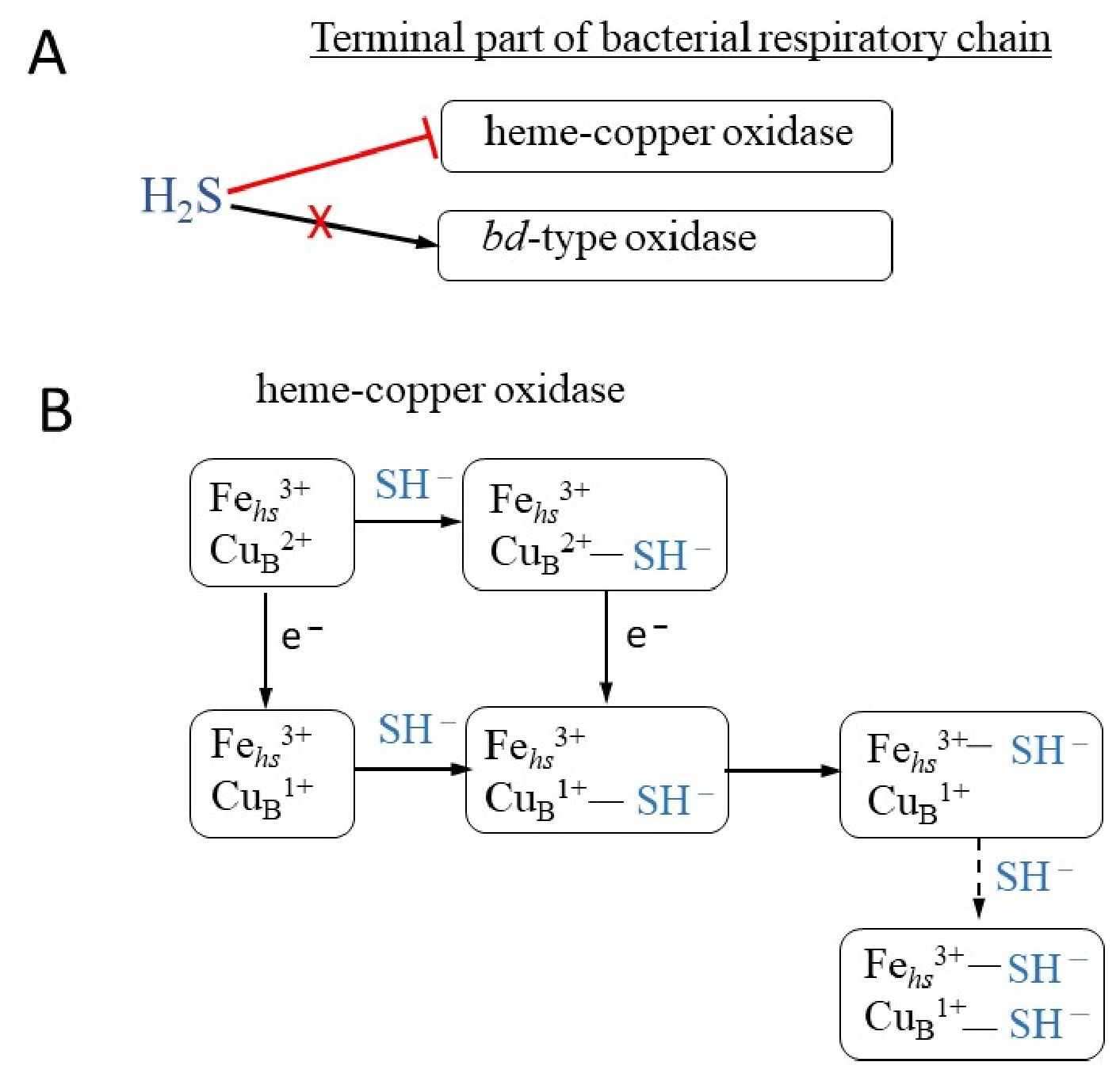
The respiratory chain of E. coli contains three terminal oxidases: cytochromes bo3, bd-I, and bd-II. Cytochrome bo3 belongs to the heme-copper oxidase superfamily, whereas copper-lacking cytochromes bd-I and bd-II are members of the bd-type oxidase superfamily [14,15]. Low micromolar concentrations of sulfide (half-maximal inhibitory concentration IC50~1–3 μM H2S) were reported to exhibit a toxic effect by inhibiting cytochromes bo3 but stimulate the transition of respiration to cytochromes bd-I and bd-II [16,17]. The transition to the less energetically efficient but sulfide-insensitive bd-type terminal oxidase allows E. coli to grow in the presence of H2S. However, at concentrations above 0.1 mM, exogenous Na2S decreased the specific growth rate of E. coli [18]. In agreement with [18], in the presence of sulfide-producing Desulfovibrio piger, PhsABC- and AsrABC-mediated H2S production by Salmonella enterica serovar Typhimurium was linked to inhibition of cytochrome bd-mediated respiration of E. coli in the murine large intestine [19]. Consistent with the E. coli data described above, in the presence of high levels of H2S, the activity of the cyanide-insensitive bd-type oxidase in Pseudomonas aeruginosa was unaltered and expression of this enzyme was enhanced. In contrast, the overall O2 consumption of all heme-copper terminal oxidases in P. aeruginosa was severely impaired by sulfide [20]. In Shewanella oneidensis, inhibition of the cbb3-type heme-copper oxidase by 0.25 mM H2S was also observed [12]. It appears that when both a heme-copper oxidase and a bd-type oxidase are present in the terminal part of the respiratory chain, bacteria respond to sulfide stress by upregulating cytochrome bd, as H2S inhibits the heme-copper oxidase (Figure 1A). In the absence of H2S and other stressors, bacterial aerobic respiration is primarily mediated by the heme-copper oxidase, which provides a higher energetic yield.
In general, sulfide toxicity, in addition to oxidative damage due to the inhibition of antioxidant proteins, may be associated with DNA damage, lipid peroxidation, protein denaturation by disruption of disulfide bonds, and inactivation of redox centers in metalloenzymes [5,12]. At the same time, the protective role of H2S against H2O2 and antibiotics has been demonstrated for different bacteria [6,7,21], which allows us to consider bacterial H2S biogenesis as a clinically important adaptive response during infections, capable of acting as a potential therapeutic target [8].

3. Sources of Endogenous H2S

In mammalian tissues, H2S concentrations are maintained at the nanomolar level, while in the intestine they can reach millimolar values (1.0–2.4 mM) [22]. In the intestine, the main source of hydrogen sulfide is commensal sulfate-reducing bacteria, which catalyze the reduction of SO42− to sulfite and then the conversion of SO32− to HS via dissimilatory sulfite reductase (Dsr) [23]. The most common sulfate-reducing bacteria found in the intestines of humans and animals are the following species: Desulfotomaculum, Desulfobulbus, Desulfomicrobium, Desulfomonas, and Desulfovibrio, with Desulfovibrio (D. desulfuricans, D. farfieldensis) being the most frequently isolated. These bacteria are not considered directly pathogenic to humans and animals, but the high concentrations of H2S they produce may be involved in gut inflammation [22].
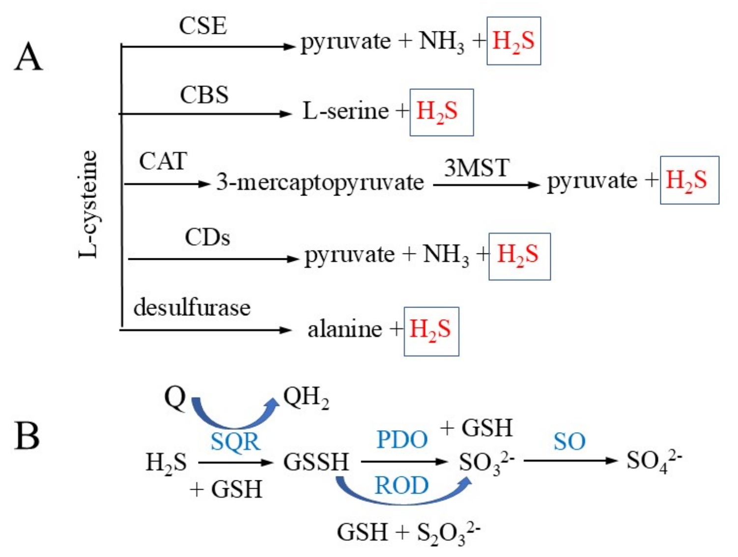
In infected mammalian tissues, sources of H2S may be the metabolism of host cells and commensal and pathogenic bacteria. Similar to mammalian cells, heterotrophic bacteria endogenously synthesize H2S primarily via the enzymes cystathionine β-synthase (CBS) and cystathionine γ-lyase (CSE) or by catabolizing cysteine via cysteine aminotransferase (CAT) to 3-mercaptopyruvate (3-MP), which is then converted to pyruvate and H2S by 3-MP sulfotransferase (3MST) via a persulfide intermediate [6,9,10]. Most bacterial genomes contain homologs of the genes encoding CBS/CSE and/or 3MST [6]. For example, Bacillus anthracis and Staphylococcus aureus have the CBS/CSE operon but lack 3MST [6]. Contrary to what was previously thought, a recent study has revealed that, in addition to CBS and CSE, 3MST also contributes to H2S production in P. aeruginosa [24]. S. oneidensis produces H2S through cysteine degradation by methionine γ-lyases MdeA, SO_1095, and SseA [12]. MdeA and SO_1095 are homologs of cystathionine γ-lyase, while SseA is a homolog of 3MST. The E. coli genome contains the mstA gene encoding 3MST but lacks genes encoding CBS and CSE; however, a mutant lacking 3MST can retain the ability to produce hydrogen sulfide. It was shown that restoration of H2S production in this mutant may occur due to a compensatory mutation in the transcriptional repressor ycjW, which leads to increased expression of the gene encoding thiosulfate sulfotransferase PspE (rhodanese) [25]. Induction of pspE expression in the suppressor strain provides an alternative mechanism for H2S biosynthesis.
In addition to CAT/MST, E. coli contains at least six enzymes (CysK, CysM, MetC, TnaA, MalY, and CyuA) with L-cysteine desulfhydrase activity, which can degrade L-cysteine into pyruvate, ammonium, and H2S [26,27,28,29]. The functional significance of these enzymes in H2S formation remains controversial. However, cysteine desulfhydrases (mainly TnaA) have been shown to be responsible for the degradation of endogenous and exogenous L-cysteine, while 3MST produces sulfane sulfur and is not a major pathway for H2S formation [30]. It has also been reported that the main enzyme catabolizing cysteine to produce H2S in E. coli under anaerobic conditions is cysteine desulfhydrase CyuA, which is controlled by the transcriptional regulator CyuR and is induced by increasing cysteine concentrations [27,28,29]. Furthermore, cysteine desulfurase (IscS), which converts cysteine to alanine and sulfane sulfur, rather than 3MST, was found to be the main source of endogenous H2S in E. coli under anaerobic conditions [31]. Apparently, H2S production in bacteria can be catalyzed by different enzymes depending on the culture conditions (Figure 2A).
The main, although not the only, source of H2S in Mycobacterium tuberculosis is cysteine desulfhydrase Cds1, which degrades cysteine to produce pyruvate, H2S, and ammonium [32]. In Treponema denticola, the majority of H2S is produced by cystalysins HlyA and HlyB, which are cystathionine β-lyases and hydrolyze cysteine to pyruvate, H2S, and ammonium [33]. Fusobacterium nucleatum has several enzymes involved in H2S production, including L-cysteine desulfhydrase, cysteine synthase, and L-methionine lyase [34]. Furthermore, a glycyl radical enzyme (isethionate sulfite lyase) was discovered from Bilophila wadsworthia, which catalyzes the cleavage of the C–S bond during taurine catabolism, producing sulfite, which is then reduced to H2S by dissimilatory sulfite reductase [35,36]. Overall, bacteria possess a large arsenal of enzymes capable of producing endogenous sulfide, but the relative contributions and physiological functions of each remain poorly understood.

4. Conditions Stimulating Endogenous H2S Production

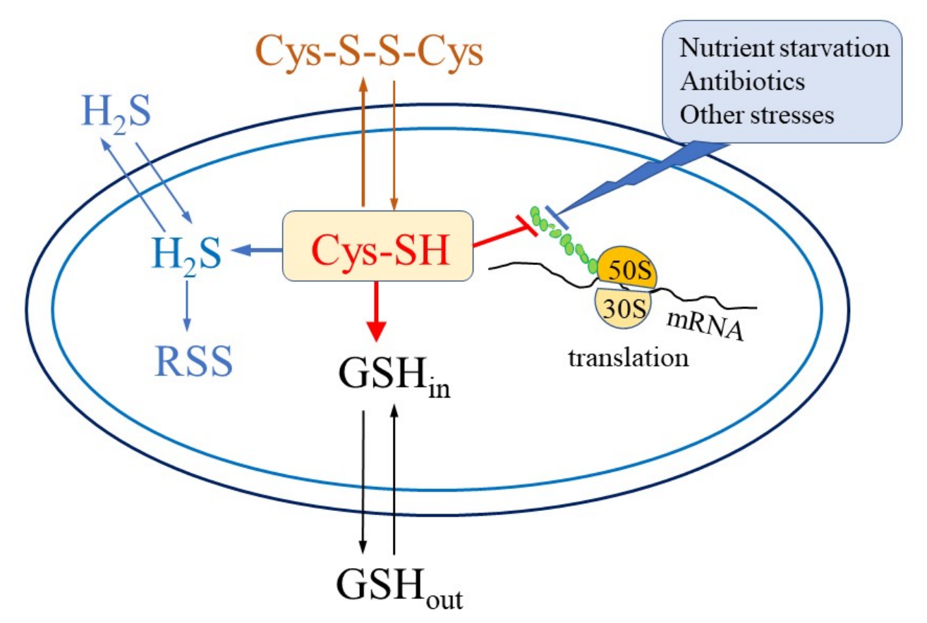
Experiments with continuous monitoring of H2S directly in a growing bacterial culture using a sensitive sulfide electrode (sensitivity threshold 5 nM) have shown that endogenous H2S production depends on the medium composition, primarily the sulfur source, and cultivation conditions [37]. When grown in a minimal medium, where sulfate is the only sulfur source, E. coli, Bacillus subtilis, and Mycobacterium smegmatis did not release sulfide during exponential growth under aerobic conditions [37,38,39]. Sulfide generation was observed in response to stressful conditions accompanied by a sharp cessation of growth and inhibition of protein synthesis. Such stresses include depletion of glucose, nitrogen, or phosphate in the medium, amino acid starvation, and exposure to a number of antibiotics [18,38,39,40,41,42]. In all these situations, there was a temporary increase in the intracellular concentration of cysteine and activation of its homeostasis mechanisms, including accelerated synthesis of glutathione (GSH) in E. coli or mycothiol (MSH) in M. smegmatis and the export of cysteine into the medium. Degradation of excess cysteine to form H2S can be considered as another mechanism of cysteine homeostasis or as an accidental consequence of an increase in its intracellular concentration (Figure 3).
Activation of these mechanisms protects bacteria from the negative consequences caused by excess cysteine: oxidative stress due to autoxidation reactions, potentiation of the Fenton reaction, and inhibition of isoleucine synthesis [43,44]. The duration and intensity of H2S production depended on the bacterial species, the growth rate preceding the stress exposure, and the type of stress. Sulfide production was reduced in the E. coli mstA mutant upon exposure to ciprofloxacin and upon ammonium or phosphate depletion, suggesting possible involvement of 3MST in the degradation of excess cysteine in these situations [41,42,45]. The absence of glutathione in the gshA mutant stimulated H2S generation compared to the parental strain under all stresses studied.
An excess of cytoplasmic cysteine and activation of cysteine homeostasis mechanisms, including H2S release, also occur upon addition of cystine to E. coli cells growing in minimal sulfate medium [16,46]. With fractional addition of cystine during cultivation, sulfide formation occurred continuously throughout the entire observation period [45]. Deletion of the cysM, mstA, and cyuA genes did not prevent H2S formation. It is possible that, under conditions of a strong excess of cytoplasmic cysteine, sulfide formation may be the result of the collective action of all enzymes capable of producing it.
In contrast to sulfate-containing minimal medium, E. coli produces H2S during normal growth in rich Luria–Bertani (LB Miller) medium (pepton—10 g/L, yeast extract—5 g/L, NaCl—10 g/L), where cystine is the main sulfur source. L-cysteine and L-cystine have been shown to be the most suitable substrates for H2S production, while GSH and methionine do not contribute significantly [30]. Simultaneous monitoring of sulfide and O2 using electrochemical sensors revealed that the onset of H2S production in E. coli BW25113 (parental strain) coincides with a sharp, reversible inhibition of respiration at an OD600 of approximately 0.35 and is likely associated with metabolic reorganization and growth retardation upon switching to a different substrate in multicomponent LB medium [37,47]. This may indicate a transient process in which excess intracellular cysteine arises, similar to that observed during inhibition of protein synthesis in E. coli in minimal medium. Slow cystine import in the tcyP and cysB mutants, which lack, respectively, the major cystine transport system TcyP and the CysB regulator, which controls both cystine importers TcyP and TcyJLN [45], resulted in almost complete cessation of sulfide production due to the lack of excess cysteine in the cytoplasm of these strains [47]. Mutants eamA, eamB, bcr, and cydD lacking cysteine exporters [48,49] exhibited multiple cycles of H2S production: the first cycle began immediately after transfer of bacteria to fresh medium, and subsequent cycles were repeated at intervals of 20–30 min, indicating that cysteine export is required for fine-tuning of cytoplasmic cysteine levels [47]. Increased H2S production in the gshA mutant confirms the important role of glutathione as a cysteine buffer, as it occurs in minimal medium [40]. The highest level of sulfide accumulation in the medium was observed in the cysK mutant, which is apparently due to a reduced ability of this strain, lacking the essential cysteine synthase, to incorporate formed H2S back into cysteine [47]. H2S production in the mstA mutant differed little from that in the parental strain. Sulfide production was reduced in the cysM, malY, tnaA, metC, cyuA, and iscS mutants lacking cysteine desulfhydrases, indicating the possible contribution of each of these enzymes to H2S formation [47].
Thus, heterotrophic bacteria release sulfide when intracellular cysteine is in excess. When cultivated in a medium where sulfate is the sole sulfur source, disruption of cysteine homeostasis can be caused by the addition of exogenous cystine or various factors that inhibit growth and the synthesis of protein, which is the primary consumer of cysteine synthesized by the cell. In LB, where cystine is a component of the medium, an increase in the intracellular cysteine concentration and, consequently, H2S formation can occur without any external factors as a result of metabolic changes and growth retardation. These differences in H2S formation, related to medium composition, must be considered when studying the role of endogenous H2S in bacterial responses to various factors, including antibiotics.

5. Mechanisms of H2S Action

In various compounds, sulfur can have oxidation states ranging from −2 to +6. At a sulfur oxidation state of −2, H2S and organic thiols (e.g., cysteine or GSH) exist in their most reduced forms and can function solely as cellular reductants [8]. Based on its chemical properties, H2S can influence cellular redox physiology through several mechanisms: neutralization of reactive oxygen and nitrogen species, reaction with metal centers of enzymes, modulation of cellular respiration, and post-translational modification of protein SH groups (S-persulfidation) [9].

5.1. The Effect of H2S on Metalloenzymes

It is well known that H2S can interact with metals, including iron, copper, nickel, and zinc, which are components of many enzymes and regulatory proteins through covalent bonding, redox interactions, or coordination [50]. At physiological concentrations, H2S can participate in the maintenance of metal homeostasis and signaling through interactions with metalloproteins [51]. Available data indicate that H2S may play a key role in attenuating iron cytotoxicity and controlling iron homeostasis under stress. Endogenous H2S generation is also often regulated by iron [52].
At the same time, higher concentrations of exogenous H2S can disrupt the functions of enzymes containing iron. The effect on cellular physiology may vary depending on the time of H2S addition. For example, in the simultaneous presence of H2S and H2O2, an increase in cytotoxicity was observed due to heme damage in catalases, while with sequential addition, H2S exerted a protective effect against H2O2 by activating OxyR and inducing antioxidant defenses [12,23,53]. Proteins modified by H2S also include zinc finger (ZF) proteins, which contain cysteine residues that coordinate zinc. In mammals, addition of H2S to the ZF protein tristetraprolin under aerobic conditions resulted in rapid persulfidation, thiol oxidation, zinc release, and inhibition of function due to loss of RNA binding capacity [54].

5.2. The Effect of H2S on Cellular Respiration

Exposure to high concentrations of H2S inhibits the mitochondrial cytochrome aa3 and bacterial heme-copper oxidases but does not affect the bd-type oxidases tested so far [55]. The model proposed by Nicholls et al. [56] for the H2S-mediated inhibition of the mitochondrial cytochrome c oxidase can be extrapolated to bacterial heme-copper oxidases (Figure 1B). According to the model, during steady-state turnover of a heme-copper oxidase, a first molecule of H2S (presumably in the form of HS) transiently binds to the CuB site (whether oxidized or reduced). It then transfers to the ferric iron ion of the high-spin heme (a3, o3, or b3). This transfer blocks the high-spin heme site from reacting with O2, halting the enzyme’s function. In this inactive state, the CuB site is reduced and possibly bound to a second HS molecule. The inhibition is completely reversible upon removal of H2S from the medium [55].

5.3. Antioxidant Effects of H2S and Persulfides

H2S can directly react with reactive oxygen and nitrogen species such as H2O2, superoxide radical, peroxynitrite, and hypochlorite, acting as an antioxidant [9,57,58]. Oxidation of H2S can lead to the formation of a variety of compounds in which the sulfur atom has oxidation states from −2 to +6. Oxidation products include sulfate (SO42−), sulfite (SO32−), thiosulfate (S2O32−), persulfides (RSS), organic and inorganic polysulfides, and elemental sulfur. All small inorganic and organic molecules derived from H2S and containing sulfur atoms in oxidation states more positive than −2 are collectively referred to as reactive sulfur species (RSS), many of which contain sulfur bound to sulfur, or “sulfane” sulfur. The organic thiol persulfide (hydropersulfide, RSSH) is of particular interest because it can function either as a nucleophile in the deprotonated state (RSS) or as an electrophile in the protonated state (RSSH). Persulfides, formed upon exposure of cells to exogenous H2S or during endogenous H2S production, readily react with oxidants such as hydrogen peroxide and peroxynitrite and are more effective one-electron reducing agents than thiols and H2S [8]. Persulfides are presumed to be responsible for the antioxidant effects observed with H2S.

5.4. Modification of Protein SH Groups

Persulfidation, the addition of a sulfur atom that converts the thiol group (–SH) of proteins to persulfide (–SSH), is a post-translational modification comparable to nitrosylation or glutathionylation [59]. This modification can protect SH groups from oxidation and alter enzymatic activity, contributing to the regulation of cellular redox homeostasis. H2S does not react directly with protein cysteines; the presence of an oxidant (e.g., hydrogen peroxide, peroxynitrite, hypochlorite, lipid peroxides) is required for this reaction to occur. The proposed mechanism for persulfide formation is the reaction of sulfur with oxidized cysteines, such as sulfenic acid (RSOH) or disulfide (RSSR) [57]. Unlike classical cysteine oxidation, the S–S–H bond remains fully reversible and can be regenerated using thioredoxins and glutaredoxins. Overall, evidence is accumulating that persulfidation is a reversible, chemically versatile, and enzymatically regulated redox switch that is at the center of sulfur signaling pathways in mammalian, plant, and bacterial cells [8,23,58,60].

6. H2S and RSS Homeostasis

To maintain physiological H2S levels in a non-toxic range, a balance between its formation and clearance is necessary. H2S breakdown occurs via a catabolic pathway in which H2S is oxidized to thiosulfate and sulfate in the presence of O2. H2S oxidation is accomplished by the enzymes sulfide:quinone oxidoreductase (SQR), persulfide dioxygenase (PDO), rhodanese, and sulfite oxidase (SO) [61,62,63]. SQR oxidizes H2S to form glutathione persulfide GSSH, and PDO catalyzes the conversion of GSSH to reduced glutathione (GSH) and sulfite (SO32−) (Figure 2B). Rhodanese can transfer sulfane sulfur between various acceptors, maintaining a pool of sulfane sulfur in the cytoplasm. It catalyzes the transfer of sulfane sulfur from GSSH to sulfite, resulting in the formation of GSH and thiosulfate, which may help minimize the accumulation of sulfane sulfur in cells and prevent disulfide stress [64]. Conversely, upon addition of exogenous thiosulfate, rhodanese PspE converts it to GSSH in E. coli cells, increasing the level of sulfane sulfur, which reduces the sensitivity of bacteria to H2O2 treatment [65]. Furthermore, sulfane sulfur can be formed during the oxidation of H2S involving heme-containing proteins, as well as during cysteine metabolism by various enzymes, including 3MST and desulfurase [58]. Continuous formation and metabolism of sulfane sulfur and H2S can maintain their concentrations within a certain range.
The H2S/RSS homeostasis mechanism is mediated by RSS sensors, specialized transcriptional regulators that can sense even small changes in cellular RSS caused by endogenous or exogenous perturbations [58]. RSS sensors belong to the copper-sensitive operon repressor (CsoR), arsenic repressor (ArsR), and Fis superfamilies. Most known RSS sensors are characterized by the presence of a pair of cysteines that form a disulfide or polysulfide bridge upon incubation with a sulfane sulfur donor, resulting in the release of repression and transcription of the targeted genes [66]. CstR proteins, which belong to the CsoR superfamily, are found mainly in Gram-positive organisms (Firmicutes) and are best characterized in S. aureus, Streptococcus pneumoniae, Streptococcus mitis, and Enterococcus faecalis [51,67]. A typical persulfide sensor belonging to the ArsR family in many Gram-negative organisms is SqrR (sulfide quinone reductase repressor), first characterized in Rhodobacter capsulatus [68]. Homologues of SqrR include BigR (biofilm repressor) in Xylella fastidiosa and A. baumannii, YgaV in E. coli, and HlyU in Vibrio spp. and probably PigS in Serratia [58]. In the chemoorganoheterotrophic bacterium Hyphomicrobium denitrificans, the use of thiosulfate as an electron donor is regulated by two sulfane-sulfur-responsive ArsR-type transcriptional repressors, sHdrR and SoxR [69]. The third major class of RSS sensors is represented by the FisR protein from the genus Cupriavidas, which is an σ54-dependent transcriptional activator [70].
RSS sensors regulate genes whose products are involved in maintaining H2S/RSS homeostasis, including SQR, PDO, flavin-dependent coenzyme A persulfide reductase, various sulfotransferases (ST), and one or more membrane transporters, including the putative sulfite exporter TauE and thiosulfate importers YedE/YeeE [58,71]. In addition, so-called secondary RSS sensors, whose primary role differs from H2S/RSS homeostasis, are distinguished [58]. They may have a specific input signal distinct from RSS and are involved, for example, in the detection and detoxification of ROS (OxyR, PerR) or in the regulation of virulence genes and can often act as global regulators. The transcriptional regulator OxyR, which responds to increased H2O2, also senses high RSS levels by activating the expression of thioredoxin, glutaredoxin, and catalase, which can remove sulfane sulfur [53,72]. The PerR protein in the cyanobacterium Synechococcus sp., a representative of Cys4 zinc finger proteins, acts not only as a ROS sensor but also as a sensor of RSS, which can modify cysteine in the Cys4:Zn2+ structural site to form Cys121-SSH, resulting in the release of the zinc atom and activation of the prxI gene expression encoding peroxiredoxin [73]. Sulfane sulfur post-translationally modifies the transcription factor AdpA, which controls secondary metabolism and morphological differentiation in Streptomyces coelicolor, to form persulfide, thereby stimulating the expression of its target genes, leading to the activation of actinorhodin biosynthesis and morphological changes [74]. In the chemolithotrophic bacterium Acidithiobacillus caldus, a novel RSS-sensitive transcription factor of the MarR family (SscR) was identified, which, together with the Cu-sensitive transcriptional repressor CsoR, regulates intracellular RSS levels, allowing A. caldus to reduce copper-induced stress and oxidative damage while preventing lethal RSS accumulation [75]. Many of the secondary RSS sensors that play important roles in bacterial virulence and antibiotic resistance are described in the following sections.
In response to exogenous H2S, bacteria increase the expression of genes encoding enzymes that oxidize sulfide (PDO, SQR), ensure sulfite export and switch respiration to sulfide-resistant cytochrome bd, and inhibit the expression of genes encoding sulfur source transporters [5,17,71]. The number of enzymes involved in ROS detoxification (catalase, superoxide dismutase, alkyl hydroperoxidase, universal stress proteins) and metal metabolism also increases [5,17]. In general, bacterial cells respond to increased H2S levels by activating defenses against disulfide and oxidative stress.
When studying the effects of H2S and RSS on bacterial virulence and antibiotic sensitivity, researchers use various methods to perturb H2S concentrations in cells: decreasing endogenous H2S production by deleting or inhibiting enzymes involved in its production from cysteine, or increasing endogenous H2S levels by supplementing the medium with cysteine/cystine or deleting enzymes involved in H2S degradation, and increasing H2S concentrations by adding sources of exogenous H2S (NaHS, Na2S, gaseous H2S, and other chemical donors). Based on the findings described in the preceding sections, different adaptive responses of cells to variations in endogenous and exogenous H2S should be expected. Endogenous H2S synthesis in heterotrophic bacteria is caused by the accumulation of excess intracellular cysteine, which is also accompanied by the activation of other mechanisms of its homeostasis, such as cysteine export into the medium, incorporation into buffering molecules (GSH, MSH), inhibition of synthesis at the biochemical level, and inhibition of gene expression at the transcriptional level. Suppression of the activity of H2S-producing enzymes can, at least in part, be compensated for by the activation of other mechanisms of cysteine homeostasis. Although an increase in intracellular cysteine levels creates a potential risk of oxidative stress, the development of disulfide stress under these conditions is less likely. In contrast, when bacterial cells are treated with sources of exogenous H2S, the risk of disulfide stress primarily arises, which triggers systems involved in H2S degradation and the restoration of H2S/RSS homeostasis. Persulfide formation can occur both during cysteine metabolism and as a result of H2S oxidation. However, despite certain parallels, differences in stress responses to increased intracellular cysteine and increased exogenous H2S may be the source of differences in the physiological response of bacteria to stress induced by antibiotics and oxidants. Furthermore, the concentration of endogenous H2S varies greatly depending on specific conditions, the bacterial species, and their physiological state, whereas the amount of added exogenous H2S typically far exceeds the level produced by the cells themselves. Differences in the effects of endogenous and exogenous H2S may be one reason for the discrepancies in the results obtained by different researchers.

7. The Effect of H2S and RSS on Bacterial Virulence

H2S and its derivatives, RSS, may promote bacterial survival in the host organism by enhancing resistance to multiple oxidative stressors produced during the antimicrobial immune response and by H2S/RSS-dependent regulation of virulence factors and biofilm formation.

7.1. Changes in H2S Concentration Modulate Bacterial Virulence

The stimulating effect of H2S on virulence has been demonstrated in various bacterial species. It has been shown that a decrease in H2S production in E. coli and S. aureus under the influence of specific inhibitors or mutations of H2S-producing enzymes increased the susceptibility of both bacterial species to rapid elimination by immune cells, while the addition of exogenous H2S mitigated this effect [76]. A cbs mutant lacking cystathionine β-synthase, the primary producer of endogenous H2S in Vibrio cholerae, exhibited a colonization defect in adult mouse experiments, whereas complementation with cbs or exogenous addition of N-acetyl-L-cysteine restored its viability in the host environment. According to the model proposed by the authors, the protective effect of endogenous H2S may be associated with the detoxification of ROS resulting from the increased post-translational activity of catalase KatB, as well as with the stimulation of the expression of several other antioxidant enzymes (SodB, KatG and AhpC, DNA protection protein DPS and Trx1) [77]. Host-derived H2S, like endogenous bacterial H2S, stimulated M. tuberculosis growth and respiration, primarily through activation of cytochrome bd oxidase, and regulated genes involved in sulfur and copper metabolism, as well as the Dos dormancy regulon [32,78,79]. Mice infected with M. tuberculosis and deficient in the H2S-producing cystathionine β-synthase lived longer, and the use of CBS inhibitors reduced bacterial load, suggesting that methods targeting H2S production may be potentially useful for tuberculosis treatment [78].
In Mycoplasma pneumoniae, H2S, produced by the bifunctional enzyme cysteine desulfurase/desulfhydrase HapE, is a virulence factor that stimulates erythrocyte lysis, inhibits human bronchial epithelial cell proliferation, arrests the cell cycle, and alters the cytokine profile of the local immune system [80]. The spirochete Treponema denticola produces H2S, which significantly contributes to its pathogenic potential by inducing apoptosis of periodontal epithelial cells and hemolysis of erythrocytes, which may play a key role in the initiation of periodontitis [33].

7.2. The Effect of H2S/RSS on the Expression of Virulence Factors

The H2S signal mediated through RSS can be sensed by global regulators and influence the expression of virulence factors. For example, in S. aureus, which is capable of expressing a wide range of virulence genes, it was demonstrated that persulfidation of the transcriptional regulator MgrA inhibits its binding to DNA, leading to altered expression of secreted virulence factors, and modulates secretome cytotoxicity. Exoprotein expression was induced by high sulfide levels and suppressed by low RSS levels [81]. In V. cholerae, the transcriptional repressor HlyU (a homolog of SqrR) regulates the expression of exotoxins required for bacterial colonization of the intestine in response to endogenously produced persulfides [82]. The activity of the quorum-sensing regulator LasR was enhanced by sulfane sulfur, which reaches its maximum level in the early stationary phase of P. aeruginosa PAO1 [83].

7.3. The Effect of H2S/RSS on Biofilm Formation

A critical factor for bacterial survival within the host is the formation of biofilms (communities of adherent cells embedded in an extracellular polymer matrix), which provide protection to bacteria from the immune system and antibiotics, making it difficult to eradicate infections [84]. Evidence is accumulating of a link between the regulation of biofilm formation and H2S/RSS homeostasis, but this issue remains poorly understood. H2S has been detected in the sputum of patients with cystic fibrosis, which represents a complex biofilm [85]. H2S has also been found to promote biofilm formation by the intestinal microbiota while simultaneously reducing the proliferation of planktonic cells [86]. Depending on their concentration, endogenous and exogenous H2S can differentially affect various characteristics of intestinal mucosal microbiota biofilms, altering the relative abundance, spatial organization, and function of bacteria in biofilms [87]. Low levels of exogenous H2S directly stabilize the intestinal mucosa and protect the intestinal microbiota, likely through iron chelation [88]. Mutation of cystathionine γ-lyase and the use of inhibitors that suppress endogenous H2S production have also been shown to reduce persister cell formation and biofilm abundance in S. aureus and P. aeruginosa. Moreover, genes associated with biofilm formation, including genes encoding alginate and other exopolysaccharides, are among the most strongly downregulated categories in H2S-deficient cells [89]. The effect of H2S on biofilm formation may be mediated through RSS. The secondary persulfide sensor BigR in A. baumannii affected the expression of genes associated with biofilm formation; furthermore, the biofilm-regulating transcriptional regulators BfmR and Crp were found to undergo strong persulfidation upon exposure to sulfide [71]. Potential targets of the RSS-sensitive transcription factor SscR in A. caldus include diguanylate cyclase and phosphodiesterase, modification of which may influence bacterial biofilm formation by regulating c-di-GMP levels [75].

8. The Impact of H2S and RSS on Bacterial Sensitivity to Antibiotics

Of particular interest is the relationship between H2S production and bacterial sensitivity to antibiotics. Endogenous or exogenous H2S has been shown to reduce the sensitivity of Bacillus anthracis, P. aeruginosa, S. aureus, and E. coli to antibiotics, while mutations and inhibitors of H2S-producing enzymes enhance the lethal activity of antimicrobials [6]. The authors suggested that H2S is a universal defense mechanism against antibiotics of different classes (quinolones, aminoglycosides, and β-lactams) in various bacterial species [6]. They also assumed that the main protective mechanism mediated by H2S is its participation in maintaining the cellular redox balance, which, according to the radical hypothesis of the action of bactericidal antibiotics, is disrupted due to increased ROS production [90,91,92]. It has been suggested that the protective effect of H2S against antibiotics is achieved by stimulating the major antioxidant enzymes catalase and SOD and reducing ROS formation, as well as by reducing cysteine levels and binding free Fe2+, which prevents the Fenton reaction that generates toxic hydroxyl radicals [6,7,9,25,93,94]. A model explaining the interaction between L-cysteine metabolism, H2S production, and oxidative stress has been proposed, in which 3MST protects E. coli from oxidative stress through L-cysteine utilization and H2S-mediated scavenging of free iron, which is required for the genotoxic Fenton reaction [7]. However, despite indirect evidence, no study has yet directly demonstrated the involvement of H2S in reducing free intracellular iron concentrations [95].
Several studies confirmed the possibility of enhancing the sensitivity of various bacterial species to antibiotics or even restoring antibiotic sensitivity in resistant strains by limiting endogenous H2S production. It was shown that chemical inhibition of H2S biosynthesis restored antibiotic sensitivity in multidrug-resistant uropathogenic E. coli isolates, while exposure to an H2S donor was accompanied by restoration of antimicrobial resistance [93]. The authors proposed that the protective effect of H2S against antibiotics could be mediated by two mechanisms: (i) down-regulation of energy-efficient cytochrome bo3 oxidase (CyoA) and induction of less-energy efficient cytochrome bd oxidase I/II (CydAB/AppBC) to maintain respiratory flux and redox balance, and (ii) augmentation of antioxidant capacity by elevating catalase and superoxide dismutase activities [93]. The ability of bacteria to maintain respiration and growth in the presence of low micromolar concentrations of sulfide by switching respiration from heme-copper cytochrome bo3 to cytochromes bd-I and bd-II [16,17] was discussed above (see also Figure 1). The role of alternative respiratory cytochrome oxidases in H2S-mediated bacterial protection from oxidative stress and bactericidal antibiotics was also studied by Seregina et al. [94]. It was shown that deletion of the cydB or cydD genes leads to hypersensitivity of E. coli to quinolones and β-lactams, whereas constitutive expression of the katG and mstA genes, which ensures increased production of catalase or H2S in these mutants, contributed to a decrease in their sensitivity to antibiotics. It has also been noted that H2S-mediated activation of catalases and superoxide dismutases may contribute to a decrease in bacterial sensitivity to antibiotics [96,97].
The effect of H2S on bacterial antimicrobial resistance may be mediated by RSS signaling, including the regulation of resistance gene expression. Growth-related changes in sulfane sulfur content have been shown to regulate the expression of antibiotic resistance genes controlled by transcriptional regulators of the MarR (multiple antibiotic resistance regulators) family [98]. These regulators are ubiquitous in bacteria, modulating numerous cellular processes, and are well known for their ability to induce resistance to multiple antibiotics, detergents, and oxidative reagents. Although several inducers have been reported, sulfane sulfur, whose concentration peaks in E. coli cells during the late logarithmic and early stationary phases of growth, is likely a key inducer regulating MarR activity. Persulfidation of thiols within the MarR protein alters its structure and affinity for DNA, leading to the derepression of the genes it controls [98]. Persulfides have also been shown to directly react with SH groups of the MexR transcriptional regulator when P. aeruginosa PAO1 enters stationary phase, leading to the derepression of the mexAB-oprM operon and the activation of the MexAB-OprM efflux pump, which plays a critical role in antibiotic resistance [99]. The MgrA regulator is modified by sulfane sulfur when S. aureus is exposed to H2S-induced stress, derepressing genes involved bacterial virulence [81]. Both MexR and MgrA belong to the MarR family. Thus, three family members sense the presence of sulfane sulfur in cells, activating antibiotic resistance genes, further confirming the signaling role of sulfane sulfur in bacteria.
In E. coli, the sulfide-responsive transcription factor YgaV (a homolog of SqrR/BigR) controls the expression of anaerobic respiration genes and is involved in H2O2 scavenging [100]. The ygaV mutant was more sensitive to antibiotics than the parental strain and, unlike it, did not respond to the presence of H2S with decreased sensitivity, indicating an important role for YgaV-dependent transcriptional regulation in maintaining redox homeostasis, ROS scavenging, and sensitivity to antibiotics [100].
Furthermore, it has been found that persulfides can participate in the direct degradation of some antibiotics. In particular, cysteine hydropersulfide (CysSSH), formed by the reaction of H2S with cystine, can directly inactivate β-lactam antibiotics of the penicillin (penicillin G and ampicillin) and carbapenem (meropenem) classes, forming ring-opened β-lactam S-acids (BL-COSH) [101]. Since E. coli and S. aureus efficiently degraded β-lactam antibiotics to form BL-COSH, the authors suggested that CysSSH-mediated degradation of β-lactams may be one of the mechanisms reducing their sensitivity to these antibiotics.
Most of the mechanisms considered for the protective effect of H2S against antibiotics are still hypothetical and require further in-depth research across species and systems. The hypothesis of oxidative stress as a general mechanism of action of bactericidal antibiotics has been challenged [102]. Accordingly, the involvement of H2S and its derivatives as antioxidants against antibiotic-generated ROS is also questionable. However, antibiotics can disrupt cysteine homeostasis as part of a universal stress response associated with the inhibition of protein synthesis [45,47]. H2S and its derivatives may be involved in maintaining cysteine homeostasis and cellular redox homeostasis in general. The involvement of H2S and RSS in cellular signaling and the activation of antibiotic resistance gene expression through persulfidation of transcriptional regulators is a very attractive mechanism but also requires further research. H2S-induced respiration switching from cytochrome oxidase bo3 to bd requires the production of micromolar concentrations of endogenous H2S, which may depend on the bacterial species and their physiological state.
The potential for using H2S production inhibitors as adjuvants for widely used antibiotics has generated considerable interest among researchers, who have sought to identify such compounds. Several compounds have been identified that enhance the antimicrobial activity of known antibiotics (Table 1). In particular, pioglitazone, a specific inhibitor of the 3MST enzyme in E. coli, suppressed H2S and sulfane sulfur production and enhanced the antimicrobial activity of gentamicin and macrophages against this bacterium [103]. Small molecules NL1, NL2, NL3 that inhibit cystathionine γ-lyase, the primary H2S generator in S. aureus and P. aeruginosa, have been discovered [89]. These inhibitors enhanced the bactericidal activity of antibiotics against these pathogens both in vitro and in mouse infection models, disrupting biofilm formation and reducing tolerance to antibiotics and the number of persister cells. It was also found that inhibition of cystathionine γ-lyase using naphthyl-substituted indole and pyrrole carboxylic acids significantly enhances the effect of antibiotics against A. baumannii, P. aeruginosa, and methicillin-resistant S. aureus [104]. Furthermore, compound 7b, based on nitrobenzofuran structures and capable of scavenging H2S, reducing its concentration, enhanced the activity of macrophages and neutrophils, suppressed biofilm formation, and also significantly increased the antimicrobial efficacy of gentamicin in models of pneumonia and skin wounds caused by P. aeruginosa [105].
However, despite intensive research into the role of H2S and the mechanisms by which it influences bacterial sensitivity to antibiotics, consensus remains elusive. Several research groups have demonstrated that the protective effect of H2S is not universal across all bacterial species and antibiotics. Analysis of the effect of H2S on the activity of antibiotics of key classes against S. aureus revealed that exogenous H2S exerts a protective effect only against aminoglycosides, whose transport into the cell depends on ΔμH+. The effect of sulfide on the bacterial respiratory chain leads to decreased uptake of aminoglycosides, weakening their antimicrobial effect [106]. It has also been shown that a deficiency in H2S production in the same strain can increase sensitivity to one type of antibiotic but does not affect sensitivity to other antibiotics. In particular, the megL mutant of F. nucleatum, lacking the essential H2S-generating enzyme L-methionine γ-lyase, exhibited increased sensitivity to nalidixic acid but demonstrated resistance to kanamycin [107]. Furthermore, the effect of H2S on antibiotic sensitivity may be highly dependent on the metabolic characteristics of the bacterial species under study. Instead of decreasing antibiotic sensitivity, exogenous H2S increases the sensitivity of A. baumannii to several classes of antibiotics and can reverse acquired resistance to gentamicin [11]. This hypersensitivity was presumably caused by H2S-induced disruption of energy metabolism and redox homeostasis in this bacterium. In M. tuberculosis, endogenous and exogenous H2S stimulates respiration and growth, regulates redox homeostasis, and increases sensitivity to the anti-tuberculosis drugs clofazimine and rifampin [32]. A recent study using mutants producing high or low levels of H2S showed that H2S production is not a protective mechanism against antibiotics of different classes in P. aeruginosa [24], which contradicts previously published data [6]. Moreover, a correlation analysis of a large collection of P. aeruginosa isolates from patients with cystic fibrosis did not reveal a protective role of H2S against antibiotic action during chronic pulmonary infection [24].
It was shown that E. coli mutants with defects in cysteine synthesis and degradation (cysK, cysM, cyuA, iscS, malY, metC, mstA, tnaA), cystine import and cysteine export (tcyP, eamA, eamB, bcr, cydD), glutathione synthesis (gshA), and regulatory proteins (cyuR and cysB) generated different amounts of H2S when grown in LB medium, where cystine is the main source of sulfur [47]. In addition to differences in H2S production, mutations caused changes in a range of other parameters: growth and respiration rates, glutathione and cysteine levels, and the degree of induction of the Fur, OxyR, and SOS regulons. A correlation was found between the expression of the Fur-controlled iucC::lacZ fusion and H2S production (r = −0.71, p < 0.05), indicating coordinated regulation of the free Fe2+ and H2S pools. Over a wide range of fluoroquinolone ciprofloxacin concentrations (0.03–10 µg/mL), a significant positive relationship was observed between the level of endogenous sulfide and the specific growth rate of bacteria (μ) after the addition of the antibiotic. However, no statistically significant relationship was found between the rate of bacterial killing in the CFU assay and H2S production. At a ciprofloxacin dose of 0.3 µg/mL, a strong direct correlation was found between the rate of bacterial death during the rapid killing phase (30 min) and the intracellular and extracellular glutathione concentrations (r = 0.8, p < 0.05). This effect was absent at higher antibiotic concentrations. This may indicate that changes in the redox status of low-molecular-weight thiols during ciprofloxacin-induced perturbations of cysteine homeostasis may contribute to the increased lethal activity of low doses of the antibiotic. At all ciprofloxacin concentrations studied, a strong inverse relationship was observed between the logarithm of CFU/mL of the studied mutants and the specific bacterial growth rate before the addition of the antibiotic. Apparently, mutants that retained higher metabolic activity when exposed to ciprofloxacin suffered more DNA damage, which complicated its repair upon resumption of growth on agar plates.
Treatment of E. coli growing in minimal medium with sulfate as the sole sulfur source with ciprofloxacin resulted in the release of H2S in the parental strain. The cysM and mstA mutants did not produce H2S, while the gshA mutant produced it in higher quantities than the parent. However, in the CFU assay, the mstA mutant was less sensitive, while the cysM and gshA mutants were more sensitive to ciprofloxacin than the parental strain, which is inconsistent with the hypothesis that H2S plays a protective role against the bactericidal action of quinolones [45]. At the same time, under conditions of continuous H2S release induced by the fractional addition of cystine, all the studied strains were lower sensitive to ciprofloxacin than in a medium without cystine, with the exception of the gshA mutant, the sensitivity of which increased 5–8 times at high doses of the antibiotic, indicating the important role of GSH in restoring the redox balance in cells growing in the presence of cystine.
To better understand the possible reasons for the existing contradictions, we compiled a summary table systematizing the available studies by bacterial species, H2S perturbation modes, antibiotic classes, experimental conditions, and observed phenotypes (Table 2). To facilitate understanding of the phenotypic effect of antibiotics, we used the unified term “change in antibiotic sensitivity”; however, since the authors employed different experimental approaches, the original papers may have referred to changes in antibiotic sensitivity, tolerance, persistence, or resistance. The analysis showed that certain contradictions exist for most of the studied species. They may be associated not only with the characteristics of the strains and the mechanism of action of antibiotics, but also with differences in the test systems used, as well as with specific cultivation conditions, sources and concentrations of H2S and antibiotics, and the physiological state of the bacteria. Some observations appear curious. A specific bCSE inhibitor (compound 2a) has been shown to increase the sensitivity of A. baumannii to β-lactams (Mer, CP, Pip + Taz) [104], while other studies claim that this bacterium maintains very low levels of H2S and, although it has 3MST, does not contain bCSE homologues in its genome [11,71].

9. Concluding Remarks

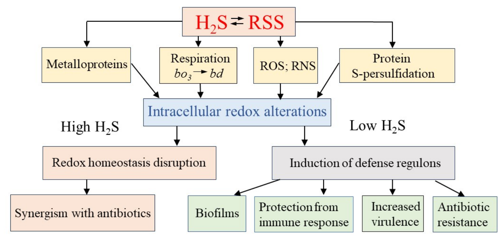
Literature analysis shows that low doses of exogenous H2S may induce regulatory redox changes in transcription factors and enzymes, accompanied by activation of bacterial defense systems and decreased sensitivity to oxidants and antibiotics. High doses of H2S, however, irreversibly disrupt the redox balance, promoting cell death, and can act synergistically with antibiotics (Figure 4). The dose required to induce a given effect can vary depending on the bacterial species and physiological state.
The production of endogenous H2S in heterotrophic bacteria is in most cases a consequence of increased intracellular cysteine concentrations under conditions that inhibit growth and protein synthesis, including various stresses and antibiotic exposure. Concurrently with H2S, the concentrations of key redox buffers (GSH and MSH) and, likely, reactive sulfur species increase, which may lead to changes in the expression of virulence and antibiotic resistance genes. A number of studies in vitro systems and mouse models have demonstrated the possibility of influencing virulence and antibiotic sensitivity in bacteria of various species, including multidrug-resistant strains, by targeted modulation of H2S levels. However, limitations related to the lack of standardization of donor systems for H2S, differences between culture media and assay systems, uncertainty in physiological sulfide concentrations, and the challenge in separating direct effects from RSS-mediated reactions lead to inconsistent results among different researchers. Many questions remain regarding the mechanism of H2S action, the influence of concentrations, culture conditions, specific stress responses in different bacterial species, interactions with host H2S, and other issues that need to be addressed, as they may require certain precautions and limitations when manipulating thiol levels during antibiotic therapy. The presence of alternative pathways for the production of endogenous H2S and RSS and the multiple mechanisms regulating cysteine homeostasis in cells of the same bacterial species, as well as differences in regulatory mechanisms across species, may require an individualized approach taking into account the metabolic characteristics of specific pathogens. Addressing these issues and elucidating the mechanisms by which H2S and RSS modulate the expression of bacterial resistance genes may facilitate the development of new therapeutic approaches based on the regulation of H2S and low-molecular-weight thiols, which is particularly relevant in the context of the widespread prevalence of antibiotic-resistant pathogens. However, addressing these issues presents significant challenges due to the strong context dependence of H2S/RSS effects, which are largely determined by bacterial physiology and experimental conditions.

Author Contributions

All authors participated in developing the review concept, analyzing the literature, writing, and editing the manuscript. All authors have read and agreed to the published version of the manuscript.

Funding

This work was supported by the State assignment # 124020500028-4 (to G.S., A.T., L.S., and O.O.), the State assignment of Lomonosov Moscow State University (project AAAA-A19-119031390114-5 to V.B.B.) and Sapienza University of Rome (projects RP124191037022F7 and RM125199C0BD5CC0 to E.F.).

Data Availability Statement

No new data were created in this study. Data sharing is not applicable to this article.

Conflicts of Interest

The authors declare no conflicts of interest.

References

  1. Wareham, L.K.; Southam, H.M.; Poole, R.K. Do nitric oxide, carbon monoxide and hydrogen sulfide really qualify as ‘gasotransmitters’ in bacteria? Biochem. Soc. Trans. 2018, 46, 1107–1118. [Google Scholar] [CrossRef]
  2. Wang, R. Gasotransmitters: Growing pains and joys. Trends Biochem. Sci. 2014, 39, 227–232. [Google Scholar] [CrossRef]
  3. Kimura, H. Production and physiological effects of hydrogen sulfide. Antioxid. Redox Signal. 2014, 20, 783–793. [Google Scholar] [CrossRef] [PubMed]
  4. Pinto, R.V.; Pinto, M.L.; Serre, C. Gasotransmitters in modern medicine: Promises and challenges in the use of porous crystalline carriers. Adv. Healthc. Mater. 2025, 14, 2404553. [Google Scholar] [CrossRef]
  5. Mendes, S.S.; Miranda, V.; Saraiva, L.M. Hydrogen sulfide and carbon monoxide tolerance in bacteria. Antioxidants 2021, 10, 729. [Google Scholar] [CrossRef] [PubMed]
  6. Shatalin, K.; Shatalina, E.; Mironov, A.; Nudler, E. H2S: A universal defense against antibiotics in bacteria. Science 2011, 334, 986–990. [Google Scholar] [CrossRef] [PubMed]
  7. Mironov, A.; Seregina, T.; Nagornykh, M.; Luhachack, L.G.; Korolkova, N.; Lopes, L.E.; Kotova, V.; Zavilgelsky, G.; Shakulov, R.; Shatalin, K.; et al. Mechanism of H2S-mediated protection against oxidative stress in Escherichia coli. Proc. Natl. Acad. Sci. USA 2017, 114, 6022–6027. [Google Scholar] [CrossRef] [PubMed]
  8. Walsh, B.J.C.; Giedroc, D.P. H2S and reactive sulfur signaling at the host-bacterial pathogen interface. J. Biol. Chem. 2020, 295, 13150–13168. [Google Scholar] [CrossRef] [PubMed]
  9. Pal, V.K.; Bandyopadhyay, P.; Singh, A. Hydrogen sulfide in physiology and pathogenesis of bacteria and viruses. IUBMB Life 2018, 70, 393–410. [Google Scholar] [CrossRef] [PubMed]
  10. Borisov, V.B.; Forte, E. Impact of hydrogen sulfide on mitochondrial and bacterial bioenergetics. Int. J. Mol. Sci. 2021, 22, 12688. [Google Scholar] [CrossRef] [PubMed]
  11. Ng, S.Y.; Ong, K.X.; Surendran, S.T.; Sinha, A.; Lai, J.J.H.; Chen, J.; Liang, J.; Tay, L.K.S.; Cui, L.; Loo, H.L.; et al. Hydrogen sulfide sensitizes Acinetobacter baumannii to killing by antibiotics. Front. Microbiol. 2020, 11, 1875. [Google Scholar] [CrossRef] [PubMed]
  12. Wu, G.; Wan, F.; Fu, H.; Li, N.; Gao, H. A matter of timing: Contrasting effects of hydrogen sulfide on oxidative stress response in Shewanella oneidensis. J. Bacteriol. 2015, 197, 3563–3572. [Google Scholar] [CrossRef] [PubMed]
  13. Fu, L.H.; Wei, Z.Z.; Hu, K.; Di Hu, L.Y.; Li, Y.H.; Chen, X.Y.; Han, Z.; Yao, G.F.; Zhang, H. Hydrogen sulfide inhibits the growth of Escherichia coli through oxidative damage. J. Microbiol. 2018, 56, 238–245. [Google Scholar] [CrossRef] [PubMed]
  14. Pereira, M.M.; Santana, M.; Teixeira, M. A novel scenario for the evolution of haem-copper oxygen reductases. Biochim. Biophys. Acta 2001, 1505, 185–208. [Google Scholar] [CrossRef] [PubMed]
  15. Murali, R.; Gennis, R.B.; Hemp, J. Evolution of the cytochrome bd oxygen reductase superfamily and the function of CydAA’ in Archaea. ISME J. 2021, 15, 3534–3548. [Google Scholar] [CrossRef] [PubMed]
  16. Korshunov, S.; Imlay, K.R.C.; Imlay, J.A. The cytochrome bd oxidase of Escherichia coli prevents respiratory inhibition by endogenous and exogenous hydrogen sulfide. Mol. Microbiol. 2016, 101, 62–77. [Google Scholar] [CrossRef] [PubMed]
  17. Forte, E.; Borisov, V.B.; Falabella, M.; Colaço, H.G.; Tinajero-Trejo, M.; Poole, R.K.; Vicente, J.B.; Sarti, P.; Giuffrè, A. The terminal oxidase cytochrome bd promotes sulfide-resistant bacterial respiration and growth. Sci. Rep. 2016, 6, 23788. [Google Scholar] [CrossRef] [PubMed]
  18. Tyulenev, A.; Smirnova, G.; Muzyka, N.; Ushakov, V.; Oktyabrsky, O. The role of sulfides in stress-induced changes of Eh in Escherichia coli cultures. Bioelectrochemistry 2018, 121, 11–17. [Google Scholar] [CrossRef] [PubMed]
  19. Larabi, A.B.; Tiffany, C.R.; Masson, H.L.P.; Nguyen, H.; Bejarano, E.J.; Liou, M.J.; Radlinski, L.C.; Demars, A.M.; Tsolis, R.M.; Baumler, A.J. Salmonella produces sulfide to compete with Escherichia coli in the gut lumen. Proc. Natl. Acad. Sci. USA 2025, 122, e2504095122. [Google Scholar] [CrossRef] [PubMed]
  20. Nastasi, M.R.; Caruso, L.; Giordano, F.; Mellini, M.; Rampioni, G.; Giuffre, A.; Forte, E. Cyanide insensitive oxidase confers hydrogen sulfide and nitric oxide tolerance to Pseudomonas aeruginosa aerobic respiration. Antioxidants 2024, 13, 383. [Google Scholar] [CrossRef] [PubMed]
  21. Szabo, C. A timeline of hydrogen sulfide (H2S) research: From environmental toxin to biological mediator. Biochem. Pharmacol. 2018, 149, 5–19. [Google Scholar] [CrossRef] [PubMed]
  22. Dordevic, D.; Jancikova, S.; Vitezova, M.; Kushkevych, I. Hydrogen sulfide toxicity in the gut environment: Meta-analysis of sulfate-reducing and lactic acid bacteria in inflammatory processes. J. Adv. Res. 2021, 27, 55–69. [Google Scholar] [CrossRef] [PubMed]
  23. Han, S.; Li, Y.; Gao, H. Generation and physiology of hydrogen sulfide and reactive sulfur species in bacteria. Antioxidants 2022, 11, 2487. [Google Scholar] [CrossRef] [PubMed]
  24. Caruso, L.; Mellini, M.; Catalano Gonzaga, O.; Astegno, A.; Forte, E.; Di Matteo, A.; Giuffrè, A.; Visca, P.; Imperi, F.; Leoni, L.; et al. Hydrogen sulfide production does not affect antibiotic resistance in Pseudomonas aeruginosa. Antimicrob. Agents Chemother. 2024, 68, e00075-24. [Google Scholar] [CrossRef] [PubMed]
  25. Luhachack, L.; Rasouly, A.; Shamovsky, I.; Nudler, E. Transcription factor YcjW controls the emergency H2S production in E. coli. Nat. Commun. 2019, 10, 2868. [Google Scholar] [CrossRef] [PubMed]
  26. Awano, N.; Wada, M.; Mori, H.; Nakamori, S.; Takagi, H. Identification and functional analysis of Escherichia coli cysteine desulfhydrases. Appl. Environ. Microbiol. 2005, 71, 4149–4152. [Google Scholar] [CrossRef] [PubMed]
  27. Shimada, T.; Tanaka, K.; Ishihama, A. Transcription factor DecR (YbaO) controls detoxification of l-cysteine in Escherichia coli. Microbiology 2016, 162, 1698–1707. [Google Scholar] [CrossRef] [PubMed]
  28. Nonaka, G.; Takumi, K. Cysteine degradation gene yhaM, encoding cysteine desulfidase, serves as a genetic engineering target to improve cysteine production in Escherichia coli. AMB Expr. 2017, 7, 90. [Google Scholar] [CrossRef] [PubMed]
  29. Loddeke, M.; Schneider, B.; Oguri, T.; Mehta, I.; Xuan, Z.; Reitzer, L. Anaerobic cysteine degradation and potential metabolic coordination in Salmonella enterica and Escherichia coli. J. Bacteriol. 2017, 199, e00117-17. [Google Scholar] [CrossRef] [PubMed]
  30. Li, K.; Xin, Y.; Xuan, G.; Zhao, R.; Liu, H.; Xia, Y.; Xun, L. Escherichia coli uses separate enzymes to produce H2S and reactive sulfane sulfur from L-cysteine. Front. Microbiol. 2019, 10, 298. [Google Scholar] [CrossRef] [PubMed]
  31. Wang, J.; Guo, X.; Li, H.; Qi, H.; Qian, J.; Yan, S.; Shi, J.; Niu, W. Hydrogen sulfide from cysteine desulfurase, not 3-mercaptopyruvate sulfurtransferase, contributes to sustaining cell growth and bioenergetics in E. coli under anaerobic conditions. Front. Microbiol. 2019, 10, 2357. [Google Scholar] [CrossRef] [PubMed]
  32. Kunota, T.T.R.; Rahman, M.A.; Truebody, B.E.; Mackenzie, J.S.; Saini, V.; Lamprecht, D.A.; Adamson, J.H.; Sevalkar, R.R.; Lancaster, J.R., Jr.; Berney, M.; et al. Mycobacterium tuberculosis H2S functions as a sink to modulate central metabolism, bioenergetics, and drug susceptibility. Antioxidants 2021, 10, 1285. [Google Scholar] [CrossRef] [PubMed]
  33. Phillips, L.; Chu, L.; Kolodrubetz, D. Multiple enzymes can make hydrogen sulfide from cysteine in Treponema denticola. Anaerobe 2020, 64, 102231. [Google Scholar] [CrossRef] [PubMed]
  34. Suwabe, K.; Yoshida, Y.; Nagano, K.; Yoshimura, F. Identification of an L-methionine-lyase involved in the production of hydrogen sulfide from L-cysteine in Fusobacterium nucleatum subsp. nucleatum ATCC 25586. Microbiology 2011, 157, 2992–3000. [Google Scholar] [CrossRef] [PubMed]
  35. Peck, S.C.; Denger, K.; Burrichter, A.; Irwin, S.M.; Balskus, E.P.; Schleheck, D. A glycyl radical enzyme enables hydrogen sulfide production by the human intestinal bacterium Bilophila wadsworthia. Proc. Natl. Acad. Sci. USA 2019, 116, 3171–3176. [Google Scholar] [CrossRef] [PubMed]
  36. Xing, M.; Wei, Y.; Zhou, Y.; Zhang, J.; Lin, L.; Hu, Y.; Hua, G.; Urs, A.N.N.; Liu, D.; Wang, F.; et al. Radical-mediated C-S bond cleavage in C2 sulfonate degradation by anaerobic bacteria. Nat. Commun. 2019, 10, 1609. [Google Scholar] [CrossRef] [PubMed]
  37. Smirnova, G.; Tyulenev, A.; Muzyka, N.; Ushakov, V.; Samoilova, Z.; Oktyabrsky, O. Influence of growth medium composition on physiological responses of Escherichia coli to the action of chloramphenicol and ciprofloxacin. BioTech 2023, 12, 43. [Google Scholar] [CrossRef] [PubMed]
  38. Tyulenev, A.; Smirnova, G.; Ushakov, V.; Kalashnikova, T.; Sutormina, L.; Oktyabrsky, O. Stress-induced sulfide production by Bacillus subtilis and Bacillus megaterium. Microorganisms 2024, 12, 1856. [Google Scholar] [CrossRef] [PubMed]
  39. Smirnova, G.; Tyulenev, A.; Kalashnikova, T.; Sutormina, L.; Ushakov, V.; Oktyabrsky, O. Cystine and antibiotic treatment alters low molecular weight thiol levels in Mycobacterium smegmatis. Front. Biosci. (Landmark Ed.) 2025, 30, 44441. [Google Scholar] [CrossRef] [PubMed]
  40. Smirnova, G.V.; Tyulenev, A.V.; Bezmaternykh, K.V.; Muzyka, N.G.; Ushakov, V.Y.; Oktyabrsky, O.N. Cysteine homeostasis under inhibition of protein synthesis in Escherichia coli cells. Amino Acids 2019, 51, 1577–1592. [Google Scholar] [CrossRef] [PubMed]
  41. Smirnova, G.V.; Tyulenev, A.V.; Bezmaternykh, K.V.; Muzyka, N.G.; Ushakov, V.Y.; Oktyabrsky, O.N. Phosphate starvation is accompanied by disturbance of intracellular cysteine homeostasis in Escherichia coli. Res. Microbiol. 2023, 174, 104108. [Google Scholar] [CrossRef] [PubMed]
  42. Smirnova, G.; Tyulenev, A.; Sutormina, L.; Kalashnikova, T.; Zhulanova, N.; Muzyka, N.; Ushakov, V.; Oktyabrsky, O. Ammonium depletion induces changes in low molecular weight thiol levels in Escherichia coli and Bacillus subtilis. Arch. Microbiol. 2025, 207, 238. [Google Scholar] [CrossRef] [PubMed]
  43. Park, S.; Imlay, J.A. High levels of intracellular cysteine promote oxidative DNA damage by driving the Fenton reaction. J. Bacteriol. 2003, 185, 1942–1950. [Google Scholar] [CrossRef] [PubMed]
  44. Korshunov, S.; Imlay, K.R.C.; Imlay, J.A. Cystine import is a valuable but risky process whose hazards Escherichia coli minimizes by inducing a cysteine exporter. Mol. Microbiol. 2020, 113, 22–39. [Google Scholar] [CrossRef] [PubMed]
  45. Smirnova, G.; Tyulenev, A.; Sutormina, L.; Kalashnikova, T.; Samoilova, Z.; Muzyka, N.; Ushakov, V.; Oktyabrsky, O. Effect of H2S and cysteine homeostasis disturbance on ciprofloxacin sensitivity of Escherichia coli in cystine-free and cystine-fed minimal medium. Arch. Microbiol. 2024, 206, 456. [Google Scholar] [CrossRef] [PubMed]
  46. Chonoles Imlay, K.R.; Korshunov, S.; Imlay, J.A. Physiological roles and adverse effects of the two cystine importers of Escherichia coli. J. Bacteriol. 2015, 197, 3629–3644. [Google Scholar] [CrossRef] [PubMed]
  47. Smirnova, G.; Tyulenev, A.; Sutormina, L.; Kalashnikova, T.; Muzyka, N.; Ushakov, V.; Samoilova, Z.; Oktyabrsky, O. Regulation of cysteine homeostasis and its effect on Escherichia coli sensitivity to ciprofloxacin in LB medium. Int. J. Mol. Sci. 2024, 25, 4424. [Google Scholar] [CrossRef] [PubMed]
  48. Daẞler, T.; Maier, T.; Winterhalter, C.; Böck, A. Identification of a major facilitator protein from Escherichia coli involved in efflux of metabolites of the cysteine pathway. Mol. Microbiol. 2000, 36, 1101–1112. [Google Scholar] [CrossRef] [PubMed]
  49. Pittman, M.S.; Corker, H.; Wu, G.H.; Binet, M.B.; Moir, A.J.G.; Poole, R.K. Cysteine is exported from Escherichia coli cytoplasm by CydDC, an ATP-binding cassette-type transporter required for cytochrome assembly. J. Biol. Chem. 2002, 277, 49841–49849. [Google Scholar] [CrossRef] [PubMed]
  50. Mantle, D.; Yang, G. Hydrogen sulfide and metal interaction: The pathophysiological implications. Mol. Cell. Biochem. 2022, 477, 2235–2248. [Google Scholar] [CrossRef] [PubMed]
  51. Capdevila, D.A.; Rondon, J.J.; Edmonds, K.A.; Rocchio, J.S.; Dujovne, M.V.; Giedroc, D.P. Bacterial metallostasis: Metal sensing, metalloproteome remodeling, and metal trafficking. Chem. Rev. 2024, 124, 13574−13659. [Google Scholar] [CrossRef] [PubMed]
  52. Arif, H.M.; Qian, Z.; Wang, R. Signaling integration of hydrogen sulfide and iron on cellular functions. Antioxid. Redox Signal. 2022, 36, 275–293. [Google Scholar] [CrossRef] [PubMed]
  53. Hou, N.; Yan, Z.; Fan, K.; Li, H.; Zhao, R.; Xia, Y.; Xun, L.; Liu, H. OxyR senses sulfane sulfur and activates the genes for its removal in Escherichia coli. Redox Biol. 2019, 26, 101293. [Google Scholar] [CrossRef] [PubMed]
  54. Lange, M.; Ok, K.; Shimberg, G.D.; Bursac, B.; Markó, L.; Ivanovic-Burmazovic, I.; Michel, S.L.J.; Filipovic, M.R. Direct zinc finger protein persulfidation by H2S is facilitated by Zn(2). Angew. Chem. Int. Ed. Engl. 2019, 58, 7997–8001. [Google Scholar] [CrossRef] [PubMed]
  55. Borisov, V.B.; Giardina, G.; Pistoia, G.; Forte, E. Cytochrome bd-type oxidases and environmental stressors in microbial physiology. Adv. Microb. Physiol. 2025, 86, 199–255. [Google Scholar] [CrossRef] [PubMed]
  56. Nicholls, P.; Marshall, D.C.; Cooper, C.E.; Wilson, M.T. Sulfide inhibition of and metabolism by cytochrome c oxidase. Biochem. Soc. Trans. 2013, 41, 1312–1316. [Google Scholar] [CrossRef] [PubMed]
  57. Andrés, C.M.C.; Pérez de la Lastra, J.M.; Andrés Juan, C.; Plou, F.J.; Pérez-Lebeña, E. Chemistry of hydrogen sulfide—Pathological and physiological functions in mammalian cells. Cells 2023, 12, 2684. [Google Scholar] [CrossRef] [PubMed]
  58. Giedroc, D.P.; Antelo, G.T.; Fakhoury, J.N.; Capdevila, D.A. Sensing and regulation of reactive sulfur species (RSS) in bacteria. Curr. Opin. Chem. Biol. 2023, 76, 102358. [Google Scholar] [CrossRef] [PubMed]
  59. Cuevasanta, E.; Möller, M.N.; Alvarez, B. Biological chemistry of hydrogen sulfide and persulfides. Arch. Biochem. Biophys. 2017, 617, 9–25. [Google Scholar] [CrossRef] [PubMed]
  60. Andres, C.M.C.; Lobo, F.; Lastra, J.M.P.; Munguira, E.B.; Juan, C.A.; Perez Lebena, E. Reactive sulfur species and protein persulfidation: An emerging redox axis in human health and disease. Curr. Issues Mol. Biol. 2025, 47, 765. [Google Scholar] [CrossRef] [PubMed]
  61. Xia, Y.; Lu, C.; Hou, N.; Xin, Y.; Liu, J.; Liu, H.; Xun, L. Sulfide production and oxidation by heterotrophic bacteria under aerobic conditions. ISME J. 2017, 11, 2754–2766. [Google Scholar] [CrossRef] [PubMed]
  62. Landry, A.P.; Ballou, D.P.; Banerjee, R. Hydrogen sulfide oxidation by sulfide quinone oxidoreductase. ChemBioChem 2021, 22, 949–960. [Google Scholar] [CrossRef] [PubMed]
  63. Giordano, F.; Troilo, F.; Nastasi, M.R.; Caruso, L.; Mellini, M.; Travaglini-Allocatelli, C.; Giardina, G.; Vicente, J.B.; Rampioni, G.; Di Matteo, A.; et al. Structure and function of persulfide dioxygenase from Pseudomonas aeruginosa: Implications for H2S homeostasis and interplay with nitric oxide. iScience 2025, 29, 114586. [Google Scholar] [CrossRef] [PubMed]
  64. Ran, M.; Li, Q.; Xin, Y.; Ma, S.; Zhao, R.; Wang, M.; Xun, L.; Xia, Y. Rhodaneses minimize the accumulation of cellular sulfane sulfur to avoid disulfide stress during sulfide oxidation in bacteria. Redox Biol. 2022, 53, 102345. [Google Scholar] [CrossRef] [PubMed]
  65. Yu, Q.; Ran, M.; Xin, Y.; Liu, H.; Liu, H.; Xia, Y.; Xun, L. The rhodanese PspE converts thiosulfate to cellular sulfane sulfur in Escherichia coli. Antioxidants 2023, 12, 1127. [Google Scholar] [CrossRef] [PubMed]
  66. Capdevila, D.A.; Walsh, B.J.C.C.; Zhang, Y.; Dietrich, C.; Gonzalez-Gutierrez, G.; Giedroc, D.P. Structural basis for persulfide-sensing specificity in a transcriptional regulator. Nat. Chem. Biol. 2021, 17, 65–70. [Google Scholar] [CrossRef] [PubMed]
  67. Luebke, J.L.; Shen, J.C.; Bruce, K.E.; Kehl-Fie, T.E.; Peng, H.; Skaar, E.P.; Giedroc, D.P. The CsoR-like sulfurtransferase repressor (CstR) is a persulfide sensor in Staphylococcus aureus. Mol. Microbiol. 2014, 94, 1343–1360. [Google Scholar] [CrossRef] [PubMed]
  68. Shimizu, T.; Shen, J.; Fang, M.; Zhang, Y.; Hori, K.; Trinidad, J.C.; Bauer, C.E.; Giedroc, D.P.; Masuda, S. Sulfide-responsive transcriptional repressor SqrR functions as a master regulator of sulfide-dependent photosynthesis. Proc. Natl. Acad. Sci. USA 2017, 114, 2355–2360. [Google Scholar] [CrossRef] [PubMed]
  69. Li, J.; Schmitte, N.E.; Torkel, K.; Dahl, C. In Hyphomicrobium denitrificans two related sulfane-sulfur responsive transcriptional repressors regulate thiosulfate oxidation and have a deep impact on nitrate respiration and anaerobic biosyntheses. Mol. Microbiol. 2025, 124, 204–220. [Google Scholar] [CrossRef] [PubMed]
  70. Li, H.; Li, J.; Lu, C.; Xia, Y.; Xin, Y.; Liu, H.; Xun, L.; Liu, H. FisR activates sigma54-dependent transcription of sulfide-oxidizing genes in Cupriavidus pinatubonensis JMP134. Mol. Microbiol. 2017, 105, 373–384. [Google Scholar] [CrossRef] [PubMed]
  71. Walsh, B.J.C.; Wang, J.; Edmonds, K.A.; Palmer, L.D.; Zhang, Y.; Trinidad, J.C.; Skaar, E.P.; Giedroc, D. The response of Acinetobacter baumannii to hydrogen sulfide reveals two independent persulfide-sensing systems and a connection to biofilm regulation. mBio 2020, 11, e01254-20. [Google Scholar] [CrossRef] [PubMed]
  72. Du, H.; Qi, Y.; Qiao, J.; Li, L.; Wei, L.; Xu, N.; Shao, L.; Liu, J. Transcription factor OxyR regulates sulfane sulfur removal and L-cysteine biosynthesis in Corynebacterium glutamicum. Appl. Environ. Microbiol. 2023, 89, e00904-23. [Google Scholar] [CrossRef] [PubMed]
  73. Liu, D.; Song, H.; Li, Y.; Huang, R.; Liu, H.; Tang, K.; Jiao, N.; Liu, J. The transcriptional repressor PerR senses sulfane sulfur by cysteine persulfidation at the structural Zn2+ site in Synechococcus sp. PCC7002. Antioxidants 2023, 12, 423. [Google Scholar] [CrossRef] [PubMed]
  74. Lu, T.; Wu, X.; Cao, Q.; Xia, Y.; Xun, L.; Liu, H. Sulfane sulfur posttranslationally modifies the global regulator AdpA to influence actinorhodin production and morphological differentiation of Streptomyces coelicolor. mBio 2022, 13, e0386221. [Google Scholar] [CrossRef] [PubMed]
  75. Huo, X.; Tong, Y.; Wang, M.; Ji, R.; Zhu, Y.; Yang, H.; Feng, S. Reactive sulfur species mediated persulfidation cascade regulation mechanism of novel transcription factor SscRAc in extremophile Acidithiobacillus caldus under extreme copper stress. Nucleic Acids Res. 2025, 53, gkaf943. [Google Scholar] [CrossRef] [PubMed]
  76. Toliver-Kinsky, T.; Cui, W.; Törö, G.; Lee, S.-J.; Shatalin, K.; Nudler, E.; Szabo, C. H2S, a bacterial defense mechanism against the host immune response. Infect. Immun. 2019, 87, e00272-18. [Google Scholar] [CrossRef] [PubMed]
  77. Ma, Y.; Yang, X.; Wang, H.; Qin, Z.; Yi, C.; Shi, C.; Luo, M.; Chen, G.; Yan, J.; Liu, X.; et al. CBS-derived H2S facilitates host colonization of Vibrio cholerae by promoting the iron-dependent catalase activity of KatB. PLoS Pathog. 2021, 17, e1009763.37. [Google Scholar] [CrossRef] [PubMed]
  78. Saini, V.; Chinta, K.C.; Reddy, V.P.; Glasgow, J.N.; Stein, A.; Lamprecht, D.A.; Rahman, M.A.; Mackenzie, J.S.; Truebody, B.E.; Adamson, J.H.; et al. Hydrogen sulfide stimulates Mycobacterium tuberculosis respiration, growth and pathogenesis. Nat. Commun. 2020, 11, 557. [Google Scholar] [CrossRef] [PubMed]
  79. Sevalkar, R.R.; Glasgow, J.N.; Pettinati, M.; Marti, M.A.; Reddy, V.P.; Basu, S.; Alipour, E.; Kim-Shapiro, D.B.; Estrin, D.A.; Lancaster, J.R., Jr.; et al. Mycobacterium tuberculosis DosS binds H2S through its Fe3+ heme iron to regulate the DosR dormancy regulon. Redox Biol. 2022, 52, 102316. [Google Scholar] [CrossRef] [PubMed]
  80. Großhennig, S.; Ischebeck, T.; Gibhardt, J.; Busse, J.; Feussner, I.; Stulke, J. Hydrogen sulfide is a novel potential virulence factor of Mycoplasma pneumoniae: Characterization of the unusual cysteine desulfurase/desulfhydrase HapE. Mol. Microbiol. 2016, 100, 42–54. [Google Scholar] [CrossRef] [PubMed]
  81. Peng, H.; Zhang, Y.; Palmer, L.D.; Kehl-Fie, T.E.; Skaar, E.P.; Trinidad, J.C.; Giedroc, D.P. Hydrogen sulfide and reactive sulfur species impact proteome S-sulfhydration and global virulence regulation in Staphylococcus aureus. ACS Infect. Dis. 2017, 3, 744–755. [Google Scholar] [CrossRef] [PubMed]
  82. Pis Diez, C.M.; Antelo, G.T.; Dalia, T.N.; Dalia, A.B.; Giedroc, D.P.; Capdevila, D.A. Increased intracellular persulfide levels attenuate HlyU-mediated hemolysin transcriptional activation in Vibrio cholerae. J. Biol. Chem. 2023, 299, 105147. [Google Scholar] [CrossRef] [PubMed]
  83. Xuan, G.; Lv, C.; Xu, H.; Li, K.; Liu, H.; Xia, Y.; Xun, L. Sulfane sulfur regulates LasR-mediated quorum sensing and virulence in Pseudomonas aeruginosa PAO1. Antioxidants 2021, 10, 1498. [Google Scholar] [CrossRef] [PubMed]
  84. Kunnath, A.P.; Suodha Suoodh, M.; Chellappan, D.K.; Chellian, J.; Palaniveloo, K. Bacterial persister cells and development of antibiotic resistance in chronic infections: An update. Br. J. Biomed. Sci. 2024, 81, 12958. [Google Scholar] [CrossRef] [PubMed]
  85. Cowley, E.S.; Kopf, S.H.; LaRiviere, A.; Ziebis, W.; Newman, D.K. Pediatric cystic fibrosis sputum can be chemically dynamic, anoxic, and extremely reduced due to hydrogen sulfide formation. mBio 2015, 6, e00767-15. [Google Scholar] [CrossRef] [PubMed]
  86. Motta, J.-P.; Flannigan, K.L.; Agbor, T.A.; Beatty, J.K.; Blackler, R.W.; Workentine, M.L.; Da Silva, G.J.; Wang, R.; Buret, A.G.; Wallace, J.L. Hydrogen sulfide protects from colitis and restores intestinal microbiota biofilm and mucus production. Inflamm. Bowel Dis. 2015, 21, 1006–1017. [Google Scholar] [CrossRef] [PubMed]
  87. Buret, A.G.; Allain, T.; Motta, J.-P.; Wallace, J.L. Effects of hydrogen sulfide on the microbiome: From toxicity to therapy. Antioxid. Redox Signal. 2022, 36, 211–219. [Google Scholar] [CrossRef] [PubMed]
  88. Buret, A.G.; Allain, T. Gut microbiota biofilms: From regulatory mechanisms to therapeutic targets. J. Exp. Med. 2023, 220, e20221743. [Google Scholar] [CrossRef] [PubMed]
  89. Shatalin, K.; Nuthanakanti, A.; Kaushik, A.; Shishov, D.; Peselis, A.; Shamovsky, I.; Pani, B.; Lechpammer, M.; Vasilyev, N.; Shatalina, E.; et al. Inhibitors of bacterial H2S biogenesis targeting antibiotic resistance and tolerance. Science 2021, 372, 1169–1175. [Google Scholar] [CrossRef] [PubMed]
  90. Kohanski, M.A.; Dwyer, D.J.; Hayete, B.; Lawrence, C.A.; Collins, J.J. A common mechanism of cellular death induced by bactericidal antibiotics. Cell 2007, 130, 797–810. [Google Scholar] [CrossRef] [PubMed]
  91. Dwyer, D.J.; Belenky, P.A.; Yang, J.H.; MacDonald, I.C.; Martell, J.D.; Takahashi, N.; Chan, C.T.; Lobritz, M.A.; Braff, D.; Schwarz, E.G.; et al. Antibiotics induce redox-related physiological alterations as part of their lethality. Proc. Natl. Acad. Sci. USA 2014, 111, E2100–E2109. [Google Scholar] [CrossRef] [PubMed]
  92. Belenky, P.; Ye, J.D.; Porter, C.B.; Cohen, N.R.; Lobritz, M.A.; Ferrante, T.; Jain, S.; Korry, B.J.; Schwarz, E.G.; Walker, G.C.; et al. Bactericidal antibiotics induce toxic metabolic perturbations that lead to cellular damage. Cell Rep. 2015, 13, 968–980. [Google Scholar] [CrossRef] [PubMed]
  93. Shukla, P.; Khodade, V.S.; SharathChandra, M.; Chauhan, P.; Mishra, S.; Siddaramappa, S.; Pradeep, B.E.; Singh, A.; Chakrapani, H. “On demand” redox buffering by H2S contributes to antibiotic resistance revealed by a bacteria-specific H2S donor. Chem. Sci. 2017, 8, 4967–4972. [Google Scholar] [CrossRef] [PubMed]
  94. Seregina, T.A.; Lobanova, K.V.; Shakulova, R.S.; Mironov, A.S. Inactivation of terminal oxidase bd-I leads to supersensitivity of E. coli to quinolone and beta-lactam antibiotics. Mol. Biol. 2022, 56, 572–579. [Google Scholar] [CrossRef]
  95. Liu, J.; Qi, Y.; Xiao, X.; Zhang, Y. The impact of endogenous hydrogen sulfide on bacterial resistance. Infect. Drug Resist. 2025, 18, 5995–6005. [Google Scholar] [CrossRef] [PubMed]
  96. Luhachack, L.; Nudler, E. Bacterial gasotransmitters: An innate defense against antibiotics. Curr. Opin. Microbiol. 2014, 21, 13–17. [Google Scholar] [CrossRef] [PubMed]
  97. Fang, D.; Wang, Z.; Liu, Y. Cystathionine γ-lyase: The Achilles heel of bacterial defense systems. Int. J. Antimicrob. Agents 2023, 62, 106845. [Google Scholar] [CrossRef] [PubMed]
  98. Xu, H.; Xuan, G.; Liu, H.; Xia, Y.; Xun, L. Sulfane sulfur is a strong inducer of the multiple antibiotic resistance regulator MarR in Escherichia coli. Antioxidants 2021, 10, 1778. [Google Scholar] [CrossRef] [PubMed]
  99. Xuan, G.; Lü, C.; Xu, H.; Chen, Z.; Li, K.; Liu, H.; Liu, H.; Xia, Y.; Xun, L. Sulfane sulfur is an intrinsic signal activating MexR-regulated antibiotic resistance in Pseudomonas aeruginosa. Mol. Microbiol. 2020, 114, 1038–1048. [Google Scholar] [CrossRef] [PubMed]
  100. Balasubramanian, R.; Hori, K.; Shimizu, T.; Kasamatsu, S.; Okamura, K.; Tanaka, K.; Ihara, H.; Masuda, S. The sulfide-responsive SqrR/BigR homologous regulator YgaV of Escherichia coli controls expression of anaerobic respiratory genes and antibiotic tolerance. Antioxidants 2022, 11, 2359. [Google Scholar] [CrossRef] [PubMed]
  101. Ono, K.; Kitamura, Y.; Zhang, T.; Tsutsuki, H.; Rahman, A.; Ihara, T.; Akaike, T.; Sawa, T. Cysteine hydropersulfide inactivates β-lactam antibiotics with formation of ring-opened carbothioic S-acids in bacteria. ACS Chem. Biol. 2021, 16, 731–739. [Google Scholar] [CrossRef] [PubMed]
  102. Liu, Y.; Imlay, J.A. Cell death from antibiotics without the involvement of reactive oxygen species. Science 2013, 339, 1210–1213. [Google Scholar] [CrossRef] [PubMed]
  103. Croppi, G.; Zhou, Y.; Yang, R.; Bian, Y.; Zhao, M.; Hu, Y.; Ruan, B.H.; Yu, J.; Wu, F. Discovery of an inhibitor for bacterial 3-mercaptopyruvate sulfurtransferase that synergistically controls bacterial survival. Cell Chem. Biol. 2020, 27, 1483–1499. [Google Scholar] [CrossRef] [PubMed]
  104. Kuzovlev, A.S.; Zybalov, M.D.; Golovin, A.V.; Gureev, M.A.; Kasatkina, M.A.; Biryukov, M.V.; Belik, A.R.; Silonov, S.A.; Yunin, M.A.; Zigangirova, N.A.; et al. Naphthyl-substituted indole and pyrrole carboxylic acids as effective antibiotic potentiators—Inhibitors of bacterial cystathionine-lyase. Int. J. Mol. Sci. 2023, 24, 16331. [Google Scholar] [CrossRef] [PubMed]
  105. Sun, J.; Wang, X.; Gao, Y.; Li, S.; Hu, Z.; Huang, Y.; Fan, B.; Wang, X.; Liu, M.; Qiao, C.; et al. H2S scavenger as a broad-spectrum strategy to deplete bacteria-derived H2S for antibacterial sensitization. Nat. Commun. 2024, 15, 9422. [Google Scholar] [CrossRef] [PubMed]
  106. Weikum, J.; Ritzmann, N.; Jelden, N.; Klöckner, A.; Herkersdorf, S.; Josten, M.; Sahl, H.-G.; Grein, F. Sulfide protects Staphylococcus aureus from aminoglycoside antibiotics but cannot be regarded as a general defense mechanism against antibiotics. Antimicrob. Agents Chemother. 2018, 62, e00602-18. [Google Scholar] [CrossRef] [PubMed]
  107. Chen, Y.-W.; Camacho, M.I.; Chen, Y.; Bhat, A.H.; Chang, C.; Peluso, E.A.; Wu, C.; Das, A.; Ton-That, H. Genetic determinants of hydrogen sulfide biosynthesis in Fusobacterium nucleatum are required for bacterial fitness, antibiotic sensitivity, and virulence. mBio 2022, 13, e01936-22. [Google Scholar] [CrossRef] [PubMed]
Figure 1. H2S and bacterial terminal oxidases. (A) Graphic sketch showing that H2S inhibits a heme-copper oxidase but does not affect the function of a bd-type oxidase. The red ‘X’ symbol indicates that H2S has no effect on a bd-type oxidase. (B) Proposed molecular mechanism for H2S-mediated inhibition of a heme-copper oxidase. Shown are different states of the binuclear center, which comprises a high-spin heme (Fehs) and a copper ion (CuB).
Ijms 27 06983 g001
Figure 2. The main pathways of endogenous H2S formation (A) and modes of its oxidation in heterotrophic bacteria (B). CSE—cystathionine γ-lyase; CBS—cystathionine β-synthase; CAT—cysteine aminotransferase; 3MST—3-mercaptopyruvate sulfotransferase; CDs—cysteine desulfhydrases; Q/QH2—quinone; GSH—glutathione; GSSH—glutathione persulfide; SQR—sulfide: quinone oxidoreductase; PDO—persulfide dioxygenase; SO—sulfite oxidase; ROD—rhodanese.
Ijms 27 06983 g002
Figure 3. Endogenous H2S production by E. coli cells in minimal sulfate medium. Stress suppresses protein synthesis. Cysteine, previously involved in protein synthesis, is redirected to glutathione synthesis and is also exported into the medium and partially degraded to form H2S. Hydrogen sulfide can freely diffuse across membranes, while GSH and cysteine/cystine undergo transmembrane circulation via their respective transporters. H2S oxidation is accompanied by the formation of reactive sulfur species (RSS). Cys-SH—cysteine; Cys-S-S-Cys—cystine; GSHin—intracellular glutathione; GSHout—extracellular glutathione.
Ijms 27 06983 g003
Figure 4. Proposed mechanisms of influence of H2S and reactive sulfur species (RSS) on bacterial virulence and antibiotic sensitivity. ROS—reactive oxygen species; RNS—reactive nitrogen species; bo3—heme-copper cytochrome oxidase; bd—copper-lacking cytochrome oxidases.
Ijms 27 06983 g004
Table 1. Potential adjuvants that enhance antibiotic activity by reducing H2S levels.
CompoundMechanism of ActionAntibiotics TestedReferences
Pioglitazone specific thiazolidinedione-type
inhibitor of E. coli 3MST
gentamycin [103]
Indole-containing compounds
NL1, NL2, NL3
selective inhibitors of bacterial cystathionine γ-lyase (bCSE) in
S. aureus and P. aeruginosa
ciprofloxacin norfloxacin
gentamycin
kanamycin
ampicillin
[89]
Naphthyl-substituted indole and pyrrole carboxylic acids
Lead compound 2a
selective inhibitors of bCSE in
S. aureus ATCC 25923
S. aureus MRSA

P. aeruginosa
A. baumannii
kanamycin
norfloxacin
ampicillin cefepime
meropenem
piperacillin + tazobactam
[104]
Compound 7b, based on nitrobenzofuran structuresH2S scavenger in P. aeruginosa,
S. aureus, S. aureus MRSA and E. coli
gentamycin ciprofloxacin ampicillin vancomycin erythromycin[105]
Table 2. Summary of the effects of H2S level perturbations on bacterial sensitivity to antibiotics.
Bacterial
Species/Strains
Modes of H2S
Perturbation
Antibiotics UsedExperimental Conditions and TestsObserved PhenotypeReferences


E. coli
MG1655


E. coli
MG1655


Uropatogenic E. coli (MDR)
E. coli BL21(DE3),
O111:B4

E. coli
MG1655


E. coli
BW25113


E. coli
ATCC25922


E. coli
BW25113


E. coli
BW25113
Reducing H2S
levels by mstA
deletion
quinolones
aminoglycosides
macrolides
β-lactams
polymyxins
antifolates
rifamycin
phenotype MicroArraygrowth suppression compared to wt[6]
NA, Gm, Amptime-kill assay (LB)increased antibiotic and H2O2 sensitivity[6]
Restoration of H2S levels due to a suppressor mutation in the ΔmstA strainNA, Gm, H2O2time-kill assay (LB)decreased antibiotic and H2O2 sensitivity compared to ΔmstA[25]
Reducing H2S
levels using 3MST
inhibitors
NA, Gm, Amptime-kill assay (LB + aspartate)increased antibiotic sensitivity[6]
Ampgrowth (OD600) (LB + aspartate)growth inhibition of
multidrug-resistant isolate
[93]
Gmtime-kill assay (LB + pioglitazone)increased sensitivity to
antibiotics and macrophages; enhanced ROS
[103]
Increasing H2S levels by adding exogenous H2S donorsNA, Gm, Amptime-kill assay (LB + 0.2 mM NaHS)decreased antibiotic sensitivity[6]
Amp, Amik, CF, H2O2time-kill assay (LB + NTR-activated H2S donor 1c)decreased antibiotic and H2O2 sensitivity;
switching from bo3 to bd-type cytochrome;
[93]
Amp, Km, Sm,
Tet
antibiotic-saturated paper disks
(LB plates + gaseous H2S)
decreased sensitivity to
antibiotics, except Km
[100]
Km, SmCFU assay 30 min after adding antibiotic (LB + 0.2 mM Na2S)decreased antibiotic sensitivity[100]
Reducing H2S levels with H2S scavenger 7bGm, CFtime-kill assay (MHB) increased sensitivity to antibiotics and macrophages[105]
Alteration of H2S levels over a wide range in mutants cysK, cysM, cyuA, iscS, malY, metC, mstA, tnaA, tcyP, eamA, eamB, bcr, cydD, gshA, cyuR, cysB CFMIC, specific growth rate (μ) and time-kill assays
(LB)
positive correlation between sulfide levels and μ after antibiotic addition;
no significant correlation between bacterial killing rate and H2S production;
strong inverse correlation between logCFU/mL and μ before CF addition
[47]
Increasing H2S production due to fractional addition of cystine CFspecific growth rate (μ) and time-kill assays
(minimal M9 medium + Cys)
decreased antibiotic sensitivity in all strains studied, except ΔgshA [45]
P. aeruginosa PA14

P. aeruginosa PAO1


P. aeruginosa PAO1


P. aeruginosa


P. aeruginosa PA14 and PAO1


P. aeruginosa clinical isolate


P. aeruginosa PAO1

Reducing H2S levels by csb/cse deletionNA, Gm, Amp, NorMIC and time-kill assays (LB)increased sensitivity to H2O2 and antibiotics [6,89]
Reducing H2S levels by csb, cse, mst, and cysI deletion Tet, Mox, Cef,
Car, Cam
antibiotic-saturated paper disks
(LB agar plates)
increased antibiotic sensitivity and repression of mexAB-oprM multidrug efflux operon, which are reversible by H2S [99]
Reducing (Δ3mst cbs cse) or increasing (Δsqr1 sqr2 pdo) H2S production in triple mutantsCar, Cam, CF,
Gm, Nor, Tet,
Col, Mer, Tob
MIC assays (MHB, or TSB-cys)
growth (OD600)
(LB or LB + NaHS)
MICs of all antibiotics for the mutants were the same as for PAO1.
Comparable growth curves of parental and mutant strains treated with sub-MIC
concentrations of all antibiotics
[24]
Clinical isolates with different H2S and antibiotic resistance levels LBAnalysis of 100 clinical isolates revealed that H2S levels are lower in resistant and MDR isolates relative to sensitive ones.[24]
Reducing H2S levels using CSB/CSE inhibitorsNA, Gm, Amptime-kill assay (LB + PAG/AOAA)increased antibiotic sensitivity[6]
CF, Nor, Amp, Gm. Kan, Tet, CamMIC and MBC assays,
growth (OD600),
time-kill assay,
lung infection model
(LB + NL1, NL2 or NL3)
increased sensitivity to antibiotics, except Tet and Cam;
reduced formation of persister cells and biofilms;
increased efficacy of Gm + NL1 in a lung infection model
[89]
Mer, CP,
Pip + Taz
MIC assay
(LB + compound 2a)
increased antibiotic sensitivity[104]
Reducing H2S levels with H2S scavenger 7bGm, CFtime-kill assay mouse models
(MHB + compound 7b)
increased sensitivity to antibiotics and macrophages; biofilm destruction;
increased efficacy of Gm + 7b in mouse model
[105]

S. aureus RN4220,
MRSA MW2, USA300, Newman


S. aureus ATCC 25923, INA00761 (MRSA)

S. aureus ATCC 29213


S. aureus HG003, RN4220, Newman, USA300

Reducing H2S levels by csb/cse deletionNA, Gm, Amp, NorMIC and time-kill assays (LB)increased antibiotic and H2O2 sensitivity[6,89]
Reducing H2S levels using CSB/CSE inhibitorsNA, Gm, Amptime-kill assay (LB + PAG/AOAA)increased antibiotic sensitivity[6]
CF, Nor, Amp, Gm. Kan, Tet, CamMIC and MBC assays,
growth (OD600),
time-kill assay,
murine sepsis model
(LB + NL1, NL2 or NL3)
increased sensitivity to antibiotics, except Tet and Cam;
reduced formation of persister cells and biofilms;
increased efficacy of Gm + NL1 in a sepsis model
[89]
Kan, Amp, NorMIC assay
(LB + compound 2a)
increased antibiotic sensitivity[104]
Reducing H2S levels with H2S scavenger 7bGm, CF, Amp, Van, Erytime-kill assay
(MHB + compound 7b)
increased sensitivity to antibiotics, photodynamic therapy and macrophages;
biofilm destruction
[105]
Increasing H2S levels by adding exogenous H2S donorsFox, Sam, Lvx, Tob, Gm, Van, Tet, Tec, Dap, Tgc, Dox, QD, Lin, Ery, Tmp, Sxt, Cam disk diffusion assay (MH agar plates + H2S gaseous);
checkerboard microdilution assays with Gm, Amp and AOAA (LB);
time-kill assay (LB + Na2S)
Sulfide protection was limited to aminoglycoside antibiotics.
In the checkerboard analysis, no synergistic effect was found between Gm, Amp and AOAA.
Low level of endogenous H2S production.
[106]
B. anthracis SterneReducing H2S levels by csb/cse deletion or with inhibitors PAG/AOAANA, Gm, Amp, NorMIC and time-kill assays (LB)increased sensitivity to H2O2 and antibiotics, reversible with NaHS [6]

A. baumannii ATCC BAA-2093TM,
clinical
isolate #8879


A. baumannii ATCC 17978


A. baumannii GIMC5509:ABT-52Ts19
Increasing H2S levels by adding NaHSGm, Col, Rif,
Clar
time-kill assay (LB + 80 or 160 μM NaHS)Absence of csb, cse, mst homologs; absence of endogenous H2S.
Exogenous H2S sensitized A. baumannii to multiple antibiotic classes, triggers a prooxidant redox disbalance, reduces membrane potential and ATP level.
[11]
Deletion of 3MST or addition of Na2S (0.2 mM) LBΔMST does not directly affect H2S levels, but it does reduce other forms of endogenous sulfur.
Addition of Na2S leads to an increase in LMW thiol persulfide (GSSH) pools and protein persulfidation, including the biofilm response regulator, BfmR. Na2S induces CydAB expression and increases the levels of enzymes involved in sulfur and metal homeostasis and ROS detoxification.
[71]
Reducing H2S levels using bCSE inhibitor compound 2aMer, CP,
Pip + Taz
MIC assay
(LB + 2a)
increased antibiotic sensitivity[104]
F. nucleatum ATCC 23726Reducing H2S levels by megL deletion Amp, Cam, Kan, NA, Metgrowth (OD600), anaerobic conditions (TSPC)increased sensitivity to NA, but decreased sensitivity to Kan;
attenuated virulence in a mouse model of preterm birth
[107]
M. tuberculosis CDC1551, TKK-01-0027, TKK-01-0047, TKK-01-0035, TKK-01-0001Reducing H2S levels by cds1 deletionCfz CFU-based survival (Middlebrook 7H9 media)decreased sensitivity of Δcds1 to Cfz versus wt.
Endogenous H2S stimulates respiration via cytochrome bd and exacerbates oxidative stress in the presence of CFZ.
[32]
Increasing H2S levels by adding NaHSCfz, Rif, INHCFU-based survival (Middlebrook 7H9 media)increased sensitivity to Cfz and Rif, but not to INH.[32]
Antibiotics and their targets: NA—nalidixic acid, CF—ciprofloxacin, Nor—norfloxacin, Lvx—levofloxacin (quinolones, DNA synthesis); Rif—rifampicin (RNA synthesis); Gm—gentamicin, Amik—amikacin, Km—kanamycin, Sm—streptomycin, Tob—tobramycin (aminoglycosides, protein synthesis); Tet—tetracycline, Tgc—tigecycline, Dox—doxycycline (tetracyclines, protein synthesis); QD—quinupristin-dalfopristin (streptogramins AB, protein synthesis); Lin—lincomycin (lincosamides, protein synthesis); Cam—chloramphenicol (protein synthesis); Ery—erythromycin, Clar—clarithromycin (macrolides, protein synthesis); Amp—ampicillin, Mox—moxalactam, Cef—cefsulodine, Car—carbenicillin, Mer—meropenem, CP—cefepime, Pip + Taz—piperacillin + tazobactam, Fox—cefoxitin, Sam—ampicillin-sulbactam (β-lactams, cell wall synthesis); Col—colistin (polymyxins, membrane destruction); Van—vancomycin, Tec—teicoplanin, Dap—daptomycin (glycopeptides, cell wall synthesis); INH—isoniazid (cell wall synthesis); Tmp—trimethoprim, Sxt—trimethoprim-sulfamethoxazole (folic acid inhibitors); Cfz—clofazimine (inhibition of replication and transcription, ROS production). LB—Luria–Bertani medium, MHB—Mueller-Hinton broth, TSB-cys—tryptic soy broth supplemented with L-cysteine, TSPC—tryptic soy broth supplemented with 1% Bacto peptone plus 0.25% freshly made cysteine; PAG—DL-propargylglycine; AOAA—aminooxyacetate.
Disclaimer/Publisher’s Note: The statements, opinions and data contained in all publications are solely those of the individual author(s) and contributor(s) and not of MDPI and/or the editor(s). MDPI and/or the editor(s) disclaim responsibility for any injury to people or property resulting from any ideas, methods, instructions or products referred to in the content.