1. Introduction
Prostate cancer represents a substantial global health burden. In the United States, an estimated 299,010 new cases and 35,250 deaths were projected for 2024 [
1], while approximately 1.5 million new cases and 397,000 deaths were estimated worldwide in 2022, making prostate cancer the second most frequently diagnosed cancer and the fifth leading cause of cancer-related death among men globally [
2]. Most prostate adenocarcinomas initially depend on androgen receptor (AR) signaling, providing the biological basis for androgen deprivation therapy and next-generation AR pathway inhibitors such as enzalutamide and abiraterone [
3,
4,
5,
6]. However, sustained AR pathway inhibition imposes strong therapeutic selection pressure, and many tumors ultimately progress to castration-resistant prostate cancer (CRPC) through AR-dependent or AR-independent resistance mechanisms, including lineage-infidelity states that reduce dependence on canonical AR signaling [
7,
8,
9]. Among AR-independent resistance states, treatment-emergent neuroendocrine prostate cancer (t-NEPC) represents a particularly aggressive form of lineage plasticity. t-NEPC commonly arises from conventional prostate adenocarcinoma under prolonged therapeutic pressure and is characterized by attenuated AR signaling, loss of luminal prostate identity, induction of neuroendocrine markers, rapid disease progression, and limited response to standard AR-directed therapies [
10,
11,
12,
13,
14,
15]. Genomic and functional studies have implicated tumor suppressor loss, MYCN activation, enhancer reprogramming, stemness programs, epigenetic remodeling, inflammatory JAK/STAT–FGFR signaling, FOXA2–KIT pathway activation, and Aurora kinase A (AURKA) signaling in this transition [
11,
12,
13,
14,
15,
16,
17,
18,
19]. Nevertheless, the post-translational mechanisms that connect chronic AR pathway inhibition to early neuroendocrine-like remodeling remain insufficiently defined. This gap is clinically important because lineage plasticity can also eliminate therapeutic targets. Prostate-specific membrane antigen (PSMA), encoded by folate hydrolase 1 (FOLH1), is a clinically important surface protein used for prostate cancer molecular imaging and radioligand therapy [
20,
21,
22,
23,
24,
25,
26,
27]. PSMA-targeted positron emission tomography (PET) improves detection and staging of clinically significant or recurrent disease [
23,
24], whereas lutetium-177–PSMA-617 has improved outcomes in PSMA-positive metastatic CRPC in both previously taxane-treated and taxane-naïve treatment settings [
25,
26]. Conversely, PSMA-low or PSMA-heterogeneous tumors are less suitable for PSMA-directed theranostic strategies [
27]. Neuroendocrine differentiation has been associated with PSMA suppression, suggesting that tumors progressing toward t-NEPC may lose a clinically actionable vulnerability [
28].
Patient-derived three-dimensional (3D) tumor models offer a strategy to study treatment adaptation in a more clinically relevant setting than conventional two-dimensional (2D) culture systems [
29,
30,
31,
32,
33]. This need is particularly acute in prostate cancer, where mechanisms of drug resistance and neuroendocrine transition have been studied largely in long-established cell lines, advanced disease specimens, or metastatic models rather than living tissue from contemporary early-stage tumors. Consequently, the early events by which treatment-naïve prostate adenocarcinoma adapts to AR pathway inhibition and progresses toward CRPC or t-NEPC-like states remain difficult to model directly. Viable 3D culture from small prostate biopsy tissues remains challenging because tissue volume, tumor-cell content, ischemic stress, and processing-related injury can limit culture success [
29,
30,
31,
32,
33]. Magnetic resonance imaging (MRI)-guided biopsy improves sampling of clinically significant prostate cancer lesions and provides an opportunity to establish living tumor cultures directly from clinically defined early disease [
34,
35]. Accordingly, MRI-guided biopsy-derived spheroids provide a clinically anchored platform to interrogate early drug adaptation, resistant outgrowth, and lineage remodeling before overt CRPC or t-NEPC is clinically established.
WD repeat and SOCS box-containing protein 1 (WSB1) is a WD-repeat and suppressor of cytokine signaling (SOCS) box-containing adaptor protein that can participate in Cullin-RING E3 ubiquitin ligase complexes [
36,
37,
38,
39,
40]. Through this E3 ligase adaptor function, WSB1 can connect substrate recognition to proteasomal degradation, thereby altering cellular programs that restrain malignant adaptation. WSB1 has been implicated in multiple cancer contexts. Prior work has linked WSB1 expression or activity to tumor development in pancreatic cancer, hepatocellular carcinoma, and salivary gland tumors, and to metastatic progression, hypoxia-associated programs, or adverse metastasis-free outcomes in melanoma, prostate cancer, urinary bladder cancer, breast cancer, colon cancer, lung adenocarcinoma, and non-small cell lung cancer specimens [
41,
42]. We previously showed that WSB1 promotes metastatic progression by targeting the von Hippel-Lindau tumor suppressor protein (pVHL) for proteasomal degradation, enhancing hypoxia-inducible factor (HIF)-associated tumor adaptation and invasion; WSB1 expression was also elevated in metastatic prostate cancer tissues compared with normal or primary prostate cancer tissues [
41]. We also demonstrated that WSB1 promotes early tumorigenic progression by targeting ataxia telangiectasia mutated (ATM), a central DNA damage response kinase, for ubiquitin-mediated degradation, thereby enabling bypass of oncogene-induced senescence [
42]. Together, these findings position WSB1 as a cancer-associated proteolytic regulator of malignant adaptation, survival, and progression. However, whether WSB1 controls prostate cancer lineage plasticity, PSMA stability, or therapy-induced neuroendocrine-like progression has remained unknown.
We hypothesized that chronic enzalutamide pressure activates a WSB1-dependent degradation program that suppresses PSMA and facilitates neuroendocrine-like transition. Given the established association of AURKA with aggressive variant prostate cancer and t-NEPC biology, we further examined whether AURKA-sensitive signaling regulates WSB1-dependent PSMA turnover [
16,
17,
43,
44,
45,
46]. Here, we establish patient-derived 3D prostate tumor spheroids from MRI-guided early prostate cancer biopsy tissues and identify an AURKA-sensitive WSB1–PSMA degradation axis associated with enzalutamide resistance and NEPC-like progression.
3. Discussion
This study establishes a biopsy-derived 3D spheroid platform for resolving how early prostate cancer adapts to sustained androgen receptor pathway inhibition and progresses toward a drug-resistant, neuroendocrine-like state. MRI-guided biopsy tissues generated viable patient-derived spheroids, and prolonged enzalutamide exposure selected a rare resistant population characterized by loss of prostate-lineage markers, induction of neuroendocrine-associated proteins, increased WSB1, and reduced PSMA. Importantly, the data define two temporally distinct intervention settings: WSB1 depletion during early enzalutamide adaptation limited subsequent progression toward a resistant PSMA-low phenotype, whereas perturbation of AURKA-, WSB1-, and EZH2-associated programs reduced the growth of fully established refractory cultures. Together, these findings support WSB1-dependent PSMA ubiquitination as a candidate post-translational mechanism linking therapeutic adaptation to neuroendocrine-like progression.
A distinctive feature of this work is the longitudinal use of patient-derived spheroids established from MRI-guided biopsy specimens of early-stage prostate cancer to model early adaptation to sustained enzalutamide pressure, rather than relying exclusively on established cell lines or advanced tumor specimens. Conventional prostate cancer models have been indispensable for defining androgen receptor signaling and therapy resistance, but they incompletely represent the heterogeneity, tissue organization, and patient-specific biology of newly diagnosed disease. Patient-derived organoids and spheroids have begun to address this limitation, particularly in advanced prostate cancer and rare histologic subtypes, yet durable ex vivo culture from small diagnostic biopsy specimens remains technically difficult [
29,
30,
31,
32,
33,
34,
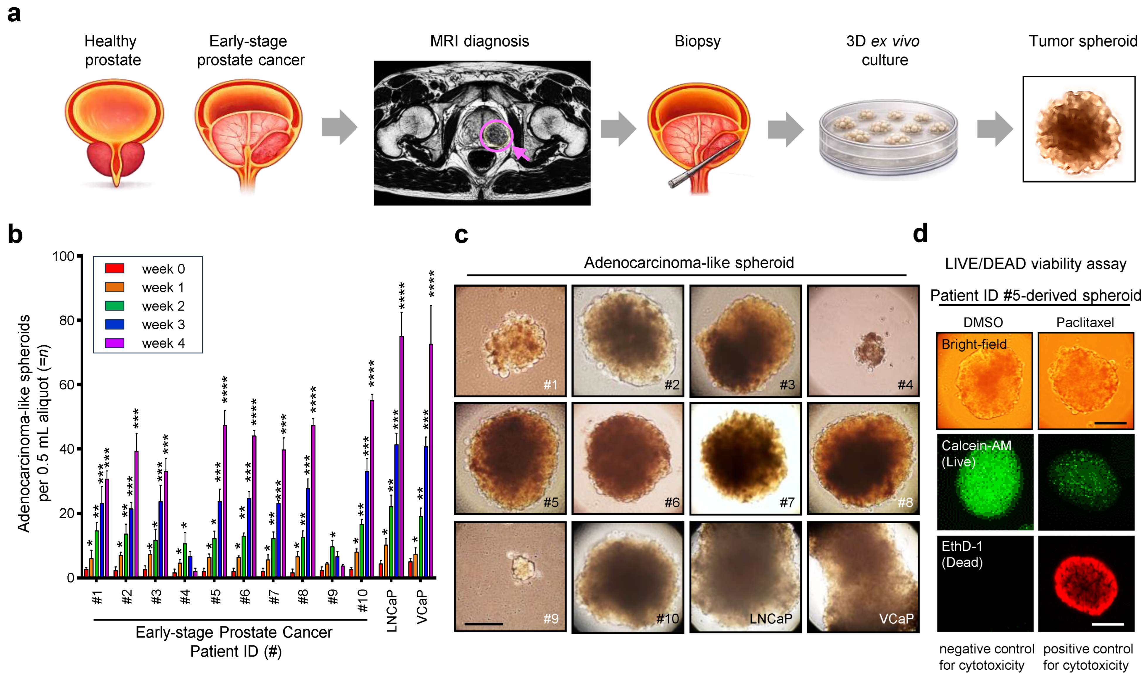
35]. In this context, successful spheroid generation from 8 of 10 MRI-guided biopsy specimens provides more than a technical platform; it creates an experimental window into early adaptive events that are usually inaccessible until after CRPC or t-NEPC has already emerged clinically.
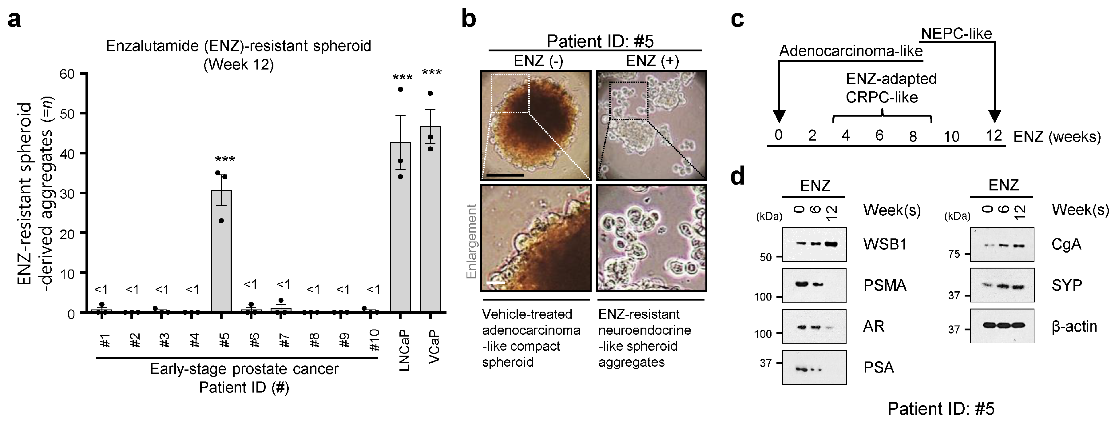
The emergence of an enzalutamide-resistant, t-NEPC-like derivative from a single patient-derived culture should be interpreted cautiously but is biologically informative. De novo NEPC is rare, accounting for less than 2% of prostate cancer at diagnosis, whereas treatment-emergent neuroendocrine or small-cell transformation has been reported in approximately 10–20% of metastatic CRPC cohorts exposed to potent androgen receptor pathway suppression [
10,
11,
12,
19,
58,
59]. These tumors are clinically aggressive, frequently androgen receptor-indifferent, often associated with low PSA relative to tumor burden, and poorly controlled by standard androgen receptor-directed therapy [
10,
11,
12,
13,
14,
15,
16,
17,
18,
19,
58,
59]. Thus, the rarity of this transition in our model is consistent with the idea that only a subset of early prostate tumors contains sufficient lineage plasticity or pre-existing drug-tolerant potential to undergo neuroendocrine-like remodeling. Our model does not imply that all early prostate cancers follow this path; instead, it provides a tractable system to study a clinically consequential but difficult-to-capture evolutionary route.
The PSMA component of this mechanism has direct clinical relevance. PSMA is highly expressed in most prostate adenocarcinomas and has become a central theranostic target for PET imaging and radioligand therapy [
20,
21,
22,
23,
24,
25,
26,
27]. Nevertheless, both PSMA expression and PSMA-ligand uptake can be heterogeneous, and low or negative signal in selected lesions may limit PSMA-based detection or eligibility for radioligand therapy [
60,
61,
62]. In advanced disease, low PSMA expression is associated with poor outcomes in PSMA-directed treatment settings [
27]. Neuroendocrine differentiation adds another layer of complexity because it is frequently associated with PSMA suppression [
28]. Therefore, PSMA loss during enzalutamide-induced neuroendocrine-like progression represents not only a lineage change but also the potential loss of a diagnostic and therapeutic entry point. Restoring or preserving PSMA may create a second therapeutic opportunity for tumors that would otherwise become less visible and less targetable.
Consistent with prior evidence that WSB1 regulates malignant adaptation and metastatic progression through ubiquitin-dependent proteostasis [
41,
42,
63,
64,
65], the present study provides convergent temporal, loss-of-function, cross-model, and reconstitution evidence linking WSB1 to the emergence of a PSMA-low resistant state. WSB1 increased as PSMA declined during enzalutamide selection (
Figure 2d), and
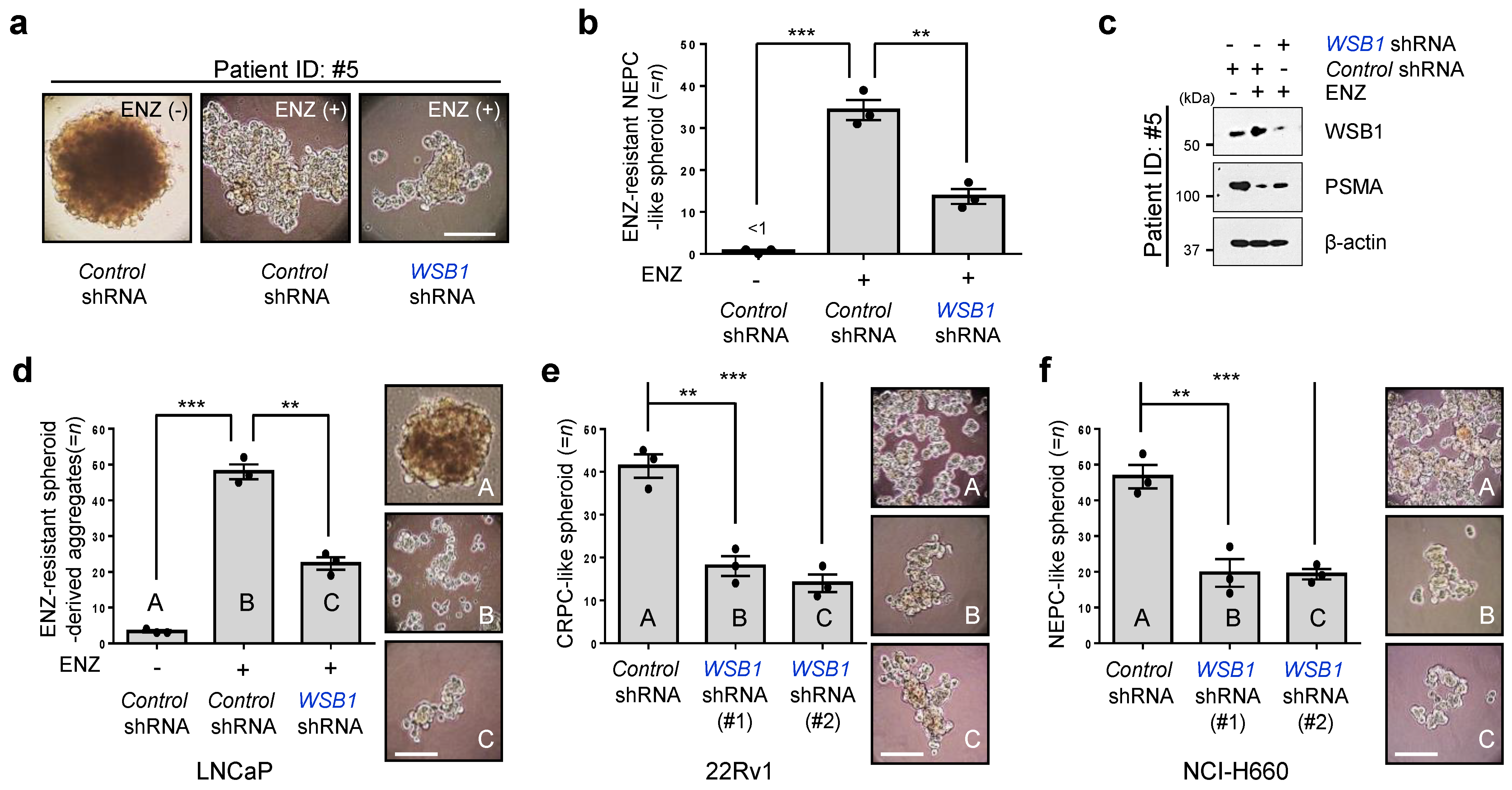
WSB1 depletion initiated at week 6, when WSB1 induction first became evident, reduced subsequent resistant aggregate formation and restored PSMA expression at week 12 in Patient ID #5-derived spheroids (
Figure 3a–c).
WSB1 silencing also reduced spheroid or aggregate growth in enzalutamide-resistant LNCaP, 22Rv1, and NCI-H660 models (
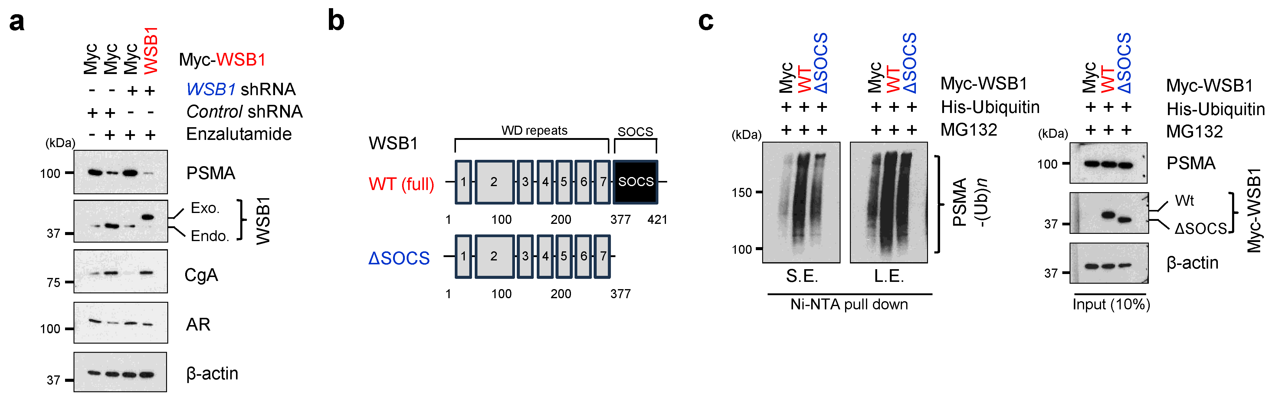
Figure 3d–f), indicating that the observed dependency was not confined to a single patient-derived culture. Conversely, WSB1 reconstitution in LNCaP cells reinstated a PSMA-low, AR-low, CgA-high molecular profile (
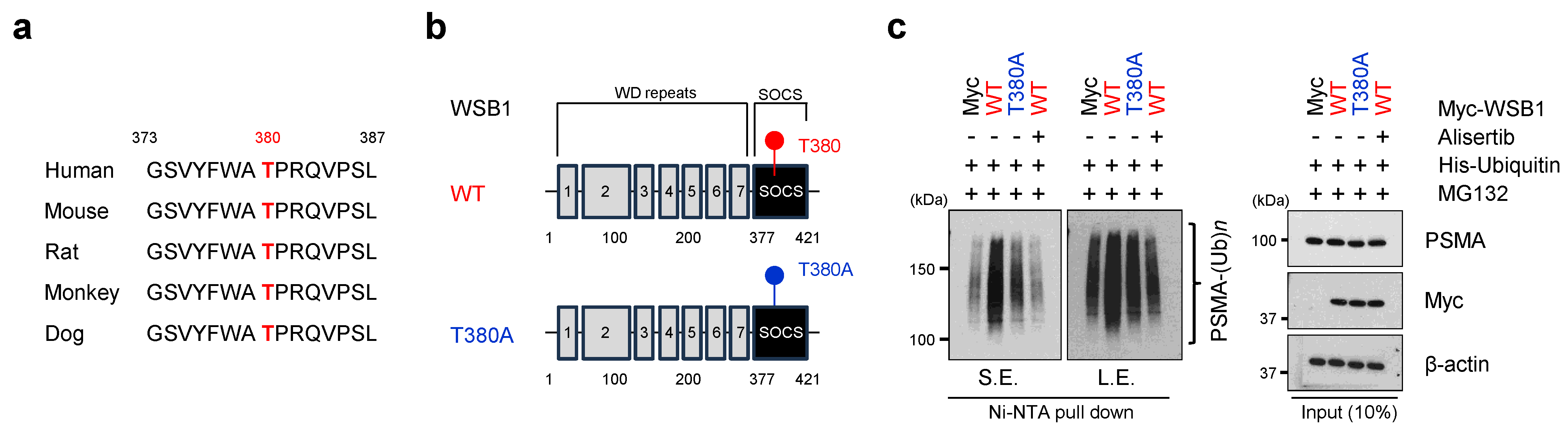
Figure 4a), supporting an on-target and reversible WSB1-dependent phenotype. Together, these findings position WSB1 as a functional contributor to progression from early enzalutamide adaptation toward a resistant PSMA-low state, rather than as a passive marker of lineage remodeling. Biochemically, the data place PSMA within a WSB1-dependent ubiquitination pathway. Wild-type WSB1 increased high-molecular-weight ubiquitinated PSMA species, whereas deletion of the SOCS box strongly reduced this activity (
Figure 4c), indicating that recruitment of the E3 ligase machinery is required for efficient PSMA ubiquitination. The T380A mutant also displayed reduced activity toward PSMA, and alisertib attenuated WSB1-associated PSMA ubiquitination under the tested conditions (
Figure 5c). These findings support a model in which the SOCS box provides the essential ubiquitin-ligase adaptor function, while the conserved T380 region and AURKA-sensitive signaling modulate WSB1 activity toward PSMA. However, the current data do not establish that AURKA directly phosphorylates WSB1 at T380, which was previously linked to CDK-dependent phosphorylation [
42]. Likewise, endogenous WSB1–PSMA interaction, PSMA protein half-life, proteasome-dependent rescue, and ubiquitin-linkage analyses will be required to establish PSMA as a bona fide WSB1 substrate.
Current treatment options for NEPC and aggressive-variant prostate cancer remain limited; platinum-based chemotherapy is commonly used, but responses are often transient and durable targeted strategies are lacking [
10,
11,
12,
13,
14,
15,
16,
17,
18,
19,
58,
59]. Against this background, the intervention studies distinguish prevention of resistant progression from treatment of an established refractory state. In
Figure 3,
WSB1 depletion initiated during early enzalutamide adaptation limited subsequent development of the PSMA-low resistant phenotype. By contrast, the experiments in
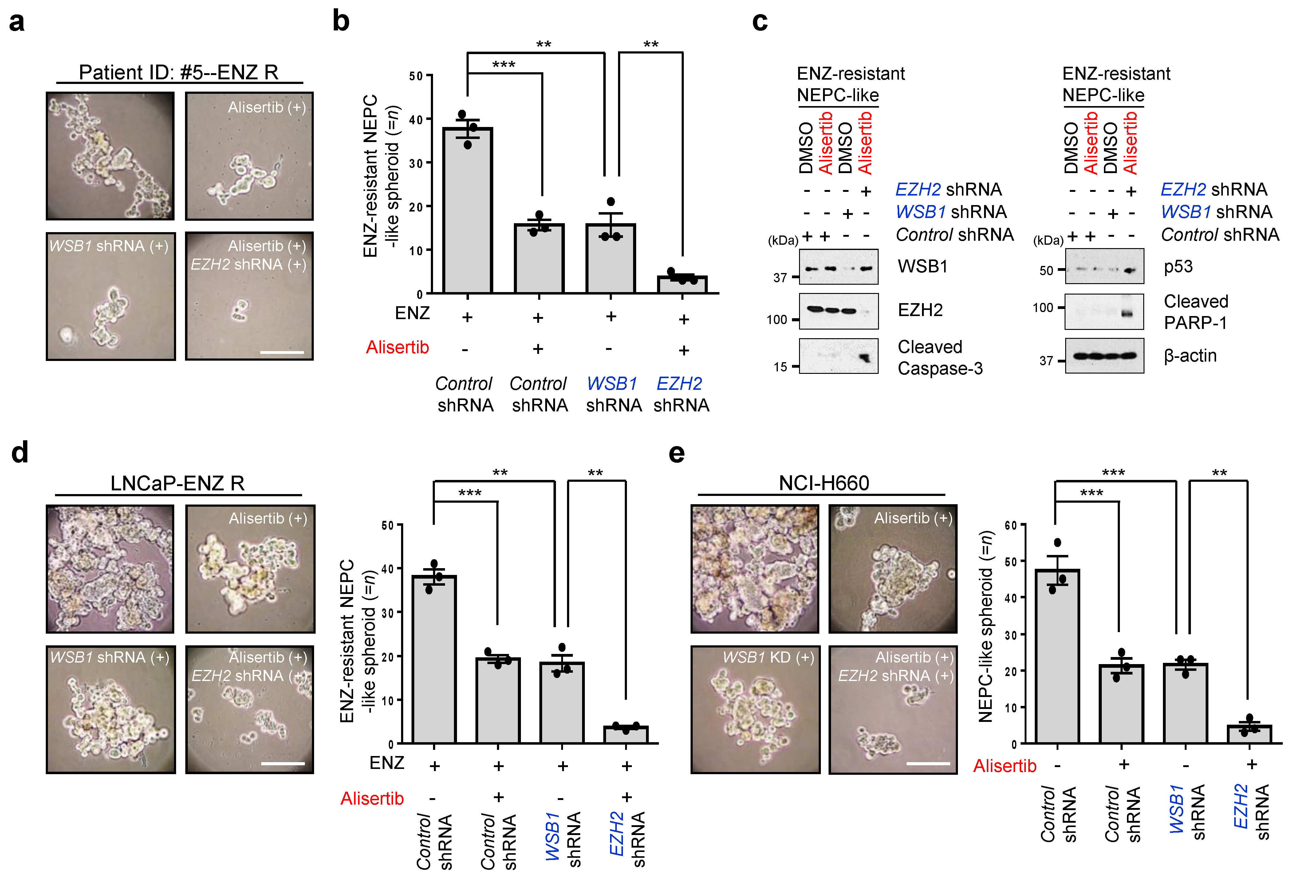
Figure 6 were performed after enzalutamide-resistant cultures had been established and therefore tested whether the refractory state remained therapeutically vulnerable. Under these conditions, AURKA inhibition with alisertib,
WSB1 suppression, and
EZH2 depletion each reduced resistant spheroid or aggregate growth, while combined alisertib treatment with
EZH2 suppression produced the greatest reduction among the tested conditions without establishing formal pharmacologic synergy. This stage-specific vulnerability is consistent with the established involvement of AURKA–MYCN signaling and EZH2-associated epigenetic reprogramming in aggressive-variant and neuroendocrine prostate cancer [
16,
17,
43,
44,
45,
46,
54,
55,
56,
57]. Rather than identifying WSB1 as a solitary determinant, the data support a model in which AURKA-sensitive signaling, WSB1-dependent proteolysis, and EZH2-associated chromatin maintenance constitute cooperating dependencies of the refractory state.
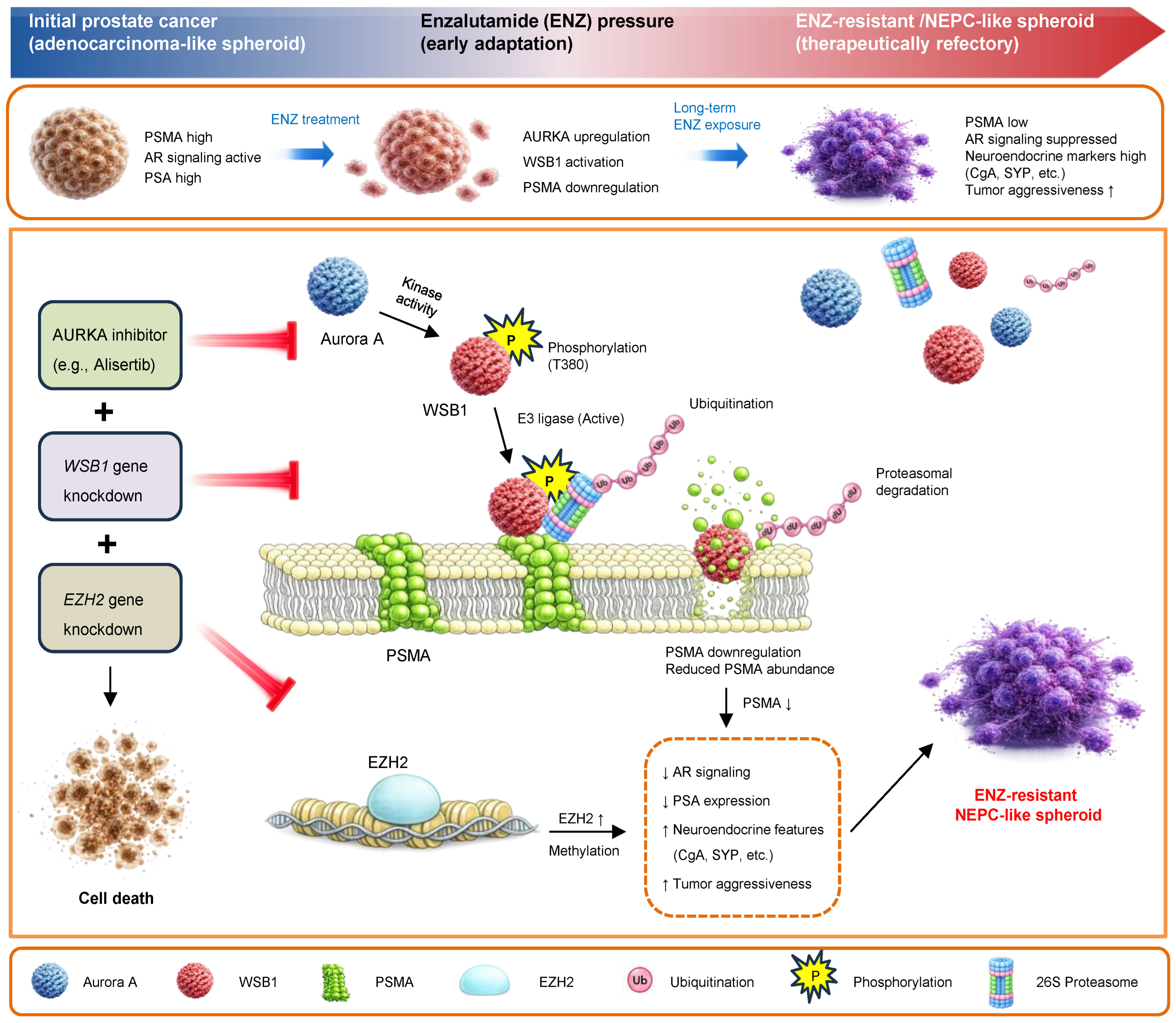
Figure 7 summarizes this framework. Intercepting WSB1 during early adaptation may constrain resistant progression, whereas targeting these convergent dependencies after resistance has emerged may reduce resistant aggregate growth and increase apoptosis-associated signaling in established NEPC-like cultures.
Several limitations should be considered. First, the strongest patient-derived resistant phenotype was observed in Patient ID #5, limiting generalizability. Because t-NEPC is heterogeneous and relatively uncommon even in metastatic CRPC cohorts, this single patient-derived transition model should be viewed as a discovery platform rather than a prevalence estimate [
10,
11,
12,
19,
58,
59]. Second, the resistant phenotype should remain designated as NEPC-like or t-NEPC-like rather than definitive t-NEPC. CgA/SYP induction with AR/PSA/PSMA loss provides a strong neuroendocrine-like signature, but definitive classification requires broader pathologic validation, including insulinoma-associated protein 1, CD56/neural cell adhesion molecule 1, achaete-scute family basic helix-loop-helix transcription factor 1, neuron-specific enolase, NKX3.1 loss, and histologic evaluation [
59,
66]. Additional MYCN, AURKA, and EZH2 profiling, together with transcriptomic comparison to clinical t-NEPC, would further strengthen lineage-state assignment [
16,
17,
43,
54,
55,
56,
57]. Additional biopsy-derived models linked to clinical pathology, genomic profiling, and treatment history will be essential to determine whether WSB1 induction is recurrent or patient-specific. Third, the therapeutic perturbation studies evaluated spheroid or aggregate burden and apoptosis-associated protein cleavage but did not include a direct post-treatment metabolic or fluorescence-based viability assay. Although the concordant reduction in aggregate growth across three prostate cancer models and the induction of cleaved caspase-3 and cleaved PARP-1 support growth-inhibitory and pro-apoptotic effects, these endpoints do not constitute a quantitative measurement of absolute cell viability or cytotoxic potency. Accordingly, the present findings should be interpreted as evidence of reduced resistant aggregate growth and apoptosis-associated signaling rather than as a direct viability endpoint.
Additional mechanistic and translational validation could address three points. First,
FOLH1 transcription, total and surface PSMA abundance, protein turnover, ligand-binding capacity, and radioligand uptake should be evaluated in parallel to distinguish transcriptional repression from post-translational degradation and to determine whether restored PSMA is functionally targetable [
28,
54,
55,
56,
57]. Second, the AURKA–WSB1 relationship requires orthogonal genetic and biochemical validation, including AURKA perturbation, kinase-dead AURKA and Aurora kinase B controls, phospho-WSB1 mapping, additional AURKA-selective inhibitors, and in vitro kinase analysis. Third, future therapeutic studies should incorporate direct three-dimensional metabolic or fluorescence-based viability assays with appropriate biological replication, together with dose–response analysis, treatment-withdrawal regrowth assays, and formal combination testing. Future animal studies should then evaluate the two temporally distinct intervention settings identified in the present study. In an early-intervention model, PSMA-high LNCaP or patient-derived adenocarcinoma-like spheroids would be implanted into male immunodeficient mice, followed by androgen deprivation and continuous enzalutamide exposure after tumor establishment. Inducible
WSB1 depletion would be initiated after early WSB1 induction and PSMA decline but before overt resistance to determine whether WSB1 suppression delays progression toward an enzalutamide-resistant, NEPC-like state. In an established-resistance model, LNCaP-derived enzalutamide-resistant and NCI-H660 xenografts, together with Patient ID #5-derived resistant spheroid xenografts if sufficient viable material and engraftment are achieved, would be randomized to vehicle, alisertib, inducible
WSB1 depletion,
EZH2 inhibition, or combined AURKA/EZH2 targeting. Longitudinal tumor growth and time to progression would be integrated with serial PSMA PET imaging and endpoint analyses of WSB1, PSMA, AR, CgA, SYP, Ki67, and cleaved caspase-3. If pathway inhibition restores tumor PSMA uptake, subsequent treatment with
177Lu-PSMA-617 would determine whether the restored PSMA is functionally accessible for radioligand therapy [
23,
24,
25,
26]. Complementary Pten/Rb1/Trp53- or MYCN/EZH2-based genetically engineered models could subsequently evaluate WSB1-dependent lineage plasticity in an immunocompetent microenvironment [
13,
14,
16,
17]. These studies will determine whether the pathway identified here represents a reproducible therapeutic dependency rather than a context-specific feature of the current models.
Overall, these findings define a stage-resolved model in which WSB1 is induced during early enzalutamide adaptation, promotes PSMA loss through a SOCS box-dependent ubiquitination program, and remains embedded within a therapeutically actionable signaling network after resistance is established. Validation in larger patient-derived cohorts and in vivo models will determine whether targeting this network can delay neuroendocrine-like progression or restore PSMA-directed theranostic sensitivity.
4. Materials and Methods
4.1. Human Prostate Cancer Biopsy Specimens and Ethical Approval
Human prostate biopsy specimens were obtained at Mayo Clinic under an Institutional Review Board-approved protocol (Mayo Clinic IRB 24-006965; approved 30 October 2024). Patient enrollment began on 18 April 2025. Written informed consent was obtained from all participants before research tissue collection. Study procedures were conducted in accordance with the Declaration of Helsinki and approved institutional guidelines. A total of 14 patients undergoing clinically indicated magnetic resonance imaging (MRI)-guided prostate biopsy for suspected or diagnosed early-stage prostate cancer were enrolled. Of these, research biopsy specimens from 10 patients were ultimately available and included in the present study for three-dimensional (3D) spheroid establishment and downstream experimental analyses. Specimens from four enrolled patients were not included because the clinical evaluation identified normal findings in some cases, biopsy scheduling was postponed in some cases, or an additional research core could not be provided because of limited biopsy core availability. Biopsy procedures were performed as part of standard clinical care using in-bore MRI-guided biopsy within a combined targeted/systematic biopsy approach. Research specimens were obtained from imaging-defined areas of clinical suspicion. After completion of clinically required diagnostic sampling, an additional research biopsy core was collected when feasible and without compromising clinical diagnostic evaluation. Because viable tissue was required for ex vivo culture, research specimens were transferred for experimental processing immediately after collection. Patient-related information was de-identified before laboratory processing, and all specimens were assigned coded study identifiers. Fresh biopsy tissues were collected in collaboration with the clinical MRI-guided biopsy team and transferred immediately for downstream experimental processing.
4.2. Biopsy Tissue Processing, Growth Factor-Enriched 3D Spheroid Culture, Morphologic Analysis, and Long-Term Enzalutamide Selection
Fresh prostate biopsy tissues allocated for research were transferred immediately after collection into pre-chilled sterile transport medium and maintained on ice until processing. Samples were transported to the laboratory at 4 °C and processed as rapidly as possible to minimize ischemic injury. Under sterile conditions, tissues were washed three times with cold phosphate-buffered saline (PBS) supplemented with 1% penicillin-streptomycin to remove blood, mucus, and tissue debris. Because MRI-guided prostate biopsy cores contain limited viable tumor material and are highly vulnerable to ischemic and mechanical stress, all procedures were performed gently to preserve multicellular tissue fragments, epithelial/tumor-cell clusters, and native cell–cell interactions. Biopsy tissues were mechanically fragmented on ice into approximately 0.5–1.0 mm tissue pieces using sterile microdissection instruments. Complete single-cell dissociation was intentionally avoided. To release compact epithelial/tumor clusters while minimizing overdigestion, tissue fragments were subjected to mild enzymatic digestion in Advanced DMEM/F12 (Gibco, Thermo Fisher Scientific, Waltham, MA, USA) containing Collagenase/Hyaluronidase (STEMCELL Technologies, Vancouver, BC, Canada; 1:10 dilution from stock), DNase I (Sigma-Aldrich, St. Louis, MO, USA; 20 μg/mL), and Y-27632 (Selleck Chemicals, Houston, TX, USA; 10 μM) for 20–30 min at 37 °C with gentle rocking. Digestion was terminated by adding excess cold spheroid culture medium, and tissue fragments were collected by low-speed centrifugation at 300× g for 5 min at 4 °C. The pellet was gently resuspended without vigorous pipetting to retain multicellular fragments and tumor-cell clusters. Processed fragments were seeded into 6-well 3D culture plates using either NunclonTM SpheraTM Low-Attachment Multidishes (Thermo Fisher Scientific, Waltham, MA, USA; Cat. No. 174932) or NanoCulture plates (SCIVAX Corporation, Kawasaki, Kanagawa, Japan). Plate type was recorded for each culture, and all within-experiment comparisons were performed using the same 3D plate format to minimize plate-dependent differences in spheroid formation.
Primary cultures were maintained in serum-free, growth factor-enriched prostate spheroid medium adapted from established prostate organoid culture systems, with modifications for low-attachment biopsy-derived spheroid culture rather than long-term extracellular matrix-embedded organoid expansion [
29,
30,
31,
32,
33]. The basal medium consisted of Advanced DMEM/F12 supplemented with 1× B27 supplement, 1.25 mM of N-acetyl-L-cysteine (Sigma-Aldrich, St. Louis, MO, USA), 10 mM of HEPES (Gibco, Waltham, MA, USA), 2 mM of GlutaMAX (Thermo Fisher Scientific, Waltham, MA, USA), and 1% penicillin-streptomycin (Gibco, Waltham, MA, USA). To support prostate epithelial survival, androgen-lineage maintenance, and recovery from biopsy-associated stress, the medium was supplemented with epidermal growth factor (EGF; 50 ng/mL; PeproTech, Cranbury, NJ, USA), fibroblast growth factor 10 (FGF10; 10 ng/mL; PeproTech), fibroblast growth factor 2 (FGF2; 5 ng/mL; PeproTech), dihydrotestosterone (DHT; 1 nM; Sigma-Aldrich), nicotinamide (10 mM; Sigma-Aldrich), A83-01 (500 nM; Tocris Bioscience, Abingdon, UK), SB202190 (10 μM; Sigma-Aldrich), Y-27632, an inhibitor of Rho-associated coiled-coil-containing protein kinases 1 and 2 (ROCK1/2) (10 μM; Selleck Chemicals), prostaglandin E2 (PGE2; 1 μM; Tocris Bioscience), recombinant R-spondin 1 (500 ng/mL; R&D Systems, Minneapolis, MN, USA), and recombinant Noggin (100 ng/mL; R&D Systems, Minneapolis, MN, USA). The medium also contained A83-01, an inhibitor of transforming growth factor-β type I receptor/ALK5 and the related ALK4 and ALK7 kinases (500 nM; Tocris Bioscience); SB202190, a p38 mitogen-activated protein kinase inhibitor (10 μM; Sigma-Aldrich); and Y-27632, a ROCK1/2 inhibitor (10 μM; Selleck Chemicals). This cocktail was selected based on prostate organoid media that support prostate epithelial and prostate cancer organoid growth through EGF/FGF signaling, Wnt potentiation, bone morphogenetic protein inhibition, transforming growth factor-β/activin receptor signaling inhibition, p38 mitogen-activated protein kinase inhibition, ROCK1/2 inhibition, and androgen pathway support [
29,
30,
31,
32,
33]. Cultures were maintained in a final volume of 4.0 mL per well. At each medium exchange, 3.5 mL of fresh growth factor-enriched spheroid medium was added together with 0.5 mL of retained autologous conditioned medium from the same well to preserve locally secreted autocrine and paracrine factors while allowing nutrient replenishment. Cultures were maintained at 37 °C in a humidified incubator containing 5% CO
2, and medium was refreshed every 3–5 days using the same conditioned-medium retention strategy.
Spheroid formation was monitored for up to 4 weeks by bright-field and phase-contrast microscopy with an EVOS M5000 imaging system (Thermo Fisher Scientific, Waltham, MA, USA). Countable tumor spheroids were operationally defined as viable-appearing, compact, rounded or near-rounded multicellular aggregates with clear outer borders and a minimum diameter of 50 μm. Single cells, blood clots, acellular debris, necrotic fragments, and loose non-compact clusters were excluded from analysis. Tumor relevance and lineage state were supported by pathology confirmation of corresponding MRI-targeted diagnostic cores and by downstream marker analysis. Parental adenocarcinoma-like spheroids were evaluated using prostate lineage markers, including androgen receptor (AR), prostate-specific antigen (PSA), and prostate-specific membrane antigen (PSMA), whereas enzalutamide-resistant neuroendocrine-like derivatives were evaluated using chromogranin A (CgA), synaptophysin (SYP), AR, PSA, PSMA, and WSB1. For spheroid quantification, conditioned medium was gently mixed to resuspend floating spheroids without disrupting compact aggregates, and a standardized 0.5 mL aliquot was collected at each indicated time point. Spheroid numbers were determined by manual counting and/or image-analysis software. Where applicable, spheroid area, equivalent diameter, circularity, solidity, and boundary roughness were quantified using ImageJ version 1.54g (National Institutes of Health, Bethesda, MD, USA) or equivalent software. Equivalent circular diameter was calculated as 2 × √(area/π). For long-term drug selection, established patient-derived spheroid cultures were subjected to continuous enzalutamide (Ambeed, Inc., Arlington Heights, IL, USA; Cat. No. A270395; CAS No. 915087-33-1) treatment at a final concentration of 10 μM for up to 12 weeks. Vehicle controls were maintained with the equivalent dimethyl sulfoxide (DMSO; Sigma-Aldrich, St. Louis, MO, USA; Cat. No. D2650) concentration, with a final concentration of approximately 0.01%. Samples were collected at week 0, week 6, and week 12 for molecular analysis. Cultures that failed to maintain viable spheroid growth under enzalutamide were classified as enzalutamide-sensitive, whereas cultures that persisted and expanded under continuous drug pressure were classified as enzalutamide-resistant spheroid-derived aggregates. Scale bars were generated using microscope-calibrated pixel-to-micrometer conversion factors and preserved during image processing.
4.3. Tumor-Spheroid Viability Assay
During culture week 4, Patient ID #5-derived spheroids cultured under the three-dimensional conditions described above were treated with vehicle (dimethyl sulfoxide [DMSO]) or paclitaxel (Sigma-Aldrich, St. Louis, MO, USA; Cat. No. T7191) at a final concentration of 10 nM for 48 h. Vehicle-treated cultures received DMSO at the same final concentration used for paclitaxel treatment. Following treatment, spheroids were gently collected and washed twice with phosphate-buffered saline (PBS). Spheroid viability was assessed using the LIVE/DEADTM Cell Viability Assay (Invitrogen, Thermo Fisher Scientific, Waltham, MA, USA; Cat. No. L3224) according to the manufacturer’s instructions. Spheroids were incubated in staining solution containing 2 μM of calcein-AM and 4 μM of ethidium homodimer-1 (EthD-1) for 45 min at room temperature in the dark. After staining, the spheroids were washed twice with PBS and immediately examined by bright-field using an EVOS M5000 Imaging System (Thermo Fisher Scientific, Waltham, MA, USA) and by confocal fluorescence microscopy using an LSM 980 confocal microscope equipped with Airyscan 2 (Carl Zeiss Microscopy GmbH, Jena, Germany). Confocal fluorescence images of vehicle- and paclitaxel-treated spheroids were acquired using ZEN software, version 3.6 (Carl Zeiss Microscopy GmbH), with identical laser power, detector gain, and acquisition settings across all experimental groups. Calcein-AM fluorescence was used to identify viable cells, whereas EthD-1 fluorescence was used to identify dead cells with compromised plasma membranes. Vehicle-treated spheroids were used to assess baseline viability, whereas paclitaxel-treated spheroids served as a positive control for cell death.
4.4. Prostate Cancer Cell Lines and Cell Line-Derived 3D Spheroid Models
Human prostate cancer cell lines were obtained from the American Type Culture Collection (ATCC, Manassas, VA, USA) and maintained according to ATCC recommendations and established literature protocols [
47,
48,
49,
50]. LNCaP cells (ATCC CRL-1740), an androgen receptor-positive prostate adenocarcinoma model, were cultured in RPMI-1640 medium (Gibco, Thermo Fisher Scientific, Waltham, MA, USA) supplemented with 10% fetal bovine serum (FBS; Gibco), 100 U/mL of penicillin, and 100 μg/mL of streptomycin (Gibco) [
47]. VCaP cells (ATCC CRL-2876), used as an additional prostate adenocarcinoma reference model, were maintained in Dulbecco’s Modified Eagle Medium (DMEM; Gibco) supplemented with 10% FBS, 100 U/mL of penicillin, and 100 μg/mL of streptomycin [
48]. 22Rv1 cells (ATCC CRL-2505), used as an androgen receptor-positive castration-resistant prostate cancer-like reference model, were cultured in RPMI-1640 medium supplemented with 10% FBS, 100 U/mL of penicillin, and 100 μg/mL of streptomycin [
49]. NCI-H660 cells (ATCC CRL-5813), used as a neuroendocrine/small-cell prostate carcinoma reference model, were maintained in HITES medium supplemented with 5% FBS, insulin (5 μg/mL; Sigma-Aldrich, St. Louis, MO, USA; Cat. No. I9278), transferrin (10 μg/mL; Sigma-Aldrich; Cat. No. T8158), sodium selenite (30 nM; Sigma-Aldrich; Cat. No. S5261), hydrocortisone (10 nM; Sigma-Aldrich; Cat. No. H0888), β-estradiol (10 nM; Sigma-Aldrich; Cat. No. E2758), additional L-glutamine to a final concentration of 4 mM (Gibco, Thermo Fisher Scientific, Waltham, MA, USA; Cat. No. 25030081), 100 U/mL of penicillin, and 100 μg/mL of streptomycin (Gibco, Thermo Fisher Scientific; Cat. No. 15140122) [
50]. NCI-H660 cultures were maintained as viable floating clusters and were not over-diluted during passaging, consistent with their aggregate-forming growth pattern. All cell cultures were maintained at 37 °C in a humidified incubator containing 5% CO
2 and were passaged before reaching overconfluence or excessive acidification of the culture medium. Cells were periodically renewed from authenticated frozen master stocks, and cultures were routinely screened for mycoplasma contamination; only mycoplasma-negative cultures were used for experiments.
For cell line-derived 3D culture experiments, LNCaP, VCaP, 22Rv1, and NCI-H660 cells were adapted to spheroid or aggregate culture using the same 3D culture plate formats used for patient-derived spheroid experiments, including NunclonTM SpheraTM Low-Attachment Multidishes or NanoCulture plates. LNCaP and VCaP cells were used as prostate adenocarcinoma spheroid controls, 22Rv1 cells were used to model a CRPC-like androgen receptor-positive state, and NCI-H660 cells were used as a neuroendocrine/small-cell prostate carcinoma aggregate reference. For 3D assays, cells were harvested under conditions that preserved viability and minimized excessive single-cell stress, counted, and seeded into 6-well 3D culture plates in their corresponding complete culture media unless otherwise indicated. LNCaP-derived enzalutamide-resistant cultures were generated by long-term enzalutamide exposure, using the same selection strategy applied to patient-derived spheroids. Cell line-derived spheroids or aggregates were monitored by bright-field microscopy and analyzed by spheroid/aggregate counting, morphologic assessment, and immunoblotting. For within-experiment comparisons, the same cell line, medium condition, plate format, seeding density, and drug-treatment schedule were used across experimental groups to minimize culture-condition-dependent variability.
4.5. Gene Knockdown and Plasmid Expression
WSB1 and
EZH2 expressions were suppressed using short hairpin RNA (shRNA)-based gene silencing. Non-targeting shRNA in the corresponding vector backbone was used as a negative control. Human
WSB1-targeting shRNA constructs were obtained from Open Biosystems (Huntsville, AL, USA) and have been described previously [
41,
42]. Myc-tagged WSB1 expression constructs were also described previously [
41,
42]. Two independent human
WSB1 shRNA constructs were used where indicated:
WSB1 shRNA #1, 5′-GCTGTTGACAGTGAGCGCGGAGTTTCTCTCGTATCGTATTAGTGAAGCCACAGATGTAATACGATACGAGAGAAACTCCATGCCTACTGCCTCGGA-3′; and
WSB1 shRNA #2, 5′-GCTGTTGACAGTGAGCGCGCTGTAAAGTGCAAGGAAATTTAGTGAAGCCACAGATGTAAATTTCCTTGCACTTTACAGCATGCCTACTGCCTCGGA-3′ [
41,
42].
EZH2 was depleted using the following human
EZH2-targeting shRNA sequence: 5′-CCGGTATGATGGTTAACGGTGATCACTCGAGTGATCACCGTTAACCATCATATTTTTG-3′. For stable
WSB1 or
EZH2 suppression, lentiviral particles were generated in HEK293T packaging cells by co-transfecting the indicated shRNA transfer plasmids with lentiviral packaging and envelope plasmids using Lipofectamine
TM 2000 or Lipofectamine
TM 3000 (Invitrogen, Thermo Fisher Scientific, Waltham, MA, USA). Viral supernatants were collected, clarified by centrifugation and filtration, and applied to prostate cancer cells or spheroid-derived cultures under aseptic conditions. For
WSB1 knockdown experiments, the non-targeting control and
WSB1-targeting shRNAs were expressed from the same puromycin-selectable vector backbone, and both groups underwent identical lentiviral transduction and antibiotic-selection procedures. For the experiments examining WSB1 function during enzalutamide adaptation, Patient ID #5-derived cultures were transduced at week 6 of continuous enzalutamide exposure, when WSB1 induction was first detected in the time-course analysis. Parallel vehicle-treated cultures were transduced at the corresponding time point. Following transduction, cultures were selected with puromycin (InvivoGen, San Diego, CA, USA; Cat. Code ant-pr-1) at a final concentration of 2 μg/mL until stable puromycin-resistant pools were established. The selected cultures were maintained under their respective vehicle or continuous enzalutamide conditions and analyzed at week 12, corresponding to six weeks after lentiviral transduction. The timing of genetic perturbation in fully established enzalutamide-resistant cultures used for therapeutic studies is described in
Section 4.8.
For rescue and gain-of-function experiments, empty Myc vector, Myc-tagged WSB1 wild type, Myc-tagged WSB1 ΔSOCS, and Myc-tagged WSB1 T380A constructs were introduced as indicated. Myc-WSB1 ΔSOCS was used to assess the requirement of the SOCS box region, whereas Myc-WSB1 T380A was used to evaluate the contribution of the conserved T380 residue. For ubiquitination assays, His-tagged ubiquitin was co-expressed with the indicated WSB1 constructs. For transient plasmid expression in LNCaP cells, DNA constructs were introduced using LipofectamineTM 2000 or LipofectamineTM 3000 (Invitrogen/Thermo Fisher Scientific) according to the manufacturer’s instructions. Transfection conditions were empirically optimized to achieve reproducible gene delivery with minimal cytotoxicity. Mock-transfected and vehicle-only controls were included where appropriate to monitor reagent-related effects. Transfection efficiency was verified by fluorescence microscopy when reporter-tagged constructs were used and by immunoblotting for target protein expression. Transiently transfected cells were typically analyzed 48–72 h after transfection. The efficiency of WSB1 depletion, EZH2 suppression, and ectopic WSB1 reconstitution was confirmed by immunoblotting for WSB1, EZH2, and Myc-tag, respectively. PSMA, AR, and CgA were analyzed as downstream molecular markers, and β-actin was used as a loading control, as appropriate.
4.6. Immunoblotting and Antibodies
Spheroids, spheroid-derived aggregates, and cultured prostate cancer cells were collected at the indicated time points and washed twice with ice-cold phosphate-buffered saline (PBS). Spheroid and aggregate cultures were collected by gentle, low-speed centrifugation to minimize mechanical disruption. Total protein lysates were prepared in radioimmunoprecipitation assay (RIPA) buffer containing 50 mM Tris-HCl, pH 7.4, 150 mM NaCl, 1% NP-40/Igepal CA-630, 0.5% sodium deoxycholate, and 0.1% sodium dodecyl sulfate (SDS), freshly supplemented with cOmplete
TM Mini, EDTA-free Protease Inhibitor Cocktail (Roche Diagnostics GmbH, Mannheim, Germany; Cat. No. 11836170001) and phosphatase inhibitors consisting of 10 mM β-glycerophosphate, 10 mM sodium fluoride, and 1 mM sodium orthovanadate. Lysates were incubated on ice for 20–30 min with intermittent mixing and clarified by centrifugation at 12,000–14,000×
g for 10–15 min at 4 °C. Protein concentrations were determined using the Bradford assay with Bio-Rad Protein Assay Dye Reagent Concentrate (Bio-Rad Laboratories, Hercules, CA, USA; Cat. No. 5000006). Immunoblotting procedures were adapted from previously described WSB1 studies, with modifications for prostate cancer spheroid and cell-line lysates [
41,
42]. Equal amounts of protein, typically 10–30 μg per lane depending on sample availability, were mixed with SDS sample buffer containing β-mercaptoethanol, denatured at 97 °C for 5 min, and immediately chilled on ice. Proteins were resolved by SDS-polyacrylamide gel electrophoresis using 4–15% Mini-PROTEAN® TGX
TM Precast Protein Gels, 15-well, 15-μL format (Bio-Rad Laboratories; Cat. No. 4561086), at 80–120 V for 1–2 h in an ice-cooled electrophoresis tank. Proteins were transferred for 7 min onto 0.2-μm nitrocellulose membranes using a Trans-Blot® Turbo
TM RTA Mini 0.2-μm Nitrocellulose Transfer Kit (Bio-Rad Laboratories; Cat. No. 1704270) and a Trans-Blot Turbo semi-dry transfer system (Bio-Rad Laboratories; Cat. No. 1704150). Membranes were blocked with 5% nonfat dry milk for 1 h at room temperature, washed three times with Tris-buffered saline containing 0.1% Tween-20 (TBS-T), and incubated overnight at 4 °C with the indicated primary antibodies, typically diluted 1:500–1:1000. After three washes with TBS-T, the membranes were incubated with the appropriate HRP-conjugated secondary antibodies for 1 h at room temperature. Depending on the host species of the primary antibody, the secondary antibodies included goat anti-rabbit IgG H&L (HRP) (Abcam, Cambridge, UK; Cat. No. ab6721), goat anti-mouse IgG H&L (HRP) (Abcam; Cat. No. ab6789), anti-rabbit IgG HRP-linked antibody (Cell Signaling Technology, Danvers, MA, USA; Cat. No. 7074), or anti-mouse IgG HRP-linked antibody (Cell Signaling Technology; Cat. No. 7076). Chemiluminescent signals were developed using Pierce
TM ECL Western Blotting Substrate (Thermo Fisher Scientific, Waltham, MA, USA; Cat. No. 32209) and recorded under nonsaturating exposure conditions on PR1MA
TM Blue autoradiography and chemiluminescence film, 8 × 10 inches (Midwest Scientific, Fenton, MO, USA; Cat. No. BX810). When required, membranes were stripped and reprobed according to standard procedures. Band intensities were quantified by densitometry using ImageJ, version 1.54g (National Institutes of Health, Bethesda, MD, USA), and normalized to β-actin unless otherwise indicated. The following primary antibodies were used: WSB1 (Proteintech, Rosemont, IL, USA, #11666-1-AP; Abcam, Cambridge, UK, ab68953; Sigma, HPA003293), prostate-specific membrane antigen (PSMA; Thermo Fisher Scientific/Invitrogen, Waltham, MA, USA, #37-3900; Abcam, AB133579), androgen receptor (AR; Cell Signaling Technology, Danvers, MA, USA, #3202; Santa Cruz Biotechnology, sc-7305), prostate-specific antigen (PSA; Abcam, #ab53774; Abcam, AB76113), chromogranin A (CgA; Santa Cruz Biotechnology, sc-393941; Novus Biologicals, Centennial, CO, USA, NBP2-33198), synaptophysin (SYP; Cell Signaling Technology, #4329; Santa Cruz Biotechnology, sc-12737), EZH2 (Cell Signaling Technology, #5246; Proteintech, # 21800-1-AP), cleaved caspase-3 (Cell Signaling Technology, #9661; Abcam, AB32042), p53 (Abcam, ab131442; Cell Signaling Technology, #9282), cleaved poly(ADP-ribose) polymerase 1 (PARP-1; Cell Signaling Technology, #9541; ABclonal, Woburn, MA, USA, A19612), Myc-tag (Cell Signaling Technology, #3400), and β-actin (Cell Signaling Technology, #3700S). Primary antibodies were used at manufacturer-recommended or empirically optimized dilutions, typically ranging from 1:500 to 1:1000 for target proteins and 1:2000 to 1:5000 for β-actin. Horseradish peroxidase-conjugated anti-rabbit and anti-mouse secondary antibodies were used at 1:2000 to 1:5000 dilutions. β-actin served as the loading control for total lysates unless otherwise specified.
4.7. Ubiquitination Assay and Denaturing Ni-NTA Pull-Down
To determine whether WSB1 promotes PSMA ubiquitination, denaturing nickel-nitrilotriacetic acid (Ni-NTA) pull-down assays were performed in LNCaP cells. This assay was adapted from previously described WSB1 ubiquitination protocols with modifications for prostate cancer cells and PSMA detection [
41,
42]. LNCaP cells were seeded to approximately 60–70% confluence and co-transfected with His-tagged ubiquitin together with empty Myc vector, Myc-tagged WSB1 wild type, Myc-tagged WSB1 ΔSOCS, or Myc-tagged WSB1 T380A constructs, as indicated. Transfections were performed using Lipofectamine
TM 2000 or Lipofectamine
TM 3000 according to the manufacturer’s instructions. Where indicated, cells were treated with alisertib (MedChemExpress, Monmouth Junction, NJ, USA; Cat. No. HY-10971) before harvest to assess Aurora kinase A-sensitive regulation of WSB1-dependent PSMA ubiquitination. To stabilize ubiquitinated intermediates, cells were treated with MG132 (Sigma-Aldrich, St. Louis, MO, USA; Cat. No. M7449) at 10 μM for 4 h before collection. Cells were washed twice with ice-cold phosphate-buffered saline (PBS) and lysed under denaturing conditions to enrich covalently ubiquitinated proteins while disrupting non-covalent protein interactions. Denaturing lysis was performed in 6 M of guanidine-HCl buffer containing 0.1 M of Na
2HPO
4/NaH
2PO
4, 0.1 M of Tris-HCl, pH 8.0, 300 mM of NaCl, 10 mM of imidazole, and 0.05% Tween-20, freshly supplemented with 20 mM of N-ethylmaleimide and 5 mM of iodoacetamide (all from Sigma-Aldrich) to inhibit deubiquitination and preserve ubiquitin conjugates. Lysates were briefly sonicated to reduce viscosity and clarified by centrifugation at 14,000×
g for 10 min at 4 °C. A fraction of each lysate was retained as input before pull-down. Clarified denatured lysates were incubated with Ni-NTA agarose beads (Qiagen, Hilden, Germany), pre-equilibrated in denaturing lysis buffer, for 4 h at room temperature with gentle rotation. Beads were washed sequentially with denaturing lysis buffer containing 20 mM of imidazole, followed by urea-based wash buffer containing 8 M of urea (Sigma-Aldrich), 0.1 M of Na
2HPO
4/NaH
2PO
4, 0.1 M of Tris-HCl, pH 6.3, 300 mM of NaCl, and 0.05% Tween-20 to reduce nonspecific binding. Bound His-ubiquitin-conjugated proteins were eluted by boiling the beads in 2× SDS sample buffer containing 200 mM of imidazole and 5% β-mercaptoethanol. Eluted proteins were resolved by SDS-polyacrylamide gel electrophoresis and analyzed by immunoblotting using anti-PSMA antibody to detect ubiquitinated PSMA species. Input lysates were analyzed in parallel for PSMA, Myc-tagged WSB1, and β-actin. Empty vector and non-His-ubiquitin controls were included where appropriate to assess nonspecific Ni-NTA binding and background signal. High-molecular-weight PSMA-reactive species in the Ni-NTA pull-down fraction were interpreted as ubiquitinated PSMA species.
4.8. Genetic and Pharmacological Perturbation of Established Enzalutamide-Resistant and Neuroendocrine-like Spheroid and Aggregate Cultures
Established enzalutamide-resistant and neuroendocrine-like prostate cancer spheroid and aggregate cultures were used for genetic and pharmacological perturbation studies. Patient ID #5-derived and LNCaP-derived enzalutamide-resistant cultures were first established by continuous enzalutamide selection for 12 weeks. After the resistant spheroid-derived aggregate phenotype had been established, cultures were transduced with lentiviruses encoding non-targeting control,
WSB1-targeting, or
EZH2-targeting shRNA and selected with puromycin at 2 μg/mL to generate stable shRNA-expressing pools. Patient ID #5-derived enzalutamide-resistant spheroid-derived aggregates and LNCaP-derived enzalutamide-resistant cultures were maintained under continuous enzalutamide pressure at 10 μM throughout treatment to preserve the drug-resistant state. NCI-H660 cells were used as a neuroendocrine/small-cell prostate carcinoma reference model and were maintained under their standard three-dimensional aggregate culture conditions without prior enzalutamide selection. Where indicated, stable control,
WSB1-targeting, or
EZH2-targeting shRNA-expressing NCI-H660 cultures were generated using the same lentiviral transduction and puromycin-selection procedure before experimental treatment. Before treatment, spheroid or aggregate cultures with comparable baseline aggregate density and morphology were allocated to the following experimental groups: non-targeting
control shRNA plus vehicle, non-targeting
control shRNA plus alisertib,
WSB1-targeting shRNA plus vehicle, or
EZH2-targeting shRNA plus alisertib. Alisertib (MLN8237), an Aurora kinase A inhibitor, was prepared as a dimethyl sulfoxide stock solution and freshly diluted into complete spheroid culture medium immediately before use. Based on published prostate cancer and neuroendocrine prostate cancer organoid/cell-line studies [
16,
17,
44], alisertib was used at a final concentration of 100 nM for 7 days, and drug-containing medium was replenished every 3–5 days during treatment. Vehicle-treated cultures received an equivalent final concentration of dimethyl sulfoxide. All comparisons within each model were performed using the same three-dimensional culture plate format, medium composition, and treatment schedule. Enzalutamide-resistant models were maintained under the same continuous enzalutamide condition throughout the experiment. At the experimental endpoint, treatment responses were evaluated by bright-field microscopy, quantitative spheroid or aggregate counting, and immunoblotting. Countable aggregates were defined as compact or semi-compact multicellular structures with clearly discernible boundaries. Single cells, cellular debris, necrotic fragments, and loose non-compact clusters were excluded from aggregate counting. Parallel cultures were collected for immunoblot analysis of WSB1, EZH2, cleaved caspase-3, p53, cleaved poly(ADP-ribose) polymerase 1 (PARP-1), and β-actin. β-actin was used as a loading control. Cleaved caspase-3 and cleaved PARP-1 were evaluated as apoptosis-associated markers, whereas p53 was assessed as a stress-response and tumor-suppressor pathway marker.
4.9. Statistical Analysis
Statistical analyses were performed using GraphPad Prism version 6.0 (GraphPad Software, Boston, MA, USA). Data are presented as mean ± standard error of the mean (SEM) unless otherwise indicated. Longitudinal spheroid counts in
Figure 1b were analyzed by two-way analysis of variance followed by Dunnett’s multiple-comparisons test, with each time point compared with week 0 within the corresponding culture. Multiple-group comparisons in
Figure 2a,
Figure 3b,d–f and
Figure 6b,d,e were analyzed by one-way analysis of variance followed by Tukey’s multiple-comparisons test. A
p value < 0.05 was considered statistically significant. Statistical significance is indicated as *
p < 0.05, **
p < 0.01, ***
p < 0.001, and ****
p < 0.0001.




 ,
,