Article

Virtual Screening, Synthesis and In Vitro Characterization of Histamine H4 Receptor Ligands Based on Pyrimidine Scaffolds

by
Olga Michalak
1,*,
Marcin Cybulski
1,
Piotr Krzeczyński
1,
Oliwia Zegrocka-Stendel
2,
Małgorzata Dutkiewicz
2,
Dorota Dymkowska
3,
Agnieszka Olejarz-Maciej
4,
Tadeusz Karcz
4,
Mariam Dubiel
5,
Pakhuri Mehta
6,†,
Marek Kubiszewski
7,
Marcin Lorkowski
6,
Jakub Jakowiecki
6,
Paweł Pasznik
6,
Przemysław Miszta
6,
Holger Stark
5,
Katarzyna Koziak
2 and
Sławomir Filipek
6,*
1
Pharmacy, Cosmetic Chemistry and Biotechnology Research Group, Łukasiewicz Research Network-Industrial Chemistry Institute, 8 Rydygiera Str., 01-793 Warsaw, Poland
2
Department of Biochemistry and Nutrition, Centre for Preclinical Research and Technologies, Medical University of Warsaw, 1b Stefana Banacha Str., 02-097 Warsaw, Poland
3
Laboratory of Cellular Metabolism, Nencki Institute of Experimental Biology, Polish Academy of Sciences, 3 Ludwika Pasteura Str., 02-093 Warsaw, Poland
4
Department of Chemical Technology and Biotechnology of Drugs, Jagiellonian University Medical College, 9 Medyczna Str., 30-688 Kraków, Poland
5
Institute for Pharmaceutical and Medicinal Chemistry, Heinrich Heine University Düsseldorf, Universitaetsstrasse 1, 40225 Duesseldorf, Germany
6
Faculty of Chemistry, Biological and Chemical Research Centre, University of Warsaw, 1 Pasteura Str., 02-093 Warsaw, Poland
7
Analytical Research Section, Pharmaceutical Analysis Laboratory, Łukasiewicz Research Network–Industrial Chemistry Institute, 8 Rydygiera Str., 01-793 Warsaw, Poland
*
Authors to whom correspondence should be addressed.
Current address: Venkateshwar Institute of Pharmacy, Sai Tirupati University, Ambua Road, Umarda, Girwa, Udaipur 313015, India.
Int. J. Mol. Sci. 2026, 27(15), 6892; https://doi.org/10.3390/ijms27156892
Submission received: 24 May 2026 / Revised: 17 July 2026 / Accepted: 23 July 2026 / Published: 1 August 2026

Abstract

Histamine is a biologically active monoamine acting through four G protein-coupled receptors (H1R–H4R), which represent attractive therapeutic targets for a range of diseases. Nowadays, H4R is recognized as a key player in inflammation and cancer. Here, we describe the design, syntheses and characterization of new H4R ligands containing pyrido[2,3-d]pyrimidine or pyrimidine scaffold. Candidate structures were scored in silico by docking to the structure of the human inactive H4R. Favorable structures were synthesized and, after confirmation of identity, their affinities were verified in in vitro screenings. Tested compounds did not exhibit any relevant cytotoxic (PrestoBlue) or antiproliferative (BrdU incorporation) effects at concentrations used in the functional assays. In a radioligand displacement assay (H4R affinity) the pyrimidine series showed lower binding, whereas several pyrido[2,3-d]pyrimidine derivatives and one pyrimidine analog produced >60% inhibition at 1 µM. Two similar compounds with the highest and moderate affinities were selected for further studies. In the Gi–cAMP pathway, both compounds behaved as moderate H4R antagonists, but in contrast, their profiles diverged in the β-arrestin pathway. These findings suggest ligand-dependent biased differences in signaling across G-protein and β-arrestin pathways at H4R. Overall, YAN-153 emerges as a promising lead structure for further optimization and for more detailed in vitro (e.g., metabolic stability, selectivity) and in vivo studies.

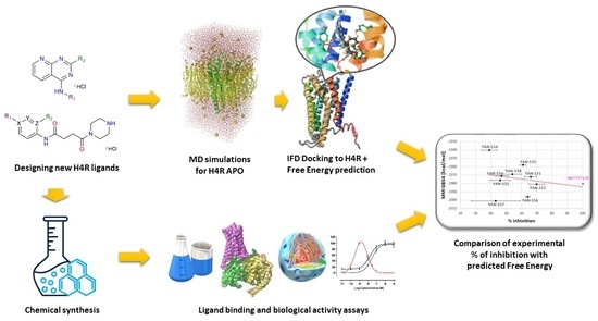
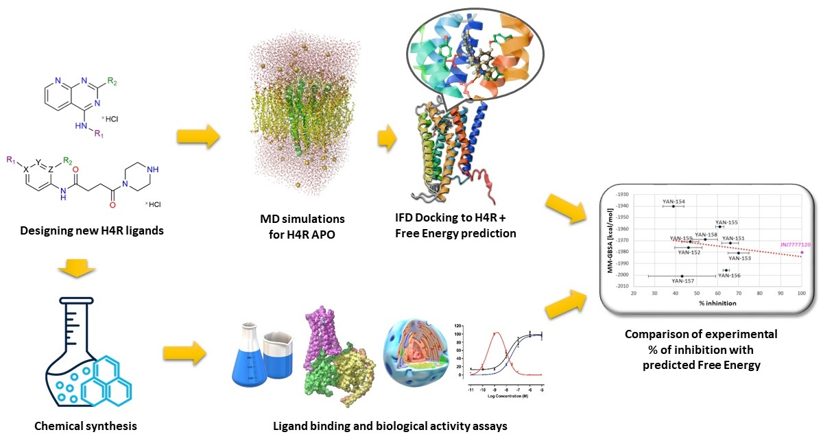
Graphical Abstract

1. Introduction

Histamine is an important biogenic amine that acts as a neurotransmitter and local mediator in numerous physiological and pathological processes. Its effects are mediated through four G protein-coupled receptors (H1R–H4R), which differ in tissue distribution, signaling properties and pharmacological profiles [1,2]. These receptors belong to the rhodopsin-like GPCR family and have emerged as attractive targets for the treatment of allergic, inflammatory, neurological and neoplastic diseases.
The human H4 receptor (H4R) was identified in 2000–2001 by several groups, based on its high sequence similarity to the histamine H3 receptor (H3R) [3,4,5,6,7]. Despite this homology, the expression pattern of H4R is clearly distinct, which indicates different physiological roles [8]. H4R is mainly expressed in cells and tissues associated with immune and inflammatory responses, including eosinophils, mast cells, monocytes, lymphocytes and macrophages [9,10,11,12]. In line with this distribution, H4R has been linked to the regulation of chemotaxis, cytokine release and other functions of immune cells.
Since its discovery, H4R has been associated with the proper functioning of the immune system and with a variety of inflammatory and allergic conditions. H4R expression has been demonstrated in synovial cells from patients with rheumatoid arthritis (RA) [13] and in inflammatory epidermal dendritic cells (IDECs) in the skin [14].
In mouse models, H4R has been shown to play a role in allergic lung inflammation by modulating T-cell responses [15]. Collectively, these findings support the involvement of H4R in diseases such as atopic dermatitis, asthma and chronic arthritis [13,14,15,16].
More recently, H4R has also been implicated in cancer. Immunohistochemical studies and gene expression analyses indicate that H4R is differentially expressed in primary tumors compared with corresponding normal tissues, suggesting a role in carcinogenesis [17,18,19]. H4R has been detected in various cancer cell lines and biopsy specimens, including breast cancer, leukemia, lymphoma, kidney cancer and lung cancer [19]. In a mouse model of triple-negative breast cancer, H4R deficiency reduced tumor growth and lung metastases, further underscoring its potential as a target in oncology [20].
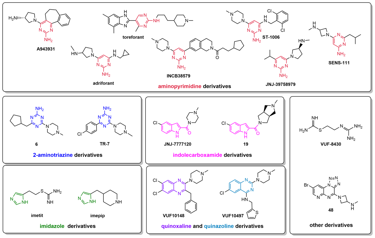
The recognition of H4R as a key player in inflammation and cancer has stimulated intensive research on H4R ligands for potential use in inflammatory, allergic and autoimmune diseases, as well as for their analgesic effects. Since the early 2000s, numerous H4R ligands have been described, including agonists, antagonists and inverse agonists. However, the high similarity in amino acid sequence between H3R and H4R led to the hypothesis, later confirmed, that many known H3R ligands are also capable of interacting with H4R [21]. As a result, H4R selectivity remains a major challenge, and there is a need for new chemotypes that provide favorable pharmacological profiles at H4R with reduced H3R liability. Promising results have been obtained for compounds containing imidazole, indolecarboxamide, aminopyrimidine, quinazoline and quinoxaline scaffolds (Figure 1) [21]. Examples include imidazole derivatives such as imepip and imetit, which are structurally related to histamine and act as potent agonists with high affinity for H3R and H4R [22,23,24,25]. Other noteworthy ligands are ST-1006, a 2-aminopyridine derivative acting as an agonist at both receptors [26], and VUF-8430, an aliphatic H4R agonist with high affinity [27].
In parallel, there has been increasing interest in H4R antagonists and inverse agonists. The first reference compound, JNJ-7777120, showed efficacy in preclinical models of asthma, allergic rhinitis and pruritus [28]. Subsequent H4R antagonists displayed anti-inflammatory effects in various in vivo models, including dermatitis (INCB38579 [29]), peritonitis (TR-7 [30], A-943931 [31]) and atopic dermatitis (adriforant and related compounds [32,33]). Compound 6 is another H4R antagonist with demonstrated analgesic effects [34]. Several H4R ligands, including SENS-111 (seliforant) for unilateral vestibulopathy [35], JNJ-39758979 and adriforant for atopic dermatitis [32,36], and toreforant for rheumatoid arthritis, have advanced to clinical trials. However, many of these studies were terminated due to limited therapeutic benefit or adverse effects.
Modern molecular docking has become a widely used tool in early phase of drug discovery. By placing three-dimensional ligand structures into the receptor binding site and estimating binding energies, docking provides insight into receptor-ligand interactions and supports hit identification and optimization [37,38]. Although the ultimate success of docking-based approaches in bringing new drugs to the market is sometimes questioned, recent improvements in computational methods, increased computing power and broad access to structural databases have greatly enhanced their utility in virtual screening campaigns [39,40].
Despite promising preclinical and early clinical data for H4R ligands in indications such as rheumatoid arthritis, asthma, dermatitis and psoriasis, no H4R-targeted drug has yet been approved for clinical use [12]. Nevertheless, H4R continues to be considered a promising target in the search for new anti-inflammatory and immunomodulatory agents [41,42]. There is still a need for structurally novel ligands that can provide favorable pharmacological profiles, improved safety and, ideally, better selectivity against H3R.
In this context, we aimed to design, synthesize and characterize new H4R ligands containing pyrido[2,3-d]pyrimidine or pyrimidine scaffold. Candidate structures were first scored in silico by docking to constructed 3D structure of the human inactive H4 receptor. Compounds with favorable predicted binding scores were selected for synthesis and in vitro verification. The synthesized compounds were structurally confirmed by NMR and HRMS and then subjected to radioligand binding and functional assays to assess their affinity and intrinsic activity at H4R.

2. Results and Discussion

2.1. Design and Virtual Screening

Quinazoline and quinoxaline scaffolds have been widely explored as chemotypes for the development of histamine H4 receptor ligands [21]. Smits et al. demonstrated that a quinoxaline fragment could be successfully incorporated into potent H4R ligands [43]. In parallel, fragment-based screening revealed that quinazoline derivatives bind to H4R with micromolar affinity, adopting binding modes consistent with the characteristic three-pocket architecture of the receptor [44]. These studies also showed that quinazoline and quinoxaline share low-energy conformations suitable for stabilizing ligand–receptor interactions. Subsequent optimization guided by pharmacophore modeling and in silico calculations yielded quinazoline derivatives acting as inverse agonists at rat H4R and displaying anti-inflammatory activity in vivo. Further QSAR-driven optimization led to the identification of quinazoline sulfonamides as H4R inverse agonists with enhanced anti-inflammatory properties [45].
Additional evidence supporting the relevance of bicyclic nitrogen-containing heterocycles for H3/H4 receptor modulation comes from studies on quinoline-based structures. Depending on substitution patterns, these compounds were characterized as H3R inverse agonists [46] or antagonists [47]. Collectively, these reports indicated that heteroaromatic systems with electron-rich bicyclic cores are well-suited for binding within the H4R orthosteric pocket.
Guided by these observations, we designed two novel structural series. The first series comprises pyrido[2,3-d]pyrimidine derivatives, representing an unexplored bicyclic scaffold with structural analogy to quinazoline and quinoxaline, yet bearing distinct physicochemical properties. The second series includes pyrimidine-substituted pyridines, selected for their more linear, flexible topology, which was hypothesized to facilitate accommodation within the H4R binding site and adjacent sub-pockets [48,49].
Both series were subjected to comprehensive computational evaluation, including docking and molecular dynamics simulations. Ligands were prepared in LigPrep (Schrödinger suite of programs) generating relevant protonation states and stereochemical variants. Docking to the human H4 receptor was performed using Glide XP, (Schrödinger suite of programs) followed by IFD-MD (Schrödinger suite of programs) refinement, enabling simultaneous optimization of ligand poses and receptor side-chain conformations, while accounting for water-mediated interactions and desolvation effects. Top-ranked poses were subsequently evaluated in 20 ns molecular dynamics simulations in an explicit POPC/cholesterol membrane environment to assess binding stability and interaction persistence.
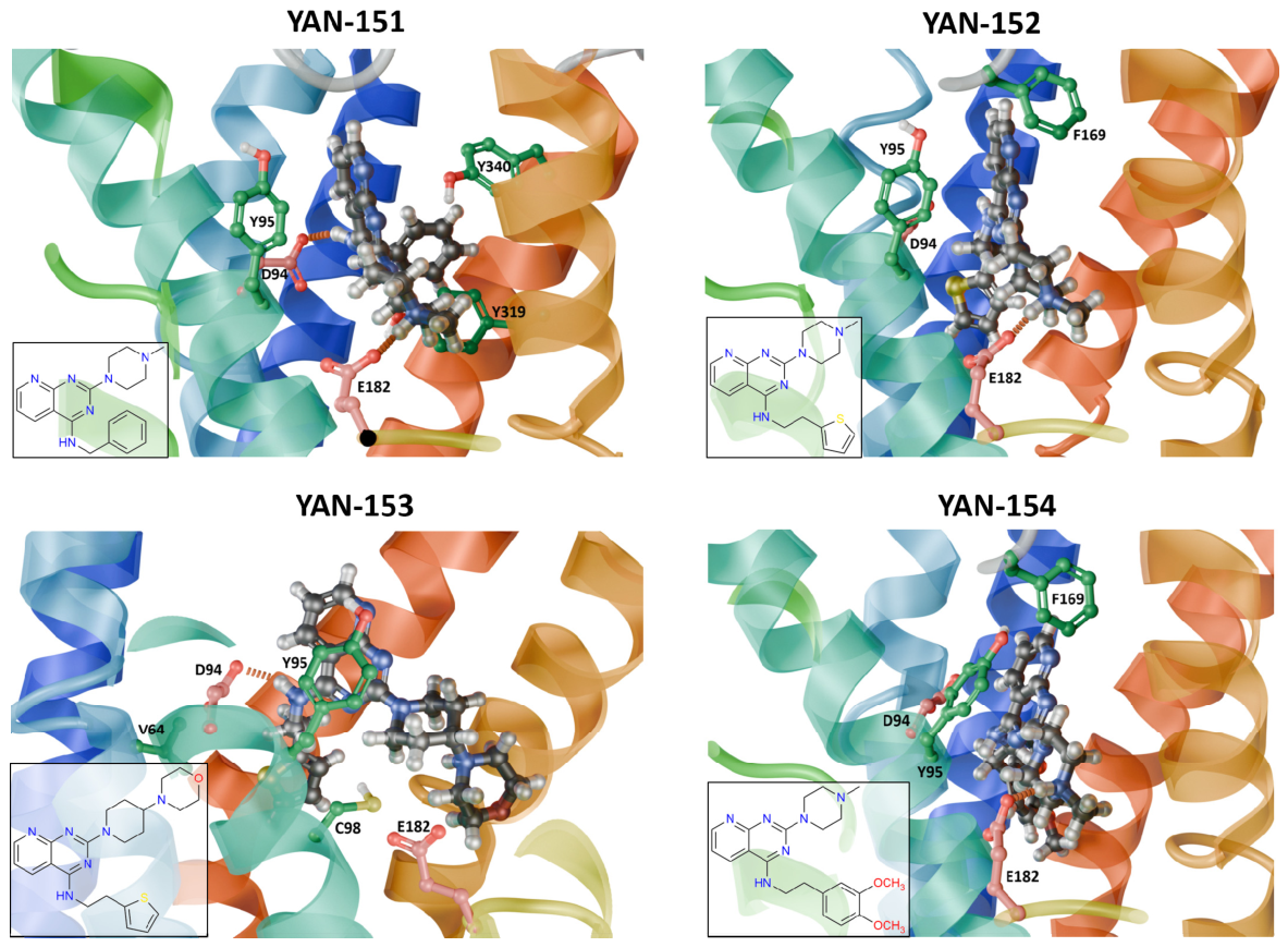
The final structures of compounds YAN-151 to YAN-154 from docking and then 20 ns MD simulations are shown in Figure 2.
The final poses of YAN-151 to YAN-154 are similar, with the protonated and positively charged amine group bound to E1825.46 via ionic interactions, but YAN-153 without a hydrogen bond to E1825.46. YAN-151 and YAN-153 additionally form a hydrogen bond with D943.32. There are also many hydrophobic and π-π interactions especially with Y953.33 and F169ELC2. YAN-152 does not form a hydrogen bond with D943.32 but the hydrophobic and π-π interactions are similar to YAN-151 since they both residue on the same binding site and their aromatic ring system is nearly in the same place. YAN-153 forms especially strong π-π interaction of its quinazoline structure with Y953.33, and additionally its thiophen ring interacts with several residues including V642.53 and C983.36. YAN-154 does not form a hydrogen bond with D943.32 but the π-π interaction of its quinazoline structure with Y953.33 is also strong as for YAN-153.

2.2. Docking and MD Results for YAN-155 to YAN-159 and JNJ-7777120

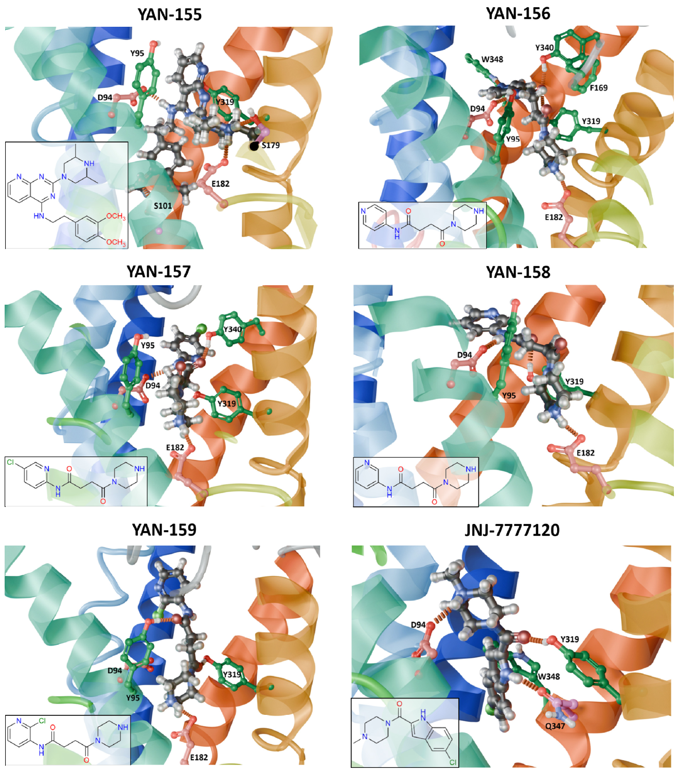
The docking poses of YAN-155 to YAN-159 and the reference antagonist JNJ-7777120 are shown in Figure 3.
All YAN compounds maintain an ionic interaction between their protonated amine and E1825.46, consistent with the binding mode of the previously described ligands in this series. YAN-155 forms three hydrogen bonds (to D943.32, S1013.39 and S1795.43), in addition to hydrophobic and aromatic contacts. YAN-156 forms hydrogen bonds with D943.32, Y3196.51, Y3407.35 and W3487.43 but displays minimal π–π stacking. YAN-157 exhibits a similar pattern but engages fewer residues in hydrogen bonds (D943.32 and Y3407.35). YAN-158 interacts through hydrogen bonding with D943.32 and Y3196.51, whereas YAN-159 forms hydrogen bonds with Y953.33 and Y3196.51, suggesting a partially shifted binding mode. JNJ-7777120 displayed several stable binding poses during docking; in the most frequently observed conformation, it forms an ionic interaction with D943.32 and not with E1825.46 (contrary to YAN compounds). The hydrogen bonds are created with Y3196.51 and Q3477.42, accompanied by notable π–π stacking with W3487.43.

2.3. Chemistry

2.3.1. Synthesis of Ligands with a Pyrido[2,3-d]pyrimidine

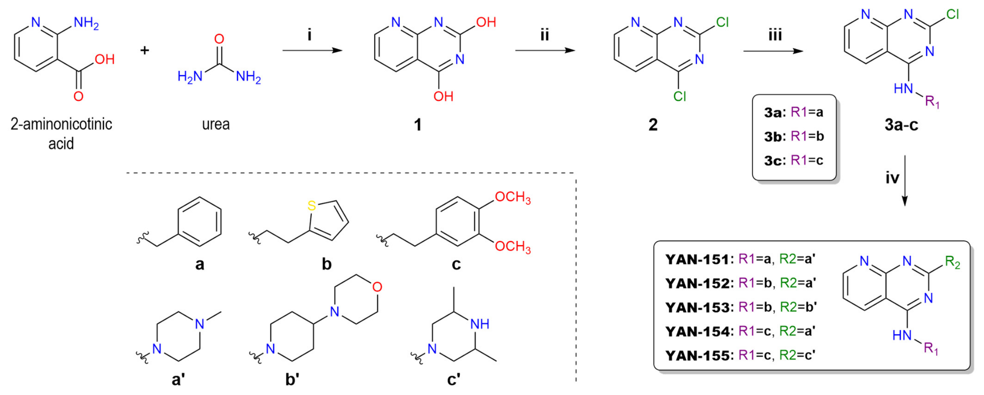
Compounds YAN-151 to YAN-155 were synthesized in four steps according to Scheme 1.
In the first step, 2-aminonicotinic acid was condensed with urea affording pyrido[2,3-d]pyrimidine-2,4-diol 1 [50], followed by hydroxyl groups substitution by chloride atoms after treatment with phosphorus oxychloride (POCl3) and catalytic amount of N,N-dimethylformamide (DMF) [51] to yield compound 2. In the next step, the amine substituents were introduced at the C-4 position of the aromatic ring of 2. The observed regioselectivity was a result of the different activation energies of the chlorides at C-2 and C-4 in 2,4-dichloropyrido[2,3-d]pyrimidine expected for the nucleophilic amine substitution. The regioselective introduction of primary amines (benzylamine a-NH2, 2-thiophenethanamine b-NH2 or 3,4-dimethoxybenzeneethanamine c-NH2) at the C-4 position of 2 was carried out at room temperature in the presence of TEA. In the next step, the amine derivatives from second group, embracing N-methylpiperazine a′-NH2, 4-(piperidin-4-yl)morpholine b′-NH2 or 2,6-dimethylpiperazine c′-NH2, were subsequently introduced at C-2 position of aromatic ring. These conversions were carried out using excess amounts of amine substrates in EtOH at elevated temperature. The expected products YAN-151YAN-155 were obtained in high yields 81–98%.

2.3.2. Synthesis of Ligands with a Pyrimidine Scaffold

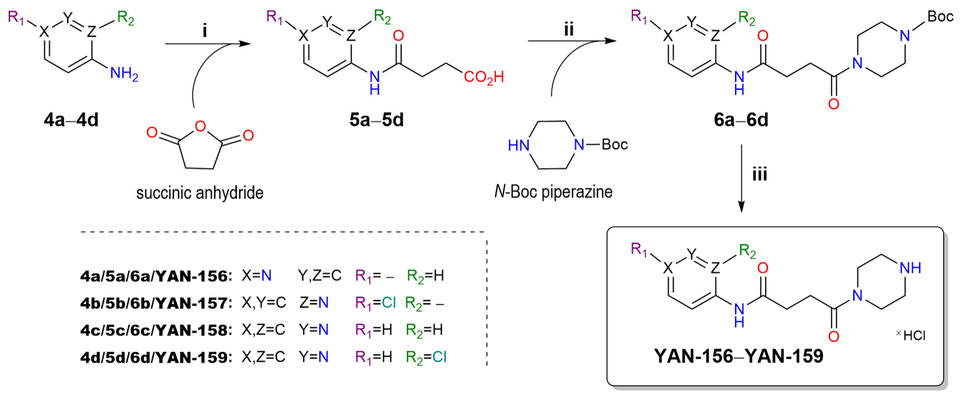
Compounds YAN-156 to YAN-159 were synthesized in three steps as shown in Scheme 2.
In the first step, a series of aminopyridine derivatives (4-aminopyridine 4a, 2-amino-5-chloropyridine 4b, 3-aminopyridine 4c and 3-amino-2-chloropyridine 4d) were condensed with succinic anhydride to obtain four 4-oxo-4-(pyridylamino)butanoic acid derivatives (5ad). The synthesis of compounds 5a and 5c was quick (within 1 h) at room temperature, while the synthesis of derivatives 5b and 5d required an increased temperature and a longer reaction time. Products 5ad were precipitated from the reaction solution. After filtering and drying under vacuum, the intermediates were used for the next step—condensation with N-Boc-piperazine. This reaction was carried out in DMF in the presence of DIPEA and EDCI as a coupling reagent. Then, reaction mixtures were extracted using a water/DCM solvent system. The crude products were isolated from the organic layers and then purified by flash chromatography (chloroform/methanol 98:2 → 95:5). The intermediates 6ad were obtained in yields ranging from 24 to 72%. In the final step, the Boc groups were removed by treating with a 6 M solution of gaseous HCl in EtOH:AcOEt (1:1 v/v). This deprotection resulted in the formation of the appropriate hydrochlorides YAN-156 to YAN-159 as final products.

2.4. In Vitro Pharmacological Studies

2.4.1. Radioligand Binding Assay

The binding affinity of compounds YAN-151YAN-159 at hH4R was evaluated in a radioligand binding assay, as previously described [48]. In brief, [3H]-histamine (10 nM) was used as the radioligand, and the hH4R receptor was expressed in Sf9 cells together with the Gαi2 and Gβ1γ2 subunits of the G protein. Although single-concentration screening at 1 µM does not provide quantitative Ki values, such an approach is widely used for rapid hit identification in early-stage drug discovery [52,53,54]. In our study, it served to prioritize compounds for further functional characterization. The results are expressed as percentage inhibition of specific [3H]histamine binding at 1 µM and are summarized in Table 1. The reference antagonist JNJ-7777120 was not screened under these conditions; it was applied at a saturating concentration (100 µM) to define non-specific binding, which was subtracted from all data. The value of 100% inhibition listed for JNJ-7777120 in Table 1 therefore represents an operational reference point rather than a potency value measured at the screening concentration and is not directly comparable with the test compounds evaluated at 1 µM. A quantitative comparison with the reference antagonist is instead provided by the functional assays, in which JNJ-7777120 was tested as a full concentration–response curve (cAMP accumulation, Section 2.4.3) or at the same concentration as the test compounds (Ca2+ mobilization, Section 2.4.5).
At the screening concentration of 1 µM, the compounds showed inhibition values ranging from 39% to 70%. This variability was dependent on both the core scaffold and the substitution pattern. In general, the pyrido[2,3-d]pyrimidine series exhibited higher inhibition, with YAN-151, YAN-153, and YAN-155 exceeding 60%. Within this group, YAN-153 reached the highest inhibition (69.9%). In contrast, YAN-152 and YAN-154, which share the same aromatic core, displayed clearly lower inhibition, indicating that modifications at R1/R2 positions strongly influence H4R binding.
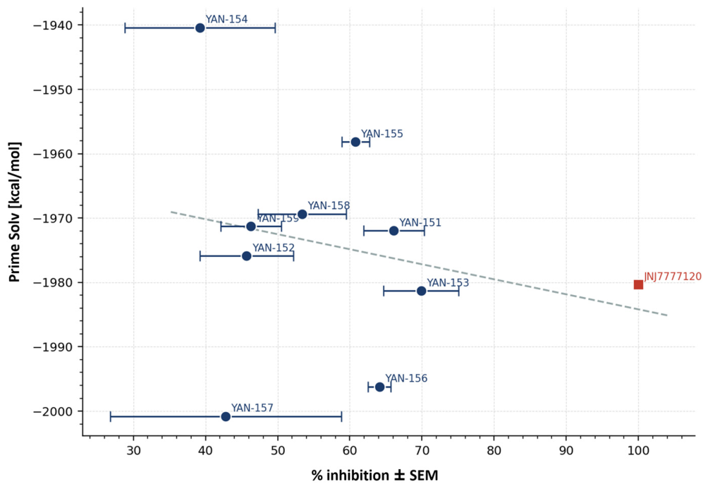
A plot showing the correlation of experimental % inhibition and the free energy prediction of ligand binding is shown in Figure 4.
The free energy values were calculated using the MM-GBSA Solvation method for all YAN compounds and the reference ligand JNJ-7777120, for which the inhibition was set to 100% and its concentration was 100 times larger than for YANs. Since the probability distribution was not normal, we calculated the Spearman rank correlation coefficient apart from the Pearson correlation coefficient. These coefficients were rather low (r = −0.241 for Pearson correlation, and ρ = −0.297 for Spearman rank correlation) but correctly predicted a direction of changes in ligand binding free energy. There are two outliers, YAN-154 and YAN-157. They have the smallest % inhibition, around 40%, and the largest standard deviation of this value (10.4 and 16.0 respectively). However, the free energy predictions are surprising: YAN-154 was predicted as the weakest ligand and YAN-157 as the strongest ligand. Such outliers indicate that the calculated binding energy is too sensitive for small changes in ligand structure and location in the receptor binding site—similar compounds YAN-157 and YAN-158 have very different predicted energies but similar % inhibition. Usage of Ki values instead of % inhibition could improve accuracy of experimental binding data but too sensitive predictions remain. The statistics would be better only with a large number of compounds.
The pyrimidine series (YAN-156YAN-159) tended to show somewhat lower inhibition. An exception was YAN-156 (64.1% inhibition), whose binding at 1 µM was comparable to the most active pyrido[2,3-d]pyrimidine derivatives. This suggests that the particular substitution pattern in YAN-156, including the position of the nitrogen atom in the pyridine moiety (para position relative to the long-chain substituent), may favor additional interactions within the receptor binding pocket.
Compounds YAN-152 and YAN-153 share the same pyrido[2,3-d]pyrimidine core and the same R1 substituent (2-(thiophen-2-yl)ethylamino), differing only in the R2 group (N-methylpiperazine in YAN-152 versus 4-(piperidin-4-yl)morpholine in YAN-153). This single modification at R2 markedly improved H4R affinity in YAN-153, highlighting the contribution of the R2 substituent to receptor binding. Therefore, two compounds were selected as a matched molecular pair for detailed functional characterization, allowing the functional consequences of a single, defined structural change to be assessed while contrasting the highest-affinity (YAN-153) and a moderate-affinity (YAN-152) member of the series. The 60% inhibition value was used only as an initial triage criterion; the pair was deliberately chosen to span it, so that YAN-152 (45.7%) provides the moderate-affinity reference required for this comparison rather than being excluded. It should be noted that radioligand displacement reports binding affinity but not intrinsic activity, since agonists and antagonists alike compete for the orthosteric site; the functional profile of each ligand was therefore established separately in the assays described below.

2.4.2. Cell Viability and Cell Proliferation Effects

To exclude non-specific effects related to cell death or altered cell growth, all synthesized compounds were first evaluated in cell viability and cell proliferation (BrdU incorporation) assays in CHO-H4R cells. At the concentration used in the functional assays (10 µM), none of the compounds affected cell viability or proliferation, and no effects were observed at 100 µM either (Figure S88). At the highest concentration tested (1 mM), a marked loss of viability, accompanied by reduced BrdU incorporation, was observed for compounds requiring 5% DMSO to reach this concentration (YAN-151YAN-155 and YAN-157); however, the vehicle alone at 5% DMSO reduced viability to a comparable extent (DMSO control, Figure S76), whereas YAN-156, YAN-158 and YAN-159, applied in 2% DMSO, remained unaffected. The effects seen at 1 mM therefore reflect the vehicle rather than the compounds.

2.4.3. Functional Characterization in cAMP Accumulation Assay

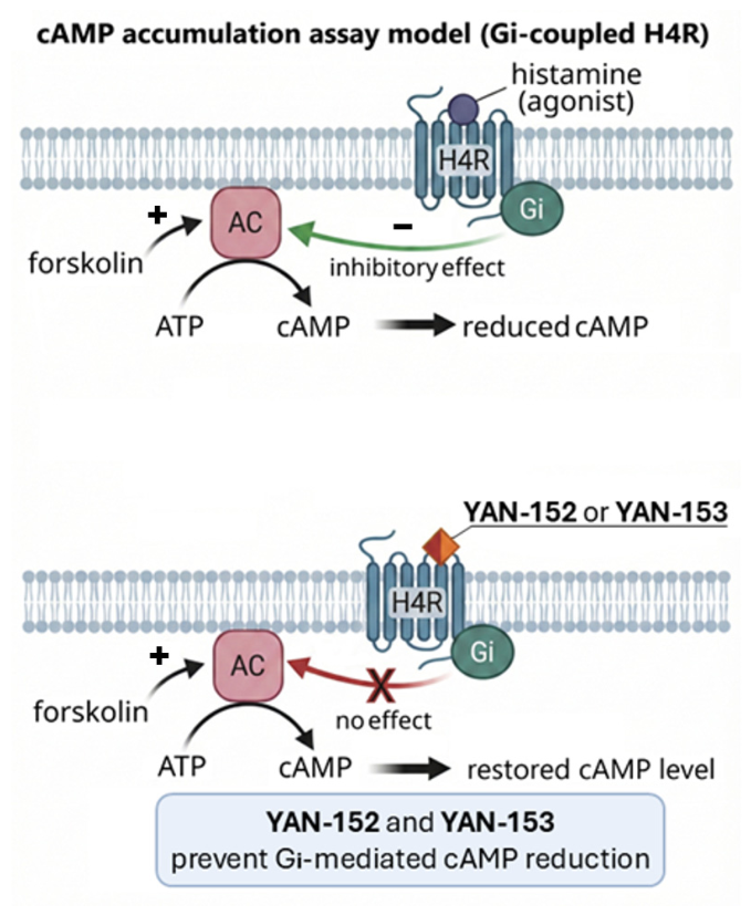
The functional activity of candidate H4 receptor ligands was assessed in a cAMP accumulation assay to evaluate their ability to block Gi-coupled H4R signaling using an antagonist-mode protocol. Upon H4R activation by histamine, Gi coupling leads to reduced intracellular cAMP; antagonists are therefore expected to prevent this decrease and restore cAMP levels in histamine and forskolin co-stimulated cells, as illustrated in Figure 5.
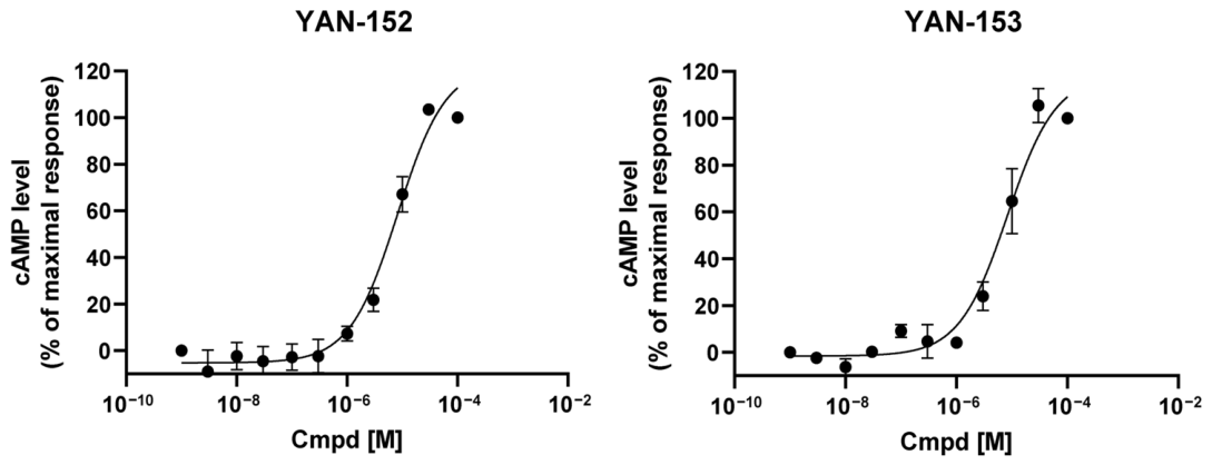
The functional activity of the tested compounds in the Gi-coupled H4 receptor pathway was evaluated using full concentration-response cAMP accumulation assay in CHO cells stably expressing human H4R. YAN-152 and YAN-153 were selected as representative examples with the lower and higher binding to hH4R, respectively. Both compound concentrations dependently reversed histamine-induced inhibition of cAMP accumulation in forskolin-stimulated cells. The IC50 values in antagonist mode were 9.91 ± 3.83 µM for YAN-152 and 8.95 ± 3.03 µM for YAN-153, compared with 0.0833 ± 0.030 µM for the reference antagonist JNJ-7777120 (Figure 6, Table 2), reflecting the differences observed in the radioligand binding assay.
These results indicate that both compounds act as moderate antagonists of H4R in the Gi-cAMP pathway. While the assay clearly demonstrates antagonistic activity, it does not provide direct information on whether the antagonism is surmountable or non-surmountable; such mechanistic characterization would require additional experiments.

2.4.4. Functional Characterization in β-Arrestin Recruitment Assay

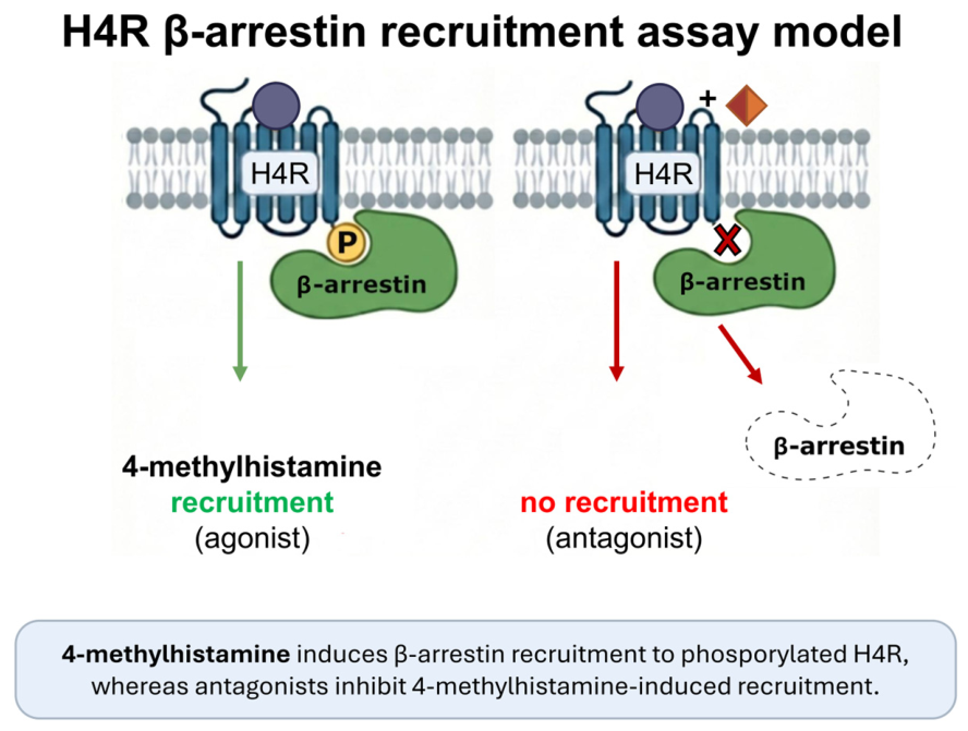
The activity of H4R ligands in the β-arrestin pathway was assessed using a cell-based LiveBLAzer assay monitoring β-arrestin recruitment to human H4R in Tango H4-bla U2OS cells. In this system, recruitment of β-arrestin to activated human H4R triggers the release of a β-lactamase transcription factor, which activates a β-lactamase reporter. Enzymatic activity is detected with a FRET-based substrate, in which cleavage of the β-lactam ring changes the fluorescent signal, providing an indirect measure of β-arrestin recruitment (Figure 7).
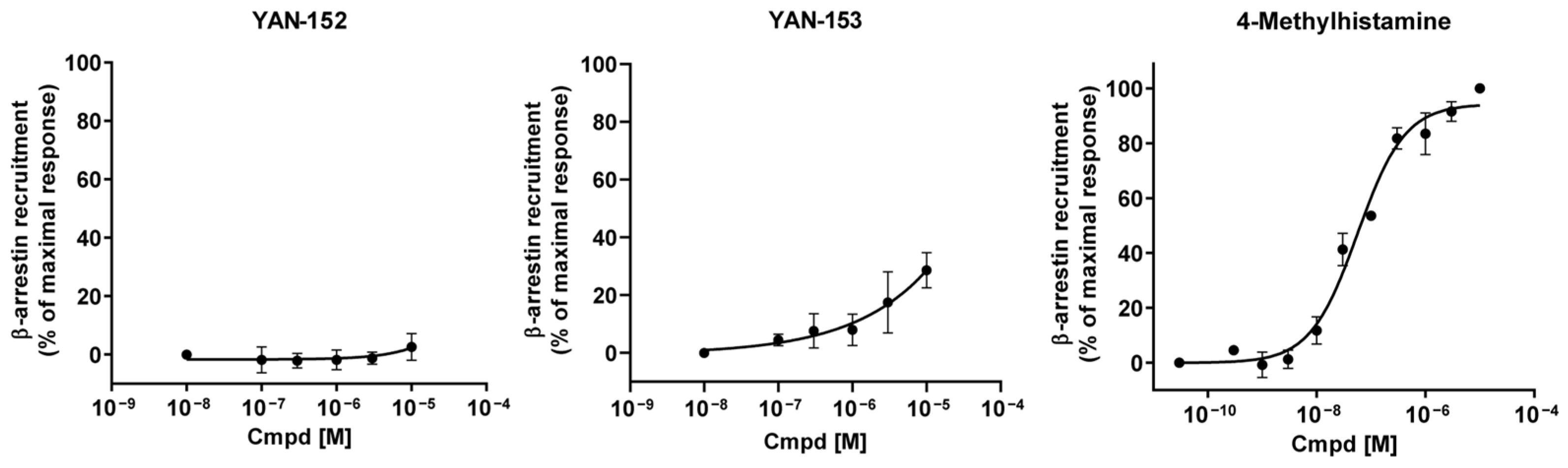
Compounds were tested in both agonist and antagonist modes. In agonist mode, YAN-153 elicited a weak, partial agonist response, reaching 29 ± 6% of the maximal 4-methylhistamine (4-MH) effect (Emax) at the highest concentration tested (10 µM), whereas YAN-152 produced only minimal activation (3 ± 5%; Table 2, Figure 8). In antagonist mode, YAN-152 inhibited 4-MH-induced β-arrestin recruitment with an IC50 of 2.37 ± 0.56 µM, while YAN-153 showed only limited inhibition (IC50 > 10 µM).
These findings indicate that YAN-152 behaves as a predominantly antagonist ligand in the β-arrestin pathway, whereas YAN-153 displays weak partial agonist properties with poor antagonist efficacy. Considered together with the cAMP data, these results reveal ligand-dependent variation in signaling across G-protein and β-arrestin pathways at H4R, consistent with the well-established concept of functional selectivity in GPCR signaling [38].

2.4.5. Cytosolic Calcium Influx

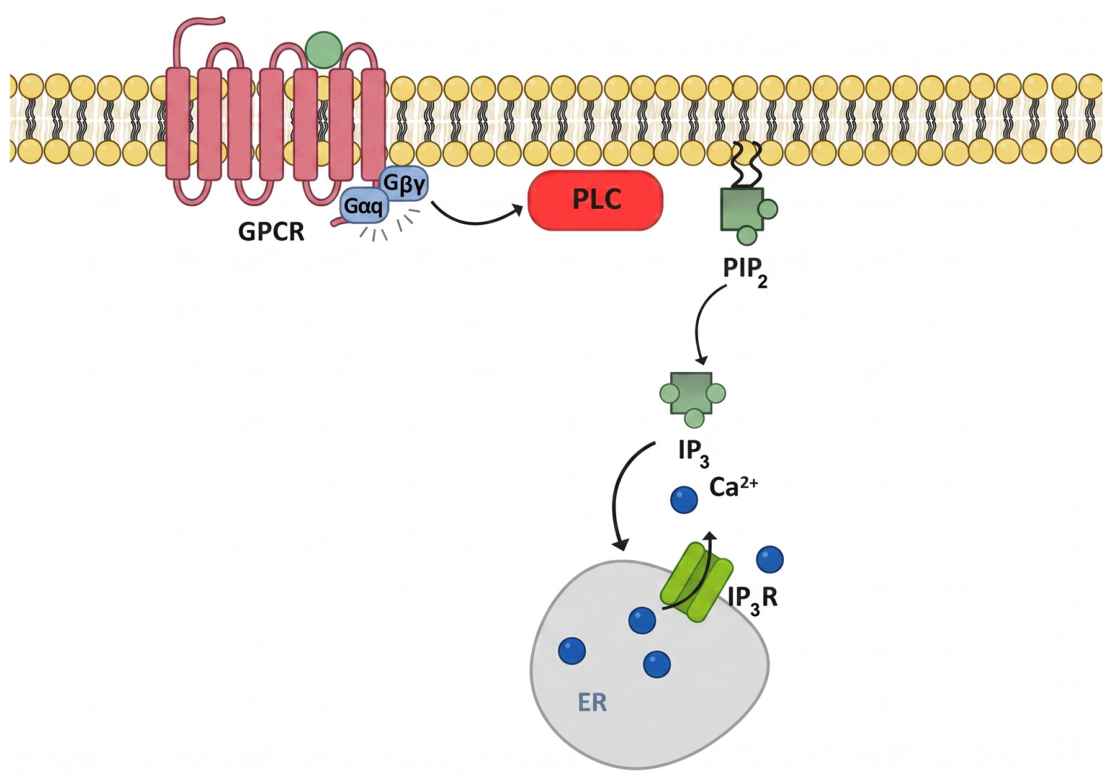
H4R is primarily coupled with Gi/o proteins. Calcium (Ca2+) mobilization occurs downstream of Gβγ subunit activation, which stimulates phospholipase C (PLC). PLC hydrolyses phosphatidylinositol-4,5-bisphosphate (PIP2) to generate inositol 1,4,5-trisphosphate (IP3). IP3 binds to IP3 receptors on the endoplasmic reticulum (ER), inducing Ca2+ release from intracellular stores; in some systems, this is followed by an additional influx of Ca2+ from the extracellular space (Figure 9).
The resulting Ca2+ response is rapid, transient, and dependent on the concentration of the H4R agonist 4-MH [7,55,56,57].
In CHO cells stably expressing human H4R (CHO-H4R), stimulation with 4-MH (10 µM) produced a marked increase in cytosolic Ca2+ concentration. Previous studies have shown that this H4R-dependent signal is abolished by the selective H4R antagonist JNJ-7777120 [58,59], confirming that Ca2+ mobilization in this model is specifically mediated by H4R activation. Therefore, Ca2+ mobilization provides a useful functional readout for studying histamine H4R ligands.
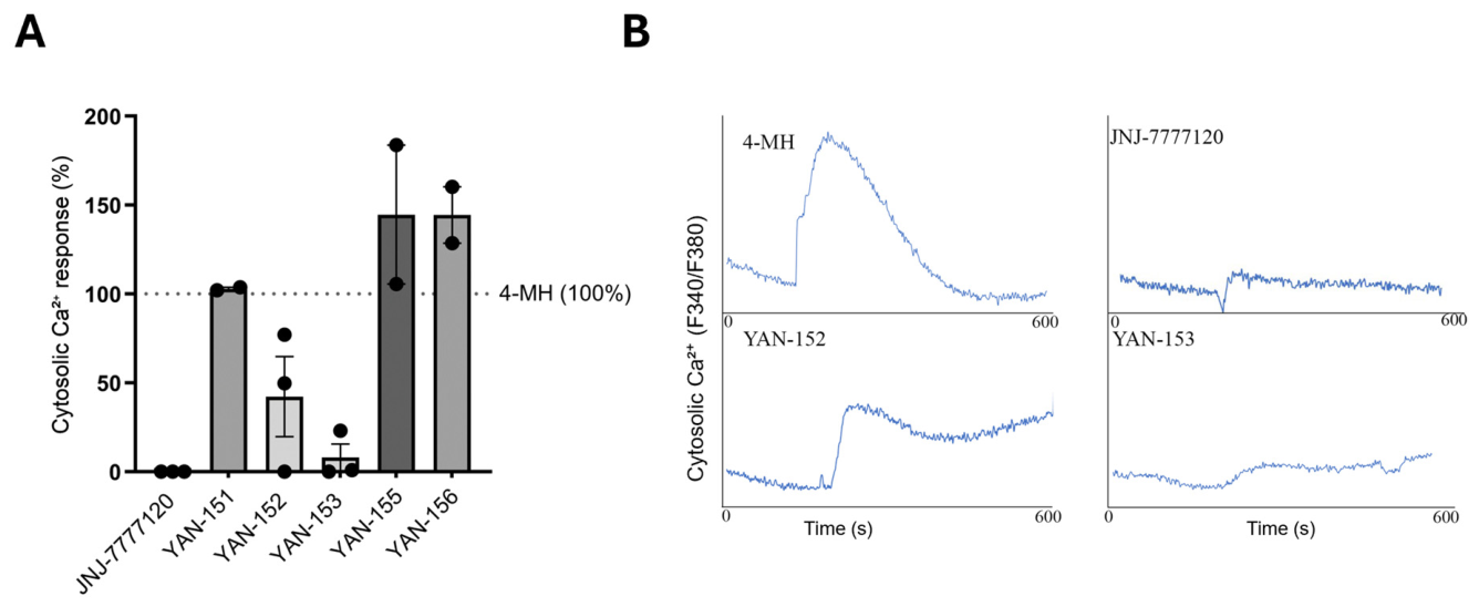
As shown in Figure 10A, stimulation of CHO-H4R cells with 10 µM 4-MH induced a rapid, transient increase in cytosolic calcium concentration, characteristic of H4R-mediated mobilization from intracellular stores. Pre-incubation with the selective H4R antagonist JNJ-7777120 (10 µM) almost completely abolished the Ca2+ signal, confirming that the response is H4R-dependent. When applied alone (without 4-MH), none of the tested compounds evoked a detectable Ca2+ response, indicating that none behaved as an H4R agonist in this assay; their effects were therefore assessed as modulation of the 4-MH-evoked response, each normalized to its paired 4-MH control (set to 100%; Figure 10A).
Quantification of peak responses (Figure 10A) revealed that YAN-153 markedly lowered the signal, to approximately 8% of the paired 4-MH control, whereas YAN-152 produced a weaker and more variable inhibition (to approximately 42% on average). Thus, both new compounds inhibited H4R-mediated Ca2+ mobilization, with YAN-153 showing the stronger and more consistent effect in this pathway. YAN-151 did not appreciably alter the response, behaving as functionally silent. In contrast, YAN-155 and YAN-156 enhanced the 4-MH-evoked Ca2+ response, an unexpected effect within the same chemotype that warrants further mechanistic investigation.

3. Materials and Methods

Reagents, solvents, and other materials were of commercial origin and used without additional operations. Reactions were monitored on silica gel TLC plates 60 F254 (Merck, Darmstadt, Germany). Visualizations were performed with UV light (254 and/or 365 nm) and then with CeMo stain and subsequent charring. The melting points were determined using the MP70 Melting Point System (Mettler-Toledo, Greifensee, Switzerland). Solvents were evaporated under reduced pressure at 40 °C on the Büchi Rotavapor (BÜCHI Labortechnik AG, Flawil, Switzerland). Flash column chromatography was performed on silica gel (200–300 mesh). The chromatographic analysis (HPLC) was performed using a Waters HPLC system (Waters Assoc., Milford, MA, USA) consisting of two Waters pump 515, Waters autosampler 717-plus, Waters column oven and Photodiode Array Detector 2996. Chromatographic parameters: stationary phase—Kromasil C-8 (250 × 4.6 mm, 5 µm), mobile phase–A (0.1% TFA in acetonitrile) and B (0.1% TFA in water) 50/50, elution-isocratic—1 mL/min, duration—15 min., column temp.—25 °C, injection volume—3 µL, UV detection wavelength—ʎ = 278 nm. The 1H and 13C NMR spectra were acquired in CDCl3, DMSO-d6, CD3OD and D2O solutions on Bruker AVANCE III HD 500 MHz spectrometer (Bruker Corporation, Billerica, MA, USA) at the temperature of 298 K. To identify the structures of all isolated products, analysis of the results of 1D and 2D NMR experiments was performed. The 1H and 13C NMR chemical shifts are given relative to the TMS signal at δ = 0.0 ppm. Mass spectra were recorded on the MaldiSYNAPT G2-S HDMS (Waters Corporation, Milford, MA, USA) spectrometer via electrospray ionization (ESI–MS). High-resolution mass spectrometry (HRMS) measurements were performed using the Synapt G2-Si mass spectrometer (Waters Corporation, Milford, MA, USA) equipped with an ESI source and a quadrupole-time-of-flight mass analyzer. The results of the measurements were processed using the MassLynx 4.1 software (Waters Corporation, Milford, MA, USA).

3.1. Virtual Screening

3.1.1. Preparing Ligand and Receptor Structures

The structure of reference compound JNJ-7777120 was taken from PubChem [60] while the 3D structures of test compounds were constructed in LigPrep module of Schrödinger suite of programs (v. 2024-2, Schrödinger LLC, Portland, OR, USA). LigPrep works by expanding tautomeric and ionization states, ring conformations, and stereoisomers. The representative structure for each test compound was set assuming neutral pH. The full structure of inactive human H4 receptor (UniProt Q9H3N8) was obtained from AlphaFold Protein Structure Database [61] since there is no structure of inactive H4R in PDB. The structure is nearly identical (Calpha RMSD of 1.354 Å over 264 aligned residues with 50.76% sequence identity) to crystal structure of human H3R in complex with antagonist PF03654746 [62]. The full structure of human H4R has 390 residues; however, for the purpose of molecular ligand docking some cytoplasmic parts of the receptor located far from the ligand binding site were removed: the long cytoplasmic loop between helices TM5 and TM6 (residues 218–286) and the C-terminus beyond helix H8 (residues 381–390). The removed parts had also a low and very low confidence values (pLDDT—the predicted local distance difference test) in AlphaFold database. The N-terminus (residues 1–10) was kept as it would be important for ligand binding.

3.1.2. Molecular Docking and Molecular Dynamics

The ligand docking was performed in Glide module (Schrödinger suite of programs) in XP (extra precision) mode and then IFD-MD (Induced Fit Docking—Molecular Dynamics) (Schrödinger suite of programs) was used. IFD-MD performs local search of the best ligand pose using the MD procedure and additionally (i) incorporates the effects that water molecules have on binding as an important component of the IFD-MD scoring function, and (ii) detects and penalizes desolvation of polar groups caused by non-native ligand poses. To check whether the ligands were stably docked to the receptor, the 20 ns classical MD simulations were performed in the explicit membrane-water environment. The receptor with appropriate ligands was immersed in a hydrated POPC bilayer (enriched by 20% (n/n) cholesterol), and then MD simulations were performed in AMBER24 using CHARMM36m force field [63]. All systems were generated in CHARMM-GUI service v. 3.8 (https://www.charmm-gui.org) [64] and the receptor orientation in the membrane was set according to the OPM database [65]. The standard system contained ~70,000 atoms, ~127 lipid molecules (cholesterol + POPC), and concentration of ions Na+ and Cl was 0.15 M. The average dimensions of a periodic cell were 74 Å × 74 Å × 140 Å. TIP3P water model parametrized to use with CHARMM force fields was employed. Before each MD simulation the system was submitted to a restrained energy minimization (5000 cycles). The first 2500 minimization cycles were performed with a steepest-descent method and after that the conjugate gradient was switched on. Next, a six-step equilibration was performed (375 ps total) at a constant pressure and temperature (NPT ensemble; 310 K, 1 bar). During equilibration the restraints were released gradually, until the last step (last 100 ps of equilibration), in which no restraints were used. In the production simulations (as well as in the last three equilibration steps) all bond lengths to hydrogen atoms were constrained using SHAKE algorithm (version in AMBER24 program) [66], which allowed us to use a longer time step (2 fs instead of 1 fs). Van der Waals and short-range electrostatic interactions cutoff = 12 Å, with 10–12 Å switching function, were used. Long-range electrostatic interactions were computed using the Particle Mesh Ewald [67] summation scheme.

3.1.3. Predictions of Free Energy of Ligand Binding

The calculations of free energy ligand binding of the poses obtained from IFD procedure were performed in Prime module of Schrödinger package using MM-GBSA (Molecular Mechanics/Generalized Born Surface Area) method. The following standard Prime energy components were used: Coulomb energy, Covalent binding energy, Van der Waals energy, Lipophilic energy, Generalized Born electrostatic solvation energy, Hydrogen-bonding correction, π–π packing correction, and Self-contact correction. The newer VSGB 2.1 (Variable Solvent Generalized Born) solvation model was used, which is a refit of VSGB 2.0 [68], and calculated in OPLS3 force field [69] (for VSGB 2.0 the calculations were done in OPLS_2005 force field).

3.1.4. The Ballesteros–Weinstein Numbering Scheme

For direct comparison of ligand–protein interactions between different GPCRs we use the Ballesteros–Weinstein numbering scheme of residues. In this numbering scheme each GPCR residue is marked with two numbers separated by a dot. The first number indicates the transmembrane helix while the second number the position of the residue relative to the most conserved residue (assigned the number 50) on the same helix in a sequential order [70].

3.2. Chemical Synthesis

3.2.1. Pyrido[2,3-d]pyrimidine-2,4(1H,3H)-dione 1

Compound 1 was obtained by melting a mixture of 2-aminonicotinic acid and urea and then heating 2 h at 200 °C [50]. The product was used in the next step without further purification. Yield 97%.

3.2.2. 2,4-Dichloropyrido[2,3-d]pyrimidine 2

Compound 2 was synthesized by heating 1 with POCl3 in DMF according to a well-established literature procedure [51]. Yield 89%.

3.2.3. Synthesis of YAN-151YAN-155—General Procedure

2-Chloro-4-(phenylmethylamino)pyrido[2,3-d]pyrimidine 3a
A total of 50 mg (0.25 mmol, 1 eq.) of 2 was dissolved in 5 mL of THF. To this solution 70 µL (0.50 mmol, 2 eq) of TEA and then 27 µL (0.25 mmol, 1 eq) of benzylamine a-NH2 were added. After a while a white solid precipitated. This suspension was stirred at room temperature overnight. Reaction completion was confirmed by TLC (DCM/MeOH 95:5 v/v). Post-reaction mixture was diluted with 15 mL of water, the product was extracted with DCM (2 × 15 mL), the organic layer was dried over magnesium sulfate and concentrated under vacuum. The crude mixture was purified on silica gel (DCM/MeOH 98:2 v/v) giving 61 mg (0.23 mmol) of the pure product. Yield: 90%; white solid, MP: 178 °C (dec); 1H NMR (25 °C, MeOH-d1): 8.92 (bs, 1H, H7Pyridine), 8.61 (dd, 1H, H5Pyridine), 7.52 (dd, 1H, H6Pyridine), 7.41 (m, 2H, H2Ph and H6Ph), 7.33 (m, 2H, H3Ph and H5Ph), 7.26 (m, 1H, H4Ph), 4.88 (s, 2H, NHCH2Ph); 13C NMR (25 °C, MeOH-d1): 163.50 (C2Pyrimidine), 160.53 (C8aPyridine), 160.25 (C4Pyrimidine) 157.18 (C7Pyridine), 139.21 (C1Ph), 134.27 (C5Pyridine), 129.62 (C3Ph/C5Ph), 129.07 (C2Ph/C6Ph), 128.53 (C4Ph), 123.03 (C6Pyridine), 46.13 (CH2); HRMS: calcd for C14H12ClN4: 271.0750 found: 271.0753 [M + H]+.
The following compounds were obtained according to the same synthetic procedure.
2-Chloro-4-(2-thiopheneethanamino)pyrido[2,3-d]pyrimidine 3b
The compound 3b was synthesized using 200 mg (1.00 mmol, 1 eq.) of (2), 114 µL (1.00 mmol, 1 eq) of 2-thiopheneethanamine b-NH2 and 278 µL (2.00 mmol, 2 eq.) of TEA. After purification on silica gel 256 mg (0.88 mmol) of the pure product was obtained. Yield: 88%; white solid, MP: 155 °C (dec); 1H NMR (25 °C, CDCl3): 8.91 (bs, 1H, H7Pyridine), 8.40 (d, 1H, H5Pyridine), 7.39 (bs, 1H, H6Pyridine), 7.14 (dd, 1H, H5Thiophene), 6.92 (dd, 1H, H4Thiophene), 6.86 (d, 1H, H3Thiophene), 3.91 (t, 2H, NHCH2CH2), 3.23 (t, 2H, NHCH2CH2); 13C NMR (25 °C, CDCl3): 161.46 (C4Pyrimidine), 161.29 (C2Pyrimidine), 158.15 (C8aPyridine), 155.36 (C7Pyridine), 140.86 (C2Thiophene), 132.92 (C5Pyridine), 127.06 (C4Thiophene), 125.55 (C3Thiophene), 124.04 (C5Thiophene), 121.49 (C6Pyridine), 108.68 (C4aPyridine), 43.05 (NHCH2CH2), 28.80 (NHCH2CH2); HRMS: calcd for C13H12ClN4S: 291.0471 found: 291.0475 [M + H]+.
2-Chloro-4-(3,4-dimethoxybenzeneethanamino)pyrido[2,3-d]pyrimidine 3c
This compound was synthesized using 100 mg (0.50 mmol, 1 eq.) of 2, 84 µL (0.50 mmol, 1 eq) of 3,4-dimethoxybenzeneethanamine c-NH2 and 139 µL (1.00 mmol, 2 eq.) of TEA. After purification on silica gel 67 mg (0.20 mmol) of the pure product was obtained. Yield: 40%; white solid, MP: 170 °C (dec); 1H NMR (25 °C, CDCl3): 8.77 (dd, 1H, H7Pyridine), 8.26 (dd, 1H, H5Pyridine), 7.26 (dd, 1H, H6Pyridine), 6.70 (m, 1H, H2Ph), 6.69 (m, 1H, H5Ph), 6.68 (m, 1H, H6Ph), 3.75 (m, 2H, NHCH2CH2), 3.73 (s, 6H, both CH3), 2.85 (t, 2H, NHCH2CH2); 13C NMR (25 °C, CDCl3): 161.77 (C4Pyrimidine), 161.26 (C2Pyrimidine), 158.80 (C8aPyridine), 155.68 (C7Pyridine), 148.77 (C3Ph), 147.47 (C4Ph), 132.32 (C5Pyridine), 131.36 (C1Ph), 121.33 (C6Pyridine), 120.74 (C6Ph), 112.00 (C2Ph), 111.25 (C5Ph), 108.71 (C4aPyridine), 55.76 (both CH3), 43.08 (NHCH2CH2), 34.21 (NHCH2CH2); HRMS: calcd for C17H18ClN4O2: 345.1118 found: 345.1121 [M + H]+.
2-(4-Methyl-1-piperazinyl)-4-(phenylmethylamino)pyrido[2,3-d]pyrimidine YAN-151
A total of 50 mg (0.19 mmol, 1 eq.) of 3a was suspended in 5 mL of EtOH and 41 µL (0.37 mmol, 2 eq) of N-methylpiperazine a′-NH2 was added. The mixture was heated for 4 h under reflux. The reaction progress was monitored by TLC (AcOEt/MeOH/TEA 90:5:5 v/v/v) until traces of starting materials were not observed. Post-reaction mixture was condensed under vacuum resulting in the crude product which was purified on silica gel (DCM/MeOH 95:5 → 90:10 v/v) obtaining 50 mg (0.15 mmol) of the pure product. Yield: 79%; Purity: 99.67% (HPLC); MP: 180 °C (dec); 1H NMR (25 °C, DMSO-d6): 8.66 (d, 1H, H7Pyridine), 8.77 (d, 1H, H5Pyridine), 7.35 (m, 2H, H2Ph and H6Ph), 7.26 (m, 1H, H4Ph), 7.21 (m, 2H, H3Ph and H5Ph), 6.93 (dd, 1H, H6Pyridine), 6.75 (bs, 1H, NH), 4.76 (d, 2H, NHCH2), 4.00 (bs, 4H, CH2Piperazine at C2/C6), 2.50 (m, 4H, CH2Piperazine at C3/C5), 2.35 (s, 3H, CH3); 13C NMR (25 °C, DMSO-d6): 160.87 (C8aPyridine), 160.39 (C2Pyrimidine), 160.36 (C4Pyrimidine), 155.07 (C7Pyridine), 138.41 (C1Ph), 131.06 (C5Pyridine), 128.60 (C3Ph and C5Ph), 127.75 (C2Ph and C6Ph), 127.45 (C4Ph), 116.37 (C6Pyridine), 105.13 (C4Pyridine), 54.56 (C3Piperazine and C5Piperazine), 45.56 (CH3), 45.32 (NHCH2), 43.16 (C2Piperazine and C6Piperazine); HRMS: calcd for C19H23N6: 335.1984 found: 335.1985 [M + H]+.
The following compounds were obtained according to the same synthetic procedure.
2-(4-Methyl-1-piperazinyl)-4-(2-thiopheneethanamino)pyrido[2,3-d]pyrimidine YAN-152
The compound YAN-152 was synthesized using 80 mg (0.28 mmol, 1 eq.) of 3b and 61 µL (0,55 mmol, 2 eq.) of N-methylpiperazine a′-NH2. After purification on silica gel 90 mg (0.25 mmol) of the pure product was obtained. Yield: 89%; Purity: 98.35% (HPLC); MP: decomposition at 180 °C; 1H NMR (25 °C, DMSO-d6): 8.62 (dd, 1H, H7Pyridine), 8.27 (dd, 1H, H5Pyridine), 7.22 (dd, 1H, H5Thiophene), 7.09 (dd, 1H, H6Pyridine), 6.94 (dd, 1H, H4Thiophene), 6.88 (d, 1H, H3Thiophene), 4.01 (bs, 4H, CH2Piperazine at C2/C6), 3.82 (t, 2H, NHCH2CH2), 3.24 (t, 2H, NHCH2CH2), 2.55 (t, 4H, CH2Piperazine at C3/C5), 2.37 (s, 3H, CH3); 13C NMR (25 °C, DMSO-d6): 162.17 (C2Pyridine + C4Pyridine), 161.95 (C8aPyridine), 155.47 (C7Pyridine), 142.83 (C2Thiophene), 133.64 (C5Pyridine), 127.87 (C4Thiophene), 126.26 (C3Thiophene), 124.72 (C5Thiophene), 117.79 (C6Pyridine), 107.12 (C4aPyridine), 55.91 (CH2Piperazine at C3/C5), 46.14 (CH3), 44.47 (CH2Piperazine at C2/C6), 44.20 (NHCH2CH2), 29.96 (NHCH2CH2); HRMS: calcd for C18H23N6S: 355.1705 found: 355.1708 [M + H]+.
2-(4-(piperidin-4-yl)morpholine)-4-(2-thiopheneethanamino)pyrido[2,3-d]pyrimidine YAN-153
The compound YAN-153 was synthesized using 80 mg (0.28 mmol, 1 eq.) of 3b and 94 mg (0,55 mmol, 2 eq.) of 4-(piperidin-4-yl)morpholine b′-NH2. After purification on silica gel 115 mg (0.27 mmol) of the pure product was obtained. Yield: 98%; Purity: 97.87% (HPLC); MP: decomposition at 98.9 °C; 1H NMR (25 °C, DMSO-d6): 8.68 (dd, 1H, H7Pyridine), 7.85 (dd, 1H, H5Pyridine), 7.18 (dd, 1H, H5Thiophene), 6.96 (dd, 1H, H4Thiophene), 6.92 (dd, 1H, H6Pyridine), 6.85 (d, 1H, H3Thiophene), 6.18 (t, 1H, NHCH2), 5.07 (d, 2H, two of CH2Piperidine at C2/C6), 3.85 (q, 2H, NHCH2), 3.73 (t, 4H, CH2Morpholine at C3/C5), 3.22 (t, 2H, NHCH2CH2), 2.91 (t, 2H, two of CH2Piperidine at C2/C6), 2.59 (t, 4H, CH2Morpholine at C2/C6), 2.50 (m, 1H, CHPiperidine at C4), 1.93 (d, 2H, two of CH2Piperidine at C3/C5), 1.50 (d, 2H, two of CH2Piperidine at C3/C5); 13C NMR (25 °C, DMSO-d6): 161.03 (C8aPyridine), 160.33 (C2Pyrimidine), 160.22 (C4Pyrimidine), 155.14 (C7Pyridine), 141.31 (C2Thiophene), 130.46 (C5Pyridine), 127.11 (C4Thiophene), 125.38 (C3Thiophene), 124.04 (C5Thiophene), 116.10 (C6Pyridine), 104.84 (C4aPyridine), 67.10 (CH2Morpholine at C3/C5), 62.64 (CHPiperidine at C4), 49.68 (CH2Morpholine at C2/C6), 43.29 (CH2Piperidine at C2/C6), 42.78 (NHCH2), 29.19 (NHCH2CH2), 28.15 (CH2Piperidine at C3/C5); HRMS: calcd for C22H29N6OS: 425.2124 found: 425.2119 [M + H]+.
2-(4-Methyl-1-piperazinyl)-4-(3,4-dimethoxybenzeneethanamino)pyrido[2,3-d]pyrimidine YAN-154
The compound YAN-154 was synthesized using 80 mg (0.23 mmol, 1 eq.) of 3c and 52 µL (0.464 mmol, 2 eq.) of N-methylpiperazine a′-NH2. After purification on silica gel 93 mg (0.22 mmol) of the pure product was obtained. Yield: 96%; Purity: 99.22% (HPLC); MP: slow decomposition from 80 °C; 1H NMR (25 °C, DMSO-d6): 8.69 (dd, 1H, H7Pyridine), 7.73 (dd, 1H, H5Pyridine), 6.82 (dd, 1H, H6Pyridine), 6.82 (d, 1H, H5Ph), 6.76 (dd, 1H, H6Ph), 6.71 (d, 1H, H2Ph), 5.83 (t, 1H, NHCH2CH2), 4.06 (bs, 4H, CH2Piperazine at C2 and C6), 3.87 (s, 3H, OCH3 at C4Ph), 3.82 (m, 2H, NHCH2CH2), 3.81 (s, 3H, OCH3 at C3Ph), 2.94 (t, 2H, NHCH2CH2), 2.53 (t, 4H, CH2Piperazine at C3 and C5), 2.37 (s, 3H, CH3); 13C NMR (25 °C, DMSO-d6): 161.04 (C8aPyridine), 160.57 (C2Pyrimidine), 160.26 (C4Pyrimidine), 155.34 (C7Pyridine), 149.04 (C3Ph), 147.74 (C4Ph), 131.30 (C1Ph), 130.08 (C5Pyridine), 120.63 (C6Ph), 116.23 (C6Pyridine), 111.87 (C2Ph), 111.36 (C5Ph), 104.84 (C4aPyrimidine), 55.89 (OCH3 at C4Ph), 55.78 (OCH3 at C3Ph), 54.97 (C3Piperazine and C5Piperazine), 46.00 (CH3), 43.51 (C2Piperazine and C6Piperazine), 42.57 (NHCH2CH2), 34.59 (NHCH2CH2); HRMS: calcd for C22H29N6O2: 409.2352 found: 409.2354 [M + H]+.
2-(2,6-Dimethylpiperazinyl)-4-(3,4-dimethoxybenzeneethanamino)pyrido[2,3-d]pyrimidine YAN-155
This compound was synthesized using 80 mg (0.23 mmol, 1 eq.) of 3c and 53 mg (0.46 mmol, 2 eq.) of 2,6-dimethylpiperazine c′-NH2. After purification on silica gel 93 mg (0.22 mmol) of the pure product was obtained. Yield: 96%; Purity: 99.74% (HPLC); MP: 165 °C (dec); 1H NMR (25 °C, DMSO-d6): 8.70 (dd, 1H, H7Pyridine), 7.68 (dd, 1H, H5Pyridine), 6.91 (dd, 1H, H6Pyridine), 6.83 (d, 1H, H5Ph), 6.77 (dd, 1H, H6Ph), 6.71 (d, 1H, H2Ph), 4.97 (bs, 2H, two of CH2Piperazine at C2 and C6), 3.87 (s, 3H, OCH3 at C4Ph), 3.83 (m, 2H, NHCH2CH2), 3.81 (s, 3H, OCH3 at C3Ph), 2.94 (m, 2H, CH2Piperazine at C3 and C5), 2.94 (m, 2H, NHCH2CH2), 2.56 (t, 2H, two of CH2Piperazine at C3 and C5), 1.18 (s, 6H, CH3); 13C NMR (25 °C, DMSO-d6): 161.14 (C8aPyridine), 160.38 (C2Pyrimidine), 160.23 (C4Pyrimidine), 155.42 (C7Pyridine), 149.06 (C3Ph), 147.77 (C4Ph), 131.31 (C1Ph), 129.93 (C5Pyridine), 120.65 (C6Ph), 116.11 (C6Pyridine), 111.89 (C2Ph), 111.37 (C5Ph), 104.75 (C4aPyrimidine), 55.90 (OCH3 at C4Ph), 55.80 (OCH3 at C3Ph), 50.96 (C3Piperazine and C5Piperazine), 50.13 (both CH2Piperazine), 42.56 (NHCH2CH2), 34.58 (NHCH2CH2), 19.27 (CH3); HRMS: calcd for C23H31N6O2: 423.2508 found: 423.2501 [M + H]+.
4-Oxo-4-(4-pyridinylamino)butanoic Acid 5a
4-Aminopyridine 4a (1.88 g, 20 mmol, 1.0 eq.) was dissolved in 40 mL of THF, forming a clear, colorless solution (A). Succinic anhydride (2.20 g, 22 mmol, 1.1 eq.) was dissolved in 20 mL of THF separately, forming a clear, colorless solution (B). The solution B was added to the solution A, while vigorously stirring at room temperature. The mixture immediately got cloudy and the white solid started to precipitate within 5 min. The resulting suspension was concentrated to approx. ⅓ of its initial volume. Precipitated solid was filtered, washed with a small volume of cold THF and dried in vacuo at 40 °C. A total of 1.78 g (9.2 mmol) of a pure product was obtained. Yield: 46%; MP: 191 °C; 1H NMR (25 °C, DMSO-d6): 9.66 (bs, 1H, CO2H), 8.00 (d, 2H, H2Pyridine and H6Pyridine), 6.75 (s, 1H, NH), 6.57 (d, 2H, H3Pyridine and H5Pyridine), 2.34 (s, 4H, both CH2); 13C NMR (25 °C, DMSO-d6): 174.51 (CO2H), 174.45 (NHC(=O)), 156.46 (C4Pyridine), 145.62 (C2Pyridine and C6Pyridine), 108.80 (C3Pyridine and C5Pyridine), 30.41 (both CH2); HRMS: calcd for C9H11N2O3: 195.0770 found: 195.0772 [M + H]+, calcd for C9H9N2O3: 193.0613 found: 193.0612 [M + H].
The following compounds were obtained according to the same synthetic protocol (reaction temperature and time are given separately in each case).
4-[(5-Chloro-2-pyridinyl)amino]-4-oxobutanoic Acid 5b
The compound 5b was synthesized using 2-amino-5-chloropyridine 4b (2.57 g, 20 mmol, 1.0 eq.) in 20 mL of THF and 2.60 g (26 mmol, 1.3 eq.) of succinic anhydride in 25 mL of THF under reflux overnight. After workup and purification as described for 5a 3.79 g (17 mmol) of the pure product was obtained. Yield: 85%. MP: 197 °C; 1H NMR (25 °C, DMSO-d6): 12.11 (s, 1H, CO2H), 10.67 (s, 1H, NH), 8.33 (d, 1H, H6Pyridine), 8.08 (d, 1H, H3Pyridine), 7.86 (dd, 1H H4Pyridine), 2.62 (m, 2H, CH2CH2CO2H), 2.50 (m, 2H, CH2CH2CO2H); 13C NMR (25 °C, DMSO-d6): 173.73 (CO2H), 171.20 (NHC(=O)), 150.71 (C2Pyridine), 146.24 (C6Pyridine), 137.81 (C4Pyridine), 124.74 (C5Pyridine), 114.39 (C3Pyridine), 30.85 (CH2CH2CO2H), 28.47 (CH2CH2CO2H); HRMS: calcd for C9H10ClN2O3: 229.0380 found: 229.0384 [M + H]+.
4-Oxo-4-(3-pyridinylamino)butanoic Acid 5c
This compound was synthesized using 3-aminopyridine 4c (1.88 g, 20 mmol, 1.0 eq.) in 10 mL of THF and 2.20 g (22 mmol, 1.1 eq.) of succinic anhydride in 20 mL of THF at room temperature for 1 h. After workup and purification as described for 5a 3.61 g (19 mmol) of the pure product was obtained. Yield: 93%. MP: 187 °C; 1H NMR (25 °C, DMSO-d6): 12.16 (s, 1H, CO2H), 10.16 (s, 1H, NH), 8.71 (d, 1H, H2Pyridine), 8.22 (dd, 1H, H6Pyridine), 8.01 (d, 1H, H4Pyridine), 7.31 (dd, 1H, H5Pyridine), 2.58 (m, 2H, CH2CH2CO2H), 2.53 (m, 2H, CH2CH2CO2H); 13C NMR (25 °C, DMSO-d6): 174.22 (CO2H), 171.18 (NHC(=O)), 144.35 (C6Pyridine), 140.98 (C2Pyridine), 136.35 (C3Pyridine), 126.23 (C4Pyridine), 124.06 (C5Pyridine), 31.37 (CH2CH2CO2H), 29.12 (CH2CH2CO2H); HRMS: calcd for C9H11N2O3: 195.0770 found: 195.0772 [M + H]+.
4-[(2-Chloro-3-pyridinyl)amino]-4-oxobutanoic Acid 5d
This compound was synthesized using 3-amino-2-chloropyridine 4d (2.57 g, 20 mmol, 1.0 eq.) in 25 mL of THF and 2.20 g (22 mmol, 1.1 eq.) of succinic anhydride in 20 mL of THF under reflux for 5 h. After workup and purification as described for 5a 2.41 g (11 mmol) of the pure product was obtained. Yield: 53%. MP: 142 °C; 1H NMR (25 °C, DMSO-d6): 12.14 (s, 1H, CO2H), 9.69 (s, 1H, NH), 8.16 (m, 1H, H4Pyridine), 8.16 (m, 1H, H6Pyridine), 7.41 (dd, 1H, H5Pyridine), 2.66 (m, 2H, CH2CH2CO2H), 2.51 (m, 2H, CH2CH2CO2H); 13C NMR (25 °C, DMSO-d6): 173.68 (CO2H), 171.07 (NHC(=O)), 145.06 (C6Pyridine), 142.77 (C2Pyridine), 133.73 (C4Pyridine), 132.02 (C3Pyridine), 123.31 (C5Pyridine), 30.64 (CH2CH2CO2H), 28.76 (CH2CH2CO2H); HRMS: calcd for C9H10ClN2O3: 229.0380 found: 229.0382 [M + H]+.
t-butyl 4-[4-(4-pyridinylamino)-1,4-dioxobutyl]-1-piperazine carboxylate 6a
EDCI × HCl (0.96 g, 5 mmol, 1.0 eq.) was suspended in 30 mL of DMF. To this suspension 1.0 mL (5 mmol, 1.0 eq.) of the DIPEA was added resulting in the slow dissolution of the initial suspension and formation of a clear solution. To this mixture were added: 0.97 g (5 mmol, 1.0 eq.) of the 4-oxo-4-(4-pyridinylamino)butanoic acid 5a and after complete dissolving of 5a, 0.88 g (4.7 mmol, 0.94 eq.) of the N-Boc-piperazine. The resulting mixture was stirred at room temperature overnight and then the solvent was removed under vacuum. The residue was treated with 25 mL of water and extracted with DCM (3 × 20 mL). Collected extracts were dried over magnesium sulphate and condensed to give a crude product, which was purified on silica gel (chloroform/methanol 98:2 → 95:5), resulting in 0.84 g (2.3 mmol) of the 6a. Yield: 46%. 1H NMR (25 °C, DMSO-d6): 9.38 (s, 1H, NH), 8.39 (d, 2H, H2Pyridine and H6Pyridine), 7.42 (d, 2H, H3Pyridine and H5Pyridine), 3.60 (m, 2H, H6Pipreazine (or H2Piperazine), 3.48 (m, 4H, H2Piperazine and H3Piperazine (or H6Piperazine and H5Piperazine)), 3.42 (m, 2H H5Piperazine (or H3Piperazine)), 2.75 (s, 4H, both CH2), 1.46 (s, 9H, CH3); 13C NMR (25 °C, DMSO-d6): 171.58 (NHC(=O)), 170.67 (C(=O)Pip), 154.42 (C(=O)Ot-Bu), 150.26 (C2Pyridine and C6Pyridine), 145.37 (C4Pyridine), 113.42 (C3Pyridine and C5Pyridine), 80.48 (C(CH3)3), 45.17 (C2Piperazine and C3Piperazine (or C6Piperazine and C5Piperazine)), 42.83 (C5Piperazine (or C3Piperazine)), 41.72 (C6Piperazine (or C2Piperazine)), 32.42 NH(C(=O)CH2CH2), 28.48 (NH(C(=O)CH2CH2), 28.30 (CH3); HRMS: calcd for C18H27N4O4: 363.2032 found [M + H]+.
The following compounds were obtained in the same manner.
t-Butyl 4-[4-(5-chloro-2-pyridinylamino)-1,4-dioxobutyl]-1-piperazine carboxylate 6b
The compound 6b was synthesized using 5b (1.14 g, 5 mmol, 1.0 eq.), 0.96 g (5 mmol, 1.0 eq.) of EDCI × HCl, 1.0 mL (5 mmol, 1.0 eq.) of DIPEA and 0.86 g (4.7 mmol, 0.94 eq.) of N-Boc-piperazine in 30 mL of DMF. After workup and purification as described for 6a 1.41 g (3.6 mmol) of the product was obtained. Yield: 72%. MP: 196 °C; 1H NMR (25 °C, CDCl3): 9.07 (s, 1H, NH), 8.19 (d, 1H, H6Pyridine), 8.16 (d, 1H, H3Pyridine), 7.62 (dd, 1H, H4Pyridine), 3.62 (m, 2H, H2Piperazine (or H6Piperazine)), 3.47 (bs, 4H, H5Piperazine and H6Piperazine (or H3Piperazine and H2Piperazine)), 3.40 (m, 2H, H3Piperazine (or H5Piperazine)), 2.79 (m, 2H, NH(C(=O)CH2CH2), 2.76 (m, 2H, NH(C(=O)CH2CH2)), 1.46 (s, 9H, CH3); 13C NMR (25 °C, CDCl3): 171.18 (NHC(=O)), 170.28 (C(=O)Pip), 154.51 (C(=O)Ot-Bu), 149.77 (C2Pyridine), 145.95 (C6Pyridine), 138.11 (C4Pyridine), 126.39 (C5Pyridine), 114.78 (C3Pyridine), 80.36 (C(CH3)3), 45.18 (C2Piperazine and C3Piperazine (or C6Piperazine and C5Piperazine)), 43.59 (C3Piperazine (or C5Piperazine)), 41.72 (C2Piperazine (or C6Piperazine)), 32.34 (NH(C(=O)CH2CH2), 28.35 (CH3), 28.27 (NH(C(=O)CH2CH2); HRMS: calcd for C18H26ClN4O4: 397.1643 found: 397.1651 [M + H]+.
t-butyl 4-[4-[(3-pyridinyl)amino]-1,4-dioxobutyl]-1-piperazine carboxylate 6c
The compound 6c was synthesized using 5c (0.97 g, 5 mmol, 1.0 eq.), 0.96 g (5 mmol, 1.0 eq.) of EDCI × HCl, 1.0 mL (5 mmol, 1.0 eq.) of DIPEA and 0.88 g (4.8 mmol, 0.94 eq.) of N-Boc-piperazine in 25 mL of DMF. After workup and purification as described for 6a 1.25 g (3.5 mmol) of the product was obtained. Yield: 70%. MP: 138 °C (dec); 1H NMR (25 °C, CDCl3): 8.96 (s, 1H, NH), 8.56 (d, 1H, H2Pyridine), 8.28 (dd, 1H, H6Pyridine), 8.08 (d, 1H, H4Pyridine), 7.19 (dd, 1H, H5Pyridine), 3.60 (m, 2H, H2Piperazine (or H6Piperazine), 3.48 (s, 4H, H6Piperazine and H5Piperazine (or H2Piperazine and H3Piperazine)), 3.41 (m, 2H, H3Piperazine (or H5Piperazine)), 2.77 (s, 4H, both CH2), 1.46 (s, 9H, CH3); 13C NMR (25 °C, CDCl3): 171.19 (NHC(=O)), 170.68 (C(=O)Pip), 154.45 (C(=O)Ot-Bu), 144.78 (C6Pyridine), 141.04 (C2Pyridine), 135.14 (C3Pyridine), 126.67 (C4Pyridine), 123.48 (C5Pyridine), 80.43 (C(CH3)3), 45.18 (C2Piperazine and C3Piperazine (or C6Piperazine and C5Piperazine)), 41.72 (C6Piperazine and C5Piperazine (or C2Piperazine and C3Piperazine)), 32.31 (NHC(=O)CH2CH2), 28.69 (NHC(=O)CH2CH2), 28.32 (CH3); HRMS: calcd for C18H27N4O4: 363.2032 found: 363.2032 [M + H]+.
t-Butyl 4-[4-(2-chloro-3-pyridinylamino)-1,4-dioxobutyl]-1-piperazine carboxylate 6d
The compound 6d was synthesized using 5d (0.38 g, 1.7 mmol, 1.0 eq.), 0.32 g (1.7 mmol, 1.0 eq.) of EDCI × HCl, 0.35 mL (1.7 mmol, 1.0 eq.) of DIPEA and 0.29 g (1.6 mmol, 0.94 eq.) of N-Boc-piperazine in 15 mL of DMF. After workup and purification as described for (6a) 0.15 g (0.4 mmol) of the product was obtained. Yield: 24%. MP: 172 °C; 1H NMR (25 °C, CDCl3): 8.70 (d, 1H, H6Pyridine), 8.48 (bs, 1H, NH), 8.09 (dd, 1H, H4Pyridine), 7.22 (dd, 1H, H5Pyridine), 3.62 (m, 2H, H5Piperazine (or H3Piperazine)), 3.48 (m, 4H, H3Piperazine and H2Piperazine (or H5Piperazine and H6Piperazine), 3.42 (m, 2H, H6Piperazine (or H2Piperazine), 2.82 (m, 2H, NHC(=O)CH2CH2), 2.77 (m, 2H, NH(C(=O)CH2CH2), 1.47 (s, 9H, CH3); 13C NMR (25 °C, CDCl3): 171.40 (NHC(=O)), 170.07 (C(=O)Pip), 154.52 (C(=O)Ot-Bu), 143.64 (C6Pyridine), 139.97 (C2Pyridine), 132.17 (C4Pyridine), 129.02 (C3Pyridine), 123.12 (C5Pyridine), 80.42 (C(CH3)3), 45.12 (C2Piperazine and C3Piperazine (or C6Piperazine and C5Piperazine)), 41.73 (C6Piperazine and C5Piperazine (or C2Piperazine and C3Piperazine)), 32.79 (NHC(=O)CH2CH2), 28.79 (NHC(=O)CH2CH2), 28.34 (CH3); HRMS: calcd for C18H24ClN4O4: 395.1486 found: 395.1489 [M + H].
4-Oxo-N-4-pyridinyl-1-piperazinebutanamide dihydrochloride YAN-156
The compound 6a (0.11 g, 0.30 mmol) was dissolved in 1 mL of chloroform. To this solution 2 mL of 6M HCl(g) in AcOEt/EtOH (1:1 v/v) was added resulting in precipitation of a solid. The product was filtered off, washed with cold MTBE and dried under vacuum obtaining 0.10 g (0.29 mmol). Yield: 97%; Purity: 99.70% (HPLC); MP: 215 °C (dec); 1H NMR (25 °C, D2O): 8.47 (d, 2H, H2Pyridine and H6Pyridine), 7.99 (d, 2H, H3Pyridine and H5Pyridine), 3.84 (t, 2H, H2Piperazine (or H6Piperazine), 3.75 (t, 2H, H6Piperazine (or H2Piperazine), 3.21 (t, 2H, H5Piperazine (or H3Piperazine), 3.30 (t, 2H, H3Piperazine (or H5Piperazine), 2.80 (s, 4H, both CH2); 13C NMR (25 °C, D2O): 175.09 (NHC(=O)), 172.73 (C(=O)Pip), 153.08 (C4Pyridine), 141.59 (C2Pyridine and C6Pyridine), 115.03 (C3Pyridine and C5Pyridine), 42.97 (C3Piperazine or C5Piperazine), 42.92 (C5Piperazine or C3Piperazine), 42.17 (C2Piperazine or C6Piperazine), 38.58 (C6Piperazine or C2Piperazine), 31.60 (NHC(=O)CH2), 27.01 (NHC(=O)CH2CH2); HRMS: calcd for C13H19N4O2: 263.1508 found: 263.1512 [M + H]+, calcd for C13H18N4O2Na: 285.1327 found: 285.1334 [M + Na]+.
4-Oxo-N-(5-chloro-2-pyridinyl)-1-piperazinebutanamide dihydrochloride YAN-157
The compound YAN-157 was obtained using 6b (0.23 g, 0.58 mmol) as described for YAN-156 and resulting in 0.17 g (0.46 mmol) of the product. Yield: 79%; Purity: 96.88% (HPLC); MP: 180 °C (dec); 1H NMR (25 °C, DMSO-d6): 10.69 (s, 1H, NH), 9.94, 9.59 (2 × s, 1H, NHPiperazine), 8.33 (d, 1H, H6Pyridine), 8.07 (d, 1H, H3Pyridine), 7.86 (dd, 1H, H4Pyridine), 3.72 (m, 2H, H2 (or H6)Piperazine), 3.65 (m, 2H, H6 (or H2)Piperazine), 3.08 (m, 2H, H3 (or H5)Piperazine), 2.99 (m, 2H, H5 (or H3)Piperazine), 2.64 (s, 4H, both CH2); 13C NMR (25 °C, DMSO-d6): 171.66 (NHC(=O)), 170.15 ((O=)CPip), 150.74 (C2Pyridine), 146.17 C6Pyridine), 137.87 C4Pyridine), 124.69 C5Pyridine), 114.41 C3Pyridine), 42.59 (C3Piperazine or C5Piperazine), 42.43 (C5Piperazine or C3Piperazine), 41.62 (C2Piperazine or C6Piperazine), 37.86 (C6Piperazine or C2Piperazine), 31.08 (NHC(=O)CH2CH2), 27.06 (NHC(=O)CH2); HRMS: calcd for C13H18ClN4O2: 297.1118 found: 297.1124 [M + H]+, calcd for C13H17ClN4O2Na: 319.0945 found: 319.0945 [M + Na]+.
4-Oxo-N-3-pyridinyl-1-piperazinebutanamide YAN-158
The compound YAN-158 was obtained using 6c (0.23 g, 0.64 mmol) as described for YAN-156 and resulting in 0.11 g (0.34 mmol) of the product. Yield: 53%; Purity: 97.00% (HPLC); MP: 175 °C; 1H NMR (25 °C, DMSO-d6): 11.41 (s, 1H, NH), 9.56 (s, 2H, NHPiperazine), 9.21 (d, 1H, 8.56 (s, 1H, H2Pyridine), 8.56 (s, 1H, H4Pyridine), 8.56 (s, 1H, H6Pyridine), 7.95 (dd, 1H, H5Pyridine), 3.74 (m, 2H, H6 (or H2)Piperazine), 3.66 (m, 2H, H2 (or H6)Piperazine), 3.10 (m, 2H, H5 (or H3)Piperazine), 3.00 (m, 2H, H3 (or H5)Piperazine), 2.70 (m, 4H, both CH2); 13C NMR (25 °C, DMSO-d6): 172.15 (NHC(=O)), 169.94 ((O=)CPip), 138.62 (C3Pyridine), 136.31 (C6Pyridine), 133.32 (C4Pyridine), 131.67 (C2Pyridine), 127.26 (C5Pyridine), 42.57 (C5Piperazine or C3Piperazine), 42.40 (C3Piperazine or C5Piperazine), 41.58 (C6Piperazine or C2Piperazine), 37.86 (C2Piperazine or C6Piperazine), 31.19 (NHC(=O)CH2CH2), 27.03 (NHC(=O)CH2CH2); HRMS: calcd for C13H19N4O2: 263.1510 found: 263.1510 [M + H]+, calcd for C13H18N4O2Na: 285.132 found: 285.1330 [M + Na]+.
4-Oxo-N-(2-chloro-3-pyridinyl)-1-piperazinebutanamide YAN-159
The compound YAN-159 was obtained using 6d (0.14 g, 0.35 mmol) as described for YAN-156 and resulting in 0.05 g (0.15 mmol) of the product. Yield: 43%; Purity: 99.56% (HPLC); MP: 186 °C (dec); 1H NMR (25 °C, DMSO-d6): 9.71 (s, 1H, NH), 9.41 (s, 2H, NHPiperazine), 8.16 (2 × d, 2H, H4Pyridine and H6Pyridine), 7.41 (dd, 1H, H5Pyridine), 3.72 (m, 2H, H2 (or H6)Piperidine), 3.67 (m, 2H, H6 (or H2)Piperidine), 3.10 (m, 2H, H5 (or H3)Piperidine), 3.00 (m, 2H, H3 (or H5)Piperidine), 2.68 (s, 4H, both CH2); 13C NMR (25 °C, DMSO-d6): 171.43 (NHC(=O)), 170.13 ((O=)CPip), 144.97 (C6Pyridine), 142.71 (C2Pyridine), 133.56 (C4Pyridine), 132.08 (C3Pyridine), 123.30 (C5Pyridine), 42.60 (C5Piperidine or C3Piperidine), 42.46 (C3Piperidine or C5Piperidine), 41.64 (C6Piperidine or C2Piperidine), 37.90 (C2Piperidine or C6Piperidine), 30.84 (NHC(=O)CH2CH2), 27.22 (NHC(=O)CH2CH2); HRMS: calcd for C13H18ClN4O2: 297.1122 found: 297.1122 [M + H]+, calcd for C13H17ClN4O2Na: 319.0938 found: 319.0941 [M + Na]+.

3.3. In Vitro Biological Studies

3.3.1. Histamine H4 Receptor Affinity

The radioligand competition binding experiments were performed in membrane fractions of Sf9 cells co-infected with baculoviruses encoding hH4R, Gαi2 and Gβ1γ1 subunit. Cell infection, cultivation and membrane preparation was conducted according to Kottke et al. [48]. Prior to the experiments, cell membranes were thawed at 4 °C, homogenized by sonication on ice and maintained in ice cold binding buffer (12.5 mM MgCl2, 1 mM EDTA and 75 mM Tris/HCl, pH 7.4). For the radioligand displacement assay, membrane fraction (40 µg/well) was incubated with [3H]-Histamine (10 nM, 16.40 Ci/mmol) and test compound (1 µM) in a total volume of 200 µL for 60 min at room temperature. To determine non-specific binding, JNJ-7777120 was used at 100 µM. Radioactivity was determined by liquid scintillation counting. Each compound was screened in triplicate one-point measurements at a test concentration of 1 µM in 2–3 separate experiments, and percent inhibition was calculated relative to total radioligand binding. Non-specific binding was subtracted from the raw data. Global mean values ± SEM of all experiments were calculated with GraphPad Prism v. 9.3.1 (San Diego, CA, USA).

3.3.2. Cell Viability and Cell Proliferation Evaluation

The effect of the tested compounds on cell viability was assessed in CHO-K1 cells stably expressing hH4R (CHO-H4R) by measuring metabolic activity using the PrestoBlueTM (Thermo Fisher Scientific/Invitrogen, Waltham, MA, USA) resazurin-based assay according to the manufacturer’s instructions. Cell proliferation was evaluated by measuring DNA synthesis in replicating cells with the BrdU Cell Proliferation Assay (Calbiochem, Merck KGaA, Darmstadt, Germany), following the manufacturer’s protocol. Compounds were dissolved in DMSO (stock concentrations: 20 mM for YAN-151YAN-155 and YAN-157; 50 mM for YAN-156, YAN-158 and YAN-159) and applied at final concentrations of 10, 100 and 1000 µM. The corresponding DMSO content was ≤0.05% at 10 µM and ≤0.5% at 100 µM, and 5% (YAN-151YAN-155 and YAN-157) or 2% (YAN-158, YAN-159) at 1000 µM. Vehicle controls containing 2% and 5% DMSO were included in every experiment. Readouts were performed after 48 h incubation of cells with the tested compounds. Each experiment was carried out in three independent repetitions, each performed in triplicate.

3.3.3. cAMP Accumulation Assay

Intrinsic activity of the tested compounds in the G protein-dependent pathway was analyzed in a cAMP accumulation assay. CHO cells stably expressing human H4R (PerkinElmer, Waltham, MA, USA) were incubated for 30 min at room temperature with histamine (140 nM), forskolin (10 µM) and the evaluated compounds (10 concentrations spanning 5 logarithmic units), in the presence of phosphodiesterase inhibitor RO-201724 (100 µM).
Intracellular cAMP levels were then measured using a homogenous TR-FRET immunoassay (LANCE® Ultra cAMP kit, PerkinElmer, Waltham, MA, USA), according to the manufacturer’s instructions. The TR-FRET signal, which is inversely proportional to the cAMP concentration in the sample, was recorded with an EnVision microplate reader (PerkinElmer, Waltham, MA, USA) and converted to absolute cAMP concentrations using a standard curve.
Sigmoidal dose–response curves were fitted with GraphPad Prism software (version 8.4.3, San Diego, CA, USA), using cAMP concentration as a function of ligand concentration. The graphs shown represent the mean of three independent experiments, each performed in triplicate.

3.3.4. β-Arrestin Recruitment Assay

The cell-based LiveBLAzer® assay (Thermo Fisher Scientific, Waltham, MA, USA) was used to determine the activity of H4R ligands in the β-arrestin pathway. In this model, recruitment of β-arrestin-2 to human H4R (hH4R) leads to the release of a β-lactamase transcription factor and activation of a β-lactamase reporter. β-lactamase activity is then detected using a FRET-based substrate, which under intact conditions emits a green fluorescent signal (detected at 530 nm). Cleavage of the β-lactam ring by β-lactamase disrupts FRET, resulting in a decrease in the 530 nm signal and a relative increase at 460 nm, which indirectly reflects β-arrestin recruitment upon receptor activation.
For the experiments, Tango-H4-bla U2OS cells were cultured in McCoy’s 5A medium supplemented according to the vendor’s recommendation (10% dialyzed FBS, 0.1 mM NEAA, 25 mM HEPES, 1 mM sodium pyruvate, 100 U/mL penicillin/streptomycin, 200 µg/mL zeocin, 100 µg/mL geneticin and 50 µg/mL hygromycin; all from Thermo Fisher Scientific, Waltham, MA, USA). One day prior to the experiment, cells were detached from culture flasks using trypsin, counted and seeded into 384-well black-wall, clear-bottom plates at 10,000 cells/well in FreeStyle medium (Thermo Fisher Scientific, Waltham, MA, USA). After 24 h incubation, cell growth and confluence were verified microscopically, and serial dilutions of the test compounds in FreeStyle medium were prepared and added to the appropriate wells.
In agonist mode, compounds were added to the wells and plates were incubated for 16 h. In antagonist mode, test compounds were first added and plates were incubated for 30 min; subsequently, histamine was added at 500 nM (corresponding to the EC80 concentration), and plates were further incubated for 16 h.
β-Arrestin recruitment was detected by the addition of the β-lactamase substrate mixture followed by fluorescence measurement at 460 nm and 530 nm after 2 h incubation, using a Spark multimode microplate reader (Tecan, Männedorf, Switzerland). All calculations were based on background-corrected fluorescence ratios (460 nm/530 nm). The resulting ratios were plotted against compound concentration, and sigmoidal dose–response curves were fitted to derive EC50/IC50 values. The data shown represent the mean of three independent experiments, each performed in triplicate.

3.3.5. Cytosolic Ca2+ Measurements

CHO-K1 cells stably expressing hH4R were cultured on glass coverslips in 35 mm dishes for 48 h. Confluent cells were loaded with 4 μM Fura-2 AM (Invitrogen/Thermo Fisher Scientific, Waltham, MA, USA) in culture medium for 30 min at 37 °C. The cells were then washed twice with the solution containing 5 mM KCl, 1 mM MgCl2, 0.5 mM Na2HPO4, 25 mM HEPES, 130 mM NaCl, 0.5 mM sodium pyruvate, 17.5 mM d-glucose, and 0.1 mM CaCl2 (pH 7.4). Coverslips were mounted in a cuvette containing 3 mL of assay solution (same composition but with 2 mM CaCl2) and placed at room temperature in a spectrofluorometer (Hitachi F-7000, Hitachi, Ltd., Tokyo, Japan). Cells were preincubated for 15 min with the tested compounds (YAN-151, YAN-152, YAN-153, YAN-155, YAN-156, each at 10 µM), after which 10 µM 4-MH was added. In parallel, each compound was also applied alone, without subsequent 4-MH, to test for intrinsic agonist activity. Fluorescence was recorded at 510 nm with excitation at 340 and 380 nm. At the end of each experiment, Fura-2 fluorescence was calibrated by the addition of 10 μM ionomycin to determine maximal fluorescence, followed by the addition of EGTA to chelate Ca2+.

3.3.6. Data Analysis

All data analyses were performed using GraphPad Prism (version 9.3.1). Concentration–response curves for cAMP accumulation and β-arrestin recruitment were fitted by nonlinear regression using a four-parameter logistic model to obtain EC50/IC50 values. Data are presented as mean ± SD or SEM, as indicated, from three independent experiments, unless stated otherwise. No formal statistical hypothesis testing was applied, given the exploratory nature of the study.

4. Conclusions

In summary, we designed and synthesized nine new pyrido[2,3-d]pyrimidine and pyrimidine analogs that did not exhibit any relevant cytotoxic or antiproliferative effects at concentrations up to 1 mM. In the single-concentration [3H]histamine binding assay at human H4R, the pyrimidine series showed generally lower binding, whereas several pyrido[2,3-d]pyrimidine derivatives (YAN-151, YAN-153, and YAN-155) and one pyrimidine analog (YAN-156) produced >60% inhibition at 1 µM. Among them, YAN-153 showed the highest inhibition (69.9%). To further explore structure–activity relationship (SAR), a matched pair from the pyrido[2,3-d]pyrimidine series differing only at the R2 position, YAN-153 (higher binding) and YAN-152 (moderate binding), was selected for detailed functional studies in cAMP and β-arrestin assays. Across the active binders, the Ca2+ assay revealed divergent functional profiles (inhibition, no effect, and enhancement of the 4-MH response), underscoring that binding affinity and functional efficacy are independent properties at H4R.
In the Gi–cAMP pathway, both compounds acted as moderate H4R antagonists, reversing histamine-induced inhibition of cAMP accumulation with similar potencies (IC50 ≈ 9–10 µM), clearly weaker than the reference antagonist JNJ-7777120. In contrast, their profiles diverged in the β-arrestin pathway as follows: YAN-152 displayed a predominantly antagonistic behavior with minimal agonist activity (Emax = 3 ± 5%), whereas YAN-153 showed weak partial agonism (Emax = 29 ± 6%) and limited antagonistic efficacy (IC50 > 10 µM). These findings suggest ligand-dependent differences in signaling across G-protein and β-arrestin pathways at H4R.
In the Ca2+ mobilization assay, both compounds reduced the 4-MH-evoked Ca2+ response, with YAN-153 producing the strongest inhibition and YAN-152 exerting a more moderate effect. Overall, YAN-153 emerges as a promising lead structure for further optimization and for more detailed in vitro (e.g., metabolic stability, selectivity) and in vivo studies, while the contrasting signaling profiles of YAN-152 and YAN-153 warrant additional investigation of potential functional selectivity at the H4 receptor.

Supplementary Materials

The following supporting information can be downloaded at: https://www.mdpi.com/article/10.3390/ijms27156892/s1.

Author Contributions

Conceptualization, O.M., M.C., P.K., K.K. and S.F.; methodology, P.K., O.M., K.K., T.K. and S.F.; software, M.L. and P.P.; validation, O.M., M.K., M.L. and O.Z.-S.; formal analysis, M.L., P.M. (Przemysław Miszta), K.K., T.K., M.D. (Mariam Dubiel) and H.S.; investigation, P.K., O.M., M.K., M.L., O.Z.-S., M.D. (Małgorzata Dutkiewicz), M.D. (Mariam Dubiel), D.D., A.O.-M., P.M. (Pakhuri Mehta) and H.S.; resources, S.F., K.K., T.K. and H.S.; data curation, M.L., O.Z.-S., D.D., A.O.-M., M.D. (Mariam Dubiel), and M.D. (Małgorzata Dutkiewicz); writing—original draft preparation, P.K., O.M. and M.C.; writing—review and editing, P.K., O.M., M.C., K.K., M.L., J.J., P.M. (Przemysław Miszta), P.M. (Pakhuri Mehta), S.F., M.D. (Małgorzata Dutkiewicz), T.K. and H.S.; visualization, M.C., J.J. and M.L.; supervision, O.M., K.K., S.F. and H.S.; project administration, O.M., K.K., S.F. and H.S.; funding acquisition, S.F. and H.S. All authors have read and agreed to the published version of the manuscript.

Funding

This research was funded by the National Science Centre Poland—NCN, grant OPUS 2022/45/B/NZ7/04246 to S.F.

Institutional Review Board Statement

Not applicable.

Informed Consent Statement

Not applicable.

Data Availability Statement

All data supporting the findings of this study are available within the article and its Supplementary Materials.

Acknowledgments

The authors acknowledge Michał Zieliński for his contribution to the preparation of the figures in this paper. We would also like to thank our colleague Joanna Tobiasz for performing HPLC analyses of the final compounds described in this article and Joanna Taperek for technical assistance with the in vitro experiments.

Conflicts of Interest

The authors declare no conflicts of interest. The funders had no role in the design of the study; in the collection, analyses, or interpretation of data; in the writing of the manuscript; or in the decision to publish the results.

Abbreviations

The following abbreviations are used in this manuscript:
Glide XPGlide extra precision
GPCRsG protein-coupled receptors
H1R–H4RHistamine receptors H1–H4
HRMSHigh resolution mass spectrometry
IDECInflammatory epidermal dendritic cells
IFD-MDInduced-fit docking molecular dynamics
4-MH4-methylhistamine
MM-GBSAMolecular mechanics/generalized born surface area
NMRNuclear magnetic resonance
POPC1-palmitoyl-2-oleoyl-sn-glycero-3-phosphocholine
QSARQuantitative structure–activity relationship
RARheumatoid arthritis
SDStandard deviation
SEMStandard error of the mean
TR-FRETTime-resolved fluorescence resonance energy transfer
VSGBVariable solvent generalized born

References

  1. Tiligada, E.; Ennis, M. Histamine Pharmacology: From Sir Henry Dale to the 21st Century. Br. J. Pharmacol. 2020, 177, 469–489. [Google Scholar] [CrossRef] [PubMed]
  2. Panula, P.; Chazot, P.L.; Cowart, M.; Gutzmer, R.; Leurs, R.; Liu, W.L.S.; Stark, H.; Thurmond, R.L.; Haas, H.L. International Union of Basic and Clinical Pharmacology. XCVIII. Histamine Receptors. Pharmacol. Rev. 2015, 67, 601–655. [Google Scholar] [CrossRef] [PubMed]
  3. Nguyen, T.; Shapiro, D.A.; George, S.R.; Setola, V.; Lee, D.K.; Cheng, R.; Rauser, L.; Lee, S.P.; Lynch, K.R.; Roth, B.L.; et al. Discovery of a Novel Member of the Histamine Receptor Family. Mol. Pharmacol. 2001, 59, 427–433. [Google Scholar] [CrossRef]
  4. Oda, T.; Morikawa, N.; Saito, Y.; Masuho, Y.; Matsumoto, S.I. Molecular Cloning and Characterization of a Novel Type of Histamine Receptor Preferentially Expressed in Leukocytes. J. Biol. Chem. 2000, 275, 36781–36786. [Google Scholar] [CrossRef] [PubMed]
  5. Liu, C.; Ma, X.J.; Jiang, X.; Wilson, S.J.; Hofstra, C.L.; Blevitt, J.; Pyati, J.; Li, X.; Chai, W.; Carruthers, N.; et al. Cloning and Pharmacological Characterization of a Fourth Histamine Receptor (H4) Expressed in Bone Marrow. Mol. Pharmacol. 2001, 59, 420–426. [Google Scholar] [CrossRef]
  6. Morse, K.L.; Behan, J.; Laz, T.M.; West, R.E.; Greenfeder, S.A.; Anthes, J.C.; Umland, S.; Wan, Y.; Hipkin, R.W.; Gonsiorek, W.; et al. Cloning and Characterization of a Novel Human Histamine Receptor. J. Pharmacol. Exp. Ther. 2001, 296, 1058–1066. [Google Scholar] [CrossRef]
  7. Zhu, Y.; Michalovich, D.; Wu, H.L.; Tan, K.B.; Dytko, G.M.; Mannan, I.J.; Boyce, R.; Alston, J.; Tierney, L.A.; Li, X.; et al. Cloning, Expression, and Pharmacological Characterization of a Novel Human Histamine Receptor. Mol. Pharmacol. 2001, 59, 434–441. [Google Scholar] [CrossRef]
  8. Kiss, R.; Noszál, B.; Rácz, Á.; Falus, A.; Eros, D.; Keseru, G.M. Binding Mode Analysis and Enrichment Studies on Homology Models of the Human Histamine H4 Receptor. Eur. J. Med. Chem. 2008, 43, 1059–1070. [Google Scholar] [CrossRef] [PubMed]
  9. Zampeli, E.; Tiligada, E. The Role of Histamine H 4 Receptor in Immune and Inflammatory Disorders. Br. J. Pharmacol. 2009, 157, 24–33. [Google Scholar] [CrossRef] [PubMed]
  10. Lane, C.A.L.; Hay, D.; Mowbray, C.E.; Paradowski, M.; Selby, M.D.; Swain, N.A.; Williams, D.H. Synthesis of Novel Histamine H4 Receptor Antagonists. Bioorg. Med. Chem. Lett. 2012, 22, 1156–1159. [Google Scholar] [CrossRef] [PubMed]
  11. Lieberman, P. The Basics of Histamine Biology. Ann. Allergy Asthma Immunol. 2011, 106, S2–S5. [Google Scholar] [CrossRef] [PubMed]
  12. Schirmer, B.; Neumann, D. The Function of the Histamine H4 Receptor in Inflammatory and Inflammation-Associated Diseases of the Gut. Int. J. Mol. Sci. 2021, 22, 6116. [Google Scholar] [CrossRef] [PubMed]
  13. Ohki, E.; Suzuki, M.; Aoe, T.; Ikawa, Y.; Negishi, E.; Ueno, K. Expression of Histamine H4 Receptor in Synovial Cells from Rheumatoid Arthritic Patients. Biol. Pharm. Bull. 2007, 30, 2217–2220. [Google Scholar] [CrossRef] [PubMed][Green Version]
  14. Dijkstra, D.; Stark, H.; Chazot, P.L.; Shenton, F.C.; Leurs, R.; Werfel, T.; Gutzmer, R. Human Inflammatory Dendritic Epidermal Cells Express a Functional Histamine H4 Receptor. J. Investig. Dermatol. 2008, 128, 1696–1703. [Google Scholar] [CrossRef] [PubMed]
  15. Dunford, P.J.; O’Donnell, N.; Riley, J.P.; Williams, K.N.; Karlsson, L.; Thurmond, R.L. The Histamine H4 Receptor Mediates Allergic Airway Inflammation by Regulating the Activation of CD4+ T Cells. J. Immunol. 2006, 176, 7062–7070. [Google Scholar] [CrossRef] [PubMed]
  16. Yamaura, K.; Shigemori, A.; Suwa, E.; Ueno, K. Expression of the Histamine H4 Receptor in Dermal and Articular Tissues. Life Sci. 2013, 92, 108–113. [Google Scholar] [CrossRef] [PubMed]
  17. Nguyen, P.L.; Cho, J.; Nguyen, P.L.; Cho, J. Pathophysiological Roles of Histamine Receptors in Cancer Progression: Implications and Perspectives as Potential Molecular Targets. Biomolecules 2021, 11, 1232. [Google Scholar] [CrossRef] [PubMed]
  18. Speisky, D.; Táquez Delgado, M.A.; Iotti, A.; Nicoud, M.B.; Ospital, I.A.; Vigovich, F.; Dezanzo, P.; Ernst, G.; Uriburu, J.L.; Medina, V.A. Histamine H4 Receptor Expression in Triple-Negative Breast Cancer: An Exploratory Study. J. Histochem. Cytochem. 2022, 70, 311–322. [Google Scholar] [CrossRef] [PubMed]
  19. Nicoud, M.B.; Formoso, K.; Medina, V.A. Pathophysiological Role of Histamine H4 Receptor in Cancer: Therapeutic Implications. Front. Pharmacol. 2019, 10, 556. [Google Scholar] [CrossRef] [PubMed]
  20. Sterle, H.A.; Nicoud, M.B.; Massari, N.A.; Táquez Delgado, M.A.; Herrero Ducloux, M.V.; Cremaschi, G.A.; Medina, V.A. Immunomodulatory Role of Histamine H4 Receptor in Breast Cancer. Br. J. Cancer 2018, 120, 128–138. [Google Scholar] [CrossRef] [PubMed]
  21. Corrêa, M.F.; Fernandes, J.P.D.S. Histamine H4 Receptor Ligands: Future Applications and State of Art. Chem. Biol. Drug Des. 2015, 85, 461–480. [Google Scholar] [CrossRef] [PubMed]
  22. De Esch, I.J.P.; Thurmond, R.L.; Jongejan, A.; Leurs, R. The Histamine H4 Receptor as a New Therapeutic Target for Inflammation. Trends Pharmacol. Sci. 2005, 26, 462–469. [Google Scholar] [CrossRef] [PubMed]
  23. Kiyohara, K.; Uta, D.; Nagaoka, Y.; Kino, Y.; Nonaka, H.; Ninomiya-Baba, M.; Fujita, T.; Kiyohara, K.; Uta, D.; Nagaoka, Y.; et al. Involvement of Histamine H3 Receptor Agonism in Premature Ejaculation Found by Studies in Rats. Int. J. Mol. Sci. 2022, 23, 2291. [Google Scholar] [CrossRef] [PubMed]
  24. Garbarg, M.; Arrang, J.M.; Rouleau, A.; Ligneau, X.; Tuong, M.D.T.; Schwartz, J.C.; Ganellin, C.R. S-[2-(4-Imidazolyl)Ethyl]Isothiourea, a Highly Specific and Potent Histamine H3 Receptor Agonist. J. Pharmacol. Exp. Ther. 1992, 263, 304–310. [Google Scholar] [CrossRef]
  25. Im, D.; Kishikawa, J.I.; Shiimura, Y.; Hisano, H.; Ito, A.; Fujita-Fujiharu, Y.; Sugita, Y.; Noda, T.; Kato, T.; Asada, H.; et al. Structural Insights into the Agonists Binding and Receptor Selectivity of Human Histamine H4 Receptor. Nat. Commun. 2023, 14, 6538. [Google Scholar] [CrossRef] [PubMed]
  26. Sander, K.; Kottke, T.; Tanrikulu, Y.; Proschak, E.; Weizel, L.; Schneider, E.H.; Seifert, R.; Schneider, G.; Stark, H. 2,4-Diaminopyrimidines as Histamine H4 Receptor Ligands—Scaffold Optimization and Pharmacological Characterization. Bioorg. Med. Chem. 2009, 17, 7186–7196. [Google Scholar] [CrossRef] [PubMed]
  27. Lim, H.D.; Smits, R.A.; Bakker, R.A.; Van Dam, C.M.E.; De Esch, I.J.P.; Leurs, R. Discovery of S-(2-Guanidylethyl)-Isothiourea (VUF 8430) as a Potent Nonimidazole Histamine H4 Receptor Agonist. J. Med. Chem. 2006, 49, 6650–6651. [Google Scholar] [CrossRef] [PubMed]
  28. Engelhardt, H.; Smits, R.A.; Leurs, R.; Haaksma, E.; de Esch, I.J. The New Generation of Antihistamines: Antagonists Histamine H4 Receptor on the Way to the Clinic. Curr. Opin. Drug Discov. Dev. 2009, 12, 628–643. [Google Scholar]
  29. Shin, N.; Covington, M.; Bian, D.; Zhuo, J.; Bowman, K.; Li, Y.; Soloviev, M.; Qian, D.Q.; Feldman, P.; Leffet, L.; et al. INCB38579, a Novel and Potent Histamine H4 Receptor Small Molecule Antagonist with Anti-Inflammatory Pain and Anti-Pruritic Functions. Eur. J. Pharmacol. 2012, 675, 47–56. [Google Scholar] [CrossRef] [PubMed]
  30. Łażewska, D.; Więcek, M.; Ner, J.; Kamińska, K.; Kottke, T.; Schwed, J.S.; Zygmunt, M.; Karcz, T.; Olejarz, A.; Kuder, K.; et al. Aryl-1, 3, 5-Triazine Derivatives as Histamine H4 Receptor Ligands. Eur. J. Med. Chem. 2014, 83, 534–546. [Google Scholar] [CrossRef] [PubMed]
  31. Cowart, M.D.; Altenbach, R.J.; Liu, H.; Hsieh, G.C.; Drizin, I.; Milicic, I.; Miller, T.R.; Witte, D.G.; Wishart, N.; Fix-Stenzel, S.R.; et al. Rotationally Constrained 2,4-Diamino-5,6-Disubstituted Pyrimidines: A New Class of Histamine H4 Receptor Antagonists with Improved Druglikeness and in Vivo Efficacy in Pain and Inflammation Models. J. Med. Chem. 2008, 51, 6547–6557. [Google Scholar] [CrossRef] [PubMed]
  32. Werfel, T.; Layton, G.; Yeadon, M.; Whitlock, L.; Osterloh, I.; Jimenez, P.; Liu, W.; Lynch, V.; Asher, A.; Tsianakas, A.; et al. Efficacy and Safety of the Histamine H4 Receptor Antagonist ZPL-3893787 in Patients with Atopic Dermatitis. J. Allergy Clin. Immunol. 2019, 143, 1830–1837.e4. [Google Scholar] [CrossRef] [PubMed]
  33. Ko, K.; Kim, H.J.; Ho, P.S.; Lee, S.O.; Lee, J.E.; Min, C.R.; Kim, Y.C.; Yoon, J.H.; Park, E.J.; Kwon, Y.J.; et al. Discovery of a Novel Highly Selective Histamine H4 Receptor Antagonist for the Treatment of Atopic Dermatitis. J. Med. Chem. 2018, 61, 2949–2961. [Google Scholar] [CrossRef] [PubMed]
  34. Olejarz-Maciej, A.; Mogilski, S.; Karcz, T.; Werner, T.; Kamińska, K.; Kupczyk, J.; Honkisz-Orzechowska, E.; Latacz, G.; Stark, H.; Kieć-Kononowicz, K.; et al. Trisubstituted 1,3,5-Triazines as Histamine H4 Receptor Antagonists with Promising Activity In Vivo. Molecules 2023, 28, 4199. [Google Scholar] [CrossRef] [PubMed]
  35. Mehta, P.; Miszta, P.; Rzodkiewicz, P.; Michalak, O.; Krzeczyński, P.; Filipek, S. Enigmatic Histamine Receptor H4 for Potential Treatment of Multiple Inflammatory, Autoimmune, and Related Diseases. Life 2020, 10, 50–66. [Google Scholar] [CrossRef] [PubMed]
  36. Thurmond, R.L.; Venable, J.; Savall, B.; La, D.; Snook, S.; Dunford, P.J.; Edwards, J.P. Clinical Development of Histamine H4 Receptor Antagonists. Handb. Exp. Pharmacol. 2017, 241, 301–320. [Google Scholar] [CrossRef] [PubMed]
  37. Jakhar, R.; Dangi, M.; Khichi, A.; Chhillar, A.K. Relevance of Molecular Docking Studies in Drug Designing. Curr. Bioinform. 2020, 15, 270–278. [Google Scholar] [CrossRef]
  38. Chen, G.; Seukep, A.J.; Guo, M.; Chen, G.; Seukep, A.J.; Guo, M. Recent Advances in Molecular Docking for the Research and Discovery of Potential Marine Drugs. Mar. Drugs 2020, 18, 545. [Google Scholar] [CrossRef] [PubMed]
  39. Phillips, M.A.; Stewart, M.A.; Woodling, D.L.; Xie, Z.-R. Has Molecular Docking Ever Brought Us a Medicine? In Molecular Docking; IntechOpen: London, UK, 2018; ISBN 978-1-78923-355-1. [Google Scholar]
  40. Śledź, P.; Caflisch, A. Protein Structure-Based Drug Design: From Docking to Molecular Dynamics. Curr. Opin. Struct. Biol. 2018, 48, 93–102. [Google Scholar] [CrossRef] [PubMed]
  41. Bhatt, H.G.; Agrawal, Y.K.; Raval, H.G.; Manna, K.; Desai, P.R. Histamine H4 Receptor: A Novel Therapeutic Target for Immune and Allergic Responses. Mini-Rev. Med. Chem. 2010, 10, 1293–1308. [Google Scholar] [CrossRef] [PubMed]
  42. Lażewska, D.; Domínguez-Alvarez, E.; Kamińska, K.; Kuder, K.; Kieć-Kononowicz, K. Monocyclic and Fused Azines and Azoles as Histamine H4 Receptor Ligands. Curr. Med. Chem. 2016, 23, 1870–1925. [Google Scholar] [CrossRef] [PubMed]
  43. Smits, R.A.; Lim, H.D.; Hanzer, A.; Zuiderveld, O.P.; Guaita, E.; Adami, M.; Coruzzi, G.; Leurs, R.; De Esch, I.J.P. Fragment Based Design of New H4 Receptor−Ligands with Anti-Inflammatory Properties in Vivo. J. Med. Chem. 2008, 51, 2457–2467. [Google Scholar] [CrossRef] [PubMed]
  44. Smits, R.A.; De Esch, I.J.P.; Zuiderveld, O.P.; Broeker, J.; Sansuk, K.; Guaita, E.; Coruzzi, G.; Adami, M.; Haaksma, E.; Leurs, R. Discovery of Quinazolines as Histamine H4 Receptor Inverse Agonists Using a Scaffold Hopping Approach. J. Med. Chem. 2008, 51, 7855–7865. [Google Scholar] [CrossRef] [PubMed]
  45. Smits, R.A.; Adami, M.; Istyastono, E.P.; Zuiderveld, O.P.; van Dam, C.M.E.; de Kanter, F.J.J.; Jongejan, A.; Coruzzi, G.; Leurs, R.; de Esch, I.J.P. Synthesis and QSAR of Quinazoline Sulfonamides As Highly Potent Human Histamine H4 Receptor Inverse Agonists. J. Med. Chem. 2010, 53, 2390–2400. [Google Scholar] [CrossRef] [PubMed]
  46. Altenbach, R.J.; Liu, H.; Banfor, P.N.; Browman, K.E.; Fox, G.B.; Fryer, R.M.; Komater, V.A.; Krueger, K.M.; Marsh, K.; Miller, T.R.; et al. Synthesis, Potency, and In Vivo Profiles of Quinoline Containing Histamine H3 Receptor Inverse Agonists. J. Med. Chem. 2007, 50, 5439–5448. [Google Scholar] [CrossRef] [PubMed]
  47. Liu, H.; Altenbach, R.J.; Diaz, G.J.; Manelli, A.M.; Martin, R.L.; Miller, T.R.; Esbenshade, T.A.; Brioni, J.D.; Cowart, M.D. In Vitro Studies on a Class of Quinoline Containing Histamine H3 Antagonists. Bioorg. Med. Chem. Lett. 2010, 20, 3295–3300. [Google Scholar] [CrossRef] [PubMed]
  48. Kottke, T.; Sander, K.; Weizel, L.; Schneider, E.H.; Seifert, R.; Stark, H. Receptor-Specific Functional Efficacies of Alkyl Imidazoles as Dual Histamine H3/H4 Receptor Ligands. Eur. J. Pharmacol. 2011, 654, 200–208. [Google Scholar] [CrossRef] [PubMed]
  49. Lee-Dutra, A.; Arienti, K.L.; Buzard, D.J.; Hack, M.D.; Khatuya, H.; Desai, P.J.; Nguyen, S.; Thurmond, R.L.; Karlsson, L.; Edwards, J.P.; et al. Identification of 2-Arylbenzimidazoles as Potent Human Histamine H4 Receptor Ligands. Bioorg. Med. Chem. Lett. 2006, 16, 6043–6048. [Google Scholar] [CrossRef] [PubMed]
  50. Sun, Z.; Wang, H.; Wen, K.; Li, Y.; Fan, E. Solvent-Free or Low-Solvent Large-Scale Preparation of Chloropyrimidine and Analogues. J. Org. Chem. 2011, 76, 4149–4153. [Google Scholar] [CrossRef] [PubMed]
  51. Sanmartín, C.; Echeverría, M.; Mendívil, B.; Cordeu, L.; Cubedo, E.; García-Foncillas, J.; Font, M.; Palop, J.A. Synthesis and Biological Evaluation of New Symmetrical Derivatives as Cytotoxic Agents and Apoptosis Inducers. Bioorg. Med. Chem. 2005, 13, 2031–2044. [Google Scholar] [CrossRef] [PubMed]
  52. Pinto, M.F.; Sirina, J.; Holliday, N.D.; McWhirter, C.L. High-Throughput Kinetics in Drug Discovery. SLAS Discov. 2024, 29, 100170. [Google Scholar] [CrossRef] [PubMed]
  53. Prudent, R.; Lemoine, H.; Walsh, J.; Roche, D. Affinity Selection Mass Spectrometry Speeding Drug Discovery. Drug Discov. Today 2023, 28, 103760. [Google Scholar] [CrossRef] [PubMed]
  54. Roy, A.; Roy, A. Early Probe and Drug Discovery in Academia: A Minireview. High-Throughput 2018, 7, 4. [Google Scholar] [CrossRef] [PubMed]
  55. Kanemaru, K.; Nakamura, Y. Activation Mechanisms and Diverse Functions of Mammalian Phospholipase C. Biomolecules 2023, 13, 915. [Google Scholar] [CrossRef] [PubMed]
  56. Hofstra, C.L.; Desai, P.J.; Thurmond, R.L.; Fung-Leung, W.P. Histamine H4 Receptor Mediates Chemotaxis and Calcium Mobilization of Mast Cells. J. Pharmacol. Exp. Ther. 2003, 305, 1212–1221. [Google Scholar] [CrossRef] [PubMed]
  57. Mammadova-Bach, E.; Braun, A. Inositol 1,3,4,5-Tetrakisphosphate: A Remarkable Second Messenger in Platelet Signaling. Res. Pract. Thromb. Haemost. 2024, 8, 102365. [Google Scholar] [CrossRef] [PubMed]
  58. Jemima, E.A.; Prema, A.; Thangam, E.B. Functional Characterization of Histamine H4 Receptor on Human Mast Cells. Mol. Immunol. 2014, 62, 19–28. [Google Scholar] [CrossRef] [PubMed]
  59. Alemany-Fornés, M.; Bori, J.; Muguerza, B.; Suárez, M. Diamine Oxidase Deficiency Implications for Health, Current Management, and Future Directions in the Treatment of Histamine Intolerance: A Review. Int. J. Biol. Macromol. 2025, 327, 147130. [Google Scholar] [CrossRef] [PubMed]
  60. Jnj-7777120|C14H16ClN3O|CID 4908365—PubChem. Available online: https://pubchem.ncbi.nlm.nih.gov/compound/4908365 (accessed on 15 January 2026).
  61. AlphaFold Protein Structure Database. Available online: https://alphafold.ebi.ac.uk/search/text/Q9H3N8 (accessed on 15 January 2026).
  62. Peng, X.; Yang, L.; Liu, Z.; Lou, S.; Mei, S.; Li, M.; Chen, Z.; Zhang, H. Structural Basis for Recognition of Antihistamine Drug by Human Histamine Receptor. Nat. Commun. 2022, 13, 6105. [Google Scholar] [CrossRef] [PubMed]
  63. Huang, J.; Rauscher, S.; Nawrocki, G.; Ran, T.; Feig, M.; De Groot, B.L.; Grubmüller, H.; MacKerell, A.D. CHARMM36m: An Improved Force Field for Folded and Intrinsically Disordered Proteins. Nat. Methods 2016, 14, 71–73. [Google Scholar] [CrossRef] [PubMed]
  64. Jo, S.; Kim, T.; Iyer, V.G.; Im, W. CHARMM-GUI: A Web-Based Graphical User Interface for CHARMM. J. Comput. Chem. 2008, 29, 1859–1865. [Google Scholar] [CrossRef] [PubMed]
  65. Lomize, M.A.; Pogozheva, I.D.; Joo, H.; Mosberg, H.I.; Lomize, A.L. OPM Database and PPM Web Server: Resources for Positioning of Proteins in Membranes. Nucleic Acids Res. 2012, 40, D370–D376. [Google Scholar] [CrossRef] [PubMed]
  66. Miyamoto, S.; Kollman, P.A. Settle: An Analytical Version of the SHAKE and RATTLE Algorithm for Rigid Water Models. J. Comput. Chem. 1992, 13, 952–962. [Google Scholar] [CrossRef]
  67. Essmann, U.; Perera, L.; Berkowitz, M.L.; Darden, T.; Lee, H.; Pedersen, L.G. A Smooth Particle Mesh Ewald Method. J. Chem. Phys. 1995, 103, 8577–8593. [Google Scholar] [CrossRef]
  68. Li, J.; Abel, R.; Zhu, K.; Cao, Y.; Zhao, S.; Friesner, R.A. The VSGB 2.0 Model: A next Generation Energy Model for High Resolution Protein Structure Modeling. Proteins Struct. Funct. Bioinform. 2011, 79, 2794–2812. [Google Scholar] [CrossRef] [PubMed]
  69. Harder, E.; Damm, W.; Maple, J.; Wu, C.; Reboul, M.; Xiang, J.Y.; Wang, L.; Lupyan, D.; Dahlgren, M.K.; Knight, J.L.; et al. OPLS3: A Force Field Providing Broad Coverage of Drug-like Small Molecules and Proteins. J. Chem. Theory Comput. 2016, 12, 281–296. [Google Scholar] [CrossRef] [PubMed]
  70. Ballesteros, J.A.; Weinstein, H. [19] Integrated Methods for the Construction of Three-Dimensional Models and Computational Probing of Structure-Function Relations in G Protein-Coupled Receptors. In Methods in Neurosciences; Academic Press: Cambridge, MA, USA, 1995; Volume 25, pp. 366–428. [Google Scholar]
Figure 1. Known H3R and H4R ligands containing: aminopyrimidine, 2-aminotriazine, indolecarboxamide, imidazole, quinazoline and quinoxaline scaffolds capable of interacting with H4R. The numbering of compounds, if names are not present, are cited from references they are presented in.
Ijms 27 06892 g001
Figure 2. The final structures of YAN-151 to YAN-154 docked to human H4 receptor. The ligands are colored orange; the residues D94 and E182 are colored purple. Coloring scheme of transmembrane helices: TM1 in blue, TM2 in light blue, TM3 in cyan, TM4 in green, TM5 in yellow, TM6 in orange and TM7 in red.
Ijms 27 06892 g002
Figure 3. The final structures of YAN-155 to YAN-159 and JNJ-7777120 docked to human H4 receptor. The ligands are colored orange; the residues D94 and E182 are colored purple. Coloring scheme of transmembrane helices: TM1 in blue, TM2 in light blue, TM3 in cyan, TM4 in green, TM5 in yellow, TM6 in orange and TM7 in red.
Ijms 27 06892 g003
Scheme 1. Synthesis of new pyrido[2,3-d]pyrimidine derivatives. Reagents and conditions: (i) 180–200 °C; (ii) POCl3, DMF, reflux, 3 h; (iii) a-NH2, b-NH2 or c-NH2, TEA, THF, rt.; (iv) a′-NH2, b′-NH2 or c’-NH2, EtOH, reflux.
Ijms 27 06892 sch001
Scheme 2. Synthesis of new pyridine derivatives. Reagents and conditions: (i) THF, rt for 5a and 5c, reflux for 5b and 5d; (ii) EDCI × HCl, DIPEA, DMF, rt, overnight; (iii) 6M HCl in EtOH:AcOEt (1:1 v/v), rt, 5 min.
Ijms 27 06892 sch002
Figure 4. The correlation of experimental % inhibition and the prediction of free energy binding of ligands by the MM-GBSA Solvation method for all YAN ligands and JNJ-7777120. The Pearson correlation coefficient r = −0.241 and the Spearman rank correlation coefficient ρ = −0.297. The dashed line corresponds to the linear regression fit.
Ijms 27 06892 g004
Figure 5. Schematic representation of the cAMP accumulation assay model. Histamine activation of Gi-coupled H4R reduces cAMP levels, whereas YAN-152 or YAN-153 block this effect and restore cAMP accumulation in histamine and forskolin co-stimulated CHO-H4R cells.
Ijms 27 06892 g005
Figure 6. Concentration–response curves illustrating antagonistic effects of YAN-152 and YAN-153 on cAMP accumulation in CHO-H4R cells. Data are mean ± SD from three independent experiments. Curves were fitted using a four-parameter logistic model.
Ijms 27 06892 g006
Figure 7. Schematic illustration of β-arrestin recruitment assay model at the H4 receptor. Agonists promote β-arrestin recruitment to phosphorylated H4R, whereas inverse agonists reverse the agonist-induced effects.
Ijms 27 06892 g007
Figure 8. Concentration–response curves for β-arrestin recruitment at human H4R in agonist mode for YAN-152, YAN-153 and the reference agonist 4-MH. Data are mean ± SD from three independent experiments (each performed in triplicate); curves were fitted using a four-parameter logistic model.
Ijms 27 06892 g008
Figure 9. Schematic representation of a GPCR-mediated calcium signaling pathway involving phospholipase C (PLC) activation and IP3-dependent Ca2+ release from the endoplasmic reticulum (ER).
Ijms 27 06892 g009
Figure 10. H4R-mediated Ca2+ mobilization in CHO-H4R cells. (A) Quantification of peak Ca2+ responses expressed as percentage of the paired 4-MH-evoked response measured in the same experiment (4-MH response set to 100%, indicated by the dashed reference line). Bars represent mean ± SEM of individual experiments (n = 2–3), with individual data points overlaid; JNJ-7777120 served as the H4R antagonist control. (B) Representative traces of cytosolic Ca2+ responses induced by 10 µM 4-MH in the absence or presence of 10 µM JNJ-7777120, YAN-152 or YAN-153, shown as Fura-2 fluorescence ratio over time.
Ijms 27 06892 g010
Table 1. Inhibition of [3H]histamine binding to human H4R by test compounds YAN-151YAN-159 and the reference antagonist JNJ-7777120. Values greater than 60% are highlighted in bold.
Compound’s SymbolInhibition at 1 μM
[%] 1
SEMn
YAN-15166.14.23
YAN-15245.76.53
YAN-15369.95.23
YAN-15439.210.42
YAN-15560.81.93
YAN-15664.11.63
YAN-15742.816.03
YAN-15853.46.13
YAN-15946.34.23
JNJ-7777120
(100 μM, non-specific binding control)
1000.43
1 Values are expressed as mean ± SEM (n = 2–3). Specific radioligand binding in the vehicle control was set to 100%. Percent inhibition was calculated as 100 minus the remaining specific [3H]histamine (10 nM) binding measured in the presence of the test compound at 1 µM. JNJ-7777120 was used at 100 µM to define non-specific binding, which was subtracted from all raw data. At the saturating concentration used, JNJ-7777120 was assumed to produce complete displacement of specific binding and thereby define non-specific binding. The value reported for JNJ-7777120 therefore represents an operational reference point rather than a potency measurement comparable with those obtained for the test compounds at 1 µM.
Table 2. Intrinsic activity of tested compounds at histamine H4 receptor in G protein-dependent and independent pathways.
CompoundcAMP Accumulation
(Antagonist Mode)
IC50 ± SD [µM]
β-Arrestin Recruitment
(Antagonist Mode)
IC50 ± SD [µM]
β-Arrestin Recruitment
(Agonist Mode)
% Emax ± SD [%] 1
4-MHN/AN/AEC50: 59.2 ± 10.1 nM
JNJ-77771200.0833 ± 0.0300.0520 ± 0.0118n.d.
YAN-1529.91 ± 3.832.37 ± 0.563 ± 5
YAN-1538.95 ± 3.03>1029 ± 6 1
1 Compared to 4-methylhistamine (4-MH) at 10 µM; n.d., not determined; N/A, not applicable. Values are expressed as mean ± SD from three independent experiments. Due to limited DMSO tolerability in the β-arrestin recruitment assay, the maximal test compounds concentration tested was 10 µM.
Disclaimer/Publisher’s Note: The statements, opinions and data contained in all publications are solely those of the individual author(s) and contributor(s) and not of MDPI and/or the editor(s). MDPI and/or the editor(s) disclaim responsibility for any injury to people or property resulting from any ideas, methods, instructions or products referred to in the content.