Article

Exploring the α-Glucosidase Inhibitory Activity of Bioactive Compounds from Persicaria odorata (Lour.) Extracts via Integrated In Vitro and In Silico Studies

1
Graduate School in Pharmaceutical Chemistry and Natural Products, Faculty of Pharmaceutical Sciences, Khon Kaen University, Khon Kaen 40002, Thailand
2
Department of Biochemistry, Faculty of Sciences, Khon Kaen University, Khon Kaen 40002, Thailand
3
Department of Pharmacology, Faculty of Science, Mahidol University, Bangkok 10400, Thailand
4
Melatonin Research Group, Khon Kaen University, Khon Kaen 40002, Thailand
5
Division of Pharmacology and Toxicology, Faculty of Pharmaceutical Sciences, Khon Kaen University, Khon Kaen 40002, Thailand
6
Integrative Pharmaceuticals and Innovation of Pharmaceutical Technology Research Unit, Faculty of Pharmacy, Mahasarakham University, Maha Sarakham 44150, Thailand
7
Division of Pharmaceutical Chemistry, Faculty of Pharmaceutical Sciences, Khon Kaen University, Khon Kaen 40002, Thailand
*
Author to whom correspondence should be addressed.
Int. J. Mol. Sci. 2026, 27(15), 6885; https://doi.org/10.3390/ijms27156885
Submission received: 27 June 2026 / Revised: 25 July 2026 / Accepted: 29 July 2026 / Published: 1 August 2026
(This article belongs to the Special Issue Exploring Molecular Properties Through Molecular Modeling)

Abstract

Persicaria odorata (Lour.) contains considerable amounts of phytochemicals, including phenolics and essential oils, which have been reported to inhibit α-glucosidase activity. However, which bioactive compounds play a key role in inhibiting the α-glucosidase activity is unclear. To address this issue, the methanolic extract of P. odorata was assayed via in vitro studies, and forty-four compounds in P. odorata extracts were elucidated using in silico studies. The extract strongly inhibited yeast α-glucosidase with an IC50 value of 0.31 µg/mL, which was 450-fold more potent than acarbose (IC50 = 139.47 µg/mL), under experimental conditions. The Lineweaver–Burk plots indicated that the inhibition type of the extract on α-glucosidase is a noncompetitive inhibitor. Molecular docking demonstrated that procyanidin B (P9) and rutin (P19), which belong to the flavonoids, were predicted by computational approaches to be bioactive compounds that have more favorable binding interactions to yeast α-glucosidase residues compared to forty-four other compounds and acarbose. Molecular dynamics simulations revealed that P9 and P19 complexes with yeast α-glucosidase remained stable during the 500 ns simulations. Moreover, both P9 and P19 were predicted to qualify as α-glucosidase inhibitors with low toxicity. These findings suggest that P. odorata could be used as an alternative edible plant to manage postprandial hyperglycemia in T2DM, and could serve as basic scientific data. Further experimental studies of the pure bioactive compounds are necessary to support these findings.

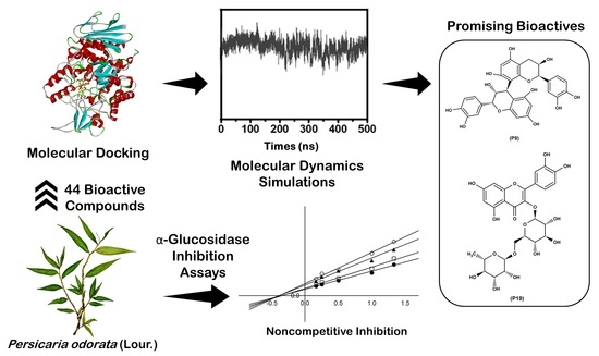
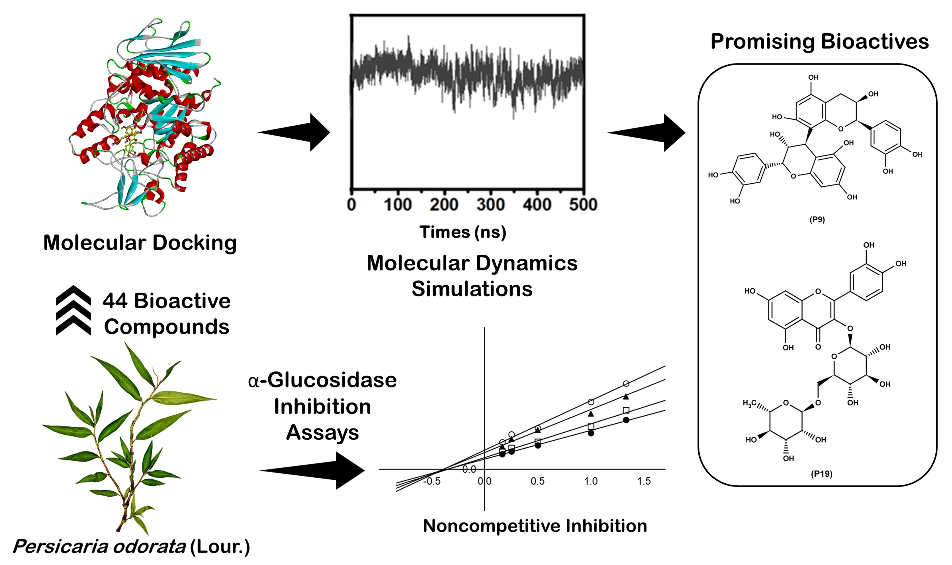
Graphical Abstract

1. Introduction

Type 2 diabetes mellitus (T2DM), one of the most prevalent chronic metabolic disorders characterized by hyperglycemia, arises when the body is unable to effectively utilize insulin or when the pancreas releases insufficient insulin levels [1]. Over 90% of diabetic patients are diagnosed with type 2 diabetes (T2D), which is non-insulin-dependent diabetes mellitus [2]. Hyperglycemia in diabetic patients could elevate the risk of vascular complications like retinopathy, nephropathy, and neuropathy [3], and also have the potential to directly elevate the risk of other deadly diseases, such as cardiovascular diseases, stroke, and cancer [2]. Controlling and suppressing postprandial hyperglycemia by inhibiting α-glucosidase activity has been implemented as one of the most effective strategies for both preventing and treating T2D [4].
α-Glucosidase, situated in the brush border of the small intestine, is a critical hydrolytic enzyme that is essential for the digestion of carbohydrates. It is responsible for the regulation of blood glucose levels by specifically hydrolyzing the 1→4-α-glucopyranosidic bond to produce α-D-glucose [5,6]. α-Glucosidase inhibitor agents (acarbose, voglibose, and miglitol) have been known to act as competitive inhibitors of the α-glucosidases [3], which can reduce both postprandial hyperglycemia and hyperinsulinemia, thereby enhancing insulin sensitivity and alleviating the stress on β-cells [3]. However, their undesired effects on the gastrointestinal tract have limited their efficacy, such as flatulence and diarrhea, which are linked to the concurrent inhibition of amylase, resulting in an increase in undigested carbohydrates in the colon [7]. Additionally, some of them may contribute to the incidence of acute hepatitis, hepatic injury, and renal tumors [8]. To resolve this issue, research for natural phytochemicals that have been known to attenuate α-glucosidase activity and have lower side effects than synthetic drugs has been conducted [8,9]. Some classes of plant phytochemicals have been reported as having the capability to attenuate α-glucosidase activity, including phenolic acids [8], flavonoids [10], terpenoids or essential oils [11], etc.
Persicaria odorata (Lour.) is an important herbal remedy that is utilized in traditional medicine to alleviate edema and inflammation, diarrhea, excessive hemorrhaging, ulcers, lesions, and wounds [12]. It has been traditionally consumed as a fresh vegetable in Southeast Asian cuisine. Meanwhile, it is employed as a traditional Thai remedy to alleviate dyspepsia and as a flavoring agent in Thailand [13]. The pharmacological benefits of P. odorata were sourced from its rich phytochemical contents [14], such as phenolics, which are categorized as phenolic acids and flavonoids [15,16,17], and essential oils or terpenoids [18,19,20]. P. odorata is known to contain high total phenolic and flavonoid contents in leaf and stem extracts, especially methanolic extract, which was linked to its antioxidant capability to scavenge free radicals at the molecular level [13,21]. Some previous studies have demonstrated that the ethanolic extract of leaves and stems [18] and the water extract of leaves [19] of P. odorata attenuated α-glucosidases activity. Unfortunately, even though phytochemical compounds were elucidated, the specific bioactive compounds responsible for the α-glucosidase inhibitory activity have not yet been elucidated. Therefore, this study aims to determine the methanolic extract of P. odorata on α-glucosidase inhibitory activity, and to investigate the bioactive compounds in P. odorata extracts as α-glucosidase inhibitors via integrated in vitro and in silico studies.

2. Results and Discussions

2.1. α-Glucosidase Inhibitory Activity

The α-glucosidase enzyme is a therapeutic target for T2DM since α-glucosidase inhibitors play a vital role in preventing the breakdown of carbohydrates to simple glucose (α-D-glucose), resulting in control of postprandial hyperglycemia in T2DM. Currently, natural products have attracted much attention as a kind of potentially safer α-glucosidase inhibitors [22]. Therefore, this study was conducted to determine the potential activity of the methanolic extract of P. odorata by in vitro studies and to explore its bioactive compounds as α-glucosidase inhibitors by in silico studies.
In this study, yeast α-glucosidase with 4-nitrophenyl-α-D-glucopyranoside as the substrate was used to initially evaluate the extract’s inhibitory activity. In addition to being widely used and cost-effective, yeast α-glucosidase catalyzes the same reaction as human α-glucosidase [23].
Inhibition of yeast α-glucosidase reflects the in vitro antidiabetic potential determined by spectrophotometry. The enzyme activity assay was conducted while taking care to avoid factors that could interfere with the enzyme inhibitory activity assay, following our protocol, including temperature, time, chemicals, and technical personnel. In the current study, the inhibition percentage values of the methanolic extract of P. odorata compared with the standard drug acarbose for yeast α-glucosidase activity are presented in Figure 1. Even though the extract contains many phytoconstituents, it could be used to assess its potential activity relative to acarbose. The in vitro results exhibited that the methanolic extract of P. odorata had a strong capability for the yeast α-glucosidase inhibition (IC50 = 0.31 ± 0.00 µg/mL) in a dose-dependent manner, which was 450-fold more potent than acarbose (IC50 = 139.47 ± 5.04 µg/mL), under experimental conditions. Further statistical analysis also indicated that the methanolic extract of P. odorata and acarbose were significantly different in inhibiting the yeast α-glucosidase activity (p < 0.05). The excellent inhibitory activity may be attributed to the phytochemicals of the crude extract, as P. odorata is known to contain high levels of phytoconstituents, so the synergistic or additive inhibition might be expected against the yeast α-glucosidase [24,25]. Previous researchers, Kee et al. [26] reported that the water extract of P. odorata has potentially inhibited both yeast and rat intestinal α-glucosidase, which was linked to an early study reporting that P. odorata inhibited both digestive enzymes, α-amylase and α-glucosidase [21]. However, due to several differences in size and amino acid sequences among yeast, rat, and human intestinal α-glucosidases, additional research linking rat and human intestinal enzymes to cell-based experiments is necessary to confirm the antidiabetic activity of the extract [27]. Moreover, further studies focused on the fractionation or isolation of pure compounds from the P. odorata extract are recommended.

2.2. Enzyme Kinetics Study Analysis

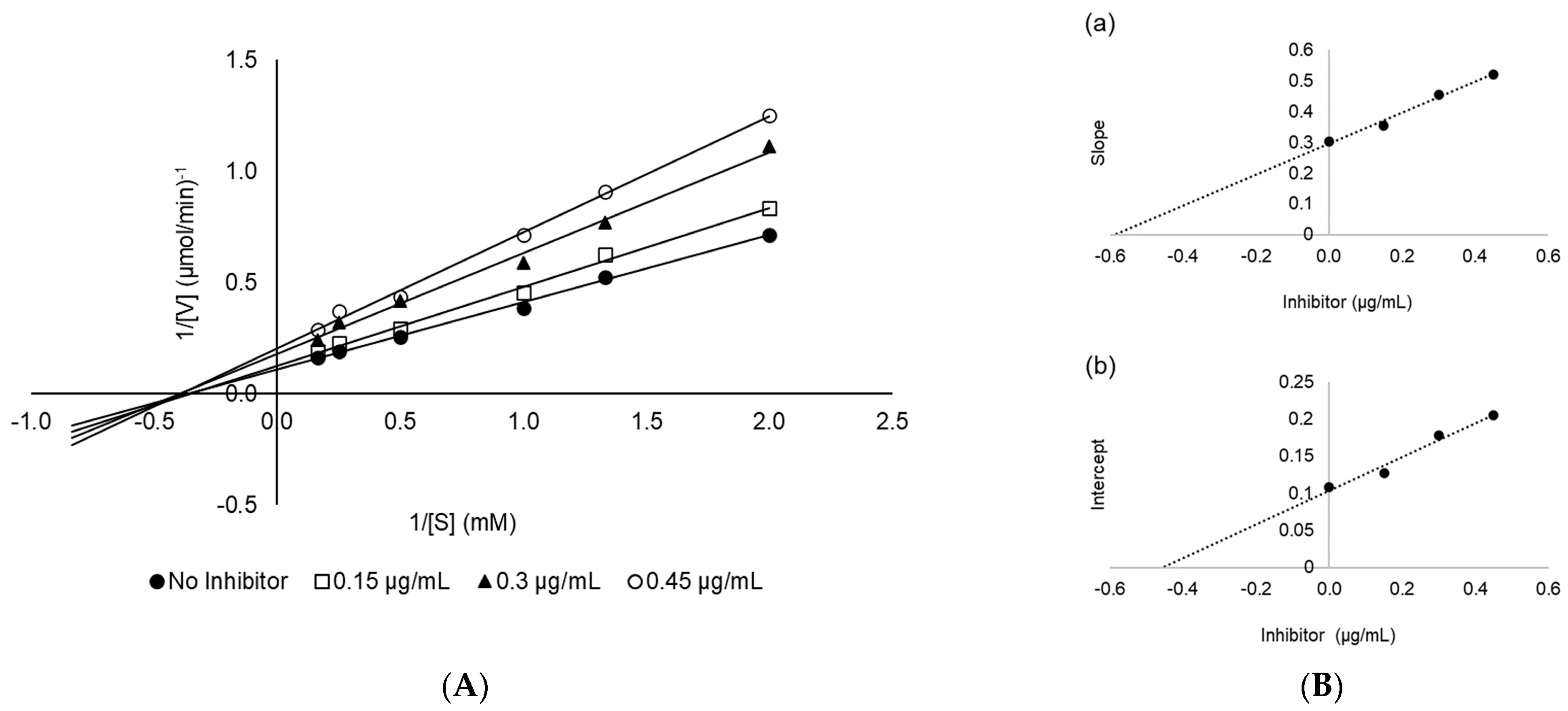
Enzymes exhibit varying inhibitor affinity, as evidenced by their inhibition constants. The dissociation constant (Ki) was obtained from the secondary plot of the slope of the linear relationships of 1/[V] vs. 1/[S] in relation to the concentration of inhibitors. The Ki value is determined by the intercept on the inhibitor axis of this plot. The Kii value of the inhibitor constant was determined by plotting 1/Vmax against the inhibitor concentration in a linear secondary plot. The Kii values are determined by the intercept of the inhibition axis [28]. Since the methanolic extract of P. odorata had strongly inhibited α-glucosidase activity, the enzyme kinetics study was performed to identify the type of inhibition of the extract. The hydrolysis reaction catalyzed by α-glucosidase was observed at a variety of substrate concentrations [S] (0.5, 0.75, 1, 2, 4, and 6 mM) to determine the initial velocity [V0] in the absence and presence of the extracts (0.15, 0.3, 0.45 µg/mL). In this current study, Lineweaver–Burk plots indicated that the methanolic extract of P. odorata inhibited the α-glucosidase with a noncompetitive inhibition (Figure 2). Noncompetitive inhibition means the inhibitor binds both the free enzyme (E0) and the enzyme–substrate (ES) complex at a different site on the enzyme [29]. Most noncompetitive inhibitors are chemically unrelated to the substrate, so increasing substrate concentration would not overcome the inhibition. In addition, noncompetitive inhibitors can decrease the concentration of active enzymes in the solution, thereby decreasing the Vmax of the reaction; however, they do not influence the Km value [30]. A noncompetitive inhibitor interacts with the enzyme at a location distinct from the actual active site. Consequently, the inhibitor’s binding does not physically obstruct the substrate binding site but can prevent subsequent reactions. An uncompetitive inhibitor binds exclusively to the ES complex, not to the free enzyme, and at a site distinct from the substrate’s active site [30].
According to Km and Vmax values obtained from Lineweaver–Burk plots, it was found that Km values remain constant, and Vmax values are decreased. In this study, α-glucosidase had a Km value of 2.797 mM for pNPG and a Vmax value of 9.259 µmoles. In the presence of 0.15, 0.30, and 0.45 µg/mL of the extracts, apparent Vmax values were found to be 7.874, 5.612, and 4.885 µmoles, and Km values remained constant at 2.791, 2.547, and 2.546 mM, respectively (Table 1). These findings indicated that the velocity [V] of the reaction catalyzed by α-glucosidase was influenced by the binding of the bioactive compounds of the extracts in a manner that was proportional to the concentration of the extracts in the reaction mixture, while the Km remained unaffected [30]. This result is linked to an early study stating that the ethanolic extract of P. odorata leaves and stems exhibited noncompetitive inhibition of α-glucosidase [21]. In contrast, acarbose is well known for inhibiting α-glucosidase competitively [3,29].

2.3. Bioactive Compounds Contained in P. odorata Extract

P. odorata contains high levels of phenolics and essential oils. In this study, there were seven phenolics from the methanolic extract of P. odorata positively detected by our group using HPLC-DAD-MS, including chlorogenic acid, sinapic acid, caffeic acid, gallic acid, protocatechuic acid, rutin, and quercetin. These compounds were selected because they are commonly found in the plant. On the other hand, as P. odorata contained high levels of phytochemicals, other bioactive compounds within any P. odorata extracts from the previous reports were selected to investigate the α-glucosidase inhibitors. A total of forty-four bioactive compounds were selected, which were divided into two main groups, namely phenolics and essential oils. Phenolics consisted of 23 compounds, including seven phenolic acids and 16 flavonoids identified by HPLC-DAD-MS, and 21 essential oils identified by GC-MS (Table 2). There were more phytochemicals found within those P. odorata extracts than in this study, especially in the essential oil group. Only essential oils with an amount higher than 1% or specifically identified were selected [18,19,20]. Table 2 presents the complete list of these compounds, while their corresponding chemical structures are depicted in Figure S1 (phenolics) and Figure S2 (essential oils). In addition, the chromatograms of phenolics in the methanolic extract of P. odorata detected by our group are presented in Figures S3–S5.

2.4. Molecular Docking

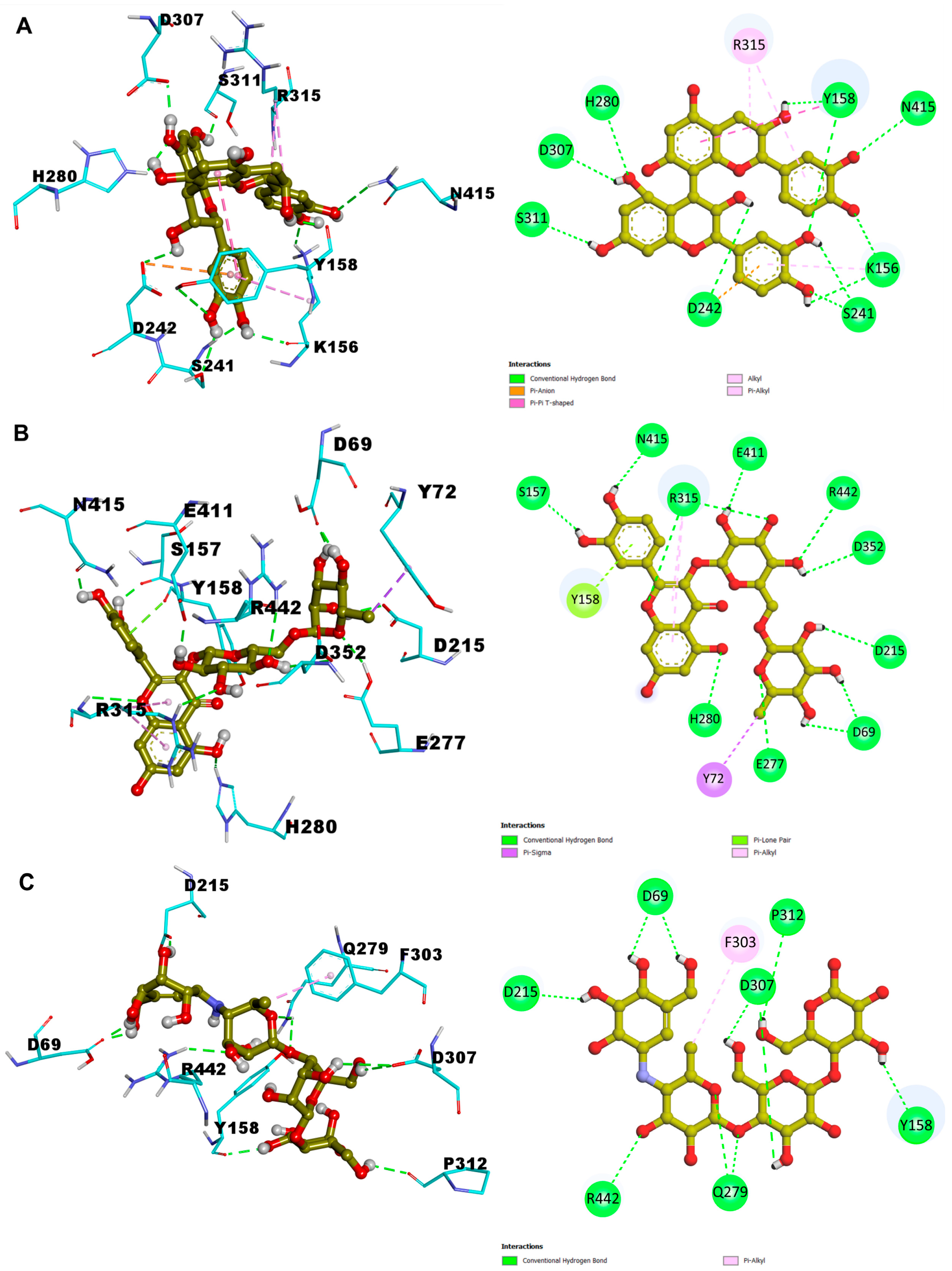
In vitro studies have demonstrated that the methanolic extract of P. odorata exhibits promising inhibitory activity against α-glucosidase, consistent with previous findings [21,26]. To further investigate this inhibitory mechanism, a molecular docking study was conducted using AutoDock4 on various bioactive compounds present in the extracts, including 23 phenolics and 21 essential oils. AutoDock4 is an excellent non-commercial docking program that is widely used. Further, it employs a stochastic Lamarckian genetic algorithm to compute ligand conformations and simultaneously minimize its scoring function, which approximates the thermodynamic stability of the ligand bound to the target protein [34]. The binding energy (ΔG) scores, inhibition constant (Ki) scores, hydrogen bond (H-bond) formations, and hydrophobic interactions between these phenolics, essential oils, and the standard drug acarbose with the amino acids of α-glucosidase’s active site are summarized. Acarbose exhibited a ΔG score of −7.30 kcal/mol. Notably, ten phenolics displayed ΔG scores lower than acarbose, including P7P9, P11, P12, P14P16, P19, and P20. Among these, P9 and P19 emerged as the most promising candidates, with the lowest ΔG scores of −9.90 kcal/mol and −9.42 kcal/mol, respectively (Table 3). Interestingly, only compound O7 from the essential oil group demonstrated the ΔG of −7.41 kcal/mol, which was lower than that of acarbose. Figure 3 shows the representative molecular docking interactions with amino acids of α-glucosidase in 3D and 2D visualizations of P9 and P19 compared to acarbose.
In the analysis of molecular binding interactions, P9 formed eight H-bonds, including K156, Y158, S241, D242, H280, D307, S311, and N415. In addition, hydrophobic interactions were formed by K156, Y158, D242, and R315 residues. Meanwhile, P19 formed 10 H-bonds, including D69, S157, D215, E277, H280, R315, D352, E411, N415, and R442. Moreover, Y72, Y158, and R315 residues also formed hydrophobic interactions. According to these results, although P9 has fewer H-bonds than P19, it was found that P9 formed double bonds via H-bonds with three amino acid residues, including K156, Y158, and S241. Meanwhile, P19 formed double bonds via H-bonds with two residues, including D69 and R315. Additionally, P9 interacted via hydrophobic interactions with four residues, while P19 interacted with three. Consequently, P9 has a higher binding affinity than P19 (Table 3 and Figure 3).
Compared to a standard drug, acarbose formed seven H-bonds with amino acid residues, including D69, Y158, D215, Q279, D307, P312, and R442, and a hydrophobic interaction with the F303 residue. This molecular docking result demonstrated that P9 shared similar interacting residues with acarbose, such as Y158 and D307, while P19 shared similar interacting residues with acarbose, which are D69, D215, and R442. These interactions indicated that P19 preferred interacting with a similar site pocket to acarbose, while P9 preferred interacting with different sites from acarbose on α-glucosidase. For more in-depth analysis, molecular dynamics simulations were conducted, and the stability of P9, P19, and acarbose with α-glucosidase enzyme affinity will be discussed below.
On the other hand, for the essential oil group, O7 formed three H-bonds with a single bond via K156, L313, and R315 residues. In addition, residues L156 and Y158 were found to form hydrophobic interactions. Most essential oils formed hydrophobic interactions and lacked H-bonds with amino acids of the active site of α-glucosidase, causing them to have low binding affinity (Table 4). The molecular interaction among other phenolics and essential oils with the amino acids of α-glucosidase’s active site in 2D visualization can be found in the Supplementary Materials, Figures S6 and S7. It is known that H-bonds have a stronger binding affinity than hydrophobic interactions. Nevertheless, it has been considered that hydrophobic interactions contributed to protein stability and enhanced the binding affinity and biological activity of the ligand–receptor complex [35,36]. Further in vitro study of this O7 compound against α-glucosidase is needed.

2.5. Molecular Dynamics Simulations Analysis

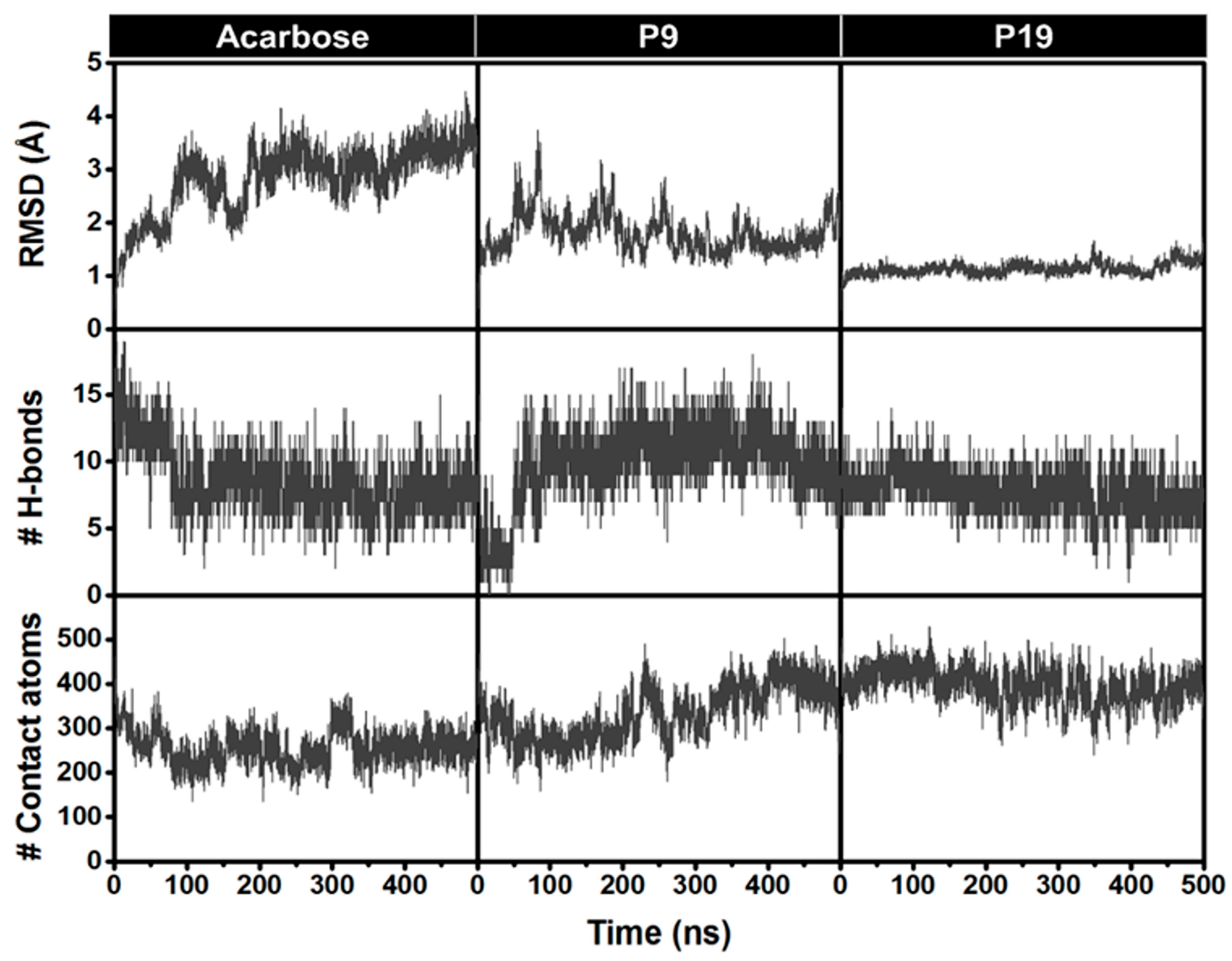
The dynamic behavior of potent compounds P9 and P19 bound to α-glucosidase was investigated using 500 ns MD simulations, compared to that of the positive control acarbose. To assess the stability of protein–ligand complexes, RMSD (root mean square deviation) of the backbone atoms within 10 Å of the active site, the # H-bonds, and the # atom contacts were calculated throughout the simulation time (Figure 4). Both acarbose and the P9 system exhibited an initial increase in RMSD values, followed by small fluctuations at ~2.5–3.5 Å and ~1.5–2.5 Å, respectively, for the remainder of the simulation. In contrast, the P19 system remained stable during the 500 ns simulation (RMSD ~1.0–1.5 Å). Over the last 100 ns, the average # H-bonds formed by acarbose, P9, and P19 were 8 ± 2, 10 ± 2, and 7 ± 1, respectively. The # atom contacts for these inhibitors ranged from ~257–401 atoms, with P9 exhibiting the highest # atom contacts, aligning with its lowest binding energy observed in the molecular docking study. This suggests that P9 could interact more favorably with α-glucosidase than the other inhibitors.
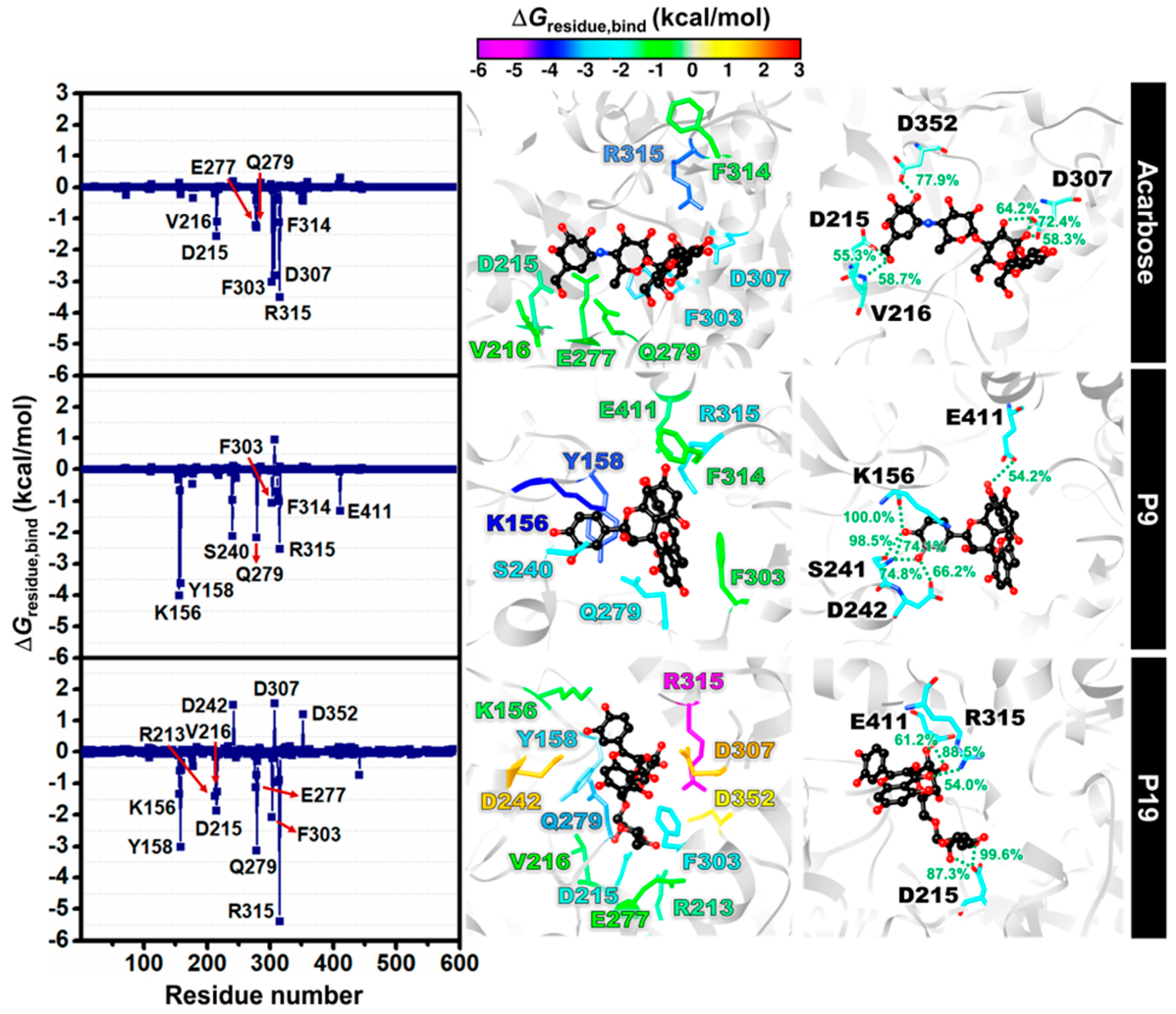
The key residues involved in inhibitor binding to α-glucosidase were identified using the MM/GBSA method (Figure 5, left). The binding orientations of each inhibitor with the active site of α-glucosidase, with hot-spot residues colored according to their ΔGresidue,bind values, are shown in Figure 5 (middle). Only residues exhibiting significant energy stabilization or destabilization (<−1.0 and >1.0 kcal/mol) are labeled and discussed. It was found that P19 interacted with 12 key residues, including K156, Y158, R213, D215, V216, D242, E277, Q279, F303, D307, R315, and D352. Acarbose interacted with eight key residues: D215, V216, E277, Q279, F303, D307, F314, and R315. Similarly, P9 interacted with eight key residues: K156, Y158, S240, Q279, F303, F314, R315, and E411. Both acarbose and P19 bind within a hydrophobic pocket formed by the D215 and E277 residues, similar to the previously reported binding mode of ursolic acid [37]. Notably, the residues Q279, F303, and R315 played crucial roles in the binding of all the compounds. Additionally, D215, a catalytic residue involved in maltose hydrolysis [38], contributed to the stabilization of both acarbose and P19 within the binding pocket through H-bonds.
To further elaborate on the insight, the percentage of H-bond occupations formed between the inhibitors and α-glucosidase residues was monitored and shown in Figure 5 (right). While H-bonds exceeding 50% occupancy were initially selected for analysis, only those exceeding 70% occupancy are discussed here to focus on strong stabilization. For the acarbose system, two strong H-bonds were identified with the residues D307 (72.4%) and D352 (77.9%). The P9 system also formed four strong H-bonds with K156 (100%) and S241 (98.5, 74.8, and 74.1%). Additionally, the P19 system exhibited three strong H-bonds with D215 (99.6 and 87.3%) and E411 (88.5%). Interestingly, the H-bonding pattern between D215 and acarbose and P19 closely resembled that observed during maltose binding to α-glucosidase [38]. These observed H-bonding interactions suggest that acarbose and P19 may inhibit α-glucosidase by binding in a manner similar to that of maltose, thereby effectively reducing the enzyme’s activity. Moreover, to confirm this activity, the promising in silico approach results, P9 and P19, were then subjected to further in vitro and in vivo investigations. It should be noted that a single 500 ns MD trajectory was performed for each protein–ligand complex in the present study. Although all systems reached stable conformations and multiple analyses consistently supported the proposed interaction mechanism, independent replicate simulations would further strengthen the statistical robustness of dynamic properties and binding free-energy estimates. To characterize protein–ligand interactions and identify key residues contributing to substrate recognition, MM/GBSA analysis was performed as previously reported [DOI: 10.1038/s41598-025-24941-5, DOI: 10.1080/07391102.2023.2201360, doi.org/10.1007/s10562-026-05443-z]. Because the aim was to assess relative interactions rather than calculate absolute binding affinities, entropy corrections were not included.

2.6. Drug-Likeness and ADMET Property Analysis

Drug-likeness and ADMET properties were predicted using computational approaches, such as SwissADME and pkCSM online software, respectively, to determine the probability of the promising bioactive compounds in P. odorata extracts for the development of drugs, especially α-glucosidase inhibitors. Lipinski’s Rule of Five (Ro5) predicted the physicochemical properties of the compounds [39], while ADMET assessed the pharmacokinetics of the compounds.
The predicted results demonstrated that P9 and P19 did not pass Lipinski’s rule because they have three Lipinski’s Ro5 violations, including high molecular weight and numerous hydrogen-bond donors and acceptors, similar to acarbose (Table 5). This may lead to poor absorption from the small intestinal tract into the systemic circulation. However, this consequence would not affect compounds targeting α-glucosidase inhibition, which are abundant on the brush border of the small intestine [40]. Additionally, 12 phenolics passed Lipinski’s Ro5, including P2P6, P8, P17, P18, P20P22, and T1, while the other 11 phenolics did not pass Lipinski’s Ro5, including P1, P7, P9P16, and P19. Most phenolics did not pass Lipinski’s Ro5 because their H-bond acceptors or donors and topological polar surface area (TPSA) were higher than standard values. Meanwhile, 13 essential oils were predicted to pass Lipinski’s Ro5, including O1, O2, O4O8, O15O18, O20, and O22. The other eight essential oils that did not pass Lipinski’s rule were O3, O9O14, and O19 due to their Mlogp values (>4.15). More details on drug-likeness predictions for phenolics and essential oils can be found in Table S1.
According to ADMET property predictions (Table 5), P9 and P19 were predicted to have lower water solubility and Caco2 permeability than acarbose. In human intestinal absorption (HIA), P9 could be absorbed in the human intestine (66.7%), which may reduce the concentration available to inhibit α-glucosidase, while P19 and acarbose were poorly absorbed, suggesting that P19 and acarbose may have a long therapeutic duration. Generally, these properties might suggest P9 and P19 to be poor candidates for systemic agents [39]. However, for intestinal α-glucosidase inhibitors, these properties may actually be advantageous and consistent with the mechanism of action [40]. HIA is an essential parameter for developing an α-glucosidase inhibitor because it provides information on how much a drug is absorbed in the human small intestine, where α-glucosidases are abundant. In addition, P9, P19, and acarbose were predicted as P-glycoprotein (P-gp) substrates. Consequently, P-gp could detect and re-efflux them from the cells back into the lumen [41]. Interestingly, P9 was also predicted to be a P-gp inhibitor I and II, whereas P19 and acarbose were not P-gp inhibitors I and II [42], meaning P9 could mitigate P-gp efflux and improve the oral absorption and bioavailability of several P-gp substrates [43]. Other ADMET properties of phenolics and essential oils can be found in Tables S2–S6.
The distribution properties predicted that P9 and acarbose had a low volume of distribution (VD). In contrast, P19 had a high VD, meaning that P9 would distribute to the plasma, while P19 would distribute instead to the tissues rather than the plasma. The blood–brain barrier (BBB) predicted that P9, P19, and acarbose were poorly distributed to the brain. The results predicted that most phenolics did not pass the BBB. Conversely, most essential oils could pass the BBB.
P9 and P19, as well as acarbose, were not substrates and inhibitors of cytochrome P450 isoforms. P9 and P19 were predicted to have lower total clearance than acarbose. P9 was a renal organic cation transporter-2 (OCT2), while P19 and acarbose were not renal OCT2. OCT2 is crucial for the renal clearance of drugs and the disposition of drugs in the kidney. Regarding toxicity, P9 and P19 were not carcinogens, were not hERG I inhibitors but hERG II inhibitors, and were safe for the liver, with properties similar to acarbose [42]. This indicates that, even though P9 was predicted to have much higher HIA than P19 and acarbose, it remains safe for systemic exposure. Therefore, these predicted results suggest that P9 and P19 may be potentially developed as α-glucosidase inhibitors.

3. Materials and Methods

3.1. Chemicals and Reagents

4-nitrophenyl-α-D-glucopyranoside or pNPG (Tokyo Chemical Industry Co., Ltd., Tokyo, Japan), the α-glucosidase enzyme (Megazyme Ltd., Wicklow, Ireland), acarbose (Fujifilm Wako Pure Chemical Corporation, Osaka, Japan), sodium bicarbonate (Na2CO3), methanol, and dimethyl sulfoxide (DMSO) were purchased from Sigma-Aldrich® Co. Ltd. (St. Louis, MO, USA). Phosphate buffer (100 mM, pH 6.8) was prepared using sodium dihydrogen phosphate (NaH2PO4·H2O) and disodium hydrogen phosphate anhydrous (Na2HPO4). Distilled water (≤18.2 mΩ) was produced using an ELGA Purelab Option-Q 15 BP (Chemoscience, Bangkok, Thailand).

3.2. Plant Collection and Extraction

P. odorata plants were purchased from the market of Maha Sarakham, Maha Sarakham Province, Thailand, in January 2023. The plant was identified by Assistant Professor Dr. Wanida Caichompoo of the Faculty of Pharmacy at Mahasarakham University. Voucher specimen was deposited at the Herbarium, Faculty of Pharmacy, Mahasarakham University with No. MSU.PH-POL-PO01. The leaves and stems were collected and washed with water, dried for one day in a heated-air oven at 45 °C, and powdered using an iron mortar and pestle before extraction. Approximately 100 g of the powdered plant was used and extracted with methanol at a 1:2 (w/v) ratio by sonication for 30 min [44]. Then, the resulting extracts were filtered with Whatman No. 1 filter paper, and the residue was extracted twice. The filtrate was desiccated at 40 °C using a rotary evaporator (Rotavapor® R-100, Buchi, Flawil, Switzerland). This extraction process yielded a value of 10.42%. Lastly, the filtrates were chilled and stored in securely sealed glass at 4 °C until analysis. All experiments, including the phytochemical screening and biological assays, were conducted using extracts from the same batch.

3.3. α-Glucosidase Inhibitory Activity Assay

The α-glucosidase inhibitory assay was conducted spectrophotometrically, with a few adjustments to the previously described procedure [45]. Briefly, 50 µg/mL of methanolic extract of P. odorata was dissolved with DMSO 5% (the final concentration was less than 1%), which did not affect enzyme activity, then mixed with 15 μL (0.5 U/mL) of the α-glucosidase solution and 20 μL of phosphate buffer (100 mM; pH 6.8). A 50 μL volume of the extract at varying concentrations (0.1, 0.2, 0.3, 0.4, 0.5, 0.75, and 1 μg/mL) was inserted into a 96-well microplate. Another 10 min was devoted to incubating the reaction mixture at 37 °C. Subsequently, 15 μL of pNPG (5 mM) was incorporated into the reaction mixture to initiate the enzymatic reaction and incubated at 37 °C for 15 min. Immediately, the reaction mixture was supplemented with 150 μL of Na2CO3 (1 M) solution to retard the enzymatic activity. In place of the sample, DMSO was employed to produce the control. Similarly, by changing the enzyme and pNPG with buffer, blank samples were generated for each sample. In order to quantify the absorbance of p-nitrophenol that was emitted during the reaction, the microplate reader was employed to measure at 405 nm. Acarbose, as an α-glucosidase inhibitor agent, was implemented as a positive control. The inhibition percentage of the α-glucosidase activity was determined by employing the following formula:
I n h i b i t i o n % = A b s o r b a n c e   o f   C o n t r o l A b s o r b a n c e   o f   S a m p l e A b s o r b a n c e   o f   C o n t r o l × 100

3.4. Enzyme Kinetics Study

The detailed procedure for the enzyme kinetics study of the α-glucosidase by the methanolic extract of P. odorata was also similar to the α-glucosidase inhibition assay described above. In order to evaluate the reversibility of enzyme inhibition, the reaction rate was calculated by increasing the substrate concentration, pNPG (0.5, 0.75, 1, 2, 4, and 6 mM), in the absence or presence of inhibitor, methanolic extract of P. odorata, at three various concentrations (0.15, 0.3, and 0.45 µg/mL). The enzyme inhibitory reaction was recorded by measuring the absorbance of the microplate reader at 405 nm for 60 min at 37 °C. To ascertain the type of enzyme inhibition induced by inhibitors, the Lineweaver–Burk plot was employed to determine the Vmax and Km, which were calculated from the results according to Michaelis–Menten kinetics [46]. From the secondary plots of the primary rate data, the enzyme inhibitor [EI] and enzyme substrate inhibitor [ESI] constants were determined through linear regression analysis. Ki values were determined by plotting the slopes of the Lineweaver–Burk plots against inhibitor concentrations. In contrast, Kii values were determined by plotting intercepts against inhibitor concentrations derived from the x-intercepts [28].

3.5. HPLC-DAD Analysis

High-performance liquid chromatography (Shimadzu Co., Tokyo, Japan) with a UV diode-array detector (HPLC-UV-DAD) was used to determine the phenolic profile of the methanolic extract of P. odorata. Analysis of the phenolic profile was performed as described in our previous study [47]. The extract was filtered using a 0.45 µm filter. The separations were realized using a Unisol C18 column as a stationary phase (4.6 mm inside diameter × 250 mm length, 5 µm particle size). Gradient elution was performed at a constant flow rate of 0.8 mL min−1, with Acetic acid 1% (A) and acetonitrile 100% (B) as the mobile phase. The spectral data were acquired at 280, 320, and 380 nm for UV detection. The retention times of the extract were identified by comparing retention times with standard phenolic acids, including gallic acid, protocatechuic acid, syringic acid, p-hydroxybenzoic acid, and caffeic acid at 280 nm, chlorogenic acid, p-coumaric acid, ferulic acid, and sinapic acid at 320 nm, and rutin and quercetin at 370 nm. The standard phenolics and extract were analyzed in triplicate. The spectra of each compound and the chromatogram are shown in Figures S3–S5.

3.6. Statistical Analysis

Each experiment was conducted in triplicate, and the results were reported as the mean ± standard deviation (SD). The statistical analysis was calculated utilizing IBM28 SPSS (version 28, Armonk, New York, NY, USA). The one-way analysis of variance (ANOVA) was implemented to assess the variations among the extracts, which was subsequently followed by Tukey’s test. A significant difference was defined as a p-value (p < 0.05).

3.7. Data Collection of Bioactive Compounds Contained in P. odorata Extracts

Phytochemical compounds of P. odorata extracts were obtained carefully from previous researchers’ reports who had screened, identified, and isolated bioactive compounds from P. odorata extracts. There were 44 compounds collected and divided into two main groups, including 23 phenolics and 21 essential oils, where phenolics consist of seven phenolic acids and 16 flavonoids. Only in silico approaches were applied for these 44 compounds to predict their binding affinity with α-glucosidase by molecular docking, then the two lowest binding affinities were chosen for analyzing their binding stability by molecular dynamics simulations, and their pharmacokinetic properties were predicted.

3.8. Molecular Docking Study

The methanolic extract of P. odorata exhibited potent inhibitory activity against α-glucosidase. To elucidate the molecular basis of this inhibition, molecular docking studies were performed using a comprehensive set of previously characterized bioactive compounds derived from this plant. Due to the unavailability of the three-dimensional (3D) structure of Saccharomyces cerevisiae α-glucosidase, the 3D crystal structure of the S. cerevisiae isomaltase (PDB ID: 3A4A), which has a resolution of 1.60 Å and shares high sequence identity with 72% identity and 85% similarity with S. cerevisiae α-glucosidase, was retrieved from the RCSB Protein Data Bank (PDB) [48,49]. The higher resolution of the receptor provides an effective receptor file [50], and the high degree of sequence homology suggests that this structure is a suitable and reliable surrogate for S. cerevisiae α-glucosidase in molecular docking studies, as demonstrated in several previous studies [49,51,52,53].
Prior to docking calculations, the protein’s 3D structure was properly prepared. Firstly, the protein and ligand were protonated at pH 7.4 to simulate physiological conditions using PDB2PQR [54] and Marvin, respectively. The identified compounds were retrieved from the PubChem database (https://pubchem.ncbi.nlm.nih.gov (accessed on 14 February 2026)). AutoDock Tools version 1.5.7 (ADT v1.5.7) was used to prepare both the protein and compound structures [34]. Second, all water molecules, ligands, and heteroatoms were eliminated from the protein. Polar hydrogens and Kollman charges were subsequently assigned to the protein [55], and missing and terminal residues in polypeptide chains were repaired using ADT v1.5.7. It was ensured that no residues contained non-integral charges, a requirement for the receptor file in AutoDock4.2. The preparation of both ligands and receptors was saved as PDBQT files for the docking simulations software [50]. The center of the predicted active site was defined as the center of a 60 × 60 × 60 Å grid box with a 0.375 Å grid spacing. The grid box was centered at coordinates X = 21.519, Y = −7.702, and Z = 23.554. Compound structures were subsequently optimized using ADT and saved in PDBQT format. Molecular docking simulations were then performed using AutoDock 4.2.6 with the Lamarckian Genetic Algorithm (LGA) method, with 100 independent runs, a population size of 150, an energy evaluation of 25 × 105, while all other parameters were kept at their default values [34]. The docking protocol was validated by re-docking the native ligand into the α-glucosidase active site. The re-docking RMSD value of 0.31 Å, which was less than 1 Å, confirmed the reliability of the protocol [56,57]. The docked conformation with the lowest binding energy was selected for subsequent interaction analysis, including hydrogen-bond and hydrophobic interactions, using BIOVIA Discovery Studio Visualizer 2021.

3.9. Molecular Dynamics Simulations

To explore the structural and dynamic behavior of a compound against α-glucosidase in an aqueous solution, all-atom MD simulations were conducted with periodic boundary conditions (PBC) using the AMBER22 software package. Initially, the selected compounds underwent optimization via the Gaussian09 program at the HF/6-31G(d) level, followed by the generation of their restrained electrostatic potential (RESP) charges using the antechamber module of AMBER22, according to our standard protocol [58,59]. The protein and ligand were assigned to the AMBER ff19SB force field [60] and the generalized AMBER force field 2 (GAFF2) [61], respectively. Subsequently, missing hydrogen atoms were added to the protein–ligand complex using the leap module. Next, the system was solvated with a TIP3P water model [62] and subjected to further minimization, first with restraints on the solvent and then with full system minimization. Non-bonded interactions were set at a 12 Å cutoff, and long-range electrostatic interactions were treated with the Particle Mesh Ewald method [63]. The target pressure (1 atm) was maintained using the Berendsen barostat [64], while covalent bonds involving hydrogen atoms were constrained using the SHAKE algorithm [65]. The system was heated up to 310 K for 100 ps, followed by a 100 ps equilibration period using the Langevin thermostat [66] with a collision frequency of 2.0 ps−1. Finally, 500 ns MD simulations of the complex systems were conducted at 310 K and 1 atm. The structural analysis focused on various parameters, including root mean square deviation (RMSD), intermolecular hydrogen bonding (# H-bonds), and the number of contact atoms (# Contact atoms), which were calculated using the CPPTRAJ module [67]. Additionally, the binding free energy decomposition per residue (ΔGresidue,bind) was determined using the Molecular Mechanics Generalized Born Surface Area (MMGBSA) [68] method based on 100 snapshots from the last 100 ns of the simulations.

3.10. ADMET Property Prediction

Both online servers of SwissADME (http://www.swissadme.ch/index.php (accessed on 14 February 2026)) was used to analyze the drug-likeness Lipinski’s rule of 5 (Ro5) and pkCSM (http://biosig.unimelb.edu.au/pkcsm/ (accessed on 14 February 2026)) was used to predict pharmacokinetic properties, which are absorption, distribution, metabolism, excretion, and toxicity (ADMET) of the bioactive compounds within P. odorata extracts using their Simplified Molecular Input Line Entry System (SMILES) [42,69].

4. Conclusions

The methanolic extract of P. odorata exhibited strong inhibitory activity against yeast α-glucosidase enzyme compared to acarbose, an α-glucosidase inhibitory agent. The type of inhibition suggests a noncompetitive inhibitor, indicating that its bioactive compounds predominantly bind to the enzyme [E0] and to the enzyme–substrate [ES] complex at sites distant from the substrate binding site, confirming that P. odorata could be used as an alternative edible plant to control postprandial hyperglycemia in T2DM. On the other hand, procyanidin B (P9) and rutin (P19), which belong to the flavonoids, were predicted by computational approaches to be bioactive compounds that have more favorable binding affinity to yeast α-glucosidase residues. Moreover, both P9 and P19 were predicted to qualify as α-glucosidase inhibitors with low toxicity. These findings could serve as basic scientific data, and further in vitro and in vivo studies are necessary to support these findings.

Supplementary Materials

The following supporting information can be downloaded at https://www.mdpi.com/article/10.3390/ijms27156885/s1.

Author Contributions

Conceptualization, P.P., B.S., K.S., T.P. and M.S.; methodology and investigation, P.P., M.S., K.S., B.N., J.R., P.S., S.D. and B.B.; software, K.S., B.N. and M.S.; validation, P.P., K.S., J.R. and M.S.; formal analysis, P.P., M.S., K.S. and J.R.; resources, B.S., B.N. and B.B.; data curation, P.P., B.S., K.S., P.S. and M.S.; writing—original draft preparation, P.P., K.S. and M.S.; writing—review and editing, P.P., K.S. and M.S.; visualization, K.S., M.S. and J.R.; supervision, P.P., B.S. and K.S.; project administration, P.P., K.S., B.S. and T.P.; funding acquisition, P.P. All authors have read and agreed to the published version of the manuscript.

Funding

This work was financially supported by the National Science, Research, and Innovation Fund (NSRF), Thailand, through the Fundamental Fund (FF), Khon Kaen University.

Institutional Review Board Statement

Not applicable.

Informed Consent Statement

Not applicable.

Data Availability Statement

The original contributions presented in this study are included in the article/Supplementary Material. Further inquiries can be directed to the corresponding author.

Acknowledgments

The authors would like to thank the National Science, Research, and Innovation Fund (NSRF), Thailand, for financial support through the Fundamental Fund (FF), Khon Kaen University. The first author, Muhammad Subhan, would like to thank the Khon Kaen University (KKU) Scholarship for ASEAN and GMS Countries’ Personnel for providing financial support during his master’s program.

Conflicts of Interest

The authors declare no conflicts of interest.

Abbreviations

The following abbreviations are used in this manuscript:
ADMETAbsorption, Distribution, Metabolism, Excretion, and Toxicity
BBBBrain–Blood Barrier
E0Free Enzyme
ESEnzyme–Substrate
H-bondHydrogen Bond
HIAHuman Intestinal Absorption
KiInhibition Constant
KmMichaelis Constant
MDMolecular Dynamics
PDBProtein Data Bank
P-gpP-glycoprotein
RMSDRoot Mean Square Deviation
TPSATopological Polar Surface Area
VDVolume Distribution
VmaxMaximum Velocity
[S]Substrate
[V]Velocity
[V0]Initial Velocity

References

  1. Kato, A.; Hayashi, E.; Miyauchi, S.; Adachi, I.; Imahori, T.; Natori, Y.; Yoshimura, Y.; Nash, R.J.; Shimaoka, H.; Nakagome, I.; et al. α-1-C-Butyl-1, 4-dideoxy-1, 4-imino-l-arabinitol as a second-generation iminosugar-based oral α-glucosidase inhibitor for improving postprandial hyperglycemia. J. Med. Chem. 2012, 55, 10347–10362. [Google Scholar] [CrossRef] [PubMed]
  2. Zeng, Z.; Yin, X.; Wang, X.; Yang, W.; Liu, X.; Hong, Y. Synthesis of water soluble pentacyclic dihydroxyterpene carboxylic acid derivatives coupled amino acids and their inhibition activities on α-glucosidase. Bioorg. Chem. 2019, 86, 277–287. [Google Scholar] [CrossRef] [PubMed]
  3. Scheen, A.J. Is there a role for α-glucosidase inhibitors in the prevention of type 2 diabetes mellitus? Drugs 2003, 63, 933–951. [Google Scholar] [CrossRef] [PubMed]
  4. Ye, G.J.; Lan, T.; Huang, Z.X.; Cheng, X.N.; Cai, C.Y.; Ding, S.M.; Xie, M.L.; Wang, B. Design and synthesis of novel xanthone-triazole derivatives as potential antidiabetic agents: α-Glucosidase inhibition and glucose uptake promotion. Eur. J. Med. Chem. 2019, 177, 362–373. [Google Scholar] [CrossRef] [PubMed]
  5. Kazmi, M.; Zaib, S.; Ibrar, A.; Amjad, S.T.; Shafique, Z.; Mehsud, S.; Saeed, A.; Iqbal, J.; Khan, I. A new entry into the portfolio of α-glucosidase inhibitors as potent therapeutics for type 2 diabetes: Design, bioevaluation and one-pot multi-component synthesis of diamine-bridged coumarinyl oxadiazole conjugates. Bioorg. Chem. 2018, 77, 190–202. [Google Scholar] [CrossRef] [PubMed]
  6. Ma, C.-M.; Hattori, M.; Daneshtalab, M.; Wang, L. Chlorogenic acid derivatives with alkyl chains of different lengths and orientations: Potent α-glucosidase inhibitors. J. Med. Chem. 2008, 51, 6188–6194. [Google Scholar] [CrossRef] [PubMed]
  7. Kumar, R.V.; Sinha, V.R. Newer insights into the drug delivery approaches of α-glucosidase inhibitors. Expert Opin. Drug Deliv. 2012, 9, 403–416. [Google Scholar] [CrossRef] [PubMed]
  8. Shobana, S.; Sreerama, Y.N.; Malleshi, N.G. Composition and enzyme inhibitory properties of finger millet (Eleusine coracana L.) seed coat phenolics: Mode of inhibition of α-glucosidase and pancreatic amylase. Food Chem. 2009, 115, 1268–1273. [Google Scholar] [CrossRef]
  9. Hiroyuki, F.; Tomohide, Y.; Kazunori, O. Efficacy and safety of Touchi extract, an α-glucosidase inhibitor derived from fermented soybeans, in non-insulin-dependent diabetic mellitus. J. Nutr. Biochem. 2001, 12, 351–356. [Google Scholar] [CrossRef] [PubMed]
  10. Tadera, K.; Minami, Y.; Takamatsu, K.; Matsuoka, T. Inhibition of α-glucosidase and α-amylase by flavonoids. J. Nutr. Sci. Vitaminol. 2006, 52, 149–153. [Google Scholar] [CrossRef] [PubMed]
  11. Salazar, M.O.; Osella, M.I.; Arcusin, D.E.J.; Lescano, L.E.; Furlan, R.L.E. New α-glucosidase inhibitors from a chemically engineered essential oil of Origanum vulgare L. Ind. Crops Prod. 2020, 156, 112855. [Google Scholar] [CrossRef]
  12. Dash, G.K.; Zakaria, Z.B. Pharmacognostic studies on Persicaria odorata (Lour.) Sojak. J. Pharm. Res. 2016, 10, 377–380. [Google Scholar]
  13. Chansiw, N.; Chotinantakul, K.; Srichairatanakool, S. Anti-inflammatory and antioxidant activities of the extracts from leaves and stems of Polygonum odoratum Lour. Anti-Inflamm. Anti-Allergy Agents Med. Chem. 2019, 18, 45–54. [Google Scholar] [CrossRef] [PubMed]
  14. Kiraman, N.A.S.; Yusof, H. A mini review on the methods for the extraction, isolation, and determination of P. odorata’s bioactive compounds. Healthscope 2022, 5, 38–44. [Google Scholar]
  15. Nanasombat, S.; Teckchuen, N. Antimicrobial, antioxidant and anticancer activities of Thai local vegetables. J. Med. Plants Res. 2009, 3, 443–449. [Google Scholar]
  16. Pawłowska, K.A.; Strawa, J.; Tomczyk, M.; Granica, S. Changes in the phenolic contents and composition of Persicaria odorata fresh and dried leaves. J. Food Compos. Anal. 2020, 91, 103507. [Google Scholar] [CrossRef]
  17. Puthongking, P.; Ratha, J.; Siriparu, P.; Subhan, M.; Panyatip, P.; Sripadung, P.; Padumanonda, T.; Tadtong, S.; Lim, V.; Sungthong, B. Sources of polyphenols with potential α-glucosidase-inhibitory activities from Thai local edible plants: In vitro and network pharmacology approaches. Plants 2026, 15, 1721. [Google Scholar] [CrossRef] [PubMed]
  18. Ridzuan, P.M.; Hairul Aini, H.; Norazian, M.H.; Shah, A.; Roesnita Aminah, K.S. Antibacterial and antifungal properties of Persicaria odorata leaf against pathogenic bacteria and fungi. Open Conf. Proc. J. 2013, 4, 71–74. [Google Scholar] [CrossRef]
  19. Sasongko, P.; Laohankunjit, N.; Kerdchoechuen, O. Antibacterial activity of the essential oil from Persicaria odorata leaves. Agric. Sci. J. 2011, 42, 105–108. [Google Scholar]
  20. Starkenmann, C.; Luca, L.; Niclass, Y.; Praz, E.; Roguet, D. Comparison of volatile constituents of Persicaria odorata (Lour.) Soják (Polygonum odoratum Lour.) and Persicaria hydropiper L. Spach (Polygonum hydropiper L.). J. Agric. Food Chem. 2006, 54, 3067–3071. [Google Scholar] [CrossRef] [PubMed]
  21. Thongra-ar, K.; Rojsanga, P.; Chewchinda, S.; Mangmool, S.; Sithisarn, P. Antioxidant, α-glucosidases and α-amylase inhibitory activities of Persicaria odorata. CMU J. Nat. Sci. 2021, 20, e2021051. [Google Scholar] [CrossRef]
  22. Peng, X.; Zhang, G.; Liao, Y.; Gong, D. Inhibitory kinetics and mechanism of kaempferol on α-glucosidase. Food Chem. 2016, 190, 207–215. [Google Scholar] [CrossRef] [PubMed]
  23. Nguyen, D.V.; Hengphasatporn, K.; Danova, A.; Suroengrit, A.; Boonyasuppayakorn, S.; Fujiki, R.; Shigeta, Y.; Rungrotmongkol, T.; Chavasiri, W. Structure-yeast α-glucosidase inhibitory activity relationship of 9-O-berberrubine carboxylates. Sci. Rep. 2023, 13, 18865. [Google Scholar] [CrossRef] [PubMed]
  24. Pan, J.; Nawaz, M.; Liu, J.; Liu, H.; Lv, Z.; Yang, W.; Jiao, Z.; Zhang, Q. Exploring synergistic inhibitory mechanisms of flavonoid mixtures on α-glucosidase by experimental analysis and molecular dynamics simulation. Food Chem. 2025, 464, 141560. [Google Scholar] [CrossRef] [PubMed]
  25. Son, H.-U.; Yoon, E.-K.; Yoo, C.-Y.; Park, C.-H.; Bae, M.-A.; Kim, T.-H.; Lee, C.H.; Lee, K.W.; Seo, H.; Kim, K.-J.; et al. Effects of synergistic inhibition on α-glucosidase by phytoalexins in soybeans. Biomolecules 2019, 9, 828. [Google Scholar] [CrossRef] [PubMed]
  26. Kee, K.T.; Koh, M.; Oong, L.X.; Ng, K. Screening culinary herbs for antioxidant and α-glucosidase inhibitory activities. Int. J. Food Sci. Technol. 2013, 48, 1884–1891. [Google Scholar] [CrossRef]
  27. Proença, C.; Rufino, A.T.; de Oliveira, J.M.P.F.; Freitas, M.; Fernandes, P.A.; Silva, A.M.S.; Fernandes, E. Inhibitory activity of flavonoids against human sucrase-isomaltase (α-glucosidase) activity in a Caco-2/TC7 cellular model. Food Funct. 2022, 13, 1108–1118. [Google Scholar] [CrossRef] [PubMed]
  28. Kalita, D.; Holm, D.G.; LaBarbera, D.V.; Petrash, J.M.; Jayanty, S.S. Inhibition of α-glucosidase, α-amylase, and aldose reductase by potato polyphenolic compounds. PLoS ONE 2018, 13, e0191025. [Google Scholar] [CrossRef] [PubMed]
  29. Abioye, R.O.; Nwamba, O.C.; Okagu, O.D.; Udenigwe, C.C. Synergistic effect of acarbose–chlorogenic acid on α-glucosidase inhibition: Kinetics and interaction studies reveal mixed-type inhibition and denaturant effect of chlorogenic acid. ACS Food Sci. Technol. 2023, 3, 1255–1268. [Google Scholar] [CrossRef]
  30. Robinson, P.K. Enzymes: Principles and biotechnological applications. Essays Biochem. 2015, 59, 1–41, Erratum in Essays Biochem. 2015, 59, 75. [Google Scholar] [CrossRef] [PubMed]
  31. Řebíčková, K.; Bajer, T.; Šilha, D.; Houdková, M.; Ventura, K.; Bajerová, P. Chemical composition and determination of the antibacterial activity of essential oils in liquid and vapor phases extracted from two different Southeast Asian herbs—Houttuynia cordata (Saururaceae) and Persicaria odorata (Polygonaceae). Molecules 2020, 25, 2432. [Google Scholar] [CrossRef] [PubMed]
  32. Settacomkul, R.; Sangpairoj, K.; Phuagkhaopong, S.; Meemon, K.; Niamnont, N.; Sobhon, P.; Vivithanaporn, P. Ethanolic extract of Halymenia durvillei induced G2/M arrest and altered the levels of cell cycle regulatory proteins of MDA-MB-231 triple-negative breast cancer cells. Res. Pharm. Sci. 2023, 18, 279. [Google Scholar] [CrossRef] [PubMed]
  33. Fujita, K.; Chavasiri, W.; Kubo, I. Anti-Salmonella activity of volatile compounds of Vietnam Coriander. Phytother. Res. 2015, 29, 1081–1087. [Google Scholar] [CrossRef] [PubMed]
  34. Morris, G.M.; Huey, R.; Lindstrom, W.; Sanner, M.F.; Belew, R.K.; Goodsell, D.S.; Olson, A.J. AutoDock4 and AutoDockTools4: Automated docking with selective receptor flexibility. J. Comput. Chem. 2009, 30, 2785–2791. [Google Scholar] [CrossRef] [PubMed]
  35. Pace, C.N.; Fu, H.; Fryar, K.L.; Landua, J.; Trevino, S.R.; Shirley, B.A.; Hendricks, M.M.; Iimura, S.; Gajiwala, K.; Scholtz, J.M.; et al. Contribution of hydrophobic interactions to protein stability. J. Mol. Biol. 2011, 408, 514–528. [Google Scholar] [CrossRef] [PubMed]
  36. Patil, R.; Das, S.; Stanley, A.; Yadav, L.; Sudhakar, A.; Varma, A.K. Optimized hydrophobic interactions and hydrogen bonding at the target-ligand interface leads the pathways of drug-designing. PLoS ONE 2010, 5, e12029. [Google Scholar] [CrossRef] [PubMed]
  37. Tuan, N.N.; Thi, H.N.; My, C.L.T.; Hai, T.X.; Trung, H.T.; Kim, A.N.T.; Tan, T.N.; Van, T.L.; Nguyen, C.Q.; Tran, Q.D.; et al. Inhibition of α-glucosidase, acetylcholinesterase, and nitric oxide production by phytochemicals isolated from Millettia speciosa—In vitro and molecular docking studies. Plants 2022, 11, 388. [Google Scholar] [CrossRef] [PubMed]
  38. Yamamoto, K.; Miyake, H.; Kusunoki, M.; Osaki, S. Crystal structures of isomaltase from Saccharomyces cerevisiae and in complex with its competitive inhibitor maltose: Crystal Structure of Isomaltase. FEBS J. 2010, 277, 4205–4214. [Google Scholar] [CrossRef] [PubMed]
  39. Lipinski, C.A. Lead- and Drug-like Compounds: The Rule-of-Five Revolution. Drug Discov. Today Technol. 2004, 1, 337–341. [Google Scholar] [CrossRef] [PubMed]
  40. Rosak, C.; Mertes, G. Critical evaluation of the role of acarbose in the treatment of diabetes: Patient considerations. Diabetes Metab. Syndr. Obes. 2012, 5, 357–367. [Google Scholar] [CrossRef] [PubMed]
  41. Abchir, O.; Khedraoui, M.; Nour, H.; Yamari, I.; Errougui, A.; Samadi, A.; Chtita, S. Integrative approach for designing novel triazole derivatives as α-glucosidase inhibitors: QSAR, molecular docking, ADMET, and molecular dynamics investigations. Pharmaceuticals 2024, 17, 261. [Google Scholar] [CrossRef] [PubMed]
  42. Pires, D.E.V.; Blundell, T.L.; Ascher, D.B. pkCSM: Predicting small-molecule pharmacokinetic and toxicity properties using graph-based signatures. J. Med. Chem. 2015, 58, 4066–4072. [Google Scholar] [CrossRef] [PubMed]
  43. Nguyen, T.-T.-L.; Duong, V.-A.; Maeng, H.-J. Pharmaceutical formulations with P-glycoprotein inhibitory effect as promising approaches for enhancing oral drug absorption and bioavailability. Pharmaceutics 2021, 13, 1103. [Google Scholar] [CrossRef] [PubMed]
  44. Javadi, N.; Abas, F.; Hamid, A.A.; Simoh, S.; Shaari, K.; Ismail, I.S.; Mediani, A.; Khatib, A. GC-MS-based metabolite profiling of Cosmos caudatus leaves possessing alpha-glucosidase inhibitory activity. J. Food Sci. 2014, 79, C1130–C1136. [Google Scholar] [CrossRef] [PubMed]
  45. Mugaranja, K.P.; Kulal, A. Alpha glucosidase inhibition activity of phenolic fraction from Simarouba glauca: An in-vitro, in-silico and kinetic study. Heliyon 2020, 6, e04392. [Google Scholar] [CrossRef] [PubMed]
  46. Kim, Y.-M.; Jeong, Y.-K.; Wang, M.-H.; Lee, W.-Y.; Rhee, H.-I. Inhibitory effect of pine extract on α-glucosidase activity and postprandial hyperglycemia. Nutrition 2005, 21, 756–761. [Google Scholar] [CrossRef] [PubMed]
  47. Siriparu, P.; Panyatip, P.; Pota, T.; Ratha, J.; Yongram, C.; Srisongkram, T.; Sungthong, B.; Puthongking, P. Effect of germination and illumination on melatonin and its metabolites, phenolic content, and antioxidant activity in mung bean sprouts. Plants 2022, 11, 2990. [Google Scholar] [CrossRef] [PubMed]
  48. Adib, M.; Peytam, F.; Rahmanian-Jazi, M.; Mahernia, S.; Bijanzadeh, H.R.; Jahani, M.; Mohammadi-Khanaposhtani, M.; Imanparast, S.; Faramarzi, M.A.; Mahdavi, M.; et al. New 6-amino-pyrido[2,3-d]pyrimidine-2,4-diones as novel agents to treat type 2 diabetes: A simple and efficient synthesis, α-glucosidase inhibition, molecular modeling and kinetic study. Eur. J. Med. Chem. 2018, 155, 353–363. [Google Scholar] [CrossRef] [PubMed]
  49. Yan, J.; Zhang, G.; Pan, J.; Wang, Y. α-Glucosidase inhibition by luteolin: Kinetics, interaction and molecular docking. Int. J. Biol. Macromol. 2014, 64, 213–223. [Google Scholar] [CrossRef] [PubMed]
  50. Akshatha, J.V.; SantoshKumar, H.S.; Prakash, H.S.; Nalini, M.S. In silico docking studies of α-amylase inhibitors from the anti-diabetic plant Leucas ciliata Benth. and an endophyte, Streptomyces longisporoflavus. 3 Biotech 2021, 11, 51. [Google Scholar] [CrossRef] [PubMed]
  51. Abudurexiti, A.; Zhang, R.; Zhong, Y.; Tan, H.; Yan, J.; Bake, S.; Ma, X. Identification of α-glucosidase inhibitors from Mulberry Using UF-UPLC-QTOF-MS/MS and molecular docking. J. Funct. Foods 2023, 101, 105362. [Google Scholar] [CrossRef]
  52. Sanachai, K.; Chamni, S.; Nutho, B.; Khammuang, S.; Ratha, J.; Choowongkomon, K.; Puthongking, P. Mechanistic study of α-mangostin derivatives as potent α-glucosidase inhibitors. Mol. Divers 2025, 29, 6293–6309. [Google Scholar] [CrossRef] [PubMed]
  53. Wang, W.L.; Zhang, K.-Y.; Yuan, M.-Q.; Yang, M.; Wang, A.-D.; Huang, L.; Li, J.L. α-Glucosidase inhibitors from the husks of Rice Oryza sativa L. Fitoterapia 2023, 171, 105688. [Google Scholar] [CrossRef] [PubMed]
  54. Dolinsky, T.J.; Nielsen, J.E.; McCammon, J.A.; Baker, N.A. PDB2PQR: An automated pipeline for the setup of Poisson–Boltzmann electrostatics calculations. Nucleic Acids Res. 2004, 32, W665–W667. [Google Scholar] [CrossRef] [PubMed]
  55. Aispuro-Pérez, A.; López-Ávalos, J.; García-Páez, F.; Montes-Avila, J.; Picos-Corrales, L.A.; Ochoa-Terán, A.; Bastidas, P.; Montaño, S.; Calderón-Zamora, L.; Osuna-Martínez, U.; et al. Synthesis and molecular docking studies of imines as α-glucosidase and α-amylase inhibitors. Bioorg. Chem. 2020, 94, 103491. [Google Scholar] [CrossRef] [PubMed]
  56. Li, S.; Yin, L.; Yi, J.; Zhang, L.-M.; Yang, L. Insight into interaction mechanism between theaflavin-3-gallate and α-glucosidase using spectroscopy and molecular docking analysis. J. Food Biochem. 2021, 45, e13550. [Google Scholar] [CrossRef] [PubMed]
  57. Sakulkeo, O.; Wattanapiromsakul, C.; Pitakbut, T.; Dej-adisai, S. Alpha-glucosidase inhibition and molecular docking of isolated compounds from traditional Thai medicinal plant, Neuropeltis racemosa Wall. Molecules 2022, 27, 639. [Google Scholar] [CrossRef] [PubMed]
  58. Nutho, B.; Tungmunnithum, D. Exploring major flavonoid phytochemicals from Nelumbo nucifera Gaertn. as potential skin anti-Aging agents: In silico and in vitro evaluations. IJMS 2023, 24, 16571. [Google Scholar] [CrossRef] [PubMed]
  59. Pinsorn, P.; Sanachai, K.; Rungrotmongkol, T.; Hoefgen, R.; Watanabe, M.; Brueckner, F.; Nakabayashi, R.; Mori, T.; Oikawa, A.; Sasaki, R.; et al. Sulfur metabolism in durian pulps: Factors contributing to the production of volatile sulfur compounds during fruit ripening. Postharvest Biol. Technol. 2023, 206, 112533. [Google Scholar] [CrossRef]
  60. Tian, C.; Kasavajhala, K.; Belfon, K.A.A.; Raguette, L.; Huang, H.; Migues, A.N.; Bickel, J.; Wang, Y.; Pincay, J.; Wu, Q.; et al. ff19SB: Amino-acid-specific protein backbone parameters trained against quantum mechanics energy surfaces in solution. J. Chem. Theory Comput. 2020, 16, 528–552. [Google Scholar] [CrossRef] [PubMed]
  61. Wang, J.; Wolf, R.M.; Caldwell, J.W.; Kollman, P.A.; Case, D.A. Development and testing of a general amber force field. J. Comput. Chem. 2004, 25, 1157–1174. [Google Scholar] [CrossRef] [PubMed]
  62. Jorgensen, W.L.; Chandrasekhar, J.; Madura, J.D.; Impey, R.W.; Klein, M.L. Comparison of simple potential functions for simulating liquid water. J. Chem. Phys. 1983, 79, 926–935. [Google Scholar] [CrossRef]
  63. Darden, T.; York, D.; Pedersen, L. Particle mesh Ewald: An N log(N) method for Ewald sums in large systems. J. Chem. Phys. 1993, 98, 10089–10092. [Google Scholar] [CrossRef]
  64. Berendsen, H.J.C.; Postma, J.P.M.; van Gunsteren, W.F.; DiNola, A.; Haak, J.R. Molecular dynamics with coupling to an external bath. J. Chem. Phys. 1984, 81, 3684–3690. [Google Scholar] [CrossRef]
  65. Ryckaert, J.-P.; Ciccotti, G.; Berendsen, H.J.C. Numerical integration of the cartesian equations of motion of a system with constraints: Molecular dynamics of n-alkanes. J. Comput. Phys. 1977, 23, 327–341. [Google Scholar] [CrossRef]
  66. Uberuaga, B.P.; Anghel, M.; Voter, A.F. Synchronization of trajectories in canonical molecular-dynamics simulations: Observation, explanation, and exploitation. J. Chem. Phys. 2004, 120, 6363–6374. [Google Scholar] [CrossRef] [PubMed]
  67. Roe, D.R.; Cheatham, T.E., III. PTRAJ and CPPTRAJ: Software for processing and analysis of molecular dynamics trajectory data. J. Chem. Theory Comput. 2013, 9, 3084–3095. [Google Scholar] [CrossRef] [PubMed]
  68. Miller, B.R., III; McGee, T.D., Jr.; Swails, J.M.; Homeyer, N.; Gohlke, H.; Roitberg, A.E. MMPBSA.py: An efficient program for end-state free energy calculations. J. Chem. Theory Comput. 2012, 8, 3314–3321. [Google Scholar] [CrossRef] [PubMed]
  69. Daina, A.; Michielin, O.; Zoete, V. SwissADME: A free web tool to evaluate pharmacokinetics, drug-likeness and medicinal chemistry friendliness of small molecules. Sci. Rep. 2017, 7, 42717. [Google Scholar] [CrossRef] [PubMed]
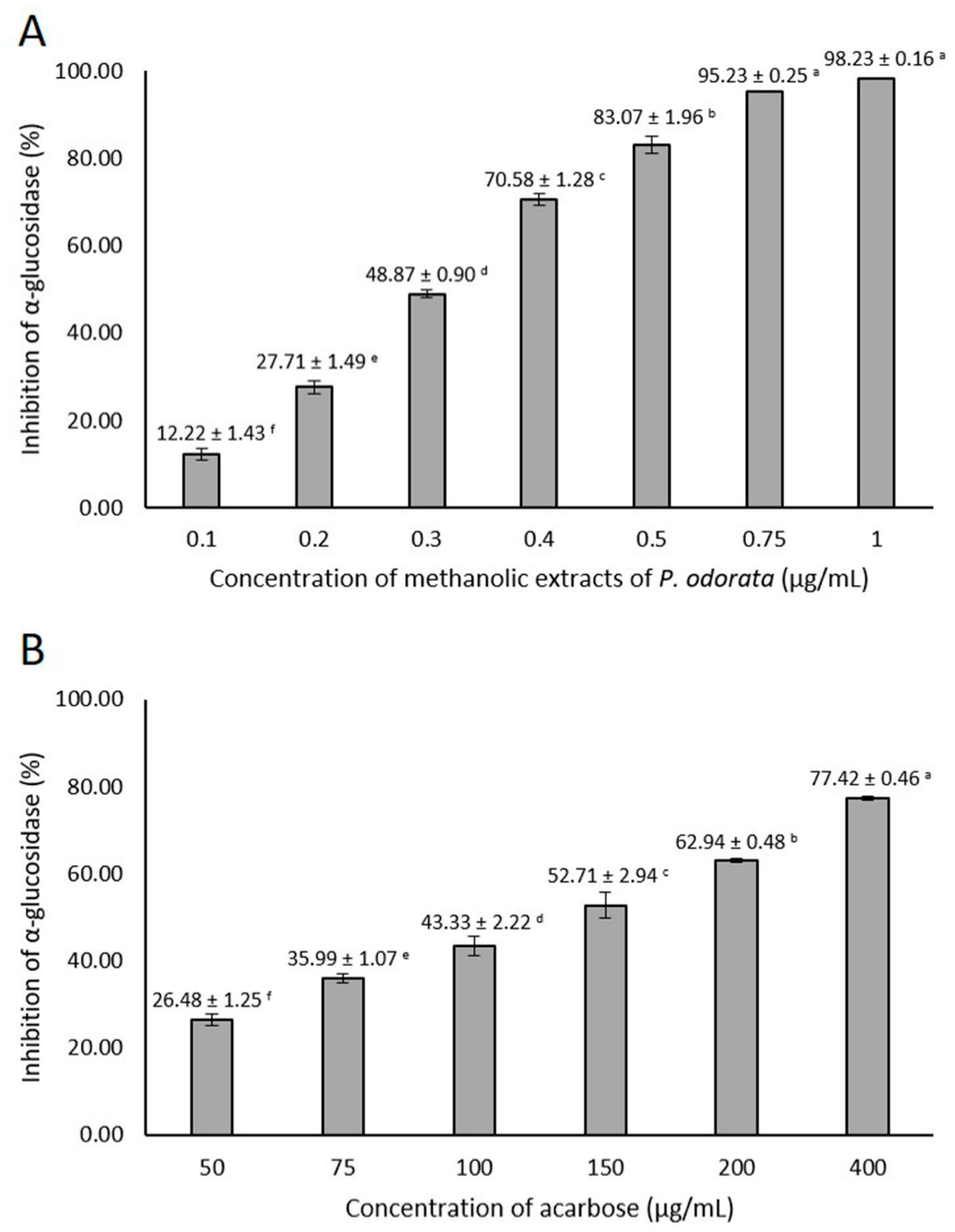
Figure 1. The inhibition percentage of (A) methanolic extract of P. odorata and (B) acarbose on the α-glucosidase activity. Data are shown as mean ± SD (n = 3). Superscript a–f means the different superscript letters in each bar are significantly different (p < 0.05).
Ijms 27 06885 g001
Figure 2. (A) Lineweaver−Burk plots of the kinetics analysis of α-glucosidase in the presence of the methanolic extract of P. odorata. (B) The slope (a) and intercept (b) of the Lineweaver–Burk plots against inhibitor concentrations. The substrate concentration was measured in the absence or presence of extracts at different concentrations. [V] and [S] are the reaction velocity and substrate concentration, respectively. (Dark circles: no inhibitor, black-lined transparent squares: 0.5IC50 concentration, dark triangles: IC50 concentration, black-lined transparent circles: 1.5IC50 concentration).
Ijms 27 06885 g002
Figure 3. The visualizations of the molecular docking interactions on the α-glucosidase’s active site. 3D visualizations (left), 2D visualizations (right), (A) P9, (B) P19, and (C) acarbose.
Ijms 27 06885 g003
Figure 4. RMSD of the backbone atoms, the number of H-bonds, and the number of atom contacts for acarbose, P9, and P19 in complex with α-glucosidase along the 500 ns MD simulations.
Ijms 27 06885 g004
Figure 5. The ΔGresidue,bind values of inhibitor-α-glucosidase complexes are depicted. Note: Residues contributing to inhibitor binding are colored according to their ΔGresidue,bind values, with the highest free energy residues colored red and the lowest free energy residues colored purple. The representative structure is used to illustrate the binding pattern of the inhibitor-α-glucosidase complexes. Green dashed lines represent H-bond formation.
Ijms 27 06885 g005
Table 1. Enzyme kinetic parameters of the methanolic extract of P. odorata are presented as Km, Vmax, Ki, Kii, and Kii/Ki values.
Extract (μg/mL)Km (mM)Vmax (μmol/min)Ki (µg/mL)Kii (µg/mL)Kii/Ki (µg/mL)
02.7979.2590.290.100.34
0.152.7917.874
0.302.5475.612
0.452.5464.885
Table 2. Bioactive compounds within P. odorata (Lour.) extracts obtained from previous reports.
Phenolics
GroupsCompoundsFormulaCodesPubChem NID 1References
Phenolic acidsChlorogenic acid 2C16H18O9P11794427[16,17]
Sinapic acid 2C11H12O5P2637775
Caffeic acid 2C9H8O4P3689043
Gallic acid 2C7H6O5P4370
Protocatechuic acid 2C7H6O4P572
Methyl gallateC8H8O5P67428
FlavonoidsQuercetin sulfateC15H8O11SP7129639308[16]
Kaempferol sulfateC15H8O10SP8129661100
Procyanidin BC30H26O12P9122738
Prodelphinidin BC30H26O14P105089687
Quercetin 3-O-β-D-galactopyranoside (Hyperoside)C21H20O12P115281643
Quercetin 3-O-β-D-glucoside (Isoquercitrin)C21H20O12P125280804
Quercetin 3-O-β-D-glucuronide (Miquelianin)C21H18O13P135274585
Quercetin 3-O-α-L-arabinopyranoside (Guajavarin)C20H18O11P145481224
Quercetin 3-O-β-D-rhamnoside (Quercitrin)C21H20O11P155280459
(Epi)catechin gallateC22H18O10P16107905
(−)-EpicatechinC15H14O6P1772276
(+)-CatechinC15H14O6P189064[15,16]
Rutin 2C27H30O16P195280805[15,17]
Quercetin 2C15H10O7P205280343
KaempferolC15H10O6P215280863[15]
IsorhamnetinC16H12O7P225281654
Phenolic acid (Indole)TryptophanC11H12N2O2T16305[17]
Essential Oils
Essential OilsDecanalC10H20OO18175[18,19,20,31]
DodecanalC12H24OO28194
β-CaryophylleneC15H24O35281515[18,19,20]
PoligodialC15H22O2O472503[32]
DrimenolC15H26OO53080551[18,19]
CitralC10H16OO6638011[18]
EuparoneC12H10O4O7104654
2,4-Heptadiene,2,6-dimethylC9H16O85370124
cis-CaryophylleneC15H24O95281522[31]
n-UndecaneC11H24O1014257
PentacosaneC25H52O1112406[33]
α-CurcumeneC15H22O1292139[19]
EremophilleneC15H24O136428416
7-Epi-α-selineneC15H24O1410726905
LedolC15H26OO1592812
NerolidolC15H26OO165284507
Caryophyllene oxideC15H24OO171742210
EupatoriochromeneC13H14O3O18100768
α-HumuleneC15H24O195281520[19,31,33]
1-DecanolC10H22OO208174
1-DedocanolC12H26OO218193
1 PubChem NID was accessed on https://pubchem.ncbi.nlm.nih.gov/ (accessed on 14 February 2026). 2 Phenolic acids and flavonoids were detected within the methanolic extract of P. odorata by our group.
Table 3. Types of interactions between phenolics and amino acids of the α-glucosidase’s active site.
Phenolics
CodesΔG (kcal/mol)Ki (μM)H-Bonds (Å)Hydrophobic Interactions
P1−6.7211.85K156 (2.15); S241 (2.19); H280 (1.76); D307 (1.93); S311 (2.18, 2.50); R315 (2.77).K156; R315.
P2−5.13173.10K156 (1.76); Y158 (3.03); S241 (2.04); D242 (1.81).K156; Y158; L177.
P3−5.8155.09K156 (1.84); Y158 (2.89); S241 (1.94, 2.09); R315 (2.38).K156.
P4−4.65391.23K156 (2.02, 2.09, 2.82); S241 (2.39); D242 (1.98, 2.17).K156.
P5−4.76326.24Y158 (2.11); L313 (2.72); N415 (1.75).F314.
P6−5.5289.65K156 (2.07); S241 (2.33); D242 (1.79, 1.96, 3.03).K156; Y158.
P7−8.250.894K156 (2.21); D307 (1.88, 1.88); R315 (2.88); E411 (2.62); N415 (1.97, 2.21).R315; F314.
P8−8.340.773K156 (1.94); S241 (1.79); R315 (2.91); E411 (1.71); R442 (3.06).Y158; F159; R315.
P9−9.900.055K156 (2.21, 2.21); Y158 (1.92, 3.04); S241 (1.82, 2.27); D242 (2.32); H280 (2.18); D307 (2.02); S311 (1.96); N415 (2.88).K156; Y158; D242; R315.
P10−6.0040.14D69 (1.72, 2.15); H112 (2.70); Y158 (2.13); D215 (2.02); D242 (2.07, 2.58); N350 (1.92); D352 (2.03); Q353 (3.04); E411 (2.86); R442 (1.97).Y158; F178; D215; V216; F303; R315; R442.
P11−8.151.06D69 (3.21); Y158 (2.16); E277 (2.23, 3.00); Q279 (2.17); H280 (2.84); D307 (2.13, 2.21); R315 (2.02, 2.38, 2.82); D352 (1.90); Q353 (2.23).Y158; F178; R315; D325; R442.
P12−7.373.97S157 (1.89, 2.73); Q279 (2.44); S311 (2.02, 2.20); E411 (2.39); N415 (1.79).Y158; R315.
P13−6.2625.94S157 (2.14); Q279 (2.44); S311 (1.98, 2.28); E411 (2.46); N415 (1.99).Y158; R316.
P14−7.622.60T306 (2.83); D307 (1.96); S311 (2.13); R315 (1.96); Q353 (1.91, 2.07); N415 (2.12).F303; R315.
P15−8.161.04E277 (1.96); Q279 (2.01); D307 (1.73, 1.99); R315 (2.58); Q353 (2.21); R442 (2.17).Y158; F178; R315; R442.
P16−8.790.359D69 (1.71); E277 (1.77); T306 (2.38); D307 (2.23); Q353 (2.16, 2.52); R442 (2.41).Y72; Y158; V216; F303; R315; D352; E411.
P17−7.304.48D69 (2.03); D215 (2.02); Q279 (2.77); R315 (2.39); Q353 (1.87); E411 (1.97); R442 (2.24).F178; V216; F303; D352; R442.
P18−7.244.95D215 (2.03, 2.14); E277 (1.72); R315 (2.73); E411 (1.75); R442 (2.26).Y72; D215; V216; F303.
P19−9.420.125D69 (1.80, 1.90); S157 (1.94); D215 (2.17); E277 (2.45); H280 (2.10); R315 (3.06, 3.08); D352 (2.74); E411 (1.89); N415 (1.81); R442 (2.79).Y72; Y158; R315.
P20−7.642.52D69 (1.77); D215 (1.97); Q279 (2.07, 2.88); R315 (2.04, 2.56); H351 (2.55); D352 (1.90); R442 (2.29).Y72; E411; R442.
P21−7.264.80D69 (1.77); H112 (2.79); Q279 (1.84); R315 (2.25); D352 (1.73); R442 (2.29).Y72; V216; D353; E411.
P22−7.073.54D215 (1.99); Q279 (1.90); R315 (2.28); D352 (1.70); R442 (2.43).D69; Y72; D215; V216; E277; E411.
T1−6.5715.22E277 (1.79); H351 (1.96); R442 (2.10).Y72; V216; D352.
Acarbose−7.304.43D69 (1.81, 1.87); Y158 (2.21); D215 (1.72); Q279 (2.31, 2.36); D307 (2.31, 3.05); P312 (2.47); R442 (2.53).F303.
G: Gibbs binding energy score, Ki: inhibition constant. The underlined amino acids are implicated in similar interactions observed with acarbose.
Table 4. Types of interactions between essential oils and amino acids of the α-glucosidase’s active site.
Essential Oils
CodesΔG (kcal/mol)Ki (µM)H-Bonds (Å)Hydrophobic Interactions
O1−4.33673.55S241 (1.90).K156; Y158; F314; R315.
O2−4.14928.11R442 (2.58, 2.72); R446 (1.88).Y72; F159; F178; V216.
O3−6.7411.38-K156; Y158.
O4−6.948.11S241 (1.76).K156; Y158; F314.
O5−6.7211.96R213 (2.14, 2.92); D352 (1.88).Y72; Y158; F159; F178; V216; F303.
O6−5.06194.35S241 (1.67).K156; F314; Y316.
O7−7.413.7K156 (2.14); L313 (2.82); R315 (2.01).K156; Y158.
O8−4.51493.36-K156; Y158; L177.
O9−6.2924.49-Y158; F159; F178; V216; F303.
O10−4.17876.17-K156; Y158; F314; R315; Y316.
O11−5.17162.55-Y72; K156; Y158; F178; V216; F314; R315.
O12−6.2327.2-K156; Y158; F314; R315; Y316.
O13−6.5615.49-K156; Y158; R315.
O14−6.324.05-K156; Y158; R315.
O15−6.5515.81N415 (2.20).K156; F314; R315; Y316.
O16−6.2725.24D69 (1.92); R442 (2.16).Y72; Y158; F178; V216; F303.
O17−6.5416.15R315 (2.11).K156; Y158; R315.
O18−6.3223.46E277 (2.10); Q279 (2.39); R442 (1.83).Y158; F178; V216; F303; D352.
O19−6.3422.71 K156; Y158.
O20−4.081010D69 (1.75); R442 (2.01).Y72; Y158; F159; F178; V216.
O21−4.18868.28D69 (2.00); H112 (1.84).Y72; F159; F178; V216.
Acarbose−7.34.43D69 (1.81, 1.87); Y158 (2.21); D215 (1.72); Q279 (2.31, 2.36); D307 (2.31, 3.05); P312 (2.47); R442 (2.53)F303.
G: Gibbs binding energy score, Ki: inhibition constant. The underlined amino acids are implicated in similar interactions observed with acarbose.
Table 5. Drug-likeness and ADMET properties of P9, P19, and acarbose.
PharmacokineticsParametersP9P19AcarboseStandard
Lipinski’s Ro5Molecular weight585.52610.52645.6<500
Rotatable bonds369<10
H-bond acceptors121619<10
H-Bond donors101014<5
TPSA220.76269.43321.17<140
MlogP−0.26−3.89−6.94<4.15
Violations/Ro53/No3/No3/No0/Yes
AbsorptionWater solubility−2.892−2.892−1.482Log mol/L
Caco2 permeability−1.225−0.949−0.481Log Papp in 10−6 cm/s
HIA (%)66.74923.4464.172% Absorbed
P-gp substateYesYesYesYes/No
P-gp inhibitor IYesNoNoYes/No
P-gp inhibitor IIYesNoNoYes/No
DistributionsHuman VDs−0.1581.663−0.836Log L/kg
BBB permeability−1.940−1.899−1.717Log BB
MetabolismCYP2D6 substrate NoNoNoYes/No
CYP3A4 substrate NoNoNoYes/No
CYP1A2 inhibitorNoNoNoYes/No
CYP2C19 inhibitorNoNoNoYes/No
CYP2C9 inhibitorNoNoNoYes/No
CYP2D6 inhibitorNoNoNoYes/No
CYP3A4 inhibitor NoNoNoYes/No
ExcretionTotal clearance−0.085−0.3690.428Log mL/min/kg
Renal OCT2 substrateYesNoNoYes/No
ToxicityAMES toxicityNoNoNoYes/No
MRTD0.4380.4520.435Log mg/kg/day
hERG I/II inhibitorsNo/YesNo/YesNo/YesYes/No
ORAT (LD50)2.4822.4912.449mol/kg
Hepatotoxicity NoNoNoYes/No
T. Pyriformis toxicity0.2850.2850.285Log μg/L
HIA: human intestinal absorption, P-gp: P-glycoprotein, VD: volume of distribution, BBB: brain–blood barrier, CYP: cytochrome P450, OCT2: organic cation transporter-2, MRTD: max. recommended tolerated dose, hERG: human ether-a-go-go-related gene, ORAT: oral rate acute toxicity, and LD50: half-lethal dose. P9: procyanidin B, and P19: rutin.
Disclaimer/Publisher’s Note: The statements, opinions and data contained in all publications are solely those of the individual author(s) and contributor(s) and not of MDPI and/or the editor(s). MDPI and/or the editor(s) disclaim responsibility for any injury to people or property resulting from any ideas, methods, instructions or products referred to in the content.