1. Introduction
Metabolic dysfunction-associated steatotic liver disease (MASLD) has emerged as the most common chronic liver disease in both adults and children, and its burden continues to rise in parallel with pediatric obesity and unhealthy dietary patterns [
1]. Among these dietary factors, excessive fructose intake, particularly through sugar-sweetened beverages and high-fructose corn syrup (HFCS)-containing processed foods, has been increasingly linked to hepatic steatosis, dyslipidemia, insulin resistance, and broader cardiometabolic risk in children and adolescents [
2,
3]. Recent experimental data further suggest that fructose-related liver injury is not merely a consequence of caloric excess but also reflects active metabolic and inflammatory disturbances, including oxidative stress, hepatic lipid accumulation, and inflammasome-associated signaling [
2,
4]. Because HFCS consumption is disproportionately high among children and adolescents and pediatric MASLD is increasing worldwide, understanding early pathogenic mechanisms in juvenile liver tissue is of particular translational importance.
Beyond metabolic dysregulation, growing evidence indicates that inflammatory signaling pathways play a pivotal role in the progression of fructose-induced liver injury. In particular, activation of the nucleotide-binding domain-like receptor protein 3 (NLRP3) inflammasome has emerged as a central mechanism linking metabolic stress to hepatic inflammation [
5,
6]. The NLRP3 inflammasome, a multiprotein complex activated by cellular stress signals such as reactive oxygen species and lipid accumulation, promotes the activation of caspase-1 (CASP1), leading to the maturation and release of pro-inflammatory cytokines, including interleukin-1β (IL-1β) [
6,
7]. In addition, CASP1 activation induces cleavage of gasdermin D (GSDMD), triggering pyroptosis, a highly inflammatory form of programmed cell death that amplifies tissue injury and immune activation in the liver [
7,
8]. Recent experimental and clinical studies have demonstrated that NLRP3-mediated pyroptosis contributes significantly to the development of hepatic steatosis, inflammation, and progression toward steatohepatitis, highlighting this pathway as a promising therapeutic target [
5,
8].
Dexpanthenol (DEX; D-panthenol) is the biologically active R-enantiomer of panthenol, with the molecular formula C
9H
19NO
4 and a molecular weight of 205.25 g/mol. Its systematic chemical name is (2R)-2,4-dihydroxy-N-(3-hydroxypropyl)-3,3-dimethylbutanamide. Structurally, DEX is the alcohol analogue of D-pantothenic acid (vitamin B5), in which the terminal carboxylic acid group is replaced by a primary alcohol group. Following biological oxidation to pantothenic acid, it contributes to the biosynthesis of coenzyme A. DEX has demonstrated antioxidant, anti-inflammatory, and cytoprotective effects in various experimental models of tissue injury [
9,
10,
11]. Previous studies have demonstrated that DEX reduces oxidative stress by enhancing endogenous antioxidant defenses, limiting lipid peroxidation, and attenuating cellular damage in liver, kidney, and intestinal injury models [
10]. In addition, DEX has been reported to modulate apoptotic signaling pathways, including caspase activation and the balance between BCL2-associated X protein (BAX) and B-cell lymphoma 2 (BCL2), thereby contributing to cellular survival and tissue regeneration [
12]. However, despite these well-established protective properties, its potential role in regulating inflammasome activation and pyroptosis, particularly in the context of fructose-induced hepatic injury, remains largely unexplored [
9,
12]. In addition to canonical pyroptotic signaling, fructose-induced hepatic injury involves closely interconnected oxidative, apoptotic, and regenerative responses. MDA reflects lipid peroxidation and oxidative membrane damage, which may act as an upstream trigger for inflammasome activation [
13,
14]. Caspase-3 (Cas-3), a key executioner of apoptosis, provides complementary information on non-pyroptotic programmed cell death occurring in parallel with inflammasome-mediated injury [
15]. Proliferating cell nuclear antigen (PCNA), as a marker of cellular proliferation and reparative activity, may indicate the regenerative response of hepatocytes following metabolic and inflammatory damage [
16].
In this study, we hypothesized that DEX ameliorates HFCS-induced liver injury in association with reduced Nlrp3, Casp1, Gsdmd, and Il-1β related transcriptional signaling, together with attenuation of lipid peroxidation, apoptosis, and injury-associated proliferative responses, as reflected by MDA, Cas-3, and PCNA immunoreactivity.
3. Discussion
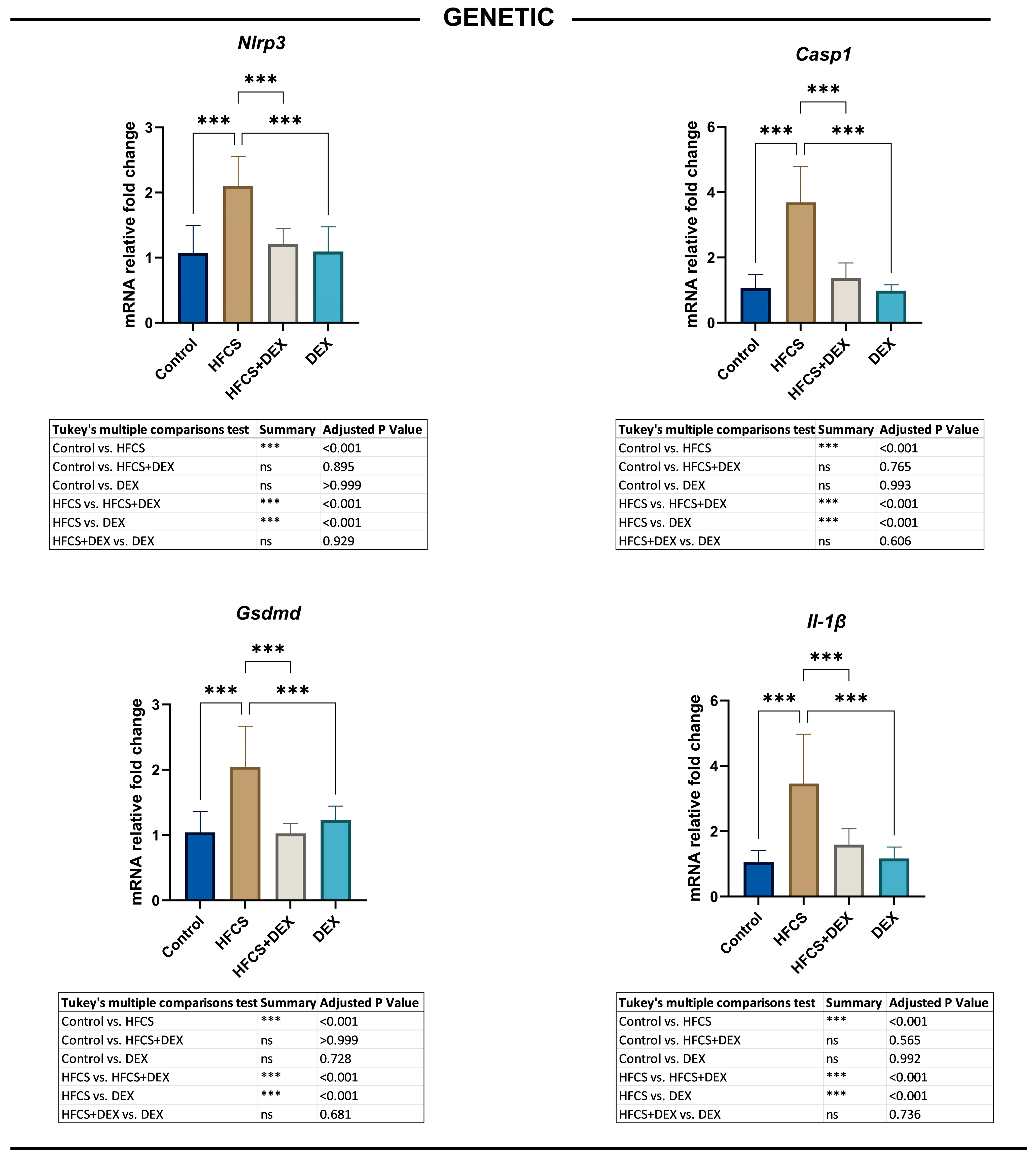
The present study demonstrated that HFCS exposure caused marked histopathological liver injury, increased hepatic oxidant burden and lipid peroxidation, enhanced apoptotic activity, and upregulated inflammasome- and pyroptosis-associated genes in young adult rats. The principal novel finding was that DEX treatment attenuated histopathological injury and significantly reduced MDA and Cas-3 immunoreactivity while downregulating Nlrp3, Casp1, Gsdmd, and Il-1β mRNA expression. These findings indicate that the hepatoprotective effects of DEX were associated with attenuation of lipid peroxidation, apoptosis, and inflammasome- and pyroptosis-associated transcriptional signaling. However, because protein cleavage, cytokine maturation, and membrane-pore formation were not directly assessed, the present results do not definitively demonstrate the activation or inhibition of pyroptotic cell death.
The pediatric relevance of these findings deserves particular emphasis. Excessive intake of fructose-containing beverages and processed foods has been closely linked to pediatric MASLD, hepatic steatosis, dyslipidemia, and cardiometabolic risk, suggesting that fructose exposure may represent an early dietary driver of metabolic liver injury in children and adolescents. Pediatric MASLD studies have also implicated oxidative stress as an important contributor to disease development and progression, while experimental adolescent models indicate that fructose-rich diets may promote hepatic lipid accumulation, inflammatory responses, oxidative stress, and activation of the NLRP3 inflammasome pathway. Therefore, the use of young adult rats in the present study provides a developmentally relevant framework for evaluating early hepatic responses to HFCS exposure. Although direct pediatric data linking fructose exposure to pyroptosis remain limited, our findings support the concept that oxidative stress, apoptosis, and inflammasome-related pyroptotic signaling may act as interconnected mechanisms in early-life fructose-induced liver injury. In this context, DEX is also of translational interest because dexpanthenol and pantothenic acid derivatives have a long history of clinical use, particularly in pediatric dermatologic and mucosal barrier-supportive formulations; however, its systemic hepatoprotective role in pediatric metabolic liver disease remains unexplored and should be investigated cautiously in future translational studies [
17,
18,
19].
Importantly, DEX treatment markedly attenuated all these pathological changes, suggesting that its hepatoprotective effects are mediated through a multi-target mechanism involving suppression of oxidative stress, inhibition of apoptosis, and modulation of inflammasome-driven pyroptosis. These findings align with recent evidence indicating that metabolic liver injury is not solely driven by lipid accumulation but also involves complex inflammatory and cell death pathways, particularly those regulated by the NLRP3 inflammasome [
5,
6,
20,
21].
Mechanistically, the findings of the present study support a sequential injury model in which HFCS-induced oxidative stress acts as an upstream trigger of inflammasome activation. Increased MDA immunoreactivity suggests enhanced lipid peroxidation and ROS generation, which are known to activate TXNIP-dependent Nlrp3 signaling. Subsequent activation of Casp1 and Gsdmd may promote pyroptotic hepatocyte death and Il-1β release, thereby amplifying hepatic inflammation. The concurrent increase in PCNA expression likely reflects a compensatory regenerative response to ongoing hepatocellular loss. DEX appears to interrupt this injury cascade at multiple levels by reducing oxidative stress, limiting apoptotic signaling, and suppressing inflammasome-associated pyroptosis. Increased PCNA expression in the HFCS group should not be interpreted solely as beneficial proliferation, but rather as a compensatory regenerative response secondary to hepatocyte injury, apoptosis, and inflammatory damage.
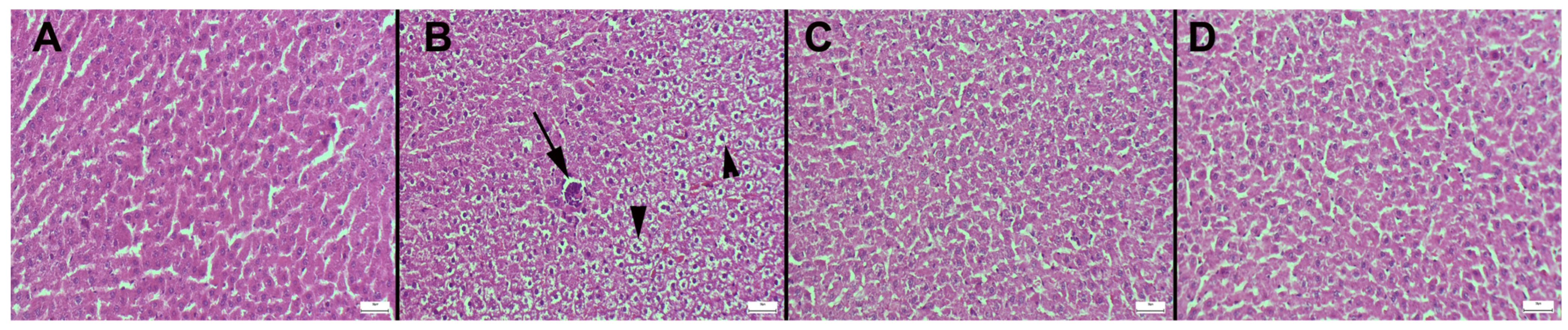
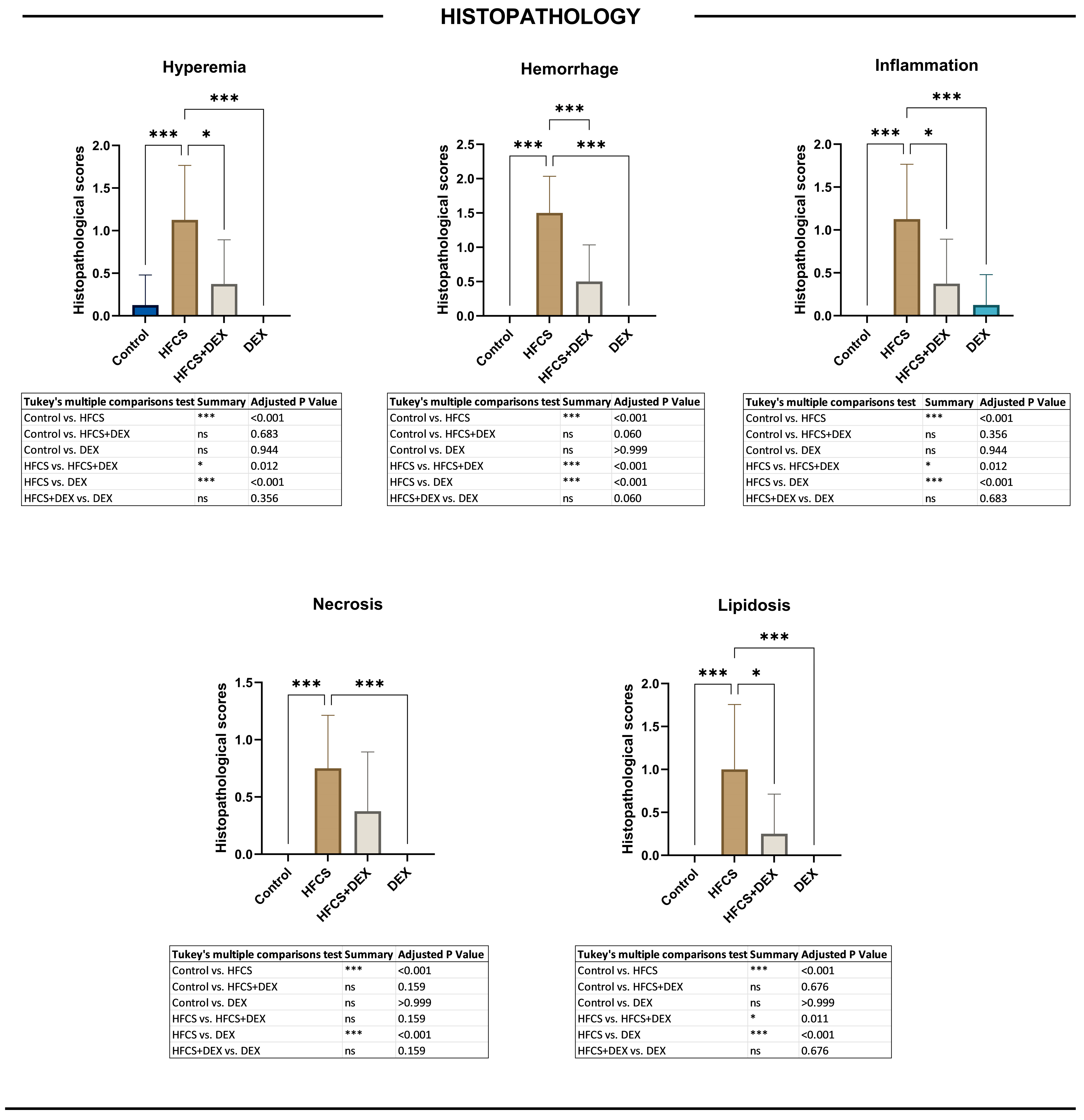
The histopathological findings of the present study are consistent with previous experimental models demonstrating that excessive fructose consumption induces hepatic steatosis, inflammatory infiltration, and hepatocellular injury. In particular, the predominance of lipidosis and necrosis in centrilobular regions observed in our study is in agreement with earlier reports showing that this zone is more susceptible to metabolic and oxidative stress due to its lower oxygenation and higher exposure to toxic metabolites [
22,
23].
High-fructose intake has been shown to promote de novo lipogenesis, increase triglyceride accumulation, and disrupt hepatic metabolic balance, ultimately leading to steatosis and structural liver damage. Moreover, fructose-driven metabolic dysregulation contributes to the progression from simple steatosis to steatohepatitis by enhancing oxidative stress and inflammatory responses [
24,
25].
The observed increase in hyperemia, hemorrhage, and inflammatory cell infiltration in the HFCS group further supports the concept that metabolic liver injury is not solely driven by lipid accumulation but involves a complex interaction between metabolic overload and inflammatory signaling pathways. Importantly, the significant improvement in histopathological parameters following DEX treatment in our study is consistent with previous findings demonstrating that antioxidant and anti-inflammatory agents can effectively attenuate fructose-induced hepatic damage [
9]. These findings suggest that DEX mitigates HFCS-induced liver injury not only by reducing lipid accumulation but also by suppressing inflammation and cellular damage, thereby contributing to the restoration of hepatic architecture.
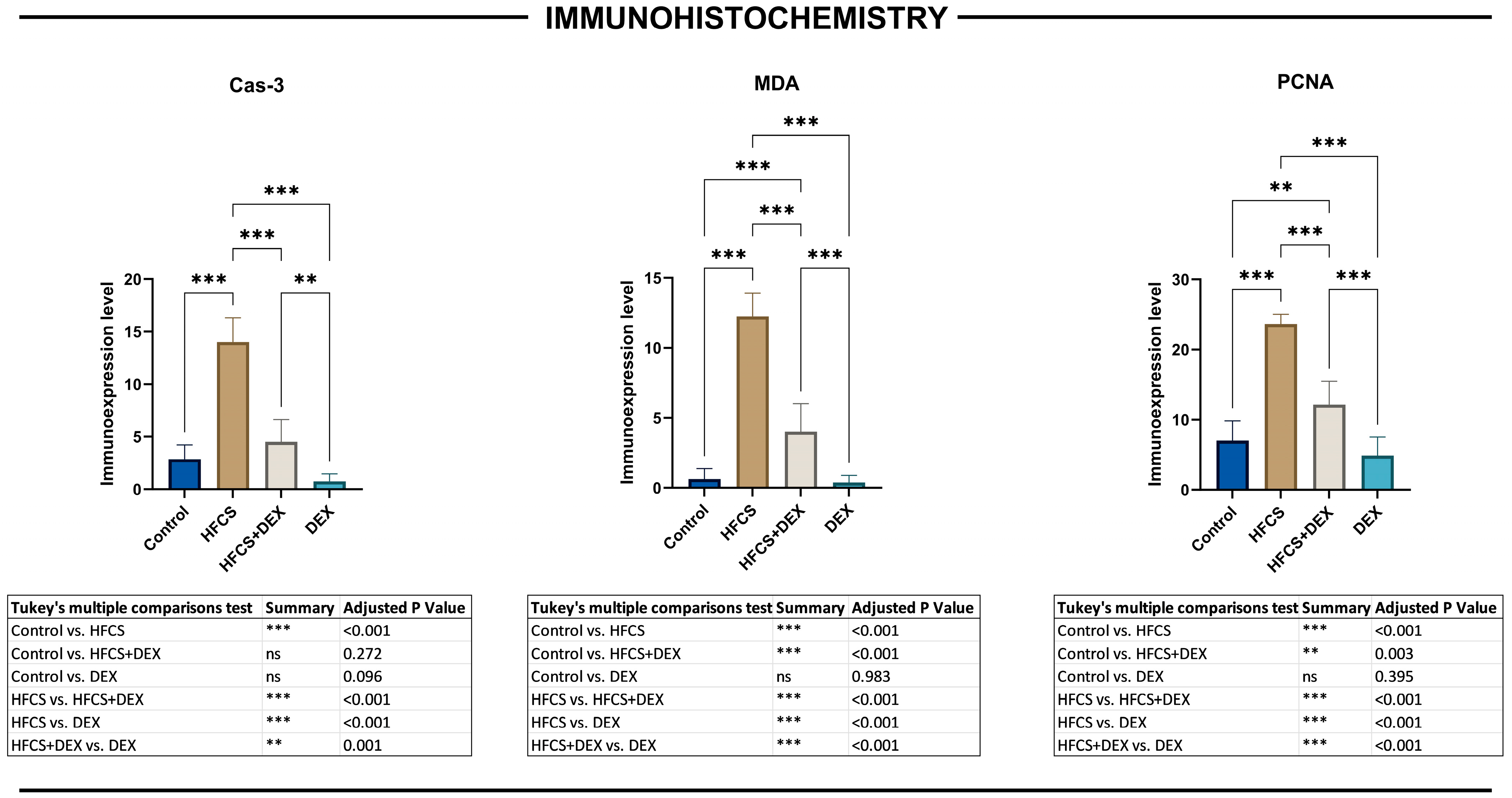
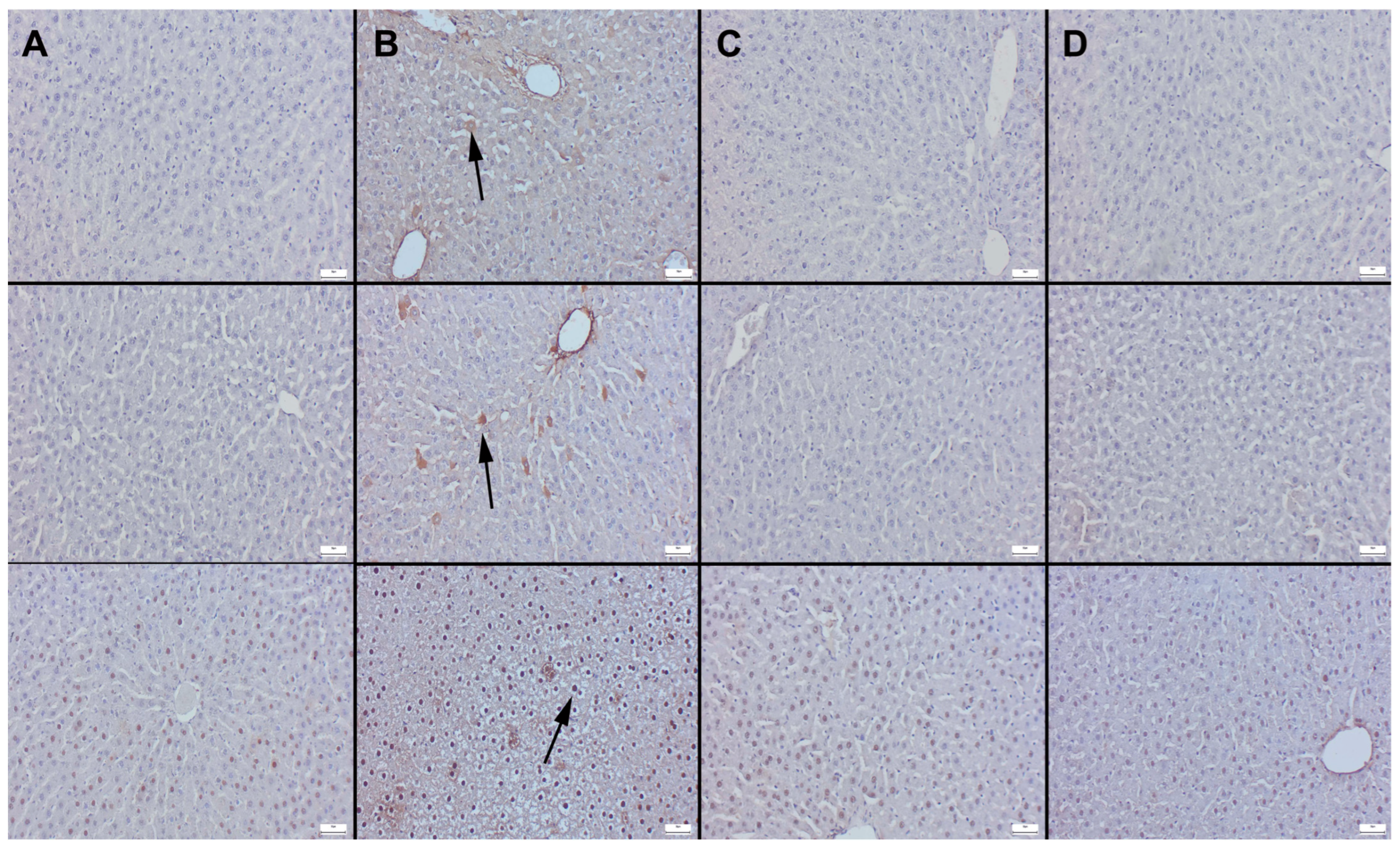
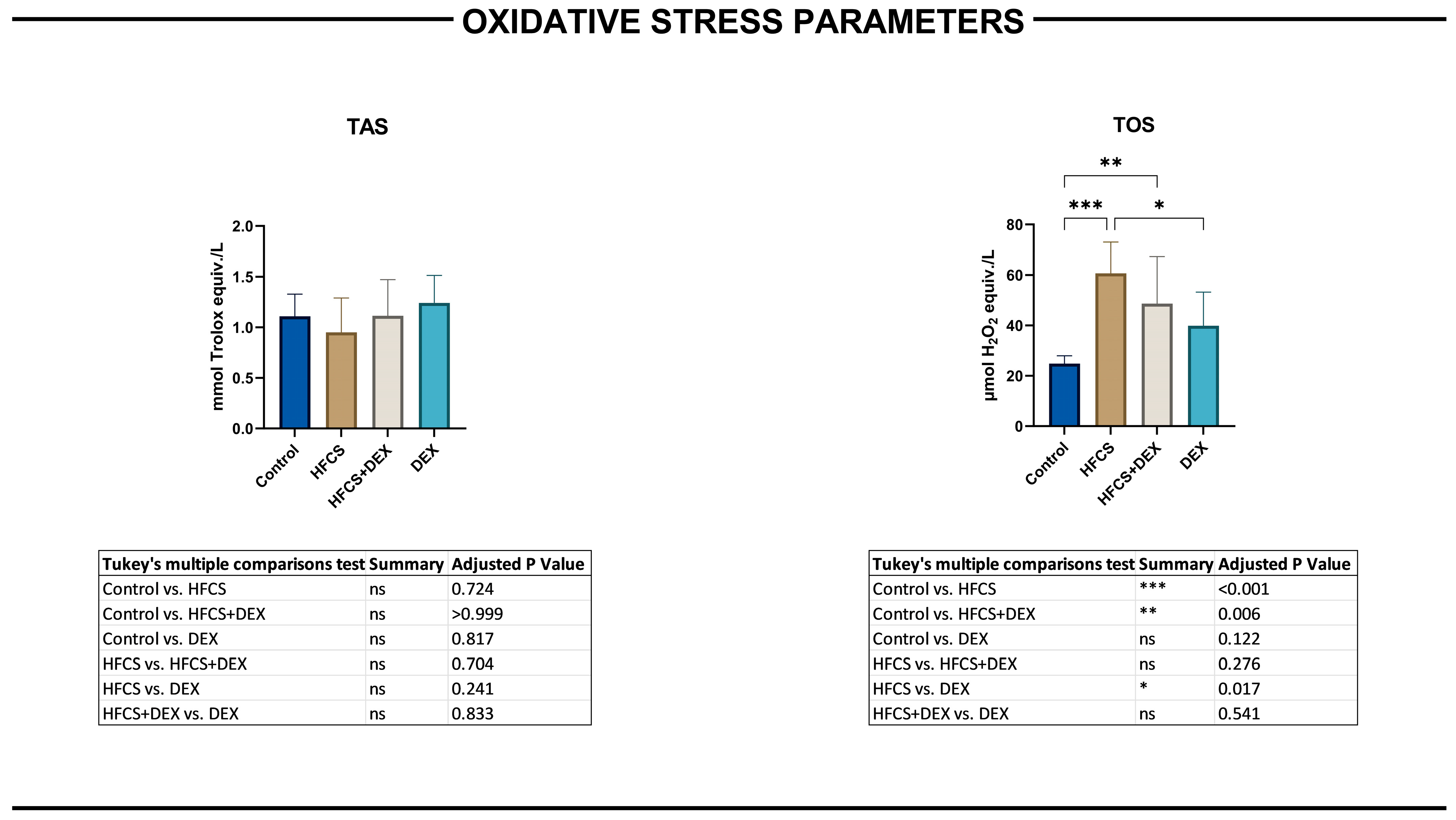
Oxidative stress is a key driver of fructose-induced liver injury and plays a central role in the transition from simple steatosis to steatohepatitis. In the present study, the significant increase in MDA expression in the HFCS group clearly indicates enhanced lipid peroxidation and oxidative damage within hepatocytes. MDA is a well-established marker of oxidative stress, reflecting the extent of membrane lipid degradation caused by ROS. These findings are consistent with previous studies demonstrating that high-fructose intake increases ROS production, impairs antioxidant defense systems, and promotes oxidative injury in liver tissue [
26,
27]. The biochemical TAS and TOS findings provide complementary evidence regarding the oxidative imbalance induced by HFCS. HFCS exposure significantly increased hepatic TOS, whereas TAS remained comparable across all groups. This pattern suggests that oxidative stress in the present model was predominantly driven by enhanced oxidant generation rather than by a detectable depletion of the overall antioxidant reserve. A preserved TAS level does not necessarily indicate adequate protection against oxidative injury, because quantitatively maintained antioxidant systems may still be insufficient to neutralize excessive reactive oxygen species production. The concurrent increase in TOS and MDA immunoreactivity therefore supports the presence of a biologically relevant pro-oxidant state in HFCS-exposed liver tissue [
14,
27,
28].
In the HFCS+DEX group, TOS was numerically lower than in the HFCS group; however, this reduction was not statistically significant, and TOS remained significantly higher than in the control group. Therefore, the TAS and TOS findings should not be interpreted as evidence that DEX completely restored global hepatic redox balance. Nevertheless, DEX treatment significantly reduced MDA immunoreactivity, indicating attenuation of lipid peroxidation at the tissue level. The apparent difference between the TOS and MDA results may be explained by the distinct biological information provided by these measurements. TOS represents a composite estimate of multiple oxidant molecules in tissue homogenates, whereas MDA immunostaining more specifically reflects lipid peroxidation and its cellular distribution within liver tissue. Differences in analytical sensitivity, biological variability, treatment duration, and the temporal kinetics of oxidant production and lipid damage may also have contributed to this divergence. Accordingly, DEX appeared to exert a more evident protective effect on lipid peroxidation and structural tissue injury than on the total hepatic oxidant pool under the present experimental conditions [
9,
10].
In parallel with oxidative stress, we observed a marked increase in Cas-3 expression in the HFCS group, indicating activation of apoptotic pathways. Cas-3 is a key executioner caspase in apoptosis and is commonly upregulated in response to mitochondrial dysfunction and oxidative damage. The coexistence of elevated MDA and Cas-3 levels in our study suggests a strong link between oxidative stress and apoptosis in HFCS-induced hepatocellular injury. This relationship has been well documented, with ROS-mediated mitochondrial damage triggering caspase activation and subsequent hepatocyte apoptosis in metabolic liver disease models [
29,
30].
Importantly, DEX treatment significantly reduced MDA and Cas-3 expression and was associated with a nonsignificant downward trend in TOS, without significantly altering TAS. These findings support the antioxidant and anti-apoptotic effects of DEX but indicate that its influence on the overall hepatic oxidant–antioxidant balance was partial rather than complete. These findings are in line with previous reports showing that DEX enhances cellular antioxidant capacity, stabilizes mitochondrial function, and reduces ROS-mediated cellular injury. By attenuating oxidative stress, DEX likely interrupts the cascade leading to apoptosis, thereby preserving hepatocyte integrity [
9,
10].
Collectively, these results suggest that oxidative stress-induced apoptosis is a major contributor to HFCS-mediated liver injury and that DEX exerts its hepatoprotective effects, at least in part, through the suppression of ROS generation and inhibition of apoptosis.
A central finding of the present study is the marked activation of the NLRP3 inflammasome and its downstream pyroptotic signaling cascade in HFCS-induced liver injury. We observed significant upregulation of
Nlrp3,
Casp1,
Gsdmd, and
Il-1β expression, indicating that inflammasome-mediated pyroptosis plays a pivotal role in hepatocellular damage in this model. The
Nlrp3 inflammasome is a cytosolic multiprotein complex that is activated in response to metabolic stress signals, including ROS, lipid accumulation, and mitochondrial dysfunction, all of which are characteristic features of fructose-induced liver injury [
20,
31].
Upon activation,
Nlrp3 promotes the cleavage and activation of
Casp1, which subsequently processes pro-
Il-1β into its active form and induces the cleavage of
Gsdmd. The N-terminal fragment of
Gsdmd forms pores in the cell membrane, leading to cellular swelling, membrane rupture, and release of pro-inflammatory cytokines, a process defined as pyroptosis. This highly inflammatory form of programmed cell death amplifies hepatic inflammation and contributes to the progression of metabolic liver disease [
7,
32].
Recent studies have demonstrated that
Nlrp3 inflammasome activation is closely associated with the development and progression of MASLD, where it links metabolic stress to innate immune activation and hepatocellular injury. Inhibition of this pathway has been shown to reduce hepatic inflammation, steatosis, and fibrosis, highlighting its importance as a therapeutic target [
5,
21].
Importantly, our findings show that DEX significantly downregulated the expression of Nlrp3, Casp1, Gsdmd, and Il-1β, suggesting that its hepatoprotective effects are mediated, at least in part, through suppression of inflammasome activation and pyroptotic cell death. Although the exact molecular mechanisms underlying this effect remain to be fully elucidated, it is likely that DEX interferes with upstream triggers of inflammasome activation, such as oxidative stress and mitochondrial dysfunction. By attenuating these signals, DEX may prevent the initiation of the inflammasome cascade and thereby reduce pyroptosis-driven inflammation.
These results position DEX as a potential modulator of inflammasome activity and highlight pyroptosis as a key therapeutic target in fructose-induced liver injury.
DEX has been widely recognized for its antioxidant, anti-inflammatory, and cytoprotective properties in various experimental models of tissue injury. Previous studies have demonstrated that DEX reduces oxidative stress, limits lipid peroxidation, and attenuates inflammatory responses in different organ systems, including liver, kidney, and intestinal tissues. Moreover, DEX has been shown to modulate apoptotic pathways by regulating key mediators such as cas-3, caspase-9, and BAX/BCL2 balance, thereby promoting cell survival and tissue repair [
9,
12].
However, the potential role of DEX in regulating inflammasome activation and pyroptosis has not been previously elucidated. To the best of our knowledge, this is the first study to demonstrate that DEX suppresses the Nlrp3–Casp1–Gsdmd axis in a model of fructose-induced liver injury. This finding represents a significant extension of the known mechanisms of DEX, shifting its role from a general antioxidant agent to a targeted modulator of inflammatory cell death pathways.
The novelty of our study lies in integrating histopathological, immunohistochemical, and molecular data to demonstrate that DEX exerts a multi-layered protective effect by simultaneously attenuating oxidative stress, apoptosis, and pyroptosis. While previous studies have primarily focused on the antioxidant and anti-apoptotic effects of DEX, our results highlight its ability to regulate upstream inflammatory signaling pathways, particularly the inflammasome complex. This is of particular importance, as pyroptosis has recently emerged as a key driver of inflammation and disease progression in metabolic liver disorders [
21,
33].
From a translational perspective, these findings suggest that DEX may offer a novel therapeutic strategy targeting both metabolic and inflammatory components of liver injury. Unlike conventional approaches that focus on downstream cytokine inhibition, DEX appears to act at an earlier stage by modulating fundamental cellular stress and death pathways. This dual-action mechanism may provide a broader and more effective therapeutic benefit, particularly in complex diseases such as metabolic-associated fatty liver disease.
In this context, our findings may have particular relevance for pediatric hepatology, as they suggest that inflammasome activation and pyroptosis may play a critical role in early-stage liver injury induced by dietary factors such as HFCS. The demonstration that DEX effectively attenuates these pathways in a young adult animal model raises the possibility that it may serve as a potential therapeutic or preventive strategy in pediatric populations at risk of metabolic liver disease.
Moreover, considering that pediatric patients often present with more aggressive disease progression and limited therapeutic options, interventions targeting early pathogenic mechanisms, such as oxidative stress and inflammasome activation, may offer significant clinical benefit. Therefore, the use of a young adult animal model not only strengthens the translational relevance of our findings but also highlights the importance of investigating age-specific mechanisms in metabolic liver disease.
This study has several limitations that should be acknowledged. First, the experimental design is based on an animal model, which may limit the direct generalizability of the findings to human disease. Although the use of young adult rats increases the translational relevance to pediatric populations, species-specific differences in metabolism and immune responses should be considered. Second, while we demonstrated significant modulation of inflammasome-related gene expression, protein-level validation (e.g., Western blot or ELISA) was not performed, which may limit the mechanistic depth of the findings. Additionally, other relevant pathways involved in metabolic liver disease, such as insulin resistance, gut–liver axis alterations, and microbiota changes [
34,
35], were beyond the scope of the present study. Serum biochemical markers were not measured in the present study. Therefore, the tissue-level findings could not be correlated with circulating markers of hepatocellular injury or cholestasis, such as ALT, AST, ALP, and GGT, or with indicators related to hepatic synthetic and excretory function, including albumin and bilirubin. Consequently, the present results demonstrate histopathological and molecular attenuation of hepatic injury but do not establish an improvement in biochemical liver function. Future studies should combine tissue analyses with a comprehensive serum liver biochemical panel.
Despite these limitations, our study provides comprehensive histopathological, immunohistochemical, and molecular evidence supporting the role of oxidative stress, apoptosis, and inflammasome-mediated pyroptosis in HFCS-induced liver injury. Future studies should aim to validate these findings in larger animal cohorts and clinical settings, incorporate protein-level analyses, and explore the long-term effects of DEX on fibrosis and disease progression. Furthermore, investigating the interaction between inflammasome activation and gut microbiota, as well as assessing combination therapies targeting multiple pathogenic pathways, may provide deeper insight into the management of metabolic liver disease [
36]. Although the present study demonstrated robust transcriptional suppression of
Nlrp3,
Casp1,
Gsdmd, and
Il-1β, future studies incorporating cleaved GSDMD, cleaved caspase-1, IL-18, and protein-level validation are required to definitively confirm pyroptotic cell death.
In conclusion, HFCS exposure caused histopathological liver injury, increased hepatic oxidant burden and lipid peroxidation, enhanced apoptotic activity, and upregulated Nlrp3-, Casp1-, Gsdmd-, and Il-1β-related transcriptional signaling in young adult rats. DEX treatment attenuated hepatic structural injury, reduced MDA and Cas-3 immunoreactivity, and downregulated inflammasome- and pyroptosis-associated gene expression. These findings support a potential multi-target hepatoprotective effect of DEX in this experimental model. However, the absence of protein-level validation and direct assessment of pyroptotic cell death requires cautious interpretation. Further mechanistic, age-comparative, and translational studies are needed before the clinical relevance of systemic DEX treatment can be determined.
4. Materials and Methods
4.1. Drugs and Chemicals
Dexpanthenol was obtained from a commercial pharmaceutical source (Bepanthen
®, Bayer, Istanbul, Türkiye). HFCS-55 solution containing 55% fructose and 41% dextrose was used to establish the fructose-induced metabolic injury model [
37]. Ketamine hydrochloride (Ketasol 10%, Richter Pharma, Wels, Austria) and xylazine hydrochloride (Rompun 2%, Bayer, Leverkusen, Germany) were used for anesthesia. Formalin, hematoxylin–eosin staining solutions, and analytical-grade laboratory consumables were purchased from standard commercial suppliers. Primary antibodies used for immunohistochemical analysis included anti-caspase-3 p12 antibody (clone EPR16888, ab179517), anti-malondialdehyde (MDA) antibody (clone 11E3, ab243066), and anti-proliferating cell nuclear antigen (PCNA) antibody (clone EPR3821, ab92552) (Abcam, Cambridge, UK). Immunohistochemical detection was performed using the Mouse and Rabbit Specific HRP/DAB IHC Detection Kit–micro-polymer (ab236466) and 3,3′-diaminobenzidine (DAB) chromogen (Abcam, Cambridge, UK). RNA isolation and RT-qPCR reagents included the GeneAll RiboEx™ RNA Isolation Kit (GeneAll Biotechnology, Seoul, Republic of Korea), A.B.T.™ cDNA Synthesis Kit, and A.B.T.™ 2X qPCR SYBR-Green MasterMix (Atlas Biotechnology, Ankara, Türkiye).
4.2. Animals and Experimental Design
A total of 32 young adult (8-week-old) male Wistar albino rats (weighing 200–250 g) were obtained from the Süleyman Demirel University Experimental Animal Research Center. The animals were approximately 16 weeks old at the time of euthanasia. Rats at approximately 8–9 weeks of age are generally considered to be transitioning from late adolescence to early adulthood, although developmental classifications may vary according to strain, sex, and the physiological outcome under investigation. Accordingly, the animals in the present study are referred to as young adult rats rather than juvenile or pediatric-equivalent animals. This age was selected to investigate HFCS-induced hepatic injury and the response to dexpanthenol in a young-adult organism while limiting potential confounding from advanced age-related metabolic and hepatic alterations [
38,
39]. Animals were housed under standard laboratory conditions (12 h light/dark cycle, temperature 22 ± 2 °C, humidity 50–60%) with free access to standard pellet diet and water. From the end of week 4 until the end of the experiment, they received physiological saline intraperitoneally once daily to provide injection-related stress comparable to the treatment groups. Rats in the HFCS group received freshly prepared 20% HFCS-55 solution in drinking water for 8 weeks and were administered physiological saline intraperitoneally once daily from the end of week 4. Rats in the HFCS+DEX group received freshly prepared 20% HFCS-55 solution in drinking water for 8 weeks and were treated with dexpanthenol at a dose of 500 mg/kg/day intraperitoneally once daily from the end of week 4 until the end of week 8. The selected dose was based on previous experimental studies demonstrating antioxidant, anti-inflammatory, and cytoprotective effects without evidence of systemic toxicity [
9,
40,
41]. Rats in the DEX group received standard drinking water for 8 weeks and were administered dexpanthenol at a dose of 500 mg/kg/day intraperitoneally once daily from the end of week 4 until the end of week 8. The 20% HFCS-55 solution was prepared by mixing 200 mL HFCS-55 with 800 mL drinking water until homogeneous. The solution was prepared fresh daily and placed equally into the drinking bottles of the HFCS-treated groups. Body weights were measured weekly to adjust dexpanthenol dose and saline volume. Water intake was recorded daily, and food intake was measured every two days. All experimental procedures were approved by the Süleyman Demirel University Animal Ethics Committee (Approval No: 15/425, Date: 12 December 2024) and conducted in accordance with ARRIVE 2.0 guidelines. Animals were randomly assigned into four groups (
n = 8 per group):
Control group: Rats received standard drinking water throughout the experimental period. From the end of the fourth week until the end of the study, animals were administered physiological saline intraperitoneally (i.p.) once daily.
HFCS group: Rats in the HFCS group received a freshly prepared 20% (
v/
v) HFCS-55 solution in drinking water for 8 weeks. The HFCS exposure protocol was adapted from a previously described high-fructose dietary model [
42]. From the end of week 4 until the end of week 8, the animals were administered 0.5–1 mL of physiological saline intraperitoneally once daily to reproduce the injection-related stress applied to the DEX-treated groups.
HFCS+DEX group: Rats were administered 20% HFCS in drinking water for 8 weeks. Beginning at the end of week 4, animals received DEX (500 mg/kg/day, i.p.) once daily and treatment was continued until the end of week 8.
DEX group: Rats received standard drinking water throughout the 8-week experimental period. From the end of week 4 until the end of week 8, the animals were administered DEX at a dose of 500 mg/kg/day intraperitoneally once daily.
Successful induction of the HFCS-associated hepatic injury model was evaluated at the end of the 8-week experimental period by comparing the HFCS group with the control group. The primary criteria were the presence of characteristic hepatic histopathological alterations, including lipidosis, inflammatory cell infiltration, hepatocellular ballooning, vascular congestion, hemorrhage, and necrotic changes, together with increased semi-quantitative histopathological scores.
At 24 h after the last administration, all animals were anesthetized with ketamine hydrochloride at 80–90 mg/kg and xylazine hydrochloride at 8–10 mg/kg, administered intraperitoneally, and euthanized by exsanguination under deep anesthesia. Liver tissues were rapidly excised, rinsed with cold saline, and divided for histopathological, immunohistochemical, and molecular analyses.
4.3. Histopathological Analysis
Liver tissue samples were fixed in 10% neutral buffered formalin for 24–48 h, processed using a standard tissue processor, and embedded in paraffin. Sections of 5 µm thickness were obtained using a rotary microtome and mounted on glass slides.
Sections were stained with hematoxylin and eosin (H&E) and examined under a light microscope (Olympus CX21, Tokyo, Japan). Histopathological evaluation included hyperemia, hemorrhage, inflammatory cell infiltration, necrosis, and lipidosis. Each feature was scored independently on a semi-quantitative scale ranging from 0 to 3 using a histopathological scoring framework adapted from Veteläinen et al. [
43]. The operational definitions and severity categories applied in the present study are provided in
Supplementary Table S1.
All slides were examined in a blinded manner by a qualified pathologist from an independent institution. For each marker, ten non-overlapping fields were randomly selected from each of the three sections obtained from each animal and evaluated at ×40 objective magnification. The field-level measurements were averaged to generate a single animal-level value. The percentage of positively stained cells was quantified using ImageJ software (National Institutes of Health, Bethesda, MD, USA; version 1.48). Measurements obtained from the evaluated fields were averaged to generate a single value for each animal, and the individual animal was considered the experimental unit for statistical analysis. Representative microphotographs were captured using the Database Manual CellSens Life Science Imaging Software System (Olympus Co., Tokyo, Japan).
4.4. Immunohistochemical Analysis
For immunohistochemical analysis, three sections were obtained from each paraffin-embedded liver block and mounted onto poly-L-lysine-coated slides to enhance tissue adherence. The HRP/DAB detection system was used to evaluate the expression of Cas-3, MDA, and PCNA. All primary antibodies were applied at a 1:100 dilution and incubated for 60 min at room temperature.
After primary antibody incubation, sections were treated with the appropriate secondary antibody and the HRP/DAB detection system according to the manufacturer’s instructions. Negative controls were prepared by replacing the primary antibody with antigen dilution solution to ensure staining specificity.
All slides were examined by a qualified pathologist who was blinded to the experimental group allocation. For each marker, ten non-overlapping fields were randomly selected from each of the three sections obtained from each animal and evaluated at ×40 objective magnification. The percentage of positively stained cells was quantified using ImageJ software (National Institutes of Health, Bethesda, MD, USA; version 1.48). Field-level measurements were averaged to generate a single value for each animal, and the individual animal was considered the experimental unit for statistical analysis. Representative microphotographs were captured using the CellSens Life Science Imaging Software System (Olympus Co., Tokyo, Japan).
4.5. Reverse Transcription Quantitative Polymerase Chain Reaction (RT-qPCR) Analysis
RNA isolation from homogenized liver tissues was performed using a commercial RNA isolation kit according to the manufacturer’s protocol. RNA concentration and purity were assessed using a BioSpec-nano spectrophotometer (Shimadzu Ltd., Kyoto, Japan). Complementary DNA was synthesized from 1 µg of total RNA using a commercial cDNA synthesis kit and a Bio-Rad T100 Thermal Cycler (Bio-Rad, Hercules, CA, USA). Primer sequences were designed based on specific mRNA sequences obtained from the NCBI database and are listed in
Table 1.
Gene expression levels were analyzed using an SYBR Green-based qPCR system. The reaction mixture was prepared in a final volume of 20 µL, and each sample was analyzed in triplicate using the Bio-Rad CFX Opus 96 real-time PCR system (Bio-Rad, Hercules, CA, USA). PCR cycling conditions were as follows: initial denaturation at 94 °C for 10 min, followed by 40 cycles of denaturation at 95 °C for 15 s and annealing/extension at 57 °C for 30 s. Relative mRNA expression levels were calculated using the 2−ΔΔCt method after normalization to GAPDH.
4.6. Biochemical Assessment of Oxidative Stress
Liver tissue samples were homogenized in phosphate-buffered saline (PBS; 10 mM sodium phosphate, pH 7.4) at a ratio of 1:9 (w/v) using a tissue homogenizer (IKA Ultra Turrax T25, Janke & Kunkel, Staufen, Germany) under cold conditions. The homogenates were centrifuged at 10,000 rpm for 10 min at 4 °C using a refrigerated centrifuge (Nüve NF 1200R, Nüve, Ankara, Türkiye), and the resulting supernatants were collected for biochemical analyses.
Total antioxidant status (TAS) and total oxidant status (TOS) were determined in liver tissue homogenates using commercially available colorimetric assay kits (Elabscience, Wuhan, China; Cat. No: E-BC-K801-M and E-BC-K802-M) according to the manufacturer’s instructions. Absorbance measurements were performed using a microplate reader (MS4 MaxiRead96 Microplate Absorbance Reader, Maxilab Biotechnology, Istanbul, Türkiye), and concentrations were calculated using standard calibration curves. TOS was selected as a composite measure of the cumulative oxidizing capacity of the liver homogenates and was expressed as H2O2 equivalents. It was not intended to identify a particular reactive oxygen species. TAS was used as an aggregate measure of antioxidant capacity, whereas MDA immunohistochemistry provided complementary information regarding lipid peroxidation and its distribution within hepatic tissue. Accordingly, these measurements were interpreted as indices of global oxidant burden, antioxidant capacity, and lipid oxidative damage rather than as direct measurements of specific reactive oxygen species.
TAS values were expressed as mmol Trolox equivalent/L, whereas TOS values were expressed as µmol H2O2 equivalent/L. These parameters were used to evaluate the overall oxidative stress status of hepatic tissue.
A schematic overview of the experimental design, including animal grouping, treatment procedures, and analytical methods, is presented in the Graphical Abstract.
4.7. Statistical Analysis
All statistical analyses were performed using GraphPad Prism software (version 10.1). The Shapiro–Wilk test was first applied to assess the normality of the data distribution. As the results indicated a normal distribution (p > 0.05), a one-way analysis of variance (ANOVA) was subsequently conducted to compare differences among the groups. To identify specific pairwise differences, Tukey’s post hoc multiple comparison test was applied. Statistical significance was set at p < 0.05. All results are presented as mean ± standard deviation (SD).




 ,
,