Article

SARS-CoV-2 Infection-Induced Alterations in ADAR Editing Patterns Differ Between Patients Who Developed Critical Compared to Non-Critical COVID-19

by
Aiswarya Mukundan Nair
1,2 and
Helen Piontkivska
1,2,3,*
1
Department of Biological Sciences, Kent State University, Kent, OH 44242, USA
2
Brain Health Research Institute, Kent State University, Kent, OH 44242, USA
3
Healthy Communities Research Institute, Kent State University, Kent, OH 44242, USA
*
Author to whom correspondence should be addressed.
Int. J. Mol. Sci. 2026, 27(15), 6809; https://doi.org/10.3390/ijms27156809
Submission received: 2 June 2026 / Revised: 9 July 2026 / Accepted: 22 July 2026 / Published: 29 July 2026
(This article belongs to the Section Molecular Genetics and Genomics)

Abstract

COVID-19, caused by the SARS-CoV-2 virus, has a wide spectrum of clinical presentations even among individuals with similar demographics. Disease severity has been linked with viral-mediated expression of interferons and interferon-stimulated genes. Among the interferon-stimulated genes are members of adenosine deaminases acting on the RNA (ADAR) family that contribute to transcriptome diversity and modulate immune response. Previous studies identified altered ADAR expression and editing patterns during SARS-CoV-2 infection, although it remains unclear whether ADAR expression and activity differ between patients with varying severities of COVID-19. We used whole-blood transcriptomes from individuals with critical or non-critical COVID-19 and matched for age, sex, and presence of comorbidities. Results show differential expression of numerous genes, including those involved in neutrophil degranulation, and upregulation of ADAR1 and its isoform ADARp110 in patients with critical COVID-19. We identified severity-specific editing events, including nonsynonymous edits, within distinct biological pathways. Differentially edited sites—that could serve as molecular markers for COVID-19 severity—were found within genes enriched in signal transduction, RNA and protein metabolism, and inflammatory pathways. Our results demonstrate differences in expression and editing patterns of ADARs between critical and non-critical patients, supporting a potential role of ADAR editing in COVID-19 pathogenesis.

1. Introduction

Coronavirus disease 2019 (COVID-19), caused by the betacoronavirus SARS-CoV-2, is characterized by a range of clinical severities even among individuals with similar demographics [1,2]. Accordingly, SARS-CoV-2 infections are categorized into asymptomatic, mild, moderate, severe, and critical illnesses [3,4]. A majority of SARS-CoV-2 infections result in asymptomatic or non-critical COVID-19 with influenzae-like symptoms such as headache, fever, cough, myalgia, fatigue, shortness of breath, and sore throat; however, a smaller proportion may progress to critical, life-threatening diseases [5,6]. Patients with critical COVID-19 often present with pneumonia, acute respiratory distress syndrome (ARDS), and/or multi-organ failure requiring external respiratory support and intensive care, which are often associated with high mortality rates [7,8,9,10,11]. Disease severity is associated with a range of host factors, such as age [12,13], sex [14,15,16], presence of comorbidities [17], blood group status [18,19], presence of neutralizing autoantibodies [20,21,22,23], and genetic variants [24,25,26]. Additionally, during active infection, a highly dysregulated innate immune response—consisting of impaired production of interferons (IFNs) and interferon-stimulated genes (ISGs), along with excessive NF-kB driven inflammatory response—is a key feature distinguishing patients with critical compared to non-critical COVID-19 [27,28,29,30].
SARS-CoV-2 is a single-stranded, positive-sense RNA virus [31]. Upon infection, viral pathogen-associated molecular patterns (PAMPs), including replication intermediaries, are detected by innate immune receptors [32,33]. Viral recognition triggers a downstream antiviral cascade resulting in the production of IFNs and ISGs. There are three main types of IFNs, namely, type I (IFN-α/β), type II (IFN-γ), and type III (IFN-λ) [34]. These IFNs bind to interferon-stimulated response elements (ISREs) to induce transcription of interferon-stimulated genes (ISGs), that act through direct and indirect mechanisms to establish an antiviral state [35,36]. Genetic variants in type I and III IFN pathway genes and autoantibodies against type I IFNs (IFN-α2 and IFN-ω) have been observed in patients with critical COVID-19 [20,22,37]. Furthermore, IFN levels vary according to disease severity and delayed or impaired production of type I (characterized by low levels and activity of IFN-α and absence of IFN-β), III IFNs and ISGs, is observed in patients with critical disease [28,38,39]. However, contrasting studies also exist where critical COVID-19 patients displayed increased levels of IFNs and ISG expression [40,41]. These differences observed in the levels of IFNs and ISGs from different studies could be attributed to the multiple factors, including differences in the populations studied, differences in the anatomical site being studied, differences in the subtype of IFN-α reported, and differences in assays used to measure the levels of cytokines (technical approaches) [30]. Nevertheless, differences in expression of IFNs and ISGs are widely observed between patients who develop critical compared to non-critical COVID-19, underscoring the importance of IFNs and their downstream effectors in determining COVID-19 severity [30].
Among hundreds of ISGs produced in response to SARS-CoV-2 infection is Adenosine Deaminase Acting on RNA 1 (ADAR1), specifically, the longer cytoplasmic isoform ADARp150, whose promoter region incorporates an ISRE. This ADAR edits RNA molecules post-transcriptionally to convert adenosines (A) to inosines (I), within double-stranded RNA (dsRNA) regions. These substitutions are then interpreted as A-to-G substitutions by cellular machinery, including elements involved in translation. Thus, ADAR-mediated editing is a key player in the dynamic regulation of gene expression and proteomic diversity through introduction of recoding sites within proteins or through its effect on various regulatory mechanisms such as alternative splicing, microRNA biogenesis and targeting [42,43]. In addition to ADARp150, ADAR1 also exists as the constitutively expressed, predominantly nuclear isoform ADARp110 [44]. It is noteworthy that ADARp110 is coexpressed with ADARp150 due to leaky ribosomal scanning downstream of the ADARp150 start codon [45]. Additionally, increased levels of ADARp110 have also been observed during SARS-CoV-2 and other viral infections [46,47]. In addition to ADAR1 (ADAR), the human genomes encode for two other ADAR genes, ADAR2 (ADARB1) and ADAR3 (ADARB2). While most viral infections are associated with changes in the expression and activity of ADAR1 and its isoforms, alterations in ADAR1 expression can also affect the expression and activity of other ADARs, such as through competition for substrates [44] and formation of heterodimers [48].
ADARs edit both viral and host transcripts, and this editing could shape the outcomes for both the virus and the host [47,49,50]. Mechanistically, it was demonstrated that the SARS-CoV-2 nucleocapsid (N) protein directed viral RNAs to ADARs (ADAR1/2), within infection-induced stress granules to promote viral editing [51]. Accordingly, widespread ADAR editing signatures are observed in the SARS-CoV-2 genome [49,52]. Deep transcriptomic studies have identified a higher proportion of ADAR edits in the minor viral RNA population compared to the consensus viral population suggesting an inverse correlation between ADAR editing and viral load [53]. Multiple nonsynonymous ADAR-edited sites have also been detected in the receptor binding motif of SARS-CoV-2 spike protein, causing structural changes that alter viral binding to host receptors, potentially affecting viral infectivity [53,54]. Host ADAR-mediated editing within the SARS-CoV-2 genome has been proposed as a major contributor to viral mutation, infectivity, transmissibility, fitness, and evolution during the pandemic [49,52,55,56,57,58].
Within the host, ADAR edits are found in both coding and non-coding regions [59,60]. Editing in these regions plays a crucial role in mediating immune responses, preventing autoimmunity, inflammation, and regulating gene expression [50,61,62]. As mentioned above, editing within protein coding regions can result in non-synonymous substitutions that may alter the structure and function of the encoded protein, thereby increasing proteomic diversity from a limited set of genes [63,64,65]. Many such highly conserved sites with significant physiological and pathological impacts have been identified in genes associated with cancer, neuronal function, and cardiovascular health [66,67,68]. However, the majority of ADAR editing occurs in the non-coding regions of transcripts, such as primate-specific Alu retrotransposons, that can form dsRNAs that are primarily located within the 3′UTR and intronic regions [69,70]. Editing reduces their dsRNA characteristics and recognition by endogenous immune sensors, thereby preventing autoimmunity [71,72]. Notably, proinflammatory responses due to altered ADAR editing is a characteristic of several inflammatory diseases, including atopic dermatitis (AD) [73], multiple sclerosis (MS) [74], Aicardi Goutières Syndrome (AGS) [75], and human inflammatory bowel disease [76]. A similar phenotype of reduced levels of ADAR editing within endogenous Alu elements has been observed in SARS-CoV-2-infected human lung cells and in lung biopsies of COVID-19 patients. In these cases, accumulation of unedited Alu RNAs was associated with activation of IRF and NF-kB driven transcriptional responses, potentially contributing to COVID-19-associated inflammation [77,78]. Similarly, changes in ADAR editing were also observed in different ocular tissues during SARS-CoV-2 infection and were linked to ocular manifestations of COVID-19 [79]. We have previously identified increased global ADAR editing levels mid SARS-CoV-2 infection that were found to persists in a subset of individuals, even after viral clearance, in patients with mild COVID-19 [46]. In a recent study, Chattopadhyay et al. (2024) [80] identified differences in ADAR editing within host long non-coding RNAs (lncRNAs) among patients infected with Delta, PreVoC, or Omicron variants, with most distinct editing patterns observed during infections with Delta variant. Their findings suggest that lncRNA editing could be variant-specific and may contribute to differences in disease severity observed between these variants [80]. Additionally, differences in host ADAR editing have also been observed in response to COVID-19 vaccinations, where these changes have been proposed to have a protective role by modulating host immunity against SARS-CoV-2 infections [81]. Findings from these studies, along with previous studies from other viral infections [50,82,83], establish RNA editing as a mechanistic link between viral infections and symptoms observed both during and post viral infections.
While it is well established that host ADAR expression and editing is altered during SARS-CoV-2 infections [46,78,84,85], it remains unknown whether the expression and activity of ADARs differ in individuals who develop critical compared to non-critical COVID-19. To address this gap, we analyzed deeply sequenced whole-blood RNA sequencing data from a young patient cohort, excluded for major comorbidities, from Carapito et al. (PRJNA722046) [86] to identify differences in ADAR expression and activity, measured as differences in ADAR editing, between patients who developed critical compared to non-critical COVID-19.

2. Results

2.1. Expression of RNA Editing Enzyme ADAR1 Is Higher in Patients with Critical COVID-19

To investigate differences in gene expression, including whether the expression of ADAR genes and isoforms differ between patients who developed critical compared to non-critical COVID-19, differential gene expression analysis was performed on whole blood RNA sequencing data using the DESeq2 package [87]. Principal component analysis (PCA) on VST (Variance Stabilizing Transformation) normalized raw counts data showed that PC 1 accounted for 19% variance and separated non-critical from critical patients (Supplementary Figure S1). After filtering out genes that were expressed at low levels (keeping those with ≥10 reads in ≥90% of samples), 4111 genes were identified as differentially expressed between the two patient groups, using the filtering criteria of log2Fold Change ≥ |0.58| (Fold Change ≥ 1.5) and an adjusted p-value < 0.05 (Figure 1A; Supplementary Table S1A). Within the differentially expressed genes, 2802 genes were upregulated (Supplementary Table S1B), while the 1309 genes were downregulated in critical compared to non-critical patients (Supplementary Table S1C).
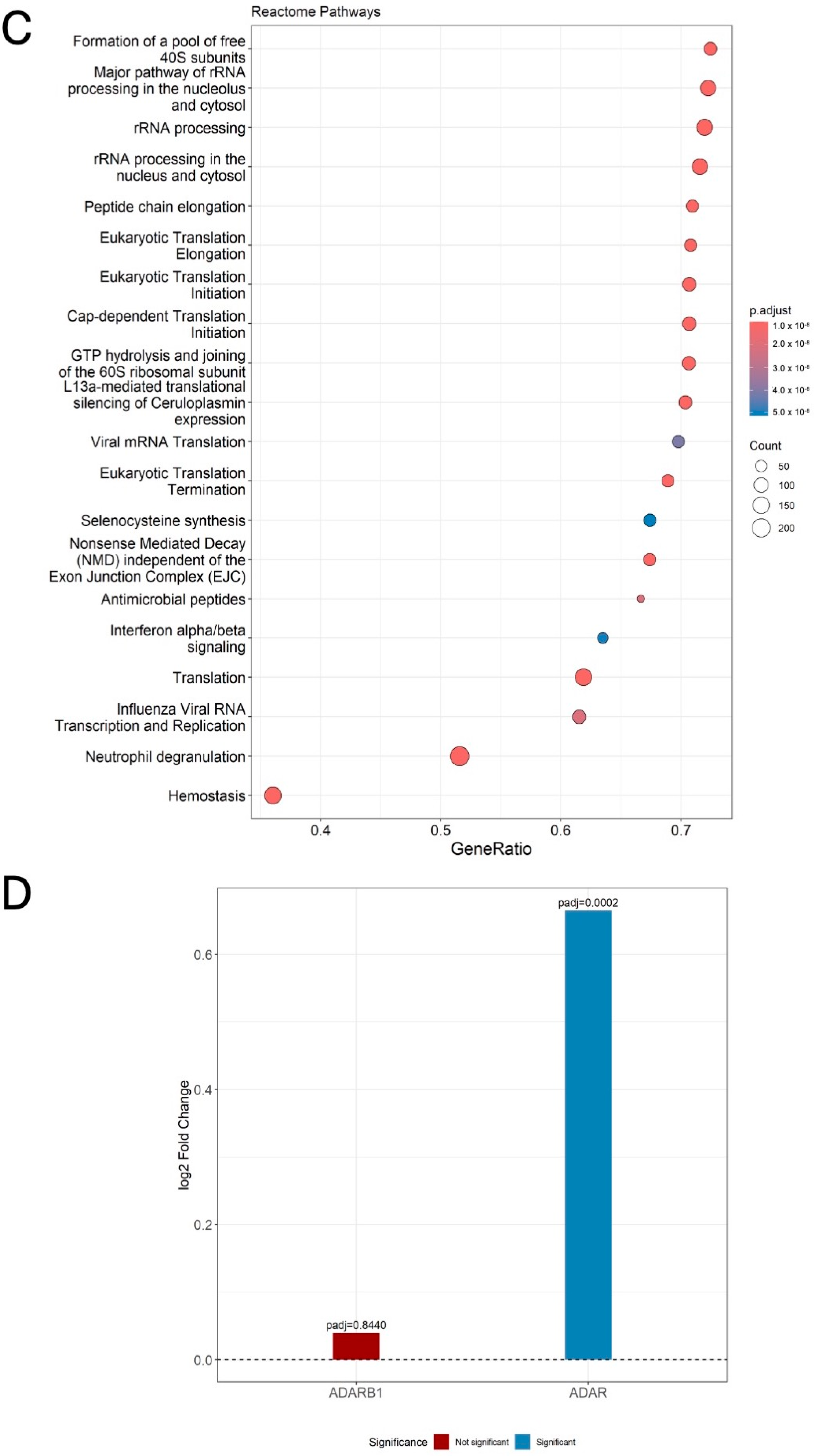
To understand which biological processes and pathways experienced differential gene expression, we performed Gene Set Enrichment Analysis (GSEA) using gene list ranked by log2Fold Change (log2FC), with gene sets from GO-BP and Reactome. Genes upregulated within critical COVID-19 patients showed enrichment for innate immune system pathways/processes including “neutrophil degranulation”, “interferon alpha/beta signaling”, “antimicrobial peptides”, “cellular response to type I interferon”, hemostasis pathways including “response to elevated platelet cytosolic Ca2+”, “platelet degranulation”, neuronal pathways including “GABA receptor activation”, “inwardly rectifying K+ channels”, and disease pathways including “disease associated with the TLR signaling cascade”. Genes downregulated within critically ill patients were enriched in RNA and protein metabolism pathways/processes such as “formation of a pool of free 40S subunits”, “MHC class II protein complex assembly” and “Nonsense Mediated Decay (NMD) independent of the Exon Junction Complex (EJC)”. The top 20 significant GO-BP and Reactome pathways (adjusted p-value < 0.05) are shown in the full list of enriched pathways/GO processes in Figure 1B,C ordered by the Nominal Enrichment Score (NES), and adjusted p-values < 0.05 are provided in the Supplementary Table S1D,E.
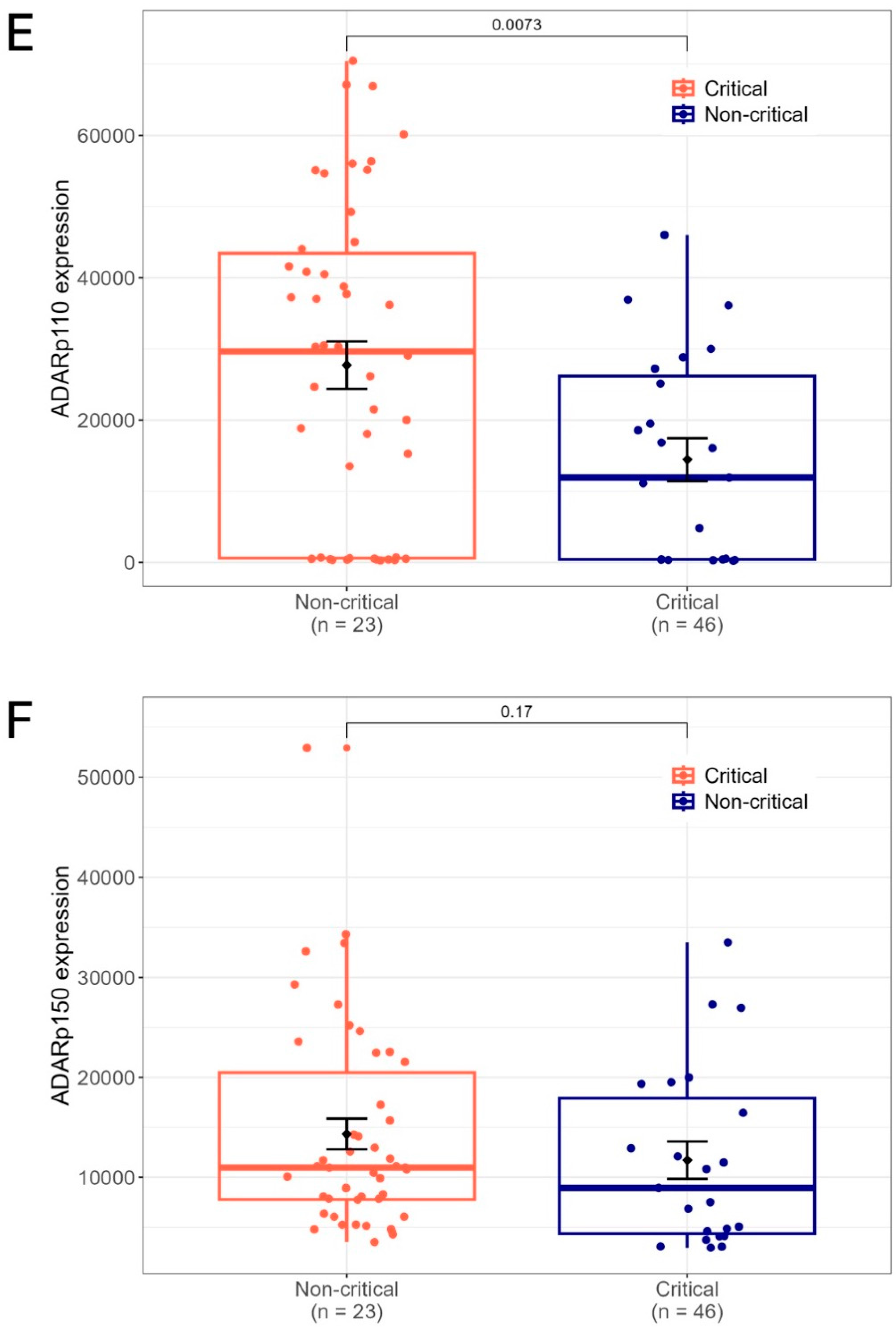
While previous studies have identified elevated expression of ADAR1 in response to SARS-CoV-2 infections [46,54], it is unknown whether this increase in expression is consistent or differs between patients with varying disease severity. We found significant differences in ADAR1 expression (log2FC = 0.6647 and adjusted p-value = 0.0002) between the two patient groups with higher expression observed in patients with critical COVID-19. Furthermore, ADAR2 expression, although without significance, was found to be nominally higher (log2FC = 0.03939 and adjusted p-value = 0.8440) in critical patients. While ADAR3 expression was not detected in our samples, this could be due to restricted expression of ADAR3 outside brain tissues [88,89] (Figure 1D; Supplementary Table S1F). Transcript-level expression of constitutively expressed ADAR1 isoform, ADARp110 and IFN-inducible isoform, ADARp150 [90,91] were examined as DESeq2 normalized counts. ADARp110 was found to be overexpressed in patients with critical illness (Wilcoxon rank-sum test: p-value = 0.0073, DESeq2: log2FC = 0.9388 and adjusted p-value = 0.2201) (Figure 1E), while expression levels of ADARp150, though minimally lower in patients with non-critical disease, did not achieve statistical significance (Wilcoxon rank-sum test: p-value = 0.17, DESeq2: log2FC = 0.2905 and adjusted p-value = 0.5304) (Figure 1F; Supplementary Table S1G,H).

2.2. ADAR Editing Patterns Differ Between Patients with Critical and Non-Critical COVID-19

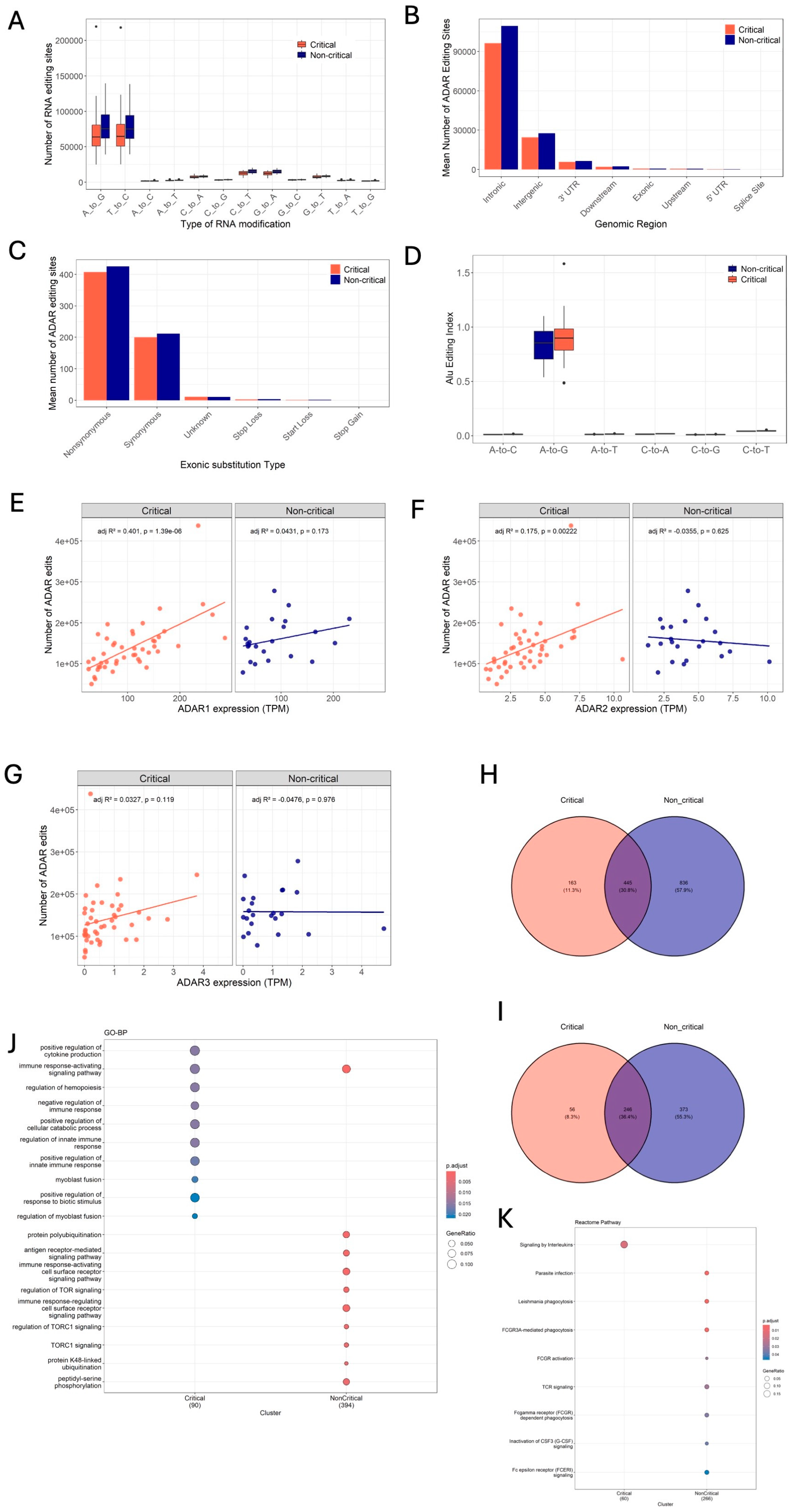
Next, we investigated whether ADAR activity differs between the two patient groups. To access this, we quantified ADAR-mediated editing within critical and non-critical patients using two different approaches: (1) by comparing the total number of ADAR-edited sites and (2) by calculating Alu Editing Index (AEI), as a measure of global levels of ADAR editing within Alu repetitive elements. In agreement with ADAR-mediated editing being the most abundant form of substitutions in humans [63,92,93], A-to-G and T-to-C edits (A nucleotides on the complementary strand) representing potential ADAR-edited sites were the most abundant form of substitutions within both groups of patients (Figure 2A, Supplementary Table S2A). However, the average number of both A-to-G and T-to-C substitutions differed between critical and non-critical COVID-19 patients. Specifically, the mean number of ADAR edits per sample were higher in patients with non-critical illness, with an average of 158,022 ± 10,234 edits, while patients with critical illness had an average of 139,285 ± 9307 edits per sample (Supplementary Figure S2 and Table S3). These sites were distributed mainly in intronic (74%), intergenic (19%), 3′UTR (4%), and downstream regions (2%), while a considerable number of sites were also distributed within the exonic regions (1%) (Figure 2B) and the mean number of editing sites within these regions differed between the two severities (Supplementary Table S2B). Notably, within exonic regions, differences were seen in the number of non-synonymous and synonymous edits between the two patient groups (Figure 2C). Further, the Alu editing index for A-to-G substitutions was highest among all other substitutions (Figure 2D). However, unlike the total number of ADAR edits, AEI for ADAR edits within Alu elements was higher in patients with critical COVID-19 (Figure 2D and Table 1). These differences suggest that ADAR activity may differ between patients who develop critical compared to non-critical COVID-19 in response to SARS-CoV-2 infections.
We further investigated whether the observed differences in total number of ADAR edits between the two patient groups were associated with differences in ADAR expression. Towards this, ADAR expression levels, measured as TPM (Transcripts Per Million) were correlated with the total number of ADAR edits within each patient group. In critical patients, the total number of ADAR edits showed a significant positive correlation with the expression levels of ADAR1 (adj R2 = 0.401, p = 1.39 × 10⁻⁶) and ADAR2 (adj R2 = 0.175, p = 0.00222). In contrast, no significant correlation was observed between expression of ADAR1 (adj. R2 = 0.0431, p = 0.173) or ADAR2 (adj. R2 = –0.0355, p = 0.625) and total editing events in non-critical patients (Figure 2E,F). Similarly, no significant correlations were observed between ADAR3 expression and total editing events in either critical (adj R2 = 0.0327, p = 0.119) or non-critical patients (adj R2 = –0.0476, p = 0.976) (Figure 2G), potentially due to negligible expression of ADAR3 in whole blood [94]. We also found ADAR editing sites that were uniquely edited within a severity group, indicating disease severity-specific editing signatures. Specifically, we observed 163 uniquely edited sites within 56 genes in critical COVID-19 patients, while there were 836 uniquely edited sites within 373 genes in non-critical patients (Figure 2H,I and Supplementary Table S2D,E). While these uniquely edited sites showed comparable genomic distribution between both groups of patients, with most sites located within the intronic, 3′UTR, and intergenic regions, the number of potential non-synonymous exonic edits varied (Supplementary Figure S2B,C). Of the four exonic edits in NBPF8, SMN1, HSPA1L, and HLA-DRB5 genes, within the non-critical patient group, HLA-DRB5 incorporated a non-synonymous edit at position Chr6: 32487309 and critical patients harbored a non-synonymous substitution within the DHRSX gene (position ChrX: 2139200). We analyzed the functional consequences of these non-synonymous edits on protein stability using AlphaFold2 [95] structure of HLA-DRB5 and DHRSX in the DDMut tool [96] (https://biosig.lab.uq.edu.au/ddmut/, accessed on 6 June 2025). In critical patients, substitution of Histidine (H) to Arginine (R) at position 292 within DHRX was predicted to result in a ∆∆G of −1.27 kcals/mol (Table 2, Supplementary Figure S2E), while in non-critical patients, the substitution of Serine (S) to Glycine (G) at position 164 within HLA-DRB5 was predicted to result in a ∆∆G of −0.08 kcal/mol (Table 2, Supplementary Figure S2D). A negative change in ∆∆G suggests that both mutations could result in destabilization of corresponding proteins, signifying distinct downstream consequences of ADAR editing between the two patient groups. Furthermore, overrepresentation analysis (ORA) of genes associated with unique edits revealed significant enrichment (adjusted p-value < 0.05, BH q-Value < 0.05) of distinct biological processes and pathways. Notably, unique edits in non-critical patients were spread across a broader range of biological processes and pathways compared to critical patients. These results suggest that these unique editing sites might have distinct functional roles in influencing COVID-19 disease severity (Figure 2J,K and Supplementary Table S2E,F).

2.3. Filtering High-Confidence ADAR Editing Sites and Identification of Differentially Edited Sites

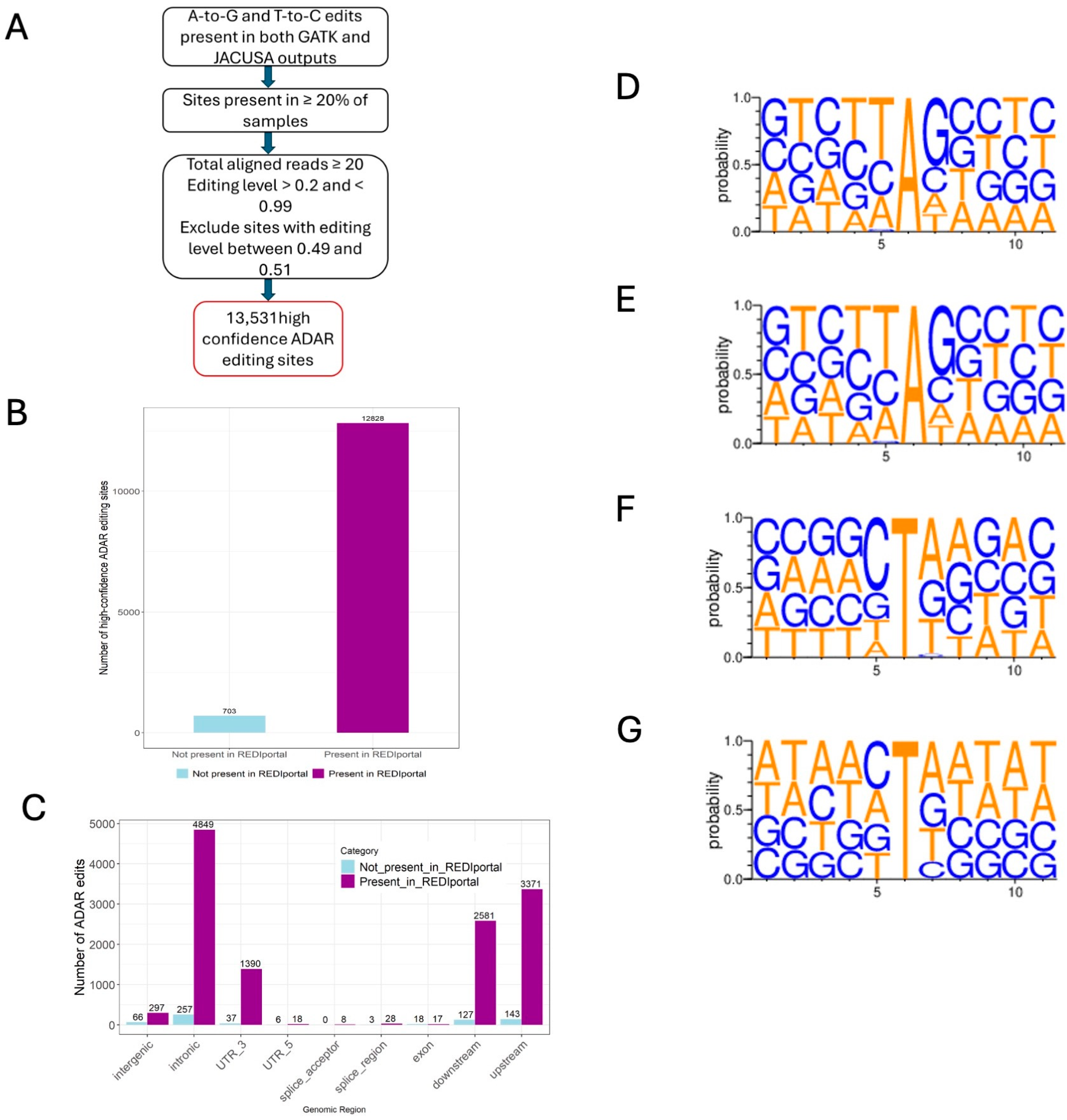
From the total ADAR editing sites, we further identified a list of high-confidence ADAR editing sites using a multistep filtering approach. First, we retained only those A-to-G and T-to-C substitutions that were present in the variant calling outputs of both GATK and JACUSA2. Additional filters were applied to this list on the total number of aligned reads and editing levels per site (refer to Methods for details). Additionally, only those sites that were present in at least 20% samples were selected. Following this filtering, a total of 13,531 high-confidence ADAR editing sites were identified within 2976 genes (Figure 3A, Supplementary Table S3A). Of these high-confidence ADAR editing sites, 12,828 sites were previously identified, confirmed editing sites present in REDIportal database (Supplementary Table S3B), while the remaining 703 were not present in REDIportal (Supplementary Table S3C), and will be referred to as novel editing sites (Figure 3B). Moreover, to ensure the robustness of the identified high-confidence sites, we examined these sites for features commonly associated with ADAR-edited sites. Previous studies have shown that ADAR editing sites are mainly located in the non-coding region, particularly within the intronic and 3′UTR regions of the transcripts [97,98] and show sequence preferences (motifs) surrounding the edited base [50]. Consistent with this, both known and novel high-confidence sites were enriched in the 3′UTR and intronic regions (Figure 3C, Supplementary Table S3A). Furthermore, for both known (sites present in REDIportal) and novel high-confidence sites, edited A’s on the positive strand had a 5′ neighboring T, C, or A and a 3′ neighboring G (Figure 3D,E). In contrast, edited T (=U, A on the complementary strand) had a 3′ neighboring A, G, or T (U), or a 5′ neighboring C (Figure 3F,G).
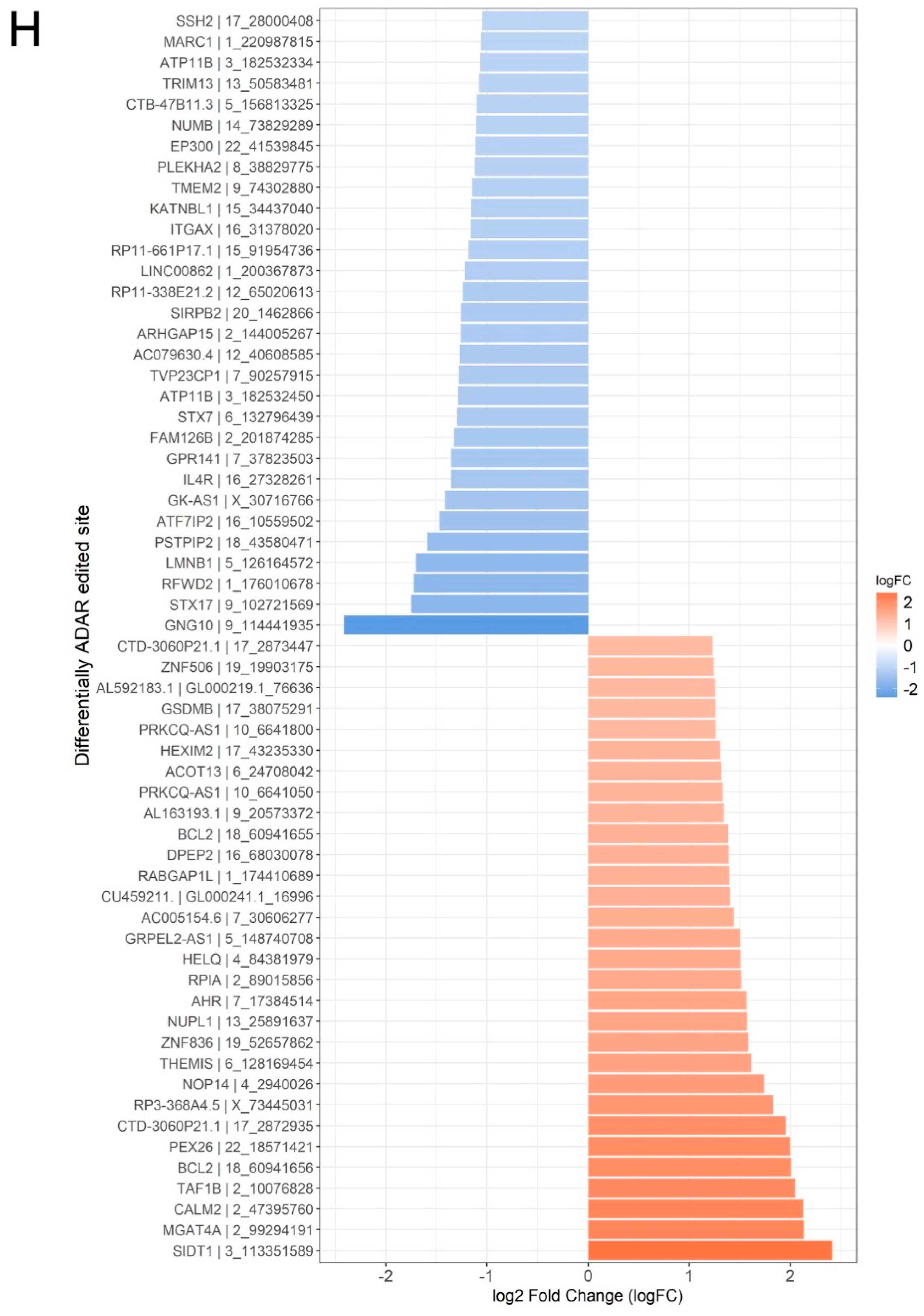
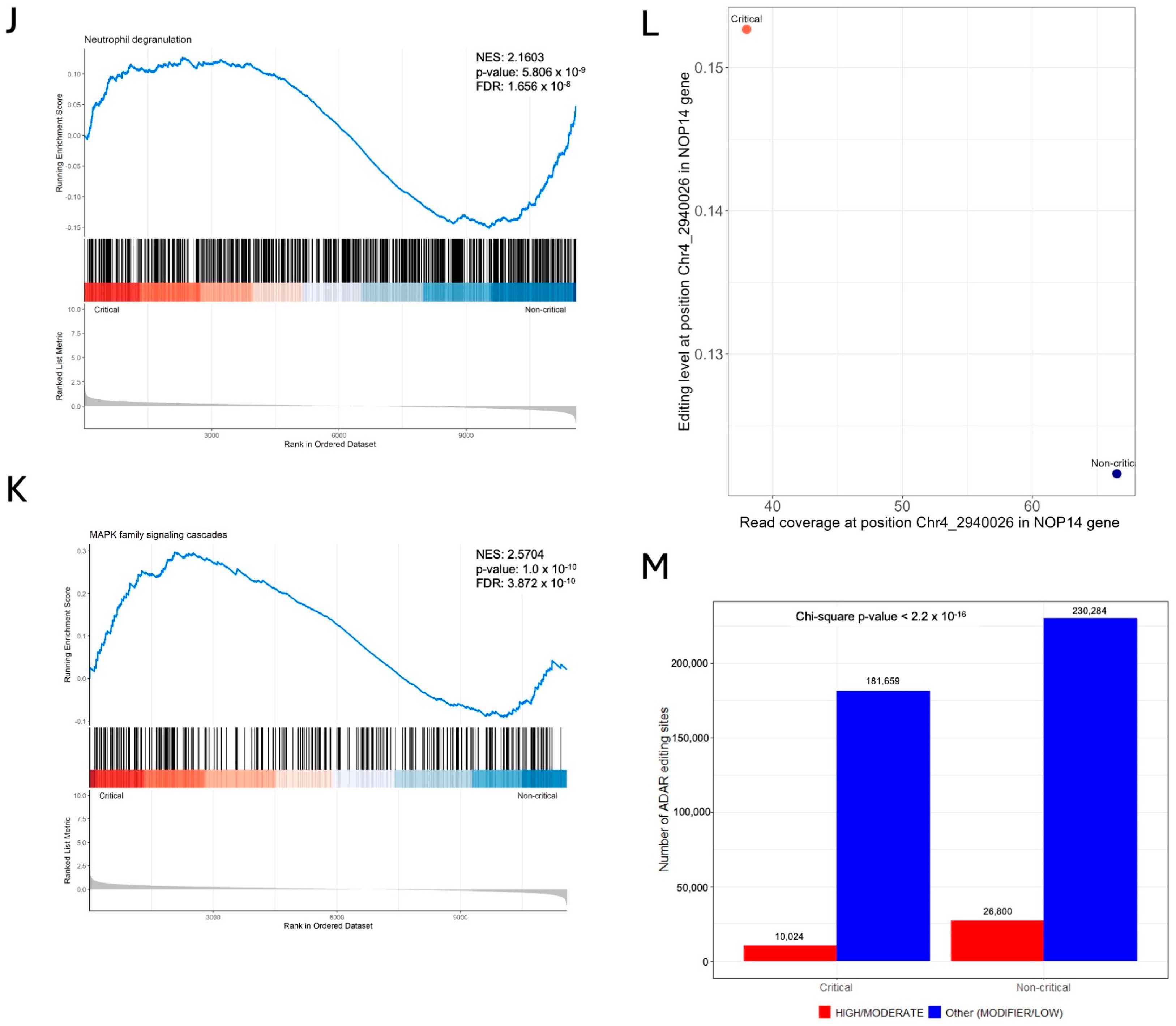
We further aimed to identify differentially edited sites within the list of high-confidence ADAR editing sites, i.e., sites with significant difference in RNA editing levels between critical and non-critical patients using the method previously described in Riemondy et al., 2018 [99]. Briefly, a generalized linear model (GLM) was applied to each high-confidence site, using edgeR, to find editing sites where the edited base (G/C) counts show significant differences between critical and non-critical patients, while controlling for unedited bases (A/T) within each sample. Using this approach, we identified a total of 140 differentially edited sites in 126 genes (edgeR log2FC > |0.58| and FDR < 0.05) within critical compared to non-critical COVID-19 patients (Supplementary Table S3D). Figure 3H shows the top 30 sites with significantly increased (log2FC > 0.58 and FDR < 0.05) and the top 30 sites with significantly decreased (log2FC < −0.58 and FDR < 0.05) editing in critical compared to non-critical patients. Of the 140 differentially edited genes, only 37 genes were found to overlap with differentially expressed genes identified above, suggesting ADAR editing could be a regulatory mechanism independent of the expression of the incorporating gene (Supplementary Table S3E). When we looked at the genomic distribution of these sites, as expected, a majority of these sites were within the Intronic (35%), 3′UTR (12%), and downstream regions (19%) of the transcript (Supplementary Figure S3). Importantly, differentially edited sites within NOP14 gene at position Chr4: 2940026 (with the higher levels of editing in critical samples—edgeR log2FC 1.7398 and FDR < 0.01) were identified as a moderate impact, missense variant (I779V) by SnpEff and ANNOVAR (Supplementary Table S3D). To explore the aforementioned possibility of ADAR editing as a regulatory mechanism independent of incorporating gene expression, we further examined whether this difference in editing within NOP14 is associated with differences in expression of its gene. Interestingly, in critical samples despite low coverage at the edited site, editing levels were higher in critical samples, while non-critical samples had higher coverage at the edited position with low editing levels (Figure 3L). Our results show that missense editing within NOP14 gene is not simply a consequence of increased expression and that editing adds another layer of regulation in COVID-19 disease severity.
Since editing within 3′UTRs could alter miRNA targeting [43], we examined whether the differentially edited sites in 3′UTRs are within known miRNA target regions using data from TargetScan release 8.0 [100]. The results showed that except for the site Chr4: 39551031 within 3′UTR of SMIM14 gene, all other sites were found to be in the known miRNA targeting regions. Specifically, 15 differentially edited sites within 12 genes were predicted to be targeted by 57 miRNAs (Table 3). Of these identified miRNAs, several—including, miR-10-5p and miR-20b-3p—have been identified as playing a role in regulating gene expression, RNA degradation and/or translation during disease states [101,102]. These findings suggest that differences in editing between critical and non-critical patients might also affect miRNA-mediated regulation in the two patient groups. While delineating the specific aspects of whether such regulatory changes occur and to what extent would require experimental validation in patient samples, a broader question of the downstream consequences of the editing changes across different miRNA binding sites should receive attention in future studies.
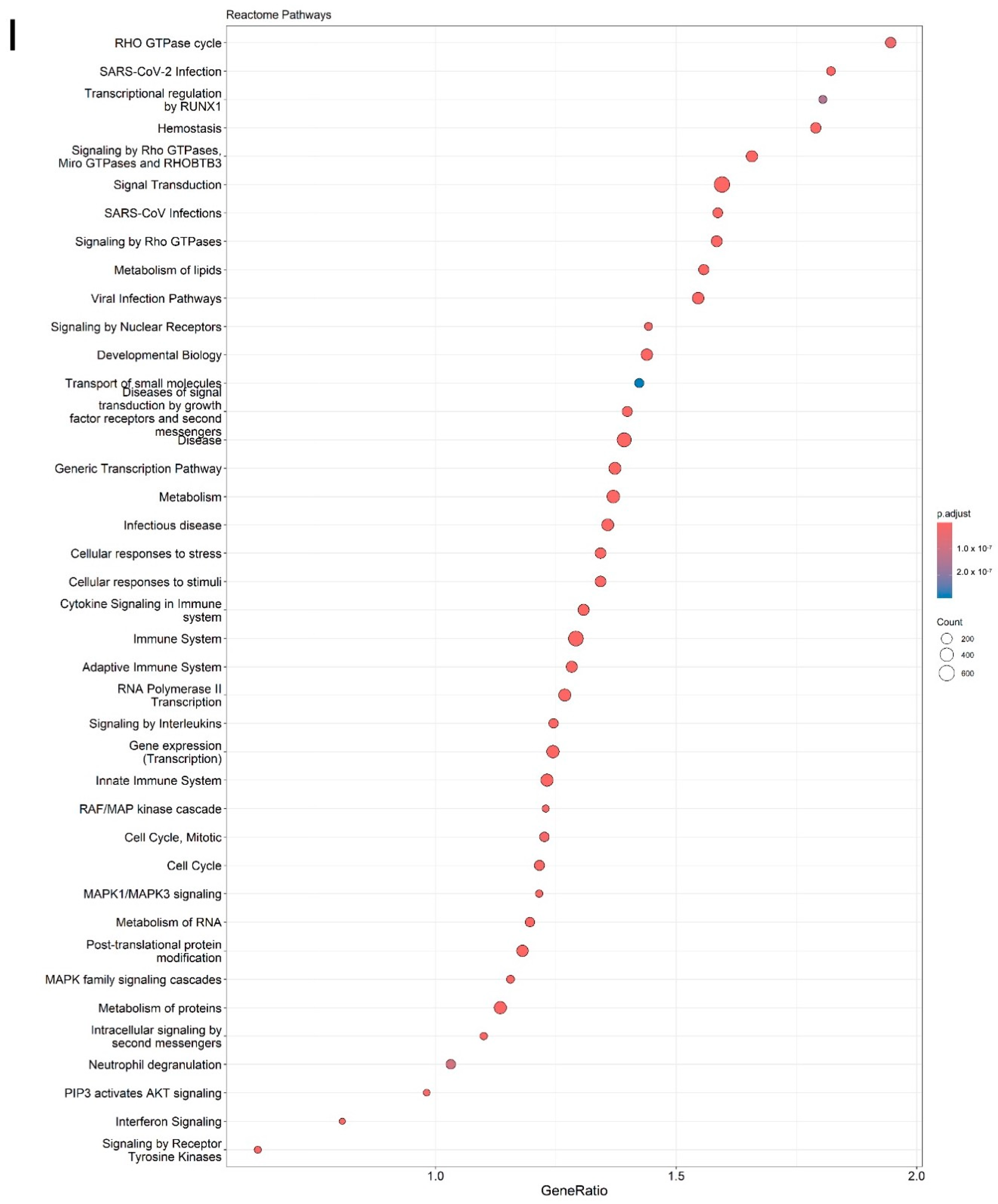
GSEA on a unique list of genes and coordinates (since the same genes could have multiple editing sites) ranked by log2FC, identified significant enrichment of multiple key pathways. Notable pathways included, immune system pathways such as “neutrophil degranulation”, “interferon signaling”, “cytokine signaling”, “signaling by interleukins”, and “activation of NF-κB in B cells”. Additionally, infectious disease pathways including “SARS-CoV-2 infection”, signal transduction pathways such as “MAPK family signaling cascade”, “signaling by Rho GTPases, Miro GTPases, and RHOBTB3”, and “insulin receptor signaling cascade” were enriched. Differentially edited genes were also enriched in pathways related to cell cycle, cellular response to stress, and RNA and protein metabolism (Figure 3I). GSEA plot shows distribution of differentially edited genes within the ranked list for “neutrophil degranulation” pathway (Figure 3J), and “MAPK family signaling cascade” (Figure 3K) that plays a key role in inflammation and shows positive enrichment in critical COVID-19 patients [122,123,124]. Interestingly, differentially edited genes were also enriched in pathways associated with cardiac conduction, and neuronal system pathways such as “neurotransmitter receptors and postsynaptic signal transmission” (Supplementary Table S3F). This suggests that ADAR editing may also contribute to cardiac and neurological manifestations observed in patients with critical COVID-19 as reported in previous studies and also identified in some patients within the present dataset (Carapito et al., 2022, Table 1) [86].

2.4. High- and Moderate-Impact Editing Events Differ Between Critical and Non-Critical COVID-19 Patients

SnpEff tool [125] predicts the functional effect and provides assessment of the putative impact of a variant, classifying it as high, moderate, modifier, and low. Here high-/moderate-impact editing events represent those editing events that can result in amino acid substitutions likely affecting the structure and function of the encoded protein. On the other hand, modifier-/low-impact editing events represent those within non-coding regions with insignificant effect on downstream protein (https://pcingola.github.io/SnpEff/snpeff/inputoutput/#eff-field-vcf-output-files (accessed on 6 June 2025)). To evaluate differences in the potential functional impact of ADAR-mediated RNA editing, we annotated the putative editing sites using SnpEff. Only those A-to-G and T-to-C sites with read depth ≥ 20 and shared across 20% of samples across a disease severity were selected. We found statistically significant differences (χ2 test, p value < 2.2 × 10⁻¹⁶) in the number of high- and moderate-impact ADAR editing sites, with a significantly lower number in patients with critical COVID-19 (Figure 3M).

2.5. Random Forest Classification Identifies Differentially Edited Gene Signatures Associated with COVID-19 Disease Severity

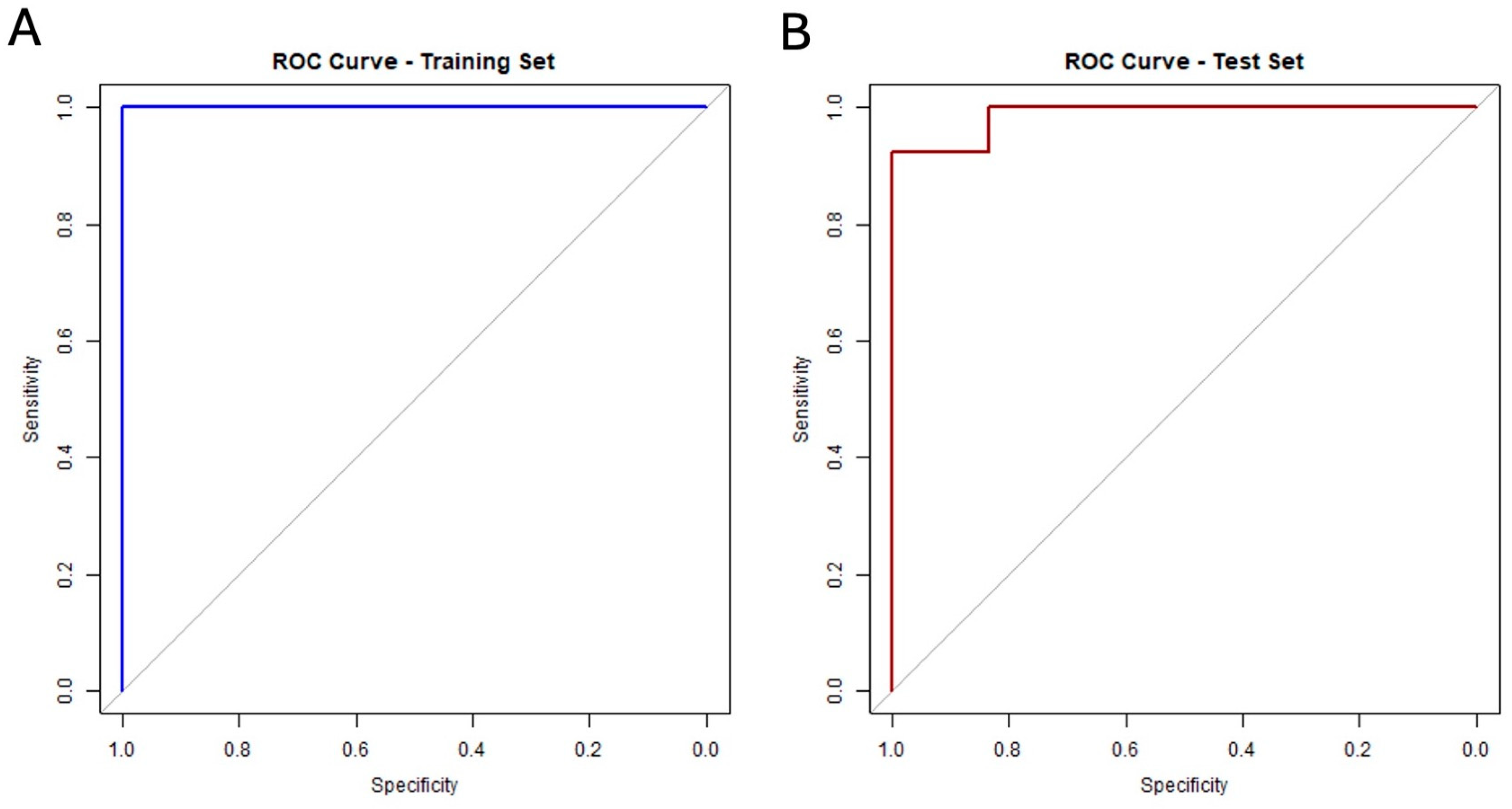
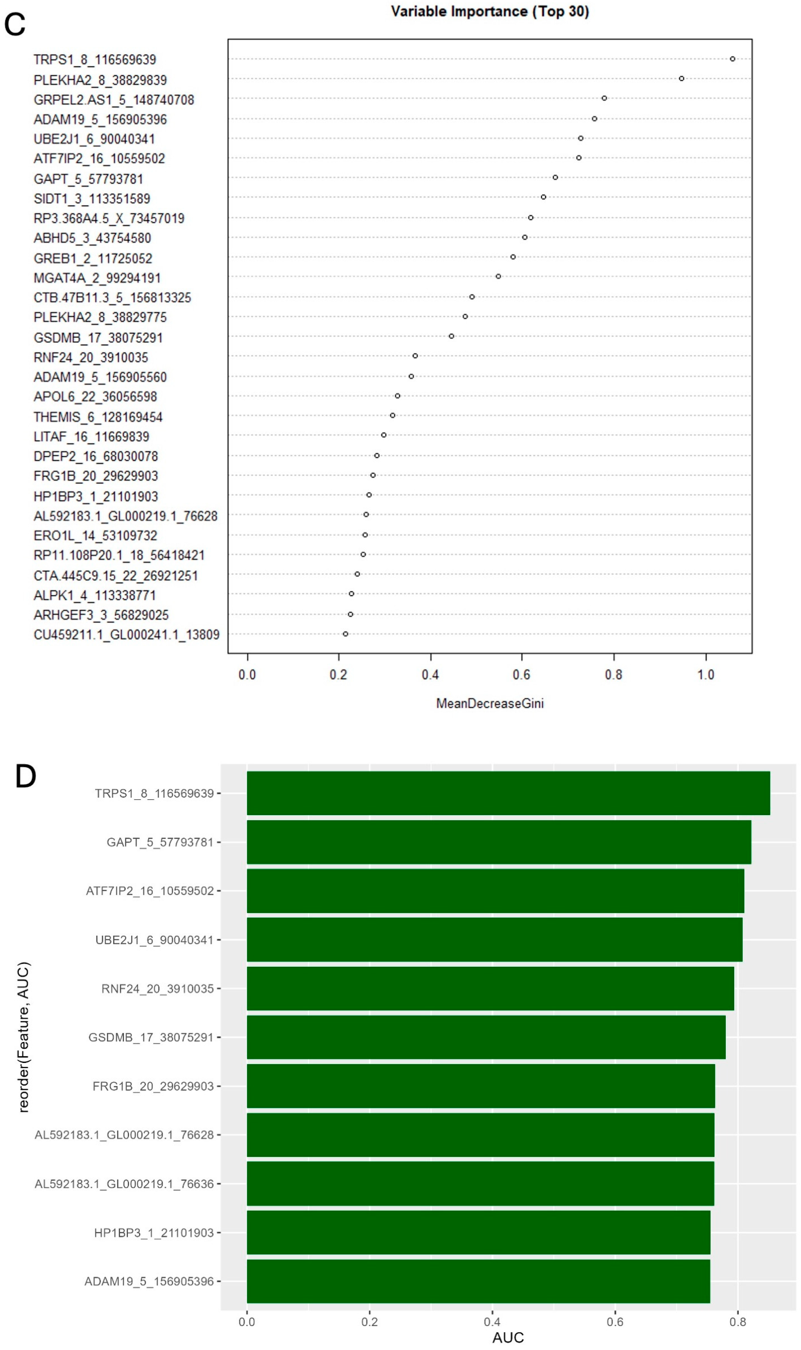
Next, to identify whether differentially edited sites can differentiate between critical and non-critical patients, we applied a random forest (RF) classifier. The dataset was randomly split to 70% training and 30% test dataset, and the RF model was trained on the training dataset using parameters described in the methods. Model performance was evaluated using receiver operating characteristics (ROC) curve analysis. The classifier showed high performance with an area under the curve (AUC) of 1 for the training dataset (Figure 4A) and 0.974 on the test dataset (Figure 4B), indicating that the model is effective in distinguishing between critical and non-critical patients and may be generalized to other datasets. To further identify key editing sites contributing to this classification, we calculated a Mean Decrease in the Gini index, a metric commonly used to access the importance of features in RF. The top 30 significant sites ordered according to the decreasing Gini index are shown in Figure 4C. Additionally, to assess the relevance of each site in distinguishing disease severity, per site AUC was computed. This resulted in 11 editing sites with AUC values >0.75 (Figure 4D, Supplementary Table S4), suggesting fair power [126] of these sites to discriminate between critical and non-critical patients.

3. Discussion

COVID-19 presents with a wide spectrum of clinical outcomes, resulting from complex interplay of various factors, including age, comorbidity status, host genetics, and immune responses [17,37,127]. Impaired IFN signaling and dysregulation of ISGs is a hallmark of critical disease [28,30]. Among ISGs, ADAR1 encodes for post transcriptional RNA editing enzyme that regulates transcriptome diversity and immune responses, and has been mechanistically linked to symptoms observed both during and post-viral infections [79,82,83]. Previous studies have shown that SARS-CoV-2 infection induces changes in host ADAR expression and editing patterns, contributing to COVID-19-associated physiological changes [46,78,85]. However, to the best of our knowledge, no studies have identified whether the expression and activity of ADARs differ with varying severities of COVID-19.
In this study we address this gap by analyzing a deeply sequenced whole-blood RNA sequencing dataset from a comparatively homogeneous cohort of individuals who developed critical COVID-19 and compared them to those who developed non-critical COVID-19, examining differences in ADAR editing patterns in response to SARS-CoV-2 infection. Our analysis revealed significantly high expression of ADAR1 and its isoform ADARp110 in patients with critical COVID-19. We further demonstrate that ADAR activity varies according to disease severity with marked differences observed in (1) the total number of ADAR-mediated edits (driven by expression of ADAR1 and ADAR2 in critical but not non-critical patients), (2) editing within repetitive Alu elements, (3) genomic distribution of edited sites, (4) the specific site being edited, including non-synonymous edits predicted to affect protein stability, (5) site-specific editing levels (that within 3′UTR may influence miRNA mediated regulation), and (6) proportion of edits with varying functional consequences. Functional enrichment analysis of uniquely and differentially edited genes in critical patients exhibited significant overrepresentation of inflammatory pathways, including neutrophil degranulation, which was also enriched among differentially expressed genes. Finally, we identified a set of differentially edited sites that could serve as molecular markers of COVID-19 disease severity. Together, our study demonstrates varying expression and editing patterns of ADARs between critical and non-critical patients and proposes a functional role for RNA editing in COVID-19 disease severity.
Extensive differences in the gene expression, including in genes involved in innate immune and inflammatory pathways, particularly neutrophil degranulation, is a key feature distinguishing patients with critical from those with non-critical COVID-19 [128,129], as was observed in the analysis of our dataset as well. While expression of ADAR1, specifically the ADAR1 isoform ADARp110, was significantly higher in patients with critical COVID-19, the expression of the IFN-inducible isoform ADARp150 did not differ significantly between the two patient groups. Considering that we are comparing virus-infected groups (i.e., disease samples) that will result in the production of IFNs and ISGs, insignificant differences in ADARp150 levels are understandable. However, it should be noted that the levels of IFNs and ISGs, including ADARp150 are known to be highly dynamic throughout the course of viral infection, and factors such as the timing of sample collection can significantly influence the measurements of expression levels [130]. Previous evidence from animal models have shown that the IFN response often peaks a few days after infection and may rapidly decline thereafter [130,131]. Therefore, it is possible that ADARp150 levels were different at some point between the two groups of patients; however, further studies that could measure the levels across different time points of SARS-CoV-2 infection are needed to confirm this. Interestingly, ADARp110, although constitutively expressed, is seen to be elevated during viral infections [47,132,133], including SARS-CoV-2 infections [46,134]. This could be due to leaky ribosomal scanning downstream of IFN-induced ADARp150 start codon, which results in the coexpression of both ADARp110 and ADARp150 [45]. It has also been proposed that persistence of SARS-CoV-2 viral transcripts could also drive sustained transcription and translation of ADARp110, resulting in elevated ADARp110 levels [135]. In line with this, an original study by Carapito et al. 2022 [86] reported the presence of viral gene transcripts within samples from patients with critical COVID-19, but not in samples from non-critical COVID-19, which could also be a potential explanation to the higher expression of ADARp110 observed in patients with critical COVID-19 in our analysis.
When comparing differences in global ADAR editing, measured as differences in the total number of ADAR edits between the two patient groups (comparison between patient groups), we found a lower number of edits within critical patients, despite significantly high expression of ADAR1, compared to non-critical patients. This is consistent with previous studies that have observed that the expression of ADAR1 does not always translate to global higher editing [136,137,138]. Nonetheless, when we applied a linear regression model to identify the relationship between expressions of all three ADARs and the total number of ADAR edits within each patient group (within group correlations), we found a significant positive correlation between the total number of ADAR edits and the expression levels of ADAR1 and ADAR2 within patients with critical COVID-19. In contrast, no significant correlations were observed between expression of any of the three ADARs and the total number of edits for patients with non-critical disease. These results suggest that the activity of ADARs differ between the two patient groups, and that ADAR expression might have a stronger influence on total editing in critical patients, while in non-critical patients factors other than editing might be contributing more to the global editing landscape. In fact, several other factors have been known to influence ADAR activity, including the formation of heterodimers between different ADARs [48], availability of cofactors such as inositol hexaphosphate (IP6) [139], post transcriptional regulation by miRNAs, post translational SUMOylation (which can reduce ADAR1 editing activity) [140], and availability and accessibility of substrates [141,142]. It is also worth noting that ADAR3 is predominantly expressed in neuronal tissues, and is known to have an inhibitory effect on the activity of the other two ADARs [143,144], though in our whole-blood dataset ADAR3 expression was negligible and showed no correlation with the total number of edits in both groups of patients. It remains possible that ADAR3 may have an impact on editing in neuronal tissues, and further studies using neuronal samples are needed to explore its role in this context. Furthermore, while the total number of ADAR editing events (i.e., events within both coding and non-coding regions) were lower in critical patients, AEI, a measure of ADAR editing activity within Alu repetitive elements in the genome, was higher in patients with critical COVID-19. Given that ADAR1 is the primary enzyme responsible for editing within Alu elements [145,146], and that the expression of ADAR1 was elevated in critical patients, this may explain the observed increase in AEI in critical patients despite a lower total number of edits.
In addition to global differences in editing in terms of total number of edits, differences were also seen in ADAR targets, as seen by the presence of genes and pathways that were uniquely edited in either critical or non-critical patients, establishing COVID-19 severity-specific ADAR activity. For instance, non-synonymous editing within exon 7 of the Dehydrogenase/Reductase X-Linked gene (DHRSX), that was predicted to result in protein destabilization, was preferentially edited in patients with critical COVID-19. DHRSX encodes for an enzyme involved in dolichol synthesis, a lipid essential for N-glycosylation [147,148]. SARS-CoV-2 spike (S) protein and the host receptor protein angiotensin converting enzyme 2 (ACE2) undergo N-glycosylation facilitating the binding between the two. Considering the importance of N-glycosylation in SARS-CoV-2 infection and the essential requirement of the DHRSX gene in N-glycosylation [149,150], it can be speculated that missense editing in this gene can impact viral entry or replication. While this could be a potential mechanistic link between DHRSX editing and disease severity, further studies are required to confirm this speculation, if any. Similarly, a unique edit was found at position 164 on exon 3 in the gene human leukocyte antigen class II heterodimer β5 (HLA-DRB5). HLA-DRB5 is a Class II major histocompatibility complex (MHC) molecule involved in antigen presentation to CD4+ T cells and is a key player in adaptive immune responses. Although the direct effect is unknown, when the role of HLA in antigen presentation is considered, it is plausible that non-synonymous editing within this gene may affect viral recognition by the host [151]. Noteworthily, the extent of protein destabilization resulting from these non-synonymous edits were relatively different, with a much larger change in ∆∆G resulting from non-synonymous editing within the DHRDX gene in critical patients, indicating that editing within DHRSX may have a greater impact on protein stability.
The remaining unique edits were predominantly distributed within the intronic and 3′UTR of transcripts, although the numbers varied between critical and non-critical COVID-19 patients. Such editing within non-coding regions may impact multiple processes regulating gene expression and ultimately influencing protein function. For instance, editing within introns can result in conversion of 5′ splice donor sites and/or splice 3′ splice acceptor site, potentially altering splicing patterns and leading to the production of alternative protein isoforms [142,152,153]. Splice site editing may also alter mRNA stability, as seen in increased stabilization of FAK transcript, promoting tumor progression in lung adenocarcinoma [154]. Additionally, intronic editing can alter biogenesis of circular RNAs (circRNAs) [155,156]. Similarly, editing within 3′UTR has been shown to impact miRNA targeting, either by creating or destroying miRNA targets [157,158]. These findings suggest that severity-specific differences in ADAR editing, including in both coding and non-coding regions, could contribute to differences in protein stability, as well as in regulatory processes (such as alternative splicing, mRNA stability, and miRNA targeting) between patients with critical compared to non-critical COVID-19.
Considering the dynamic and heterogenous nature of ADAR editing between individuals and disease conditions [44], we identified a set of high-confidence ADAR editing events, with signatures consistent with that of ADAR-edited sites. While the majority of these were previously reported sites, a subset of these sites were novel, not previously reported sites, and may represent editing events specifically induced by SARS-CoV-2 infection irrespective of disease severity. Notably, we found that many such high-confidence sites were differentially edited between patients with critical and non-critical disease, indicating that the site-specific editing rates also varies with COVID-19 disease severity. For instance, we identified differential editing within multiple genes related to SARS-CoV-2 infection and disease severity. One such differentially edited site was within the 3′UTR of APOL6 (Apolipoprotein L6) gene that has been previously implicated in immune responses to COVID-19 vaccinations and was proposed to contribute to antiviral defense and immune regulation [81]. Our identification of differentially edited sites within APOL6 gene reinforces its significance in SARS-CoV-2-associated immune responses. We also detected a differentially edited site within DDX6 (DDX DEAD box RNA helicase 6), an RNA helicase involved in RNA metabolism and p-body formation. DDX6 has been shown to be essential for SARS-CoV-2 replication. The viral nucleocapsid (N) protein is known to interact with DDX6 and hijacks it to promote viral replication. This suggests that differential editing within DDX6 may influence its function or regulation and impact SARS-CoV-2 replication thereby impacting disease severity [159]. Furthermore, several intronic edits were found within the BCL2 (B-Cell Leukemia/Lymphoma 2) gene, a key anti-apoptotic gene from the intrinsic (mitochondrial) apoptotic pathway. Notably, low levels of BCL2 have been reported in patients with critical disease (specifically in non-surviving patients), which is associated with higher cellular damage due to apoptosis. Although the functional consequences of intronic editing remain unclear, it is possible that such edits may influence BCL2 expression or splicing [160]. We also identified a differentially edited site within NOP14 gene that results in a non-synonymous edit. Importantly, this editing event was independent of the expression of the host gene, suggesting that this editing is not simply a consequence of increased expression. Moreover, Peng et al. [161] had previously identified the exact site to be differentially edited in patients with lung adenocarcinoma, further underscoring the importance of ADAR editing at this site in diseases. Differential editing was also observed in genes previously identified to undergo editing across various diseases. For instance, editing within PGAM5 has been associated with heart diseases [162], ITGAX has been shown to be differentially edited in children with Mycoplasma pneumoniae pneumonia [163], NLRC5 has been reported to be edited in the autoimmune condition, primary Sjögren’s syndrome [164]. The consistent editing within disease related genes indicates that differentially edited sites identified in our analysis may hold clinical significance.
Overall, this study takes advantage of a deeply sequenced and relatively homogenous cohort in terms of age and comorbidity status, factors that can impact severity and editing patterns [17,165,166], thereby enabling a comparison of differences in ADAR editing across varying severities of COVID-19. Moreover, these patients were sampled during the first wave of the COVID-19 pandemic in France, indicating the infections were potentially due to an ancestral SARS-CoV-2 strain [86]. Furthermore, these samples were obtained prior to widespread use of corticosteroids in COVID-19 treatment. Corticosteroids are anti-inflammatory drugs known to reduce mortality and morbidity in critical patients [167,168]. However, these drugs are known to influence ADAR editing rates [169]; therefore, sampling before the use of corticosteroids reduces the risk of treatment related factors confounding the results. We also would like to point out the relatively high sequencing depth of this dataset. The reliability of detected RNA editing events is known to increase with the coverage, and a depth of 80–100 million reads is typically recommended for Illumina sequencing [170]. Our dataset had an average depth of ~208 million reads per sample, sufficient to detect editing even within lowly expressed genes.
Nonetheless, despite these strengths, our study also has some limitations. A limitation of our approach to identifying high-confidence editing sites is that the stringent identification criteria we used may have reduced sensitivity for detecting low-level RNA editing events. While our approach required the sites to be detected by GATK and JACUSA2 and to pass further stringent downstream filters, which may have improved the specificity of our identified editing sites, it also may have excluded biologically relevant but low-level editing events. However, these filtering criteria were applied consistently across both critical and non-critical groups, minimizing potential disease group-specific bias. Therefore, while the number of detected edits could be a conservative estimate of the total edits in each group, the difference that we identified between the groups reflects comparisons made under similar filtering strategies. Furthermore, a lack of detailed clinical metadata, including underlying comorbidities, viral load, and other host factors associated with COVID-19 severity, were not available for all individuals. Therefore, we cannot completely exclude the possibility that additional clinical variables—including differences in viral load at the time of infection or that at the time of disease progressing in severity—have contributed to the observed differences between critical and non-critical groups. Host genetics are also known to play a very significant role in deciding COVID-19 disease severity; however, the presence of disease-affecting variants, if any, was not known in these patients. Moreover, two patients with critical COVID-19 were reported to have anti-type I IFN autoantibodies in the parent study [86]; thus, it is possible that this may influence ADAR expression and downstream editing. The parent study also stated that the presence of obesity alone in the patients was not considered as an exclusion criteria under comorbidity; however, multiple studies have identified obesity as a major comorbidity affecting COVID-19 severity and can also alter editing patterns [171,172]. Furthermore, it should be noted that ADAR editing is highly variable between tissues, and thus, blood ADAR editing signatures and presumed downstream effects may not fully represent the physiological changes within other tissues, especially those in neuronal tissues where editing is highly enriched and expression of the catalytically inactive enzyme ADAR3 impacts the activity of other two ADARs. Nonetheless, our findings show that patterns of ADAR editing could indeed serve as prognostic and/or diagnostic markers for COVID-19 disease severity.

4. Materials and Methods

  • Transcriptomic dataset of COVID-19 patients with critical and non-critical COVID-19
In this study, we used deeply sequenced transcriptomic data, with an average depth of ~208 million reads per sample (Supplementary Table S5), from Carapitio et al.’s study [86], which is publicly available at NCBI GEO under the accession GSE172114 (BioProject PRJNA722046). Briefly, the dataset includes whole-blood RNA sequencing samples from 69 young patients (under the age of 50), without comorbidities, hospitalized for varying severities of COVID-19 (confirmed using qRT-PCR test of nasopharyngeal swabs) at a university hospital network in northeast France from March to April 2020 (first wave of pandemic in France). These 69 patients were categorized into two groups based on severities of COVID-19 presentations. The first group consists of 23 patients with non-critical COVID-19, referred to as those who stayed at a non-critical care ward and may or may not have required low-flow supplemental oxygen. The second group consists of 46 patients with critical COVID-19 defined as those hospitalized in the Intensive Care Unit (ICU) due to moderate or severe ARDS (berlin criteria) and/or requiring high-flow nasal oxygen and mechanical ventilation due to respiratory failure (Table 1). The group also included patients who succumbed to the disease. Detailed summary of patient characteristics including drug and supportive treatments (if used) is available in Carapito et al. (2022), Table 1 [86].
  • RNA-seq data analysis and identification of RNA editing sites
RNA sequencing dataset of paired end 151 base pairs (bp) long reads from total RNA, depleted for ribosomal and globin RNA, from Carapito et al. (2022) [86], were downloaded from NCBI SRA. Quality check, read mapping, and assembly were performed using the computational pipeline Automated Isoform Diversity Detector (AIDD) [173]. Briefly, raw FASTQ files were quality-checked using FASTQC version 0.11.5 (https://www.bioinformatics.babraham.ac.uk/projects/fastqc/ (accessed on 6 June 2025)). Following trimming, reads were aligned to GRCh37 (human reference genome Ensembl build release 75) annotated with splice sites and genomic single-nucleotide polymorphisms using HISAT2 version 2.1.0 [174]. On average, approximately 200 million reads were aligned to the genome (Supplementary Table S5). Stringtie version 1.3.5 [175] was used to perform transcriptome assembly, the resulting ballgown files with gene- and transcript-level information were analyzed using a custom bash script to generate gene- and transcript-level raw count matrices. Both gene and transcript counts were analyzed using DESeq2 (v1.44.0) [87] to identify differentially expressed genes, including ADARs, between critical and non-critical patients. Genes that met the criteria of an adjusted p-value (padj) < 0.05 and log2 Fold Change (log2FC) ≥ |0.58| were considered differentially expressed. ADAR isoform expression was measured as DESeq2 normalized counts. FDR correction for multiple testing was performed using the Benjamini and Hochberg (BH) method. Two different tools, Genome Analysis Toolkit (GATK) version 3.8.1.0 [176] haplotype caller and JACUSA2 version 2.0.4 [177] were used to identify all potential RNA editing events. GATK analysis followed the best practice settings, as defined in Plonski et al., [173] to identify putative ADAR editing sites from the BAM files. Single-nucleotide polymorphisms (SNPs) defined in NCBI database of SNPs (dbSNP build 138) were filtered out [178]. Java framework for accurate SNV assessment (JACUSA2 version 2.0.4, [177]), filtering out sites with less than 20 reads, was used to identify putative ADAR editing sites and count the number of bases observed at each such site. ADAR editing events were defined as substitutions that included both A-to-G and U (T)-to-C, the latter representing editing events on the complementary strand, referred here to as T-to-C substitutions. Thus, identified editing site matrixes were annotated, and their potential functional consequences were predicted using snpEff variant annotation and effect prediction tool version 4.3t [125] and ANNOVAR (https://annovar.openbioinformatics.org/en/latest/ (accessed on 6 June 2025)) gene symbols using RefGene [179].
  • Calculation of Alu editing index (AEI)
Alu editing index (AEI) represents the weighted average of all the A-to-G mismatches within Alu elements to the total coverage of adenosines within a genomic region. Alu editing indexer method described in Roth et al., 2019 [180], using default parameters on STAR mapped BAM files as input, was used to determine differences in editing levels within the Alu repetitive elements, between the two patient groups.
  • Selection of high-confidence ADAR editing sites and differential RNA editing analysis
High-confidence ADAR editing sites were identified using multiple filters on GATK version 3.8.1.0 [176] and JACUSA2 version 2.0.4 [177] generated VCF files. First, GATK and JACUSA2 generated VCF files were filtered to include only those A-to-G and T-to-C substitutions that were identified by both the tools. Furthermore, this only included those substitutions that were present in at least 20% of all samples and with greater than or equal to 20 total aligned reads (read coverage), editing level (defined as the proportion of G reads at a site with an A reference or C reads at a site with T reference) greater than 0.2, less than 0.99 and not between 0.49 and 0.51. The latter filtration was done to include only sites with at least a 20% editing level at each edited site and to exclude potential noise, homozygous and heterogenous genomic variants respectively. Further, editing level filters of >0.2 were used to retain high-confidence editing events and reduce potential false positives that could be associated with editing sites with low frequency. Similar editing level filters have been used in previous studies on ADAR editing to identify high-confidence editing events from RNA seq data [181]. The remaining sites were further mapped to REDIportal [182], a comprehensive database providing curated collection of ADAR editing events across multiple tissues including whole blood to identify known ADAR editing sites. Sites that were not present in REDIportal were classified as novel sites. Next, to identify differentially edited sites between critical and non-critical patients we applied a statistical model using the edgeR package (version 4.2.2) [183], as described previously in Riemondy et al., 2018 [99]. Briefly, for each high-confidence ADAR editing site, we extracted the reference and alternative base counts from each sample, generating a count matrix consisting of separate counts for each base per sample. A generalized linear model (GLM) was then constructed, ~0 + sample_ID + condition:allele, where “sample_ID” accounts for individual-specific effects, “condition” represents disease severity, and “allele” indicates reference or alternative base. Dispersion estimates were calculated and the GLM was fit to the above count data. Differential editing analysis was performed using a likelihood ratio test (glmLRT) to identify significant differences in edited base count across conditions, while controlling for within-sample differences in reference base count. Differentially edited sites were defined as those with log2FC > |0.58| and FDR < 0.05.
  • Enrichment analysis
To identify functionally relevant pathways and biological processes within our gene list, we performed both overrepresentation analysis (ORA) and Gene Set Enrichment Analysis (GSEA) using the R package “cluster profiler” (version 4.12.6). ORA was performed on the list of genes unique to disease severity to identify Gene Ontology (GO) terms and Reactome pathways enriched within the unranked list of genes. GSEA was performed on ranked gene lists generated from both DESeq2 analysis (to identify differentially expressed genes) and edgeR analysis (to identify differentially edited ADAR editing sites). In both analyses, for GSEA, genes were ranked based on log2FC values, and 1000 permutations were used to access statistical significance. Gene annotations were provided using the org.Hs.eg.db human gene annotation database. Pathways with an adjusted p-value < 0.05 were considered significantly enriched and ordered according to enrichment score.
  • Motif Analysis
Both the novel and known high-confidence ADAR editing sites (A-to-G and T-to-C analyzed separately) were converted to BED format. BED files were then used with BEDtools [184] to extract 5 bp upstream and downstream sequence flanking the edited bases from the unmasked human reference genome (Homo_sapiens.GRCh37.dna_sm.primary_assembly.fa). The resulting FASTA sequence was then used to generate sequence logos using Weblogo (version 3.7.12) [185] using a custom bash script.
  • In silico prediction of effect of missense edits on protein stability
DDMut [96], with AlphaFold2 [95,186] predicted protein structure as input, was used to predict changes in protein stability caused by the missense edits. DDMut is a deep learning model that predicts changes to protein stability, quantified as changes in Gibbs free energy (∆∆G) in kcal/mol, based on atomic interactions in the wild type and mutant amino acid residue. A negative ∆∆G value (<0) indicates protein destabilization while positive ∆∆G (>0) indicates protein stabilization.
  • Analysis of miRNAs with targets overlapping with differentially edited sites within 3′UTRs
Because editing within 3′UTRs can alter miRNA targeting [43], we examined whether the differentially edited sites in 3′UTRs are located within known miRNA target regions using data from TargetScan [100]. All pre-identified miRNA target sites were obtained from TargetScan (v8.0) by downloading a All_Target_Locations.hf19.bed file from https://www.targetscan.org/vert_80/vert_80_data_download/All_Target_Locations.hg19.bed.zip (accessed on 6 June 2025). Differentially edited sites within 3′UTRs were compared to the miRNA target sites using a custom R script. Genomic coordinates of 3′UTR editing sites and miRNA target regions were matched using R package GenomicRanges [187].
  • Random forest and ROC analysis
To assess whether ADAR editing could distinguish between critical and non-critical patients, we used a random forest (RF) classifier. The ADAR editing matrix containing editing levels for all the identified differentially edited sites was used as features. Samples were randomly partitioned to test (70%) and training (30%) and the random forest was trained on the training data using the randomForest [188] package in R, with 1000 tree default parameters. Model performance was evaluated using receiver operating characteristics (ROC) curve and area under the curve (AUC) computed using the pROC package [189]. Editing sites that were most important for classification were calculated using the Mean Decrease in the Gini index, a measure of variable importance in RF [190]. Additionally, site-specific predictive power of a site was calculated using AUC values, where each site was used as a predictor for binary outcome. Editing sites with AUC >0.75 was considered to have fair discriminatory power [126].
  • Statistical analysis of expression and editing data
Statistical analyses were performed in R version 4.2.2 (R Foundation for Statistical Computing, Vienna, Austria). The Wilcoxon rank-sum test was used to compare the expression of ADAR1 isoforms, ADARp110 and ADARp150, as DESeq2 normalized values between critical and non-critical patients. Pearson’s Chi-squared test with Yates’ continuity correction was used to compare the number of high/moderate or other editing events between the two patient groups. To examine whether ADAR gene expression is associated with the total number of ADAR editing events within each patient group, we performed a linear regression analysis using expression values of all three ADARs in transcripts per million (TPMs) as the independent variable and total number of ADAR edits within each group as the dependent variable. The strength and significance of these associations were assessed using adjusted R2 and p-values (<0.05). Statistical analyses and data visualization were conducted using the ggplot2 (version 3.5.2), ggpubr (version 0.6.0), and dplyr (version 1.1.4) packages in R.

Supplementary Materials

The following supporting information can be downloaded at https://www.mdpi.com/article/10.3390/ijms27156809/s1.

Author Contributions

Conceptualization, A.M.N. and H.P.; methodology, A.M.N. and H.P.; software, A.M.N.; formal analysis, A.M.N.; resources, H.P.; data curation, A.M.N.; writing—original draft preparation, A.M.N.; writing—review and editing, A.M.N. and H.P.; visualization, A.M.N.; supervision, H.P.; project administration, H.P. All authors have read and agreed to the published version of the manuscript.

Funding

This research received no external funding.

Institutional Review Board Statement

As a secondary analysis of publicly available, unrestricted access, data, this study was determined exempt human subjects research under the Category 4 of the Common Rule (45 CFR 46) by the Kent State University Institutional Review Board (protocol 2535).

Informed Consent Statement

As a secondary analysis of publicly available, unrestricted access, data, this study is exempt from the consent requirements, under the Category 4 of the Common Rule (45 CFR 46).

Data Availability Statement

The dataset used in this study is publicly available in the NCBI SRA/BioProject repository, as BioProject PRJNA722046 (Carapito et al., 2022, [86]). Supplementary Materials and code are shared at https://github.com/RNAdetective/Editing_in_critical-non-critical-COVID-19 (accessed on 1 July 2026) and in the Zenodo archive at https://doi.org/10.5281/zenodo.20514089.

Conflicts of Interest

The authors declare no conflicts of interest.

Abbreviations

The following abbreviations are used in this manuscript:
ADARAdenosine Deaminase Acting on RNA
IFNInterferon
ISGInterferon-stimulated gene
ISREInterferon-stimulated response element
UTRUntranslated region
SARS-CoV-2Severe acute respiratory syndrome coronavirus 2
COVID-19Coronavirus disease 2019
ARDSAcute respiratory distress syndrome
ssRNASingle-stranded RNA
dsRNADouble-stranded RNA
SINEShort interspaced repetitive elements
PAMPPathogen-associated molecular patterns
PCAPrincipal component analysis

References

  1. Huang, C.; Wang, Y.; Li, X.; Ren, L.; Zhao, J.; Hu, Y.; Zhang, L.; Fan, G.; Xu, J.; Gu, X.; et al. Clinical Features of Patients Infected with 2019 Novel Coronavirus in Wuhan, China. Lancet 2020, 395, 497–506. [Google Scholar] [CrossRef] [PubMed]
  2. Lowery, S.A.; Sariol, A.; Perlman, S. Innate Immune and Inflammatory Responses to SARS-CoV-2: Implications for COVID-19. Cell Host Microbe 2021, 29, 1052–1062. [Google Scholar] [CrossRef] [PubMed]
  3. CDC-COVID-19 Clinical Presentation. Available online: https://www.cdc.gov/covid/hcp/clinical-care/covid19-presentation.html (accessed on 15 March 2025).
  4. Montenegro, A.F.L.; Clementino, M.A.F.; Yaochite, J.N.U. Type I Interferon Pathway Genetic Variants in Severe COVID-19. Virus Res. 2024, 342, 199339. [Google Scholar] [CrossRef] [PubMed]
  5. Kim, G.-U.; Kim, M.-J.; Ra, S.H.; Lee, J.; Bae, S.; Jung, J.; Kim, S.-H. Clinical Characteristics of Asymptomatic and Symptomatic Patients with Mild COVID-19. Clin. Microbiol. Infect. 2020, 26, 948.e1–948.e3. [Google Scholar] [CrossRef] [PubMed]
  6. Sauerwald, N.; Zhang, Z.; Ramos, I.; Nair, V.D.; Soares-Schanoski, A.; Ge, Y.; Mao, W.; Alshammary, H.; Gonzalez-Reiche, A.S.; van de Guchte, A.; et al. Pre-Infection Antiviral Innate Immunity Contributes to Sex Differences in SARS-CoV-2 Infection. Cell Syst. 2022, 13, 924–931.e4. [Google Scholar] [CrossRef] [PubMed]
  7. Gebremeskel, G.G.; Tadesse, D.B.; Haile, T.G. Mortality and Morbidity in Critically Ill COVID-19 Patients: A Systematic Review and Meta-Analysis. J. Infect. Public Health 2024, 17, 102533. [Google Scholar] [CrossRef] [PubMed]
  8. Goh, K.J.; Choong, M.C.; Cheong, E.H.; Kalimuddin, S.; Duu Wen, S.; Phua, G.C.; Chan, K.S.; Haja Mohideen, S. Rapid Progression to Acute Respiratory Distress Syndrome: Review of Current Understanding of Critical Illness from Coronavirus Disease 2019 (COVID-19) Infection. Ann. Acad. Med. Singap. 2020, 49, 108–118. [Google Scholar] [CrossRef]
  9. Guan, W.; Ni, Z.; Hu, Y.; Liang, W.; Ou, C.; He, J.; Liu, L.; Shan, H.; Lei, C.; Hui, D.S.C.; et al. Clinical Characteristics of Coronavirus Disease 2019 in China. N. Engl. J. Med. 2020, 382, 1708–1720. [Google Scholar] [CrossRef] [PubMed]
  10. Oran, D.P.; Topol, E.J. Prevalence of Asymptomatic SARS-CoV-2 Infection. Ann. Intern. Med. 2020, 173, 362–367. [Google Scholar] [CrossRef] [PubMed]
  11. Wang, D.; Hu, B.; Hu, C.; Zhu, F.; Liu, X.; Zhang, J.; Wang, B.; Xiang, H.; Cheng, Z.; Xiong, Y.; et al. Clinical Characteristics of 138 Hospitalized Patients with 2019 Novel Coronavirus–Infected Pneumonia in Wuhan, China. JAMA 2020, 323, 1061–1069, Correction in JAMA 2021, 325, 1113. [Google Scholar] [CrossRef] [PubMed]
  12. Davies, N.G.; Klepac, P.; Liu, Y.; Prem, K.; Jit, M.; Eggo, R.M. Age-Dependent Effects in the Transmission and Control of COVID-19 Epidemics. Nat. Med. 2020, 26, 1205–1211. [Google Scholar] [CrossRef] [PubMed]
  13. Liu, Y.; Mao, B.; Liang, S.; Yang, J.-W.; Lu, H.-W.; Chai, Y.-H.; Wang, L.; Zhang, L.; Li, Q.-H.; Zhao, L.; et al. Association between Age and Clinical Characteristics and Outcomes of COVID-19. Eur. Respir. J. 2020, 55, 2001112. [Google Scholar] [CrossRef] [PubMed]
  14. Chen, N.; Zhou, M.; Dong, X.; Qu, J.; Gong, F.; Han, Y.; Qiu, Y.; Wang, J.; Liu, Y.; Wei, Y.; et al. Epidemiological and Clinical Characteristics of 99 Cases of 2019 Novel Coronavirus Pneumonia in Wuhan, China: A Descriptive Study. Lancet 2020, 395, 507–513. [Google Scholar] [CrossRef] [PubMed]
  15. Meng, Y.; Wu, P.; Lu, W.; Liu, K.; Ma, K.; Huang, L.; Cai, J.; Zhang, H.; Qin, Y.; Sun, H.; et al. Sex-Specific Clinical Characteristics and Prognosis of Coronavirus Disease-19 Infection in Wuhan, China: A Retrospective Study of 168 Severe Patients. PLoS Pathog. 2020, 16, e1008520. [Google Scholar] [CrossRef] [PubMed]
  16. Takahashi, T.; Ellingson, M.K.; Wong, P.; Israelow, B.; Lucas, C.; Klein, J.; Silva, J.; Mao, T.; Oh, J.E.; Tokuyama, M.; et al. Sex Differences in Immune Responses That Underlie COVID-19 Disease Outcomes. Nature 2020, 588, 315–320. [Google Scholar] [CrossRef] [PubMed]
  17. Biswas, M.; Rahaman, S.; Biswas, T.K.; Haque, Z.; Ibrahim, B. Association of Sex, Age, and Comorbidities with Mortality in COVID-19 Patients: A Systematic Review and Meta-Analysis. Intervirology 2020, 64, 36–47. [Google Scholar] [CrossRef] [PubMed]
  18. Ellinghaus, D. COVID-19 Host Genetics and ABO Blood Group Susceptibility. Camb. Prism. Precis. Med. 2023, 1, e10. [Google Scholar] [CrossRef] [PubMed]
  19. Liu, N.; Zhang, T.; Ma, L.; Zhang, H.; Wang, H.; Wei, W.; Pei, H.; Li, H. The Impact of ABO Blood Group on COVID-19 Infection Risk and Mortality: A Systematic Review and Meta-Analysis. Blood Rev. 2021, 48, 100785. [Google Scholar] [CrossRef] [PubMed]
  20. Bastard, P.; Rosen, L.B.; Zhang, Q.; Michailidis, E.; Hoffmann, H.-H.; Zhang, Y.; Dorgham, K.; Philippot, Q.; Rosain, J.; Béziat, V.; et al. Autoantibodies against Type I IFNs in Patients with Life-Threatening COVID-19. Science 2020, 370, eabd4585. [Google Scholar] [CrossRef] [PubMed]
  21. Chauvineau-Grenier, A.; Bastard, P.; Servajean, A.; Gervais, A.; Rosain, J.; Jouanguy, E.; Cobat, A.; Casanova, J.-L.; Rossi, B. Autoantibodies Neutralizing Type I Interferons in 20% of COVID-19 Deaths in a French Hospital. J. Clin. Immunol. 2022, 42, 459–470. [Google Scholar] [CrossRef] [PubMed]
  22. Koning, R.; Bastard, P.; Casanova, J.-L.; Brouwer, M.C.; van de Beek, D. Autoantibodies against Type I Interferons Are Associated with Multi-Organ Failure in COVID-19 Patients. Intensive Care Med. 2021, 47, 704–706. [Google Scholar] [CrossRef] [PubMed]
  23. Troya, J.; Bastard, P.; Planas-Serra, L.; Ryan, P.; Ruiz, M.; de Carranza, M.; Torres, J.; Martínez, A.; Abel, L.; Casanova, J.-L.; et al. Neutralizing Autoantibodies to Type I IFNs in >10% of Patients with Severe COVID-19 Pneumonia Hospitalized in Madrid, Spain. J. Clin. Immunol. 2021, 41, 914–922. [Google Scholar] [CrossRef] [PubMed]
  24. Niemi, M.E.K.; Karjalainen, J.; Liao, R.G.; Neale, B.M.; Daly, M.; Ganna, A.; Pathak, G.A.; Andrews, S.J.; Kanai, M.; Veerapen, K.; et al. Mapping the Human Genetic Architecture of COVID-19. Nature 2021, 600, 472–477. [Google Scholar] [CrossRef] [PubMed]
  25. Pathak, G.A.; Karjalainen, J.; Stevens, C.; Neale, B.M.; Daly, M.; Ganna, A.; Andrews, S.J.; Kanai, M.; Cordioli, M.; Polimanti, R.; et al. A First Update on Mapping the Human Genetic Architecture of COVID-19. Nature 2022, 608, E1–E10. [Google Scholar] [CrossRef] [PubMed]
  26. Severe Covid-19 GWAS Group; Ellinghaus, D.; Degenhardt, F.; Bujanda, L.; Buti, M.; Albillos, A.; Invernizzi, P.; Fernández, J.; Prati, D.; Baselli, G.; et al. Genomewide Association Study of Severe Covid-19 with Respiratory Failure. N. Engl. J. Med. 2020, 383, 1522–1534. [Google Scholar] [CrossRef] [PubMed]
  27. Del Valle, D.M.; Kim-Schulze, S.; Huang, H.-H.; Beckmann, N.D.; Nirenberg, S.; Wang, B.; Lavin, Y.; Swartz, T.H.; Madduri, D.; Stock, A.; et al. An Inflammatory Cytokine Signature Predicts COVID-19 Severity and Survival. Nat. Med. 2020, 26, 1636–1643. [Google Scholar] [CrossRef] [PubMed]
  28. Hadjadj, J.; Yatim, N.; Barnabei, L.; Corneau, A.; Boussier, J.; Smith, N.; Péré, H.; Charbit, B.; Bondet, V.; Chenevier-Gobeaux, C.; et al. Impaired Type I Interferon Activity and Inflammatory Responses in Severe COVID-19 Patients. Science 2020, 369, 718–724. [Google Scholar] [CrossRef] [PubMed]
  29. Merad, M.; Blish, C.A.; Sallusto, F.; Iwasaki, A. The Immunology and Immunopathology of COVID-19. Science 2022, 375, 1122–1127. [Google Scholar] [CrossRef] [PubMed]
  30. Smith, N.; Possémé, C.; Bondet, V.; Sugrue, J.; Townsend, L.; Charbit, B.; Rouilly, V.; Saint-André, V.; Dott, T.; Pozo, A.R.; et al. Defective Activation and Regulation of Type I Interferon Immunity Is Associated with Increasing COVID-19 Severity. Nat. Commun. 2022, 13, 7254. [Google Scholar] [CrossRef] [PubMed]
  31. V’kovski, P.; Kratzel, A.; Steiner, S.; Stalder, H.; Thiel, V. Coronavirus Biology and Replication: Implications for SARS-CoV-2. Nat. Rev. Microbiol. 2021, 19, 155–170. [Google Scholar] [CrossRef] [PubMed]
  32. Kim, Y.-M.; Shin, E.-C. Type I and III Interferon Responses in SARS-CoV-2 Infection. Exp. Mol. Med. 2021, 53, 750–760. [Google Scholar] [CrossRef] [PubMed]
  33. Zhang, S.; Wang, L.; Cheng, G. The Battle between Host and SARS-CoV-2: Innate Immunity and Viral Evasion Strategies. Mol. Ther. 2022, 30, 1869–1884. [Google Scholar] [CrossRef] [PubMed]
  34. Lazear, H.M.; Schoggins, J.W.; Diamond, M.S. Shared and Distinct Functions of Type I and Type III Interferons. Immunity 2019, 50, 907–923. [Google Scholar] [CrossRef] [PubMed]
  35. Ivashkiv, L.B.; Donlin, L.T. Regulation of Type I Interferon Responses. Nat. Rev. Immunol. 2014, 14, 36–49. [Google Scholar] [CrossRef] [PubMed]
  36. Minkoff, J.M.; tenOever, B. Innate Immune Evasion Strategies of SARS-CoV-2. Nat. Rev. Microbiol. 2023, 21, 178–194. [Google Scholar] [CrossRef] [PubMed]
  37. Zhang, Q.; Bastard, P.; Liu, Z.; Le Pen, J.; Moncada-Velez, M.; Chen, J.; Ogishi, M.; Sabli, I.K.D.; Hodeib, S.; Korol, C.; et al. Inborn Errors of Type I IFN Immunity in Patients with Life-Threatening COVID-19. Science 2020, 370, eabd4570. [Google Scholar] [CrossRef] [PubMed]
  38. Blanco-Melo, D.; Nilsson-Payant, B.E.; Liu, W.-C.; Uhl, S.; Hoagland, D.; Møller, R.; Jordan, T.X.; Oishi, K.; Panis, M.; Sachs, D.; et al. Imbalanced Host Response to SARS-CoV-2 Drives Development of COVID-19. Cell 2020, 181, 1036–1045.e9. [Google Scholar] [CrossRef] [PubMed]
  39. Galani, I.-E.; Rovina, N.; Lampropoulou, V.; Triantafyllia, V.; Manioudaki, M.; Pavlos, E.; Koukaki, E.; Fragkou, P.C.; Panou, V.; Rapti, V.; et al. Untuned Antiviral Immunity in COVID-19 Revealed by Temporal Type I/III Interferon Patterns and Flu Comparison. Nat. Immunol. 2021, 22, 32–40. [Google Scholar] [CrossRef] [PubMed]
  40. Broggi, A.; Ghosh, S.; Sposito, B.; Spreafico, R.; Balzarini, F.; Lo Cascio, A.; Clementi, N.; De Santis, M.; Mancini, N.; Granucci, F.; et al. Type III Interferons Disrupt the Lung Epithelial Barrier upon Viral Recognition. Science 2020, 369, 706–712. [Google Scholar] [CrossRef] [PubMed]
  41. Lucas, C.; Wong, P.; Klein, J.; Castro, T.B.R.; Silva, J.; Sundaram, M.; Ellingson, M.K.; Mao, T.; Oh, J.E.; Israelow, B.; et al. Longitudinal Analyses Reveal Immunological Misfiring in Severe COVID-19. Nature 2020, 584, 463–469. [Google Scholar] [CrossRef] [PubMed]
  42. Hsiao, Y.-H.E.; Bahn, J.H.; Yang, Y.; Lin, X.; Tran, S.; Yang, E.-W.; Quinones-Valdez, G.; Xiao, X. RNA Editing in Nascent RNA Affects Pre-mRNA Splicing. Genome Res. 2018, 28, 812–823. [Google Scholar] [CrossRef] [PubMed]
  43. Tomaselli, S.; Bonamassa, B.; Alisi, A.; Nobili, V.; Locatelli, F.; Gallo, A. ADAR Enzyme and miRNA Story: A Nucleotide That Can Make the Difference. Int. J. Mol. Sci. 2013, 14, 22796–22816. [Google Scholar] [CrossRef] [PubMed]
  44. Savva, Y.A.; Rieder, L.E.; Reenan, R.A. The ADAR Protein Family. Genome Biol. 2012, 13, 252. [Google Scholar] [CrossRef] [PubMed]
  45. Sun, T.; Yu, Y.; Wu, X.; Acevedo, A.; Luo, J.-D.; Wang, J.; Schneider, W.M.; Hurwitz, B.; Rosenberg, B.R.; Chung, H.; et al. Decoupling Expression and Editing Preferences of ADAR1 P150 and P110 Isoforms. Proc. Natl. Acad. Sci. USA 2021, 118, e2021757118. [Google Scholar] [CrossRef] [PubMed]
  46. Nair, A.M.; Piontkivska, H. SARS-CoV-2-Induced Dysregulation in ADAR Editing Patterns Persists Post Viral Clearance in Individuals with Mild COVID-19. Front. Cell. Infect. Microbiol. 2026, 16, 1857062. [Google Scholar] [CrossRef] [PubMed]
  47. Tariq, A.; Piontkivska, H. Reovirus Infection Induces Transcriptome-Wide Unique A-to-I Editing Changes in the Murine Fibroblasts. Virus Res. 2024, 346, 199413. [Google Scholar] [CrossRef] [PubMed]
  48. Cenci, C.; Barzotti, R.; Galeano, F.; Corbelli, S.; Rota, R.; Massimi, L.; Di Rocco, C.; O’Connell, M.A.; Gallo, A. Down-regulation of RNA editing in pediatric astrocytomas: ADAR2 editing activity inhibits cell migration and proliferation. J. Biol. Chem. 2008, 283, 7251–7260. [Google Scholar] [CrossRef] [PubMed]
  49. Di Giorgio, S.; Martignano, F.; Torcia, M.G.; Mattiuz, G.; Conticello, S.G. Evidence for Host-Dependent RNA Editing in the Transcriptome of SARS-CoV-2. Sci. Adv. 2020, 6, eabb5813. [Google Scholar] [CrossRef] [PubMed]
  50. Piontkivska, H.; Wales-McGrath, B.; Miyamoto, M.; Wayne, M.L. ADAR Editing in Viruses: An Evolutionary Force to Reckon with. Genome Biol. Evol. 2021, 13, evab240. [Google Scholar] [CrossRef] [PubMed]
  51. Li, Z.; Luo, L.; Ju, X.; Huang, S.; Lei, L.; Yu, Y.; Liu, J.; Zhang, P.; Chi, T.; Ma, P.; et al. Viral N Protein Hijacks Deaminase-Containing RNA Granules to Enhance SARS-CoV-2 Mutagenesis. EMBO J. 2024, 43, 6444–6468. [Google Scholar] [CrossRef] [PubMed]
  52. Picardi, E.; Mansi, L.; Pesole, G. Detection of A-to-I RNA Editing in SARS-COV-2. Genes 2022, 13, 41. [Google Scholar] [CrossRef] [PubMed]
  53. Ringlander, J.; Fingal, J.; Kann, H.; Prakash, K.; Rydell, G.; Andersson, M.; Martner, A.; Lindh, M.; Horal, P.; Hellstrand, K.; et al. Impact of ADAR-Induced Editing of Minor Viral RNA Populations on Replication and Transmission of SARS-CoV-2. Proc. Natl. Acad. Sci. USA 2022, 119, e2112663119. [Google Scholar] [CrossRef] [PubMed]
  54. Song, Y.; He, X.; Yang, W.; Wu, Y.; Cui, J.; Tang, T.; Zhang, R. Virus-Specific Editing Identification Approach Reveals the Landscape of A-to-I Editing and Its Impacts on SARS-CoV-2 Characteristics and Evolution. Nucleic Acids Res. 2022, 50, 2509–2521. [Google Scholar] [CrossRef] [PubMed]
  55. Azgari, C.; Kilinc, Z.; Turhan, B.; Circi, D.; Adebali, O. The Mutation Profile of SARS-CoV-2 Is Primarily Shaped by the Host Antiviral Defense. Viruses 2021, 13, 394. [Google Scholar] [CrossRef] [PubMed]
  56. Kosuge, M.; Furusawa-Nishii, E.; Ito, K.; Saito, Y.; Ogasawara, K. Point Mutation Bias in SARS-CoV-2 Variants Results in Increased Ability to Stimulate Inflammatory Responses. Sci. Rep. 2020, 10, 17766. [Google Scholar] [CrossRef] [PubMed]
  57. Mourier, T.; Sadykov, M.; Carr, M.J.; Gonzalez, G.; Hall, W.W.; Pain, A. Host-Directed Editing of the SARS-CoV-2 Genome. Biochem. Biophys. Res. Commun. 2021, 538, 35–39. [Google Scholar] [CrossRef] [PubMed]
  58. Wang, R.; Hozumi, Y.; Zheng, Y.-H.; Yin, C.; Wei, G.-W. Host Immune Response Driving SARS-CoV-2 Evolution. Viruses 2020, 12, 1095. [Google Scholar] [CrossRef] [PubMed]
  59. Gabay, O.; Shoshan, Y.; Kopel, E.; Ben-Zvi, U.; Mann, T.D.; Bressler, N.; Cohen-Fultheim, R.; Schaffer, A.A.; Roth, S.H.; Tzur, Z.; et al. Landscape of Adenosine-to-Inosine RNA Recoding across Human Tissues. Nat. Commun. 2022, 13, 1184. [Google Scholar] [CrossRef] [PubMed]
  60. Nishikura, K. A-to-I Editing of Coding and Non-Coding RNAs by ADARs. Nat. Rev. Mol. Cell Biol. 2016, 17, 83–96. [Google Scholar] [CrossRef] [PubMed]
  61. Wang, Q.; Li, X.; Qi, R.; Billiar, T. RNA Editing, ADAR1, and the Innate Immune Response. Genes 2017, 8, 41. [Google Scholar] [CrossRef] [PubMed]
  62. Yuan, J.; Xu, L.; Bao, H.-J.; Wang, J.; Zhao, Y.; Chen, S. Biological Roles of A-to-I Editing: Implications in Innate Immunity, Cell Death, and Cancer Immunotherapy. J. Exp. Clin. Cancer Res. 2023, 42, 149. [Google Scholar] [CrossRef] [PubMed]
  63. Eisenberg, E.; Levanon, E.Y. A-to-I RNA Editing—Immune Protector and Transcriptome Diversifier. Nat. Rev. Genet. 2018, 19, 473–490. [Google Scholar] [CrossRef] [PubMed]
  64. Hood, J.L.; Emeson, R.B. Editing of Neurotransmitter Receptor and Ion Channel RNAs in the Nervous System. In Adenosine Deaminases Acting on RNA (ADARs) and A-to-I Editing; Samuel, C.E., Ed.; Springer: Berlin/Heidelberg, Germany, 2012; pp. 61–90. ISBN 978-3-642-22801-8. [Google Scholar]
  65. Rosenthal, J.J.C.; Seeburg, P.H. A-to-I RNA Editing: Effects on Proteins Key to Neural Excitability. Neuron 2012, 74, 432–439. [Google Scholar] [CrossRef] [PubMed]
  66. Hu, X.; Wan, S.; Ou, Y.; Zhou, B.; Zhu, J.; Yi, X.; Guan, Y.; Jia, W.; Liu, X.; Wang, Q.; et al. RNA Over-Editing of BLCAP Contributes to Hepatocarcinogenesis Identified by Whole-Genome and Transcriptome Sequencing. Cancer Lett. 2015, 357, 510–519. [Google Scholar] [CrossRef] [PubMed]
  67. Jain, M.; Mann, T.D.; Stulić, M.; Rao, S.P.; Kirsch, A.; Pullirsch, D.; Strobl, X.; Rath, C.; Reissig, L.; Moreth, K.; et al. RNA Editing of Filamin A pre-mRNA Regulates Vascular Contraction and Diastolic Blood Pressure. EMBO J. 2018, 37, e94813. [Google Scholar] [CrossRef] [PubMed]
  68. Pinto, Y.; Cohen, H.Y.; Levanon, E.Y. Mammalian Conserved ADAR Targets Comprise Only a Small Fragment of the Human Editosome. Genome Biol. 2014, 15, R5. [Google Scholar] [CrossRef] [PubMed]
  69. Kim, D.D.Y.; Kim, T.T.Y.; Walsh, T.; Kobayashi, Y.; Matise, T.C.; Buyske, S.; Gabriel, A. Widespread RNA Editing of Embedded Alu Elements in the Human Transcriptome. Genome Res. 2004, 14, 1719–1725. [Google Scholar] [CrossRef] [PubMed]
  70. Solomon, O.; Di Segni, A.; Cesarkas, K.; Porath, H.T.; Marcu-Malina, V.; Mizrahi, O.; Stern-Ginossar, N.; Kol, N.; Farage-Barhom, S.; Glick-Saar, E.; et al. RNA Editing by ADAR1 Leads to Context-Dependent Transcriptome-Wide Changes in RNA Secondary Structure. Nat. Commun. 2017, 8, 1440. [Google Scholar] [CrossRef] [PubMed]
  71. Jiao, Y.; Xu, Y.; Liu, C.; Miao, R.; Liu, C.; Wang, Y.; Liu, J. The Role of ADAR1 through and beyond Its Editing Activity in Cancer. Cell Commun. Signal 2024, 22, 42. [Google Scholar] [CrossRef] [PubMed]
  72. Liddicoat, B.J.; Piskol, R.; Chalk, A.M.; Ramaswami, G.; Higuchi, M.; Hartner, J.C.; Li, J.B.; Seeburg, P.H.; Walkley, C.R. RNA Editing by ADAR1 Prevents MDA5 Sensing of Endogenous dsRNA as Nonself. Science 2015, 349, 1115–1120. [Google Scholar] [CrossRef] [PubMed]
  73. Karmon, M.; Kopel, E.; Barzilai, A.; Geva, P.; Eisenberg, E.; Levanon, E.Y.; Greenberger, S. Altered RNA Editing in Atopic Dermatitis Highlights the Role of Double-Stranded RNA for Immune Surveillance. J. Investig. Dermatol. 2023, 143, 933–943.e8. [Google Scholar] [CrossRef] [PubMed]
  74. Tossberg, J.T.; Heinrich, R.M.; Farley, V.M.; Crooke, P.S., III; Aune, T.M. Adenosine-to-Inosine RNA Editing of Alu Double-Stranded (Ds)RNAs Is Markedly Decreased in Multiple Sclerosis and Unedited Alu dsRNAs Are Potent Activators of Proinflammatory Transcriptional Responses. J. Immunol. 2020, 205, 2606–2617. [Google Scholar] [CrossRef] [PubMed]
  75. Chung, H.; Calis, J.J.A.; Wu, X.; Sun, T.; Yu, Y.; Sarbanes, S.L.; Dao Thi, V.L.; Shilvock, A.R.; Hoffmann, H.-H.; Rosenberg, B.R.; et al. Human ADAR1 Prevents Endogenous RNA from Triggering Translational Shutdown. Cell 2018, 172, 811–824.e14. [Google Scholar] [CrossRef] [PubMed]
  76. Aune, T.M.; Tossberg, J.T.; Heinrich, R.M.; Porter, K.P.; Crooke, P.S. Alu RNA Structural Features Modulate Immune Cell Activation and A-to-I Editing of Alu RNAs Is Diminished in Human Inflammatory Bowel Disease. Front. Immunol. 2022, 13, 818023. [Google Scholar] [CrossRef] [PubMed]
  77. Crooke, P.S.; Tossberg, J.T.; Porter, K.P.; Aune, T.M. Reduced A-to-I Editing of Endogenous Alu RNAs in Lung after SARS-CoV-2 Infection1. Curr. Res. Immunol. 2021, 2, 52–59. [Google Scholar] [CrossRef] [PubMed]
  78. Crooke, P.S., III; Tossberg, J.T.; Porter, K.P.; Aune, T.M. Cutting Edge: Reduced Adenosine-to-Inosine Editing of Endogenous Alu RNAs in Severe COVID-19 Disease. J. Immunol. 2021, 206, 1691–1696. [Google Scholar] [CrossRef] [PubMed]
  79. Jin, Y.-Y.; Liang, Y.-P.; Huang, W.-H.; Guo, L.; Cheng, L.-L.; Ran, T.-T.; Yao, J.-P.; Zhu, L.; Chen, J.-H. Ocular A-to-I RNA Editing Signatures Associated with SARS-CoV-2 Infection. BMC Genom. 2024, 25, 431. [Google Scholar] [CrossRef] [PubMed]
  80. Chattopadhyay, P.; Mehta, P.; Kanika; Mishra, P.; Liu, C.S.C.; Tarai, B.; Budhiraja, S.; Pandey, R. RNA Editing in Host lncRNAs as Potential Modulator in SARS-CoV-2 Variants-Host Immune Response Dynamics. iScience 2024, 27, 109846. [Google Scholar] [CrossRef] [PubMed]
  81. Jin, Y.-Y.; Liang, Y.-P.; Pan, J.-Q.; Huang, W.-H.; Feng, Y.-M.; Sui, W.-J.; Yu, H.; Tang, X.-D.; Zhu, L.; Chen, J.-H. RNA Editing in Response to COVID-19 Vaccines: Unveiling Dynamic Epigenetic Regulation of Host Immunity. Front. Immunol. 2024, 15, 1413704. [Google Scholar] [CrossRef] [PubMed]
  82. Piontkivska, H.; Plonski, N.-M.; Miyamoto, M.M.; Wayne, M.L. Explaining Pathogenicity of Congenital Zika and Guillain-Barre Syndromes: Does Dysregulation of RNA Editing Play a Role? Bioessays 2019, 41, e1800239. [Google Scholar] [CrossRef] [PubMed]
  83. Tsivion-Visbord, H.; Kopel, E.; Feiglin, A.; Sofer, T.; Barzilay, R.; Ben-Zur, T.; Yaron, O.; Offen, D.; Levanon, E.Y. Increased RNA Editing in Maternal Immune Activation Model of Neurodevelopmental Disease. Nat. Commun. 2020, 11, 5236. [Google Scholar] [CrossRef] [PubMed]
  84. Huang, M.; Mark, A.; Pham, J.; Vera, K.; Saravia-Butler, A.M.; Beheshti, A.; Jiang, Q.; Fisch, K.M. RNA Editing Regulates Host Immune Response and T Cell Homeostasis in SARS-CoV-2 Infection. PLoS ONE 2024, 19, e0307450. [Google Scholar] [CrossRef] [PubMed]
  85. Merdler-Rabinowicz, R.; Gorelik, D.; Park, J.; Meydan, C.; Foox, J.; Karmon, M.; Roth, H.S.; Cohen-Fultheim, R.; Shohat-ophir, G.; Eisenberg, E.; et al. Elevated A-to-I RNA Editing in COVID-19 Infected Individuals. NAR Genom. Bioinform. 2023, 5, lqad092. [Google Scholar] [CrossRef] [PubMed]
  86. Carapito, R.; Li, R.; Helms, J.; Carapito, C.; Gujja, S.; Rolli, V.; Guimaraes, R.; Malagon-Lopez, J.; Spinnhirny, P.; Lederle, A.; et al. Identification of Driver Genes for Critical Forms of COVID-19 in a Deeply Phenotyped Young Patient Cohort. Sci. Transl. Med. 2022, 14, eabj7521. [Google Scholar] [CrossRef] [PubMed]
  87. Love, M.I.; Huber, W.; Anders, S. Moderated Estimation of Fold Change and Dispersion for RNA-Seq Data with DESeq2. Genome Biol. 2014, 15, 550. [Google Scholar] [CrossRef] [PubMed]
  88. Melcher, T.; Maas, S.; Herb, A.; Sprengel, R.; Higuchi, M.; Seeburg, P.H. RED2, a Brain-Specific Member of the RNA-Specific Adenosine Deaminase Family. J. Biol. Chem. 1996, 271, 31795–31798. [Google Scholar] [CrossRef] [PubMed]
  89. Raghava Kurup, R.; Oakes, E.K.; Manning, A.C.; Mukherjee, P.; Vadlamani, P.; Hundley, H.A. RNA Binding by ADAR3 Inhibits Adenosine-to-Inosine Editing and Promotes Expression of Immune Response Protein MAVS. J. Biol. Chem. 2022, 298, 102267. [Google Scholar] [CrossRef] [PubMed]
  90. Rehwinkel, J.; Mehdipour, P. ADAR1: From Basic Mechanisms to Inhibitors. Trends Cell Biol. 2025, 35, 59–73. [Google Scholar] [CrossRef] [PubMed]
  91. Samuel, C.E. Adenosine Deaminases Acting on RNA (ADARs) Are Both Antiviral and Proviral Dependent upon the Virus. Virology 2011, 411, 180–193. [Google Scholar] [CrossRef] [PubMed]
  92. Mannion, N.; Arieti, F.; Gallo, A.; Keegan, L.P.; O’Connell, M.A. New Insights into the Biological Role of Mammalian ADARs; the RNA Editing Proteins. Biomolecules 2015, 5, 2338–2362. [Google Scholar] [CrossRef] [PubMed]
  93. Walkley, C.R.; Li, J.B. Rewriting the Transcriptome: Adenosine-to-Inosine RNA Editing by ADARs. Genome Biol. 2017, 18, 205. [Google Scholar] [CrossRef] [PubMed]
  94. Giacopuzzi, E.; Gennarelli, M.; Sacco, C.; Filippini, A.; Mingardi, J.; Magri, C.; Barbon, A. Genome-Wide Analysis of Consistently RNA Edited Sites in Human Blood Reveals Interactions with mRNA Processing Genes and Suggests Correlations with Cell Types and Biological Variables. BMC Genom. 2018, 19, 963. [Google Scholar] [CrossRef] [PubMed]
  95. Jumper, J.; Evans, R.; Pritzel, A.; Green, T.; Figurnov, M.; Ronneberger, O.; Tunyasuvunakool, K.; Bates, R.; Žídek, A.; Potapenko, A.; et al. Highly Accurate Protein Structure Prediction with AlphaFold. Nature 2021, 596, 583–589. [Google Scholar] [CrossRef] [PubMed]
  96. Zhou, Y.; Pan, Q.; Pires, D.E.V.; Rodrigues, C.H.M.; Ascher, D.B. DDMut: Predicting Effects of Mutations on Protein Stability Using Deep Learning. Nucleic Acids Res. 2023, 51, W122–W128. [Google Scholar] [CrossRef] [PubMed]
  97. Bahn, J.H.; Lee, J.-H.; Li, G.; Greer, C.; Peng, G.; Xiao, X. Accurate Identification of A-to-I RNA Editing in Human by Transcriptome Sequencing. Genome Res. 2012, 22, 142–150. [Google Scholar] [CrossRef] [PubMed]
  98. Hundley, H.A.; Bass, B.L. ADAR Editing in Double-Stranded UTRs and Other Non-Coding RNA Sequences. Trends Biochem. Sci. 2010, 35, 377–383. [Google Scholar] [CrossRef] [PubMed]
  99. Riemondy, K.A.; Gillen, A.E.; White, E.A.; Bogren, L.K.; Hesselberth, J.R.; Martin, S.L. Dynamic Temperature-Sensitive A-to-I RNA Editing in the Brain of a Heterothermic Mammal during Hibernation. RNA 2018, 24, 1481–1495. [Google Scholar] [CrossRef] [PubMed]
  100. McGeary, S.E.; Lin, K.S.; Shi, C.Y.; Pham, T.M.; Bisaria, N.; Kelley, G.M.; Bartel, D.P. The Biochemical Basis of microRNA Targeting Efficacy. Science 2019, 366, eaav1741. [Google Scholar] [CrossRef] [PubMed]
  101. Singh, R.; Ha, S.E.; Yu, T.Y.; Ro, S. Dual Roles of miR-10a-5p and miR-10b-5p as Tumor Suppressors and Oncogenes in Diverse Cancers. Int. J. Mol. Sci. 2025, 26, 415. [Google Scholar] [CrossRef] [PubMed]
  102. Ali, S.; Dwivedi, Y. Stress-Associated Changes in MiR-20b-3p as a Potential Predictor of Underlying Psychopathology in the Depressed Brain: Mechanistic Insights from a Rat Model of Chronic Restraint Stress. Mol. Neurobiol. 2026, 63, 55. [Google Scholar] [CrossRef] [PubMed]
  103. Cheng, M.; Lin, N.; Dong, D.; Ma, J.; Su, J.; Sun, L. PGAM5: A Crucial Role in Mitochondrial Dynamics and Programmed Cell Death. Eur. J. Cell Biol. 2021, 100, 151144. [Google Scholar] [CrossRef] [PubMed]
  104. Sievers, B.L.; Cheng, M.T.K.; Csiba, K.; Meng, B.; Gupta, R.K. SARS-CoV-2 and Innate Immunity: The Good, the Bad, and the “Goldilocks”. Cell. Mol. Immunol. 2024, 21, 171–183. [Google Scholar] [CrossRef] [PubMed]
  105. Stewart, H.; Lu, Y.; O’Keefe, S.; Valpadashi, A.; Cruz-Zaragoza, L.D.; Michel, H.A.; Nguyen, S.K.; Carnell, G.W.; Lukhovitskaya, N.; Milligan, R.; et al. The SARS-CoV-2 Protein ORF3c Is a Mitochondrial Modulator of Innate Immunity. iScience 2023, 26, 108080. [Google Scholar] [CrossRef] [PubMed]
  106. Weller, S.; Cajigas, I.; Morrell, J.; Obie, C.; Steel, G.; Gould, S.J.; Valle, D. Alternative Splicing Suggests Extended Function of PEX26 in Peroxisome Biogenesis. Am. J. Hum. Genet. 2005, 76, 987–1007. [Google Scholar] [CrossRef] [PubMed]
  107. Wang, C.; Chen, S.; Li, X.; Fan, L.; Zhou, Z.; Zhang, M.; Shao, Y.; Shang, Z.; Niu, Y. TEAD3 Inhibits the Proliferation and Metastasis of Prostate Cancer via Suppressing ADRBK2. Biochem. Biophys. Res. Commun. 2023, 654, 120–127. [Google Scholar] [CrossRef] [PubMed]
  108. Fan, Z.; Liu, Y.; Wang, X.; Xu, Y.; Huang, R.; Shi, W.; Qu, Y.; Ruan, J.; Zhou, C.; Zhao, X.; et al. APOL6 Predicts Immunotherapy Efficacy of Bladder Cancer by Ferroptosis. BMC Cancer 2024, 24, 1046. [Google Scholar] [CrossRef] [PubMed]
  109. Liu, K.; Chen, Y.; Li, B.; Li, Y.; Liang, X.; Lin, H.; Luo, L.; Chen, T.; Dai, Y.; Pang, W.; et al. Upregulation of Apolipoprotein L6 Improves Tumor Immunotherapy by Inducing Immunogenic Cell Death. Biomolecules 2023, 13, 415. [Google Scholar] [CrossRef] [PubMed]
  110. Larigot, L.; Juricek, L.; Dairou, J.; Coumoul, X. AhR Signaling Pathways and Regulatory Functions. Biochim. Open 2018, 7, 1–9. [Google Scholar] [CrossRef] [PubMed]
  111. Liu, X.; Liu, L.; Yan, J.; He, J.; Wan, B.; Ge, R.; Xiao, J.; Yang, G. Neurodevelopmental Impact of Bipolar Disorder Genetic Risk on Cortical Thickness and Network Topology in Adolescents. bioRxiv 2025, 2025.06.07.658415. [Google Scholar] [CrossRef]
  112. Chen, P.; Chen, Y.; Sharma, A.; Gonzalez-Carmona Maria, A.; Schmidt-Wolf, I.G.H. Inhibition of ERO1L Induces Autophagy and Apoptosis via Endoplasmic Reticulum Stress in Colorectal Cancer. Cell. Signal. 2025, 127, 111560. [Google Scholar] [CrossRef] [PubMed]
  113. Zhang, J.; Yang, J.; Lin, C.; Liu, W.; Huo, Y.; Yang, M.; Jiang, S.-H.; Sun, Y.; Hua, R. Endoplasmic Reticulum Stress-Dependent Expression of ERO1L Promotes Aerobic Glycolysis in Pancreatic Cancer. Theranostics 2020, 10, 8400–8414. [Google Scholar] [CrossRef] [PubMed]
  114. Wu, Q.; Fang, L.; Wang, Y.; Yang, P. Unraveling the Role of ZNF506 as a Human PBS-pro-Targeting Protein for ERVP Repression. Nucleic Acids Res. 2023, 51, 10309–10325. [Google Scholar] [CrossRef] [PubMed]
  115. Nakshatri, S.; Dinh, P.C.; Einhorn, L.H.; Feldman, D.R.; Hamilton, R.J.; Vaughn, D.J.; Fung, C.; Kollmannsberger, C.; Huddart, R.A.; Travis, L.B.; et al. Impact of Population Pharmacogenomics on Cisplatin-Induced Neurotoxicities in Testicular Cancer Survivors. Cancer Med. 2025, 14, e71218. [Google Scholar] [CrossRef] [PubMed]
  116. Borase, H.; Patil, C.D.; Valyi-Nagy, T.; Shukla, D. HPSE-Mediated Proinflammatory Signaling Contributes to Neurobehavioral Deficits Following Intranasal HSV-1 Infection. mBio 2025, 16, e03765-24. [Google Scholar] [CrossRef] [PubMed]
  117. Goldberg, R.; Meirovitz, A.; Hirshoren, N.; Bulvik, R.; Binder, A.; Rubinstein, A.M.; Elkin, M. Versatile Role of Heparanase in Inflammation. Matrix Biol. 2013, 32, 234–240. [Google Scholar] [CrossRef] [PubMed]
  118. Kurohara, K.; Komatsu, K.; Kurisaki, T.; Masuda, A.; Irie, N.; Asano, M.; Sudo, K.; Nabeshima, Y.; Iwakura, Y.; Sehara-Fujisawa, A. Essential Roles of Meltrin β (ADAM19) in Heart Development. Dev. Biol. 2004, 267, 14–28. [Google Scholar] [CrossRef] [PubMed]
  119. Melenhorst, W.B.W.H.; van den Heuvel, M.C.; Timmer, A.; Huitema, S.; Bulthuis, M.; Timens, W.; van Goor, H. ADAM19 Expression in Human Nephrogenesis and Renal Disease: Associations with Clinical and Structural Deterioration. Kidney Int. 2006, 70, 1269–1278. [Google Scholar] [CrossRef] [PubMed]
  120. Bao, G.; Xu, L.; Xu, X.; Zhai, L.; Duan, C.; Xu, D.; Song, J.; Liu, Z.; Tao, R.; Cui, Z.; et al. SGTB Promotes the Caspase-Dependent Apoptosis in Chondrocytes of Osteoarthritis. Inflammation 2016, 39, 601–610. [Google Scholar] [CrossRef] [PubMed]
  121. Cao, M.; Xu, W.; Yu, J.; Zheng, H.; Tan, X.; Li, L.; Rui, Y.; Xu, G.; Cui, G.; Xu, J.; et al. Up-Regulation of SGTB Is Associated with Neuronal Apoptosis after Neuroinflammation Induced by Lipopolysaccharide. J. Mol. Histol. 2013, 44, 507–518. [Google Scholar] [CrossRef] [PubMed]
  122. Cusato, J.; Manca, A.; Palermiti, A.; Mula, J.; Costanzo, M.; Antonucci, M.; Trunfio, M.; Corcione, S.; Chiara, F.; De Vivo, E.D.; et al. COVID-19: A Possible Contribution of the MAPK Pathway. Biomedicines 2023, 11, 1459. [Google Scholar] [CrossRef] [PubMed]
  123. McKenna, E.; Wubben, R.; Isaza-Correa, J.M.; Melo, A.M.; Mhaonaigh, A.U.; Conlon, N.; O’Donnell, J.S.; Ní Cheallaigh, C.; Hurley, T.; Stevenson, N.J.; et al. Neutrophils in COVID-19: Not Innocent Bystanders. Front. Immunol. 2022, 13, 864387. [Google Scholar] [CrossRef] [PubMed]
  124. Reusch, N.; De Domenico, E.; Bonaguro, L.; Schulte-Schrepping, J.; Baßler, K.; Schultze, J.L.; Aschenbrenner, A.C. Neutrophils in COVID-19. Front. Immunol. 2021, 12, 652470. [Google Scholar] [CrossRef] [PubMed]
  125. Cingolani, P.; Platts, A.; Wang, L.L.; Coon, M.; Nguyen, T.; Wang, L.; Land, S.J.; Lu, X.; Ruden, D.M. A Program for Annotating and Predicting the Effects of Single Nucleotide Polymorphisms, SnpEff. Fly 2012, 6, 80–92. [Google Scholar] [CrossRef] [PubMed]
  126. Çorbacıoğlu, Ş.K.; Aksel, G. Receiver Operating Characteristic Curve Analysis in Diagnostic Accuracy Studies: A Guide to Interpreting the Area under the Curve Value. Turk. J. Emerg. Med. 2023, 23, 195–198. [Google Scholar] [CrossRef] [PubMed]
  127. Adab, P.; Haroon, S.; O’Hara, M.E.; Jordan, R.E. Comorbidities and Covid-19. BMJ 2022, 377, o1431. [Google Scholar] [CrossRef] [PubMed]
  128. Aschenbrenner, A.C.; Mouktaroudi, M.; Krämer, B.; Oestreich, M.; Antonakos, N.; Nuesch-Germano, M.; Gkizeli, K.; Bonaguro, L.; Reusch, N.; Baßler, K.; et al. Disease Severity-Specific Neutrophil Signatures in Blood Transcriptomes Stratify COVID-19 Patients. Genome Med. 2021, 13, 7. [Google Scholar] [CrossRef] [PubMed]
  129. Gómez-Carballa, A.; Rivero-Calle, I.; Pardo-Seco, J.; Gómez-Rial, J.; Rivero-Velasco, C.; Rodríguez-Núñez, N.; Barbeito-Castiñeiras, G.; Pérez-Freixo, H.; Cebey-López, M.; Barral-Arca, R.; et al. A Multi-Tissue Study of Immune Gene Expression Profiling Highlights the Key Role of the Nasal Epithelium in COVID-19 Severity. Environ. Res. 2022, 210, 112890. [Google Scholar] [CrossRef] [PubMed]
  130. Chiale, C.; Greene, T.T.; Zuniga, E.I. Interferon Induction, Evasion, and Paradoxical Roles during SARS-CoV-2 Infection. Immunol. Rev. 2022, 309, 12–24. [Google Scholar] [CrossRef] [PubMed]
  131. Greene, T.T.; Zuniga, E.I. Type I Interferon Induction and Exhaustion during Viral Infection: Plasmacytoid Dendritic Cells and Emerging COVID-19 Findings. Viruses 2021, 13, 1839. [Google Scholar] [CrossRef] [PubMed]
  132. Nachmani, D.; Zimmermann, A.; Oiknine Djian, E.; Weisblum, Y.; Livneh, Y.; Khanh Le, V.T.; Galun, E.; Horejsi, V.; Isakov, O.; Shomron, N.; et al. MicroRNA Editing Facilitates Immune Elimination of HCMV Infected Cells. PLoS Pathog. 2014, 10, e1003963. [Google Scholar] [CrossRef] [PubMed]
  133. Zhang, K.; Wang, S.; Chen, T.; Tu, Z.; Huang, X.; Zang, G.; Wu, C.; Fan, X.; Liu, J.; Tian, Y.; et al. ADAR1p110 Promotes Enterovirus D68 Replication through Its Deaminase Domain and Inhibition of PKR Pathway. Virol. J. 2022, 19, 222. [Google Scholar] [CrossRef] [PubMed]
  134. Peng, X.; Luo, Y.; Li, H.; Guo, X.; Chen, H.; Ji, X.; Liang, H. RNA Editing Increases the Nucleotide Diversity of SARS-CoV-2 in Human Host Cells. PLoS Genet. 2022, 18, e1010130. [Google Scholar] [CrossRef] [PubMed]
  135. Fan, B.; Li, Y.; Wang, Y.; Yang, S.; Peng, Q.; Qian, J.; Wang, C.; Zhang, X.; Xu, H.; Liu, S.; et al. Coronavirus S Protein Alters dsRNA Accumulation and Stress Granule Formation through Regulation of ADAR1-P150 Expression. Nucleic Acids Res. 2024, 52, 13174–13191. [Google Scholar] [CrossRef] [PubMed]
  136. Deffit, S.N.; Hundley, H.A. To Edit or Not to Edit: Regulation of ADAR Editing Specificity and Efficiency. WIREs RNA 2016, 7, 113–127. [Google Scholar] [CrossRef] [PubMed]
  137. Schaffer, A.A.; Kopel, E.; Hendel, A.; Picardi, E.; Levanon, E.Y.; Eisenberg, E. The Cell Line A-to-I RNA Editing Catalogue. Nucleic Acids Res. 2020, 48, 5849–5858. [Google Scholar] [CrossRef] [PubMed]
  138. Silberberg, G.; Lundin, D.; Navon, R.; Öhman, M. Deregulation of the A-to-I RNA Editing Mechanism in Psychiatric Disorders. Hum. Mol. Genet. 2012, 21, 311–321. [Google Scholar] [CrossRef] [PubMed]
  139. Macbeth, M.R.; Schubert, H.L.; Vandemark, A.P.; Lingam, A.T.; Hill, C.P.; Bass, B.L. Inositol Hexakisphosphate Is Bound in the ADAR2 Core and Required for RNA Editing. Science 2005, 309, 1534–1539. [Google Scholar] [CrossRef] [PubMed]
  140. Desterro, J.M.P.; Keegan, L.P.; Jaffray, E.; Hay, R.T.; O’Connell, M.A.; Carmo-Fonseca, M. SUMO-1 Modification Alters ADAR1 Editing Activity. Mol. Biol. Cell 2005, 16, 5115–5126. [Google Scholar] [CrossRef] [PubMed]
  141. Barbon, A.; Fumagalli, F.; La Via, L.; Caracciolo, L.; Racagni, G.; Riva, M.A.; Barlati, S. Chronic Phencyclidine Administration Reduces the Expression and Editing of Specific Glutamate Receptors in Rat Prefrontal Cortex. Exp. Neurol. 2007, 208, 54–62. [Google Scholar] [CrossRef] [PubMed]
  142. Bass, B.L. RNA Editing by Adenosine Deaminases That Act on RNA. Annu. Rev. Biochem. 2002, 71, 817–846. [Google Scholar] [CrossRef] [PubMed]
  143. Mladenova, D.; Barry, G.; Konen, L.M.; Pineda, S.S.; Guennewig, B.; Avesson, L.; Zinn, R.; Schonrock, N.; Bitar, M.; Jonkhout, N.; et al. Adar3 Is Involved in Learning and Memory in Mice. Front. Neurosci. 2018, 12, 243. [Google Scholar] [CrossRef] [PubMed]
  144. Wang, Y.; Chung, D.H.; Monteleone, L.R.; Li, J.; Chiang, Y.; Toney, M.D.; Beal, P.A. RNA Binding Candidates for Human ADAR3 from Substrates of a Gain of Function Mutant Expressed in Neuronal Cells. Nucleic Acids Res. 2019, 47, 10801–10814. [Google Scholar] [CrossRef] [PubMed]
  145. Tan, M.H.; Li, Q.; Shanmugam, R.; Piskol, R.; Kohler, J.; Young, A.N.; Liu, K.I.; Zhang, R.; Ramaswami, G.; Ariyoshi, K.; et al. Dynamic Landscape and Regulation of RNA Editing in Mammals. Nature 2017, 550, 249–254. [Google Scholar] [CrossRef] [PubMed]
  146. Zhan, D.; Zheng, C.; Cai, W.; Li, H.; Duan, Y. The Many Roles of A-to-I RNA Editing in Animals: Functional or Adaptive? FBL 2023, 28, 256. [Google Scholar] [CrossRef] [PubMed]
  147. Kentache, T.; Althoff, C.R.; Caligiore, F.; Souche, E.; Schulz, C.; Graff, J.; Pieters, E.; Stanley, P.; Contessa, J.N.; Van Schaftingen, E.; et al. Absence of the Dolichol Synthesis Gene DHRSX Leads to N-Glycosylation Defects in Lec5 and Lec9 Chinese Hamster Ovary Cells. J. Biol. Chem. 2024, 300, 107875. [Google Scholar] [CrossRef] [PubMed]
  148. Wilson, M.P.; Kentache, T.; Althoff, C.R.; Schulz, C.; de Bettignies, G.; Mateu Cabrera, G.; Cimbalistiene, L.; Burnyte, B.; Yoon, G.; Costain, G.; et al. A Pseudoautosomal Glycosylation Disorder Prompts the Revision of Dolichol Biosynthesis. Cell 2024, 187, 3585–3601.e22. [Google Scholar] [CrossRef] [PubMed]
  149. Das, T.; Luo, S.; Tang, H.; Fang, J.; Mao, Y.; Yen, H.-H.; Dash, S.; Shajahan, A.; Pepi, L.; Huang, S.; et al. N-Glycosylation of the SARS-CoV-2 Spike Protein at Asn331 and Asn343 Is Involved in Spike-ACE2 Binding, Virus Entry, and Regulation of IL-6. Microbiol. Immunol. 2024, 68, 165–178. [Google Scholar] [CrossRef] [PubMed]
  150. Zhao, P.; Praissman, J.L.; Grant, O.C.; Cai, Y.; Xiao, T.; Rosenbalm, K.E.; Aoki, K.; Kellman, B.P.; Bridger, R.; Barouch, D.H.; et al. Virus-Receptor Interactions of Glycosylated SARS-CoV-2 Spike and Human ACE2 Receptor. bioRxiv 2020. [Google Scholar] [CrossRef] [PubMed]
  151. Ye, Q.; Ying, Q.; Chen, Y.; Liao, C.; Li, A. HLA-DRB5 Promotes Immune Thrombocytopenia via Activating CD8+ T Cells. Open Med. 2024, 19, 20240955. [Google Scholar] [CrossRef] [PubMed]
  152. Tang, S.J.; Shen, H.; An, O.; Hong, H.; Li, J.; Song, Y.; Han, J.; Tay, D.J.T.; Ng, V.H.E.; Bellido Molias, F.; et al. Cis- and Trans-Regulations of Pre-mRNA Splicing by RNA Editing Enzymes Influence Cancer Development. Nat. Commun. 2020, 11, 799. [Google Scholar] [CrossRef] [PubMed]
  153. Zhang, D.; Zhu, L.; Gao, Y.; Wang, Y.; Li, P. RNA Editing Enzymes: Structure, Biological Functions and Applications. Cell Biosci. 2024, 14, 34. [Google Scholar] [CrossRef] [PubMed]
  154. Amin, E.M.; Liu, Y.; Deng, S.; Tan, K.S.; Chudgar, N.; Mayo, M.W.; Sanchez-Vega, F.; Adusumilli, P.S.; Schultz, N.; Jones, D.R. The RNA-Editing Enzyme ADAR Promotes Lung Adenocarcinoma Migration and Invasion by Stabilizing FAK. Sci. Signal 2017, 10, eaah3941. [Google Scholar] [CrossRef] [PubMed]
  155. Ivanov, A.; Memczak, S.; Wyler, E.; Torti, F.; Porath, H.T.; Orejuela, M.R.; Piechotta, M.; Levanon, E.Y.; Landthaler, M.; Dieterich, C.; et al. Analysis of Intron Sequences Reveals Hallmarks of Circular RNA Biogenesis in Animals. Cell Rep. 2015, 10, 170–177. [Google Scholar] [CrossRef] [PubMed]
  156. Shen, H.; An, O.; Ren, X.; Song, Y.; Tang, S.J.; Ke, X.-Y.; Han, J.; Tay, D.J.T.; Ng, V.H.E.; Molias, F.B.; et al. ADARs Act as Potent Regulators of Circular Transcriptome in Cancer. Nat. Commun. 2022, 13, 1508. [Google Scholar] [CrossRef] [PubMed]
  157. Kawahara, Y.; Zinshteyn, B.; Sethupathy, P.; Iizasa, H.; Hatzigeorgiou, A.G.; Nishikura, K. Redirection of Silencing Targets by Adenosine-to-Inosine Editing of miRNAs. Science 2007, 315, 1137–1140. [Google Scholar] [CrossRef] [PubMed]
  158. Tomaselli, S.; Galeano, F.; Alon, S.; Raho, S.; Galardi, S.; Polito, V.A.; Presutti, C.; Vincenti, S.; Eisenberg, E.; Locatelli, F.; et al. Modulation of microRNA Editing, Expression and Processing by ADAR2 Deaminase in Glioblastoma. Genome Biol. 2015, 16, 5. [Google Scholar] [CrossRef] [PubMed]
  159. Ariumi, Y. Host Cellular RNA Helicases Regulate SARS-CoV-2 Infection. J. Virol. 2022, 96, e0000222. [Google Scholar] [CrossRef] [PubMed]
  160. Lorente, L.; Martín, M.M.; González-Rivero, A.F.; Pérez-Cejas, A.; Argueso, M.; Perez, A.; Ramos-Gómez, L.; Solé-Violán, J.; Marcos Y Ramos, J.A.; Ojeda, N.; et al. Blood Concentrations of Proapoptotic sFas and Antiapoptotic Bcl2 and COVID-19 Patient Mortality. Expert Rev. Mol. Diagn. 2021, 21, 837–844. [Google Scholar] [CrossRef] [PubMed]
  161. Peng, L.; Lee, L.J.; Xiong, H.; Su, H.; Rao, J.; Xiao, D.; He, J.; Wu, K.; Liu, D. Characterization of RNA Editome in Primary and Metastatic Lung Adenocarcinomas. Oncotarget 2016, 8, 11517–11529. [Google Scholar] [CrossRef] [PubMed]
  162. Chen, J.; Liu, H.; Qiao, L.; Wang, F.; Wang, L.; Lin, Y.; Liu, J. Global RNA Editing Identification and Characterization during Human Pluripotent-to-Cardiomyocyte Differentiation. Mol. Ther. Nucleic Acids 2021, 26, 879–891. [Google Scholar] [CrossRef] [PubMed]
  163. Jin, Y.-Y.; Guo, Y.; Xiong, S.-W.; Zhang, N.; Chen, J.-H.; Liu, F. BALF Editome Profiling Reveals A-to-I RNA Editing Associated with Severity and Complications of Mycoplasma Pneumoniae Pneumonia in Children. mSphere 2025, 10, e0101224. [Google Scholar] [CrossRef] [PubMed]
  164. Wang, X.; Zhu, L.; Ying, S.; Liao, X.; Zheng, J.; Liu, Z.; Gao, J.; Niu, M.; Xu, X.; Zhou, Z.; et al. Increased RNA Editing Sites Revealed as Potential Novel Biomarkers for Diagnosis in Primary Sjögren’s Syndrome. J. Autoimmun. 2023, 138, 103035. [Google Scholar] [CrossRef] [PubMed]
  165. Nicholas, A.; de Magalhaes, J.P.; Kraytsberg, Y.; Richfield, E.K.; Levanon, E.Y.; Khrapko, K. Age-Related Gene-Specific Changes of A-to-I mRNA Editing in the Human Brain. Mech. Ageing Dev. 2010, 131, 445–447. [Google Scholar] [CrossRef] [PubMed]
  166. Szymczak, F.; Cohen-Fultheim, R.; Thomaidou, S.; de Brachène, A.C.; Castela, A.; Colli, M.; Marchetti, P.; Levanon, E.; Eizirik, D.; Zaldumbide, A. ADAR1-Dependent Editing Regulates Human β Cell Transcriptome Diversity during Inflammation. Front. Endocrinol. 2022, 13, 1058345. [Google Scholar] [CrossRef] [PubMed]
  167. Barnes, P.J. Corticosteroids: The Drugs to Beat. Eur. J. Pharmacol. 2006, 533, 2–14. [Google Scholar] [CrossRef] [PubMed]
  168. Van den Eynde, E.; Gasch, O.; Oliva, J.C.; Prieto, E.; Calzado, S.; Gomila, A.; Machado, M.L.; Falgueras, L.; Ortonobes, S.; Morón, A.; et al. Corticosteroids and Tocilizumab Reduce In-Hospital Mortality in Severe COVID-19 Pneumonia: A Retrospective Study in a Spanish Hospital. Infect. Dis. 2021, 53, 291–302. [Google Scholar] [CrossRef] [PubMed]
  169. Vlachogiannis, N.I.; Gatsiou, A.; Silvestris, D.A.; Stamatelopoulos, K.; Tektonidou, M.G.; Gallo, A.; Sfikakis, P.P.; Stellos, K. Increased Adenosine-to-Inosine RNA Editing in Rheumatoid Arthritis. J. Autoimmun. 2020, 106, 102329. [Google Scholar] [CrossRef] [PubMed]
  170. Diroma, M.A.; Ciaccia, L.; Pesole, G.; Picardi, E. Elucidating the Editome: Bioinformatics Approaches for RNA Editing Detection. Brief. Bioinform. 2019, 20, 436–447. [Google Scholar] [CrossRef] [PubMed]
  171. Cui, X.-B.; Fei, J.; Chen, S.; Edwards, G.L.; Chen, S.-Y. ADAR1 Deficiency Protects against High-Fat Diet-Induced Obesity and Insulin Resistance in Mice. Am. J. Physiol. Endocrinol. Metab. 2021, 320, E131–E138. [Google Scholar] [CrossRef] [PubMed]
  172. Nagy, É.; Cseh, V.; Barcs, I.; Ludwig, E. The Impact of Comorbidities and Obesity on the Severity and Outcome of COVID-19 in Hospitalized Patients—A Retrospective Study in a Hungarian Hospital. Int. J. Environ. Res. Public Health 2023, 20, 1372. [Google Scholar] [CrossRef] [PubMed]
  173. Plonski, N.-M.; Johnson, E.; Frederick, M.; Mercer, H.; Fraizer, G.; Meindl, R.; Casadesus, G.; Piontkivska, H. Automated Isoform Diversity Detector (AIDD): A Pipeline for Investigating Transcriptome Diversity of RNA-Seq Data. BMC Bioinform. 2020, 21, 578. [Google Scholar] [CrossRef] [PubMed]
  174. Kim, D.; Langmead, B.; Salzberg, S.L. HISAT: A Fast Spliced Aligner with Low Memory Requirements. Nat. Methods 2015, 12, 357–360. [Google Scholar] [CrossRef] [PubMed]
  175. Pertea, M.; Pertea, G.M.; Antonescu, C.M.; Chang, T.-C.; Mendell, J.T.; Salzberg, S.L. StringTie Enables Improved Reconstruction of a Transcriptome from RNA-Seq Reads. Nat. Biotechnol. 2015, 33, 290–295. [Google Scholar] [CrossRef] [PubMed]
  176. McKenna, A.; Hanna, M.; Banks, E.; Sivachenko, A.; Cibulskis, K.; Kernytsky, A.; Garimella, K.; Altshuler, D.; Gabriel, S.; Daly, M.; et al. The Genome Analysis Toolkit: A MapReduce Framework for Analyzing next-Generation DNA Sequencing Data. Genome Res. 2010, 20, 1297–1303. [Google Scholar] [CrossRef] [PubMed]
  177. Piechotta, M.; Naarmann-de Vries, I.S.; Wang, Q.; Altmüller, J.; Dieterich, C. RNA Modification Mapping with JACUSA2. Genome Biol. 2022, 23, 115. [Google Scholar] [CrossRef] [PubMed]
  178. Sherry, S.T.; Ward, M.-H.; Kholodov, M.; Baker, J.; Phan, L.; Smigielski, E.M.; Sirotkin, K. dbSNP: The NCBI Database of Genetic Variation. Nucleic Acids Res. 2001, 29, 308–311. [Google Scholar] [CrossRef] [PubMed]
  179. Wang, K.; Li, M.; Hakonarson, H. ANNOVAR: Functional Annotation of Genetic Variants from High-Throughput Sequencing Data. Nucleic Acids Res. 2010, 38, e164. [Google Scholar] [CrossRef] [PubMed]
  180. Roth, S.H.; Levanon, E.Y.; Eisenberg, E. Genome-Wide Quantification of ADAR Adenosine-to-Inosine RNA Editing Activity. Nat. Methods 2019, 16, 1131–1138. [Google Scholar] [CrossRef] [PubMed]
  181. Dredge, W.H.; Li, J.; Fan, X.; Kozenkov, A.; Lalli, M.; Khalique, S.; Dracheva, S.; Mukamel, E.A.; Breen, M.S. Cellular and Genetic Drivers of RNA Editing Variation in the Human Brain. Nat. Commun. 2022, 13, 2997. [Google Scholar] [CrossRef] [PubMed]
  182. Picardi, E.; D’Erchia, A.M.; Lo Giudice, C.; Pesole, G. REDIportal: A Comprehensive Database of A-to-I RNA Editing Events in Humans. Nucleic Acids Res. 2017, 45, D750–D757. [Google Scholar] [CrossRef] [PubMed]
  183. Robinson, M.D.; McCarthy, D.J.; Smyth, G.K. edgeR: A Bioconductor Package for Differential Expression Analysis of Digital Gene Expression Data. Bioinformatics 2010, 26, 139–140. [Google Scholar] [CrossRef] [PubMed]
  184. Quinlan, A.R.; Hall, I.M. BEDTools: A Flexible Suite of Utilities for Comparing Genomic Features. Bioinformatics 2010, 26, 841–842. [Google Scholar] [CrossRef] [PubMed]
  185. Crooks, G.E.; Hon, G.; Chandonia, J.-M.; Brenner, S.E. WebLogo: A Sequence Logo Generator. Genome Res. 2004, 14, 1188–1190. [Google Scholar] [CrossRef] [PubMed]
  186. Cheng, J.; Novati, G.; Pan, J.; Bycroft, C.; Žemgulytė, A.; Applebaum, T.; Pritzel, A.; Wong, L.H.; Zielinski, M.; Sargeant, T.; et al. Accurate Proteome-Wide Missense Variant Effect Prediction with AlphaMissense. Science 2023, 381, eadg7492. [Google Scholar] [CrossRef] [PubMed]
  187. Lawrence, M.; Huber, W.; Pagès, H.; Aboyoun, P.; Carlson, M.; Gentleman, R.; Morgan, M.T.; Carey, V.J. Software for Computing and Annotating Genomic Ranges. PLoS Comput. Biol. 2013, 9, e1003118. [Google Scholar] [CrossRef] [PubMed]
  188. Liaw, A.; Wiener, M. Classification and Regression by randomForest. R News 2002, 2, 18–22. Available online: https://journal.r-project.org/articles/RN-2002-022/ (accessed on 6 June 2025).
  189. Robin, X.; Turck, N.; Hainard, A.; Tiberti, N.; Lisacek, F.; Sanchez, J.-C.; Müller, M. pROC: An Open-Source Package for R and S+ to Analyze and Compare ROC Curves. BMC Bioinform. 2011, 12, 77. [Google Scholar] [CrossRef] [PubMed]
  190. Nembrini, S.; König, I.R.; Wright, M.N. The Revival of the Gini Importance? Bioinformatics 2018, 34, 3711–3718. [Google Scholar] [CrossRef] [PubMed]
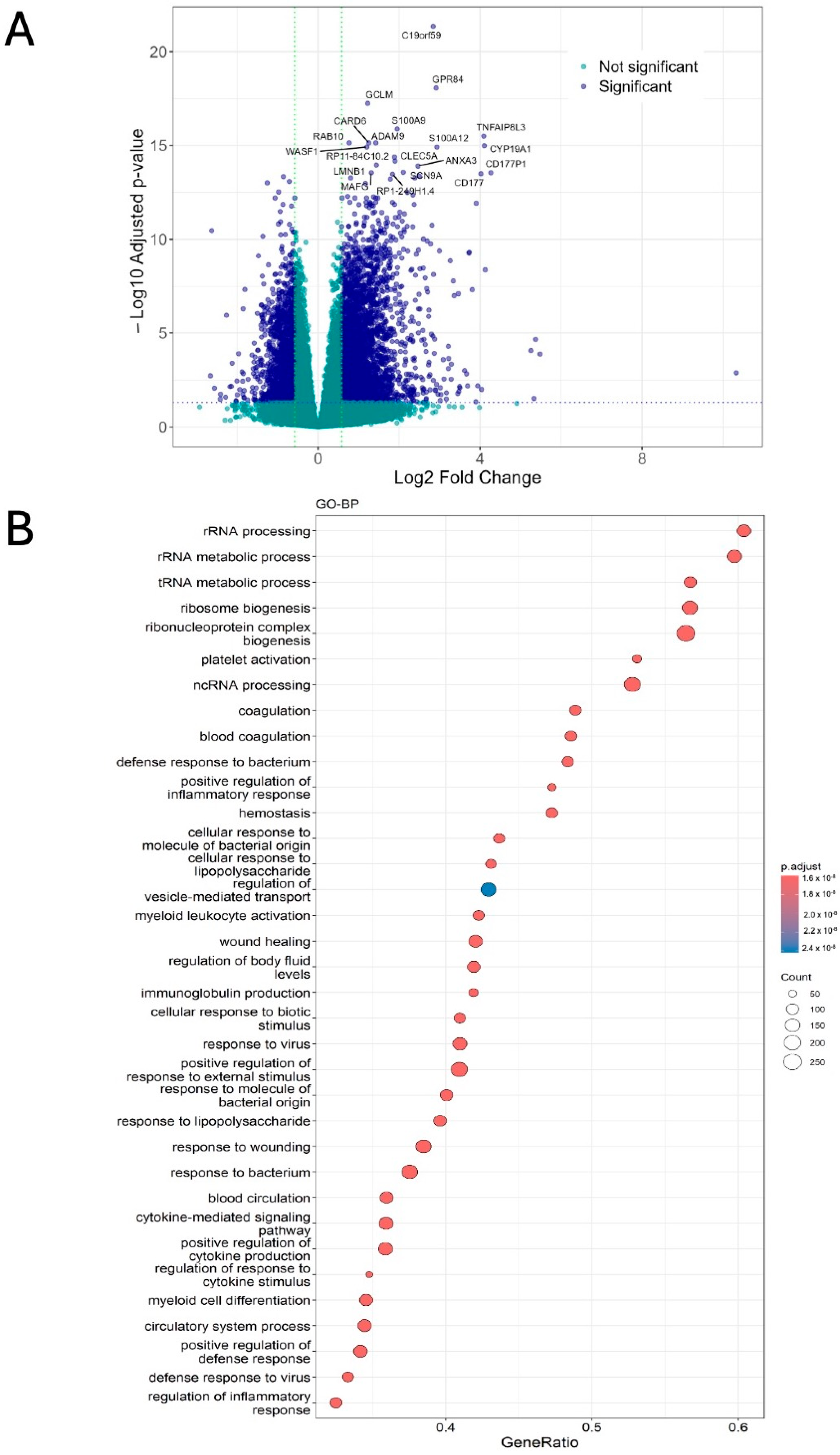
Figure 1. Differences in gene expression, including changes in the expression of ADARs and ADAR1 isoforms in patients with critical compared to non-critical COVID-19. (A) Volcano plot showing differentially expressed genes (log2Fold Change ≥ |0.58| and an adjusted p-value < 0.05), with a higher number of genes upregulated in patients with critical COVID-19. Blue dots represent genes with significant difference in expression between the two patient groups. The top 20 differentially expressed genes are labeled on the plot. GSEA of differentially expressed genes ranked by log2FC with gene sets from (B) GO–biological processes (GO-BP) and (C) Reactome pathway. (D) DESeq2 analysis results for ADAR genes: ADAR1 (ADAR) and ADAR2 (ADARb1). ADAR1 is significantly higher in critical (log2FC = 0.6647 and adjusted p-value = 0.0002) compared to non-critical patients. While expression of ADAR2 (log2FC = 0.03939 and adjusted p-value = 0.8440) is minimally different between critical and non-critical patients, this difference was not significant. DESeq2 normalized expression of ADAR1 isoforms: ADARp110 (E) and ADARp150 (F). ADARp110 expression was significantly upregulated in critical patients compared with non-critical patients (critical: 27,714 ± 3325, n = 46; non-critical: 14,458 ± 3027, n = 23; Wilcoxon rank-sum test, p = 0.0073). In contrast, ADARp150 expression was only modestly higher in critical patients (critical: 14,326 ± 1527, n = 46; non-critical: 11,713 ± 1864, n = 23), and this difference was not statistically significant (Wilcoxon rank-sum test, p = 0.17).
Ijms 27 06809 g001aIjms 27 06809 g001bIjms 27 06809 g001c
Figure 2. Differences in ADAR editing between critical and non-critical patients. (A) Bar plot shows the number of different substitutions identified by GATK. A-to-G and T-to-C substitutions, representing potential ADAR editing sites, are the most abundant type of substitutions in both groups of patients; however, the number of these substitutions are higher among patients with non-critical COVID-19. (B) The distribution of editing sites across genic regions shows the average number of edits across all regions was higher for non-critical patients. (C) The number of different exonic substitution types shows non-synonymous substitutions were the most abundant in both groups with higher counts in patients with non-critical COVID-19. (D) The comparison of AEI between patient groups shows A-to-G index was higher in patients with critical disease. The correlation between the expression of ADAR1 (E), ADAR2 (F), and ADAR3 (G) measured in TPM, and total number of ADAR edits in critical and non-critical patients shows ADAR1 (adj R2 = 0.401, p = 1.39 × 10⁻⁶) and ADAR2 (adj R2 = 0.175, p = 0.00222) expressions were significantly positively correlated with the total number of ADAR edits within critically ill patients. Venn diagram comparing the number of ADAR editing sites (H) and corresponding genes (I) that are shared and unique between the two patient groups. Overrepresentation analysis of GO–biological processes (J) and pathways enriched (K) among genes are shown with unique edits in each group. Genes incorporating unique edits were enriched for distinct biological functions.
Ijms 27 06809 g002
Figure 3. High-confidence ADAR editing sites and differential editing. (A) Flow chart of methods/tools and filters used to identify high-confidence sites. (B) Bar plot showing the number of high-confidence ADAR editing sites that are present in REDIportal, representing previously identified ADAR editing sites, and novel sites, not present in REDIportal. (C) Genic distribution of both known and novel high-confidence edits. Sequence logo for edited A’s on the positive strand for both known (D) and novel (E) editing sites. Sequence logo for edited T (=U, A on the complementary strand) for both known (F) and novel (G) editing sites. (H) Bar plot of top 30 sites with significantly increased (log2FC) > 0.58 and FDR < 0.05) and top 30 sites with significantly decreased (log2FC < −0.58 and FDR < 0.05) editing in critical compared to non-critical patients. (I) GSEA results showing Reactome pathways enriched within genes containing differentially edited sites. GSEA plots show positive enrichment of neutrophil degranulation (J) and MAPK family signaling cascade (K) both positively enriched in critical patients. (L) Relationship between read coverage and editing levels (values represented as median) at position Chr4: 2940026 within NOP14 gene. (M) Differences in the number of high- and moderate-impact editing events between critical and non-critical patients.
Ijms 27 06809 g003aIjms 27 06809 g003bIjms 27 06809 g003cIjms 27 06809 g003d
Figure 4. Random forest model identifies RNA editing sites predictive of disease severity. ROC curves showing the performance of random forest classifier in distinguishing critical from non-critical patients in training (A) and test (B) dataset, based on editing levels of differentially edited sites. (C) Variable importance plot showing the top 30 RNA editing sites ranked by Mean Decrease in Gini index. (D) Bar plot of top RNA editing sites with AUC > 0.75.
Ijms 27 06809 g004aIjms 27 06809 g004b
Table 1. Characteristics of BioProject PRJNA722046 from Carapito et al. (2022) [86] used in this study. Values for the number of ADAR editing sites are represented as mean and standard error of mean (SEM) for respective severity group.
COVID-19 Disease SeveritySample CountAverage Number of Aligned ReadsMean Number of Putative ADAR Editing Sites (+/− SEM) per Sample for Different Severities of COVID-19Mean A-to-G Alu Editing Index (AEI) (+/− SEM) per Sample for Different Severities of COVID-19
Non-critical23203,697,837158,022.21 ± 10,234.060.847 ± 0.0333
Critical46197,249,840139,285.2174 ± 9307.700.893 ± 0.0285
Table 2. The position and potential consequences of non-synonymous substitution within DHRSX gene (unique to critical patients) and HLA-DRB5 (unique to non-critical patients), on corresponding protein stability as predicted by DDMut [96]. (H = Histidine; R = Arginine; S = Serine; G = Glycine; ∆∆G = change in Gibbs free energy).
Disease SeverityGene NameExon/PositionWildtype ResidueMutant ResiduePredicted Stability Change (∆∆GStability wt>mt)
CriticalDHRSX (dehydrogenase/reductase X-linked)Exon 7; 292HR−1.27 kcal/mol
Non-criticalHLA-DRB5 (major histocompatibility complex, class II, DR beta 5)Exon 3; 164SG−0.08 kcal/mol
Table 3. List of differentially edited genes with corresponding ADAR editing sites located within 3′UTR, the microRNAs predicted to target these regions, and significance of the edited genes.
GeneEditing SitemiRNAs with Target SiteSignificance of the Edited Gene
PGAM5Chr12: 133298349miR-548c-3pRegulates mitochondrial dynamics and cellular senescence. SARS-CoV-2 ORF3c interacts with PGAM5 to disrupt innate immune response by promoting MAVS cleavage [103,104,105]
PEX26Chr22: 18571421miR-625-3p, miR-1295b-3p, miR-4759, miR-8083, miR-3117-3p, miR-3169Involved in the assembly and function of peroxisomes. Mutations in PEX26 gene has been associated with Zellweger spectrum disorder [106]
ADRBK2Chr22: 26120928miR-365a-5p/365b-5pEncodes for G protein-coupled Receptor Kinase 3K (GRK3) a crucial component of hedgehog pathway required for normal embryonic development [107]
Chr22: 26121801miR-562, miR-3199/8052
APOL6Chr22: 36056598miR-641/3617-5pMember of apolipoprotein L gene family; APOL6 regulates lipid metabolism and is associated with apoptosis and autophagy particularly in cancer [108,109]
AHRChr7: 17384514miR-30b-3p/1273h-5p/3689-3p/6779-5p/6780a-5p, miR-887-5p, miR-30c-3p/6788-5p, miR-450a-1-3p, miR-6513-5p, miR-3122/3913-5p, miR-383-3pEncodes for AHR protein, a ligan-activated transcription factor, and is a crucial regulator of xenobiotic (foreign chemicals) metabolism [110]
PLEKHA2Chr8: 38829775miR-7158-3pLocalized to post-synaptic membrane PLEKHA2, it is involved in regulating post-synaptic membrane stability, neuronal excitability and is also identified as a risk gene for bipolar disorder during adolescence [111]
Chr8: 38829839miR-4540, miR-6838-3p
ERO1LChr14: 53109732miR-7-3p, miR-5692a, miR-4775, miR-590-3pEncodes for Ero1-like proteins, primarily localized in ER, which are involved in the disulfide bond formation of secreted and cell surface proteins. Highly expressed in various malignancies, ERO1L overexpression is associated with poor outcomes [112,113]
ZNF506Chr19: 19903175miR-214-5p, miR-2114-5p, miR-6811-3p, miR-6514-3p, miR-20b-3p, miR-5588-3pInvolved in maintaining genome stability through transcriptional repression of endogenous retroviruses (ERVs) [114]
RNF24Chr20: 3910035miR-10-5p, miR-339-5p, miR-339-5p, miR-4725-5p, miR-4421/5699-3pBelonging to the family of E3 ubiquitin ligases. RNF24 expression is elevated in multiple cancers where it affects immune cell infiltration and is associated with poor prognosis [115]
HPSEChr4: 84215723miR-1229-5p, miR-1225-5p, miR-555, miR-4803Encodes for heparinase enzyme involved in tissue remodeling. HPSE plays a crucial role in metastasis, angiogenesis, and inflammation, specifically neuroinflammation during herpes simplex virus-1 (HSV-1) infections. HPSE is also a therapeutic target for neuroinflammation and associated neurobehavioral deficits [116,117]
ADAM19Chr5: 156905396miR-627-3p, miR-6738-3p, miR-544a-3pMember of type-1 transmembrane glycoprotein; it is involved in cellular interactions. It is essential during cardiovascular morphogenesis, neurogenesis, nephrogenesis, and other developmental processes [118,119]
Chr5: 156905560miR-3149
SGTBChr5: 64964718miR-499a-5p, miR-4432, miR-8087Plays a key role in protein–protein interactions and regulates several physiological processes. Upregulation of SGTB is associated with neuronal apoptosis following neuroinflammation, induced by lipopolysaccharide (LPS) administration [120,121]
Disclaimer/Publisher’s Note: The statements, opinions and data contained in all publications are solely those of the individual author(s) and contributor(s) and not of MDPI and/or the editor(s). MDPI and/or the editor(s) disclaim responsibility for any injury to people or property resulting from any ideas, methods, instructions or products referred to in the content.