Review

Decoding the Collagenome in Breast Cancer: Mechanotransduction, Microenvironment, and Translational Opportunities


by
Noelia Vigo-Díaz
1,
Rubén López-Cortés
2,
Laura Rodríguez-Silva
1,
Marcelino Maneiro
1 and
Cristina Núñez
1,*
1
Inorganic Chemistry Department, Institute for Research in Global Health and Sustainable Development (iTERRA), Faculty of Sciences, Campus Terra, University of Santiago de Compostela, 27002 Lugo, Spain
2
Research Unit, Hospital Universitario Lucus Augusti (HULA), Servizo Galego de Saúde (SERGAS), 27002 Lugo, Spain
*
Author to whom correspondence should be addressed.
Int. J. Mol. Sci. 2026, 27(15), 6794; https://doi.org/10.3390/ijms27156794
Submission received: 8 June 2026 / Revised: 23 July 2026 / Accepted: 27 July 2026 / Published: 29 July 2026

Abstract

Breast cancer (BC) progression is strongly influenced by the extracellular matrix (ECM), whose remodelling regulates tumour growth, invasion, metastasis, immune modulation, and therapeutic response. Among ECM components, collagens have emerged as both structural proteins and active mediators of mechanotransduction, stromal interactions, and tumour cell behaviour. This narrative review analyses collagen families and collagen-associated proteins implicated in BC, integrating evidence on their expression patterns, biological functions, clinical significance, and translational potential. We examine fibrillar and non-fibrillar collagens, including fibril-associated collagens with interrupted triple helices (FACITs), membrane-associated collagens with interrupted triple helices (MACITs), basement membrane (BM) collagens, and multiplexins, together with their interactions with cancer-associated fibroblasts (CAFs), immune cells, and signalling pathways involved in tumour progression. Alterations in collagen composition, organization, crosslinking, and degradation regulate ECM stiffness, epithelial–mesenchymal transition (EMT), invasion, metastatic dissemination, and therapy resistance. Several collagen types and collagen-derived fragments also show promise as prognostic biomarkers and therapeutic targets, particularly in aggressive BC subtypes such as human epidermal growth factor receptor 2 (HER2)-positive and triple-negative breast cancer (TNBC). Overall, this review highlights the collagenome as a dynamic component of the breast tumour microenvironment (TME) and supports collagen-informed strategies for improved patient stratification and targeted therapies.

Graphical Abstract

1. Introduction

Solid tumours are complex multicellular entities characterized by the coexistence of malignant cells and a heterogeneous microenvironment. Beyond cancer cells, the TME comprises immune and inflammatory cells, fibroblasts (including CAFs), endothelial and lymphatic cells, adipocytes, and ECM. The ECM is not a passive scaffold. Instead, it constitutes a dynamic and highly organized molecular network that continuously remodels its composition, architecture, and mechanical properties, thereby influencing tumour evolution. Aberrant ECM remodelling is now recognized as a hallmark of many solid cancers, and in BC, it strongly influences tumour initiation, local invasion, metastatic dissemination, therapeutic response, and relapse [1,2].
Throughout this review, the term collagenome refers to the complete repertoire of collagen molecules that constitute the breast tumour ECM, including their supramolecular organization, biologically active collagen-derived fragments generated during ECM remodelling, and their functional interactions with collagen-binding receptors and associated signalling pathways. Thus, the collagenome is considered not merely as the collection of collagen genes or proteins, but as a dynamic structural and signalling network that actively regulates tumour progression, stromal communication, immune modulation, and therapeutic response. Although proteins containing collagen-like domains are mentioned where relevant, the primary focus of this review is on human collagen families and their biological roles in breast cancer.
Collagen-rich matrices exhibit marked structural heterogeneity, and increasing evidence indicates that collagen architecture actively regulates tumour invasion through mechanotransduction. For example, a Yes-associated protein (YAP)-centred mechanotransduction circuit couples collagen organization with collective BC invasion, reinforcing the concept that collagen architecture functions as an instructive regulator of tumour progression rather than merely providing structural support [2,3]. Likewise, physiological tissue-remodelling processes, such as postpartum mammary gland involution, further support the concept that ECM remodelling contributes to tumour progression by creating a transient pro-remodelling microenvironment [4].
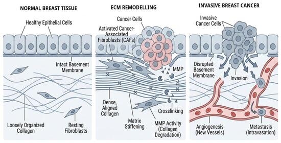
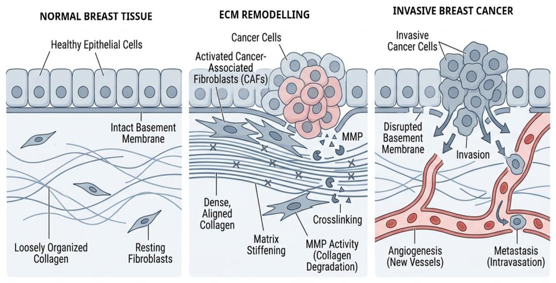
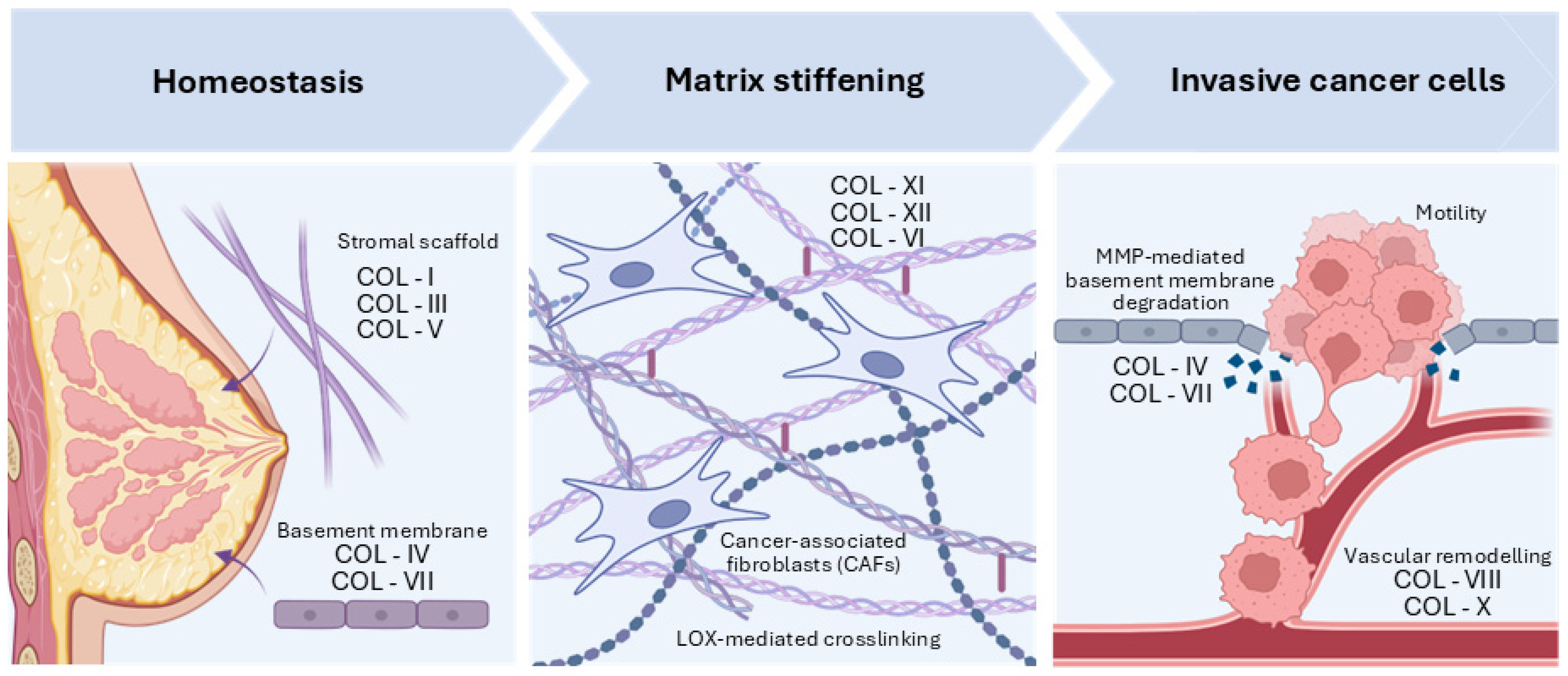
Collagen remodelling is coordinated through synthesis, enzymatic processing, crosslinking, and proteolytic degradation. Matrix metalloproteinases (MMPs) regulate collagen turnover and the release of bioactive ECM fragments, whereas lysyl oxidase (LOX) family enzymes promote collagen crosslinking and tissue stiffening, thereby contributing to mechanotransduction, tumour progression, and therapy resistance. In parallel, CAFs represent the major collagen-producing cells within BC, and play a central role in ECM remodelling, stromal organization, and tumour–stroma communication [5,6,7,8]. Together, these interconnected processes progressively transform the breast ECM from a homeostatic scaffold into a mechanically active and invasion-promoting microenvironment, as summarized in Figure 1.
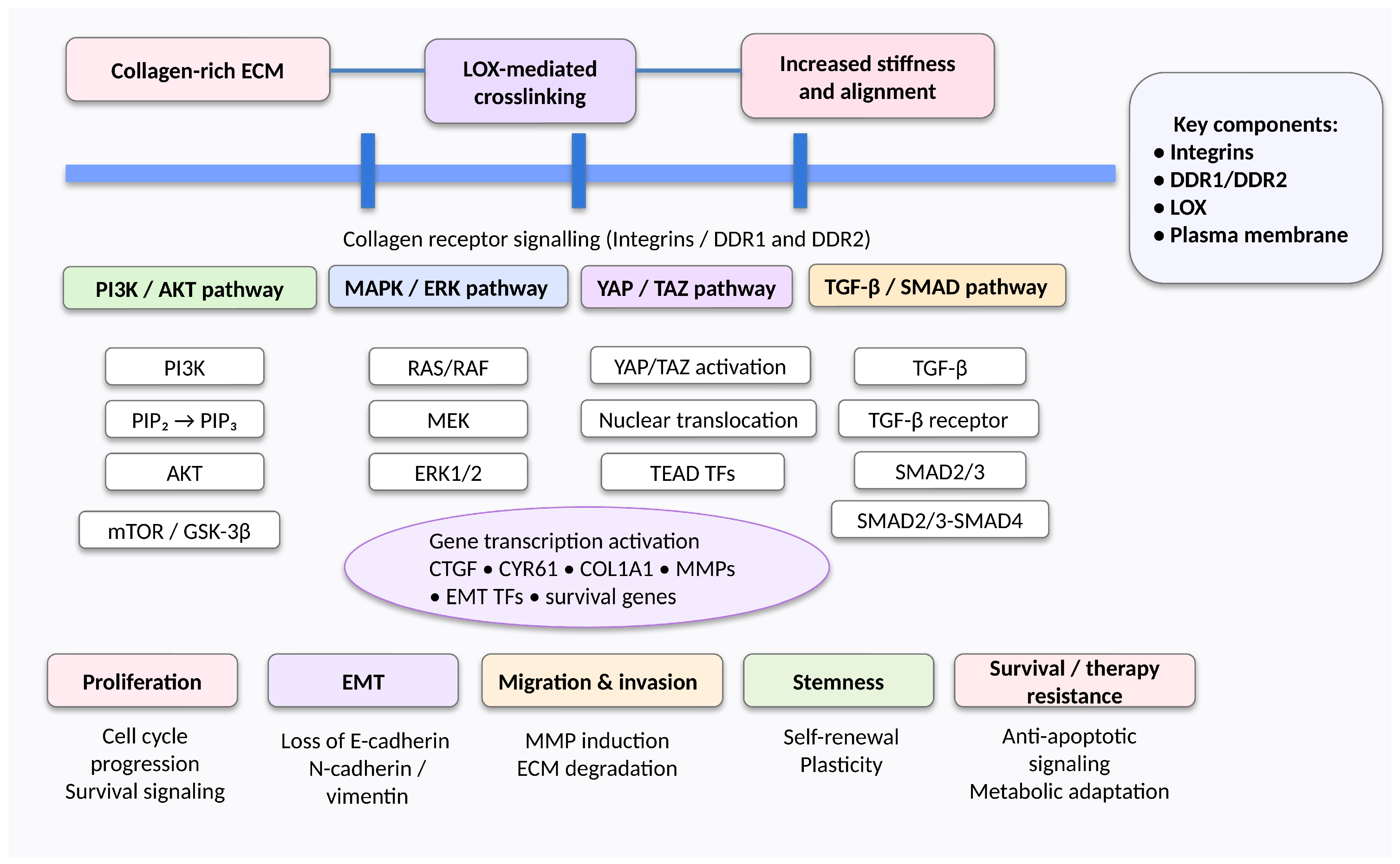
Beyond its structural role, collagen remodelling influences multiple biological processes involved in BC progression. Alterations in collagen organization regulate tissue mechanics, mechanotransduction, metabolic adaptation, stromal communication, and clinically relevant phenotypes associated with prognosis and therapeutic response. The principal signalling pathways activated by collagen remodelling are summarized in Figure 2 [2,3,7].
Collagen alterations are also increasingly recognized as clinically relevant biomarkers. Both tissue-based collagen signatures and circulating markers reflecting ECM turnover have shown potential for prognostic stratification and monitoring stromal activation, supporting the concept that collagen remodelling may be exploited through imaging, tissue analysis, and liquid biopsy approaches [8,9,10].
Altogether, these observations support the view that the collagenome constitutes a dynamic structural and signalling network integrating collagen diversity, ECM remodelling, and collagen-mediated cellular communication. Because collagen remodelling may differ across clinicopathological BC subgroups defined by hormone receptor and HER2 status, available evidence should be interpreted according to the strength and type of supporting studies. Table 1 summarizes the principal collagen-associated features reported for the main BC subtypes.
Before discussing the individual collagen families, it is important to emphasize that the evidence available for each collagen type derives from different experimental approaches. Throughout this review, we distinguish, whenever possible, whether the reported findings originate from transcriptomic analyses, proteomic studies, immunohistochemistry, ECM imaging, serum-based assays, or functional experimental models. This distinction is particularly relevant because changes in collagen gene expression do not necessarily reflect collagen protein abundance, ECM organization, or biological function.
Accordingly, this narrative review examines the spectrum of collagen types implicated in BC, integrating recent evidence regarding their cellular sources, biological functions, mechanistic contributions, and translational potential [2,5,9,10,18]. Given the remarkable diversity of collagen molecules within the breast TME, a collagen-type-resolved framework is required to understand their distinct biological functions.
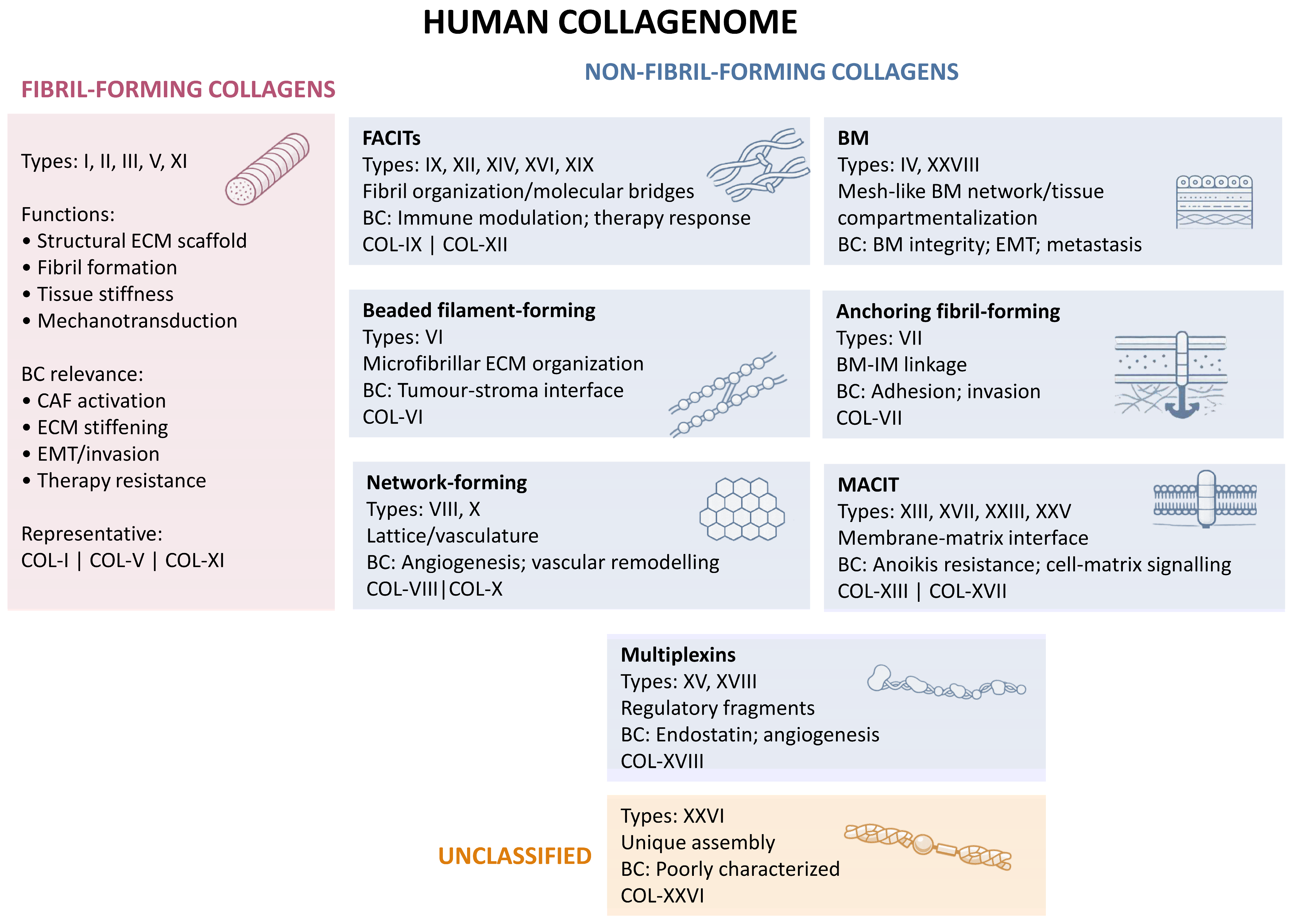
Although collagens are traditionally classified according to their supramolecular organization, increasing evidence indicates that their functions extend far beyond structural support. Different collagen subtypes contribute differentially to ECM stiffness, mechanotransduction, immune regulation, angiogenesis, metastatic dissemination, and therapeutic resistance in a context-dependent manner. Human collagens are currently classified into several supramolecular subgroups, including fibrillar collagens, FACITs, BM collagens, network-forming collagens, MACITs, and multiplexins [22,23]. Their principal characteristics and reported implication in BC are summarized in Table 2.
Figure 3 provides a schematic overview of this hierarchical classification and illustrates the principal functional implications of each collagen subgroup in BC. This framework serves as the basis for the collagen-type-resolved discussion developed throughout the following sections [22,23,30].

2. The Collagen Mechanobiology Axis in BC

2.1. Collagen Accumulation and ECM Expansion

The earliest stages of ECM remodelling in BC are characterized by progressive collagen accumulation and expansion of the stromal compartment. Instead of representing a passive consequence of tumour growth, excessive collagen deposition actively reshapes the TME, creating a mechanically dynamic niche that promotes malignant progression. This process is driven primarily by CAFs, which become persistently activated through reciprocal interactions with tumour cells, inflammatory cytokines, and transforming growth factor-β (TGF-β). Activated CAFs have been identified by transcriptomic, proteomic, and immunohistochemical studies as the principal producers of fibrillar collagens, particularly collagen I, III, and V. These studies also demonstrate increased transcript and protein expression of matrix-remodelling enzymes associated with CAF activation, establishing a self-reinforcing fibrotic programme that progressively modifies ECM composition and architecture [50].
Collagen accumulation is accompanied by profound changes in the cellular composition of the tumour stroma. In addition to CAFs, tumour-associated adipocytes, endothelial cells, infiltrating immune cells, and, in some cases, BC cells themselves contribute to matrix remodelling by producing collagen molecules or by secreting cytokines and growth factors that further stimulate stromal activation. Consequently, collagen deposition should be regarded as the integrated output of multiple cellular populations rather than the exclusive product of fibroblasts. This reciprocal communication establishes a positive feedback loop in which tumour cells stimulate stromal collagen production, while the altered ECM provides biochemical and biomechanical signals that further enhance tumour aggressiveness [109].
Histopathological analyses, together with second harmonic generation (SHG) imaging studies, have consistently shown that the transition from normal mammary tissue to invasive carcinoma is associated with progressive stromal fibrosis, increased collagen deposition, higher collagen fibre density, and expansion of desmoplastic regions. These structural changes profoundly modify the physical properties of the breast TME. They are also closely related to mammographic breast density, one of the strongest independent risk factors for BC development and progression. Accordingly, mammographic breast density should be interpreted as a macroscopic surrogate of collagen-rich stromal remodelling rather than a direct indicator of tumour aggressiveness. Importantly, collagen accumulation alone does not fully explain the biological consequences of ECM remodelling. Instead, the quantity of collagen provides the structural substrate upon which subsequent architectural reorganization, enzymatic crosslinking, and mechanical stiffening take place. Thus, collagen abundance represents the first hierarchical step in a cascade of biomechanical events that ultimately governs tumour behaviour [110].
Experimental studies have demonstrated that excessive collagen deposition increases the probability of cell–matrix interactions by enlarging the available collagen surface that can be recognized by collagen receptors, including integrins and DDRs. These interactions not only reinforce cell adhesion, but also facilitate the transmission of mechanical forces from the extracellular environment to the cytoskeleton, thereby priming tumour cells for mechanotransduction. Nevertheless, collagen abundance alone is insufficient to generate the highly invasive phenotype observed in advanced BC. The biological impact of collagen deposition critically depends on the spatial organization of collagen fibres and on the degree of enzymatic crosslinking that determines matrix stiffness. Therefore, collagen accumulation should be viewed as the initiating event of a sequential mechanobiological process rather than as an isolated determinant of tumour progression [111].

2.2. Collagen Fibre Alignment and Architectural Remodelling

SHG imaging, multiphoton microscopy, and computational image-analysis studies have demonstrated that the three-dimensional organization of collagen fibres, rather than collagen abundance alone, is a major determinant of BC progression. During tumour evolution, these imaging approaches reveal that collagen fibres undergo profound architectural reorganization, becoming progressively straighter, thicker, and more anisotropically aligned. These structural alterations transform the ECM from a compliant scaffold into a highly organized mechanical network that actively regulates tumour cell behaviour. Therefore, collagen architecture has emerged as an important prognostic feature, complementing conventional molecular biomarkers and histopathological parameters [112].
One of the best-characterized examples of matrix imaging in BC is the progressive appearance of Tumour-Associated Collagen Signatures (TACSs). These collagen architectural patterns were originally identified by SHG microscopy and subsequently validated using complementary multiphoton imaging approaches. TACS-1 is characterized by locally increased collagen deposition surrounding early tumour lesions, whereas TACS-2 consists of stretched collagen fibres arranged tangentially around the expanding tumour mass. As invasion progresses, collagen fibres become radially aligned perpendicular to the tumour boundary, giving rise to the TACS-3 phenotype, which is consistently associated with enhanced tumour invasion, lymph node metastasis, and poor clinical outcome. More recently, additional collagen architectural patterns (TACS-4 to TACS-8) have been described, reflecting the increasing spatial complexity of the invasive front, and further emphasizing that collagen organization evolves continuously during BC progression [112].
The biological significance of collagen alignment resides in its ability to provide contact-guidance cues for migrating tumour cells. Unlike randomly organized collagen networks, aligned collagen bundles generate preferential migration tracks that facilitate persistent and directional cell movement toward blood vessels and surrounding tissues. This phenomenon is driven by the physical orientation of collagen fibrils rather than by biochemical gradients alone, allowing tumour cells to migrate collectively or individually along pre-existing collagen highways. Accordingly, collagen alignment not only promotes local invasion, but also facilitates intravasation and the early stages of metastatic dissemination. This concept is supported by experimental mechanobiology models combining collagen-matrix engineering and live-cell imaging, which have consistently demonstrated that increased collagen anisotropy enhances cell polarity, directional migration, and invasive capacity, highlighting ECM architecture as an active regulator of tumour dissemination rather than a passive structural framework [50].
Collagen fibre orientation also profoundly influences the tumour immune microenvironment. Recent imaging studies together with functional experimental models have demonstrated that highly aligned collagen bundles may function as a physical barrier restricting immune-cell infiltration into tumour nests. In particular, collagen organization mediated by discoidin domain receptor 1 (DDR1) promotes the formation of densely packed collagen architectures that limit T-cell access to malignant epithelial regions. Experimental blockade of DDR1 disrupts collagen alignment, increases immune-cell penetration, and enhances antitumour immune responses, indicating that collagen architecture contributes not only to tumour invasion, but also to immune escape. These observations reinforce the concept that ECM organization represents a dynamic regulator of both tumour progression and antitumour immunity [112].
The clinical relevance of collagen architecture has increased considerably owing to advances in imaging technologies. SHG microscopy, polarized light microscopy, multiphoton microscopy, and computational image analysis now enable quantitative assessment of collagen alignment, fibre orientation, anisotropy, and microstructural organization in tumour specimens. These approaches have shown that collagen architectural parameters frequently outperform collagen quantity alone in predicting disease-free survival, metastatic risk, and therapeutic response. Accordingly, collagen alignment is increasingly recognized as a promising imaging biomarker that captures biomechanical properties of the TME not reflected by conventional histopathology [112]. Notably, these imaging approaches characterize collagen architecture and fibre organization rather than collagen gene expression or protein abundance, providing complementary information on ECM biomechanics.
Importantly, collagen alignment should not be considered an isolated phenomenon, but rather the intermediate step linking collagen deposition with matrix stiffening and mechanotransduction. As collagen fibres become progressively aligned, they provide an optimal substrate for enzymatic crosslinking by LOX family members, resulting in increased ECM rigidity and enhanced transmission of mechanical forces to tumour cells. Therefore, architectural remodelling of collagen constitutes the structural bridge between ECM expansion and activation of intracellular mechanosensitive signalling pathways that drive BC progression [50].

2.3. LOX-Mediated Collagen Crosslinking and ECM Stiffening

Although collagen deposition and fibre alignment establish the structural framework of the TME, the acquisition of its biomechanical properties is primarily determined by collagen crosslinking. This process is catalysed by the LOX family of copper-dependent amine oxidases (LOX and LOXL1–LOXL4), which generate covalent intermolecular crosslinks between collagen molecules, thereby increasing fibrillar stability, tensile strength, and resistance to proteolytic degradation. Thus, collagen crosslinking transforms the ECM into a mechanically rigid scaffold that profoundly influences tumour cell behaviour. Growing evidence indicates that this increase in matrix stiffness is not merely a consequence of tumour progression, but an active driver of BC initiation, invasion, and metastatic dissemination [50].
Transcriptomic and mechanistic experimental studies have shown that LOX expression is tightly regulated by several tumour-associated stimuli, particularly hypoxia and transforming growth factor-β (TGF-β). Under hypoxic conditions, stabilization of hypoxia-inducible factor-1α (HIF-1α) promotes LOX transcription, whereas activated CAFs further increase LOX production through paracrine signalling. These mechanisms generate a positive feedback loop in which fibrosis, collagen deposition, and matrix stiffening continuously reinforce one another during tumour progression. Biochemical and ECM imaging studies indicate that increased collagen crosslinking protects collagen fibres from MMP-mediated degradation, thereby contributing to the persistence of a dense desmoplastic microenvironment [110].
Biomechanical studies using engineered collagen matrices have demonstrated that the mechanical consequences of collagen crosslinking extend beyond increased tissue rigidity. Crosslinked collagen fibres exhibit higher resistance to deformation and transmit mechanical forces more efficiently to adherent cells. This enhanced force transmission promotes focal adhesion maturation, actomyosin contractility, and cytoskeletal tension, enabling tumour cells to continuously sense changes in ECM mechanics. Thus, collagen crosslinking represents the crucial physical event that converts structural ECM remodelling into biologically relevant mechanical signals. In BC, experimental studies have demonstrated that increased matrix stiffness enhances cell proliferation, survival, EMT, migration, and invasion while simultaneously promoting angiogenesis, stemness, and resistance to systemic therapies [110].
Mechanobiology studies have demonstrated that ECM stiffening initiates mechanotransduction through integrin-dependent signalling pathways. Increased collagen rigidity promotes integrin clustering and focal adhesion assembly, resulting in the activation of FAK, Src family kinases, and RhoA/ROCK-mediated actomyosin contractility. These signalling cascades ultimately regulate the nuclear localization of the mechanosensitive transcriptional co-activators YAP and transcriptional co-activator with PDZ-binding motif (TAZ), which coordinate gene expression programmes involved in proliferation, ECM remodelling, stem-cell maintenance, and therapeutic resistance. Importantly, YAP activation also induces the transcription of ECM-remodelling genes, including collagen-modifying enzymes, thereby establishing a feed-forward loop that further enhances matrix stiffening and tumour progression [3].
Functional organoid-based mechanobiology studies have recently shown that collagen I does not simply provide a permissive substrate for invasion, but actively participates in a reciprocal mechanical dialogue with tumour cells. Contact with collagen I induces YAP activation in basal-like BC cells, whereas activated YAP promotes transcriptional programmes that increase collagen remodelling and fibre alignment, generating localized regions of elevated mechanical tension that further amplify YAP signalling. This self-reinforcing mechanotransduction circuit facilitates leader-cell formation and collective invasion, providing direct experimental evidence that collagen remodelling and intracellular mechanosignalling constitute an integrated biological process rather than independent events [3].
The translational implications of these findings are considerable. Preclinical experimental studies suggest that pharmacological inhibition of LOX activity, interference with collagen crosslinking, or blockade of stiffness-sensitive signalling pathways may reduce tumour fibrosis, improve drug penetration, and enhance the efficacy of chemotherapy, targeted therapies, and immunotherapy. Although most of these strategies remain in preclinical or early clinical development, they support the concept that targeting tumour biomechanics represents a promising therapeutic approach complementary to conventional anticancer treatments [109].

2.4. Collagen Sensing Through Integrins and Discoidin Domain Receptors

Functional mechanobiology studies have demonstrated that the biological effects of collagen remodelling ultimately depend on the ability of cells to sense changes in ECM composition and mechanics. This function is mediated primarily by two major classes of collagen receptors: integrins and DDRs. Although both receptor families recognize collagen molecules, they bind distinct collagen motifs and activate complementary signalling pathways, enabling cells to integrate biochemical and biomechanical information from the surrounding ECM. Rather than functioning independently, increasing evidence indicates that integrins and DDRs cooperate to orchestrate cellular responses to collagen remodelling, thereby regulating tumour progression, immune evasion, and therapeutic resistance [111].
Functional cell biology and mechanobiology studies have established that integrins constitute the principal mechanoreceptors responsible for sensing matrix stiffness. In BC, collagen-binding integrins, particularly α1β1, α2β1, α10β1, and α11β1, interact with fibrillar collagens and promote the assembly of focal adhesions, which mechanically couple the ECM to the actin cytoskeleton. Increased collagen density, fibre alignment, and matrix stiffening enhance integrin clustering, thereby promoting the recruitment of FAK, Src family kinases, and integrin-linked kinase (ILK). These signalling complexes activate downstream pathways, including PI3K/AKT, MAPK/ERK, and RhoA/ROCK, leading to cytoskeletal remodelling, increased cellular contractility, and enhanced proliferation, migration, and survival. As a result, integrins function as the primary sensors that convert ECM stiffness into intracellular biochemical signals, initiating mechanotransduction programmes that promote tumour progression [113].
Unlike integrins, DDRs are receptor tyrosine kinases that are activated directly by native triple-helical collagen molecules. Transcriptomic analyses together with immunohistochemical studies indicate that DDR1 is predominantly expressed in epithelial tumour cells, whereas DDR2 is mainly associated with stromal fibroblasts and mesenchymal-like tumour cells. Following collagen binding, DDRs undergo a slow but sustained activation that differs from the rapid signalling characteristic of integrins. This prolonged signalling regulates multiple biological processes, including ECM remodelling, epithelial differentiation, cell survival, and migration. Recent evidence further suggests that DDR1 also functions as a mechanosensor capable of responding to extracellular stiffness and cytoskeletal tension, thereby extending its role beyond collagen recognition to the regulation of tissue mechanics [114].
DDR1 and DDR2 exhibit distinct but complementary functions during BC progression. DDR1 contributes to tumour growth by promoting cell survival, maintaining epithelial plasticity, and regulating collagen organization. Remarkably, matrix imaging studies combined with functional experimental models have recently demonstrated that DDR1-mediated alignment of collagen fibres generates a dense peritumoural collagen barrier that limits T-cell infiltration, thereby facilitating immune exclusion and reducing antitumour immunity. These imaging and functional studies also provide a mechanistic explanation for the current evidence that collagen architecture and DDR1-mediated matrix organization may influence responsiveness to immune checkpoint blockade by restricting cytotoxic T-cell access to tumour cells. Conversely, DDR2 plays a central role in tumour invasion by stabilizing the EMT transcription factor SNAIL1 through Src-dependent ERK2 signalling, promoting EMT, and facilitating migration through collagen-rich stroma. These observations highlight that collagen receptors not only regulate tumour cell behaviour, but also shape the immune and stromal compartments of the TME [50].
Current evidence from functional experimental models indicates that integrins and DDRs should not be viewed as independent signalling systems, but rather as components of an integrated collagen-sensing network. Integrin activation rapidly transduces mechanical forces generated by ECM stiffening, whereas DDR signalling provides sustained biochemical responses to collagen engagement. Crosstalk between these receptors amplifies mechanotransduction, coordinates cytoskeletal remodelling, and regulates transcriptional programmes associated with proliferation, invasion, stemness, and therapeutic resistance. This coordinated signalling enables tumour cells to continuously adapt to dynamic changes in collagen abundance, fibre organization, and matrix stiffness throughout tumour progression [111].
Activation of collagen receptors ultimately converges on several evolutionarily conserved signalling pathways, including FAK/Src, PI3K/AKT, MAPK/ERK, and the Hippo effectors YAP/TAZ. These downstream pathways integrate extracellular mechanical information with transcriptional responses controlling cell proliferation, metabolic adaptation, ECM remodelling, and metastatic dissemination. More importantly, recent organoid-based mechanobiology studies and in vivo experimental models have demonstrated that YAP activation further stimulates collagen remodelling and LOX expression, thereby reinforcing matrix stiffening and generating a self-sustaining feed-forward circuit that promotes collective invasion. For this reason, collagen receptors represent the critical molecular interface linking ECM remodelling with intracellular mechanotransduction, and constitute attractive therapeutic targets for disrupting tumour–stroma interactions [3].

2.5. Downstream Mechanotransduction Pathways: Integrating Biochemical and Biomechanical Signalling

Functional mechanobiology studies have demonstrated that mechanical information generated by collagen remodelling is ultimately translated into coordinated intracellular signalling networks that regulate virtually every aspect of tumour biology. Rather than functioning as isolated cascades, mechanotransduction pathways constitute a highly interconnected signalling system in which integrins, DDRs, growth factor receptors, and cytoskeletal mechanosensors cooperate to integrate biochemical and biomechanical cues derived from the ECM. This signalling network enables tumour cells to continuously adapt to changes in collagen abundance, fibre architecture, and matrix stiffness, thereby promoting phenotypic plasticity during BC progression [115].
Functional cell biology studies have established that one of the earliest intracellular events following collagen receptor activation is the assembly of focal adhesions. Engagement of collagen-binding integrins induces rapid recruitment and autophosphorylation of FAK, which serves as the principal signalling hub connecting extracellular mechanical stimuli with intracellular kinase cascades. Biochemical and functional experimental studies have shown that activated FAK associates with Src family kinases to regulate focal adhesion turnover, cytoskeletal remodelling, and actomyosin contractility, while simultaneously initiating downstream PI3K/AKT and MAPK/ERK signalling. Together, these pathways promote cell-cycle progression, survival, metabolic adaptation, and migration, thereby enabling tumour cells to respond efficiently to mechanical changes within the surrounding stroma. Persistent activation of FAK/Src signalling has been consistently associated with increased invasiveness, metastatic dissemination, and poor clinical outcome in BC, underscoring its central role in collagen-dependent mechanotransduction [113].
Mechanical signalling further converges on the Rho family of small GTPases, particularly RhoA, which regulates actomyosin contractility through activation of Rho-associated coiled-coil kinase (ROCK). Increased collagen stiffness enhances RhoA/ROCK activity, leading to stress-fibre formation, elevated intracellular tension, and maturation of focal adhesions. These cytoskeletal rearrangements reinforce integrin signalling and establish a positive mechanical feedback loop in which increasing matrix stiffness promotes greater cellular contractility, which in turn further remodels the through enhanced traction forces exerted by both tumour cells and CAFs. Therefore, ECM remodelling and intracellular force generation become tightly coupled processes that continuously reinforce one another throughout tumour progression [110].
Among the downstream effectors of mechanotransduction, the Hippo pathway transcriptional co-activators YAP and TAZ have emerged as master regulators of mechanical signalling. Under compliant matrix conditions, YAP and TAZ remain predominantly cytoplasmic and transcriptionally inactive. In contrast, increasing ECM stiffness, collagen crosslinking, and actomyosin tension promote their nuclear translocation, where they interact with TEAD transcription factors to regulate genes involved in proliferation, ECM remodelling, epithelial plasticity, stem-cell maintenance, and therapeutic resistance. Unlike transient kinase activation, YAP/TAZ signalling provides sustained transcriptional adaptation to persistent mechanical stimulation, allowing tumour cells to acquire long-term phenotypic changes in response to alterations in the ECM [3].
Recent experimental studies have demonstrated that YAP signalling is not only activated by collagen remodelling, but also actively contributes to its maintenance. Nuclear YAP promotes the expression of ECM proteins, LOX family members, and multiple CAF-derived profibrotic mediators, thereby reinforcing collagen deposition, fibre alignment, and matrix stiffening. This feed-forward circuit establishes a self-perpetuating biomechanical programme in which ECM remodelling continuously enhances intracellular mechanotransduction, while activated mechanotransduction pathways further remodel the extracellular environment. Such reciprocal regulation has emerged as one of the defining hallmarks of tumour mechanobiology, and provides a mechanistic explanation for the progressive increase in tissue stiffness observed during BC evolution [3].
Importantly, mechanotransduction does not operate independently of canonical oncogenic signalling pathways. Mechanical activation of integrins and DDRs cooperates with receptor tyrosine kinases such as epidermal growth factor receptor (EGFR), HER2, and TGF-β receptors, amplifying proliferative and survival signals through shared downstream effectors. As a results, collagen remodelling not only influences tumour mechanics, but also modifies the intensity and duration of growth factor signalling, thereby contributing to tumour heterogeneity and variable therapeutic responses. This extensive crosstalk explains why alterations in ECM composition frequently affect sensitivity to chemotherapy, endocrine therapy, HER2-targeted agents, and immune checkpoint inhibitors, even in the absence of additional genetic alterations [115].
Overall, these observations support the concept that collagen-dependent mechanotransduction represents an integrated signalling network rather than a collection of independent pathways. Mechanical cues generated by collagen accumulation, fibre alignment, and matrix stiffening are sensed through collagen receptors and transmitted via interconnected FAK/Src, PI3K/AKT, MAPK/ERK, RhoA/ROCK, and YAP/TAZ signalling pathways, ultimately regulating tumour cell plasticity, stromal interactions, and disease progression. However, these mechanobiological responses are not mediated uniformly by all collagens. Instead, they depend on the specific structural, biochemical, and signalling properties of individual collagen molecules. Accordingly, the following sections examine the principal collagen families and discuss their distinct contributions to ECM remodelling and BC biology.

3. Fibrillar Collagens

Fibrillar collagens are the principal structural determinants of ECM architecture and mechanical properties in BC. Alterations in their abundance, organization, and crosslinking directly influence tissue stiffness, tumour–stroma interactions, and invasive behaviour.
In healthy breast tissue, fibrillar collagens are tightly organized within interstitial matrix (IM) and BM compartments. However, tumour progression is accompanied by profound alterations in collagen architecture and spatial organization.
The collagen composition of the ECM is highly tissue-specific and spatially heterogeneous. In healthy breast tissue, the expression of collagens I, III, and V depends on fibroblasts located in IM, whereas collagens IV and VII are mainly produced by epithelial cells associated with the BM. Multiple stromal and tumour-associated cell populations can contribute to collagen production. Thus, collagen production is not restricted to resident breast cells, as multiple tumour-associated and stromal cell populations also contribute to ECM remodelling [30].
Of note, breast density is a clinically relevant parameter associated with BC risk and progression. In addition, the ECM actively regulates stromal-cell and immune-cell behaviour [116].

3.1. Collagen I

In healthy breast tissue, collagen I fibrils display a highly organized parallel arrangement. During BC progression, CAFs promote excessive collagen I deposition and collagen crosslinking, leading to progressive ECM disorganization and increased tissue stiffness. These alterations contribute to tumour progression and immune evasion, and collagen I remodelling is currently being explored as an imaging- and histology-based biomarker for BC stratification and prognostic assessment [24,117].
As a major determinant of ECM stiffness and mechanotransduction, collagen I actively shapes tumour cell adhesion, protease secretion, and invasive behaviour. Increased collagen I deposition enhances MMP expression, promotes cell–matrix interactions, and facilitates metastatic dissemination [25]. However, another study reported that collagen I induced apoptosis in luminal BC cells, but not in the basal-like subtype, likely due to differential MMP-14 expression [26]. These apparently conflicting observations likely reflect the context-dependent and subtype-specific functions of collagen I during BC progression.
Functional experimental studies suggest that COL1A1 may act as an oncogenic driver in BC, as experimental downregulation of COL1A1 inhibits tumour-associated fibroblast activation and ECM remodelling [27]. Together, these findings reinforce the concept that ECM remodelling actively shapes tumour cell behaviour and adaptive tumour progression. Recent studies have shown that low COL1A1 mRNA and protein levels correlate with unfavourable tumour characteristics in BC and may predict chemotherapy sensitivity [12]. These findings suggest that collagen I does not act as a universally pro-tumoural cue, but rather exerts subtype-dependent effects shaped by receptor expression, protease activity, and stromal context.

3.2. Collagen II

The functional interplay between the ECM and tumour cell behaviour is further illustrated by the example of another fibrillar collagen, collagen II. Transcriptomic analyses have shown that increased COL2A1 mRNA expression is associated with reduced response to neoadjuvant anti-HER2 therapy in HER2-positive BC, even when combined with phosphoinositide 3-kinase (PI3K) inhibitors. Mechanistically, this resistance appears to involve activation of the integrin-β1/proto-oncogene tyrosine-protein kinase Src (Src) signalling pathway, which promotes tumour cell motility and proliferation [14]. Notably, resistance was reversed following treatment with a collagen synthesis inhibitor, further supporting the direct contribution of ECM remodelling to therapeutic resistance.

3.3. Collagen III

Transcriptomic analyses indicate that COL1A1 and COL3A1 mRNA expression levels are low in normal tissue and benign lesions, but increase during tumour progression, likely reflecting enhanced ECM turnover [28]. However, accumulating evidence indicates that collagen III exerts context-dependent functions. Dormant tumours from lymph node-negative head and neck carcinoma patients displayed collagen III-enriched matrices [32], consistent with BC murine studies showing increased tumour formation in COL3A1-deficient mice [33]. Mechanistically, collagen III was shown to suppress key metastatic processes in experimental models.
Nevertheless, other studies suggest that collagen III may exert pro-migratory effects under specific microenvironmental conditions. For example, another study found that the presence of collagen α1(III) chain in conditioned medium from bone mesenchymal stem-cells promoted BC-cell migration in Transwell assays [29]. Notably, this response was maximal at two hours and then declined rapidly.
In contrast, the presence of the collagen α1(I) chain induced a sustained migratory response. Other ECM components, such as fibronectin or laminin 421, induced migratory responses that progressively increased over time. A recent study showed that collagen III-deficient fibroblasts produce tumour-permissive collagen matrices that drive cell proliferation and suppress apoptosis in BC cell lines [34].
Therefore, collagen III appears to display context-dependent functions, acting as a tumour-restrictive matrix component in some settings, while supporting migration under specific stromal conditions.

3.4. Collagen V

Collagen V is considered a minor component of the fibrillar IM. Nevertheless, its biological role remains poorly understood. Collagen V co-polymerizes with collagen I to regulate fibril assembly and fibril diameter [35]. Therefore, collagen V is likely to play an important role in tumour progression. In fact, a Kyoto Encyclopedia of Genes and Genomes (KEGG) analysis identified that collagen V participates in focal adhesion, regulation of the actin cytoskeleton, and cell-to-ECM interactions [36]. Nevertheless, the prognostic relevance of collagen V in BC remains unclear, as both tumour-promoting and tumour-suppressive functions have been reported, and correlations with clinical outcome remain inconsistent [36,37,38,39].
Collagen V monomers can interact with ECM ligands such as transforming growth factor beta (TGF-β1) and with cell-surface molecules including the proteoglycan (PG) glypican-1, thereby contributing to downstream oncogenic signalling pathways associated with tumour cell adhesion and proliferation. Recent evidence identified COL5A1 as a driver of tumour growth, metastasis, and doxorubicin resistance in TNBC models [19]. Mechanistically, interleukin 6 (IL-6) secretion by COL5A1-overexpressing tumour cells promoted M2 macrophage polarization. In turn, macrophage-derived TGF-β enhanced chemoresistance through activation of the TGF-β/Smad3/COL5A1 axis, thereby establishing a tumour–stroma feedback loop.
These findings suggest that collagen V may function as more than a structural regulator of fibrillogenesis. By linking ECM organization with inflammatory signalling, macrophage polarization and TGF-β-dependent pathways, collagen V emerges as a potential mediator of tumour–stroma crosstalk, particularly in aggressive BC subtypes such as TNBC.

3.5. Collagen XI

Compared with other minor fibrillar collagens, collagen XI has emerged as an important component of the tumour ECM in BC. Transcriptomic analyses together with immunohistochemical studies indicate that COL11A1 is predominantly expressed by CAFs, although immunohistochemical evidence also supports expression by subsets of BC cells, suggesting a role in ECM remodelling and tumour–stroma interactions [40,41].
Transcriptomic analyses reported that tumours with prominent lymphocytic infiltration exhibited more than a 100-fold increase in COL11A1 mRNA expression [42]. Transcriptomic and immunohistochemical studies indicate that both CAFs and BC cells are able to produce collagen XI, which may explain the marked variability observed in COL11A1 expression across different stages of disease progression [41,43]. Clinical transcriptomic cohort studies have associated elevated COL11A1 expression with poor prognosis and altered patterns of tumour immune infiltration, although the underlying biological mechanisms remain under investigation [44].
A recent transcriptomic analysis demonstrated a significant correlation between BC progression and changes in COL11A1 mRNA expression [45]. Higher COL11A1 mRNA levels in primary tumours correlate with poor prognosis, whereas lower transcript levels in lymph-node metastases suggest stage-dependent regulation during metastatic dissemination.
In summary, transcriptomic, immunohistochemical, and functional studies suggest that, rather than acting as a passive stromal marker, collagen XI reflects dynamic interactions between CAFs, tumour cells, and immune populations. Its variable expression across disease stages suggests that collagen XI may participate in ECM remodelling programmes that evolve during tumour progression and metastatic dissemination.

3.6. Collagens XXIV and XXVII

Compared with other fibrillar collagens, collagens XXIV and XXVII remain poorly characterized in BC. These collagens appear to belong to a distinct evolutionary branch and display structural features associated with ancestral collagen forms [47,48]. In murine models, they form thin fibrillar structures and are predominantly expressed during early embryonic development [49].
Regarding their potential role in cancer, mRNA expression analyses have shown increased COL24A1 expression in head and neck cancer patients [46]. As for collagen XXVII, its serum concentration was markedly increased in dogs with hemangiosarcoma suffering large metastatic burdens [118]. Although evidence remains limited, further studies are needed to determine whether collagen XXIV and collagen XXVII contribute to early breast tumourigenesis, metastatic dissemination, or adaptation to the metastatic niche.
Overall, fibrillar collagens emerge as the principal determinants of ECM architecture and biomechanical signalling in BC. Although individual collagen types display distinct functions, a common theme is their ability to regulate tissue stiffness, stromal communication, and invasive behaviour through context-dependent interactions with tumour and stromal cells.

4. Non-Fibrillar Collagens

Beyond their structural contribution to ECM organization, non-fibrillar collagens actively regulate cell–matrix communication, mechanotransduction, angiogenesis, and immune interactions within the breast TME. Rather than functioning solely as architectural components, these collagens regulate fibril organization, signalling pathways, immune landscapes, and therapy-related phenotypes in a highly dynamic manner.
Among non-fibrillar collagens, FACITs constitute a highly diverse subgroup involved in ECM organization, stability, and cell–matrix communication (Table 2). Instead of forming independent fibrils, FACIT collagens associate with pre-existing collagen networks and regulate tissue architecture, biomechanical signalling, and stromal communication.
Accumulating evidence indicates that several FACIT collagens actively contribute to BC progression. Their reported functions include tumour cell invasion, immune modulation, metastatic dissemination, and therapeutic response. The major FACIT collagens currently implicated in BC include collagens IX, XII, XIV, XVI, XIX, XX, XXI, and XXII.

4.1. FACITs

Current evidence supports the involvement of FACIT collagens in metastatic dissemination, immune modulation, and therapy response in BC. Their biological functions extend beyond ECM organization and include the regulation of tumour–stroma communication, mechanotransduction, and subtype-specific signalling programmes.

4.1.1. Collagen IX

Collagen IX contributes to collagen scaffold organization and maintenance of ECM architecture. In BC, altered collagen IX expression has been associated with trastuzumab resistance in HER2-positive tumours, metabolic rewiring in TNBC/basal-like subtypes, and SRY-box transcription factor 10 (SOX10)-associated transcriptional programmes in luminal tumours [119]. These findings suggest that collagen IX may participate in subtype-specific ECM signalling and therapy-resistance programmes.

4.1.2. Collagen XII

Collagen XII is a FACIT collagen characterized by a short collagenous region and a large non-collagenous domain containing multiple interaction modules. Its splice variants display distinct glycosaminoglycan modifications and ligand-binding properties [51]. Beyond its structural functions, collagen XII regulates ECM organization, cell adhesion, and tumour progression by promoting a pro-invasive stromal microenvironment in BC [5]. Recent transcriptomic studies have shown that miRNA-mediated regulation of COL12A1 mRNA expression influences patient prognosis, while chemotherapy agents, including doxorubicin, cisplatin, and tamoxifen, can also modulate COL12A1 transcript level [11,53,120].
Emerging evidence also links collagen XII to tumour-associated immunosuppression. Transcriptomic analyses demonstrated that activation of the kynurenine pathway increases COL12A1 mRNA expression while promoting regulatory T-cell differentiation and immunosuppressive signalling [121].
Clinical transcriptomic cohort studies have shown that elevated COL12A1 mRNA expression correlates with M2 macrophage infiltration and TGF-β-associated markers. Moreover, elevated COL12A1 levels predict poor response to anti-PD-1/PD-L1 immunotherapy [52]. These findings support the potential value of collagen XII as a biomarker of immune remodelling and therapeutic response in BC.
Altogether, these observations position collagen XII as a stromal organizer and immune-modulatory collagen with particular relevance for metastatic progression and immunotherapy resistance.

4.1.3. Collagen XIV

Transcriptomic studies indicate that COL14A1 displays stage-dependent mRNA expression during BC progression. Although its transcript levels have been reported to decrease from grade I to grade III tumours [54], other transcriptomic studies have reported markedly increased COL14A1 mRNA expression in BC patients with high lymphatic infiltration, metastasis and, particularly, brain metastasis [55,122]. In these patients, altered expression of collagens III and VI was also observed.
Beyond altered expression, genomic alterations involving COL14A1 may also contribute to BC biology. The identification of the COL14A1-SKAP1 fusion gene suggests a potential connection between collagen remodelling and immune-cell regulation [123]. This association is particularly relevant considering the role of SKAP1 in T-cell activation and differentiation. Nevertheless, the prognostic significance of these alterations remains unclear. Interestingly, EPS15 overexpression has also been associated with favourable prognosis in BC. Moreover, EPS15 alterations frequently co-occur with SKAP1 abnormalities [124]. However, the mechanistic relationship between EPS15, SKAP1, and COL14A1-dependent pathways remains unresolved.

4.1.4. Collagen XVI

The functions of collagen XVI in BC remain poorly understood. Similar to other FACIT collagens, collagen XVI contributes to fibrillar ECM organization and mediates cell–ECM interactions through integrin-β1 binding [56,57]. Therefore, altered collagen XVI-dependent adhesion mechanisms may influence tumour invasiveness.
In glioblastoma models, collagen XVI overexpression enhanced cell adhesion without significantly increasing migration [57], whereas in oral cancer, it promoted proliferation through Kindlin-1/integrin-β1 signalling [58].
More recently, collagen XVI was included in an eight-protein signature associated with progression risk in ovarian cancer subtype C1, a molecular subtype defined by its metabolic profile [125]. These effects may partly result from concomitant MMP-9 overexpression, which facilitates ECM degradation and tumour cell migration [59]. In BC, increased mammographic density correlates with elevated collagen XVI expression [68]. However, its precise contribution to ECM organization, tumour stiffness, and disease progression remains insufficiently explored [126].

4.1.5. Collagens XX, XXI, and XXII

The biological functions of collagens XX, XXI, and XXII remain poorly understood. collagen XX is predicted to interact with other collagen fibrils, collagen XXI participates in blood vessel assembly, and collagen XXII contributes to myotendinous junction organization through interactions with integrins α2β1 and α11β1 [67,69,127]. Emerging evidence suggests that these collagens may contribute to several cancer types.
In brain tumour-initiating cells, COL20A1 was upregulated [128]. COL21A1 mRNA levels were altered in response to chemotherapy or radiotherapy in ovarian cancer, multiple myeloma, and colorectal cancer [129,130,131]. Circulating collagen XX levels were elevated in sera from patients with several solid cancers and were associated with poor prognosis in pancreatic ductal carcinoma [68].
Interestingly, increased COL22A1 mRNA levels correlated with lymph node metastases in head and neck cancer patients [47]. In parallel, mutations in COL22A1 were more frequently detected in pre-treatment samples from TNBC/basal-like BC patients who achieved pathological complete response (pCR) after chemotherapy [21,46]. Therefore, although these collagens display relatively restricted expression patterns, they may still contribute to tumour progression and treatment response in specific cancer contexts.

4.1.6. Collagen XIX

Collagen XIX is a BM-associated FACIT collagen that is highly expressed during embryonic development and subsequently becomes restricted to selected tissues, particularly the nervous system, where it contributes to synaptic maintenance [60]. Consistent with this tissue distribution, altered collagen XIX expression has been reported in neurological diseases. Collagen XIX dysregulation has also been described in several cancer types, including BC, where it is produced by myoepithelial cells [61]. In contrast to several other collagens discussed above, COL19A1 mRNA expression decreases from the early stages of BC progression. This reduction has been attributed to its susceptibility to proteolytic degradation during BM remodelling [61].
Proteolytic cleavage between the triple-helical collagenous domain 1 (COL1) and the short C-terminal non-collagenous domain 1 (NC1) releases bioactive fragments known as matrikines [62]. Unlike many collagen-derived fragments, these matrikines are generated by plasmin rather than metalloproteinases, and display strong anti-angiogenic activity through vascular endothelial growth factor (VEGF) inhibition [63]. Similarly, NC1-derived fragments reduce phosphorylation and activation of the FAK/PI3K/AKT/mTOR pathway, thereby decreasing tumour cell proliferation and migration [64]. Current evidence suggests that collagen XIX-derived fragments may exert tumour-restrictive effects, whereas reduced COL19A1 transcript expression may disrupt this protective balance during BC progression.
Further studies are required to clarify the mechanisms underlying collagen XIX degradation in BC and the clinical relevance of plasmin-mediated matrikine release. Preliminary evidence from a multicancer study primarily conducted in patients with non-small cell lung cancer suggests that circulating collagen XIX-derived fragments may have biomarker potential. However, because BC was not specifically investigated, these findings cannot be directly extrapolated to BC, and require dedicated validation in BC cohorts before any clinical relevance can be inferred [65]. Interpreting the available evidence on collagen XIX remains challenging. Its biological effects appear to depend on ECM organization, proteolytic processing, and tumour-stage-specific expression patterns. In BC, reduced collagen XIX expression has been reported during early tumour progression and before BM invasion [61,65,132].
FACIT collagens display remarkable functional diversity in BC, contributing to ECM remodelling, metastatic dissemination, and therapy-associated stromal reprogramming. Their context-dependent functions reinforce the need to interpret FACIT alterations within specific tumour subtypes, stromal niches, and disease stages.
A recurring theme across FACIT collagens is their ability to couple ECM organization with regulatory functions that extend beyond structural support. By modulating fibrillar collagen networks and influencing cell–matrix communication, FACIT members contribute to biomechanical signalling, immune-cell behaviour, and therapy-associated stromal reprogramming. These shared properties position FACIT collagens as emerging indicators of microenvironmental dynamics and potential biomarkers of disease progression and treatment response in BC.

4.2. Multiplexins

Because collagen XIX also shares functional similarities with multiplexins through the generation of bioactive anti-angiogenic fragments, it provides a conceptual bridge toward other multiplexin collagens such as collagen XV and collagen XVIII. The growing interest in collagen XIX in BC also supports consideration of collagens XV and XVIII because of their structural and functional similarities. Both collagens have been implicated in ECM homeostasis, angiogenesis, and tumour progression, although through distinct biological mechanisms [77].
Proteolytic processing of collagens XV and XVIII can release their respective C-terminal non-collagenous fragments, namely, the NC10 domain of collagen XV (historically referred to as restin) and endostatin derived from collagen XVIII. Throughout this review, the term restin refers exclusively to the collagen XV-derived NC10 fragment and should not be confused with the unrelated intracellular MAGE-family protein MAGEH1, which has also been designated “restin” in the earlier literature. Collagen XV, for which four isoforms have been described, has been described as a potential anti-angiogenic factor, whereas endostatin also inhibits cell proliferation by binding to heparan sulfate PGs involved in growth factor signalling. The anti-angiogenic activity of the collagen XV-derived NC10 fragment (restin) was initially proposed based primarily on its sequence homology with endostatin rather than on direct functional evidence. Consequently, the biological functions of this extracellular fragment remain incompletely characterized, and should not be extrapolated from studies on the unrelated intracellular protein MAGEH1, historically also termed “restin”. Therefore, the present review discusses only evidence directly related to collagen XV and its NC10-derived fragment [133].

4.2.1. Collagen XV

One study compared full-length collagen XV with several truncated variants lacking either the C-terminal NC10 domain (collagen XV-derived restin) or specific collagenous domains [97]. The results indicated that the effects on tumour growth and cell adhesion were mainly associated with the collagenous region rather than with the NC10/restin domain itself.
In a mammary carcinoma model, COL15A1 deletion altered the tumour ECM and was associated with increased tumour growth, despite reduced tumour vascularization [98]. These findings indicate that collagen XV may exert context-dependent effects on tumour progression, and that changes in angiogenesis do not necessarily translate into reduced tumour growth. Recombinant human collagen XV has also been shown to regulate cell adhesion and migration, including interactions with collagen I, whereas no comparable adhesion was observed with collagen IV [99].
Overall, the biological functions of collagen XV and its proteolytically released NC10 fragment (restin) remain incompletely characterized. Importantly, this extracellular collagen XV-derived fragment is distinct from the intracellular MAGE-family protein MAGEH1, which has historically also been referred to as “restin”. Although sequence homology with endostatin initially suggested possible anti-angiogenic activity, direct evidence supporting a clinically relevant anti-tumour function of collagen XV-derived restin in BC remains limited. Further studies are required to distinguish the functions of full-length collagen XV from those of its proteolytically released NC10 fragment and to determine their respective contributions to basement-membrane organization, angiogenesis, and tumour progression.

4.2.2. Collagen XVIII

The functional relationship between collagen XVIII and endostatin appears more complex. Collagen XVIII is part of the epithelial and vascular BM, although different isoforms exist depending on the tissue, including a long isoform highly expressed in the liver [100]. This may partly explain why much of the available evidence derives from liver studies. Toxicological studies identified a link between collagen XVIII and integrin-β1/AKT signalling, as deficiency in some of these components increases hepatocyte susceptibility to toxicants [101]. Consequently, collagen XVIII has been proposed to participate in provisional ECM assembly during tissue injury and wound healing.
Recent transcriptomic studies indicate increased COL18A1 mRNA expression in HER2-positive and TNBC/basal-like BCs [15]. Together, these observations suggest that collagen XVIII contributes to ECM remodelling and proliferative signalling during BC progression.
By contrast, the biological effects of endostatin are considerably better characterized. Endostatin binds strongly to mammary duct BM cells and exerts potent anti-angiogenic activity, which has been exploited in several therapeutic strategies with promising results, particularly in advanced BC and TNBC/basal-like tumours [102,103,104,105,106,107]. These studies support an important role for endostatin in restricting neovascularization, a process particularly relevant in tumours exposed to radiotherapy and radiation necrosis. In addition, vasculogenic mimicry in TNBC/basal-like BC, characterized by the formation of functional vascular-like channels lined by tumour cells, should also be considered when evaluating endostatin-based therapeutic approaches.
An intriguing feature of collagen XVIII is its dual biological behaviour. Whereas full-length collagen XVIII promotes oncogenic signalling through HER2-, EGFR-, and integrin-dependent pathways, its cleavage product endostatin exerts anti-angiogenic and tumour-restrictive effects. This functional dichotomy illustrates how collagen processing can generate biologically distinct molecules with opposing effects on BC progression.

4.3. BM Collagens

BM collagens are essential regulators of epithelial integrity, tissue compartmentalization, and tumour–stroma interactions in BC. Beyond their structural functions, BM collagens actively participate in mechanotransduction, EMT, angiogenesis, and metastatic dissemination through dynamic remodelling of the BM architecture. Accordingly, alterations in BM collagen expression and proteolytic processing represent critical events during tumour invasion and metastatic progression.

4.3.1. Collagen IV

Collagen IV is the principal structural component of the BM, particularly within the lamina densa. By forming a mesh-like network that separates the epithelial compartment from the breast stroma, collagen IV maintains tissue compartmentalization. Its degradation is therefore closely associated with tumour invasion and disease progression. This association is supported by previous studies showing that elevated serum levels of C4M (MMP-generated collagen IV fragment), as well as C1M and C3M (MMP-generated collagen I and collagen III fragments, respectively), were correlated with worse prognosis in BC patients [134]. Similar studies reached equivalent conclusions, indicating that circulating collagen IV fragments reflect increased ECM turnover and enhanced metastatic dissemination [70,71].
Mechanistically, collagen IV contributes to tumour progression through signalling pathways involved in EMT, cytoskeletal remodelling, and invasive behaviour [71,99]. Functional experimental studies demonstrated that tumour-associated adipocytes enhance collagen IV production and promote BM remodelling within the TME [81]. Collagen IV overexpression induces E-cadherin downregulation together with upregulation of SNAIL1/2, Stat5, and Sip1 transcription factors, all of which are implicated in tumourigenesis and EMT [72,73]. In addition, DDR1 and DDR2 are activated by collagen IV, which in turn induces further production of MMP-2, and -9, and CD9 through a Src-dependent pathway [74,75]. Src activation is strongly linked to signalling pathways regulating cell proliferation, cytoskeletal reorganization, and tumour cell migration.
These signalling pathways are tightly regulated. In ER+ BC cell lines, adhesion to collagen IV reduces cell migration through the transcription factor AF-1, whereas inhibition of Src activity reverses the suppression of cell invasiveness [76]. These observations may partly explain the comparatively lower invasiveness of ER+ luminal subtypes. Subsequent studies further demonstrated that integrin-αv expression plays a major role in regulating motility in ER + BC cells [77].
Interestingly, binding of collagen IV to integrin-αvβ3 promotes invadopodia formation through dishevelled-associated activator of morphogenesis 1 (DAAM1)-mediated actin polymerization [111]. Invadopodia have attracted considerable attention in recent years because of their central role in tumour invasion and their potential therapeutic tractability [135]. Moreover, collagen IV contributes to vasculogenic mimicry by forming part of the vascular-like channels generated by tumour cells [136].
These observations highlight collagen IV as a central regulator of BM dynamics during BC progression. Beyond its structural role, collagen IV integrates EMT-associated signalling, invadopodia formation, protease activation, and tumour cell plasticity, positioning BM remodelling as a critical step in the transition from local growth to invasive disease.

4.3.2. Collagen XXVIII

Very limited information is currently available regarding the BM collagen XXVIII. It can be abnormally expressed in some types of cancer such as lung cancer or glioblastoma, and it may participate in fibroproliferative disorders [137,138]. Intriguingly, although observing changes in collagen stiffness is a common risk event in BC, there is currently no evidence regarding the expression or functional relevance of collagen XXVIII in BC.

4.4. Beaded Filament-Forming Collagens: Collagen VI

Collagen VI is the only beaded collagen identified to date, and forms a disorganized microfibrillar network between epithelial cells in the BM and collagens of the IM. Collagen VI has been implicated in vasculogenic mimicry in melanoma cells [139]. In a pan-cancer proteomic study that included BC cases, two molecular subgroups were proposed based on collagen VI expression and its interacting proteins [140]. Another proteomic-based analysis found that collagen VI dysregulation was identified as a common event in BC, and these alterations were associated with defective organization of cell junctions and EMT [141]. Consequently, collagen VI directly participates in regulatory and organizational ECM remodelling processes. Transcriptomic analyses identified aberrant COL6A6 transcript expression associated with early pathological stage in BC [142].
Of note, collagen VI further illustrates the importance of stromal compartments in shaping tumour behaviour, as it can be produced by fibroblasts and adipocytes [78,79]. This close interaction between stromal cell populations resembles the intercellular relationships discussed in previous sections, and several ligands act as common mediators. Among these mediators, CCL5 is particularly relevant because it promotes macrophage recruitment while stimulating collagen VI production by fibroblasts and adipocytes [79].
The tumour-promoting consequences of collagen VI upregulation may partly result from the formation of supramolecular linear structures that facilitate tumour cell migration. In addition, the α3(VI) chain of collagen VI acts as a substrate for MMP11, a metalloproteinase secreted by adipocytes, which is required for the correct extracellular folding of collagen VI [78]. The authors showed that adipose-tissue-derived MMP11 promotes tumour progression through localized collagenolytic activity targeting collagen VI at the tumour–adipocyte interface [80].
The biological relevance of collagen VI extends beyond ECM architecture. Its production by fibroblasts and adipocytes places this collagen at the centre of tumour–stroma communication, linking adipose tissue remodelling, inflammatory signalling, and migratory niches that may facilitate tumour progression.

4.5. Anchoring Fibril-Forming Collagens: Collagen VII

Similar to collagen VI, collagen VII is the only representative of its collagen subgroup. In this context, collagen VII functions as an ECM-to-ECM anchoring fibre between the lamina densa layer and the major collagens of the IM, collagens I and III. Based on currently available evidence, in which it has been found to be produced preferentially by keratinocytes and fibroblasts, it may represent a reinforcing collagen predominantly found in partially keratinized epithelia. This would explain its absence in several types of cancer, except for bladder cancer, squamous carcinomas of the lung, cervical cancers, and head and neck cancer [82].
Very limited evidence regarding collagen VII dysregulation in BC is currently available, although sporadic expression has been reported. Moreover, most available evidence is indirect and does not derive from BC-specific functional studies focused on collagen VII. Nevertheless, the available evidence suggests that collagen VII expression would have a tumour-suppressive role, as inactivating-hypermethylation and COL7A1 gene mutations were found in BC, and both events correlated with poor prognosis of those patients [84,85].
A recent study hypothesizes that collagen VII is a favourable prognostic marker in BC, as it was associated with reduced proliferation of the BC cell line MCF-7 in vitro, as well as with several positive prognostic markers in human BC tissue [83].

4.6. Network-Forming Collagens

Although collagens VIII and X both belong to the network-forming collagen subgroup, their biological functions are fundamentally different. Collagen VIII forms hexagonal lattice-like networks and contributes to vascular architecture and angiogenic regulation, whereas collagen X assembles into short-chain networks and participates in physiological cartilage mineralization. Therefore, their biological functions are fundamentally distinct.

4.6.1. Collagen VIII

Few studies have investigated the role of collagen VIII in BC, although available evidence supports its involvement in angiogenesis and vascular remodelling [86]. Transcriptomic analyses demonstrated that BC patients had higher COL8A1 mRNA expression levels than healthy volunteers. Although COL8A1 showed only moderate discriminatory capacity between these cohorts, elevated mRNA levels identified a subgroup of patients with poorer prognosis [87].
Although further research is required, collagen VIII overexpression may promote vascular remodelling within tumour-associated vessels, and may facilitate metastatic dissemination, a function comparable with other collagens. A possible association between COL8A2 and Procollagen C-proteinase enhancer protein (PCOLCE), an enhancer of collagen maturation, has been reported in relation to bone metastatic disease. However, the available evidence derives mainly from osteosarcoma, and its relevance to BC remains to be established. In that study, increased PCOLCE mRNA expression was associated with poorer prognosis and a higher risk of bone metastasis, whereas COL8A2 and COL10A1 were also found to be overexpressed [143].

4.6.2. Collagen X

Compared with collagen VIII, more information is available regarding the role of collagen X in BC. Immunohistochemical studies have demonstrated increased collagen X protein expression within the vascular network of several cancer types, including BC [88,89]. Furthermore, its expression co-localizes with that of elastin fibres, another major component of the IM and blood vessel walls [90].
Notably, the prognostic relevance of collagen X appears to be strongly subtype-dependent, such that it is selectively expressed in ER+ and HER2+ subtypes, whereas the correlation with the TNBC/basal-like subtype is less well defined [90,91]. Patients with these subtypes and with collagen X overexpression had a worse prognosis. Collagen X may also serve as a biomarker for predicting response to neoadjuvant anti-HER2 therapy in HER2-positive and ER-positive BC subtypes, and its predictive value appears to increase when tumour-infiltrating lymphocytes (TILs) are also considered, as patients with collagen X overexpression and low lymphocyte infiltration had worse response to this therapy [13,17].
In this sense, the importance of the TME and the composition of the matrisome is clearly evident. A recent study showed that knocking down COL10A1 inhibited the PI3K/AKT signalling pathway via its direct interacting protein integrin-β1 (ITGB1), thereby restraining TNBC cell proliferation, migration, invasion, and suppressing tumour growth and lung metastasis [20].

4.7. MACITs

MACIT is a non-fibrillar collagen subgroup that includes four collagens (collagens XIII, XVII, XXIII, and XXV) with a common structure: a short cytosolic N-terminal domain, the transmembrane domain, and a larger C-terminal ectodomain that contains the collagenous domains. Their transmembrane localization enables MACIT collagens to function as molecular interfaces between intracellular signalling pathways and the ECM.
Among them, collagens XIII and XVII are the best-characterized MACIT collagens in BC, although alterations in other MACIT members were detected in other cancers, or even different diseases. Collagen XIII induces integrin-β1 expression, enhances cancer cell stemness, and, notably, promotes anoikis resistance, thereby enabling tumour cell survival after detachment from the primary tumour site, a prerequisite for metastatic dissemination [92]. Inhibition of either integrin-β1 or collagen XIII reduced metastatic potential.
On the other hand, collagen XVII downregulation in advanced stages of BC is common, and it correlates with worse prognosis [93]. Likewise, axillary lymph node metastasis from TNBC/basal-like tumours had a comparatively lower COL17A1 expression [94]. Together, these observations support the existence of a regulatory mechanism for collagen XVII in a p53-dependent manner, as the expression of this collagen is involved in the epidermal development [95].
An alternative mechanism for this tumour-related suppressing activity of collagen XVII in BC was proposed after finding its expression down-regulates the activity of several effectors of the AKT/mTOR signalling pathway [96]. This downregulation appears to be mediated by the hypermethylation of the COL17A1 promoter [93].
Overall, non-fibrillar collagens emerge as central coordinators of ECM remodelling and tumour–stroma crosstalk in BC. Beyond their structural functions, these collagens actively influence mechanotransduction, immune modulation, angiogenesis, metastatic dissemination, and therapeutic resistance through highly dynamic tumour–stroma interactions. Their remarkable functional diversity reinforces the concept that collagen remodelling actively drives BC progression rather than merely representing a passive consequence of tumour growth.

5. Unclassified Collagens: Collagen XXVI

Finally, collagen XXVI cannot currently be assigned to any established collagen subgroup because of its unique sequence characteristics and supramolecular organization. Its expression has mainly been detected in the ovary and testis, as well as in neonatal genital tissues, where it is present at higher levels. Accordingly, it has been proposed to participate in embryonic development and tissue modelling during growth [108].
Very limited information is currently available regarding collagen XXVI, and no studies have specifically investigated its role in BC. A transient supportive role during IM development has also been observed in perinatal kidney extracts in a recent study [144]. In addition, collagen XXVI appears to be tightly regulated in lymphocytes, where it may participate in cell–cell adhesion and intercellular communication [145].
These observations are particularly interesting because proteins involved in embryogenesis are often re-expressed during tumourigenesis and tumour progression. Whether collagen XXVI undergoes similar re-expression during BC progression remains completely unexplored.

6. Collagen-like Domain Proteins

Although the primary focus of this review is the human collagen family, several proteins containing collagen-like domains have also been implicated in BC. Unlike structural collagens, these molecules do not constitute major ECM components, but rather regulate immune responses, ECM homeostasis, and tumour–stroma communication, thereby indirectly influencing collagen-dependent processes within the TME. Because their contribution is complementary to that of canonical collagens, only the principal proteins currently associated with BC are summarized below.
Complement component C1q has attracted considerable interest because of its dual role in innate immunity and tumour biology [146]. Although C1q may exert either tumour-promoting or tumour-suppressive effects depending on the cancer type, most evidence in BC supports a protective role through regulation of immune surveillance, angiogenesis, and complement activation [147,148]. C1q also interacts with ADAM28, a metalloendopeptidase implicated in BC progression and metastasis [149]. Formation of the ADAM28–C1q complex reduces the pro-apoptotic activity of C1q, suggesting that the biological effects of both proteins depend on their spatial distribution within the TME [150,151]. Beyond its immunological functions, C1q also contributes to angiogenic regulation [152] and may participate in the therapeutic efficacy of trastuzumab/pertuzumab-based anti-HER2 therapy by enhancing complement-mediated immune responses [16].
Other collagen-like lectins involved in innate immunity have also been associated with BC. Ficolins (ficolin-1, -2, and -3) participate in complement activation through the lectin pathway and regulate macrophage polarization and antitumour immune responses [146]. Reduced circulating ficolin levels have been reported in BC patients, and experimental evidence suggests that restoration of ficolin activity may enhance macrophage- and lymphocyte-mediated antitumour responses [153,154]. Likewise, altered ficolin levels have also been associated with infection susceptibility in haematological malignancies, highlighting their broader immunological relevance [155]. Although this aspect remains poorly investigated in BC, infectious complications are clinically relevant in these patients [156].
Similarly, mannose-binding lectin (MBL2) has been linked to BC susceptibility and treatment outcome [157,158,159]. Genetic polymorphisms affecting MBL2 expression have been associated with differences in cancer susceptibility and increased risk of severe chemotherapy-related infections [160]. In addition, elevated circulating MBL2 levels have been associated with poorer survival and failure to achieve pCR following neoadjuvant treatment, although these findings probably reflect systemic tissue damage rather than a direct tumour-promoting role [161].
Ectodysplasin, a tumour necrosis factor-like ligand involved in mammary gland development, regulates fibroblast growth factor 20 (FGF20) and NF-κB signalling pathways that control epithelial morphogenesis [162,163,164].
Consistent with its developmental functions, ectodysplasin signalling has also been implicated in BC. A recent study demonstrated increased activation of the ectodysplasin receptor in ER-negative BC subtypes, which was associated with increased tumour burden and more extensive squamous metaplasia [165]. Recent studies have also linked increased ectodysplasin receptor activation with ER-negative BC, suggesting that developmental pathways may become reactivated during tumour progression [166].
Finally, elastin microfibril interface proteins (emilins) contribute to ECM organization by connecting cells with elastic fibres and regulating transforming growth factor beta (TGF-β) bioavailability [167,168]. Reduced EMILIN-1 expression has been associated with increased tumour growth and metastatic dissemination, supporting the concept that alterations in elastin-associated matrix components cooperate with collagen remodelling during BC progression.
The evidence discussed throughout this review highlights that collagens and collagen-associated pathways exert clinically relevant and subtype-dependent effects in BC progression, immune modulation, metastatic dissemination, and therapeutic response. To facilitate comparison across collagen families, Table 3 summarizes the current clinical and translational evidence supporting their involvement in BC. Prognostic associations, predictive biomarker evidence, and experimental mechanisms are presented separately, together with the principal source and overall strength of the available evidence. This organization allows a clearer distinction between clinically supported observations and mechanistic findings derived from experimental studies.

7. Conclusions and Future Perspectives

Collagens have emerged as central regulators of BC (BC) biology, extending far beyond their classical structural role within the ECM. The evidence summarized throughout this review demonstrates that collagen remodelling is a dynamic and spatially organized process that influences virtually every stage of disease progression, including tumour initiation, stromal activation, local invasion, metastatic dissemination, immune modulation, and therapeutic response. Through coordinated alterations in collagen deposition, fibrillar organization, enzymatic crosslinking, and proteolytic remodelling, the collagenome actively regulates ECM stiffness, mechanotransduction, and tumour–stroma communication.
Importantly, the biological functions of collagens are highly context-dependent. Individual collagen types may exert tumour-promoting or tumour-restrictive effects depending on their supramolecular organization, anatomical localization, cellular source, tumour subtype, and disease stage. Fibrillar collagens primarily regulate IM remodelling and tissue mechanics, whereas BM collagens preserve epithelial compartmentalization and influence invasion. FACIT, MACIT, and multiplexin collagens further increase this complexity by regulating collagen fibrillogenesis, cell–matrix signalling, immune-cell interactions, and the generation of biologically active matrikines. These observations emphasize that the collagenome should be regarded as an integrated and highly dynamic signalling network rather than as a collection of isolated structural proteins.
From a translational perspective, collagen-associated alterations provide multiple opportunities for improving BC management. Tissue collagen signatures, circulating collagen-derived fragments, and imaging-based assessment of collagen architecture may contribute to more accurate prognostic stratification and prediction of therapeutic response. Likewise, therapeutic strategies targeting collagen deposition, LOX-mediated crosslinking, matrix stiffness, collagen–integrin signalling, CAF activation, or ECM remodelling have emerged as promising approaches, particularly for aggressive BC subtypes such as HER2-positive and triple-negative BC. The current translational landscape of collagen-derived biomarkers and collagen-targeted therapeutic strategies discussed throughout this review is summarized in Table 4. Nevertheless, the context-dependent nature of collagen biology indicates that future therapeutic strategies should be guided by tumour subtype, spatial ECM organization, and molecular context.
Despite these advances, several important challenges remain. Many collagen types remain poorly characterized in BC, particularly collagens XX, XXI, XXII, XXIII, XXV, XXVI, and XXVIII, while the temporal evolution of collagen remodelling throughout tumour initiation, progression, metastasis, and treatment response is still incompletely understood. Furthermore, the molecular mechanisms linking collagen architecture with mechanotransduction, immune regulation, metabolic adaptation, and therapeutic resistance require further clarification.
Future research should therefore move beyond the analysis of individual collagen molecules and adopt systems-level approaches integrating spatial transcriptomics, single-cell sequencing, quantitative proteomics, advanced imaging, biomechanical analyses, and clinically annotated patient cohorts. Such multidisciplinary strategies will improve our understanding of collagen-producing cell populations and their interactions with cancer cells, CAFs, adipocytes, endothelial cells, and immune infiltrates within the TME. In parallel, prospective multicentre studies will be required to validate collagen-derived biomarkers and determine the clinical utility of collagen-targeted therapies.
Overall, the collagenome represents one of the most biologically informative and therapeutically actionable components of the breast TME. A deeper understanding of collagen diversity and ECM remodelling will not only improve our knowledge of BC biology, but also facilitate the development of more precise biomarkers and innovative stromal-targeted therapeutic strategies, ultimately contributing to more personalized management of patients with BC.
As summarized in Table 4, although numerous collagen-targeted strategies have demonstrated encouraging activity in preclinical BC models, most remain at an early stage of development. Many approaches have only been evaluated in cell culture systems or animal models, whereas robust prospective clinical validation is still limited. Moreover, previous attempts to therapeutically target the tumour stroma, particularly broad-spectrum MMP inhibitors, have shown limited efficacy and unacceptable toxicity in clinical trials, highlighting the complexity of ECM biology. Likewise, inhibition of collagen crosslinking, integrin signalling, FAK, DDRs, or CAFs may interfere with physiological ECM remodelling, wound healing, and tissue homeostasis. Consequently, these strategies should currently be regarded as promising investigational approaches rather than established therapeutic options. Future studies will need to identify appropriate patient populations, optimize treatment combinations, and demonstrate clinical benefit in prospective trials before routine clinical implementation.

8. Literature Search Strategy

This narrative review was based on literature retrieved from PubMed, Scopus, and Web of Science. The search was primarily focused on publications available up to June 2026, and used combinations of keywords including breast cancer, extracellular matrix, collagen, collagenome, tumour microenvironment, mechanotransduction, cancer-associated fibroblasts, metastasis, angiogenesis, immune modulation, biomarkers, and therapy. Additional relevant publications were identified through manual screening of the reference lists of selected articles. Priority was given to original research articles, systematic reviews, meta-analyses, and high-quality review papers published in peer-reviewed journals, with particular emphasis on recent studies while also including seminal publications considered essential for understanding collagen biology and its role in BC progression. The final selection of references was based on their scientific relevance and their contribution to the biological, mechanistic, translational, and clinical understanding of collagen families in BC. Studies not directly related to BC, collagen biology, or the TME, as well as publications lacking sufficient mechanistic or translational relevance, were not considered.

Author Contributions

Conceptualization, C.N.; writing—original draft preparation, N.V.-D., R.L.-C., L.R.-S., M.M. and C.N.; writing—review and editing, N.V.-D., R.L.-C., L.R.-S., M.M. and C.N.; visualization, C.N.; supervision, C.N. All authors have read and agreed to the published version of the manuscript.

Funding

This research was funded by the Instituto de Salud Carlos III (ISCIII) through grant PI22/00025, by the Spanish Ministry of Science, Innovation and Universities through the Consolidación Investigadora 2024 programme (CNS2024-154855), and by the Consellería de Cultura, Educación e Ordenación Universitaria, Xunta de Galicia, through grant GRC GI-1584 (ED431C 2023/02), co-funded by the European Regional Development Fund (ERDF).

Institutional Review Board Statement

Not applicable.

Informed Consent Statement

Not applicable.

Data Availability Statement

No new data were created or analyzed in this study. Data sharing is not applicable to this article.

Acknowledgments

R.L.C. acknowledges the “Fundación Instituto de Investigación Sanitaria de Santiago de Compostela (FIDIS)” for financial support through his research contract. We wish to apologize to those whose relevant publications could not be included in this review due to space limitations. Figure 1 was created in BioRender.

Conflicts of Interest

The authors declare no conflicts of interest.

Abbreviations

The following abbreviations are used in this manuscript:
ADAMA disintegrin and metalloproteinase
AKTProtein kinase B
AP-1Activator protein-1
BCBreast cancer
BMBasement membrane
CAFCancer-associated fibroblast
DAAM1Dishevelled-associated activator of morphogenesis 1
DDRDiscoidin domain receptor
DDR1/DDR2Discoidin domain receptor 1/2
ECMExtracellular matrix
EGFREpidermal growth factor receptor
EMTEpithelial–mesenchymal transition
EREstrogen receptor
ERKExtracellular signal-regulated kinase
FACITFibril-associated collagens with interrupted triple helices
FAKFocal adhesion kinase
FGF20Fibroblast growth factor 20
HER2Human epidermal growth factor receptor 2
HIF-1αHypoxia-inducible factor-1α
IL-6Interleukin 6
ILKIntegrin-linked kinase
IMInterstitial matrix
ITGB1Integrin beta-1
KEGGKyoto Encyclopedia of Genes and Genomes
LOXLysyl oxidase
LOXL2Lysyl oxidase-like 2
MACITMembrane-associated collagens with interrupted triple helices
MAPKMitogen-activated protein kinase
MBL2Mannose-binding lectin 2
MMPMatrix metalloproteinase
MRIMagnetic resonance imaging
mTORMechanistic target of rapamycin
NC1Non-collagenous domain 1
NF-κBNuclear factor kappa-light-chain-enhancer of activated B cells
pCRPathological complete response
PD-1Programmed cell death protein 1
PD-L1Programmed death-ligand 1
PGProteoglycan
PI3KPhosphoinositide 3-kinase
SHGSecond harmonic generation
ROCKRho-associated coiled-coil containing protein kinase
SOX10SRY-box transcription factor 10
SrcProto-oncogene tyrosine-protein kinase Src
TACSsTumour-associated collagen signatures
TAZTranscriptional co-activator with PDZ-binding motif
TGF-βTransforming growth factor beta
TILsTumour-infiltrating lymphocytes
TMETumour microenvironment
TNBCTriple-negative breast cancer
VEGFVascular endothelial growth factor
YAPYes-associated protein

References

  1. Huang, J.; Zhang, L.; Wan, D.; Zhou, L.; Zheng, S.; Lin, S.; Qiao, Y. Extracellular Matrix and Its Therapeutic Potential for Cancer Treatment. Signal Transduct. Target Ther. 2021, 6, 153. [Google Scholar] [CrossRef] [PubMed]
  2. Prakash, J.; Shaked, Y. The Interplay between Extracellular Matrix Remodeling and Cancer Therapeutics. Cancer Discov. 2024, 14, 1375–1388. [Google Scholar] [CrossRef] [PubMed]
  3. Khalil, A.A.; Smits, D.; Haughton, P.D.; Koorman, T.; Jansen, K.A.; Verhagen, M.P.; Van Der Net, M.; Van Zwieten, K.; Enserink, L.; Jansen, L.; et al. A YAP-Centered Mechanotransduction Loop Drives Collective Breast Cancer Cell Invasion. Nat. Commun. 2024, 15, 4866. [Google Scholar] [CrossRef] [PubMed]
  4. Jindal, S.; Pennock, N.D.; Sun, D.; Horton, W.; Ozaki, M.K.; Narasimhan, J.; Bartlett, A.Q.; Weinmann, S.; Goss, P.E.; Borges, V.F.; et al. Postpartum Breast Cancer Has a Distinct Molecular Profile That Predicts Poor Outcomes. Nat. Commun. 2021, 12, 6341. [Google Scholar] [CrossRef] [PubMed]
  5. Papanicolaou, M.; Parker, A.L.; Yam, M.; Filipe, E.C.; Wu, S.Z.; Chitty, J.L.; Wyllie, K.; Tran, E.; Mok, E.; Nadalini, A.; et al. Temporal Profiling of the Breast Tumour Microenvironment Reveals Collagen XII as a Driver of Metastasis. Nat. Commun. 2022, 13, 4587. [Google Scholar] [CrossRef] [PubMed]
  6. Kwon, M.J. Matrix Metalloproteinases as Therapeutic Targets in Breast Cancer. Front. Oncol. 2023, 12, 1108695. [Google Scholar] [CrossRef] [PubMed]
  7. Nazemi, M.; Yanes, B.; Martinez, M.L.; Walker, H.J.; Pham, K.; Collins, M.O.; Bard, F.; Rainero, E. The Extracellular Matrix Supports Breast Cancer Cell Growth under Amino Acid Starvation by Promoting Tyrosine Catabolism. PLoS Biol. 2024, 22, e3002406. [Google Scholar] [CrossRef] [PubMed]
  8. Xi, G.; Guo, W.; Kang, D.; Ma, J.; Fu, F.; Qiu, L.; Zheng, L.; He, J.; Fang, N.; Chen, J.; et al. Large-Scale Tumor-Associated Collagen Signatures Identify High-Risk Breast Cancer Patients. Theranostics 2021, 11, 3229–3243. [Google Scholar] [CrossRef] [PubMed]
  9. Li, Z.; Kang, D.; Xu, S.; Xi, G.; Li, L.; Zheng, L.; Guo, W.; Fu, F.; Wang, C.; Ma, J.; et al. Collagen Signature Adds Prognostically Significant Information to Staging for Breast Cancer. ESMO Open 2024, 9, 103990. [Google Scholar] [CrossRef] [PubMed]
  10. Crespo-Bravo, M.; Hettich, A.; Thorlacius-Ussing, J.; Cox, T.R.; Karsdal, M.A.; Willumsen, N. Type XII Collagen Is Elevated in Serum from Patients with Solid Tumors: A Non-Invasive Biomarker of Activated Fibroblasts. Clin. Exp. Med. 2024, 24, 166. [Google Scholar] [CrossRef] [PubMed]
  11. Verghese, E.T.; Drury, R.; Green, C.A.; Holliday, D.L.; Lu, X.; Nash, C.; Speirs, V.; Thorne, J.L.; Thygesen, H.H.; Zougman, A.; et al. MiR-26b Is down-Regulated in Carcinoma-Associated Fibroblasts from ER-Positive Breast Cancers Leading to Enhanced Cell Migration and Invasion: MiR-26b in Breast Cancer CAFs. J. Pathol. 2013, 231, 388–399. [Google Scholar] [CrossRef] [PubMed]
  12. Jansson, M.; Lindberg, J.; Rask, G.; Svensson, J.; Billing, O.; Nazemroaya, A.; Berglund, A.; Wärnberg, F.; Sund, M. Stromal Type I Collagen in Breast Cancer: Correlation to Prognostic Biomarkers and Prediction of Chemotherapy Response. Clin. Breast Cancer 2024, 24, e360–e369.e4. [Google Scholar] [CrossRef] [PubMed]
  13. Brodsky, A.S.; Xiong, J.; Yang, D.; Schorl, C.; Fenton, M.A.; Graves, T.A.; Sikov, W.M.; Resnick, M.B.; Wang, Y. Identification of Stromal ColXα1 and Tumor-Infiltrating Lymphocytes as Putative Predictive Markers of Neoadjuvant Therapy in Estrogen Receptor-Positive/HER2-Positive Breast Cancer. BMC Cancer 2016, 16, 274. [Google Scholar] [CrossRef] [PubMed]
  14. Hanker, A.B.; Estrada, M.V.; Bianchini, G.; Moore, P.D.; Zhao, J.; Cheng, F.; Koch, J.P.; Gianni, L.; Tyson, D.R.; Sánchez, V.; et al. Extracellular Matrix/Integrin Signaling Promotes Resistance to Combined Inhibition of HER2 and PI3K in HER2+ Breast Cancer. Cancer Res. 2017, 77, 3280–3292, Correction in Cancer Res. 2019, 79, 873. [Google Scholar] [CrossRef] [PubMed]
  15. Devarajan, R.; Izzi, V.; Peltoketo, H.; Rask, G.; Kauppila, S.; Väisänen, M.-R.; Ruotsalainen, H.; Martínez-Nieto, G.; Karppinen, S.-M.; Väisänen, T.; et al. Targeting Collagen XVIII Improves the Efficiency of ErbB Inhibitors in Breast Cancer Models. J. Clin. Investig. 2023, 133, e159181, Corrigendum in J. Clin. Investig. 2025, 135, e196478. [Google Scholar] [CrossRef] [PubMed]
  16. Tsao, L.-C.; Crosby, E.J.; Trotter, T.N.; Wei, J.; Wang, T.; Yang, X.; Summers, A.N.; Lei, G.; Rabiola, C.A.; Chodosh, L.A.; et al. Trastuzumab/Pertuzumab Combination Therapy Stimulates Antitumor Responses through Complement-Dependent Cytotoxicity and Phagocytosis. JCI Insight 2022, 7, e155636. [Google Scholar] [CrossRef] [PubMed]
  17. Zhao, C.L.; Singh, K.; Brodsky, A.S.; Lu, S.; Graves, T.A.; Fenton, M.A.; Yang, D.; Sturtevant, A.; Resnick, M.B.; Wang, Y. Stromal ColXα1 Expression Correlates with Tumor-Infiltrating Lymphocytes and Predicts Adjuvant Therapy Outcome in ER-Positive/HER2-Positive Breast Cancer. BMC Cancer 2019, 19, 1036. [Google Scholar] [CrossRef] [PubMed]
  18. Cetin, M.; Saatci, O.; Rezaeian, A.-H.; Rao, C.N.; Beneker, C.; Sreenivas, K.; Taylor, H.; Pederson, B.; Chatzistamou, I.; Buckley, B.; et al. A Highly Potent Bi-Thiazole Inhibitor of LOX Rewires Collagen Architecture and Enhances Chemoresponse in Triple-Negative Breast Cancer. Cell Chem. Biol. 2024, 31, 1926–1941.e11. [Google Scholar] [CrossRef] [PubMed]
  19. Chen, X.; Ma, C.; Li, Y.; Liang, Y.; Chen, T.; Han, D.; Luo, D.; Zhang, N.; Zhao, W.; Wang, L.; et al. COL5A1 Promotes Triple-Negative Breast Cancer Progression by Activating Tumor Cell-Macrophage Crosstalk. Oncogene 2024, 43, 1742–1756, Correction in Oncogene 2025, 44, 3525–3526. [Google Scholar] [CrossRef] [PubMed]
  20. Liu, J.; Chai, X.-X.; Qiu, X.-R.; Sun, W.-J.; Tian, Y.-L.; Guo, W.-H.; Yin, D.-C.; Zhang, C.-Y. Type X Collagen Knockdown Inactivate ITGB1/PI3K/AKT to Suppress Chronic Unpredictable Mild Stress-Stimulated Triple-Negative Breast Cancer Progression. Int. J. Biol. Macromol. 2024, 273, 133074. [Google Scholar] [CrossRef] [PubMed]
  21. Ademuyiwa, F.O.; Chen, I.; Luo, J.; Rimawi, M.F.; Hagemann, I.S.; Fisk, B.; Jeffers, G.; Skidmore, Z.L.; Basu, A.; Richters, M.; et al. Immunogenomic Profiling and Pathological Response Results from a Clinical Trial of Docetaxel and Carboplatin in Triple-Negative Breast Cancer. Breast Cancer Res. Treat. 2021, 189, 187–202. [Google Scholar] [CrossRef] [PubMed]
  22. Gelse, K. Collagens—Structure, Function, and Biosynthesis. Adv. Drug Deliv. Rev. 2003, 55, 1531–1546. [Google Scholar] [CrossRef] [PubMed]
  23. Ricard-Blum, S. The Collagen Family. Cold Spring Harb. Perspect. Biol. 2011, 3, a004978. [Google Scholar] [CrossRef] [PubMed]
  24. Egnell, L.; Vidić, I.; Jerome, N.P.; Bofin, A.M.; Bathen, T.F.; Goa, P.E. Stromal Collagen Content in Breast Tumors Correlates with In Vivo Diffusion-Weighted Imaging: A Comparison of Multi b -Value DWI with Histologic Specimen from Benign and Malignant Breast Lesions. J. Magn. Reson. Imaging 2020, 51, 1868–1878. [Google Scholar] [CrossRef] [PubMed]
  25. Kim, S.H.; Lee, H.Y.; Jung, S.P.; Kim, S.; Lee, J.E.; Nam, S.J.; Bae, J.W. Role of Secreted Type I Collagen Derived from Stromal Cells in Two Breast Cancer Cell Lines. Oncol. Lett. 2014, 8, 507–512. [Google Scholar] [CrossRef] [PubMed]
  26. Maquoi, E.; Assent, D.; Detilleux, J.; Pequeux, C.; Foidart, J.-M.; Noël, A. MT1-MMP Protects Breast Carcinoma Cells against Type I Collagen-Induced Apoptosis. Oncogene 2012, 31, 480–493. [Google Scholar] [CrossRef] [PubMed]
  27. Ma, B.; Li, F.; Ma, B. Down-Regulation of COL1A1 Inhibits Tumor-Associated Fibroblast Activation and Mediates Matrix Remodeling in the Tumor Microenvironment of Breast Cancer. Open Life Sci. 2023, 18, 20220776. [Google Scholar] [CrossRef] [PubMed]
  28. Kauppila, S.; Stenbäck, F.; Risteli, J.; Jukkola, A.; Risteli, L. Aberrant Type I and Type III Collagen Gene Expression in Human Breast Cancerin In Vivo. J. Pathol. 1998, 186, 262–268. [Google Scholar]
  29. Graf, F.; Horn, P.; Ho, A.D.; Boutros, M.; Maercker, C. The Extracellular Matrix Proteins Type I Collagen, Type III Collagen, Fibronectin, and Laminin 421 Stimulate Migration of Cancer Cells. FASEB J. 2021, 35, e21692. [Google Scholar] [CrossRef] [PubMed]
  30. Xu, S.; Xu, H.; Wang, W.; Li, S.; Li, H.; Li, T.; Zhang, W.; Yu, X.; Liu, L. The Role of Collagen in Cancer: From Bench to Bedside. J. Transl. Med. 2019, 17, 309. [Google Scholar] [CrossRef] [PubMed]
  31. Li, X.; Jin, Y.; Xue, J. Unveiling Collagen’s Role in Breast Cancer: Insights into Expression Patterns, Functions and Clinical Implications. Int. J. Gen. Med. 2024, 17, 1773–1787. [Google Scholar] [CrossRef] [PubMed]
  32. Di Martino, J.S.; Nobre, A.R.; Mondal, C.; Taha, I.; Farias, E.F.; Fertig, E.J.; Naba, A.; Aguirre-Ghiso, J.A.; Bravo-Cordero, J.J. A Tumor-Derived Type III Collagen-Rich ECM Niche Regulates Tumor Cell Dormancy. Nat. Cancer 2021, 3, 90–107. [Google Scholar] [CrossRef] [PubMed]
  33. Brisson, B.K.; Mauldin, E.A.; Lei, W.; Vogel, L.K.; Power, A.M.; Lo, A.; Dopkin, D.; Khanna, C.; Wells, R.G.; Puré, E.; et al. Type III Collagen Directs Stromal Organization and Limits Metastasis in a Murine Model of Breast Cancer. Am. J. Pathol. 2015, 185, 1471–1486. [Google Scholar] [CrossRef] [PubMed]
  34. Stewart, D.C.; Brisson, B.K.; Dekky, B.; Berger, A.C.; Yen, W.; Mauldin, E.A.; Loebel, C.; Gillette, D.; Assenmacher, C.-A.; Quincey, C.; et al. Prognostic and Therapeutic Implications of Tumor-Restrictive Type III Collagen in the Breast Cancer Microenvironment. npj Breast Cancer 2024, 10, 86. [Google Scholar] [CrossRef] [PubMed]
  35. Wenstrup, R.J.; Florer, J.B.; Brunskill, E.W.; Bell, S.M.; Chervoneva, I.; Birk, D.E. Type V Collagen Controls the Initiation of Collagen Fibril Assembly. J. Biol. Chem. 2004, 279, 53331–53337. [Google Scholar] [CrossRef] [PubMed]
  36. Wu, M.; Sun, Q.; Mo, C.; Pang, J.; Hou, J.; Pang, L.; Lu, H.; Dang, Y.; Fang, S.; Tang, D.; et al. Prospective Molecular Mechanism of COL5A1 in Breast Cancer Based on a Microarray, RNA Sequencing and Immunohistochemistry. Oncol. Rep. 2019, 42, 151–175. [Google Scholar] [CrossRef] [PubMed]
  37. Ren, W.; Zhang, Y.; Zhang, L.; Lin, Q.; Zhang, J.; Xu, G. Overexpression of Collagen Type V A1 Chain in Human Breast Invasive Ductal Carcinoma Is Mediated by TGF-β1. Int. J. Oncol. 2018, 52, 1694–1704. [Google Scholar] [CrossRef] [PubMed]
  38. Balancin, M.L.; Teodoro, W.R.; Baldavira, C.M.; Prieto, T.G.; Farhat, C.; Velosa, A.P.; Da Costa Souza, P.; Yaegashi, L.B.; Ab’Saber, A.M.; Takagaki, T.Y.; et al. Different Histological Patterns of Type-V Collagen Levels Confer a Matrices-Privileged Tissue Microenvironment for Invasion in Malignant Tumors with Prognostic Value. Pathol. Res. Pract. 2020, 216, 153277. [Google Scholar] [CrossRef] [PubMed]
  39. Huang, G.; Ge, G.; Izzi, V.; Greenspan, D.S. A3 Chains of Type V Collagen Regulate Breast Tumour Growth via Glypican-1. Nat. Commun. 2017, 8, 14351. [Google Scholar] [CrossRef] [PubMed]
  40. Liu, Z.; Lai, J.; Jiang, H.; Ma, C.; Huang, H. Collagen XI Alpha 1 Chain, a Potential Therapeutic Target for Cancer. FASEB J. 2021, 35, e21603. [Google Scholar] [CrossRef] [PubMed]
  41. Toss, M.S.; Miligy, I.M.; Gorringe, K.L.; Aleskandarany, M.A.; Alkawaz, A.; Mittal, K.; Aneja, R.; Ellis, I.O.; Green, A.R.; Rakha, E.A. Collagen (XI) Alpha-1 Chain Is an Independent Prognostic Factor in Breast Ductal Carcinoma in Situ. Mod. Pathol. 2019, 32, 1460–1472. [Google Scholar] [CrossRef] [PubMed]
  42. Ellsworth, R.E.; Seebach, J.; Field, L.A.; Heckman, C.; Kane, J.; Hooke, J.A.; Love, B.; Shriver, C.D. A Gene Expression Signature That Defines Breast Cancer Metastases. Clin. Exp. Metastasis 2009, 26, 205–213. [Google Scholar] [CrossRef] [PubMed]
  43. Giussani, M.; Landoni, E.; Merlino, G.; Turdo, F.; Veneroni, S.; Paolini, B.; Cappelletti, V.; Miceli, R.; Orlandi, R.; Triulzi, T.; et al. Extracellular Matrix Proteins as Diagnostic Markers of Breast Carcinoma. J. Cell. Physiol. 2018, 233, 6280–6290. [Google Scholar] [CrossRef] [PubMed]
  44. Luo, Q.; Li, J.; Su, X.; Tan, Q.; Zhou, F.; Xie, S. COL11A1 Serves as a Biomarker for Poor Prognosis and Correlates with Immune Infiltration in Breast Cancer. Front. Genet. 2022, 13, 935860. [Google Scholar] [CrossRef] [PubMed]
  45. Wang, Y.; Wang, J. The Dynamic Changes of COL11A1 Expression During the Carcinogenesis and Development of Breast Cancer and as a Candidate Diagnostic and Prognostic Marker. Breast J. 2025, 2025, 7861864. [Google Scholar] [CrossRef] [PubMed]
  46. Misawa, K.; Kanazawa, T.; Imai, A.; Endo, S.; Mochizuki, D.; Fukushima, H.; Misawa, Y.; Mineta, H. Prognostic Value of Type XXII and XXIV Collagen mRNA Expression in Head and Neck Cancer Patients. Mol. Clin. Oncol. 2014, 2, 285–291. [Google Scholar] [CrossRef] [PubMed]
  47. Pace, J.M.; Corrado, M.; Missero, C.; Byers, P.H. Identification, Characterization and Expression Analysis of a New Fibrillar Collagen Gene, COL27A1. Matrix Biol. 2003, 22, 3–14. [Google Scholar] [CrossRef] [PubMed]
  48. Boot-Handford, R.P.; Tuckwell, D.S.; Plumb, D.A.; Rock, C.F.; Poulsom, R. A Novel and Highly Conserved Collagen (Proα1(XXVII)) with a Unique Expression Pattern and Unusual Molecular Characteristics Establishes a New Clade within the Vertebrate Fibrillar Collagen Family. J. Biol. Chem. 2003, 278, 31067–31077. [Google Scholar] [CrossRef] [PubMed]
  49. Plumb, D.A.; Dhir, V.; Mironov, A.; Ferrara, L.; Poulsom, R.; Kadler, K.E.; Thornton, D.J.; Briggs, M.D.; Boot-Handford, R.P. Collagen XXVII Is Developmentally Regulated and Forms Thin Fibrillar Structures Distinct from Those of Classical Vertebrate Fibrillar Collagens. J. Biol. Chem. 2007, 282, 12791–12795. [Google Scholar] [CrossRef] [PubMed]
  50. Wu, D.; Liu, L.; Jiang, Y.; Qian, Z.; You, Y.; Ning, X.; Zhang, F.; Wang, Y.; Zhang, Y. Collagen Dynamics in the Breast Cancer Tumor Microenvironment and Therapeutic Perspectives. Discov. Oncol. 2025, 17, 9. [Google Scholar] [CrossRef] [PubMed]
  51. Koch, M.; Bohrmann, B.; Matthison, M.; Hagios, C.; Trueb, B.; Chiquet, M. Large and Small Splice Variants of Collagen XII: Differential Expression and Ligand Binding. J. Cell Biol. 1995, 130, 1005–1014. [Google Scholar] [CrossRef] [PubMed]
  52. Yan, Y.; Liang, Q.; Liu, Y.; Zhou, S.; Xu, Z. COL12A1 as a Prognostic Biomarker Links Immunotherapy Response in Breast Cancer. Endocr. Relat. Cancer 2023, 30, e230012. [Google Scholar] [CrossRef] [PubMed]
  53. Xu, Y.-H.; Deng, J.-L.; Wang, L.-P.; Zhang, H.-B.; Tang, L.; Huang, Y.; Tang, J.; Wang, S.-M.; Wang, G. Identification of Candidate Genes Associated with Breast Cancer Prognosis. DNA Cell Biol. 2020, 39, 1205–1227. [Google Scholar] [CrossRef] [PubMed]
  54. Uddin, M.N.; Wang, X. Identification of Key Tumor Stroma-Associated Transcriptional Signatures Correlated with Survival Prognosis and Tumor Progression in Breast Cancer. Breast Cancer 2022, 29, 541–561. [Google Scholar] [CrossRef] [PubMed]
  55. Goto, R.; Nakamura, Y.; Takami, T.; Sanke, T.; Tozuka, Z. Quantitative LC-MS/MS Analysis of Proteins Involved in Metastasis of Breast Cancer. PLoS ONE 2015, 10, e0130760. [Google Scholar] [CrossRef] [PubMed]
  56. Grässel, S.; Bauer, R.J. Collagen XVI in Health and Disease. Matrix Biol. 2013, 32, 64–73. [Google Scholar] [CrossRef] [PubMed]
  57. Senner, V.; Ratzinger, S.; Mertsch, S.; Grässel, S.; Paulus, W. Collagen XVI Expression Is Upregulated in Glioblastomas and Promotes Tumor Cell Adhesion. FEBS Lett. 2008, 582, 3293–3300. [Google Scholar] [CrossRef] [PubMed]
  58. Ratzinger, S.; Grässel, S.; Dowejko, A.; Reichert, T.E.; Bauer, R.J. Induction of Type XVI Collagen Expression Facilitates Proliferation of Oral Cancer Cells. Matrix Biol. 2011, 30, 118–125. [Google Scholar] [CrossRef] [PubMed]
  59. Bedal, K.B.; Grässel, S.; Oefner, P.J.; Reinders, J.; Reichert, T.E.; Bauer, R. Collagen XVI Induces Expression of MMP9 via Modulation of AP-1 Transcription Factors and Facilitates Invasion of Oral Squamous Cell Carcinoma. PLoS ONE 2014, 9, e86777. [Google Scholar] [CrossRef] [PubMed]
  60. Calvo, A.; Moreno, L.; Moreno, L.; Toivonen, J.; Manzano, R.; Molina, N.; De La Torre, M.; López, T.; Miana-Mena, F.; Muñoz, M.; et al. Type XIX Collagen: A Promising Biomarker from the Basement Membranes. Neural Regen. Res. 2020, 15, 988. [Google Scholar] [CrossRef] [PubMed]
  61. Amenta, P.S.; Hadad, S.; Lee, M.T.; Barnard, N.; Li, D.; Myers, J.C. Loss of Types XV and XIX Collagen Precedes Basement Membrane Invasion in Ductal Carcinoma of the Female Breast. J. Pathol. 2003, 199, 298–308. [Google Scholar] [CrossRef] [PubMed]
  62. Monboisse, J.C.; Oudart, J.B.; Ramont, L.; Brassart-Pasco, S.; Maquart, F.X. Matrikines from Basement Membrane Collagens: A New Anti-Cancer Strategy. Biochim. Biophys. Acta Gen. Subj. 2014, 1840, 2589–2598. [Google Scholar] [CrossRef] [PubMed]
  63. Oudart, J.-B.; Brassart-Pasco, S.; Vautrin, A.; Sellier, C.; Machado, C.; Dupont-Deshorgue, A.; Brassart, B.; Baud, S.; Dauchez, M.; Monboisse, J.-C.; et al. Plasmin Releases the Anti-Tumor Peptide from the NC1 Domain of Collagen XIX. Oncotarget 2015, 6, 3656–3668. [Google Scholar] [CrossRef] [PubMed]
  64. Oudart, J.-B.; Doué, M.; Vautrin, A.; Brassart, B.; Sellier, C.; Dupont-Deshorgue, A.; Monboisse, J.-C.; Maquart, F.-X.; Brassart-Pasco, S.; Ramont, L. The Anti-Tumor NC1 Domain of Collagen XIX Inhibits the FAK/PI3K/Akt/mTOR Signaling Pathway through Avβ3 Integrin Interaction. Oncotarget 2016, 7, 1516–1528. [Google Scholar] [CrossRef] [PubMed]
  65. Thorlacius-Ussing, J.; Manon-Jensen, T.; Sun, S.; Leeming, D.J.; Sand, J.M.; Karsdal, M.; Willumsen, N. Serum Type XIX Collagen Is Significantly Elevated in Non-Small Cell Lung Cancer: A Preliminary Study on Biomarker Potential. Cancers 2020, 12, 1510. [Google Scholar] [CrossRef] [PubMed]
  66. Oudart, J.-B.; Monboisse, J.-C.; Maquart, F.-X.; Brassart, B.; Brassart-Pasco, S.; Ramont, L. Type XIX Collagen: A New Partner in the Interactions between Tumor Cells and Their Microenvironment. Matrix Biol. 2017, 57–58, 169–177. [Google Scholar] [CrossRef] [PubMed]
  67. Willumsen, N.; Karsdal, M.A. Type XX Collagen. In Biochemistry of Collagens, Laminins and Elastin; Elsevier: Amsterdam, The Netherlands, 2016; pp. 127–129. ISBN 978-0-12-809847-9. [Google Scholar]
  68. Thorlacius-Ussing, J.; Jensen, C.; Madsen, E.A.; Nissen, N.I.; Manon-Jensen, T.; Chen, I.M.; Johansen, J.S.; Diab, H.M.H.; Jørgensen, L.N.; Karsdal, M.A.; et al. Type XX Collagen Is Elevated in Circulation of Patients with Solid Tumors. Int. J. Mol. Sci. 2022, 23, 4144. [Google Scholar] [CrossRef] [PubMed]
  69. Kehlet, S.N.; Karsdal, M.A. Type XXII Collagen. In Biochemistry of Collagens, Laminins and Elastin; Elsevier: Amsterdam, The Netherlands, 2016; pp. 135–137. ISBN 978-0-12-809847-9. [Google Scholar]
  70. Mazouni, C.; Arun, B.; André, F.; Ayers, M.; Krishnamurthy, S.; Wang, B.; Hortobagyi, G.N.; Buzdar, A.U.; Pusztai, L. Collagen IV Levels Are Elevated in the Serum of Patients with Primary Breast Cancer Compared to Healthy Volunteers. Br. J. Cancer 2008, 99, 68–71. [Google Scholar] [CrossRef] [PubMed]
  71. Lindgren, M.; Jansson, M.; Tavelin, B.; Dirix, L.; Vermeulen, P.; Nyström, H. Type IV Collagen as a Potential Biomarker of Metastatic Breast Cancer. Clin. Exp. Metastasis 2021, 38, 175–185. [Google Scholar] [CrossRef] [PubMed]
  72. Espinosa Neira, R.; Salazar, E.P. Native Type IV Collagen Induces an Epithelial to Mesenchymal Transition-like Process in Mammary Epithelial Cells MCF10A. Int. J. Biochem. Cell Biol. 2012, 44, 2194–2203. [Google Scholar] [CrossRef] [PubMed]
  73. Robledo, T.; Arriaga-Pizano, L.; Lopez-Pérez, M.; Salazar, E.P. Type IV Collagen Induces STAT5 Activation in MCF7 Human Breast Cancer Cells. Matrix Biol. 2005, 24, 469–477. [Google Scholar] [CrossRef] [PubMed]
  74. Castro-Sanchez, L.; Soto-Guzman, A.; Navarro-Tito, N.; Martinez-Orozco, R.; Salazar, E.P. Native Type IV Collagen Induces Cell Migration through a CD9 and DDR1-Dependent Pathway in MDA-MB-231 Breast Cancer Cells. Eur. J. Cell Biol. 2010, 89, 843–852. [Google Scholar] [CrossRef] [PubMed]
  75. Castro-Sanchez, L.; Soto-Guzman, A.; Guaderrama-Diaz, M.; Cortes-Reynosa, P.; Salazar, E.P. Role of DDR1 in the Gelatinases Secretion Induced by Native Type IV Collagen in MDA-MB-231 Breast Cancer Cells. Clin. Exp. Metastasis 2011, 28, 463–477. [Google Scholar] [CrossRef] [PubMed]
  76. Sisci, D.; Aquila, S.; Middea, E.; Gentile, M.; Maggiolini, M.; Mastroianni, F.; Montanaro, D.; Andò, S. Fibronectin and Type IV Collagen Activate ERα AF-1 by c-Src Pathway: Effect on Breast Cancer Cell Motility. Oncogene 2004, 23, 8920–8930. [Google Scholar] [CrossRef] [PubMed][Green Version]
  77. Sisci, D.; Middea, E.; Morelli, C.; Lanzino, M.; Aquila, S.; Rizza, P.; Catalano, S.; Casaburi, I.; Maggiolini, M.; Andò, S. 17β-Estradiol Enhances A5 Integrin Subunit Gene Expression through ERα–Sp1 Interaction and Reduces Cell Motility and Invasion of ERα-Positive Breast Cancer Cells. Breast Cancer Res. Treat. 2010, 124, 63–77. [Google Scholar] [CrossRef] [PubMed][Green Version]
  78. Motrescu, E.R.; Blaise, S.; Etique, N.; Messaddeq, N.; Chenard, M.-P.; Stoll, I.; Tomasetto, C.; Rio, M.-C. Matrix Metalloproteinase-11/Stromelysin-3 Exhibits Collagenolytic Function against Collagen VI under Normal and Malignant Conditions. Oncogene 2008, 27, 6347–6355. [Google Scholar] [CrossRef] [PubMed]
  79. Brett, E.; Sauter, M.; Timmins, É.; Azimzadeh, O.; Rosemann, M.; Merl-Pham, J.; Hauck, S.M.; Nelson, P.J.; Becker, K.F.; Schunn, I.; et al. Oncogenic Linear Collagen VI of Invasive Breast Cancer Is Induced by CCL5. J. Clin. Med. 2020, 9, 991. [Google Scholar] [CrossRef] [PubMed]
  80. Wishart, A.L.; Conner, S.J.; Guarin, J.R.; Fatherree, J.P.; Peng, Y.; McGinn, R.A.; Crews, R.; Naber, S.P.; Hunter, M.; Greenberg, A.S.; et al. Decellularized Extracellular Matrix Scaffolds Identify Full-Length Collagen VI as a Driver of Breast Cancer Cell Invasion in Obesity and Metastasis. Sci. Adv. 2020, 6, eabc3175. [Google Scholar] [CrossRef] [PubMed]
  81. Choi, J.; Cha, Y.J.; Koo, J.S. Adipocyte Biology in Breast Cancer: From Silent Bystander to Active Facilitator. Prog. Lipid Res. 2018, 69, 11–20. [Google Scholar] [CrossRef] [PubMed]
  82. Wetzels, R.H.; Robben, H.C.; Leigh, I.M.; Schaafsma, H.E.; Vooijs, G.P.; Ramaekers, F.C. Distribution Patterns of Type VII Collagen in Normal and Malignant Human Tissues. Am. J. Pathol. 1991, 139, 451–459. [Google Scholar] [PubMed]
  83. Pérez-Díaz, S.; Lindberg, J.; Anerillas, L.O.; Kingham, P.J.; Sund, M.; Rask, G.; Svensson, J.; Jansson, M.; Wiberg, R. The Potential Role of Collagen Type VII in Breast Cancer Proliferation. Cancer Cell Int. 2024, 24, 254. [Google Scholar] [CrossRef] [PubMed]
  84. Wood, L.D.; Parsons, D.W.; Jones, S.; Lin, J.; Sjöblom, T.; Leary, R.J.; Shen, D.; Boca, S.M.; Barber, T.; Ptak, J.; et al. The Genomic Landscapes of Human Breast and Colorectal Cancers. Science 2007, 318, 1108–1113. [Google Scholar] [CrossRef] [PubMed]
  85. Chan, T.A.; Glockner, S.; Yi, J.M.; Chen, W.; Van Neste, L.; Cope, L.; Herman, J.G.; Velculescu, V.; Schuebel, K.E.; Ahuja, N.; et al. Convergence of Mutation and Epigenetic Alterations Identifies Common Genes in Cancer That Predict for Poor Prognosis. PLoS Med. 2008, 5, e114. [Google Scholar] [CrossRef] [PubMed]
  86. Hansen, N.U.B.; Willumsen, N.; Sand, J.M.B.; Larsen, L.; Karsdal, M.A.; Leeming, D.J. Type VIII Collagen Is Elevated in Diseases Associated with Angiogenesis and Vascular Remodeling. Clin. Biochem. 2016, 49, 903–908. [Google Scholar] [CrossRef] [PubMed]
  87. Peng, W.; Li, J.-D.; Zeng, J.-J.; Zou, X.-P.; Tang, D.; Tang, W.; Rong, M.-H.; Li, Y.; Dai, W.-B.; Tang, Z.-Q.; et al. Clinical Value and Potential Mechanisms of COL8A1 Upregulation in Breast Cancer: A Comprehensive Analysis. Cancer Cell Int. 2020, 20, 392. [Google Scholar] [CrossRef] [PubMed]
  88. Chapman, K.B.; Prendes, M.J.; Sternberg, H.; Kidd, J.L.; Funk, W.D.; Wagner, J.; West, M.D. COL10A1 Expression Is Elevated in Diverse Solid Tumor Types and Is Associated with Tumor Vasculature. Future Oncol. 2012, 8, 1031–1040. [Google Scholar] [CrossRef] [PubMed]
  89. Yang, W.; Wu, X.; Zhou, F. Collagen Type X Alpha 1 (COL10A1) Contributes to Cell Proliferation, Migration, and Invasion by Targeting Prolyl 4-Hydroxylase Beta Polypeptide (P4HB) in Breast Cancer. Med. Sci. Monit. 2021, 27, e928919. [Google Scholar] [CrossRef] [PubMed]
  90. Wang, Y.; Lu, S.; Xiong, J.; Singh, K.; Hui, Y.; Zhao, C.; Brodsky, A.S.; Yang, D.; Jolly, G.; Ouseph, M.; et al. ColXα1 Is a Stromal Component That Colocalizes with Elastin in the Breast Tumor Extracellular Matrix. J. Pathol. Clin. Res. 2019, 5, 40–52. [Google Scholar] [CrossRef] [PubMed]
  91. Zhang, M.; Chen, H.; Wang, M.; Bai, F.; Wu, K. Bioinformatics Analysis of Prognostic Significance of COL10A1 in Breast Cancer. Biosci. Rep. 2020, 40, BSR20193286. [Google Scholar] [CrossRef] [PubMed]
  92. Zhang, H.; Fredericks, T.; Xiong, G.; Qi, Y.; Rychahou, P.G.; Li, J.-D.; Pihlajaniemi, T.; Xu, W.; Xu, R. Membrane Associated Collagen XIII Promotes Cancer Metastasis and Enhances Anoikis Resistance. Breast Cancer Res. 2018, 20, 116. [Google Scholar] [CrossRef] [PubMed]
  93. Thangavelu, P.U.; Krenács, T.; Dray, E.; Duijf, P.H.G. In Epithelial Cancers, Aberrant COL17A1 Promoter Methylation Predicts Its Misexpression and Increased Invasion. Clin. Epigenet. 2016, 8, 120. [Google Scholar] [CrossRef] [PubMed]
  94. Srour, M.K.; Gao, B.; Dadmanesh, F.; Carlson, K.; Qu, Y.; Deng, N.; Cui, X.; Giuliano, A.E. Gene Expression Comparison between Primary Triple-negative Breast Cancer and Paired Axillary and Sentinel Lymph Node Metastasis. Breast J. 2020, 26, 904–910. [Google Scholar] [CrossRef] [PubMed]
  95. Yodsurang, V.; Tanikawa, C.; Miyamoto, T.; Lo, P.H.Y.; Hirata, M.; Matsuda, K. Identification of a Novel P53 Target, COL17A1, That Inhibits Breast Cancer Cell Migration and Invasion. Oncotarget 2017, 8, 55790–55803. [Google Scholar] [CrossRef] [PubMed]
  96. Lothong, M.; Sakares, W.; Rojsitthisak, P.; Tanikawa, C.; Matsuda, K.; Yodsurang, V. Collagen XVII Inhibits Breast Cancer Cell Proliferation and Growth through Deactivation of the AKT/mTOR Signaling Pathway. PLoS ONE 2021, 16, e0255179. [Google Scholar] [CrossRef] [PubMed]
  97. Mutolo, M.J.; Morris, K.J.; Leir, S.-H.; Caffrey, T.C.; Lewandowska, M.A.; Hollingsworth, M.A.; Harris, A. Tumor Suppression by Collagen XV Is Independent of the Restin Domain. Matrix Biol. 2012, 31, 285–289. [Google Scholar] [CrossRef] [PubMed]
  98. Martínez-Nieto, G.; Heljasvaara, R.; Heikkinen, A.; Kaski, H.-K.; Devarajan, R.; Rinne, O.; Henriksson, C.; Thomson, E.; Von Hertzen, C.; Miinalainen, I.; et al. Deletion of Col15a1 Modulates the Tumour Extracellular Matrix and Leads to Increased Tumour Growth in the MMTV-PyMT Mouse Mammary Carcinoma Model. Int. J. Mol. Sci. 2021, 22, 9978. [Google Scholar] [CrossRef] [PubMed]
  99. Hurskainen, M.; Ruggiero, F.; Hägg, P.; Pihlajaniemi, T.; Huhtala, P. Recombinant Human Collagen XV Regulates Cell Adhesion and Migration. J. Biol. Chem. 2010, 285, 5258–5265. [Google Scholar] [CrossRef] [PubMed]
  100. Saarela, J.; Rehn, M.; Oikarinen, A.; Autio-Harmainen, H.; Pihlajaniemi, T. The Short and Long Forms of Type XVIII Collagen Show Clear Tissue Specificities in Their Expression and Location in Basement Membrane Zones in Humans. Am. J. Pathol. 1998, 153, 611–626. [Google Scholar] [CrossRef] [PubMed]
  101. Duncan, M.B.; Yang, C.; Tanjore, H.; Boyle, P.M.; Keskin, D.; Sugimoto, H.; Zeisberg, M.; Olsen, B.R.; Kalluri, R. Type XVIII Collagen Is Essential for Survival during Acute Liver Injury in Mice. Dis. Model. Mech. 2013, 6, 942–951. [Google Scholar] [CrossRef] [PubMed]
  102. Aydemir, E.A.; Şimşek, E.; Korcum, A.F.; Fişkin, K. Endostatin and Irradiation Modifies the Activity of ADAM10 and Neprilysin in Breast Cancer Cells. Mol. Med. Rep. 2016, 14, 2343–2351. [Google Scholar] [CrossRef] [PubMed]
  103. Xing, S.; Fan, Z.; Shi, L.; Yang, Z.; Bai, Y. Successful Treatment of Brain Radiation Necrosis Resulting from Triple-Negative Breast Cancer with Endostar and Short-Term Hyperbaric Oxygen Therapy: A Case Report. OncoTargets Ther. 2019, 12, 2729–2735. [Google Scholar] [CrossRef] [PubMed]
  104. Tan, A.; Wang, H.; Nong, L.; Jia, Y.; Liu, Y.; Zhong, W.; Qin, F.; Wang, H.; Tang, J.; Zhou, W.; et al. Efficacy and Safety of Continuous Infusion of Rh-Endostatin Combined with Platinum-Based Chemotherapy for Advanced Triple-Negative Breast Cancer. Ann. Palliat. Med. 2021, 10, 12101–12112. [Google Scholar] [CrossRef] [PubMed]
  105. Chen, J.; Yao, Q.; Huang, M.; Wang, B.; Zhang, J.; Wang, T.; Ming, Y.; Zhou, X.; Jia, Q.; Huan, Y.; et al. A Randomized Phase III Trial of Neoadjuvant Recombinant Human Endostatin, Docetaxel and Epirubicin as First-line Therapy for Patients with Breast Cancer (CBCRT 01). Int. J. Cancer 2018, 142, 2130–2138. [Google Scholar] [CrossRef] [PubMed]
  106. Zhang, X.; Zhang, Z.; Cao, M.; Liu, B.; Mori, M.; Luoh, S.-W.; Bergan, R.; Liu, Y.; Liu, Y. A Randomized Parallel Controlled Phase II Trial of Recombinant Human Endostatin Added to Neoadjuvant Chemotherapy for Stage III Breast Cancer. Clin. Breast Cancer 2020, 20, 291–299.e3. [Google Scholar] [CrossRef] [PubMed]
  107. Wang, X.; Shi, Y.; Jia, Y.; Zhao, W.; Zhang, L.; Bai, G.; Ren, Y.; Chen, Y.-Z.; Tong, Z. Tolerance and Pharmacokinetics of Recombinant Human Endostatin Administered as Single-Dose or Multiple-Dose Infusions in Patients with Advanced Solid Tumors: A Phase I Clinical Trial. Technol. Cancer Res. Treat. 2021, 20, 15330338211064434. [Google Scholar] [CrossRef] [PubMed]
  108. Sato, K.; Yomogida, K.; Wada, T.; Yorihuzi, T.; Nishimune, Y.; Hosokawa, N.; Nagata, K. Type XXVI Collagen, a New Member of the Collagen Family, Is Specifically Expressed in the Testis and Ovary. J. Biol. Chem. 2002, 277, 37678–37684. [Google Scholar] [CrossRef] [PubMed]
  109. Hall, M.; Amobi, O.; Khalaf, J.; Olanrewaju, A.J.; Brantley, E. Targeting Matrix Stiffness and Mechanotransduction in Breast Cancer: Implications for Emerging Therapies. Int. J. Mol. Sci. 2026, 27, 1510. [Google Scholar] [CrossRef] [PubMed]
  110. Xu, R.; Yin, P.; Wei, J.; Ding, Q. The Role of Matrix Stiffness in Breast Cancer Progression: A Review. Front. Oncol. 2023, 13, 1284926. [Google Scholar] [CrossRef] [PubMed]
  111. Al Azim, M.; Di Martino, J.S. ECM, Integrins, and DDRs: A Nexus of Cancer Progression, Therapy, and Future Directions. Matrix Biol. 2025, 138, 27–43. [Google Scholar] [CrossRef] [PubMed]
  112. Heydari, S.; Tajik, F.; Safaei, S.; Kamani, F.; Karami, B.; Dorafshan, S.; Madjd, Z.; Ghods, R. The Association between Tumor-Stromal Collagen Features and the Clinical Outcomes of Patients with Breast Cancer: A Systematic Review. Breast Cancer Res. 2025, 27, 69. [Google Scholar] [CrossRef] [PubMed]
  113. Yuan, Z.; Lin, B.; Wang, C.; Yan, Z.; Yang, F.; Su, H. Collagen Remodeling-Mediated Signaling Pathways and Their Impact on Tumor Therapy. J. Biol. Chem. 2025, 301, 108330. [Google Scholar] [CrossRef] [PubMed]
  114. Liu, J.; Chien, S.; Zhou, J. Discoidin Domain Receptor 1 (DDR1): An Emerged Novel Mechanosensor. Fundam. Res. 2026, 6, 1887–1892. [Google Scholar] [CrossRef] [PubMed]
  115. Trono, P.; Masi, I.; Ottavi, F.; Rosanò, L. Decoding Collagen Cues: The Interplay of Integrins and Discoidin Domain Receptors in Health and Disease. J. Biomed. Sci. 2026, 33, 8, Correction in J. Biomed. Sci. 2026, 33, 32. [Google Scholar] [CrossRef] [PubMed]
  116. Gao, H.; Tian, Q.; Zhu, L.; Feng, J.; Zhou, Y.; Yang, J. 3D Extracellular Matrix Regulates the Activity of T Cells and Cancer Associated Fibroblasts in Breast Cancer. Front. Oncol. 2021, 11, 764204. [Google Scholar] [CrossRef] [PubMed]
  117. Natal, R.D.A.; Paiva, G.R.; Pelegati, V.B.; Marenco, L.; Alvarenga, C.A.; Vargas, R.F.; Derchain, S.F.; Sarian, L.O.; Franchet, C.; Cesar, C.L.; et al. Exploring Collagen Parameters in Pure Special Types of Invasive Breast Cancer. Sci. Rep. 2019, 9, 7715. [Google Scholar] [CrossRef] [PubMed]
  118. Kirby, G.M.; Mackay, A.; Grant, A.; Woods, P.; McEwen, B.; Khanna, C.; Macri, J.; Hayes, M.A.; Stalker, M. Concentration of Lipocalin Region of Collagen XXVII Alpha 1 in the Serum of Dogs with Hemangiosarcoma. J. Vet. Intern. Med. 2011, 25, 497–503. [Google Scholar] [CrossRef] [PubMed]
  119. Gao, Y.; Wu, N.; Wang, S.; Yang, X.; Wang, X.; Xu, B. Concurrent Mutations Associated with Trastuzumab-Resistance Revealed by Single Cell Sequencing. Breast Cancer Res. Treat. 2021, 187, 613–624. [Google Scholar] [CrossRef] [PubMed]
  120. Zhang, G.-M.; Goyal, H.; Song, L.-L. Bioinformatics Analysis of Differentially Expressed miRNA-Related mRNAs and Their Prognostic Value in Breast Carcinoma. Oncol. Rep. 2018, 39, 2865–2872, Corrigendum in Oncol. Rep. 2018, 40, 1197. [Google Scholar] [CrossRef] [PubMed]
  121. Ala, M. The Footprint of Kynurenine Pathway in Every Cancer: A New Target for Chemotherapy. Eur. J. Pharmacol. 2021, 896, 173921. [Google Scholar] [CrossRef] [PubMed]
  122. Zeng, C.; Lin, M.; Jin, Y.; Zhang, J. Identification of Key Genes Associated with Brain Metastasis from Breast Cancer: A Bioinformatics Analysis. Med. Sci. Monit. 2022, 28, e935071. [Google Scholar] [CrossRef] [PubMed]
  123. Schulte, I.; Batty, E.M.; Pole, J.C.; Blood, K.A.; Mo, S.; Cooke, S.L.; Ng, C.; Howe, K.L.; Chin, S.-F.; Brenton, J.D.; et al. Structural Analysis of the Genome of Breast Cancer Cell Line ZR-75-30 Identifies Twelve Expressed Fusion Genes. BMC Genom. 2012, 13, 719. [Google Scholar] [CrossRef] [PubMed]
  124. Dai, X.; Liu, Z.; Zhang, S. Over-Expression of EPS15 Is a Favorable Prognostic Factor in Breast Cancer. Mol. BioSyst. 2015, 11, 2978–2985. [Google Scholar] [CrossRef] [PubMed]
  125. Wang, L.; Li, X. Identification of an Energy Metabolism-related Gene Signature in Ovarian Cancer Prognosis. Oncol. Rep. 2020, 43, 1755–1770. [Google Scholar] [CrossRef] [PubMed]
  126. McConnell, J.C.; O’Connell, O.V.; Brennan, K.; Weiping, L.; Howe, M.; Joseph, L.; Knight, D.; O’Cualain, R.; Lim, Y.; Leek, A.; et al. Increased Peri-Ductal Collagen Micro-Organization May Contribute to Raised Mammographic Density. Breast Cancer Res. 2016, 18, 5. [Google Scholar] [CrossRef] [PubMed]
  127. Kehlet, S.N.; Karsdal, M.A. Type XXI Collagen. In Biochemistry of Collagens, Laminins and Elastin; Elsevier: Amsterdam, The Netherlands, 2016; pp. 131–133. ISBN 978-0-12-809847-9. [Google Scholar]
  128. Ishihara, E.; Takahashi, S.; Fukaya, R.; Ohta, S.; Yoshida, K.; Toda, M. Identification of KLRC2 as a Candidate Marker for Brain Tumor-Initiating Cells. Neurol. Res. 2019, 41, 1043–1049. [Google Scholar] [CrossRef] [PubMed]
  129. Januchowski, R.; Świerczewska, M.; Sterzyńska, K.; Wojtowicz, K.; Nowicki, M.; Zabel, M. Increased Expression of Several Collagen Genes Is Associated with Drug Resistance in Ovarian Cancer Cell Lines. J. Cancer 2016, 7, 1295–1310. [Google Scholar] [CrossRef] [PubMed]
  130. Mutlu, P.; Ural, A.U.; Gündüz, U. Differential Gene Expression Analysis Related to Extracellular Matrix Components in Drug-Resistant RPMI-8226 Cell Line. Biomed. Pharmacother. 2012, 66, 228–231. [Google Scholar] [CrossRef] [PubMed]
  131. Ge, C.; Wu, M.; Chen, G.; Yu, G.; Ji, D.; Wang, S. Identification of Molecular Characteristics Induced by Radiotherapy in Rectal Cancer Based on Microarray Data. Oncol. Lett. 2017, 13, 2777–2783. [Google Scholar] [CrossRef] [PubMed]
  132. Määttä, M.; Virtanen, I.; Burgeson, R.; Autio–Harmainen, H. Comparative Analysis of the Distribution of Laminin Chains in the Basement Membranes in Some Malignant Epithelial Tumors: The A1 Chain of Laminin Shows a Selected Expression Pattern in Human Carcinomas. J. Histochem. Cytochem. 2001, 49, 711–725. [Google Scholar] [CrossRef] [PubMed]
  133. Ramchandran, R.; Dhanabal, M.; Volk, R.; Waterman, M.J.F.; Segal, M.; Lu, H.; Knebelmann, B.; Sukhatme, V.P. Antiangiogenic Activity of Restin, NC10 Domain of Human Collagen XV: Comparison to Endostatin. Biochem. Biophys. Res. Commun. 1999, 255, 735–739. [Google Scholar] [CrossRef] [PubMed]
  134. Lipton, A.; Leitzel, K.; Ali, S.M.; Polimera, H.V.; Nagabhairu, V.; Marks, E.; Richardson, A.E.; Krecko, L.; Ali, A.; Koestler, W.; et al. High Turnover of Extracellular Matrix Reflected by Specific Protein Fragments Measured in Serum Is Associated with Poor Outcomes in Two Metastatic Breast Cancer Cohorts. Int. J. Cancer 2018, 143, 3027–3034. [Google Scholar] [CrossRef] [PubMed]
  135. Meirson, T.; Gil-Henn, H. Targeting Invadopodia for Blocking Breast Cancer Metastasis. Drug Resist. Updat. 2018, 39, 1–17. [Google Scholar] [CrossRef] [PubMed]
  136. Hao, X.; Sun, B.; Zhang, S.; Zhao, X. Correlation between the expression of collagen IV, VEGF and vasculogenic mimicry. Zhonghua Zhong Liu ZA Zhi 2003, 25, 524–526. [Google Scholar] [PubMed]
  137. Reese-Petersen, A.L.; Willumsen, N.; Palau, P.; Nunez, J.; Sun, S.; Jensen, T.M.; Kamall, B.; Karsdal, M.A.; Genovese, F. Evaluation of a Novel Biomarker of Type XXVIII Collagen Formation, PRO-C28, in Samples from Cancer and Heart Failure with Preserved Ejection Fraction Patients. J. Pharm. Biomed. Anal. 2021, 204, 114272. [Google Scholar] [CrossRef] [PubMed]
  138. Yang, H.; Jin, L.; Sun, X. A Thirteen-Gene Set Efficiently Predicts the Prognosis of Glioblastoma. Mol. Med. Rep. 2019, 19, 1613–1621. [Google Scholar] [CrossRef] [PubMed]
  139. Maniotis, A.J.; Folberg, R.; Hess, A.; Seftor, E.A.; Gardner, L.M.G.; Pe’er, J.; Trent, J.M.; Meltzer, P.S.; Hendrix, M.J.C. Vascular Channel Formation by Human Melanoma Cells in Vivo and in Vitro: Vasculogenic Mimicry. Am. J. Pathol. 1999, 155, 739–752. [Google Scholar] [CrossRef] [PubMed]
  140. Chen, F.; Chandrashekar, D.S.; Varambally, S.; Creighton, C.J. Pan-Cancer Molecular Subtypes Revealed by Mass-Spectrometry-Based Proteomic Characterization of More than 500 Human Cancers. Nat. Commun. 2019, 10, 5679. [Google Scholar] [CrossRef] [PubMed]
  141. Moriggi, M.; Giussani, M.; Torretta, E.; Capitanio, D.; Sandri, M.; Leone, R.; De Palma, S.; Vasso, M.; Vozzi, G.; Tagliabue, E.; et al. ECM Remodeling in Breast Cancer with Different Grade: Contribution of 2D-DIGE Proteomics. Proteomics 2018, 18, 1800278. [Google Scholar] [CrossRef] [PubMed]
  142. Yeh, M.-H.; Tzeng, Y.-J.; Fu, T.-Y.; You, J.-J.; Chang, H.-T.; Ger, L.-P.; Tsai, K.-W. Extracellular Matrix–Receptor Interaction Signaling Genes Associated with Inferior Breast Cancer Survival. Anticancer Res. 2018, 38, 4593–4605. [Google Scholar] [CrossRef] [PubMed]
  143. Wang, S.; Zhong, L.; Li, Y.; Xiao, D.; Zhang, R.; Liao, D.; Lv, D.; Wang, X.; Wang, J.; Xie, X.; et al. Up-Regulation of PCOLCE by TWIST1 Promotes Metastasis in Osteosarcoma. Theranostics 2019, 9, 4342–4353. [Google Scholar] [CrossRef] [PubMed]
  144. Lipp, S.N.; Jacobson, K.R.; Hains, D.S.; Schwarderer, A.L.; Calve, S. 3D Mapping Reveals a Complex and Transient Interstitial Matrix During Murine Kidney Development. J. Am. Soc. Nephrol. 2021, 32, 1649–1665. [Google Scholar] [CrossRef] [PubMed]
  145. You, L.; Liu, A.; Sang, X.; Gao, X.; Li, T.; Zhang, S.; Li, K.; Wang, W.; Huang, G.; Wang, T.; et al. Integrated Analyses of lncRNA and mRNA Profiles Reveal Characteristic and Functional Changes of Leukocytes in Qi-Deficiency Constitution and Pi-Qi-Deficiency Syndrome of Chronic Superficial Gastritis. Evid. Based Complement. Altern. Med. 2020, 2020, 8518053. [Google Scholar] [CrossRef] [PubMed]
  146. Macor, P.; Capolla, S.; Tedesco, F. Complement as a Biological Tool to Control Tumor Growth. Front. Immunol. 2018, 9, 2203. [Google Scholar] [CrossRef] [PubMed]
  147. Bai, J.; Zhang, X.; Kang, X.; Jin, L.; Wang, P.; Wang, Z. Screening of Core Genes and Pathways in Breast Cancer Development via Comprehensive Analysis of Multi Gene Expression Datasets. Oncol. Lett. 2019, 18, 5821–5830. [Google Scholar] [CrossRef] [PubMed]
  148. Mangogna, A.; Agostinis, C.; Bonazza, D.; Belmonte, B.; Zacchi, P.; Zito, G.; Romano, A.; Zanconati, F.; Ricci, G.; Kishore, U.; et al. Is the Complement Protein C1q a Pro- or Anti-Tumorigenic Factor? Bioinformatics Analysis Involving Human Carcinomas. Front. Immunol. 2019, 10, 865. [Google Scholar] [CrossRef] [PubMed]
  149. Mitsui, Y.; Mochizuki, S.; Kodama, T.; Shimoda, M.; Ohtsuka, T.; Shiomi, T.; Chijiiwa, M.; Ikeda, T.; Kitajima, M.; Okada, Y. ADAM28 Is Overexpressed in Human Breast Carcinomas: Implications for Carcinoma Cell Proliferation through Cleavage of Insulin-like Growth Factor Binding Protein-3. Cancer Res. 2006, 66, 9913–9920, Correction in Cancer Res. 2015, 75, 4437–4438. [Google Scholar] [CrossRef] [PubMed]
  150. Miyamae, Y.; Mochizuki, S.; Shimoda, M.; Ohara, K.; Abe, H.; Yamashita, S.; Kazuno, S.; Ohtsuka, T.; Ochiai, H.; Kitagawa, Y.; et al. ADAM28 Is Expressed by Epithelial Cells in Human Normal Tissues and Protects from C1q-induced Cell Death. FEBS J. 2016, 283, 1574–1594. [Google Scholar] [CrossRef] [PubMed]
  151. Gérard, C.; Hubeau, C.; Carnet, O.; Bellefroid, M.; Sounni, N.E.; Blacher, S.; Bendavid, G.; Moser, M.; Fässler, R.; Noel, A.; et al. Microenvironment-Derived ADAM28 Prevents Cancer Dissemination. Oncotarget 2018, 9, 37185–37199. [Google Scholar] [CrossRef] [PubMed]
  152. Bandini, S.; Macagno, M.; Hysi, A.; Lanzardo, S.; Conti, L.; Bello, A.; Riccardo, F.; Ruiu, R.; Merighi, I.F.; Forni, G.; et al. The Non-Inflammatory Role of C1q during Her2/Neu-Driven Mammary Carcinogenesis. OncoImmunology 2016, 5, e1253653. [Google Scholar] [CrossRef] [PubMed]
  153. Frankenberger, M.; Schwaeble, W.; Ziegler-Heitbrock, L. Expression of M-Ficolin in Human Monocytes and Macrophages. Mol. Immunol. 2008, 45, 1424–1430. [Google Scholar] [CrossRef] [PubMed]
  154. Ding, Q.; Shen, Y.; Li, D.; Yang, J.; Yu, J.; Yin, Z.; Zhang, X.-L. Ficolin-2 Triggers Antitumor Effect by Activating Macrophages and CD8+ T Cells. Clin. Immunol. 2017, 183, 145–157. [Google Scholar] [CrossRef] [PubMed]
  155. Ameye, L.; Paesmans, M.; Thiel, S.; Jensenius, J.C.; Aoun, M. M-Ficolin Levels Are Associated with the Occurrence of Severe Infections in Patients with Haematological Cancer Undergoing Chemotherapy. Clin. Exp. Immunol. 2012, 167, 303–308. [Google Scholar] [CrossRef] [PubMed]
  156. Brand, J.S.; Colzani, E.; Johansson, A.L.V.; Giesecke, J.; Clements, M.; Bergh, J.; Hall, P.; Czene, K. Infection-Related Hospitalizations in Breast Cancer Patients: Risk and Impact on Prognosis. J. Infect. 2016, 72, 650–658. [Google Scholar] [CrossRef] [PubMed]
  157. Bernig, T.; Boersma, B.J.; Howe, T.M.; Welch, R.; Yadavalli, S.; Staats, B.; Mechanic, L.E.; Chanock, S.J.; Ambs, S. The Mannose-Binding Lectin (MBL2) Haplotype and Breast Cancer: An Association Study in African-American and Caucasian Women. Carcinogenesis 2006, 28, 828–836. [Google Scholar] [CrossRef] [PubMed][Green Version]
  158. Zanetti, K.A.; Haznadar, M.; Welsh, J.A.; Robles, A.I.; Ryan, B.M.; McClary, A.C.; Bowman, E.D.; Goodman, J.E.; Bernig, T.; Chanock, S.J.; et al. 3′-UTR and Functional Secretor Haplotypes in Mannose-Binding Lectin 2 Are Associated with Increased Colon Cancer Risk in African Americans. Cancer Res. 2012, 72, 1467–1477. [Google Scholar] [CrossRef] [PubMed]
  159. Kalia, N.; Singh, J.; Sharma, S.; Kaur, M. Impact of SNPs Interplay across the Locus of MBL2, between MBL and Dectin-1 Gene, on Women’s Risk of Developing Recurrent Vulvovaginal Infections. Cell Biosci. 2019, 9, 35. [Google Scholar] [CrossRef] [PubMed]
  160. Jamieson, D.; Sunter, N.; Muro, S.; Pouché, L.; Cresti, N.; Lee, J.; Sludden, J.; Griffin, M.J.; Allan, J.M.; Verrill, M.W.; et al. Pharmacogenetic Association of MBL2 and CD95 Polymorphisms with Grade 3 Infection Following Adjuvant Therapy for Breast Cancer with Doxorubicin and Cyclophosphamide. Eur. J. Cancer 2017, 71, 15–24. [Google Scholar] [CrossRef] [PubMed]
  161. Gwark, S.; Ahn, H.-S.; Yeom, J.; Yu, J.; Oh, Y.; Jeong, J.H.; Ahn, J.-H.; Jung, K.H.; Kim, S.-B.; Lee, H.J.; et al. Plasma Proteome Signature to Predict the Outcome of Breast Cancer Patients Receiving Neoadjuvant Chemotherapy. Cancers 2021, 13, 6267. [Google Scholar] [CrossRef] [PubMed]
  162. Elo, T.; Lindfors, P.H.; Lan, Q.; Voutilainen, M.; Trela, E.; Ohlsson, C.; Huh, S.-H.; Ornitz, D.M.; Poutanen, M.; Howard, B.A.; et al. Ectodysplasin Target Gene Fgf20 Regulates Mammary Bud Growth and Ductal Invasion and Branching during Puberty. Sci. Rep. 2017, 7, 5049. [Google Scholar] [CrossRef] [PubMed]
  163. Voutilainen, M.; Lindfors, P.H.; Trela, E.; Lönnblad, D.; Shirokova, V.; Elo, T.; Rysti, E.; Schmidt-Ullrich, R.; Schneider, P.; Mikkola, M.L. Ectodysplasin/NF-κB Promotes Mammary Cell Fate via Wnt/β-Catenin Pathway. PLoS Genet. 2015, 11, e1005676. [Google Scholar] [CrossRef] [PubMed]
  164. Lindfors, P.H.; Voutilainen, M.; Mikkola, M.L. Ectodysplasin/NF-κB Signaling in Embryonic Mammary Gland Development. J. Mammary Gland Biol. Neoplasia 2013, 18, 165–169. [Google Scholar] [CrossRef] [PubMed]
  165. Williams, R.; Jobling, S.; Sims, A.H.; Mou, C.; Wilkinson, L.; Collu, G.M.; Streuli, C.H.; Gilmore, A.P.; Headon, D.J.; Brennan, K. Elevated EDAR Signalling Promotes Mammary Gland Tumourigenesis with Squamous Metaplasia. Oncogene 2022, 41, 1040–1049. [Google Scholar] [CrossRef] [PubMed]
  166. Rabajdova, M.; Urban, P.; Spakova, I.; Saksun, L.; Dudic, R.; Ostro, A.; Caprnda, M.; Kruzliak, P.; Adamek, M.; Marekova, M. The Crucial Role of Emilin 1 Gene Expression during Progression of Tumor Growth. J. Cancer Res. Clin. Oncol. 2016, 142, 2397–2402. [Google Scholar] [CrossRef] [PubMed]
  167. Shen, C.; Lu, X.; Li, Y.; Zhao, Q.; Liu, X.; Hou, L.; Wang, L.; Chen, S.; Huang, J.; Gu, D. Emilin1 Gene and Essential Hypertension: A Two-Stage Association Study in Northern Han Chinese Population. BMC Med. Genet. 2009, 10, 118. [Google Scholar] [CrossRef] [PubMed]
  168. Shimodaira, M.; Nakayama, T.; Sato, N.; Naganuma, T.; Yamaguchi, M.; Aoi, N.; Sato, M.; Izumi, Y.; Soma, M.; Matsumoto, K. Association Study of the Elastin Microfibril Interfacer 1 (EMILIN1) Gene in Essential Hypertension. Am. J. Hypertens. 2010, 23, 547–555. [Google Scholar] [CrossRef] [PubMed]
  169. Nissen, N.I.; Karsdal, M.; Willumsen, N. Collagens and Cancer Associated Fibroblasts in the Reactive Stroma and Its Relation to Cancer Biology. J. Exp. Clin. Cancer Res. 2019, 38, 115. [Google Scholar] [CrossRef] [PubMed]
  170. Wu, W.; Warner, M.; Wang, L.; He, W.-W.; Zhao, R.; Guan, X.; Botero, C.; Huang, B.; Ion, C.; Coombes, C.; et al. Drivers and Suppressors of Triple-Negative Breast Cancer. Proc. Natl. Acad. Sci. USA 2021, 118, e2104162118. [Google Scholar] [CrossRef] [PubMed]
  171. Ivanov, S.V.; Panaccione, A.; Nonaka, D.; Prasad, M.L.; Boyd, K.L.; Brown, B.; Guo, Y.; Sewell, A.; Yarbrough, W.G. Diagnostic SOX10 Gene Signatures in Salivary Adenoid Cystic and Breast Basal-like Carcinomas. Br. J. Cancer 2013, 109, 444–451. [Google Scholar] [CrossRef] [PubMed]
  172. Chen, A.; Beetham, H.; Black, M.A.; Priya, R.; Telford, B.J.; Guest, J.; Wiggins, G.A.R.; Godwin, T.D.; Yap, A.S.; Guilford, P.J. E-Cadherin Loss Alters Cytoskeletal Organization and Adhesion in Non-Malignant Breast Cells but Is Insufficient to Induce an Epithelial-Mesenchymal Transition. BMC Cancer 2014, 14, 552. [Google Scholar] [CrossRef] [PubMed]
Figure 1. Schematic overview of collagen-mediated ECM remodelling during BC progression. Normal mammary tissue displays organized interstitial and BM collagens that maintain ECM homeostasis. During tumour progression, CAFs, progressive collagen deposition, collagen fibre remodelling, LOX-mediated crosslinking, and BM remodelling promote ECM stiffening, aberrant mechanotransduction, tumour invasion, and metastatic dissemination. The collagen types indicated in each panel are representative examples selected on the basis of their predominant experimentally and/or clinically supported functional involvement in the biological processes illustrated, rather than their exclusive expression, localization, or stage-specific occurrence. Their inclusion does not imply exclusive expression, localization, degradation, or stage-specific occurrence, since several collagen types participate in multiple and overlapping aspects of BC progression. The schematic integrates available evidence on collagen deposition and stromal expansion, collagen fibre organization, ECM stiffening, BM remodelling, and vascular invasion, as described in previous studies [2,3,5,6,7]. Created in BioRender. Vigo, N. (2026) https://BioRender.com/vmoojta (accessed on 3 March 2026). Abbreviations: LOX, lysyl oxidase; MMP, matrix metalloproteinase; COL, collagen (Roman numerals indicate the corresponding collagen type).
Ijms 27 06794 g001
Figure 2. Mechanotransduction pathways induced by collagen remodelling in breast cancer. Collagen deposition by CAFs, together with LOX-mediated collagen crosslinking, promotes ECM stiffening and collagen fibre alignment. These biochemical and mechanical cues are sensed through collagen-binding receptors, including integrins and discoidin domain receptors (DDR1 and DDR2), leading to the activation of interconnected mechanotransduction pathways, including PI3K/AKT, MAPK/ERK, YAP/TAZ, and TGF-β/SMAD signalling. The coordinated activation of these pathways promotes transcriptional programmes associated with tumour progression, including increased proliferation, EMT, migration, invasion, stemness, metabolic adaptation, and therapy resistance. Abbreviations: ECM, extracellular matrix; CAFs, cancer-associated fibroblasts; LOX, lysyl oxidase; DDR, discoidin domain receptor; PI3K, phosphoinositide 3-kinase; AKT, protein kinase B; mTOR, mechanistic target of rapamycin; GSK-3β, glycogen synthase kinase-3β; MAPK, mitogen-activated protein kinase; ERK, extracellular signal-regulated kinase; YAP, Yes-associated protein; TAZ, transcriptional co-activator with PDZ-binding motif; TEAD, TEA domain transcription factor; TGF-β, transforming growth factor beta; SMAD, mothers against decapentaplegic homolog; CTGF, connective tissue growth factor; CYR61, cysteine-rich angiogenic inducer 61 (CCN1); COL1A1, collagen type I alpha 1 chain; MMPs, matrix metalloproteinases; EMT, epithelial–mesenchymal transition.
Ijms 27 06794 g002
Figure 3. Classification of the human collagenome and its functional relevance in breast cancer. Human collagens are classified into fibril-forming, non-fibrillar, and unclassified collagen families according to their structural organization and biological functions. Fibril-forming collagens constitute the major structural framework of the ECM, whereas non-fibrillar collagens comprise specialised subfamilies involved in basement membrane organization, fibril regulation, cell–matrix interactions, and tissue homeostasis. The lower panel summarizes the principal biological functions of each collagen family together with representative collagen types and their reported relevance in BC, including extracellular matrix remodelling, tumour progression, EMT, angiogenesis, invasion, immune modulation, and therapy resistance. Abbreviations: BC, breast cancer; ECM, extracellular matrix; BM, basement membrane; IM, interstitial matrix; CAF, cancer-associated fibroblast; EMT, epithelial–mesenchymal transition; FACIT, fibril-associated collagens with interrupted triple helices; MACIT, membrane-associated collagens with interrupted triple helices; COL, collagen (Roman numerals indicate the corresponding collagen type).
Ijms 27 06794 g003
Table 1. Collagen-associated ECM features reported across clinicopathological BC subgroups. The included studies differ in design, methodology, cohort composition, and sample size; therefore, the summarized associations should be interpreted as evidence-based trends rather than universally established characteristics of each subgroup.
Clinicopathological BC SubgroupMain Collagen-Associated FeaturesEvidence TypeEvidence LevelReferences
HR+/HER2−Altered expression and organization of fibrillar and BM collagens have been reported in selected HR-positive cohorts and experimental models. These alterations are associated with stromal activation, ECM remodelling, tumour cell migration, and possible differences in prognosis or treatment responseHuman tumour cohorts supported by experimental ER-positive BC modelsModerate[11,12,13]
HER2+Selected collagens and collagen-dependent signalling pathways have been associated with resistance to HER2-targeted therapy, tumour–ECM signalling, and treatment responseRetrospective clinical cohorts together with experimental HER2-positive BC modelsModerate[14,15,16,17]
TNBCExtensive collagen remodelling, LOX-dependent matrix stiffening, and altered expression of several collagens have been associated with EMT, immune modulation, metastatic dissemination, and resistance to chemotherapy or immunotherapyHuman tumour cohorts, transcriptomic studies, and experimental TNBC modelsModerate–High[18,19,20,21]
Evidence level: High, consistent findings from multiple independent human BC studies supported by experimental evidence; moderate, limited or heterogeneous human evidence supported by experimental BC models; the strength of evidence differs among the individual collagen types and mechanisms summarized for TNBC. References listed correspond to the principal studies supporting the associations summarized in each row; the additional supporting literature is discussed throughout the main text.
Table 2. Classification of human collagen types and their reported implication in BC. Human collagen types are classified according to their supramolecular organization and biological functions in the breast ECM. The table summarizes collagen subgroups, gene symbols, and experimental evidence regarding their implication in BC progression.
Collagen NameGene SymbolTrimer CompositionEvidence LevelReferences
Fibril-forming collagens
Collagen ICOL1A1, COL1A2[α1(I)]2α2(I)/[α1(I)]3High[12,24,25,26,27,28,29]
Collagen IICOL2A1[α1(II)]3Moderate[22,23,30,31]
Collagen IIICOL3A1[α1(III)]3High[28,29,32,33,34]
Collagen VCOL5A1, COL5A2, COL5A3[α1(V)]2α2(V)/[α1(V)]3/ [α1(XI)α1(V)α3(XI)]High[19,35,36,37,38,39]
Collagen XICOL11A1, COL11A2, COL2A1[α1(XI)α2(XI)α3(XI)]/
[α1(XI)α1(V)α3(XI)] *
High[13,17,40,41,42,43,44,45]
Collagen XXIVCOL24A1[α1(XXIV)]3Indirect[46]
Collagen XXVIICOL27A1[α1(XXVII)]3Indirect[47,48,49]
Non-fibril-forming collagens
FACITs collagens
Collagen IXCOL9A1, COL9A2, COL9A3α1(IX)α2(IX)α3(IX)Moderate[22,23,30,50]
Collagen XIICOL12A1[α1(XII)]3High[5,10,51,52]
Collagen XIVCOL14A1[α1(XIV)]3Moderate[53,54,55]
Collagen XVICOL16A1[α1(XVI)]3Low[56,57,58,59]
Collagen XIXCOL19A1[α1(XIX)]3Moderate[60,61,62,63,64,65,66]
Collagen XXCOL20A1[α1(XX)]3Indirect[67,68]
Collagen XXICOL21A1[α1(XXI)]3Indirect[69]
Collagen XXIICOL22A1[α1(XXII)]3Low[46,69]
BM collagens
Collagen IVCOL4A1, COL4A2, COL4A3, COL4A4, COL4A5, COL4A6[α1(IV)]2α2(IV)/
α3(IV)α4(IV)α5(IV)/
[α5(IV)]2α6(IV)
High[70,71,72,73,74,75,76,77]
Collagen XXVIIICOL28A1[α1(XXVIII)]3Indirect[69]
Beaded filament-forming collagens
Collagen VICOL6A1, COL6A2, COL6A3α1(VI)α2(VI)α3(VI)High[78,79,80,81]
Anchoring fibril-forming collagens
Collagen VIICOL7A1[α1(VII)]3Low[82,83,84,85]
Network-forming collagens
Collagen VIIICOL8A1, COL8A2[α1(VIII)]2α2(VIII)/
α1(VIII)[α2(VIII)]2/
[α1(VIII)]3/[α2(VIII)]3
Moderate[86,87]
Collagen XCOL10A1[α1(X)]3High[20,88,89,90,91]
MACIT collagens
Collagen XIIICOL13A1[α1(XIII)]3High[92]
Collagen XVIICOL17A1[α1(XVII)]3High[93,94,95,96]
Collagen XXIIICOL23A1[α1(XXIII)]3Low-
Collagen XXVCOL25A1[α1(XXV)]3Indirect-
Multiplexins
Collagen XVCOL15A1[α1(XV)]3Moderate[61,97,98,99]
Collagen XVIIICOL18A1[α1(XVIII)]3High[15,100,101,102,103,104,105,106,107]
Unclassified
Collagen XXVICOL26A1[α1(XXVI)]3Indirect[108]
Hybrid type V/XI heterotrimers have been reported in selected human tissues because collagen V and collagen XI belong to the same fibrillar collagen subfamily and may share α-chain assembly. * The α3(XI) and α1(II) chains are encoded by the same gene, COL2A1, but undergo different post-translational processing. Evidence level: high, multiple independent studies in human BC cohorts with supporting experimental evidence; moderate, limited human BC evidence supported by experimental BC models; low, evidence mainly derived from experimental models or isolated BC studies; indirect, evidence derived predominantly from other tumour types, developmental biology, or veterinary studies. Representative references are provided to identify the principal studies supporting each summarized association. The additional supporting literature is discussed in the corresponding sections of the main text.
Table 3. Clinical and translational evidence supporting the role of collagen families in BC. Prognostic evidence refers to reported associations with patient outcome, clinicopathological characteristics, or disease progression. Predictive biomarker evidence summarizes studies evaluating treatment response or therapeutic stratification. Experimental mechanism describes biological functions demonstrated in BC cell lines, animal models, or mechanistic studies. The strength of evidence reflects the overall consistency of the available literature.
Collagen/FactorSubgroupPrognostic EvidencePredictive Biomarker EvidenceExperimental MechanismEvidence SourceOverall EvidenceReferences
Collagen I (COL1A1/
COL1A2)
Fibrillar; IMLow COL1A1 linked to poor prognosisMay predict chemotherapy sensitivityCAF-mediated ECM stiffening, mechanotransduction, and invasionHuman cohorts; in vitroModerate[12,24,25,26,27,117]
Collagen II (COL2A1)FibrillarNo consistent prognostic valueReduced response to neoadjuvant anti-HER2 therapyIntegrin-β1/Src activation; collagen inhibition restores sensitivityClinical studies; in vitroModerate
Collagen III FibrillarContext-dependent prognostic valueNo validated evidenceRegulates dormancy, apoptosis, and migrationHuman cohorts; in vitroModerate[28,29,32,33,34]
Collagen V FibrillarCOL5A1 associated with metastasisAssociated with doxorubicin resistance in TNBCIL-6/TGF-β signalling, M2 polarization, and chemoresistanceHuman cohorts; in vitro; in vivoModerate[19,35,36,37,38,39]
Collagen XI FibrillarHigh COL11A1 associated with poor prognosisPossible radiotherapy biomarkerCAF-mediated ECM remodelling and immune regulationHuman cohorts; in vitro; in vivoHigh[40,41,42,43,44,45,169]
Collagen IX FACITNo consistent prognostic valueAssociated with trastuzumab resistance ECM signalling and metabolic adaptationClinical studies; in vitroModerate[119,170,171]
Collagen XII FACITHigh COL12A1 associated with poor prognosis Predicts poor response to anti-PD-1/PD-L1 therapyStromal remodelling, immune suppression, and TGF-β signallingHuman cohorts; in vitro; in vivoHigh[11,52,53,120,121,172]
Collagen XIV FACITAssociated with lymphatic and brain metastasisNo validated evidenceDynamic stromal remodelling; immune-related functionsHuman cohorts; in vivoModerate[55,122,123,124]
Collagen XVI FACITAssociated with high mammographic densityNo validated evidenceIntegrin-mediated ECM remodellingHuman cohorts; in vitroLow[56,57,58,59,125,126]
Collagen XIXFACITReduced during tumour progressionNo validated evidenceAnti-angiogenic matrikines suppress VEGF signallingExperimental modelsModerate[60,61,62,63,64,65,132]
Collagen VIBeaded filamentAssociated with poor prognosis and EMTPotential biomarker Fibroblast/adipocyte-derived ECM remodellingHuman cohorts; in vitro; in vivoModerate[78,79,80,139,140,141,142]
Collagen VII Anchoring fibrilsEpigenetic alterations linked to poor prognosisNo validated evidencePutative tumour suppressorHuman cohorts; in vitroLow[82,83,84,85]
Collagen VIII/ PCOLCENetwork-formingLimited prognostic evidencePotential biomarkerAngiogenesis and vascular remodellingHuman cohorts; in vitroModerate[86,87,143]
Collagen XNetwork-formingHigh COL10A1 associated with poor prognosisPredicts response to neoadjuvant anti-HER2 therapyITGB1/PI3K/AKT signalling and immune modulationHuman cohorts; clinical studies; in vitroHigh[13,17,20,88,89,90,91]
Collagen XIII MACITAssociated with metastatic competenceNo validated evidenceAnoikis resistance, integrin signalling, and stemnessHuman cohorts; in vitroModerate[92]
Collagen XVII MACITReduced expression associated with advanced diseaseNo validated evidencep53-regulated tumour suppressor; inhibits AKT/mTORHuman cohorts; in vitroModerate[93,94,95,96]
Collagen XVIIIMultiplexinHigh COL18A1 in aggressive HER2-positive and basal-like BEndostatin evaluated as therapeutic biomarkerFull-length collagen XVIII promotes HER2/EGFR signalling; endostatin is anti-angiogenicHuman cohorts; clinical studies; in vitro; in vivoHigh[15,100,101,102,103,104,105,106,107]
Evidence level: High, consistent findings from multiple independent human BC studies supported by complementary experimental evidence; moderate, limited or heterogeneous human BC evidence supported by experimental BC models; low, evidence derived predominantly from isolated clinical studies or preclinical models requiring further independent validation. Prognostic evidence refers to associations with patient outcome, survival, or clinicopathological characteristics. Predictive biomarker evidence refers to associations with treatment response, patient stratification, or therapeutic decision-making. Experimental mechanism summarizes biological mechanisms demonstrated in BC experimental models. Evidence source: Human cohorts, observational studies performed in BC patient samples; clinical studies, investigations evaluating treatment response or therapeutic biomarkers; in vitro, mechanistic studies performed in BC cell lines; in vivo, preclinical animal models. The evidence level reflects the overall consistency of the currently available literature rather than the importance of the biological effect. References listed correspond to the principal studies supporting the associations summarized in each row; the additional supporting literature is discussed throughout the main text.
Table 4. Current translational status of collagen-targeted therapeutic strategies in BC. The table summarizes the biological rationale, current level of evidence, stage of development, principal limitations, and safety concerns associated with collagen-directed therapeutic approaches. Although several strategies have demonstrated encouraging activity in preclinical BC models, most remain investigational and require prospective clinical validation before routine clinical implementation.
Therapeutic StrategyBiological RationaleCurrent EvidenceDevelopment StageMain Limitations/Safety ConcernsTranslational PerspectiveReferences
Endostatin (COL18A1-derived fragment)Inhibits angiogenesis and tumour vascularizationClinical and extensive preclinical evidence; modest, context-dependent benefitEarly-phase clinical trials/exploratory evidenceVariable efficacy; no validated predictive biomarkers; not established in BCPromising adjunctive strategy requiring patient selection and combination therapies[15,104,105,106]
LOX inhibitionReduces collagen crosslinking, ECM stiffness, and mechanotransductionStrong preclinical evidence; minimal clinical validationCell studies + animal studiesPossible effects on connective tissue remodelling and wound healingPromising experimental approach requiring clinical validation[18,109]
FAK/Src inhibitionBlocks collagen-mediated mechanotransduction Strong preclinical evidence; limited early clinical evaluationAnimal studies + early clinical trialsLimited monotherapy efficacy; toxicity under evaluationInvestigational strategy; not yet suitable for routine use[14,111]
Integrin-targeted therapiesInhibit tumour-cell adhesion, migration, and ECM signallingExtensive preclinical evidence; inconsistent clinical resultsCell studies + animal studies + limited clinical trialsPathway redundancy; possible effects on tissue repair and immunityInvestigational approach requiring biomarker-guided selection[14,111,115]
DDR1/DDR2 inhibitionBlocks collagen receptor signalling involved in invasion, EMT, and therapy resistancePredominantly preclinical evidenceCell studies + animal studiesNo prospective validation; long-term safety unknownPromising preclinical target[111,114,115]
CAF-directed therapiesReduce collagen deposition and stromal remodellingMainly experimental evidenceCell studies + animal studiesCAF heterogeneity; possible impairment of tissue repair Requires selective targeting before translation[10,11,116]
Broad-spectrum MMP inhibitionPrevents collagen degradation and ECM remodellingEvaluated in multiple clinical trials with native outcomesClinical trials Poor efficacy, musculoskeletal toxicity, and limited specificityBroad inhibition abandoned; focus to selective MMPs targeting[6,78]
Collagen-directed drug deliveryExploits collagen-rich ECM for targeted drug deliveryProof-of-concept experimental evidenceCell studies + animal studiesManufacturing complexity, tumour heterogeneity, and lack of clinical validationInnovative platform at an early translational stage[1,2,113]
Development stage: Cell studies, evidence derived exclusively from in vitro experiments; animal studies, validated in preclinical animal models; retrospective human cohorts, observational studies using existing patient cohorts; prospective validation, prospective observational studies; clinical trials, interventional clinical studies. The assigned stage corresponds to the highest level of evidence currently available for each strategy. Translational perspective: Strategies described as investigational, experimental, or early translational should not be interpreted as established clinical therapies, but rather as approaches currently undergoing preclinical or clinical evaluation. References listed correspond to the principal studies supporting the associations summarized in each row; the additional supporting literature is discussed throughout the main text.
Disclaimer/Publisher’s Note: The statements, opinions and data contained in all publications are solely those of the individual author(s) and contributor(s) and not of MDPI and/or the editor(s). MDPI and/or the editor(s) disclaim responsibility for any injury to people or property resulting from any ideas, methods, instructions or products referred to in the content.