Intracellular Ca2+ Modulates PKA Compartmentalization and Dynamics in Human iPSC-Derived Cardiomyocytes
 , , ,
, , , 
Abstract
1. Introduction
2. Results
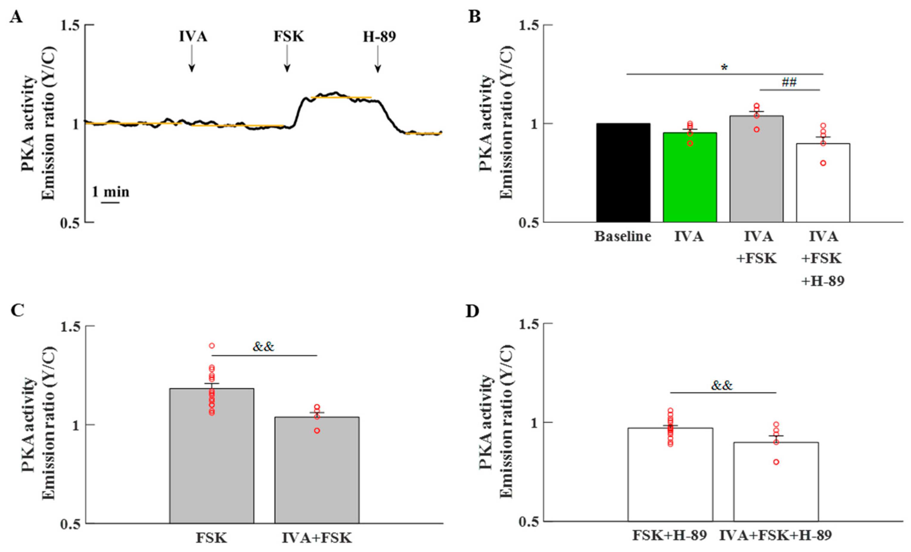
2.1. The Role of Ca2+-Activated, PKA-Dependent Signaling in hiPSC-CMs
2.2. Cytosolic PKA Dynamics in hiPSC-CMs
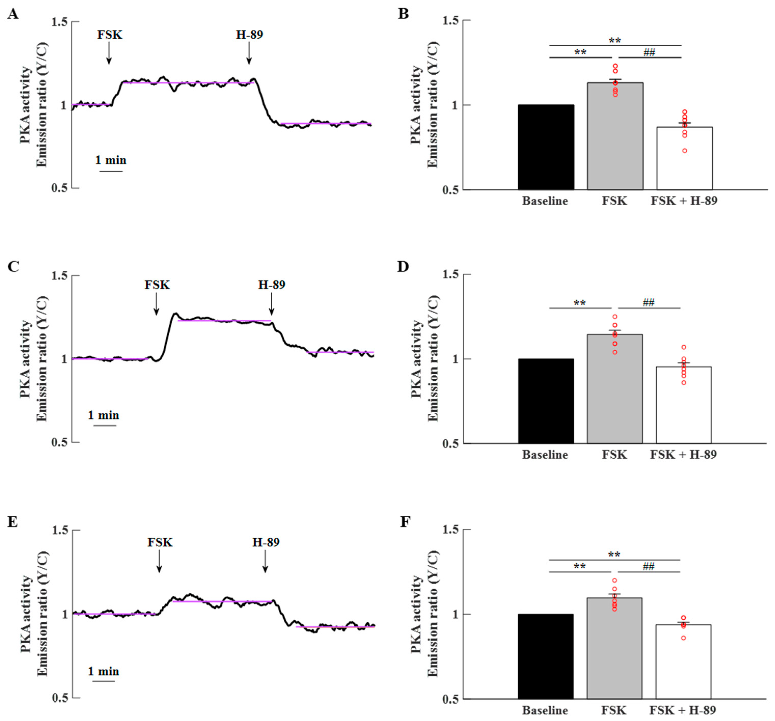
2.3. PKA Compartmentalization in the Cytosol, MM, and OMM in hiPSC-CMs
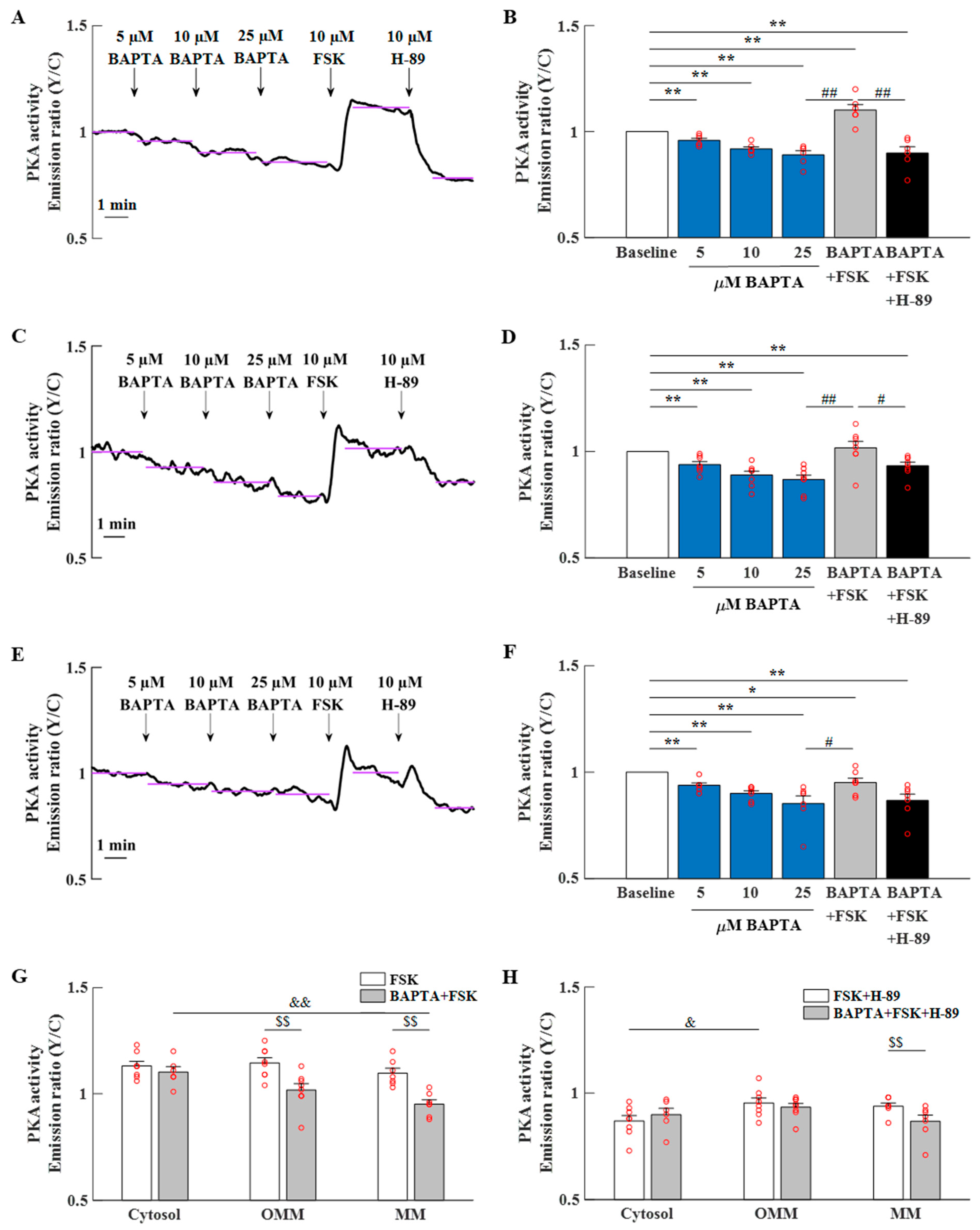
2.4. Ca2+-Activated AC Is an Important Regulator of PKA Activity in hiPSC-CMs
2.5. Mitochondrial Ca2+-Activated AC Is an Important Regulator of PKA Activity in hiPSC-CMs
2.6. PKA Activity Is Affected by the Perturbation of the M Clock in hiPSC-CMs
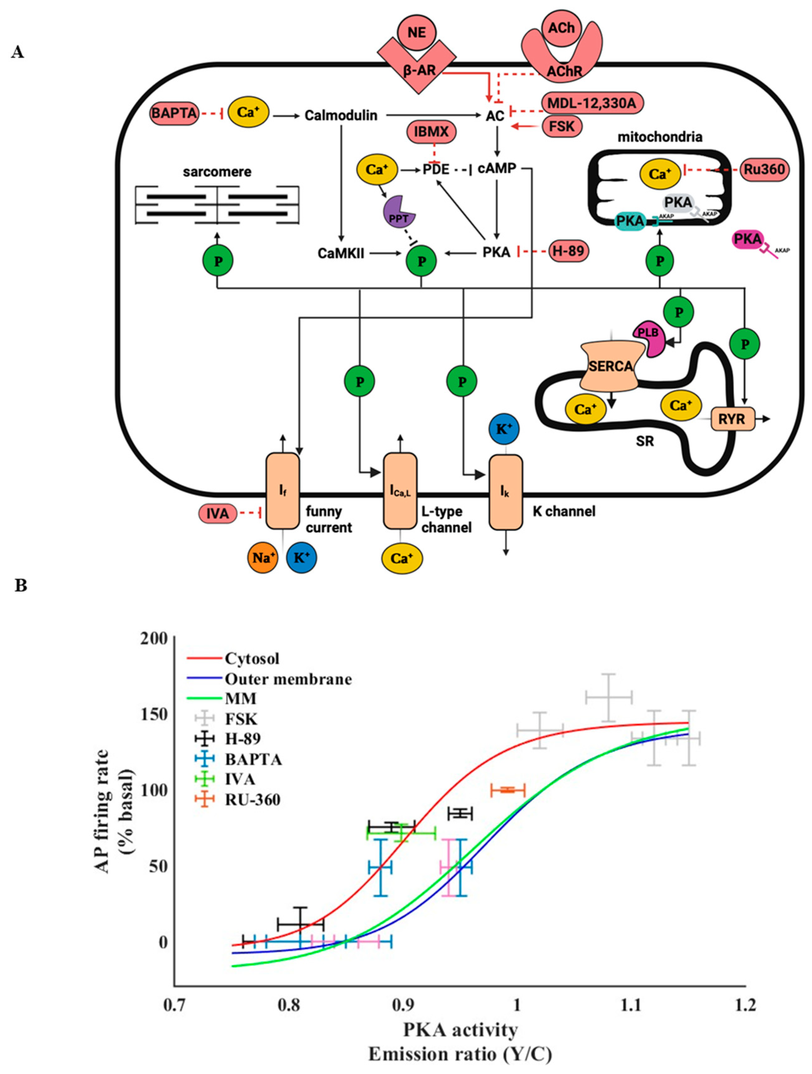
2.7. PKA Activity in the Cytosol Correlates with the Beating Rate of hiPSC-CMs
2.8. PKA Compartmentalization in the SANC Cytosol, MM, and OMM
2.9. Ca2+-Activated AC Is an Important Regulator of PKA Activity in SANCs
3. Discussion
3.1. Ca2+-Activated, PKA-Dependent Signaling Is an Important Regulator of Energy Balance
3.2. High PKA Activity in hiPSC-CMs Is Distributed Differently in Mitochondrial vs. Cytosolic Compartments
3.3. The Role of cAMP/PKA Activity in hiPSC-CMs
3.4. PKA Activity and Its Compartmentalization Are Affected by Ca2+
3.5. PKA Dynamics and Their Ca2+-Dependent Activity Are Similar in Spontaneously Beating hiPSC-CMs and Rabbit SANCs
3.6. Study Limitations
4. Materials and Methods
4.1. hiPSC Generation and Differentiation into Cardiomyocytes
4.2. Isolation of Rabbit SANCs
4.3. Measurement of PKA Dynamics
4.4. Staining of Outer Mitochondrial Membrane and Mitochondrial Matrix
4.5. Measuring the Spontaneous Beating Rate and Ca2+ Characteristics of hiPSC-CMs
4.6. AC1 and AC8 Staining in hiPSC-CMs
4.7. Flavoprotein Autofluorescence Recordings
4.8. Drugs
4.9. Statistics
5. Conclusions
Supplementary Materials
Author Contributions
Funding
Institutional Review Board Statement
Informed Consent Statement
Data Availability Statement
Conflicts of Interest
Abbreviations
| hiPSC-CMs | Human-induced Pluripotent Stem Cell-derived cardiomyocytes |
| LCRs | Local Ca2+ releases |
| cAMP | Cyclic adenosine monophosphate |
| PKA | Protein kinase A |
| SANCs | Sinoatrial node cells |
| M | Membrane |
| SR | Sarcoplasmic reticulum |
| AC | Adenylyl cyclase |
| FRET | Fluorescence resonance energy transfer |
| OMM | Outer mitochondrial membrane |
| MM | Mitochondrial matrix |
| CFP | Cyan fluorescent protein |
| RT | Room temperature |
| FSK | Forskolin |
| PDE | Phosphodiesterase |
| IBMX | Inhibitor 3-isobutyl-1-methylxanthine |
| IVA | Ivabradine |
| MDL | MDL-12,330A |
| If | Funny current |
References
- Mandel, Y.; Weissman, A.; Schick, R.; Barad, L.; Novak, A.; Meiry, G.; Goldberg, S.; Lorber, A.; Rosen, M.R.; Itskovitz-Eldor, J.; et al. Human Embryonic and Induced Pluripotent Stem Cell-Derived Cardiomyocytes Exhibit Beat Rate Variability and Power-Law Behavior. Circulation 2012, 125, 883–893. [Google Scholar] [CrossRef] [Scilit] [PubMed]
- Mazgaoker, S.; Weiser-Bitoun, I.; Brosh, I.; Binah, O.; Yaniv, Y. CAMP-PKA Signaling Modulates the Automaticity of Human IPSC-Derived Cardiomyocytes. J. Gen. Physiol. 2023, 155, e202213153. [Google Scholar] [CrossRef] [Scilit] [PubMed]
- Yaniv, Y.; Sirenko, S.; Ziman, B.D.; Spurgeon, H.A.; Maltsev, V.A.; Lakatta, E.G. New Evidence for Coupled Clock Regulation of the Normal Automaticity of Sinoatrial Nodal Pacemaker Cells: Bradycardic Effects of Ivabradine Are Linked to Suppression of Intracellular Ca2+ Cycling. J. Mol. Cell. Cardiol. 2013, 62, 80–89. [Google Scholar] [CrossRef] [Scilit] [PubMed]
- Tsutsui, K.; Monfredi, O.J.; Sirenko-Tagirova, S.G.; Maltseva, L.A.; Bychkov, R.; Kim, M.S.; Ziman, B.D.; Tarasov, K.V.; Tarasova, Y.S.; Zhang, J.; et al. A Coupled-Clock System Drives the Automaticity of Human Sinoatrial Nodal Pacemaker Cells. Sci. Signal. 2018, 11, eaap7608. [Google Scholar] [CrossRef] [Scilit] [PubMed]
- Groenke, S.; Larson, E.D.; Alber, S.; Zhang, R.; Lamp, S.T.; Ren, X.; Nakano, H.; Jordan, M.C.; Karagueuzian, H.S.; Roos, K.P.; et al. Complete Atrial-Specific Knockout of Sodium-Calcium Exchange Eliminates Sinoatrial Node Pacemaker Activity. PLoS ONE 2013, 8, e81633. [Google Scholar] [CrossRef] [Scilit] [PubMed]
- Neco, P.; Torrente, A.G.; Mesirca, P.; Zorio, E.; Liu, N.; Priori, S.G.; Napolitano, C.; Richard, S.; Benitah, J.P.; Mangoni, M.E.; et al. Paradoxical Effect of Increased Diastolic Ca2+ Release and Decreased Sinoatrial Node Activity in a Mouse Model of Catecholaminergic Polymorphic Ventricular Tachycardia. Circulation 2012, 126, 392–401. [Google Scholar] [CrossRef] [Scilit] [PubMed]
- Koschinski, A.; Zaccolo, M. Micro-2D Cell Culture for CAMP Measurements Using FRET Reporters in Human IPSC-Derived Cardiomyocytes. Methods Mol. Biol. 2022, 2483, 141–165. [Google Scholar] [CrossRef] [Scilit] [PubMed]
- Ben-Ari, M.; Schick, R.; Barad, L.; Novak, A.; Ben-Ari, E.; Lorber, A.; Itskovitz-Eldor, J.; Rosen, M.R.; Weissman, A.; Binah, O. From Beat Rate Variability in Induced Pluripotent Stem Cell-Derived Pacemaker Cells to Heart Rate Variability in Human Subjects. Heart Rhythm 2014, 11, 1808–1818. [Google Scholar] [CrossRef] [Scilit] [PubMed]
- Ebert, A.; Joshi, A.U.; Andorf, S.; Dai, Y.; Sampathkumar, S.; Chen, H.; Li, Y.; Garg, P.; Toischer, K.; Hasenfuss, G.; et al. Proteasome-Dependent Regulation of Distinct Metabolic States During Long-Term Culture of Human IPSC-Derived Cardiomyocytes. Circ. Res. 2019, 125, 90–103. [Google Scholar] [CrossRef] [Scilit] [PubMed]
- Segal, S.; Arbel-Ganon, L.; Mazgaoker, S.; Davoodi, M.; Yaniv, Y. Increase in Ca2+-Activated CAMP/PKA Signaling Prevents Hydroxychloroquine-Induced Bradycardia of the Cardiac Pacemaker. Front. Physiol. 2022, 13, 839140. [Google Scholar] [CrossRef] [Scilit] [PubMed]
- Lakatta, E.G.; Maltsev, V.A.; Vinogradova, T.M. A Coupled SYSTEM of Intracellular Ca2+ Clocks and Surface Membrane Voltage Clocks Controls the Timekeeping Mechanism of the Heart’s Pacemaker. Circ. Res. 2010, 106, 659–673. [Google Scholar] [CrossRef] [Scilit] [PubMed]
- Vinogradova, T.M.; Lyashkov, A.E.; Zhu, W.; Ruknudin, A.M.; Sirenko, S.; Yang, D.; Deo, S.; Barlow, M.; Johnson, S.; Caffrey, J.L.; et al. High Basal Protein Kinase A-Dependent Phosphorylation Drives Rhythmic Internal Ca2+ Store Oscillations and Spontaneous Beating of Cardiac Pacemaker Cells. Circ. Res. 2006, 98, 505–514. [Google Scholar] [CrossRef] [Scilit] [PubMed]
- Younes, A.; Lyashkov, A.E.; Graham, D.; Sheydina, A.; Volkova, M.V.; Mitsak, M.; Vinogradova, T.M.; Lukyanenko, Y.O.; Li, Y.; Ruknudin, A.M.; et al. Ca2+-Stimulated Basal Adenylyl Cyclase Activity Localization in Membrane Lipid Microdomains of Cardiac Sinoatrial Nodal Pacemaker Cells. J. Biol. Chem. 2008, 283, 14461–14468. [Google Scholar] [CrossRef] [Scilit] [PubMed]
- Mattick, P.; Parrington, J.; Odia, E.; Simpson, A.; Collins, T.; Terrar, D. Ca2+-Stimulated Adenylyl Cyclase Isoform AC1 Is Preferentially Expressed in Guinea-Pig Sino-Atrial Node Cells and Modulates the I(f) Pacemaker Current. J. Physiol. 2007, 582, 1195–1203. [Google Scholar] [CrossRef] [Scilit] [PubMed]
- Ren, L.; Thai, P.N.; Gopireddy, R.R.; Timofeyev, V.; Ledford, H.A.; Woltz, R.L.; Park, S.; Puglisi, J.L.; Moreno, C.M.; Santana, L.F.; et al. Adenylyl Cyclase Isoform 1 Contributes to Sinoatrial Node Automaticity via Functional Microdomains. JCI Insight 2022, 7, e162602. [Google Scholar] [CrossRef] [Scilit] [PubMed]
- Bourque, K.; Jones-Tabah, J.; Pétrin, D.; Martin, R.D.; Tanny, J.C.; Hébert, T.E. Comparing the Signaling and Transcriptome Profiling Landscapes of Human IPSC-Derived and Primary Rat Neonatal Cardiomyocytes. Sci. Rep. 2023, 13, 12248. [Google Scholar] [CrossRef] [Scilit] [PubMed]
- Yaniv, Y.; Juhaszova, M.; Lyashkov, A.E.E.; Spurgeon, H.A.; Sollott, S.J.J.; Lakatta, E.G.G. Ca2+-Regulated-CAMP/PKA Signaling in Cardiac Pacemaker Cells Links ATP Supply to Demand. J. Mol. Cell. Cardiol. 2011, 51, 740–748. [Google Scholar] [CrossRef] [Scilit] [PubMed]
- Yaniv, Y.; Spurgeon, H.A.A.; Lyashkov, A.E.E.; Yang, D.; Ziman, B.D.D.; Maltsev, V.A.A.; Lakatta, E.G.G. Crosstalk between Mitochondrial and Sarcoplasmic Reticulum Ca2+ Cycling Modulates Cardiac Pacemaker Cell Automaticity. PLoS ONE 2012, 7, e37582. [Google Scholar] [CrossRef] [Scilit] [PubMed]
- Yaniv, Y.; Ganesan, A.; Yang, D.; Ziman, B.D.; Lyashkov, A.E.; Levchenko, A.; Zhang, J.; Lakatta, E.G. Real-Time Relationship between PKA Biochemical Signal Network Dynamics and Increased Action Potential Firing Rate in Heart Pacemaker Cells: Kinetics of PKA Activation in Heart Pacemaker Cells. J. Mol. Cell. Cardiol. 2015, 86, 168–178. [Google Scholar] [CrossRef] [Scilit] [PubMed]
- Segal, S.; Kirschner Peretz, N.; Arbel-Ganon, L.; Liang, J.; Li, L.; Marbach, D.; Yang, D.; Wang, S.Q.; Yaniv, Y. Eliminating Contraction during Culture Maintains Global and Local Ca2+ Dynamics in Cultured Rabbit Pacemaker Cells. Cell Calcium 2019, 78, 35–47. [Google Scholar] [CrossRef] [Scilit] [PubMed]
- Lefkimmiatis, K.; Leronni, D.; Hofer, A.M. The Inner and Outer Compartments of Mitochondria Are Sites of Distinct CAMP/PKA Signaling Dynamics. J. Cell Biol. 2013, 202, 453–462. [Google Scholar] [CrossRef] [Scilit] [PubMed]
- Kirschner Peretz, N.; Segal, S.; Weiser-Bitoun, I.; Yaniv, Y. Distinct PKA Signaling in Cytosolic and Mitochondrial Compartments in Electrically Paced Atrial Myocytes. Cells 2022, 11, 2261. [Google Scholar] [CrossRef] [Scilit] [PubMed]
- Covian, R.; Balaban, R.S. Cardiac Mitochondrial Matrix and Respiratory Complex Protein Phosphorylation. Am. J. Physiol. Heart Circ. Physiol. 2012, 303, H940–H966. [Google Scholar] [CrossRef] [Scilit] [PubMed]
- Valenti, D.; Tullo, A.; Caratozzolo, M.F.; Merafina, R.S.; Scartezzini, P.; Marra, E.; Vacca, R.A. Impairment of F1F0-ATPase, Adenine Nucleotide Translocator and Adenylate Kinase Causes Mitochondrial Energy Deficit in Human Skin Fibroblasts with Chromosome 21 Trisomy. Biochem. J. 2010, 431, 299–310. [Google Scholar] [CrossRef] [Scilit] [PubMed]
- Kostic, M.; Ludtmann, M.H.R.; Bading, H.; Hershfinkel, M.; Steer, E.; Chu, C.T.; Abramov, A.Y.; Sekler, I. PKA Phosphorylation of NCLX Reverses Mitochondrial Calcium Overload and Depolarization, Promoting Survival of PINK1-Deficient Dopaminergic Neurons. Cell Rep. 2015, 13, 376–386. [Google Scholar] [CrossRef] [Scilit] [PubMed]
- Yaniv, Y.; Spurgeon, H.A.A.; Ziman, B.D.D.; Lyashkov, A.E.E.; Lakatta, E.G.G. Mechanisms That Match ATP Supply to Demand in Cardiac Pacemaker Cells during High ATP Demand. Am. J. Physiol. Heart Circ. Physiol. 2013, 304, H1428–H1438. [Google Scholar] [CrossRef] [Scilit] [PubMed]
- Faleeva, M.; Diakonov, I.; Srivastava, P.; Ramuz, M.; Calamera, G.; Andressen, K.W.; Bork, N.; Tsansizi, L.; Cosson, M.V.; Bernardo, A.S.; et al. Compartmentation of CGMP Signaling in Induced Pluripotent Stem Cell Derived Cardiomyocytes during Prolonged Culture. Cells 2022, 11, 3257. [Google Scholar] [CrossRef] [Scilit] [PubMed]
- Behar, J.; Ganesan, A.; Zhang, J.; Yaniv, Y. The Autonomic Nervous System Regulates the Heart Rate through CAMP-PKA Dependent and Independent Coupled-Clock Pacemaker Cell Mechanisms. Front. Physiol. 2016, 7, 419. [Google Scholar] [CrossRef] [Scilit] [PubMed]
- Heijman, J.; Dewenter, M.; El-Armouche, A.; Dobrev, D. Function and Regulation of Serine/Threonine Phosphatases in the Healthy and Diseased Heart. J. Mol. Cell. Cardiol. 2013, 64, 90–98. [Google Scholar] [CrossRef] [Scilit] [PubMed]
- Yaniv, Y.; Maltsev, V.A.; Ziman, B.D.; Spurgeon, H.A.; Lakatta, E.G. The “Funny” Current (If) Inhibition by Ivabradine at Membrane Potentials Encompassing Spontaneous Depolarization in Pacemaker Cells. Molecules 2012, 17, 8241–8254. [Google Scholar] [CrossRef] [Scilit] [PubMed]
- Saleem, U.; Ismaili, D.; Mannhardt, I.; Pinnschmidt, H.; Schulze, T.; Christ, T.; Eschenhagen, T.; Hansen, A. Regulation of ICa,L and Force by PDEs in Human-Induced Pluripotent Stem Cell-Derived Cardiomyocytes. Br. J. Pharmacol. 2020, 177, 3036–3045. [Google Scholar] [CrossRef] [Scilit] [PubMed]
- Omori, K.; Kotera, J. Overview of PDEs and Their Regulation. Circ. Res. 2007, 100, 309–327. [Google Scholar] [CrossRef] [Scilit] [PubMed]
- Vinogradova, T.M.; Kobrinsky, E.; Lakatta, E.G. Dual Activation of Phosphodiesterases 3 and 4 Regulates Basal Spontaneous Beating Rate of Cardiac Pacemaker Cells: Role of Compartmentalization? Front. Physiol. 2018, 9, 1301. [Google Scholar] [CrossRef] [Scilit] [PubMed]
- Bers, D.M. Calcium Cycling and Signaling. Annu. Rev. Physiol. 2008, 70, 23–49. [Google Scholar] [CrossRef] [Scilit] [PubMed]
- Protze, S.I.; Liu, J.; Nussinovitch, U.; Ohana, L.; Backx, P.H.; Gepstein, L.; Keller, G.M. Sinoatrial Node Cardiomyocytes Derived from Human Pluripotent Cells Function as a Biological Pacemaker. Nat. Biotechnol. 2016, 35, 56–68. [Google Scholar] [CrossRef] [Scilit] [PubMed]
- Ben-Ari, M.; Naor, S.; Zeevi-Levin, N.; Schick, R.; Ben Jehuda, R.; Reiter, I.; Raveh, A.; Grijnevitch, I.; Barak, O.; Rosen, M.R.; et al. Developmental Changes in Electrophysiological Characteristics of Human-Induced Pluripotent Stem Cell–Derived Cardiomyocytes. Heart Rhythm 2016, 13, 2379–2387. [Google Scholar] [CrossRef] [Scilit] [PubMed]
- Giannetti, F.; Benzoni, P.; Campostrini, G.; Milanesi, R.; Bucchi, A.; Baruscotti, M.; Dell’Era, P.; Rossini, A.; Barbuti, A. A Detailed Characterization of the Hyperpolarization-Activated “Funny” Current (If) in Human-Induced Pluripotent Stem Cell (IPSC)-Derived Cardiomyocytes with Pacemaker Activity. Pflug. Arch. 2021, 473, 1009–1021. [Google Scholar] [CrossRef] [Scilit] [PubMed]
- Yehezkel, S.; Rebibo-Sabbah, A.; Segev, Y.; Tzukerman, M.; Shaked, R.; Huber, I.; Gepstein, L.; Skorecki, K.; Selig, S. Reprogramming of Telomeric Regions during the Generation of Human Induced Pluripotent Stem Cells and Subsequent Differentiation into Fibroblast-like Derivatives. Epigenetics 2011, 6, 63–75. [Google Scholar] [CrossRef] [Scilit] [PubMed]
- Novak, A.; Barad, L.; Lorber, A.; Gherghiceanu, M.; Reiter, I.; Eisen, B.; Eldor, L.; Itskovitz-Eldor, J.; Eldar, M.; Arad, M.; et al. Functional Abnormalities in IPSC-Derived Cardiomyocytes Generated from CPVT1 and CPVT2 Patients Carrying Ryanodine or Calsequestrin Mutations. J. Cell. Mol. Med. 2015, 19, 2006–2018. [Google Scholar] [CrossRef] [Scilit] [PubMed]
- Lian, X.; Zhang, J.; Azarin, S.M.; Zhu, K.; Hazeltine, L.B.; Bao, X.; Hsiao, C.; Kamp, T.J.; Palecek, S.P. Directed Cardiomyocyte Differentiation from Human Pluripotent Stem Cells by Modulating Wnt/β-Catenin Signaling under Fully Defined Conditions. Nat. Protoc. 2013, 8, 162–175. [Google Scholar] [CrossRef] [Scilit] [PubMed]
- Davoodi, M.; Segal, S.; Kirschner Peretz, N.; Kamoun, D.; Yaniv, Y. Semi-Automated Program for Analysis of Local Ca2+ Spark Release with Application for Classification of Heart Cell Type. Cell Calcium 2017, 64, 83–90. [Google Scholar] [CrossRef] [Scilit] [PubMed]


Disclaimer/Publisher’s Note: The statements, opinions and data contained in all publications are solely those of the individual author(s) and contributor(s) and not of MDPI and/or the editor(s). MDPI and/or the editor(s) disclaim responsibility for any injury to people or property resulting from any ideas, methods, instructions or products referred to in the content. |
© 2026 by the authors. Licensee MDPI, Basel, Switzerland. This article is an open access article distributed under the terms and conditions of the Creative Commons Attribution (CC BY) license.
Share and Cite
Rotschield, A.; Mazgaoker, S.; Segal, S.; Weiser-Bitoun, I.; Brosh, I.; Binah, O.; Yaniv, Y. Intracellular Ca2+ Modulates PKA Compartmentalization and Dynamics in Human iPSC-Derived Cardiomyocytes. Int. J. Mol. Sci. 2026, 27, 6559. https://doi.org/10.3390/ijms27156559
Rotschield A, Mazgaoker S, Segal S, Weiser-Bitoun I, Brosh I, Binah O, Yaniv Y. Intracellular Ca2+ Modulates PKA Compartmentalization and Dynamics in Human iPSC-Derived Cardiomyocytes. International Journal of Molecular Sciences. 2026; 27(15):6559. https://doi.org/10.3390/ijms27156559
Chicago/Turabian StyleRotschield, Anat, Savyon Mazgaoker, Sofia Segal, Ido Weiser-Bitoun, Inbar Brosh, Ofer Binah, and Yael Yaniv. 2026. "Intracellular Ca2+ Modulates PKA Compartmentalization and Dynamics in Human iPSC-Derived Cardiomyocytes" International Journal of Molecular Sciences 27, no. 15: 6559. https://doi.org/10.3390/ijms27156559
APA StyleRotschield, A., Mazgaoker, S., Segal, S., Weiser-Bitoun, I., Brosh, I., Binah, O., & Yaniv, Y. (2026). Intracellular Ca2+ Modulates PKA Compartmentalization and Dynamics in Human iPSC-Derived Cardiomyocytes. International Journal of Molecular Sciences, 27(15), 6559. https://doi.org/10.3390/ijms27156559