Review

Neuroinvasive Free-Living Amoebae Pathogenesis, Neuroinflammation and Therapeutic Challenges

by
Oliwia Pawelec-Pęciak
,
Karolina Kot
,
Danuta Kosik-Bogacka
and
Natalia Łanocha-Arendarczyk
*
Department of Biology, Parasitology, and Pharmaceutical Botany, Pomeranian Medical University in Szczecin, Powstańców Wielkopolskich 72, 70-111 Szczecin, Poland
*
Author to whom correspondence should be addressed.
Int. J. Mol. Sci. 2026, 27(13), 6056; https://doi.org/10.3390/ijms27136056
Submission received: 29 May 2026 / Revised: 28 June 2026 / Accepted: 2 July 2026 / Published: 6 July 2026

Abstract

Neuroinvasive free-living amoebae (FLA), particularly Naegleria fowleri and Acanthamoeba spp., are responsible for rare but devastating infections of the central nervous system (CNS). Approximately 480 cases of primary amoebic meningoencephalitis (PAM) and fewer than 200 well-documented cases of Acanthamoeba-associated granulomatous amoebic encephalitis (GAE) have been reported worldwide. Mortality rates frequently exceed 90%. PAM typically develops following exposure to warm freshwater contaminated with N. fowleri and progresses rapidly in otherwise healthy individuals. In contrast, GAE usually follows a more indolent course and occurs predominantly in immunocompromised hosts. Despite their distinct clinical courses, both infections are characterized by CNS invasion, amoeba-mediated tissue destruction, blood–brain barrier (BBB) disruption, and host inflammatory responses. These processes drive neuroinflammation, neuronal injury, and neurological deterioration. Early diagnosis remains challenging because clinical manifestations are nonspecific and disease progression can be either fulminant or initially subtle. Therapeutic management is hindered by poor CNS drug penetration, limited efficacy of currently available therapies, treatment-related toxicity, and the absence of standardized treatment protocols or controlled clinical trials. This narrative review critically synthesizes current evidence on CNS invasion, neuroinflammation, neuropathology, diagnostic challenges, and therapeutic strategies in neuroinvasive FLA infections. It also highlights key translational priorities, including earlier diagnosis, standardized treatment protocols, stronger clinical evidence, and improved CNS-targeted drug delivery.

1. Introduction

Neuropathogenic protozoa possess diverse mechanisms that enable penetration of the blood–brain barrier (BBB) and subsequent colonization of the central nervous system (CNS) [1]. Although the CNS has long been considered an immunologically privileged site, it remains vulnerable to parasite-induced inflammatory responses that disrupt neuronal homeostasis [2]. Free-living amoebae (FLA) are ubiquitous environmental protozoa capable of persisting in diverse aquatic, terrestrial, and human-associated environments [1,2,3,4,5]. Some amphizoic species can cause opportunistic human infections. Among them, Naegleria fowleri and Acanthamoeba spp. are the most clinically significant neuroinvasive FLA, causing primary amoebic meningoencephalitis and granulomatous amoebic encephalitis, respectively [2,3,4,5,6,7]. Neuroinvasive FLA infections are rare, globally reported, and associated with exceptionally high mortality. To date, approximately 480 cases of PAM and fewer than 200 well-documented cases of Acanthamoeba-associated GAE have been described worldwide, with case fatality rates frequently exceeding 90% [7,8,9,10,11,12]. Despite their distinct clinical courses, both infections involve CNS invasion, amoeba-mediated tissue destruction, BBB disruption, and host inflammatory responses that contribute to neuroinflammation, neuronal injury, and neurological deterioration [2,3]. In this review, neuropathogenesis refers to the processes through which FLA invade the CNS, interact with neural and immune cells, and induce tissue injury and pathology [13]. Neuroinflammation refers to an inflammatory response within the CNS triggered by infection or tissue injury, involving activation of microglia and astrocytes and the release of immune mediators [14]. In Acanthamoeba infection, this response may also include granulomatous inflammation, defined as organized aggregates of macrophages, lymphocytes, and other immune cells surrounding persistent pathogens or damaged tissue [7,8]. Despite decades of research, many aspects of CNS invasion, immune dysregulation, inflammatory injury, and tissue damage in FLA infections remain incompletely understood. This knowledge gap has direct clinical relevance because early diagnosis is often hindered by nonspecific manifestations, whereas effective treatment is limited by poor CNS drug penetration, treatment-related toxicity, and the lack of controlled clinical trials [7,9,10].
This narrative review examines current evidence on host–pathogen interactions, neuroinflammatory mechanisms, and neuropathological outcomes in neuroinvasive FLA infections and discusses their implications for diagnosis and treatment. In addition, it highlights key translational priorities, including earlier diagnosis, standardized treatment protocols, stronger clinical evidence, and improved CNS-directed drug delivery.
Literature Search Strategy
This manuscript was developed as a narrative, evidence-based review integrating clinical, experimental, and translational literature on neuroinvasive infections caused by FLA. Relevant publications were identified through searches of PubMed, Scopus, and Web of Science, covering studies published up to early 2026. The analysis included clinical case reports, experimental in vitro and in vivo studies, recent reviews, and available meta-analyses, with emphasis on neuropathogenesis, neuroinflammation, neuropathology, diagnosis, and treatment. Seminal earlier publications were also included when they provided foundational insights into the field.

2. Naegleria fowleri as the Causative Agent of Primary Amoebic Meningoencephalitis

Of the more than 45 described Naegleria species, only Naegleria fowleri is recognized as a human pathogen capable of infecting both humans and several animal species [9]. This thermophilic free-living amoeba occurs predominantly in warm freshwater environments, including lakes, rivers, thermal waters, and inadequately disinfected recreational water systems, particularly during warmer months of the year [10,11]. For PAM, the estimated risk following recreational freshwater exposure is extremely low, at approximately one case per 2.5 million exposed swimmers [11,12].
Clinically, PAM represents a medical emergency because of its abrupt onset, aggressive CNS invasion, and rapid progression to life-threatening neurological failure [12,15]. Most published reports originate from the United States, Central America, Australia, and several Asia countries, although cases have also been documented in Europe [5,16]. One of the best-documented European outbreaks occurred in Ústí nad Labem in the former Czechoslovakia, where fatal PAM cases were linked to exposure to a heated indoor swimming pool [17]. The expanding geographic distribution of N. fowleri is influenced by both environmental and water-system conditions. Climate warming and thermal pollution may expand suitable habitats for this thermophilic amoeba by increasing surface-water temperatures [18]. Impaired water quality, including low disinfectant residuals, turbidity, biofilm formation, and microbial or algal overgrowth, may further facilitate its persistence in natural and engineered aquatic systems [19]. This risk is illustrated by a PAM case linked to an artificial whitewater river, where warm, turbid water, low chlorine concentrations, and extensive algal growth favored N. fowleri persistence [20]. These observations support the inclusion of environmental and water-quality parameters in assessments of FLA transmission risk. Certain cultural or religious practices involving nasal exposure to untreated water may also increase the risk of PAM in endemic regions or settings characterized by high ambient temperatures [21]. Considering the thermophilic nature of N. fowleri, the relatively low number of reported cases in tropical regions likely reflects substantial underdiagnosis.
The life cycle of N. fowleri comprises three morphological stages: the trophozoite (amoeboid form, 7–35 μm), the flagellate (10–16 μm), and the cyst (7–10 μm) [22]. The trophozoite is the invasive and proliferative stage responsible for human infection, whereas the cyst is an environmentally resistant form that enables survival under unfavorable conditions. Importantly, only trophozoites are observed in human brain tissue during PAM, as cyst formation does not occur within the CNS [22,23].

2.1. Transmission to the Host and Clinical Manifestations of PAM

PAM typically results from nasal exposure to water contaminated with N. fowleri trophozoites, particularly during recreational or domestic contact with warm freshwater [24,25]. Young males are disproportionately affected, most likely because of greater exposure to aquatic environments and potential age-related anatomical susceptibility of the olfactory route [15,26]. After entering the nasal cavity, trophozoites attach to the olfactory epithelium and migrate through the cribriform plate to the olfactory bulbs, thereby gaining direct access to the brain [5,24,27]. Clinical symptoms usually develop within 1–7 days and initially include headache, fever, nausea, fatigue, and occasionally anosmia. As CNS invasion progresses, patients may develop neck stiffness, seizures, confusion, photophobia, hallucinations, personality changes, coma, and signs of increased intracranial pressure [24,28]. Early nonspecific manifestations often resemble bacterial or viral meningitis, which may delay timely diagnosis and therapeutic intervention [9]. PAM typically progresses rapidly, with death occurring within 1–2 weeks of symptom onset. Consequently, many cases are diagnosed only post-mortem [5].

2.2. Macroscopic and Microscopic Findings in PAM

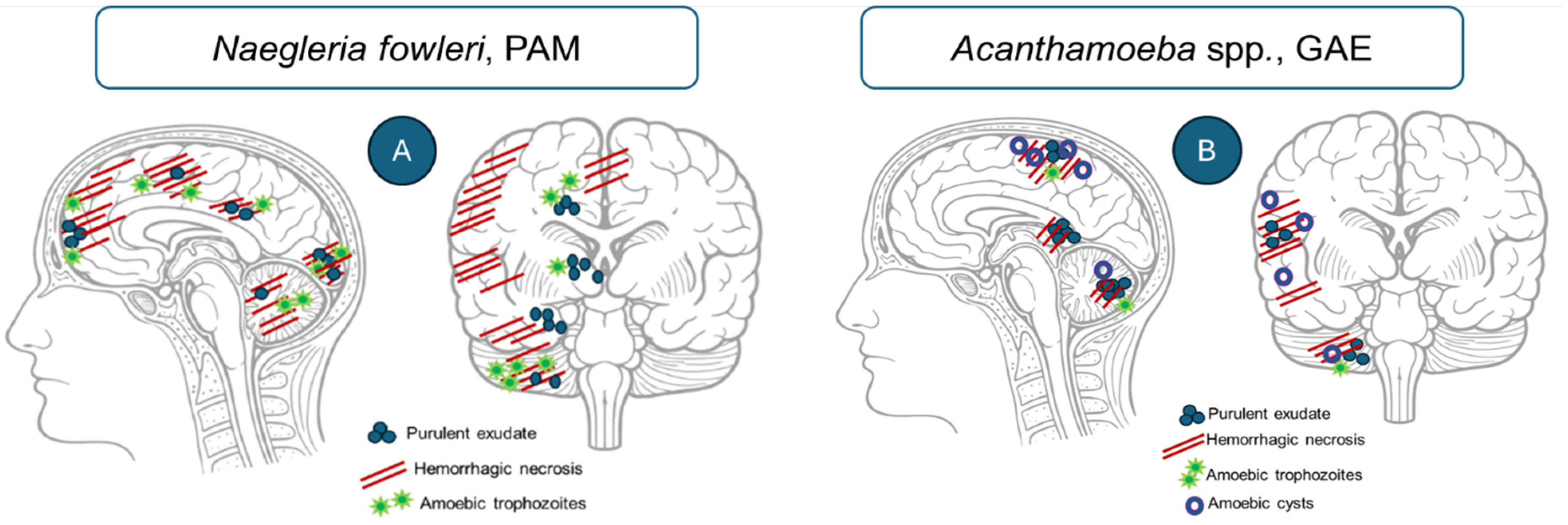
The neuropathology of PAM reflects rapid amoebic invasion through the olfactory pathway, and extensive tissue destruction within the anterior regions of the brain. Macroscopically, the brain is typically swollen and edematous, with evidence of increased intracranial pressure and, in severe cases, herniation of the uncus or cerebellar tonsils (Figure 1). The most characteristic lesions include hemorrhage, necrosis, and inflammation involving the olfactory bulbs, olfactory tracts, frontal cortex, and adjacent frontal and temporal regions [11,29,30]. The cerebral hemispheres may appear soft, hyperemic, and congested, and hemorrhagic exudate may be present along the meningeal surfaces [5,11]. Microscopically, PAM is characterized by acute necrotizing and hemorrhagic meningoencephalitis, with extensive destruction of cortical gray matter and inflammatory involvement of the leptomeninges [31]. Tissue damage may affect multiple CNS regions, including the cerebral hemispheres, brainstem, cerebellum, and upper spinal cord [31]. Motile N. fowleri trophozoites can be detected in cerebrospinal fluid (CSF) or brain tissue, whereas cysts are not observed in human brain tissue during PAM [32,33]. Experimental models have similarly demonstrated extensive hemorrhagic injury and necrotic changes, particularly within the olfactory bulbs, olfactory nerves, and cerebellum [34,35].

2.3. Virulence Mechanisms of Naegleria fowleri

2.3.1. Adhesion and Phagocytosis

Naegleria fowleri deploys a broad repertoire of virulence mechanisms that enable CNS access, tissue invasion, and rapid cerebral damage [7]. These mechanisms include contact-dependent processes, such as adhesion and phagocytosis, as well as contact-independent mechanisms, including protease secretion, extracellular vesicle release, and cytolytic activity [5,36]. Together, they support a stepwise pathogenic sequence involving attachment to host tissues, invasion of anatomical barriers, tissue destruction, and adaptation to host-derived stress signals. N. fowleri trophozoites exhibit high-affinity binding to extracellular matrix (ECM) components, including laminin-1, fibronectin, and type I collagen [37]. This adhesive capacity is facilitated by specialized cytoskeletal structures, notably lamellipodia, which increase the contact surface area, and focal adhesion-like complexes that mediate stable interactions with the ECM [38]. In a murine model, a ~72 kDa surface protein that interacts with the nasal epithelium has been identified. Immunofluorescence studies localized this protein to the trophozoite membrane and pseudopodia, supporting its role in host cell adhesion [39].
Adhesion is further enhanced by cathepsin B and N. fowleri adhesion protein 1 (Nfa1), both of which are localized to pseudopodia and specialized feeding structures termed amoebastomes (“food cups”), where they facilitate attachment, invasion, and nutrient acquisition [40,41]. Nfa1 represents a key virulence determinant through its roles in adhesion, phagocytosis, and amoebastome formation. Moreover, Nfa1 interacts with cytoskeletal and stress-response proteins, including heat shock protein 70 (Hsp70), actin (Nf-actin), and cathepsin-like proteases, thereby enhancing pathogenicity [41]. Cathepsin B contributes to host tissue adherence and may directly mediate neural tissue damage [42]. Functional inhibition of cathepsin B has been associated with improved host survival, whereas anti-Nfa1 antibodies reduce N. fowleri-induced cytotoxicity, supporting their relevance as promising preclinical targets for therapeutic or immunological intervention [40,42,43].

2.3.2. Invasion and Tissue Penetration

Following these adhesion events, the parasite begins to interact more extensively with host tissues, initiating invasion processes that rely on both mechanical penetration and enzymatic activity [25]. Importantly, this invasive process is not restricted to phagocytosis. Experimental studies indicate that N. fowleri can compromise epithelial barrier integrity by disrupting tight-junction organization and increasing paracellular permeability, thereby facilitating penetration through the olfactory mucosa. This route of invasion is supported by amoeboid motility, cytoskeletal remodeling, adhesion to extracellular matrix components, and protease-mediated degradation of junctional and structural proteins [25,40,44]. In addition to direct host–parasite interactions, N. fowleri employs multiple contact-independent mechanisms that promote tissue degradation, facilitate traversal of anatomical barriers, and induce distal cytotoxic effects [5]. The amoeba secretes a variety of proteolytic enzymes, including matrix metalloproteinases-2 and -9 (MMP-2 and MMP-9, respectively), which degrade extracellular matrix components and facilitate disruption of the nasal mucosa, promoting progression along the olfactory route toward the CNS [40,45,46]. Collectively, these proteases promote extracellular matrix degradation, compromise nasal mucosal integrity, and facilitate penetration through the cribriform plate, thereby enabling CNS invasion [40,46]. Once epithelial barriers are breached, additional enzymatic mechanisms contribute to deeper tissue injury. Cysteine proteases may disrupt tight-junction integrity in BBB-associated endothelial cells, alter cytoskeletal organization, modulate the actin cytoskeleton, and facilitate trophozoite transmigration into the CNS [7,10,47]. Additional enzymatic activities, including hydrolases, phospholipases, neuraminidases, and phosphatases, contribute to host cell destruction and neural tissue damage and have also been implicated in demyelination [40]. Pore-forming polypeptides constitute another class of virulence factors that induce lysis of nucleated host cells through membrane disruption [48]. Together, these mechanisms indicate that N. fowleri tissue penetration is a coordinated process involving adhesion, amoeboid migration, paracellular barrier disruption, enzymatic extracellular matrix remodeling, and direct cytolytic injury [44].

2.3.3. Extracellular Vesicles, Cytotoxicity, and Stress Adaptation

In addition to tissue destruction and invasion, immune evasion represents an important component of N. fowleri pathogenicity, enabling survival within the host and facilitating disease progression. Increasing attention has also focused on extracellular vesicles (EVs) released by N. fowleri, which constitute an important component of contact-independent pathogenicity. These vesicles exhibit hemolytic activity against erythrocytes, possess proteolytic activity, induce necrosis, and increase paracellular ion permeability, as demonstrated in Madin–Darby canine kidney (MDCK) cell models [49]. EVs are enriched in hydrolytic enzymes and cytoskeletal proteins, facilitating tissue degradation, promoting adhesion and migration, and mediating intercellular communication between amoebae and host cells [50]. In parallel, Nf23 has emerged as another virulence-associated molecule. Its expression increases after brain invasion in vivo, suggesting infection-dependent regulation and a role in disease progression [51]. Neutralization with anti-Nf23 antibodies reduces amoeba-induced cytotoxicity, further supporting its contribution to virulence [51]. Finally, although increased nitric oxide (NO) production is observed within the trophozoite microenvironment during infection, N. fowleri demonstrates relative resistance to NO-mediated cytotoxicity. This adaptation likely promotes parasite survival under oxidative stress and helps sustain pathogenic potential [5].
Overall, the pathogenicity of N. fowleri reflects the coordinated action of adhesion, phagocytosis, proteolytic degradation, barrier disruption, EV-mediated cytotoxicity, immune evasion, and resistance to host-derived stress signals. Among these mechanisms, cathepsin B, Nfa1, Nf23, and EV-associated pathways appear to be particularly promising preclinical targets for future therapeutic or immunological intervention. Collectively, these virulence factors accelerate olfactory migration, CNS invasion, edema formation, hemorrhage, necrosis, and intracranial pressure. This multifactorial pathogenic strategy helps explain the fulminant course of PAM and the narrow window available for diagnosis and treatment.

2.4. Immune Response to Naegleria fowleri

Exposure of the nasal cavity to N. fowleri trophozoites initiates a rapid mucosal immune response, although several stages of this process remain incompletely understood. After nasal entry, the amoeba must overcome physical barriers, including mucus and epithelial tight junctions [7,16]. Disruption of tight-junction proteins facilitates epithelial penetration and represents an early step in tissue invasion [52].
The first immunological barrier is mediated by respiratory antibodies, including immunoglobulin A (IgA), immunoglobulin M (IgM), and immunoglobulin G (IgG), which may reduce amoebic motility, inhibit epithelial adhesion, and promote clearance from the upper airways [53]. Experimental studies further support the protective role of antibody-mediated immunity. In a murine model of nasal infection, Fc gamma receptor III (FcγRIII) activation was associated with enhanced neutrophil-mediated amoebic clearance, reduced parasite migration, and lower risk of PAM [54]. Natural IgG and IgA antibodies against N. fowleri antigens have also been detected in residents of endemic areas, suggesting prior environmental exposure and potential relevance for serological diagnostics and preventive strategies [55]. Rodríguez-Mera et al. [55] further demonstrated that these antibodies recognize structurally distinct antigens of N. fowleri located within the cell membrane, pseudopodia, food cups, and extracellular vesicles. The immunogenicity of these antigens highlights their potential utility in serological diagnostics and, potentially, vaccine development for endemic regions.
Although antibodies may promote complement activation, N. fowleri exhibit resistance to complement-mediated lysis and can secrete proteases that degrade immunoglobulins, representing an important mechanism of immune evasion [7,56]. When antibody-mediated responses are insufficient, a rapid innate immune response develops, characterized by early recruitment of neutrophils to the olfactory epithelium [52,53]. Neutrophils respond through the production of reactive oxygen species (ROS), NO, and neutrophil extracellular traps. If trophozoites penetrate deeper tissues, macrophages and monocytes are also activated. Nevertheless, some amoebae evade local immune clearance, reach the lamina propria, and migrate along olfactory nerve bundles toward the brain, where neuronal pathways may partially shield them from immune effector mechanisms [53].
Once within the CNS, infection triggers a fulminant inflammatory response characterized by infiltration of neutrophils, monocytes, and eosinophils, together with activation of microglia and astrocytes [40]. At this stage, neurological injury is shaped not only by direct amoebic cytotoxicity but by a rapidly escalating pathogenic circuit in which parasite-mediated tissue destruction and host-driven immunopathology reinforce one another [5,40]. Neutrophil-rich inflammation, microglial activation, astrocyte reactivity, cytokine and chemokine release, oxidative stress, and inflammasome activation collectively amplify BBB disruption, cerebral edema, hemorrhagic–necrotic tissue injury, and increased intracranial pressure [57]. Thus, PAM represents a paradigmatic example of fulminant innate neuroinflammation, in which antimicrobial defense mechanisms become insufficient to restrict parasite invasion while simultaneously accelerating irreversible CNS damage. This response can escalate into severe neuroinflammation and widespread tissue destruction. Microglia stimulated by N. fowleri-derived extracellular vesicles adopt a pro-inflammatory phenotype and produce cytokines such as interleukin-6 (IL-6), interleukin-1β (IL-1β), interleukin-23 (IL-23), and tumor necrosis factor- α (TNF-α), as well as ROS and NO [58]. Interleukin-10 (IL-10) production has also been observed, suggesting an attempted regulatory response that is usually insufficient to prevent immunopathology [59]. Activated astrocytes further amplify inflammation by releasing cytokines and chemokines involved in immune-cell recruitment [60,61].
At the molecular level, amoebic trophozoites can induce ROS production, which promotes epidermal growth factor receptor (EGFR)-dependent interleukin-8 (IL-8) upregulation and EGFR-independent IL-1β expression [62]. ROS may also contribute to NLR family pyrin domain-containing 3 (NLRP3) inflammasome activation, leading to caspase-1 activation and maturation of IL-1β [62,63]. In addition, N. fowleri has been shown to induce ROS-dependent necroptosis in Jurkat cells, suggesting that inflammatory cell death may contribute to irreversible CNS damage [64].
Overall, the immune response to N. fowleri has a dual role. Early mucosal and innate responses may limit parasite migration and dissemination, whereas excessive CNS inflammation contributes to edema, tissue destruction, and irreversible neurological injury. Effective control of infection therefore likely depends on a delicate balance between protective immunity and immunopathology.

3. Acanthamoeba spp. as Etiological Agents of Granulomatous Amoebic Encephalitis

Acanthamoeba spp. are globally distributed FLA that persist in diverse environmental and human-associated habitats, including aquatic systems, soil, tap water, recreational waters, medical devices, air-conditioning systems, and contact lenses [65,66,67,68,69]. Widespread human exposure is reflected by the detection of anti-Acanthamoeba IgG antibodies in up to approximately 80% of healthy individuals [70]. To date, at least 24 species and approximately 23 genotypes (T1–T23), defined by 18S rDNA sequencing, have been described. Genotype T4 is most frequently associated with human pathogenicity, although T1, T2, T4, T5, T10, T12, and T18 have also been implicated in GAE [71,72,73]. The clinical burden of GAE remains difficult to define. Since the first reported case in the United States in the 1970s, sporadic cases have been documented worldwide. Between 1990 and 2026, 81 cases were identified in the PubMed-indexed literature, whereas U.S. surveillance data documented 122 cases by 2020, including 20 confirmed post-mortem [74,75,76,77,78]. This discrepancy suggests substantial underestimation of GAE in the published literature and likely reflects diagnostic challenges, underreporting, and post-mortem recognition. Unlike PAM, GAE is usually reported as sporadic cases rather than outbreak-associated infection; therefore, this section focuses on representative epidemiological patterns rather than individual case descriptions.
Unlike N. fowleri, Acanthamoeba spp. have two life-cycle stages: a motile trophozoite (25–40 μm) and a resistant cyst (13–20 μm), and they lack a flagellated stage [71]. Trophozoites are metabolically active forms characterized by pseudopodia and acanthopodia, whereas cysts possess a double-layered wall composed of an outer ectocyst and an inner endocyst. This wall, rich in cellulose and chitin, enables long-term survival under adverse environmental conditions and confers resistance to many disinfectants [71,79,80]. Such resilience promotes persistence in natural and engineered water systems as well as human-associated environments, increases opportunities for exposure, and complicates prevention and eradication strategies.

3.1. Transmission Routes and Clinical Manifestations of Acanthamoeba-Associated GAE

GAE occurs predominantly in immunocompromised individuals, including patients receiving immunosuppressive or anticancer therapy, transplant recipients, and individuals living with HIV [8,74,81]. However, CNS involvement has also been reported in immunocompetent hosts and, rarely, in infants [82,83,84].
Infection may occur through inhalation of amoebae into the nasal cavity or lower respiratory tract, or through entry via damaged skin exposed to contaminated water or soil [16,85]. The parasite may subsequently disseminate to the CNS through hematogenous spread from primary sites in the lungs or skin, or less commonly, through direct invasion via the olfactory epithelium [86,87,88]. Nasal exposure to untreated water, including nasal irrigation with unboiled tap water or the use of inadequately cleaned continuous positive airway pressure (CPAP) devices, has been recognized as a clinically relevant risk factor [87]. Additional routes of neuroinvasion remain less well defined. Experimental studies suggest that Acanthamoeba spp. may migrate between the CNS and ocular structures along neural pathways, including the optic nerve [89,90]. However, direct clinical evidence supporting cornea-to-brain migration is currently lacking. Therefore, ocular-to-CNS spread should be regarded as a biologically plausible but unproven mechanism rather than an established route of GAE.
The initial manifestations of GAE are nonspecific and may mimic bacterial or viral meningoencephalitis [91]. Patients commonly present with headache, low-grade fever, nausea, dizziness, irritability, or subtle behavioral changes [92]. The disease usually follows a subacute to chronic course, with an incubation period ranging from several weeks to several months [93,94]. As CNS dissemination progresses, neurological symptoms develop in most patients and may include altered mental status, seizures, focal neurological deficits, aphasia, ataxia, diplopia, hallucinations, or meningeal signs [95,96,97,98,99]. Progressive neurological deterioration may ultimately lead to death, most often as a consequence of increased intracranial pressure [92,98]. The evolution of these clinical manifestations parallels the development of characteristic neuropathological changes within the CNS, which ultimately drive neurological deterioration and diseases progression.

3.2. Neuropathological Features of GAE

Clinical and neuropathological studies indicate that GAE lesions most commonly involve the cerebral cortex, particularly the frontal and temporal lobes, although deep brain structures, the brainstem, cerebellum, and posterior fossa may also be affected [75,93,99,100]. The principal neuropathological findings include focal necrotic or abscess-like lesions; granulomatous inflammation; vasculitis; and, occasionally, hydrocephalus resulting from impaired cerebrospinal fluid outflow [31,75,93]. Acanthamoeba cysts and trophozoites are frequently located in perivascular regions and may be surrounded by macrophages, lymphocytes, and multinucleated giant cells [31,101,102]. In immunosuppressed patients, granulomas may be poorly formed or absent, whereas necrosis and vasculitis often predominate [99].
Experimental models provide additional insight into early CNS involvement. Following intranasal inoculation in mice, cerebral hyperemia, hemorrhagic lesions, and marked damage to the olfactory bulbs have been observed, supporting the relevance of the olfactory route in CNS invasion [103]. Meningeal involvement and neuronal injury have also been reported, although the translation of these findings to human GAE requires cautious interpretation [103,104].
Histopathological confirmation is rarely available during life. Consequently, diagnosis often relies on neuroimaging; CSF analysis; molecular testing; and, when feasible, brain biopsy. Neuroimaging typically demonstrates multiple well-demarcated ring-enhancing lesions with perilesional edema, while leptomeningeal enhancement may be observed when the meninges are involved [75,93,105,106,107]. Imaging findings may vary according to immune status. Immunocompetent patients may present with disseminated tumor-like or hemorrhagic lesions, whereas immunosuppressed patients may show atypical lesions mimicking stroke, neoplasia, or other CNS infections [75,93,105,106,107]. Early-stage disease may show no detectable neuroimaging abnormalities, and amoebae are often difficult to identify in CSF, even with molecular methods [93,108]. The parasite may be present only transiently or in low numbers, which explains why brain biopsy is frequently required for definitive diagnosis [108]. CSF analysis typically reveals elevated protein concentrations, hypoglycorrhachia, and predominantly lymphocytic pleocytosis, although atypical cellular profiles, including neutrophil predominance, have been reported in selected cases [82,105,106].
Overall, the clinical and neuropathological manifestations of neuroinvasive FLA infections are strongly species-dependent. N. fowleri typically causes an acute, fulminant meningoencephalitis characterized by rapid neurological deterioration, whereas Acanthamoeba spp. more commonly produce subacute or chronic granulomatous encephalitis associated with heterogeneous neurological manifestations and multifocal CNS lesions. Table 1 summarizes these species-dependent differences, including clinical presentation, neuropathological features, diagnostic challenges, and therapeutic implications.

3.3. Virulence Mechanisms of Acanthamoeba spp.

The virulence of Acanthamoeba spp. reflects a stepwise process involving adhesion to host cells, active invasion and phagocytosis, enzymatic barrier disruption of host barriers, and induction of host-cell death. Together, these mechanisms promote local tissue injury and facilitate dissemination to the CNS, particularly in immunocompromised hosts.

3.3.1. Adhesion to Host Cells

The invasive potential of Acanthamoeba spp. is largely determined by their ability to adhere to host tissues. This process is supported by abundant acanthopodia, with pathogenic strains exhibiting substantially more surface protrusions than non-pathogenic isolates [109,110]. Adhesion is further mediated by parasite surface proteins, including the mannose-binding protein (MBP) and laminin-binding protein (LBP), which recognize host glycoproteins and extracellular matrix components [111].
MBP, a transmembrane adhesin that recognizes mannose residues on host cells, promotes attachment and activates downstream virulence pathways involving serine proteases and metalloproteinases [112]. In vitro studies demonstrate a strong correlation between MBP expression and cytopathogenicity, with highly cytopathic strains exhibiting increased MBP levels [113,114,115]. Although direct experimental evidence in GAE remains limited, MBP-mediated adhesion to endothelial cells of the BBB is considered to represent an early step in CNS invasion [109,116].

3.3.2. Host Cell Invasion, Phagocytosis, Paracellular Migration and Dissemination

Following attachment, Acanthamoeba spp. actively invade host tissues through phagocytic and endocytic mechanisms. Amoebae initiate actin-dependent phagocytosis, accompanied by cytoskeletal remodeling regulated by tyrosine kinase and phosphatidylinositol 3-kinase (PI3K) signaling pathways [71,117]. Subsequent phagosome maturation, vesicular trafficking and lysosomal fusion enable degradation of internalized material and contribute to host-cell injury [118,119]. In addition to direct cytolysis and phagocytosis, Acanthamoeba spp. may promote tissue invasion through paracellular routes. This process involves disruption of intercellular junctions; increased barrier permeability; and redistribution or degradation of tight-junction proteins, including zonula occludens-1 (ZO-1) and occludin [16,44,120,121]. Together with adhesion to extracellular matrix components and protease-mediated degradation of structural proteins, these mechanisms facilitate amoebic migration across epithelial and endothelial barriers [65]. Barrier damage may also support dissemination from peripheral sites toward the CNS. This is particularly relevant in GAE, where disease progression is thought to involve tissue invasion, hematogenous dissemination and subsequent interaction with the BBB.

3.3.3. Enzymatic Mechanisms of Tissue Damage

Tissue destruction during Acanthamoeba spp. infection is strongly driven by secreted hydrolytic enzymes that degrade host structural and barrier-associated molecules. Serine and cysteine proteases, metalloproteinases and phospholipases target extracellular matrix components, cellular membranes and junctional proteins, thereby promoting intercellular migration, barrier penetration and host-cell injury [71,109]. Serine proteases can disrupt endothelial tight-junction proteins, including occludin, claudin-1 and ZO-1, as well as structural and immune proteins such as collagen, fibronectin, fibrinogen and immunoglobulins [122,123]. This protease-dependent destabilization of tight junctions is considered a key mechanism facilitating paracellular traversal of the BBB in GAE [98]. Additional virulence factors include parasite-derived and host-induced matrix metalloproteinases, whose dysregulated activity may amplify extracellular matrix degradation, BBB dysfunction and neuroinflammatory injury [124]. In parallel, phospholipases, including phospholipase A2 (PLA2) and phospholipase D (PLD), contribute to host-cell membrane damage, with encephalitis-associated genotypes demonstrating increased enzymatic activity [125]. Neuraminidase production may further alter CNS glycolipid composition and contribute to neuronal vulnerability, although this mechanism remains less well characterized [126].

3.3.4. Host Cell Death, Apoptosis and Dissemination

Host-cell death represents a final convergent mechanism linking tissue injury, barrier disruption, and dissemination. Programmed cell death pathways are activated during infection and further contribute to tissue destruction and loss of barrier integrity. Virulent genotypes, particularly T1-T4, can induce apoptosis in endothelial and neuronal cells through calcium dysregulation, Bax-dependent signaling, caspase activation, and proinflammatory cytokine release [71,127].
In addition to apoptosis, extracellular adenosine diphosphate (ADP) released by or generated during the activity of Acanthamoeba trophozoites may promote Ca2+ influx, membrane disruption, and calcium-dependent host-cell death. This mechanism may amplify local tissue injury and facilitate parasite dissemination by weakening cellular barriers [128,129]. Together, adhesion, phagocytosis, protease-mediated barrier disruption, phospholipase activity, and ADP-associated host-cell death provide a mechanistic basis for tissue invasion, CNS dissemination, and progression of GAE. These converging mechanisms allow Acanthamoeba spp. to transition from environmental persistence to invasive CNS infection, particularly in susceptible hosts.

3.4. Immune Response to Acanthamoeba spp.

The CNS is regulated by specialized immune mechanisms shaped by the BBB and the restricted access of peripheral immune cells. During CNS invasion by Acanthamoeba spp. and development of GAE, these features strongly influence the dynamics and effectiveness of host defense [7]. The first line of CNS defense is mediated by innate immunity, with microglia, the resident macrophage-like immune cells of neural tissue, playing a central role [130].
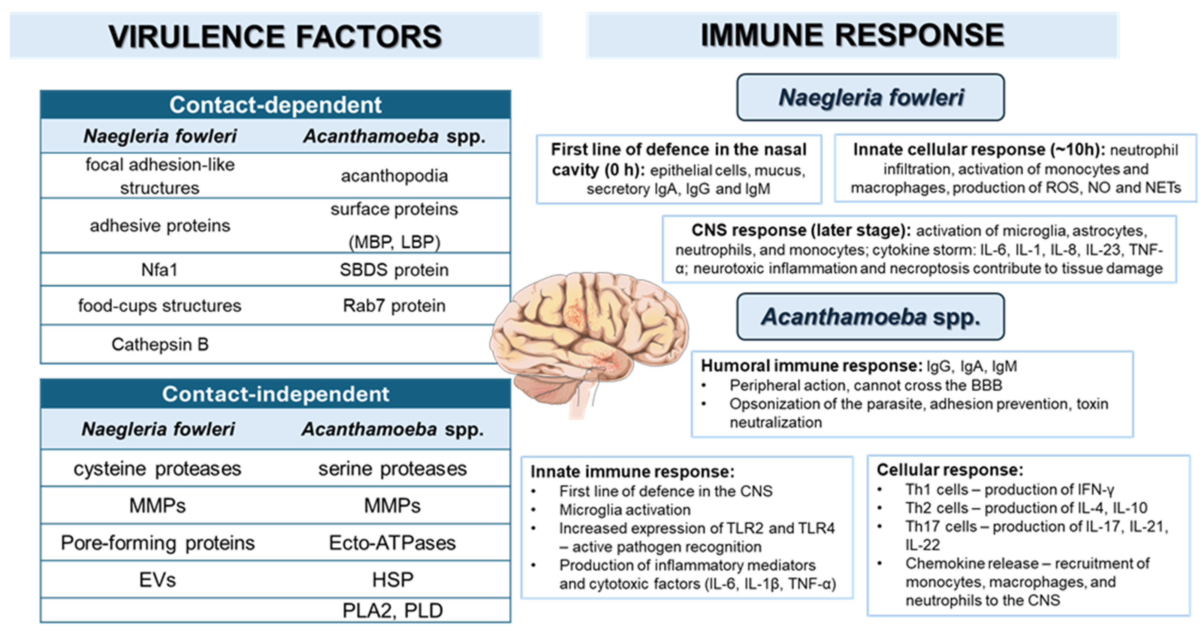
Importantly, neuroinflammation in GAE appears to be temporally heterogeneous and may remain limited during the earliest stages of CNS invasion. In an experimental intranasal model of Acanthamoeba genotype T4 infection, trophozoites crossed the nasal and olfactory epithelium through slow, contact-dependent migration between intercellular junctions, without overt cytolysis or prominent host inflammation. These findings suggest that early Acanthamoeba neuroinfection may be initially driven by mechanically mediated tissue penetration before a fully developed inflammatory response emerges [131]. As infection progresses and amoebae reach deeper CNS compartments, innate immune mechanisms become increasingly relevant. Microglia recognize pathogens through pattern recognition receptors, including Toll-like receptors (TLRs), leading to cellular activation and inflammatory signaling [8,132]. In murine infection models, increased expression of TLR2 and TLR4 has been detected in neural and vascular CNS-associated cells, indicating active pathogen recognition during infection [133]. Activated microglia can phagocytose Acanthamoeba trophozoites and release pro-inflammatory cytokines, including TNF-α, IL-1β, and IL-6 [132,134,135]. In vitro studies using Acanthamoeba castellanii genotype T4 have also shown induction of both pro-inflammatory cytokines and IL-10 in human monocytes and macrophages, suggesting simultaneous inflammatory activation and immune modulation [136]. Adaptive immune responses during CNS Acanthamoeba infection appear limited and predominantly cell-mediated. T helper 1 (Th1) responses, characterized by interferon gamma (IFN-γ), enhance microglial and macrophage activity and may amplify inflammatory signaling [86,137,138,139]. In immunosuppressed mice, Th1-skewed responses may contribute to greater tissue injury, whereas immunocompetent hosts also demonstrate T helper 2 (Th2) and T helper 17 (Th17) activity [86]. Th2-associated cytokines, including interleukin-4 (IL-4) and IL-10, may help limit excessive inflammation and protect neural tissue [140]. Th17 responses may be either protective or pathogenic depending on the cytokine profile and inflammatory context [141,142]. Interleukin-17 receptor (IL-17R)-mediated chemokine production by CNS-resident and endothelial cells promotes recruitment of monocytes, macrophages, neutrophils, and lymphocytes, supporting parasite clearance but potentially increasing BBB permeability and secondary inflammation [142,143]. Thus, balanced Th1/Th2/Th17 activity, potentially modulated by neurotrophic signaling, may be important for infection control while limiting CNS damage [144]. Humoral immunity appears to play a more limited role in cerebral acanthamoebiasis because antibody penetration across the BBB is restricted and antibodies are generally insufficient to eliminate amoebae within neural tissue [145]. Nevertheless, infection can stimulate IgG, IgM, and IgA responses that may neutralize trophozoites in blood or peripheral tissues, inhibit adhesion and motility, and reduce amoebic cytotoxicity [85,146,147]. Antibodies and complement may also promote lysis or opsonization in the presence of phagocytes, thereby supporting peripheral parasite clearance [65,148,149]. Overall, cellular immunity appears to be the principal determinant of CNS defense, whereas humoral responses may contribute mainly during early or peripheral stages of infection. Key virulence factors and host immune responses underlying the pathogenesis of PAM and GAE are summarized in Figure 2.

4. Diagnostic Strategies for Free-Living Amoebae Infections

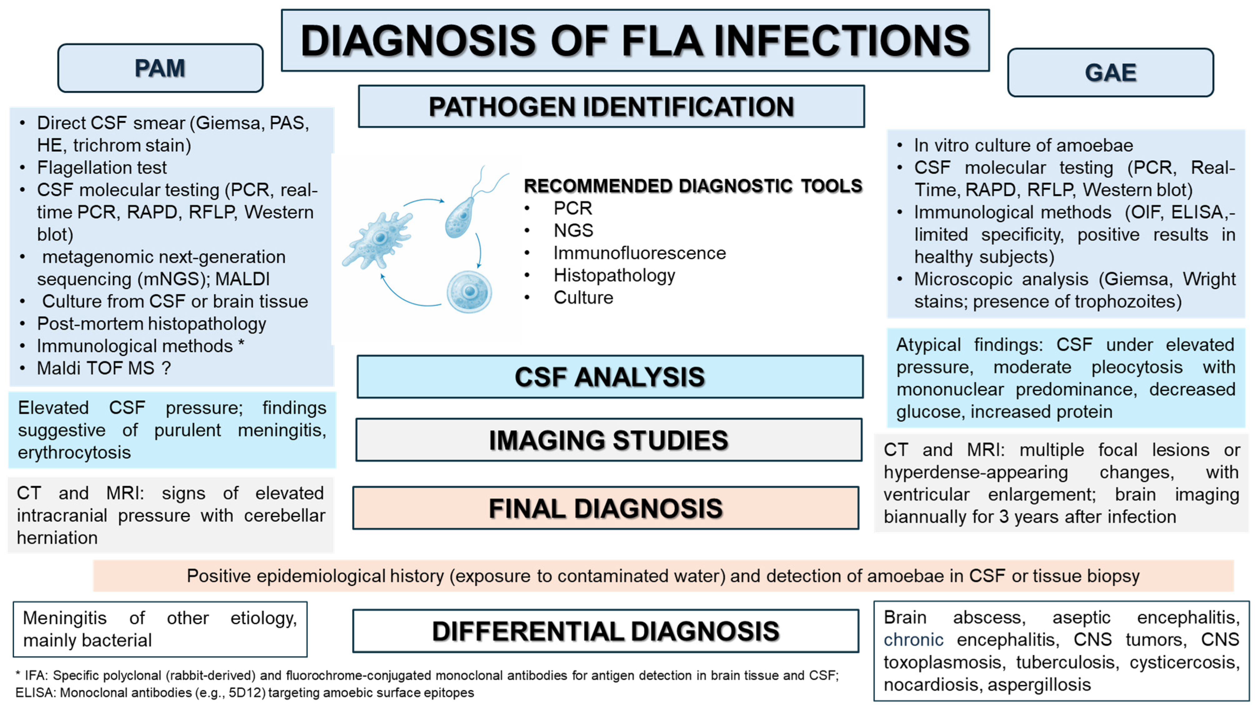
Diagnostic evaluation of FLA infections combines both conventional and advanced diagnostic approaches, such as microscopy, culture, PCR-based assays, direct immunofluorescence, and immunohistochemistry [22,40]. Given the high mortality associated with FLA infections, rapid and accurate diagnosis is essential for timely therapeutic intervention [150]. Figure 3 outlines the proposed diagnostic algorithm.
In clinical practice, initial assessment typically relies on microscopy and culture of biopsy specimens and cerebrospinal fluid, increasingly complemented by molecular diagnostic techniques [15]. Conventional and real-time PCR methods provide high sensitivity and specificity, whereas multiplex PCR enables rapid simultaneous detection of multiple pathogens, thereby improving diagnostic efficiency and clinical decision-making [40,151]. Despite advances in molecular diagnostics, early detection of FLA infections remains challenging due to nonspecific clinical manifestations and limited clinical awareness. These limitations highlight the need for improved rapid diagnostic approaches.

5. Treatment

Treatment of central nervous system infections caused by FLA remains highly challenging, as no uniformly effective therapy has been established. Current management relies largely on multidrug combinations derived from limited clinical guidelines and case reports [152,153]. Delayed diagnosis, poor BBB penetration, systemic toxicity, and the limited in vivo efficacy of compounds demonstrating activity only in vitro contribute to mortality rates approaching 80–98% [23,154]. These limitations highlight the urgent need for novel therapeutic strategies and improved drug delivery systems capable of overcoming BBB-related pharmacological barriers. An important determinant of therapeutic efficacy in FLA infections is the developmental stage of the amoeba. Trophozoites are metabolically active, invasive forms responsible for tissue destruction and are generally more susceptible to pharmacological treatment. In contrast, cysts are dormant or low-metabolic forms protected by a resistant wall, which limits drug penetration and increases tolerance to anti-amoebic agents [155]. This distinction is clinically important in Acanthamoeba spp. infections, where both trophozoites and cysts may contribute to persistence and relapse. By contrast, N. fowleri does not form cysts in human tissue; therefore, PAM therapy is directed primarily against trophozoites [9]. For GAE, an ideal regimen should target both trophozoites and cysts; however, no standardized stage-specific treatment strategy currently exists.
Therapeutic approaches can be broadly categorized into (i) conventional regimens based on the Centers for Disease Control and Prevention (CDC) recommendations; (ii) therapeutic combinations and surgical interventions associated with reported survival; and (iii) emerging and experimental therapies, including repurposed drugs, metal-based systems, drug conjugates, and phytochemical-enhanced strategies.

5.1. Conventional Regimens (CDC-Based Therapy)

Current treatment of PAM and GAE relies on aggressive multidrug combinations recommended by CDC [23,152,156,157,158]. For PAM, CDC-recommended regimens typically include amphotericin B; azithromycin; an azole antifungal, such as fluconazole or posaconazole; rifampicin; miltefosine; and adjunctive dexamethasone to mitigate cerebral inflammation and reduce intracranial pressure [156]. Therapeutic hypothermia and aggressive management of cerebral edema have also been used in selected survivors as supportive interventions [159]. For GAE, CDC-recommended multidrug therapy includes pentamidine; sulfadiazine; flucytosine; miltefosine; and an azole antifungal, most commonly fluconazole or voriconazole [23,152,157,158]. Additional agents, including amphotericin B, metronidazole, trimethoprim–sulfamethoxazole, rifampicin, ethambutol, acyclovir, or corticosteroids, have been used in selected cases depending on disease severity, host immune status, drug availability, and clinical response [152,157]. Despite aggressive multidrug therapy, outcomes remain poor, highlighting the urgent need for novel therapeutic strategies, improved CNS drug delivery approaches, and early diagnostic intervention.

5.2. Reported Survival Outcomes and Survival-Associated Therapeutic Combinations

Survival from PAM remains exceedingly rare. Documented survivors have generally been treated with early, aggressive multidrug therapy, most commonly including amphotericin B, azithromycin, fluconazole, rifampicin, miltefosine, and adjunctive dexamethasone [156]. Laboratory-confirmed survival cases are summarized in Table 2. Although approximately 32 PAM survivors have been described in the broader literature, only nine cases fulfilled laboratory criteria confirming N. fowleri infection. In the remaining reports, the causative organism was not definitively confirmed, and diagnosis relied largely on indirect or morphological evidence [160,161,162]. This distinction is important because infections caused by other FLA cannot be excluded, particularly in regions with limited access to confirmatory molecular diagnostics [162]. Across confirmed survival cases, favorable outcomes appear to be associated with early recognition, rapid initiation of combination therapy, aggressive management of intracranial complications, and access to miltefosine-containing regimens when available. In GAE, survival is also uncommon. By 2020, only 19 survival cases had been reported among immunocompetent and immunosuppressed patients [96,108,163]. Reported favorable outcomes were associated with multidrug therapy, including combinations of miltefosine, metronidazole, rifampicin, azithromycin, pentamidine isethionate, and co-trimoxazole. In selected immunocompetent patients, surgical excision or debulking of focal abscess-like lesions combined with pharmacological therapy, particularly miltefosine-based regimens, was associated with survival [76,96,108,163]. By contrast, fatal outcomes were frequently reported in patients receiving less intensive regimens, such as fluconazole and co-trimoxazole alone [75]. Collectively, reported survival cases suggest that favorable outcomes in PAM and GAE are associated with early diagnosis; rapid initiation of multidrug therapy; effective control of intracranial complications; and, in selected GAE cases, surgical excision or debulking of focal lesions. However, these observations are derived mainly from case reports and small case series and should therefore be interpreted as survival-associated patterns rather than evidence-based standardized treatment regimens.

5.3. Emerging and Experimental Therapeutic Strategies

Several emerging compounds and delivery platforms have demonstrated anti-amoebic activity and may improve future therapeutic outcomes when incorporated into rational combination regimens. However, at present, most evidence remains preclinical; therefore, these approaches should be interpreted as experimental strategies rather than established treatment options. Novel drug delivery strategies are also being explored to improve CNS penetration and reduce systemic toxicity. Intranasal administration of amphotericin B and dexamethasone using automated nebulization systems may enable controlled delivery along the olfactory route, potentially bypassing the BBB and increasing drug accumulation in the olfactory bulb and frontal cortex [25,170]. Although still experimental, this approach directly addresses two major therapeutic barriers in PAM: delayed CNS drug exposure and dose-limiting systemic toxicity.

5.3.1. Repurposed Drugs

Drug repurposing represents an attractive strategy because several approved compounds have well-characterized pharmacological profiles and may be rapidly redirected toward rare CNS infections. Nitroxoline has demonstrated potent in vitro activity against both N. fowleri and Acanthamoeba spp. and has been evaluated within Investigational New Drug (IND) protocols [156,157,171,172]. Auranofin, a gold-containing antirheumatic drug, exhibits inhibitory activity against both Acanthamoeba spp. and N. fowleri and has attracted interest because of its reported BBB penetration [173,174]. Other repurposed agents, including statins, prochlorperazine-based combinations, digoxin, and amlodipine, have demonstrated amoebicidal activity in vitro through disruption of cellular homeostasis or apoptosis-like mechanisms [175,176]. Although these agents have not yet been shown to improve survival in clinical settings, their pharmacological properties and anti-amoebic activity support further evaluation as components of future combination regimens.

5.3.2. Stage-Specific Anti-Amoebic Biocides, Diamidines and Photodynamic Therapy

Several established anti-amoebic agents used primarily for the treatment of Acanthamoeba keratitis have provided important experimental data on stage-specific susceptibility. Biguanides, particularly polyhexamethylene biguanide (PHMB) and chlorhexidine, as well as diamidines including propamidine, hexamidine, dibromopropamidine, and pentamidine, have shown activity against Acanthamoeba trophozoites and cysts in vitro [177,178]. This distinction is clinically relevant because cysts may survive therapy, subsequently excyst, and contribute to infection recurrence. However, the efficacy of some agents, including miltefosine and selected diamidines, appears variable depending on assay conditions and the developmental stage tested [177]. Photodynamic therapy, including photodynamic therapy based on chlorin e6 (chlorin e6-based PDT), has also demonstrated dose-dependent amoebicidal effects against both trophozoites and cysts, although this approach remains experimental and its applicability to CNS disease is uncertain [178]. Together, these findings highlight the need to evaluate candidate therapies against both developmental stages rather than trophozoites alone.

5.3.3. Nanotechnology-Based Systems: Metal-Based, Drug-Conjugate and Phytochemical-Enhanced Approaches

Nanotechnology-based systems are being explored to improve drug stability, bioavailability, CNS delivery, and host-cell selectivity in amoebic infections [179,180]. These approaches can be grouped into three main categories. First, metal and metal oxide nanoparticles (NPs), including zinc oxide nanoparticles (ZnO NPs), silver nanoparticles (AgNPs), and gold nanoparticles (AuNPs), exhibit intrinsic anti-amoebic activity and may exert their effects through membrane disruption and oxidative stress-related mechanisms [181,182,183,184,185,186]. Second, drug–nanoparticle conjugates, such as ZnO–β-cyclodextrin–ceftriaxone systems, have been shown to reduce the growth of Acanthamoeba castellanii, cytopathogenicity, and encystation with limited host-cell toxicity [187]. Related ZnO-based conjugates have also demonstrated enhanced activity against N. fowleri while preserving host-cell viability [181]. Third, phytochemical-enhanced nanoparticle systems, including nanoparticles combined with curcumin, terpenes, or other plant-derived compounds, may increase anti-amoebic efficacy, including activity against cyst stages, while reducing toxicity [25,182,183,184]. Although these platforms remain preclinical, they may help address two major barriers to survival: inadequate CNS drug delivery and systemic toxicity.

5.3.4. Plant-Derived Compounds and Immunological Strategies

Plant-derived compounds represent an additional experimental therapeutic avenue. Flavonoids, terpenoids, and other secondary metabolites, including demethoxycurcumin, kaempferol, resveratrol, betulinic acid, and extracts from Pinus densiflora, Artemisia annua, and other plants, have demonstrated amoebicidal activity against N. fowleri and Acanthamoeba spp. in vitro [188,189,190,191,192,193]. These compounds may reduce trophozoite viability, interfere with encystation as well as adhesion, and decrease cytopathogenicity while maintaining relatively low host-cell toxicity. However, their clinical translation requires standardized extract characterization, pharmacokinetic evaluation, toxicity assessment, and validation in relevant in vivo CNS infection models.
Immunological strategies, including multi-epitope vaccine candidates against N. fowleri, have also been proposed using in silico approaches [194,195]. These platforms aim to induce humoral and cellular immune responses; however, they remain at an early stage and require experimental validation. Overall, emerging therapies may improve future survival by enhancing CNS delivery, reducing systemic toxicity, targeting cyst persistence, and expanding the range of drugs available for rational combination therapy. At present, however, most approaches remain preclinical and should not be regarded as substitutes for rapid diagnosis, aggressive multidrug treatment, and effective management of intracranial complications.

6. Conclusions and Future Directions: A Translational Roadmap

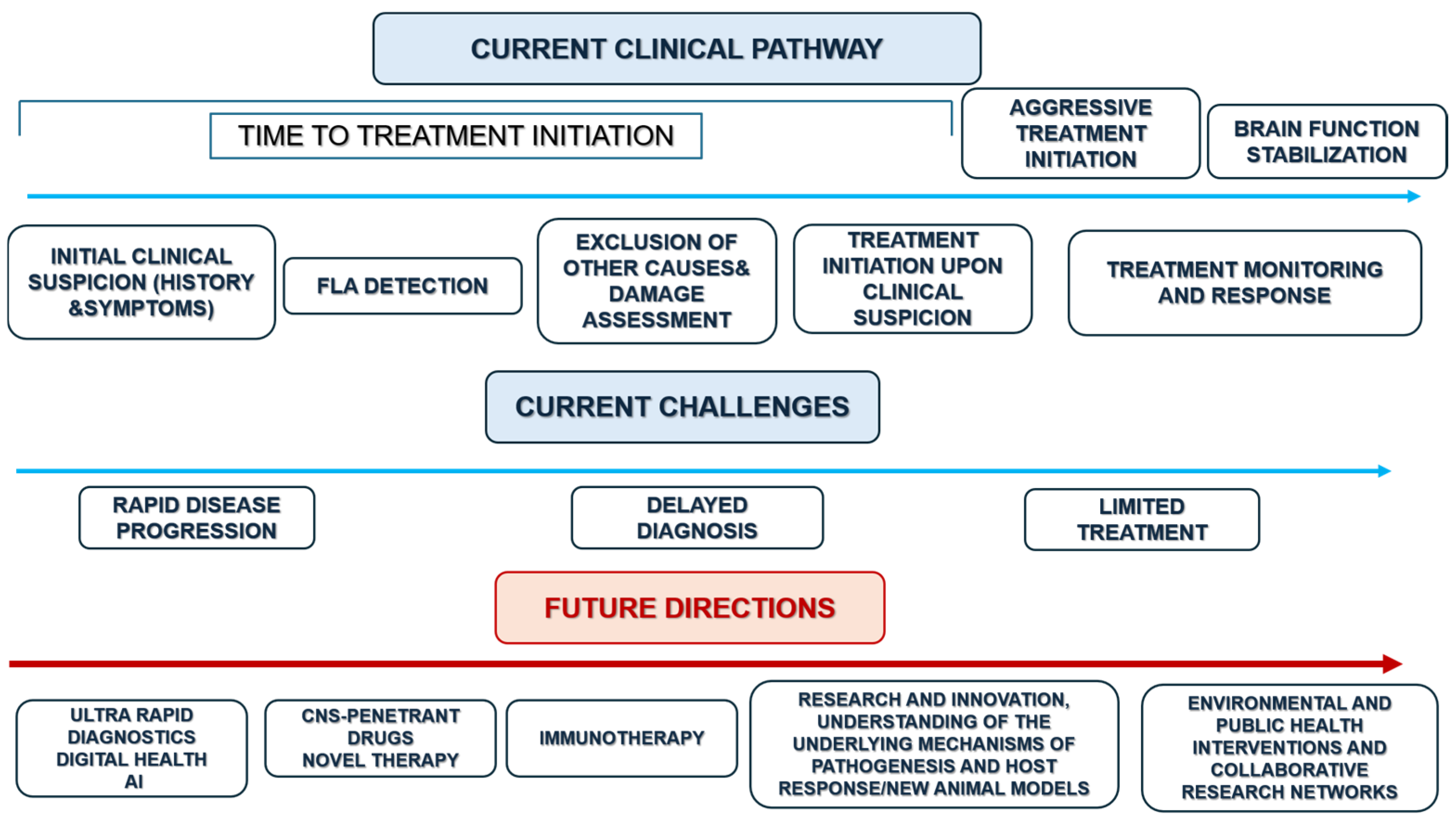
Free-living amoebae, particularly N. fowleri and Acanthamoeba spp., remain among the most devastating causes of rare central nervous system infections, with persistently high mortality driven by rapid disease progression, delayed diagnosis, and limited therapeutic efficacy. Despite advances in molecular diagnostics, pathogen biology, and experimental therapies, substantial improvements in patient outcomes will require a shift from reactive management toward proactive strategies based on early recognition, rapid initiation of combination therapy, and improved understanding of host–pathogen interactions. Future progress should prioritize ultra-rapid diagnostic platforms, novel BBB-penetrating therapies, host-directed and immunomodulatory approaches, and more predictive experimental models, supported by enhanced environmental surveillance and global clinical awareness within the One Health framework. Given that PAM and GAE are rare but frequently fatal diseases, progress will require coordinated international collaboration, shared clinical registries, harmonized diagnostic workflows, and standardized treatment reporting. As illustrated in Figure 4, an integrated translational roadmap linking diagnostics, therapeutic innovation, surveillance, and collaborative research is urgently needed to translate emerging scientific advances into improved survival.

Author Contributions

Conceptualization, O.P.-P. and N.Ł.-A.; methodology, O.P.-P.; investigation and data curation, O.P.-P. and N.Ł.-A.; writing—original draft preparation O.P.-P., N.Ł.-A. and K.K.; writing—review and editing, O.P.-P., N.Ł.-A., K.K. and D.K.-B.; visualization O.P.-P. and N.Ł.-A., supervision, N.Ł.-A. and K.K. All authors have read and agreed to the published version of the manuscript.

Funding

This research received no external funding.

Institutional Review Board Statement

Not applicable. This review article did not involve any studies with human participants or animals performed by the authors.

Informed Consent Statement

Not applicable.

Data Availability Statement

No new data were created or analyzed in this study. Data sharing is not applicable to this article.

Conflicts of Interest

The authors declare no conflicts of interest.

Abbreviations

The following abbreviations are used in this manuscript:
ADP adenosine diphosphate
BBBblood–brain barrier
CDCCenters for Disease Control and Prevention
CNScentral nervous system
CSFcerebrospinal fluid
ECMextracellular matrix
EGFRepidermal growth factor receptor
EVsextracellular vesicles
FLAfree-living amoeba
GAEgranulomatous amoebic encephalitis
HSP70heat shock protein 70
IFN-γinterferon gamma
ILinterleukin
LBPlaminin-binding protein
MALDI-TOF MSmatrix-assisted laser desorption/ionization time-of-flight mass spectrometry
MBPmannose-binding protein
MMPsmatrix metalloproteinases
mNGSmetagenomic next-generation sequencing
NETsneutrophil extracellular traps
Nfa1Naegleria fowleri adhesion protein 1
NOnitric oxide
OIFindirect immunofluorescence
PAMprimary amoebic meningoencephalitis
PI3Kphosphatidylinositol 3-kinase
PLA2phospholipase A2
PLDphospholipase D
RAPDrandom amplified polymorphic DNA
RFLPrestriction fragment length polymorphism
ROSreactive oxygen species
ThT helper cell
TLRsToll-like receptors
TNF-αtumor necrosis factor alpha
ZO-1zonula occludens-1

References

  1. Elsheikha, H.M.; Khan, N.A. Protozoa traversal of the blood-brain barrier to invade the central nervous system. FEMS Microbiol. Rev. 2010, 34, 532–553. [Google Scholar] [CrossRef] [PubMed]
  2. de Brito Duval, I.; Cardozo, M.E.; Souza, J.L.N.; de Medeiros Brito, R.M.; Fujiwara, R.T.; Bueno, L.L.; Magalhães, L.M.D. Parasite infections: How inflammation alters brain function. Trends Parasitol. 2025, 41, 115–128. [Google Scholar] [CrossRef] [PubMed]
  3. Garcia, H.H. Parasitic Infections of the Nervous System. Continuum 2021, 27, 943–962. [Google Scholar] [CrossRef] [PubMed]
  4. Wohlfert, E.A.; Blader, I.J.; Wilson, E.H. Brains and Brawn: Toxoplasma Infections of the Central Nervous System and Skeletal Muscle. Trends Parasitol. 2017, 33, 519–531. [Google Scholar] [CrossRef] [PubMed]
  5. Güémez, A.; García, E. Primary Amoebic Meningoencephalitis by Naegleria fowleri: Pathogenesis and Treatments. Biomolecules 2021, 11, 1320. [Google Scholar] [CrossRef] [PubMed]
  6. Figarella, K.; Wolburg, H.; Garaschuk, O.; Duszenko, M. Microglia in neuropathology caused by protozoan parasites. Biol. Rev. Camb. Philos. Soc. 2020, 95, 333–349. [Google Scholar] [CrossRef] [PubMed]
  7. Siddiqui, R.; Maciver, S.K.; Khan, N.A. Different Roads to the Brain: A Brief Overview of Divergent Pathways and Convergent Mechanisms in Amoebic Neuroinvasion. Infect. Genet. Evol. 2026, 138, 105888. [Google Scholar] [CrossRef] [PubMed]
  8. Kot, K.; Łanocha-Arendarczyk, N.; Kosik-Bogacka, D. Immunopathogenicity of Acanthamoeba spp. in the Brain and Lungs. Int. J. Mol. Sci. 2021, 22, 1261. [Google Scholar] [CrossRef] [PubMed]
  9. Vijayan, V.; Tristram, D.; Sundareshan, V. Naegleria Infection and Primary Amebic Meningoencephalitis. In StatPearls; StatPearls Publishing: Treasure Island, FL, USA, 2026. Available online: https://www.ncbi.nlm.nih.gov/books/NBK535447/ (accessed on 28 June 2026).
  10. Siddiqui, R.; Khan, N.A. Primary amoebic meningoencephalitis caused by Naegleria fowleri: An old enemy presenting new challenges. PLoS Negl. Trop. Dis. 2014, 8, e3017. [Google Scholar] [CrossRef] [PubMed]
  11. Chimelli, L. A morphological approach to the diagnosis of protozoal infections of the central nervous system. Pathol. Res. Int. 2011, 2011, 290853. [Google Scholar] [CrossRef] [PubMed]
  12. Marri, A.R.; Hamer, D.H.; Sherchan, S.; Shioda, K. Naegleria fowleri and the future of surveillance: A one-health call to action. One Health 2025, 21, 101215. [Google Scholar] [CrossRef] [PubMed]
  13. DiSabato, D.J.; Quan, N.; Godbout, J.P. Neuroinflammation: The devil is in the details. J. Neurochem. 2016, 139, 136–153. [Google Scholar] [CrossRef] [PubMed]
  14. Rîpă, C.; Cobzaru, R.G.; Rîpă, M.R.; Maștaleru, A.; Oancea, A.; Cumpăt, C.M.; Leon, M.M. Naegleria fowleri Infections: Bridging Clinical Observations and Epidemiological Insights. J. Clin. Med. 2025, 14, 526. [Google Scholar] [CrossRef] [PubMed]
  15. Alanazi, A.; Younas, S.; Ejaz, H.; Alruwaili, M.; Alruwaili, Y.; Mazhari, B.B.Z.; Atif, M.; Junaid, K. Advancing the understanding of Naegleria fowleri: Global epidemiology, phylogenetic analysis, and strategies to combat a deadly pathogen. J. Infect. Public Health 2025, 18, 102690. [Google Scholar] [CrossRef] [PubMed]
  16. Khan, N.A. Acanthamoeba: Biology and increasing importance in human health. FEMS Microbiol. Rev. 2006, 30, 564–595. [Google Scholar] [CrossRef] [PubMed]
  17. Indolfi, C.; Colosimo, S.; Mondillo, G.; Perrotta, A.; Frattolillo, V.; Decimo, F.; del Giudice, M.M. Naegleria fowleri, the “brain-eating amoeba”: An invisible enemy in nasal irrigation with untreated water. Ital. J. Pediatr. Allergy Immunol. 2025, 38, 3–7. [Google Scholar] [CrossRef]
  18. DeFelice, J. Naegleria fowleri and Risk of Primary Amoebic Meningoencephalitis in a Changing Climate: A Scoping Review of Biomedical Literature. Int. J. Environ. Res. Public Health 2026, 23, 764. [Google Scholar] [CrossRef] [PubMed]
  19. Sarantopoulos, A.; Quattrocchi, A.; Kopsidas, I.; Cornely, O.A.; Seidel, D.; Grotto, I.; Pana, Z.D. Global Review on Naegleria fowleri Cases: Contemporary Epidemiology, Diagnosis, Treatment and Outcomes. Infect. Dis. Rep. 2026, 18, 25. [Google Scholar] [CrossRef] [PubMed]
  20. Cope, J.R.; Murphy, J.; Kahler, A.; Gorbett, D.G.; Ali, I.; Taylor, B.; Corbitt, L.; Roy, S.; Lee, N.; Roellig, D.; et al. Primary Amebic Meningoencephalitis Associated with Rafting on an Artificial Whitewater River: Case Report and Environmental Investigation. Clin. Infect. Dis. 2018, 66, 548–553. [Google Scholar] [CrossRef] [PubMed]
  21. Phatthararangrong, N.; Chantratong, N.; Jitsurong, S. Bacteriological quality of holywater from Thai temples in Songkhla Province, southern Thailand. J. Med. Assoc. Thail. 1998, 81, 547–550. [Google Scholar]
  22. Shaukat, A.; Khaliq, N.; Riaz, R.; Munsab, R.; Ashraf, T.; Raufi, N.; Shah, H. Noninvasive diagnostic biomarkers, genomic profiling, and advanced microscopic imaging in the early detection and characterization of Naegleria fowleri infections leading to primary amebic meningoencephalitis (PAM). Ann. Med. Surg. 2024, 86, 2032–2048. [Google Scholar] [CrossRef] [PubMed]
  23. Mungroo, M.R.; Khan, N.A.; Maciver, S.; Siddiqui, R. Opportunistic free-living amoebal pathogens. Pathog. Glob. Health 2022, 116, 70–84. [Google Scholar] [CrossRef] [PubMed]
  24. Nadeem, A.; Malik, I.A.; Afridi, E.K.; Shariq, F. Naegleria fowleri outbreak in Pakistan: Unveiling the crisis and path to recovery. Front. Public Health 2023, 11, 1266400. [Google Scholar] [CrossRef] [PubMed]
  25. Siddiqui, R.; Maciver, S.K.; Khan, N.A. Naegleria fowleri: Emerging therapies and translational challenges. Expert Rev. Anti-Infect. Ther. 2025, 23, 753–761. [Google Scholar] [CrossRef] [PubMed]
  26. Mitra, A.; Brito Goulart, D. From nose to neurons: The lethal journey of the brain-eating amoeba Naegleria fowleri. Microbe 2025, 8, 100537. [Google Scholar] [CrossRef]
  27. Guerlais, V.; Allouch, N.; Moseman, E.A.; Wojciechowska, A.W.; Wojciechowski, J.W.; Marcelino, I. Transcriptomic profiling of “brain-eating amoeba” Naegleria fowleri infection in mice: The host and the protozoa perspectives. Front. Cell. Infect. Microbiol. 2024, 14, 1490280. [Google Scholar] [CrossRef] [PubMed]
  28. Kuang, Y.; Li, Q.; Li, H.; Guo, Y.; Wang, J.; Peng, C.; He, P.; Chen, S.; Nian, S.; Feng, L. A case report of primary amebic meningoencephalitis in a Chinese adult male. Parasitol. Res. 2026, 125, 32. [Google Scholar] [CrossRef] [PubMed]
  29. Kasprzak, W. Free-Living Amoeba with Pathogenic Properties to Human and Animals, 1st ed.; Państwowe Wydawnictwo Naukowe: Warsaw, Poland, 1985; pp. 8–15. [Google Scholar]
  30. Borkens, Y. The Pathology of the Brain Eating Amoeba Naegleria fowleri. Indian J. Microbiol. 2024, 64, 1384–1394. [Google Scholar] [CrossRef] [PubMed]
  31. Guarner, J.; Bartlett, J.; Shieh, W.J.; Paddock, C.D.; Visvesvara, G.S.; Zaki, S.R. Histopathologic spectrum and immunohistochemical diagnosis of amebic meningoencephalitis. Mod. Pathol. 2007, 20, 1230–1237. [Google Scholar] [CrossRef] [PubMed]
  32. Kou, Y.; Zhang, J.; Wang, D.; Cui, L.; Sun, Q.; Lv, Y.; Liu, Y.; He, Z.; Zhao, Y.; Zhang, H. Rare Naegleria fowleri meningoencephalitis diagnosed via combined molecular biology and metagenomic sequencing techniques: A case report. Infect. Dis. Poverty 2025, 14, 69. [Google Scholar] [CrossRef] [PubMed]
  33. Phung, N.T.N.; Pham, H.T.; Tran, T.T.; Dinh, V.H.; Tran, N.M.; Tran, N.A.N.; Ngo, M.Q.N.; Nguyen, H.T.T.; Tran, D.K.; Le, T.K.T.; et al. Naegleria fowleri: Portrait of a Cerebral Killer. Diagnostics 2025, 15, 89. [Google Scholar] [CrossRef] [PubMed]
  34. Pimentel, L.A.; Dantas, A.F.; Uzal, F.; Riet-Correa, F. Meningoencephalitis caused by Naegleria fowleri in cattle of northeast Brazil. Res. Vet. Sci. 2012, 93, 811–812. [Google Scholar] [CrossRef] [PubMed]
  35. Henker, L.C.; Lorenzett, M.P.; Dos Santos, D.L.; Virginio, V.G.; Driemeier, D.; Rott, M.B.; Pavarini, S.P. Naegleria fowleri-associated meningoencephalitis in a cow in Southern Brazil—First molecular detection of N. fowleri in Brazil. Parasitol. Res. 2021, 120, 2873–2879. [Google Scholar] [CrossRef] [PubMed]
  36. Sohn, H.J.; Ham, A.J.; Park, A.Y.; Lee, J.H.; Park, S.; Shin, H.J.; Kim, J.H. Cloning of nf-profilin and intercellular interaction with nf-actin in Naegleria fowleri cysts. Sci. Rep. 2025, 15, 7015. [Google Scholar] [CrossRef] [PubMed]
  37. Jamerson, M.; da Rocha-Azevedo, B.; Cabral, G.A.; Marciano-Cabral, F. Pathogenic Naegleria fowleri and non-pathogenic Naegleria lovaniensis exhibit differential adhesion to, and invasion of, extracellular matrix proteins. Microbiology 2012, 158, 791–803. [Google Scholar] [CrossRef] [PubMed]
  38. Chao-Pellicer, J.; Arberas-Jiménez, I.; Sifaoui, I.; Díaz-Marrero, A.R.; Fernández, J.J.; Jamerson, M.; Piñero, J.E.; Lorenzo-Morales, J. Potential Inhibitors of Human–Naegleria fowleri Interactions: An In Vitro Extracellular Matrix-Based Model. Mar. Drugs 2025, 23, 306. [Google Scholar] [CrossRef] [PubMed]
  39. Flores Suárez, B.; Bonilla Lemus, P.; Rojas Hernández, S.; Terrazas Valdés, L.L.; Carrasco Yépez, M.M. The 72-KDA protein of Naegleria fowleri plays an important role in the adhesion of trophozoites to BALB/c mice nasal epithelium. J. Parasitol. 2024, 110, 360–370. [Google Scholar] [CrossRef] [PubMed]
  40. Dai, L.; Guo, X.R.; Chen, X.R.; Ma, M.H.; Liu, Z.H.; Lai, J.; Lu, J.; Feng, M.; Liu, X.X.; Yang, S.H. A review of the mechanism, diagnosis, and treatment of Naegleria fowleri infection. Front. Microbiol. 2025, 16, 1686695. [Google Scholar] [CrossRef] [PubMed]
  41. Kim, J.H.; Sohn, H.J.; Shin, H.J.; Walz, S.E.; Jung, S.Y. Understanding the pathogenicity of Naegleria fowleri in association with N. fowleri antigen-1 (Nfa1). Parasites Hosts Dis. 2024, 62, 385–398. [Google Scholar] [CrossRef] [PubMed]
  42. Rodríguez-Mera, I.B.; Carrasco-Yépez, M.M.; Vásquez-Moctezuma, I.; Correa-Basurto, J.; Salinas, G.R.; Castillo-Ramírez, D.A.; Rosales-Cruz, É.; Rojas-Hernández, S. Role of cathepsin B of Naegleria fowleri during primary amebic meningoencephalitis. Parasitol. Res. 2022, 121, 3287–3303. [Google Scholar] [CrossRef] [PubMed]
  43. Kang, S.Y.; Song, K.J.; Jeong, S.R.; Kim, J.H.; Park, S.; Kim, K.; Kwon, M.H.; Shin, H.J. Role of the Nfa1 protein in pathogenic Naegleria fowleri cocultured with CHO target cells. Clin. Diagn. Lab. Immunol. 2005, 12, 873–876. [Google Scholar] [CrossRef] [PubMed][Green Version]
  44. Betanzos, A.; Bañuelos, C.; Orozco, E. Host Invasion by Pathogenic Amoebae: Epithelial Disruption by Parasite Proteins. Genes 2019, 10, 618. [Google Scholar] [CrossRef] [PubMed]
  45. Dziankowska-Bartkowiak, B.; Żebrowska, A.; Joss-Wichmann, E. Stężenie metaloproteinazy 2 i 9 (MMP-2 i MMP-9) w surowicy chorych na twardzinę układową—Porównanie z ich ekspresją w chorobowo zmienionej skórze—Badania wstępne. Post. Dermatol. Alergol. 2007, 2, 73. [Google Scholar]
  46. Lam, C.; Jamerson, M.; Cabral, G.; Carlesso, A.M.; Marciano-Cabral, F. Expression of matrix metalloproteinases in Naegleria fowleri and their role in invasion of the central nervous system. Microbiology 2017, 163, 1436–1444. [Google Scholar] [CrossRef] [PubMed]
  47. Coronado-Velázquez, D.; Betanzos, A.; Serrano-Luna, J.; Shibayama, M. An In Vitro Model of the Blood-Brain Barrier: Naegleria fowleri Affects the Tight Junction Proteins and Activates the Microvascular Endothelial Cells. J. Eukaryot. Microbiol. 2018, 65, 804–819. [Google Scholar] [CrossRef] [PubMed]
  48. Herbst, R.; Ott, C.; Jacobs, T.; Marti, T.; Marciano-Cabral, F.; Leippe, M. Pore-forming polypeptides of the pathogenic protozoon Naegleria fowleri. J. Biol. Chem. 2002, 277, 22353–22360. [Google Scholar] [CrossRef] [PubMed]
  49. Castelan-Ramírez, I.; Flores-Maldonado, C.; Hernández-Martínez, D.; Salazar-Villatoro, L.; Saucedo-Campos, A.D.; Segura-Cobos, D.; Méndez-Cruz, A.R.; Omaña-Molina, M. Advances in the study of extracellular vesicles of Naegleria fowleri and their role in contact-independent pathogenic mechanisms. Parasit. Vectors 2025, 18, 164. [Google Scholar] [CrossRef] [PubMed]
  50. Chacón Camacho, N.; Steller Espinoza, M.F.; Alvarado Ocampo, J.; Osuna, A.; Retana Moreira, L.; Abrahams Sandí, E. Characterization of Naegleria fowleri from two human cases: Insights into its excretion/secretion products. Front. Cell. Infect. Microbiol. 2025, 15, 1585448. [Google Scholar] [CrossRef] [PubMed]
  51. Flores-Huerta, N.; Sánchez-Monroy, V.; Rodríguez, M.A.; Serrano-Luna, J.; Shibayama, M. A comparative study of the membrane proteins from Naegleria species: A 23-kDa protein participates in the virulence of Naegleria fowleri. Eur. J. Protistol. 2020, 72, 125640. [Google Scholar] [CrossRef] [PubMed]
  52. Chen, C.-W.; Moseman, E.A. Pro-inflammatory cytokine responses to Naegleria fowleri infection. Front. Trop. Dis. 2022, 3, 1082334. [Google Scholar] [CrossRef] [PubMed]
  53. Moseman, E.A. Battling brain eating amoeba: Enigmas surrounding immunity to Naegleria fowleri. PLoS Pathog. 2020, 16, e1008406. [Google Scholar] [CrossRef] [PubMed]
  54. Rojas-Ortega, S.; Lazcano-Hernández, G.; Rivera-Aguilar, V.; De Jesús Paredes Catalán, M.; Lares-Villa, F.; Serrano-Luna, J. Role of FcγRIII in the nasal cavity of BALB/c mice in the primary amebic meningoencephalitis protection model. Parasit. Res. 2023, 122, 2147–2160. [Google Scholar] [CrossRef] [PubMed]
  55. Rodríguez-Mera, I.B.; Ortiz-Avalos, M.; Rojas-Hernández, S.; Hernández-Rivas, R.; Serrano-Luna, J. Identification of Naegleria fowleri antigens recognized by serum antibodies from people of Mexicali Valley, México. Parasit. Res. 2025, 124, 12345–12356. [Google Scholar] [CrossRef] [PubMed]
  56. Whiteman, L.Y.; Marciano-Cabral, F. Resistance of highly pathogenic Naegleria fowleri amoebae to complement-mediated lysis. Infect. Immun. 1989, 57, 3869–3875. [Google Scholar] [CrossRef] [PubMed]
  57. Jahangeer, M.; Mahmood, Z.; Munir, N.; Waraich, U.E.; Tahir, I.M.; Akram, M.; Ali Shah, S.M.; Zulfqar, A.; Zainab, R. Naegleria fowleri: Sources of Infection, Pathophysiology, Diagnosis, and Management: A Review. Clin. Exp. Pharmacol. Physiol. 2020, 47, 199–212. [Google Scholar] [CrossRef] [PubMed]
  58. Lê, H.G.; Kang, J.M.; Võ, T.C.; Na, B.K. Naegleria fowleri Cathepsin B induces a pro-inflammatory immune response in BV-2 microglial cells via NF-κB and AP-1 dependent-MAPK signaling pathway. Int. J. Mol. Sci. 2022, 23, 8388. [Google Scholar] [CrossRef] [PubMed]
  59. Retana Moreira, L.; González-Morales, J.C.; Guevara-Flores, A.; Bonilla-Lemus, P.; Rodríguez-Tovar, L.E.; Ramón-Luing, L.A. Providing an in vitro depiction of microglial cells challenged with immunostimulatory extracellular vesicles of Naegleria fowleri. Front. Microbiol. 2024, 14, 1346021. [Google Scholar] [CrossRef] [PubMed]
  60. Kim, J.H.; Sohn, J.H.; Lee, S.H.; Kwon, D.; Shin, H.J. Induction of interleukin-8 by Naegleria fowleri lysates requires activation of extracellular signal-regulated kinase in human astroglial cells. Parasit. Res. 2012, 111, 587–592. [Google Scholar] [CrossRef] [PubMed]
  61. Kim, J.H.; Song, A.R.; Sohn, H.J.; Lee, J.; Yoo, J.K.; Kwon, D.; Shin, H.J. IL-1β and IL-6 activate inflammatory responses of astrocytes against Naegleria fowleri infection via the modulation of MAPKs and AP-1. Parasite Immunol. 2013, 35, 120–128. [Google Scholar] [CrossRef] [PubMed]
  62. Jeong, S.R.; Lee, S.C.; Song, K.J.; Park, S.; Kim, K.; Kwon, M.H.; Im, K.I.; Shin, H.J. Expression of the nfa1 gene cloned from pathogenic Naegleria fowleri in nonpathogenic N. gruberi enhances cytotoxicity against CHO target cells in vitro. Infect. Immun. 2005, 73, 4098–4105. [Google Scholar] [CrossRef] [PubMed]
  63. Kim, J.H.; Sohn, H.J.; Yoo, J.K.; Kang, H.; Seong, G.S.; Chwae, Y.J.; Kim, K.; Park, S.; Shin, H.J. NLRP3 inflammasome activation in THP-1 target cells triggered by pathogenic Naegleria fowleri. Infect. Immun. 2016, 84, 2422–2428. [Google Scholar] [CrossRef] [PubMed]
  64. Song, K.J.; Jang, Y.S.; Lee, Y.A.; Kim, K.A.; Lee, S.K.; Shin, M.H. Reactive oxygen species-dependent necroptosis in Jurkat T cells induced by pathogenic free-living Naegleria fowleri. Parasite Immunol. 2011, 33, 390–400. [Google Scholar] [CrossRef] [PubMed]
  65. Siddiqui, R.; Khan, N.A. Biology and pathogenesis of Acanthamoeba. Parasit. Vectors 2012, 5, 6. [Google Scholar] [CrossRef] [PubMed]
  66. Koyun, İ.; Kolören, Z.; Karaman, Ü.; Tsiami, A.; Karanis, P. Acanthamoeba spp. in river water samples from the Black Sea region, Turkey. J. Water Health 2020, 18, 186–199. [Google Scholar] [CrossRef] [PubMed]
  67. Karaman, U.; Koloren, Z.; Karanis, P. Survey and first report of Acanthamoeba T4 genotype in natural spring water resources in the Black Sea, Turkey. J. Water Health 2022, 20, 193–204. [Google Scholar] [CrossRef] [PubMed]
  68. Hassan, A.; Farouk, H.; Hassanein, F.; Abdul-Ghani, R.; Abdelhady, A.H. Acanthamoeba contamination of hemodialysis and dental units in Alexandria, Egypt: A neglected potential source of infection. J. Infect. Public Health 2012, 5, 304–310. [Google Scholar] [CrossRef] [PubMed]
  69. Gabr, N.S.; Mohamed, R.M.; Belal, U.S.; Abdel-Fatah, M.M.; Ahmed, R.F.; Abdel Gaber, N.A.T.; Abdel-Hafeez, E.H. Isolation and Identification of Pathogenic Acanthamoeba Species from Air Conditioning Systems, Egypt. Jpn. J. Infect. Dis. 2021, 74, 180–186. [Google Scholar] [CrossRef] [PubMed]
  70. Huang, L.; Suhler, E.B.; Rosenberg, C.; Ta Kim, D.; Winthrop, K.L.; Doan, T.; Lin, P. Acanthamoeba-associated retinitis successfully treated with intravitreal and systemic antimicrobials. Am. J. Ophthalmol. Case Rep. 2023, 32, 101902. [Google Scholar] [CrossRef] [PubMed]
  71. Wang, Y.; Jiang, L.; Zhao, Y.; Ju, X.; Wang, L.; Jin, L.; Fine, R.D.; Li, M. Biological characteristics and pathogenicity of Acanthamoeba. Front. Microbiol. 2023, 14, 1147077. [Google Scholar] [CrossRef] [PubMed]
  72. Putaporntip, C.; Kuamsab, N.; Nuprasert, W.; Rojrung, R.; Pattanawong, U.; Tia, T.; Yanmanee, S.; Jongwutiwes, S. Analysis of Acanthamoeba genotypes from public freshwater sources in Thailand reveals a new genotype, T23 Acanthamoeba bangkokensis sp. nov. Sci. Rep. 2021, 11, 17290. [Google Scholar] [CrossRef] [PubMed]
  73. Corsaro, D. Correction to: Update on Acanthamoeba phylogeny. Parasit. Res. 2021, 120, 1927–1928. [Google Scholar] [CrossRef] [PubMed]
  74. Haston, J.C.; O’Laughlin, K.; Matteson, K.; Roy, S.; Qvarnstrom, Y.; Ali, I.K.M.; Cope, J.R. The epidemiology and clinical features of non-keratitis Acanthamoeba infections in the United States, 1956–2020. Open Forum Infect. Dis. 2023, 10, ofac682. [Google Scholar] [CrossRef] [PubMed]
  75. Belaidi, K.; Nissrin, L.; Zahlane, S.; Chraa, M.; Kissani, N. Challenges in diagnosing and treating granulomatous amebic encephalitis: A case report of fatal Acanthamoeba spp. encephalitis in an immunocompetent patient. J. Neurol. Res. 2025, 15, 51–55. [Google Scholar] [CrossRef]
  76. Keane, N.A.; Lane, L.M.; Canniff, E.; Hare, D.; Doran, S.; Wallace, E.; Hutchinson, S.; Healy, M.L.; Hennessy, B.; Meaney, J.; et al. A surviving case of Acanthamoeba granulomatous amebic encephalitis in a hematopoietic stem cell transplant recipient. Am. J. Case Rep. 2020, 21, e923219. [Google Scholar] [CrossRef] [PubMed]
  77. Walochnik, J.; Scheikl, U.; Haller-Schober, E.M. Twenty years of Acanthamoeba diagnostics in Austria. J. Eukaryot. Microbiol. 2015, 62, 3–11. [Google Scholar] [CrossRef] [PubMed]
  78. Centers for Disease Control and Prevention (CDC). Free-Living Amebic Infections. CDC DPDx. Available online: https://www.cdc.gov/dpdx/freelivingamebic/index.html (accessed on 8 April 2026).
  79. Anwar, A.; Khan, N.A.; Siddiqui, R. Combating Acanthamoeba spp. cysts: What are the options? Parasit. Vectors 2018, 11, 26. [Google Scholar] [CrossRef] [PubMed]
  80. Choaji, M.; Samba-Louaka, A.; Fechtali-Moute, Z.; Aucher, W.; Pomel, S. Encystment and excystment processes in Acanthamoeba castellanii: An emphasis on cellulose involvement. Pathogens 2025, 14, 268. [Google Scholar] [CrossRef] [PubMed]
  81. Shirwadkar, C.G.; Samant, R.; Sankhe, M.; Deshpande, R.; Yagi, S.; Schuster, F.L.; Sriram, R.; Visvesvara, G.S. Acanthamoeba encephalitis in patient with systemic lupus, India. Emerg. Infect. Dis. 2006, 12, 984–986. [Google Scholar] [CrossRef] [PubMed]
  82. Aparicio, D.V.; Bejarano, J.I.C.; de Los Santos, A.M.; Ramírez-Cortinas, S.; de la O Cavazos, M. Case report: Granulomatous amebic encephalitis due to Acanthamoeba spp. in an immunocompetent pediatric patient. Am. J. Trop. Med. Hyg. 2021, 105, 167–170. [Google Scholar] [CrossRef] [PubMed]
  83. Benoit, P.; Wang, S.; Wang, C.; Chakravarti, A.; Villalba, J.A.; Ali, I.K.M.; Roy, S.; Sapp, S.G.; Reagan-Steiner, S.; Nelson, K.; et al. Brainstorm: A case of granulomatous encephalitis. J. Assoc. Med. Microbiol. Infect. Dis. Can. 2024, 9, 113–120. [Google Scholar] [CrossRef] [PubMed]
  84. Srivastava, C.; Rao, S.K.; Saroj, A.K.; Tilak, R.; Khurana, S.; Verma, A. Survivors of Acanthamoeba granulomatous encephalitis in children: A case series and review of literature. Pediatr. Infect. Dis. J. 2026, 45, e277–e278. [Google Scholar] [CrossRef] [PubMed]
  85. Marciano-Cabral, F.; Cabral, G. Acanthamoeba spp. as agents of disease in humans. Clin. Microbiol. Rev. 2003, 16, 273–307. [Google Scholar] [CrossRef] [PubMed]
  86. Łanocha-Arendarczyk, N.; Kolasa-Wołosiuk, A.; Wojciechowska-Koszko, I.; Kot, K.; Roszkowska, P.; Krasnodębska-Szponder, B.; Paczkowska, E.; Machaliński, B.; Łuczkowska, K.; Wiszniewska, B.; et al. Changes in the immune system in experimental acanthamoebiasis in immunocompetent and immunosuppressed hosts. Parasit. Vectors 2018, 11, 517. [Google Scholar] [CrossRef] [PubMed]
  87. Haston, J.C.; Ali, I.K.; Roy, S.; Roundtree, A.; Hofstetter, J.; Pierson, S.; Helmrich, E.; Torres, P.; Lockey, K.; Martines, R.B.; et al. Notes from the field: Fatal Acanthamoeba encephalitis in a patient who regularly used tap water in an electronic nasal irrigation device and a CPAP machine at home—New Mexico, 2023. MMWR Morb. Mortal. Wkly. Rep. 2025, 74, 179–180. [Google Scholar] [CrossRef] [PubMed]
  88. Schuster, F.L.; Visvesvara, G.S. Amebae and ciliated protozoa as causal agents of waterborne zoonotic disease. Vet. Parasitol. 2004, 126, 91–120. [Google Scholar] [CrossRef] [PubMed]
  89. Alfawaz, A. Radial Keratoneuritis as a Presenting Sign in Acanthamoeba Keratitis. Middle East Afr. J. Ophthalmol. 2011, 18, 252–255. [Google Scholar] [CrossRef] [PubMed]
  90. Feng, X.; Zheng, W.; Wang, Y.; Zhao, D.; Jiang, X.; Lv, S. A Rabbit Model of Acanthamoeba Keratitis That Better Reflects the Natural Human Infection. Anat. Rec. 2015, 298, 1509–1517. [Google Scholar] [CrossRef] [PubMed]
  91. Atamna, A.; Franken, L.; Grossman, T.; Rozenblatt, S.; Tobar, A.; Fichman-Horn, S.; Zvi, H.B.; Auriel, E.; Bishara, J. Acanthamoeba healyi (genotype T12) meningoencephalitis in an adult—First case in Israel. Diagn. Microbiol. Infect. Dis. 2025, 113, 116988. [Google Scholar] [CrossRef] [PubMed]
  92. Zhang, H.; Cheng, X. Various brain-eating amoebae: The protozoa, the pathogenesis, and the disease. Front. Med. 2021, 15, 842–866. [Google Scholar] [CrossRef] [PubMed]
  93. Ong, T.Y.Y.; Khan, N.A.; Siddiqui, R. Brain-eating amoebae: Predilection sites in the brain and disease outcome. J. Clin. Microbiol. 2017, 55, 1989–1997. [Google Scholar] [CrossRef] [PubMed]
  94. Duggal, S.D.; Rongpharpi, S.R.; Duggal, A.K.; Kumar, A.; Biswal, I. Role of Acanthamoeba in granulomatous encephalitis: A review. J. Infect. Dis. Immune Ther. 2017, 1, 1–7. [Google Scholar]
  95. Das, S.; Saha, R.; Rani, M.; Goyal, R.; Shah, D.; Asish, J.K. Central nervous system infection due to Acanthamoeba: A case series. Trop. Parasitol. 2016, 6, 88–91. [Google Scholar] [CrossRef] [PubMed]
  96. Chowdhury, S.; Priyanka, Y.; Agarwal, A.; Garg, D.; Garg, A.; Jangir, H.; Srivastava, A.K. Relapsing granulomatous amoebic encephalitis. Pract. Neurol. 2026, 26, 150–152. [Google Scholar] [CrossRef] [PubMed]
  97. Asokan, S.; Choudekar, A.; Makeshkumar, V.; Rudresh, S.M.; Abbas, R.K.; Hadi, Z.S.; Vijayan, S.; Atiyah, M.M.; Rajeswary, D.; Cherian, T. Amoebic meningoencephalitis in Kerala: Insights for strengthening global health preparedness. Mass Gather. Med. 2025, 4, 100037. [Google Scholar] [CrossRef]
  98. Borges, S.d.L.; de Macedo, E.d.S.; da Silva, F.A.V.; Ataíde, B.J.d.A.; Mendes, N.d.S.F.; Passos, A.d.C.F.; de Moraes, S.A.S.; Herculano, A.M.; Oliveira, K.R.H.M.; Bahia, C.P.; et al. Experimental granulomatous amebic encephalitis caused by Acanthamoeba castellanii. Trop. Med. Infect. Dis. 2024, 9, 145. [Google Scholar] [CrossRef] [PubMed]
  99. Damhorst, G.L.; Watts, A.; Hernandez-Romieu, A.; Mel, N.; Palmore, M.; Ali, I.K.M.; Neill, S.G.; Kalapila, A.; Cope, J.R. Acanthamoeba castellanii encephalitis in a patient with AIDS: A case report and literature review. Lancet Infect. Dis. 2022, 22, e59–e65. [Google Scholar] [CrossRef] [PubMed]
  100. Chan, A.; Smith, S.; Tan, E.; Kuruvath, S. Case report: First successful treatment of Acanthamoeba brain abscess with combination surgical excision and miltefosine-led antimicrobial therapy. Am. J. Trop. Med. Hyg. 2022, 106, 861–866. [Google Scholar] [CrossRef] [PubMed]
  101. Sütçü, M.; Aktürk, H.; Gülümser-Şişko, S.; Acar, M.; Erol, O.B.; Somer, A.; Bilgiç, B.; Salman, N. Granulomatous amebic encephalitis caused by Acanthamoeba in an immunocompetent child. Turk. J. Pediatr. 2018, 60, 340–343. [Google Scholar] [CrossRef] [PubMed]
  102. Vijayan, V.; Tristram, D.; Anilkumar, A.C. Infections Caused by Free-Living Amebas. In StatPearls; StatPearls Publishing: Treasure Island, FL, USA, 2026. Available online: https://www.ncbi.nlm.nih.gov/books/NBK430754/ (accessed on 28 June 2026).
  103. Gieryng, H.; Gieryng, R. Histological changes in the murine brain caused by experimental infection with amoeba of the Limax group. Ann. Univ. Mariae Curie-Skłodowska Med. 1987, 42, 103–109. [Google Scholar] [PubMed]
  104. Górnik, K.; Kuźna-Grygiel, W. Histological studies of selected organs of mice experimentally infected with Acanthamoeba spp. Folia Morphol. 2005, 64, 161–167. [Google Scholar]
  105. Vargas-Urbina, J.; Martinez-Silva, R.; Rojas-Panta, G.; Ponce-Manrique, G.; Flores-Castillo, J.; Martínez, D.Y.; Cabello-Vilchez, A.M.; Cabrera-Enriquez, J.A. Encephalitis-causing free-living amoebic infections in children: A rare and fatal disease. Surg. Neurol. Int. 2025, 16, 166. [Google Scholar] [CrossRef] [PubMed]
  106. Das, D.; Biswas, K.; Banerjee, K.; Bhattacharya, B.; Roy, A.; Khurana, S.; Biswas, A. Unfurling a case of encephalitis with Acanthamoeba after a near-drowning event. Rare Dis. 2024, 2, 100035. [Google Scholar] [CrossRef]
  107. McKellar, M.S.; Mehta, L.R.; Greenlee, J.E.; Hale, D.C.; Booton, G.C.; Kelly, D.J.; Fuerst, P.A.; Sriram, R.; Visvesvara, G.S. Fatal granulomatous Acanthamoeba encephalitis mimicking a stroke, diagnosed by correlation of MRI, biopsy, culture, immunofluorescence and molecular analysis. J. Clin. Microbiol. 2006, 44, 4265–4269. [Google Scholar] [CrossRef] [PubMed]
  108. Kung, V.M.; Vargas Barahona, L.; Benamu Sultan, E.; Ramanan, P.; Kleinschmidt-DeMasters, B.K.; McCollister, B.D.; Madinger, N.E. The Brief Case: Acanthamoeba meningoencephalitis in a transplant recipient. J. Clin. Microbiol. 2025, 63, e0035024. [Google Scholar] [CrossRef] [PubMed]
  109. Siddiqui, R.; Emes, R.; Elsheikha, H.; Khan, N.A. Area 51: How do Acanthamoeba invade the central nervous system? Trends Parasitol. 2011, 27, 185–189. [Google Scholar] [CrossRef] [PubMed]
  110. Lorenzo-Morales, J.; Martín-Navarro, C.M.; López-Arencibia, A.; Arnalich-Montiel, F.; Piñero, J.E.; Valladares, B. Acanthamoeba keratitis: An emerging disease gathering importance worldwide? Trends Parasitol. 2013, 29, 181–187. [Google Scholar] [CrossRef] [PubMed]
  111. Corsaro, D. Acanthamoeba mannose and laminin binding proteins variation across species and genotypes. Microorganisms 2022, 10, 2162. [Google Scholar] [CrossRef] [PubMed]
  112. Panjwani, N. Pathogenesis of Acanthamoeba keratitis. Ocul. Surf. 2010, 8, 70–79. [Google Scholar] [CrossRef] [PubMed]
  113. Garate, M.; Marchant, J.; Cubillos, I.; Cao, Z.; Khan, N.A.; Panjwani, N. In vitro pathogenicity of Acanthamoeba is associated with the expression of the mannose-binding protein. Investig. Ophthalmol. Vis. Sci. 2006, 47, 1056–1062. [Google Scholar] [CrossRef] [PubMed]
  114. Ng, S.L.; Nordin, A.; Abd Ghafar, N.; Suboh, Y.; Ab Rahim, N.; Chua, K.H. Acanthamoeba-mediated cytopathic effect correlates with MBP and AhLBP mRNA expression. Parasit. Vectors 2017, 10, 625. [Google Scholar] [CrossRef] [PubMed]
  115. Yoo, K.T.; Jung, S.Y. Effects of mannose on pathogenesis of Acanthamoeba castellanii. Korean J. Parasitol. 2012, 50, 365–369. [Google Scholar] [CrossRef] [PubMed]
  116. Alsam, S.; Kim, K.S.; Stins, M.; Rivas, A.O.; Sissons, J.; Khan, N.A. Acanthamoeba interactions with human brain microvascular endothelial cells. Microb. Pathog. 2003, 35, 235–241. [Google Scholar] [CrossRef] [PubMed]
  117. Alsam, S.; Sissons, J.; Dudley, R.; Khan, N.A. Mechanisms associated with Acanthamoeba castellanii (T4) phagocytosis. Parasitol. Res. 2005, 96, 402–409. [Google Scholar] [CrossRef] [PubMed]
  118. Uribe-Querol, E.; Rosales, C. Phagocytosis: Our current understanding of a universal biological process. Front. Immunol. 2020, 11, 1066. [Google Scholar] [CrossRef] [PubMed]
  119. Kim, M.J.; Moon, E.K.; Jo, H.J.; Quan, F.S.; Kong, H.H. Phagocytosis-associated genes in Acanthamoeba castellanii feeding on Escherichia coli. Parasites Hosts Dis. 2023, 61, 397–404. [Google Scholar] [CrossRef] [PubMed]
  120. Piña-Vázquez, C.; Reyes-López, M.; Ortíz-Estrada, G.; de la Garza, M.; Serrano-Luna, J. Host–Parasite Interaction: Parasite-Derived and -Induced Proteases That Degrade Human Extracellular Matrix. J. Parasitol. Res. 2012, 2012, 748206. [Google Scholar] [CrossRef] [PubMed]
  121. Flores-Maldonado, C.; González-Robles, A.; Salazar-Villatoro, L.; Omaña-Molina, M.; Gallardo, J.M.; González-Lázaro, M.; Hernández-Ramírez, V.I.; Talamás-Rohana, P.; Lorenzo-Morales, J.; Martínez-Palomo, A. Acanthamoeba (T4) Trophozoites Cross the MDCK Epithelium without Cell Damage but Increase Paracellular Permeability and Transepithelial Resistance by Modifying Tight Junction Composition. Exp. Parasitol. 2017, 183, 69–75. [Google Scholar] [CrossRef] [PubMed]
  122. Cho, J.H.; Na, B.K.; Kim, T.S.; Song, C.Y. Purification and characterization of an extracellular serine proteinase from Acanthamoeba castellanii. IUBMB Life 2000, 50, 209–214. [Google Scholar] [CrossRef] [PubMed]
  123. Coronado-Velázquez, D.; Silva-Olivares, A.; Castro-Muñozledo, F.; Lares-Jiménez, L.F.; Rodríguez-Anaya, L.Z.; Shibayama, M.; Serrano-Luna, J. Acanthamoeba mauritaniensis genotype T4D: An environmental isolate displays pathogenic behavior. Parasitol. Int. 2020, 74, 102002. [Google Scholar] [CrossRef] [PubMed]
  124. Łanocha-Arendarczyk, N.; Baranowska-Bosiacka, I.; Gutowska, I.; Kolasa-Wołosiuk, A.; Kot, K.; Łanocha, A.; Metryka, E.; Wiszniewska, B.; Chlubek, D.; Kosik-Bogacka, D. The activity of matrix metalloproteinases (MMP-2, MMP-9) and their tissue inhibitors (TIMP-1, TIMP-3) in the cerebral cortex and hippocampus in experimental acanthamoebiasis. Int. J. Mol. Sci. 2018, 19, 4128. [Google Scholar] [CrossRef] [PubMed]
  125. Matin, A.; Jung, S.Y. Phospholipase activities in clinical and environmental isolates of Acanthamoeba. Korean J. Parasitol. 2011, 49, 1–8. [Google Scholar] [CrossRef] [PubMed]
  126. Pellegrin, J.L.; Ortega-Barria, E.; Barza, M.; Baum, J.; Pereira, M.E. Neuraminidase activity in Acanthamoeba species trophozoites and cysts. Investig. Ophthalmol. Vis. Sci. 1991, 32, 3061–3066. [Google Scholar]
  127. Sissons, J.; Kim, K.S.; Stins, M.; Jayasekera, S.; Alsam, S.; Khan, N.A. Acanthamoeba castellanii induces host cell death via a phosphatidylinositol 3-kinase-dependent mechanism. Infect. Immun. 2005, 73, 2704–2708. [Google Scholar] [CrossRef] [PubMed]
  128. Mattana, A.; Tozzi, M.G.; Costa, M.; Delogu, G.; Fiori, P.L.; Cappuccinelli, P. By releasing ADP, Acanthamoeba castellanii causes an increase in cytosolic free calcium concentration and apoptosis in WISH cells. Infect. Immun. 2001, 69, 4134–4140. [Google Scholar] [CrossRef] [PubMed][Green Version]
  129. Mattana, A.; Cappai, V.; Alberti, L.; Serra, C.; Fiori, P.L.; Cappuccinelli, P. ADP and other metabolites released from Acanthamoeba castellanii lead to human monocytic cell death through apoptosis and stimulate the secretion of proinflammatory cytokines. Infect. Immun. 2002, 70, 4424–4432, Erratum in Infect. Immun. 2002, 70, 5900. https://doi.org/10.1128/iai.70.10.5900.2002-a. [Google Scholar] [CrossRef] [PubMed]
  130. Harrison, J.L.; Ferreira, G.A.; Raborn, E.S.; Lafrenaye, A.D.; Marciano-Cabral, F.; Cabral, G.A. Acanthamoeba culbertsoni elicits soluble factors that exert anti-microglial cell activity. Infect. Immun. 2010, 78, 4001–4011. [Google Scholar] [CrossRef] [PubMed][Green Version]
  131. Omaña-Molina, M.; Hernandez-Martinez, D.; Sanchez-Rocha, R.; Cardenas-Lemus, U.; Salinas-Lara, C.; Mendez-Cruz, A.R.; Colin-Barenque, L.; Aley-Medina, P.; Espinosa-Villanueva, J.; Moreno-Fierros, L.; et al. In Vivo CNS Infection Model of Acanthamoeba Genotype T4: The Early Stages of Infection Lack Presence of Host Inflammatory Response and Are a Slow and Contact-Dependent Process. Parasitol. Res. 2017, 116, 725–733. [Google Scholar] [CrossRef] [PubMed]
  132. Rodríguez, A.M.; Rodríguez, J.; Giambartolomei, G.H. Microglia at the crossroads of pathogen-induced neuroinflammation. ASN Neuro 2022, 14, 17590914221104566. [Google Scholar] [CrossRef] [PubMed]
  133. Wojtkowiak-Giera, A.; Derda, M.; Kolasa-Wołosiuk, A.; Hadaś, E.; Kosik-Bogacka, D.; Solarczyk, P.; Jagodziński, P.P.; Wandurska-Nowak, E. Toll-like receptors in the brain of mice following infection with Acanthamoeba spp. Parasitol. Res. 2016, 115, 4335–4344. [Google Scholar] [CrossRef] [PubMed]
  134. Benedetto, N.; Rossano, F.; Gorga, F.; Folgore, A.; Rao, M.; Romano Carratelli, C. Defense mechanisms of IFN-gamma and LPS-primed murine microglia against Acanthamoeba castellanii infection. Int. Immunopharmacol. 2003, 3, 825–834. [Google Scholar] [CrossRef] [PubMed]
  135. Marciano-Cabral, F.; Ludwick, C.; Puffenbarger, R.A.; Cabral, G.A. Differential stimulation of microglial pro-inflammatory cytokines by Acanthamoeba culbertsoni versus Acanthamoeba castellanii. J. Eukaryot. Microbiol. 2004, 51, 472–479. [Google Scholar] [CrossRef] [PubMed]
  136. Mattana, A.; Sanna, M.; Cano, A.; Delogu, G.; Erre, G.; Roberts, C.W.; Henriquez, F.L.; Fiori, P.L.; Cappuccinelli, P. Acanthamoeba castellanii genotype T4 stimulates the production of interleukin-10 as well as proinflammatory cytokines in THP-1 cells, human peripheral blood mononuclear cells, and human monocyte-derived macrophages. Infect. Immun. 2016, 84, 2953–2962. [Google Scholar] [CrossRef] [PubMed][Green Version]
  137. Massilamany, C.; Marciano-Cabral, F.; Rocha-Azevedo, B.D.; Jamerson, M.; Gangaplara, A.; Steffen, D.; Zabad, R.; Illes, Z.; Sobel, R.A.; Reddy, J. SJL mice infected with Acanthamoeba castellanii develop central nervous system autoimmunity through the generation of cross-reactive T cells for myelin antigens. PLoS ONE 2014, 9, e98506. [Google Scholar] [CrossRef] [PubMed]
  138. Lin, A.A.; Tripathi, P.K.; Sholl, A.; Jordan, M.B.; Hildeman, D.A. Gamma interferon signaling in macrophage lineage cells regulates central nervous system inflammation and chemokine production. J. Virol. 2009, 83, 8604–8615. [Google Scholar] [CrossRef] [PubMed]
  139. Ng, C.; Hakim Abdullah, M.; Fong, L. Interferon-gamma (IFN-γ): Reviewing its mechanisms and signaling pathways on the regulation of endothelial barrier function. Cytokine 2023, 166, 156208. [Google Scholar] [CrossRef] [PubMed]
  140. Berger, A. Th1 and Th2 responses: What are they? BMJ 2000, 321, 424. [Google Scholar] [CrossRef] [PubMed]
  141. Guglani, L.; Khader, S.A. Th17 cytokines in mucosal immunity and inflammation. Curr. Opin. HIV AIDS 2010, 5, 120–127. [Google Scholar] [CrossRef] [PubMed]
  142. Park, E.; Ciofani, M. Th17 cell pathogenicity in autoimmune disease. Exp. Mol. Med. 2025, 57, 1913–1927. [Google Scholar] [CrossRef] [PubMed]
  143. Kuwabara, T.; Ishikawa, F.; Kondo, M.; Kakiuchi, T. The role of IL-17 and related cytokines in inflammatory autoimmune diseases. Mediat. Inflamm. 2017, 2017, 3908061. [Google Scholar] [CrossRef] [PubMed]
  144. Sredni-Kenigsbuch, D. TH1/TH2 cytokines in the central nervous system. Int. J. Neurosci. 2002, 112, 665–703. [Google Scholar] [CrossRef] [PubMed]
  145. Schwinghamer, K.; Siahaan, T.J. Enhancing antibody exposure in the central nervous system: Mechanisms of uptake, clearance, and strategies for improved brain delivery. J. Nanotheranostics 2023, 4, 463–479. [Google Scholar] [CrossRef] [PubMed]
  146. Kim, M.J.; Ham, A.J.; Park, A.Y.; Sohn, H.J.; Shin, H.J.; Quan, F.S.; Kong, H.H.; Moon, E.K. Detection of Acanthamoeba from Acanthamoeba keratitis mouse model using Acanthamoeba-specific antibodies. Microorganisms 2022, 10, 1711. [Google Scholar] [CrossRef] [PubMed]
  147. Alizadeh, H.; Apte, S.; El-Agha, M.S.; Li, L.; Hurt, M.; Howard, K.; Cavanagh, H.D.; McCulley, J.P.; Niederkorn, J.Y. Tear IgA and serum IgG antibodies against Acanthamoeba in patients with Acanthamoeba keratitis. Cornea 2001, 20, 622–627. [Google Scholar] [CrossRef] [PubMed]
  148. Asuka, E.; Launico, M.V. Physiology, Opsonization. In StatPearls; StatPearls Publishing: Treasure Island, FL, USA, 2023. Available online: https://www.ncbi.nlm.nih.gov/books/NBK534215/ (accessed on 28 June 2026).
  149. Weinstein, J.R.; Quan, Y.; Hanson, J.F.; Colonna, L.; Iorga, M.; Honda, S.; Shibuya, K.; Shibuya, A.; Elkon, K.B.; Möller, T. IgM-dependent phagocytosis in microglia is mediated by complement receptor 3, not Fcα/μ receptor. J. Immunol. 2015, 195, 5309–5317. [Google Scholar] [CrossRef] [PubMed]
  150. Burqi, A.M.K.; Satti, L.; Mahboob, S.; Anwar, S.O.Z.; Bizanjo, M.; Rafique, M.; Ghanchi, N.K. Successful treatment of confirmed Naegleria fowleri primary amebic meningoencephalitis. Emerg. Infect. Dis. 2024, 30, 803–805. [Google Scholar] [CrossRef] [PubMed]
  151. da Rocha-Azevedo, B.; Tanowitz, H.B.; Marciano-Cabral, F. Diagnosis of infections caused by pathogenic free-living amoebae. Interdiscip. Perspect. Infect. Dis. 2009, 2009, 251406. [Google Scholar] [CrossRef] [PubMed]
  152. Spottiswoode, N.; Haston, J.C.; Hanners, N.W.; Gruenberg, K.; Kim, A.; DeRisi, J.L.; Wilson, M.R. Challenges and advances in the medical treatment of granulomatous amebic encephalitis. Ther. Adv. Infect. Dis. 2024, 11, 20499361241228340. [Google Scholar] [CrossRef] [PubMed]
  153. Centers for Disease Control and Prevention (CDC). Non-Keratitis Acanthamoeba Infections. 2025. Available online: https://www.cdc.gov/acanthamoeba/about/non-keratitis-acanthamoeba-infections.html (accessed on 8 April 2026).
  154. Kalra, S.K.; Sharma, P.; Shyam, K.; Tejan, N.; Ghoshal, U. Acanthamoeba and its pathogenic role in granulomatous amebic encephalitis. Exp. Parasitol. 2020, 208, 107788. [Google Scholar] [CrossRef] [PubMed]
  155. Kofman, A.; Guarner, J. Infections Caused by Free-Living Amoebae. J. Clin. Microbiol. 2022, 60, e0022821. [Google Scholar] [CrossRef] [PubMed]
  156. Centers for Disease Control and Prevention (CDC). Clinical Care of Naegleria Fowleri Infection. 2025. Available online: https://www.cdc.gov/naegleria/hcp/clinical-care/index.html (accessed on 8 April 2026).
  157. Taravaud, A.; Fechtali-Moute, Z.; Loiseau, P.M.; Pomel, S. Drugs used for the treatment of cerebral and disseminated infections caused by free-living amoebae. Clin. Transl. Sci. 2021, 14, 791–805. [Google Scholar] [CrossRef] [PubMed]
  158. Centers for Disease Control and Prevention. Clinical Treatment of Non-Keratitis Acanthamoeba Infections. U.S. Department of Health and Human Services. 2025. Available online: https://www.cdc.gov/acanthamoeba/hcp/clinical-care/index.html (accessed on 9 April 2026).
  159. Linam, W.M.; Ahmed, M.; Cope, J.R.; Chu, C.; Visvesvara, G.S.; da Silva, A.J.; Qvarnstrom, Y.; Green, J. Successful treatment of an adolescent with Naegleria fowleri primary amebic meningoencephalitis. Pediatrics 2015, 135, e744–e748. [Google Scholar] [CrossRef] [PubMed]
  160. Beevi, A.; Raveendran, R.; Anish, A.L. Primary Amoebic Meningoencephalitis—A Case Review. Int. J. Sci. Dev. Res. 2025, 10, a741–a744. Available online: https://ijsdr.org/papers/IJSDR2507081.pdf (accessed on 9 April 2026).
  161. Martínez, D.Y.; Bravo-Cossio, F.; Valdivia-Tapia, M.D.C.; Carreazo, N.Y.; Cabello-Vilchez, A.M. Successful treatment of primary amoebic meningoencephalitis using a novel therapeutic regimen including miltefosine and voriconazole. Acta Parasitol. 2022, 67, 1421–1424. [Google Scholar] [CrossRef] [PubMed]
  162. Gharpure, R.; Bliton, J.; Goodman, A.; Ali, I.K.M.; Yoder, J.; Cope, J.R. Epidemiology and clinical characteristics of primary amebic meningoencephalitis caused by Naegleria fowleri: A global review. Clin. Infect. Dis. 2021, 73, e19–e27. [Google Scholar] [CrossRef] [PubMed]
  163. Mishra, J.; Paul, D.; Chatterjee, C.; Ray, R. Granulomatous amoebic encephalitis in an immunocompetent young female: A rare case with microbiological confirmation and survival. Indian J. Case Rep. 2025, 11, 503–506. [Google Scholar] [CrossRef]
  164. Anderson, K.; Jamieson, A. Primary amoebic meningoencephalitis. Lancet 1972, 2, 379. [Google Scholar] [CrossRef] [PubMed]
  165. Seidel, J.S.; Harmatz, P.; Visvesvara, G.S.; Cohen, A.; Edwards, J.; Turner, J. Successful treatment of primary amebic meningoencephalitis. N. Engl. J. Med. 1982, 306, 346–348. [Google Scholar] [CrossRef] [PubMed]
  166. Vargas-Zepeda, J.; Gómez-Alcalá, A.V.; Vásquez-Morales, J.A.; Licea-Amaya, L.; De Jonckheere, J.F.; Lares-Villa, F. Successful treatment of Naegleria fowleri meningoencephalitis using intravenous amphotericin B, fluconazole and rifampicin. Arch. Med. Res. 2005, 36, 83–86. [Google Scholar] [CrossRef] [PubMed]
  167. Cope, J.R.; Conrad, D.A.; Cohen, N.; Cotilla, M.; DaSilva, A.; Jackson, J.; Visvesvara, G.S. Use of the novel therapeutic agent miltefosine for the treatment of primary amebic meningoencephalitis: Report of 1 fatal and 1 surviving case. Clin. Infect. Dis. 2016, 62, 774–776. [Google Scholar] [CrossRef] [PubMed]
  168. Ghanchi, N.K.; Jamil, B.; Khan, E.; Ansar, Z.; Samreen, A.; Zafar, A.; Hasan, Z. Case series of Naegleria fowleri primary amoebic meningoencephalitis from Karachi, Pakistan. Am. J. Trop. Med. Hyg. 2017, 97, 1600–1602. [Google Scholar] [CrossRef] [PubMed]
  169. Dunn, A.L.; Reed, T.; Stewart, C.; Levy, R.A. Naegleria fowleri causing primary amoebic meningoencephalitis: Rapid diagnosis and rare survival in a 12-year-old girl. Lab. Med. 2016, 47, 149–154. [Google Scholar] [CrossRef] [PubMed]
  170. Siddiqui, R.; Khan, N.A. Intranasal route for the delivery of anti-amebic drugs against brain-eating amoeba. Ther. Deliv. 2023, 14, 175–177. [Google Scholar] [CrossRef] [PubMed]
  171. Noor, A.; Preuss, C.V. Amphotericin B. In StatPearls; StatPearls Publishing: Treasure Island, FL, USA, 2024. Available online: https://www.ncbi.nlm.nih.gov/books/NBK482327/ (accessed on 28 June 2026).
  172. Siddiqui, R.; Abouleish, M.Y.; Khamis, M.; Ibrahim, T.; Khan, N.A. Potential application of vaporized drugs via nasal inhalers to prevent mortality and CNS damage caused by Naegleria fowleri. ACS Pharmacol. Transl. Sci. 2021, 4, 1249–1252. [Google Scholar] [CrossRef] [PubMed]
  173. Peroutka-Bigus, N.; Bellaire, B.H. Antiparasitic activity of auranofin against pathogenic Naegleria fowleri. J. Eukaryot. Microbiol. 2019, 66, 684–688. [Google Scholar] [CrossRef] [PubMed]
  174. Loufouma Mbouaka, A.; Leitsch, D.; Koehsler, M.; Walochnik, J. Antimicrobial effect of auranofin against Acanthamoeba spp. Int. J. Antimicrob. Agents 2021, 58, 106425. [Google Scholar] [CrossRef] [PubMed]
  175. Kulsoom, H.; Baig, A.M.; Siddiqui, R.; Khan, N.A. Combined drug therapy in the management of granulomatous amoebic encephalitis due to Acanthamoeba spp. and Balamuthia mandrillaris. Exp. Parasitol. 2014, 145, S115–S120. [Google Scholar] [CrossRef] [PubMed]
  176. Rodríguez-Expósito, R.L.; Sifaoui, I.; Reyes-Batlle, M.; Maciver, S.K.; Piñero, J.E.; Lorenzo-Morales, J. Statins induce actin cytoskeleton disassembly and an apoptosis-like process in Acanthamoeba spp. Antibiotics 2022, 11, 280. [Google Scholar] [CrossRef] [PubMed]
  177. Shi, L.; Stachon, T.; Latta, L.; Elhawy, M.I.; Gunaratnam, G.; Orosz, E.; Kiderlen, A.F.; Seitz, B.; Bischoff, M.; Szentmáry, N. Comparison of In Vitro Assays to Study the Effectiveness of Antiparasitics against Acanthamoeba castellanii Trophozoites and Cysts. Acta Microbiol. Immunol. Hung. 2019, 67, 23–32. [Google Scholar] [CrossRef] [PubMed]
  178. Shi, L.; Muthukumar, V.; Stachon, T.; Latta, L.; Elhawy, M.I.; Gunaratnam, G.; Orosz, E.; Seitz, B.; Kiderlen, A.F.; Bischoff, M.; et al. The Effect of Anti-Amoebic Agents and Ce6-PDT on Acanthamoeba castellanii Trophozoites and Cysts, In Vitro. Transl. Vis. Sci. Technol. 2020, 9, 29. [Google Scholar] [CrossRef] [PubMed]
  179. Mungroo, M.R.; Khan, N.A.; Anwar, A.; Siddiqui, R. Nanovehicles in the improved treatment of infections due to brain-eating amoebae. Int. Microbiol. 2022, 25, 225–235. [Google Scholar] [CrossRef] [PubMed]
  180. Sharma, G.; Kalra, S.K.; Tejan, N.; Ghoshal, U. Nanoparticles based therapeutic efficacy against Acanthamoeba: Updates and future prospect. Exp. Parasitol. 2020, 218, 108008. [Google Scholar] [CrossRef] [PubMed]
  181. Siddiqui, R.; Boghossian, A.; Akbar, N.; Jabri, T.; Aslam, Z.; Shah, M.R.; Alharbi, A.M.; Alfahemi, H.; Khan, N.A. Zinc oxide nanoconjugates against brain-eating amoebae. Antibiotics 2022, 11, 1281. [Google Scholar] [CrossRef] [PubMed]
  182. Rajendran, K.; Anwar, A.; Khan, N.A.; Siddiqui, R. Brain-eating amoebae: Silver nanoparticle conjugation enhanced efficacy of anti-amoebic drugs against Naegleria fowleri. ACS Chem. Neurosci. 2017, 8, 2626–2630. [Google Scholar] [CrossRef] [PubMed]
  183. Rajendran, K.; Ahmed, U.; Meunier, A.C.; Shaikh, M.F.; Siddiqui, R.; Anwar, A. Nanoparticle-terpene fusion: A game-changer in combating primary amoebic meningoencephalitis caused by Naegleria fowleri. ACS Omega 2024, 9, 11597–11607. [Google Scholar] [CrossRef] [PubMed]
  184. Mungroo, M.R.; Anwar, A.; Khan, N.A.; Siddiqui, R. Gold-conjugated curcumin as a novel therapeutic agent against brain-eating amoebae. ACS Omega 2020, 5, 12467–12475. [Google Scholar] [CrossRef] [PubMed]
  185. Sirelkhatim, A.; Mahmud, S.; Seeni, A.; Kaus, N.H.M.; Ann, L.C.; Bakhori, S.K.M.; Hasan, H.; Mohamad, D. Review on zinc oxide nanoparticles: Antibacterial activity and toxicity mechanism. Nano-Micro Lett. 2015, 7, 219–242. [Google Scholar] [CrossRef] [PubMed]
  186. Xie, Y.; He, Y.; Irwin, P.L.; Jin, T.; Shi, X. Antibacterial activity and mechanism of action of zinc oxide nanoparticles against Campylobacter jejuni. Appl. Environ. Microbiol. 2011, 77, 2325–2331. [Google Scholar] [CrossRef] [PubMed]
  187. Makhlouf, Z.; Akbar, N.; Khan, N.A.; Shah, M.R.; Alharbi, A.M.; Alfahemi, H.; Siddiqui, R. Antiamoebic properties of ceftriaxone and zinc-oxide-cyclodextrin-conjugated ceftriaxone. Antibiotics 2022, 11, 1721. [Google Scholar] [CrossRef] [PubMed]
  188. Lê, H.G.; Võ, T.C.; Kang, J.M.; Nguyễn, T.H.; Hwang, B.S.; Oh, Y.T.; Na, B.K. Antiamoebic activities of flavonoids against pathogenic free-living amoebae, Naegleria fowleri and Acanthamoeba species. Parasites Hosts Dis. 2023, 61, 449–454. [Google Scholar] [CrossRef] [PubMed]
  189. Lê, H.G.; Kim, W.; Kang, J.M.; Võ, T.C.; Yoo, W.G.; Cheong, H.; Na, B.K. The anti-amoebic activity of Pinus densiflora leaf extract against the brain-eating amoeba Naegleria fowleri. Parasites Hosts Dis. 2024, 62, 169–179. [Google Scholar] [CrossRef] [PubMed]
  190. Siddiqui, R.; Akbar, N.; Khatoon, B.; Kawish, M.; Ali, M.S.; Shah, M.R.; Khan, N.A. Novel plant-based metabolites as disinfectants against Acanthamoeba castellanii. Antibiotics 2022, 11, 248. [Google Scholar] [CrossRef] [PubMed]
  191. Siddiqui, R.; Boghossian, A.; Khatoon, B.; Kawish, M.; Alharbi, A.M.; Shah, M.R.; Alfahemi, H.; Khan, N.A. Antiamoebic properties of metabolites against Naegleria fowleri and Balamuthia mandrillaris. Antibiotics 2022, 11, 539. [Google Scholar] [CrossRef] [PubMed]
  192. Derda, M.; Hadaś, E.; Cholewiński, M.; Skrzypczak, Ł.; Grzondziel, A.; Wojtkowiak-Giera, A. Artemisia annua L. as a plant with potential use in the treatment of acanthamoebiasis. Parasitol. Res. 2016, 115, 1635–1639. [Google Scholar] [CrossRef] [PubMed]
  193. Abdullah Hamad, A. In Vitro Evaluation of the Efficacy of Some New Plant Extracts and Biocides on the Viability of Acanthamoeba castellanii. Protist 2023, 174, 125966. [Google Scholar] [CrossRef] [PubMed]
  194. Sarfraz, A.; Wara, T.U.; Sheheryar; Chen, K.; Ansari, S.H.; Zaman, A.; Nishan, U.; Iqbal, A.; Ullah, R.; Ali, E.A.; et al. Structural informatics approach for designing an epitope-based vaccine against the brain-eating Naegleria fowleri. Front. Immunol. 2023, 14, 1284621, Correction in Front. Immunol. 2023, 14, 1345435. https://doi.org/10.3389/fimmu.2023.1345435. [Google Scholar] [CrossRef] [PubMed]
  195. Naveed, M.; Ali, U.; Aziz, T.; Jabeen, K.; Arif, M.H.; Alharbi, M.; Alasmari, A.F.; Albekairi, T.H. Development and immunological evaluation of an mRNA-based vaccine targeting Naegleria fowleri for the treatment of primary amoebic meningoencephalitis. Sci. Rep. 2024, 14, 767. [Google Scholar] [CrossRef] [PubMed]
Figure 1. Representative histopathological alterations in brain sections. (A) Histopathological changes characteristic of primary amoebic meningoencephalitis (PAM). (B) Histopathological changes characteristic of granulomatous amoebic encephalitis (GAE). (The figure was prepared by the authors using Microsoft PowerPoint LTSC MSO 2021 (Version 16.0.14334.20756, 64-bit; Microsoft Corporation). Only the graphical brain illustration was generated with the assistance of ChatGPT (OpenAI); the scientific content, labels, and interpretation were prepared and verified by the authors).
Ijms 27 06056 g001
Figure 2. Comparative overview of virulence mechanisms, pathogenic pathways, and host immune responses involved in CNS infections caused by Naegleria fowleri and Acanthamoeba spp. (The figure was prepared by the authors using Microsoft PowerPoint LTSC MSO 2021 (Version 16.0.14334.20756, 64-bit; Microsoft Corporation). Brain image adapted from Servier Medical Art by Servier, CC BY 4.0.). Abbreviations: BBB, blood–brain barrier; CNS, central nervous system; Ecto-ATPases, ecto-adenosine triphosphatases; EVs, extracellular vesicles; h, hours; HSP, heat shock protein; IFN-γ, interferon gamma; IgA, immunoglobulin A; IgG, immunoglobulin G; IgM, immunoglobulin M; IL-1β, interleukin-1 beta; IL-4, interleukin-4; IL-6, interleukin-6; IL-8, interleukin-8; IL-10, interleukin-10; IL-17, interleukin-17; IL-21, interleukin-21; IL-22, interleukin-22; IL-23, interleukin-23; LBP, laminin-binding protein; MBP, mannose-binding protein; MMPs, matrix metalloproteinases; NETs, neutrophil extracellular traps; Nfa1, Naegleria fowleri adhesion protein 1; NO, nitric oxide; PLA2, phospholipase A2; PLD, phospholipase D; Rab7, Ras-related protein Rab-7; ROS, reactive oxygen species; SBDS protein, Shwachman–Bodian–Diamond syndrome protein; spp., species plural; Th1, T helper 1; Th2, T helper 2; Th17, T helper 17; TLR2, Toll-like receptor 2; TLR4, Toll-like receptor 4; TNF-α, tumor necrosis factor-alpha.
Ijms 27 06056 g002
Figure 3. Overview of the diagnostic strategy for central nervous system infections caused by free-living amoebae. The schematic highlights differential diagnostic features of PAM and GAE and integrates clinical presentation, epidemiological exposure, laboratory findings, neuroimaging characteristics, and confirmatory molecular diagnostics. The figure was prepared by the authors using Microsoft PowerPoint LTSC MSO 2021 (Version 16.0.14334.20756, 64-bit; Microsoft Corporation). Only the graphical illustration was generated with the assistance of ChatGPT (OpenAI, https://chatgpt.com); the scientific content, labels, and interpretation were prepared and verified by the authors. Abbreviations: CNS, central nervous system; CSF, cerebrospinal fluid; CT, computed tomography; ELISA, enzyme-linked immunosorbent assay; FLA, free-living amoebae; GAE, granulomatous amoebic encephalitis; H&E, hematoxylin and eosin; MALDI-TOF MS, matrix-assisted laser desorption/ionization time-of-flight mass spectrometry; mNGS, metagenomic next-generation sequencing; MRI, magnetic resonance imaging; OIF, indirect immunofluorescence; PAM, primary amoebic meningoencephalitis; PAS, periodic acid–Schiff; PCR, polymerase chain reaction; RAPD, random amplified polymorphic DNA; RFLP, restriction fragment length polymorphism.
Ijms 27 06056 g003
Figure 4. Proposed translational roadmap for improving outcomes in PAM and GAE, illustrating key milestones, current limitations, and future priorities in diagnostics, therapy development, environmental surveillance, One Health strategies, and international collaborative research.
Ijms 27 06056 g004
Table 1. Species-dependent clinical manifestations of neuroinvasive free-living amoebae infections.
Disease EntityPrimary Amoebic MeningoencephalitisGranulomatous Amoebic Encephalitis
FeatureNaegleria fowleriAcanthamoeba spp.
Typical host profileUsually previously healthy immunocompetent individuals, often children or young adults with freshwater exposureMainly immunocompromised patients; occasional cases in immunocompetent hosts have also been reported
Disease courseShort incubation period followed by acute symptom onset and fulminant neurological progressionInsidious onset with subacute or chronic progression
Dominant neurological presentationMeningoencephalitis-like illness with fever, severe headache, neck stiffness, seizures, altered mental status, and comaNonspecific neurological symptoms, seizures, focal neurological deficits, altered mental status, ataxia, aphasia, behavioral changes, or coma
Neuroimaging/clinical mimicryOften resembles acute bacterial meningitisMay mimic tumor, abscess, stroke, lymphoma, or other opportunistic CNS infections
Predominant neuropathologyAcute hemorrhagic and necrotizing meningoencephalitis, often involving the olfactory bulbs and frontal lobesGranulomatous and necrotizing encephalitis, vasculitis, abscess-like lesions, and perivascular amoebae
Type of inflammationneutrophilicgranulomatous
Key diagnostic challengeRapid progression leaves limited time for diagnosis and treatmentNonspecific and slowly progressive presentation often delays recognition
Therapeutic implicationTreatment mainly targets trophozoites, as cysts are not formed in human brain tissueTherapy should ideally target both trophozoites and drug-resistant cysts
Table 2. Laboratory-confirmed survival cases of primary amoebic meningoencephalitis (PAM) caused by Naegleria fowleri. The table presents patient demographics, exposure history, treatment strategies, and clinical reports documenting successful outcomes.
Case
Report
Country of
Exposure
YearAge/SexWater-Related ActivitiesTreatmentReference
1Australia197114 y; M-Amphotericin B[164]
2USA19789 y; FBathing in hot springsAmphotericin B, miconazole, rifampicin; dexamethasone and phenytoin (symptomatic)[165]
3Mexico200310 y; MSwimming in an irrigation canalAmphotericin B, fluconazole, rifampicin, dexamethasone[166]
4USA201312 y; FSwimming in an outdoor water parkAmphotericin B, fluconazole, rifampicin, azithromycin, miltefosine, dexamethasone[159]
5USA20138 y; MPlaying on the riverbankAmphotericin B, fluconazole, rifampicin, azithromycin, miltefosine, dexamethasone[167]
6Pakistan201525 y; MSwimming in a riverAmphotericin B, fluconazole, azithromycin, miltefosine, rifampicin, chlorpromazine[168]
7USA201616 y; MSwimming in a freshwater water parkAmphotericin B, fluconazole, azithromycin, miltefosine, rifampicin, dexamethasone[169]
8Pakistan202322 y; M-Amphotericin B, fluconazole, azithromycin, miltefosine, rifampicin[150]
9India202423 y; Mswimming and bathing
in a pond
Amphotericin B, rifampicin, fluconazole, miltefosine, azithromycin[160]
Abbreviations: F, female; M, male.
Disclaimer/Publisher’s Note: The statements, opinions and data contained in all publications are solely those of the individual author(s) and contributor(s) and not of MDPI and/or the editor(s). MDPI and/or the editor(s) disclaim responsibility for any injury to people or property resulting from any ideas, methods, instructions or products referred to in the content.