Article

Anisakis simplex: The Exclusive Member of Anisakidae Family Infecting Fish Consumed by Humans in Chile Is a Mosaic of Allergens

1
Molecular Parasitology Unit, Medical Technology Departament, Faculty of Health Sciences, University of Antofagasta, Antofagasta 1240000, Chile
2
Departament of Biochemistry and Molecular Biology, Michael Smith Laboratories, University of British Columbia, Vancouver, BC V3T1Z4, Canada
3
Laboratório de Biologia Molecular e Sistemica de Tripanossomatídeos (Labtryp), Instituto Carlos Chagas Fiocruz (ICC-Fiocruz), Curitiba 81310-020, Paraná, Brazil
4
Center for Immunology and Biomedical Biotechnology of Antofagasta, University of Antofagasta, Antofagasta 1240000, Chile
*
Author to whom correspondence should be addressed.
These authors contributed equally to this work.
Int. J. Mol. Sci. 2026, 27(13), 5922; https://doi.org/10.3390/ijms27135922
Submission received: 1 April 2026 / Revised: 25 May 2026 / Accepted: 27 May 2026 / Published: 30 June 2026
(This article belongs to the Special Issue State-of-the-Art Molecular Immunology in Chile, 2nd Edition)

Abstract

This study aims to determine the prevalence of infection by larvae from the Anisakidae family in fish commonly consumed in the north of Chile. Then, 2968 specimens belonging to 22 different fish genera were studied. Anisakis spp. third-stage larvae were collected and used for PCR and proteomics studies. Trachurus murphyi was the most parasitized species (51.6%), whereas Scomber japonicus (21.3%) and Isacia conceptions (9.5%) were also found parasitized. PCR studies showed that the only species detected was Anisakis simplex. By LC-MS/MS, we identified a total of 8119 peptide precursors, which correspond to 1919 proteins. Gene Ontology analysis indicated that, among molecular functions, catalytic and binding activities were the most highly expressed. Among biological processes, cellular and metabolic processes were the most highly expressed, while among cellular components, cellular anatomical entities and complex-containing proteins were the most highly detected. By in silico analyses, novel putative allergens were detected through comparative analyses with related genera. Among them, apolipophorin is proposed as a potential new allergen. These findings are of relevance for advancing the understanding of allergen–host immune system interactions. Proteomics and bioinformatics studies strongly suggest that A. simplex is a mosaic of allergens whose implications for public health must be properly evaluated. Finally, a One Health approach is proposed to mitigate Anisakidae infections by integrating multisectoral prevention across human and animal interfaces while concurrently preserving aquatic ecosystem integrity.

1. Introduction

Anisakids are nematode helminths belonging to the family Anisakidae, which includes the genera Anisakis, Pseudoterranova, Contracaecum, and Hysterothylacium [1]. These genera are geographically distributed throughout the world’s oceans across all continents. Consequently, the larvae of the Anisakidae family found in various fish species commonly consumed by humans represent a potential public health risk. However, at present only three Anisakis species are of greatest clinical relevance in humans: Anisakis simplex sensu stricto, Anisakis pegreffii, and Anisakis physeteris [2,3].
These helminths have a complex life cycle involving multiple hosts. The adult stages of Anisakis spp. are found in the stomach of marine mammals, where they become embedded in the mucosal tissues. The unembryonated eggs produced by adult females are released via the feces of these mammals and subsequently embryonate in the ocean, developing into first-stage larvae (L1) within the eggs. The larvae then molt and successively develop into second- and third-stage larvae (L2 and L3). The L3 larvae hatch from the eggs and swim freely until they are ingested by krill, in which they continue to develop to the L3 stage, the infective form for fish and squid. Through predation, the larvae are transferred to these hosts [4].
Following fish capture and once the host dies, the larvae migrate from the intestine to the abdominal cavity and eventually into muscle tissues, where they grow to lengths of up to 3 cm. Humans become infected by consuming raw or undercooked marine fish and cephalopods containing third-stage larvae. This makes humans accidental hosts, as the worms cannot reproduce or reach the adult stage, and they typically die within approximately three weeks [5].
Anisakidosis, predominantly associated with A. simplex and A. pegreffii, exhibits a global incidence of 0.32/100,000 [6]. Historically, 90% of 20,000 worldwide cases were localized in Japan, which reports 2000–3000 annual infections [7,8]. In Europe, marked discrepancies exist between confirmed cases and risk projections; for instance, Spanish hospital records cited 2471 cases over 18 years, while models estimate the possible incidence could reach 20,978 annual infections [9]. Seroprevalence in asymptomatic populations fluctuates from 0.4% in Norway to 22.1% in Spain, escalating to 46.4% among occupationally exposed fish industry workers [10,11]. Allergic anisakidosis prevalence peaks in Portugal and Norway at 18.45–22.50% [12]. Ultimately, the disease remains significantly underreported globally due to non-standardized methodologies and non-specific clinical presentations [4]. However, these parasites can cause a gastric disease in humans known as anisakiasis [13]. Anisakiasis (or anisakidosis) is an emerging, cosmopolitan, and highly underdiagnosed disease [14]. Its presence in humans results in several gastrointestinal clinical manifestations, including acute gastric, chronic gastric, and acute intestinal anisakiasis [15]. Invasive intestinal anisakiasis represents a severe manifestation, characterized by luminal stenosis potentially progressing to obstruction. Critical complications include intestinal perforation, peritonitis, and intussusception [16,17,18]. Furthermore, ectopic anisakidosis involves extra-gastrointestinal larval migration to visceral organs, inducing eosinophilic granulomas or tumorous formations that mimic internal malignancies [19,20,21,22]. The diagnosis of gastrointestinal anisakidosis necessitates a comprehensive clinical history focusing on epidemiological data and recent raw fish ingestion. Gastric manifestations typically occur within hours and are definitively diagnosed via upper endoscopy by identifying filiform larvae embedded in the inflamed mucosa [23,24]. Conversely, intestinal involvement presents diagnostic challenges due to delayed symptom onset and frequent anatomical inaccessibility, often mimicking acute surgical pathologies. Radiographic findings, such as CT-detected localized bowel edema or ultrasound demonstrating the characteristic “corn sign,” serve as critical diagnostic adjuncts [23,24]. While invasive, exploratory laparotomy may be utilized for confirmation in cases where clinical presentations are non-specific [23,24]. Ultimately, parasitological examination of extracted larvae using PCR, Western blots or serological detection of specific recombinant allergens provides the definitive genus-level identification [25,26,27,28,29,30].
Additionally, allergic anisakiasis has been described, characterized as an IgE-mediated hypersensitivity reaction [31]. Three forms of allergic anisakiasis are recognized: (i) gastroallergic anisakiasis, involving both gastric symptoms and allergic reaction; (ii) classical allergic reactions, in which patients develop clinical manifestations ranging from mild to severe, such as urticaria, asthma, dermatitis, rhino-conjunctivitis, and, in some cases, anaphylaxis; and (iii) a proposed form occurring in healthy sensitized individuals or asymptomatic patients who exhibit elevated anti-Anisakis IgE levels, likely due to a previous subclinical and undiagnosed infection without overt allergic symptoms [32]. To date, A. simplex remains the main member of the Anisakidae family demonstrated to induce allergic responses in humans [33]. However, more recent evidence has also suggested the allergenic potential of Pseudoterranova decipiens [34,35].
Several allergens have been described in the literature, designated as Ani s 1–14. According to various reports, the most clinically relevant allergens include the proteins Ani s 1, Ani s 2, Ani s 3, and Ani s 4 [36]. Then, within hours after consuming raw or improperly cooked fish infected with Anisakis, patients may develop a type I hypersensitivity reaction, whose severity ranges from cutaneous involvement with urticaria and angioedema to anaphylactic shock or asthma [15]. Infections caused by Anisakis spp. are considered emerging diseases, and according to the European Food Safety Authority (EFSA), the parasite has been classified as a biohazardous organism [7,37].
In Chile, human anisakiasis was first reported by Sapunar et al. (1976) [38]. Since then, human cases have been sporadically documented. Most reported cases correspond to gastric anisakiasis caused by larvae of P. decipiens [39,40]. However, only a limited number of studies have been published on this topic, and the epidemiological status of infections caused by larvae of the Anisakidae family in fish commonly consumed by humans remains poorly understood. No extensive studies have been conducted to determine which Anisakis species are most prevalent in Chile or to characterize their protein expression profiles and allergen repertoires. Likewise, no information is available regarding allergic reactions induced by helminths of the Anisakidae family, and the prevalence of Anisakis infection in commercially consumed fish is not well known.
Therefore, determining the prevalence of Anisakis spp. in fish commonly consumed in the city of Antofagasta is essential for generating reliable knowledge that may guide the design of control and prevention strategies for this parasitosis. In this study, we present a molecular epidemiology, proteomic, and bioinformatic analysis demonstrating that A. simplex is the only prevalent species, infecting primarily Chilean jack mackerel (Trachurus murphyi) and, to a lesser extent, other fish species such as Chub mackerel (Scomber japonius) and Cabinza grunt (Isacia conceptionis). A. simplex exhibits a complex proteome in which, in addition to the traditional allergens already described, we identified a substantial number of allergenic proteins not previously reported in the literature, including apolipoprotein. Furthermore, we advocate for the implementation of a One Health framework. One Health constitutes an integrated and unifying paradigm aimed at sustainably optimizing the health of humans, animals, and ecosystems. This framework recognizes the fundamental interdependence existing between anthropogenic populations, domestic and wild biota, and their shared environmental matrices. By fostering multisectoral collaboration, the approach mitigates complex biosecurity threats, including zoonotic emergence and food safety vulnerabilities, while preserving ecological integrity [41].

2. Results

2.1. Parasitism by Larvae of the Anisakidae Family in Commonly Consumed Fish and PCR Determination

In this study, samples were collected from the abdominal cavity and viscera of fish species from the Antofagasta region in northern Chile. The parasites were identified morphologically based on the fundamental macroscopic and microscopic characteristics of third-stage larvae (L3) of the genus Anisakis. These features included a cylindrical body measuring approximately 12.30–18 mm in length and 0.34–0.45 mm in width. A total of 987 helminths were collected, all of which corresponded to L3 Anisakis larvae, displaying diagnostic structures such as the mucron at the posterior end, the excretory pore, and the ventricle. Nematode larvae were detected in only six of these species (Table 1). Chilean jack mackerel (Trachurus murphyi) was the most heavily infected species, with 62 out of 120 examined specimens testing positive for Anisakis larvae (51.6%). In contrast, Chub mackerel (S. japonicus) showed a much lower parasite burden, with 10 of 47 specimens infected (21.3%), followed by Cabinza grunt (Isacia conceptionis) with 4 of 42 (9.5%), Painted comber (Paralabrax humeralis) with 3 of 87 (3.5%), and Palm ruff (Seriolella violacea) with 2 of 148 infected specimens (1.4%). In bonito (Sarda chilensis), larvae were found in only 1 of 167 individuals collected (0.60%) (Table 1).
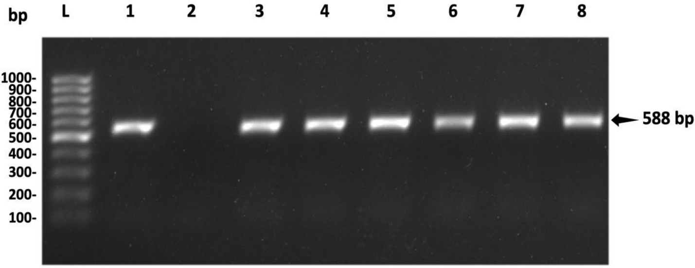
All 987 larvae extracted were submitted to conventional PCR in order to identify the genus and species of the larvae collected. Of the total parasites evaluated by PCR, all of them (100%) were identified as A. simplex (Figure 1).

2.2. Global Proteome Identification

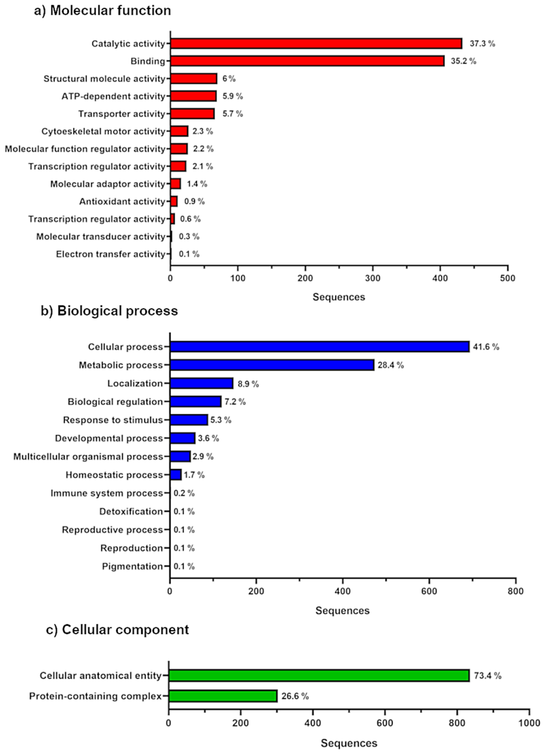
Mass spectrometry analysis identified 8119 peptide precursors from A. simplex (Supplementary Table S1), which correspond to 1919 identified proteins (Supplementary Table S2), which corresponds to a total of 1615 gene entries whose GO term annotations were determined using the PANTHER classification system (Figure 2). Among these, the most represented categories within molecular function were catalytic activity (GO:0003824), accounting for 37.3%, and binding (GO:0005488), accounting for 35.2%. Similarly, within biological processes, the most common categories were cellular processes (GO:0009987) with 41.6% and metabolic processes (GO:0008152) with 28.4%. Finally, regarding cellular components, 73.4% of proteins were associated with the category “cellular anatomical entity” (GO:0110165), whereas 26.6% were associated with “protein-containing complex” (GO:0032991) (Figure 2).
Bioinformatic analyses were also conducted using the DAVID platform to determine functional gene classifications and enrichment scores for specific groups. The DAVID functional annotation clustering analysis yielded a total of 140 gene clusters (Supplementary Table S3), with the two clusters displaying the highest enrichment scores being 17.25 and 15.44.
These clusters contained genes with enriched functional annotations corresponding to structural constituents of ribosomes, including ribosomal proteins and ribonucleoproteins in Cluster 1, and translation-related components, ribonucleoprotein complexes, and ribosomal elements in Cluster 2. Subsequently, and with a marked decrease in enrichment score, a cluster with a score of 7.06 was identified, containing genes with functional annotations associated with the parasite’s mitochondrion, including components of the electron transport chain (Table 2).

2.3. Allergen Bioinformatic Analysis

From the 1919 proteins identified, a search was conducted to detect putative proteins with allergenic characteristics already described in other species. For this purpose, the AllergenFP tool was used, allowing the identification of a total of 436 proteins with predicted allergenic potential (Supplementary Table S4). Among these, 422 proteins corresponded to allergens previously identified in other species, while 14 proteins were classified as A. simplex allergens. Within this latter group, the known allergens Ani s 1, Ani s 2, Ani s 3, Ani s 4, Ani s 5, and Ani s 7 were detected (Table 3). The 422 proteins were subsequently filtered using phylogenetically related organisms to A. simplex, resulting in a subset of 105 proteins. These were separated into three groups: (i) potential allergens from human or animal helminths, (ii) allergens from protozoan parasites or allergenic mites, and (iii) a group composed of proteins recognized as potential allergens in organisms such as bees, cockroaches, crustaceans, peanuts, or hazelnuts (Supplementary Table S5).
Of these 105 proteins, duplicate results were removed and a total of 92 proteins were analyzed in AlgPred v. 2.0, to predict and classify allergens, where a hybrid score (ML Score, MERCI Score, BLAST Score) with a threshold value of 1.0 was used to predict highly allergenic proteins in Anisakis, and a total of 12 proteins (hybrid score > 1.0) were obtained (Table 4). In addition, further localization analysis performed on the DeepLoc v. 2.1 server showed that 10 proteins have cytoplasmic localization and 2 have extracellular localization (A0A0M3KE55 and A0A0M3IXZ4), all of which are soluble and therefore not membrane proteins (Table 5).
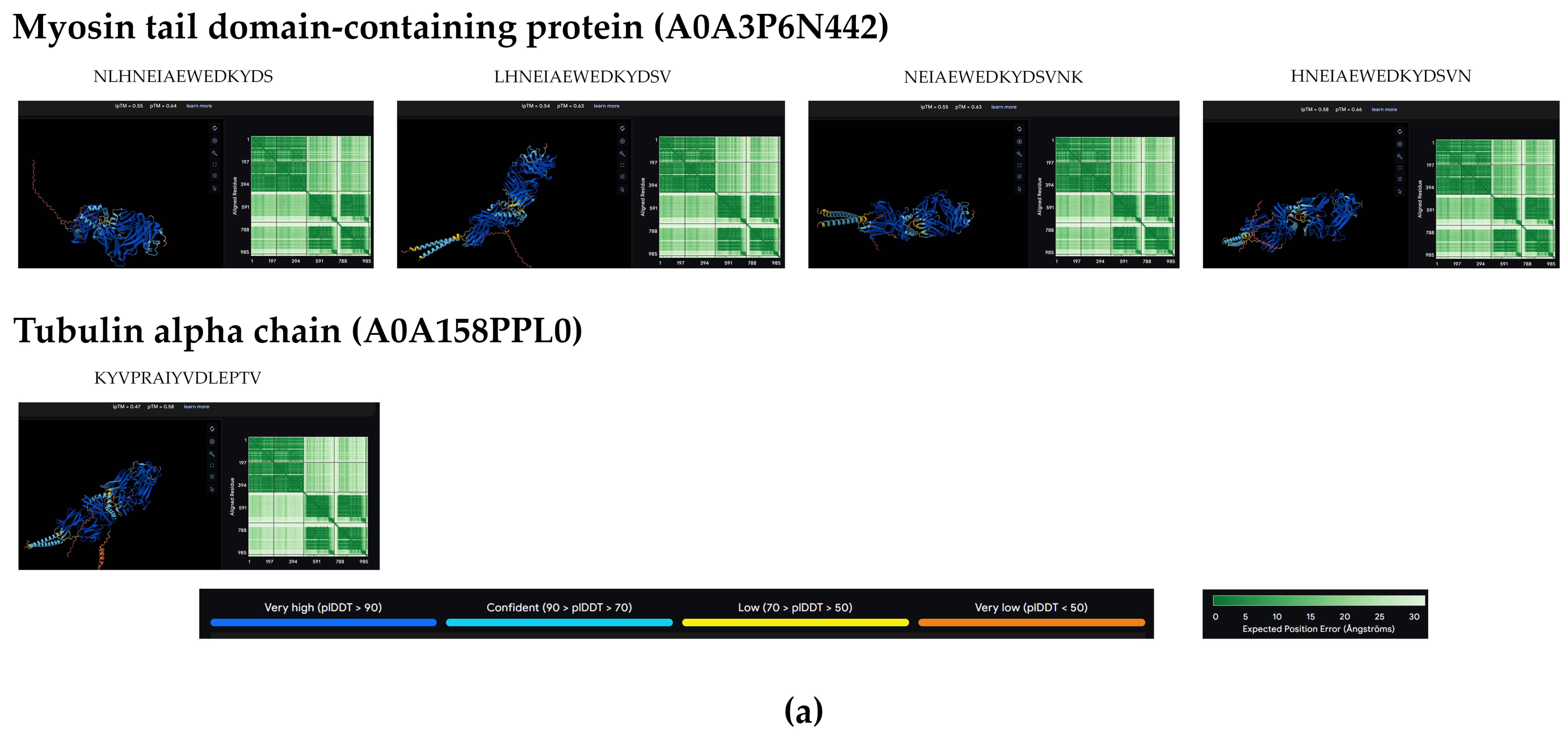
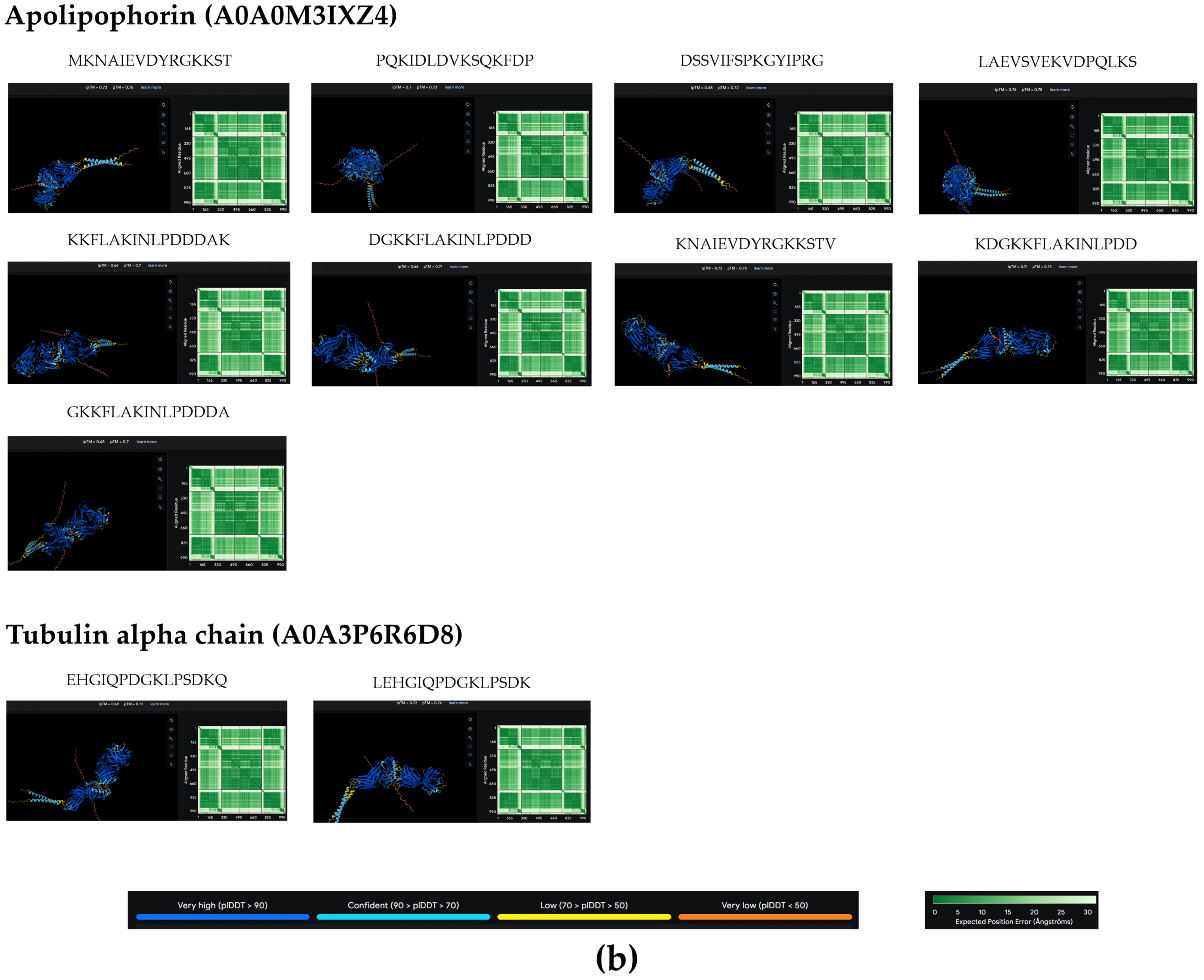
Because the type of response it can generate in humans is MHC-II-dependent, the MHC II ligands were mapped for the 12 allergenic proteins using the NetMHCIIpan-4.3 server. Considering the percentile rank ≤ 0.02, peptide size of 15 mers, and relevant human MHC II alleles of the DR, DQ, and DP gene loci (Figure 3), the analysis identified a total of 27 strong MHC II-binding peptides present in the allergens Myosin tail domain-containing protein (A0A3P6N442), Tubulin alpha chain (A0A158PPL0, A0A3P6R6D8), Tubulin_C domain-containing protein (A0A0M3KB72), and Apolipophorins (A0A0M3IXZ4) allergens (Table 6). Of note is the Apolipophorins (A0A0M3IXZ4) protein, with a total of 16 strong-binding peptides.
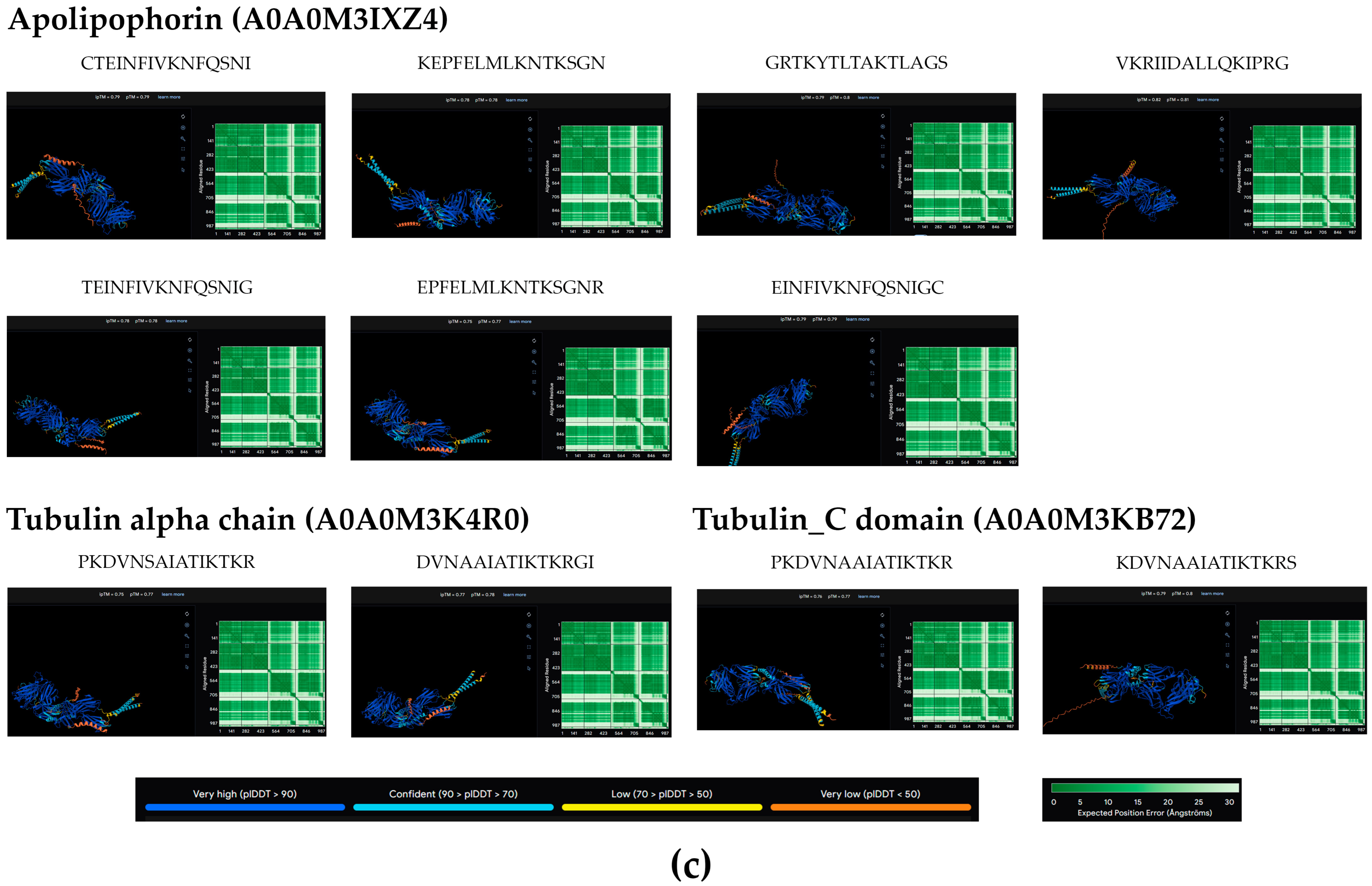
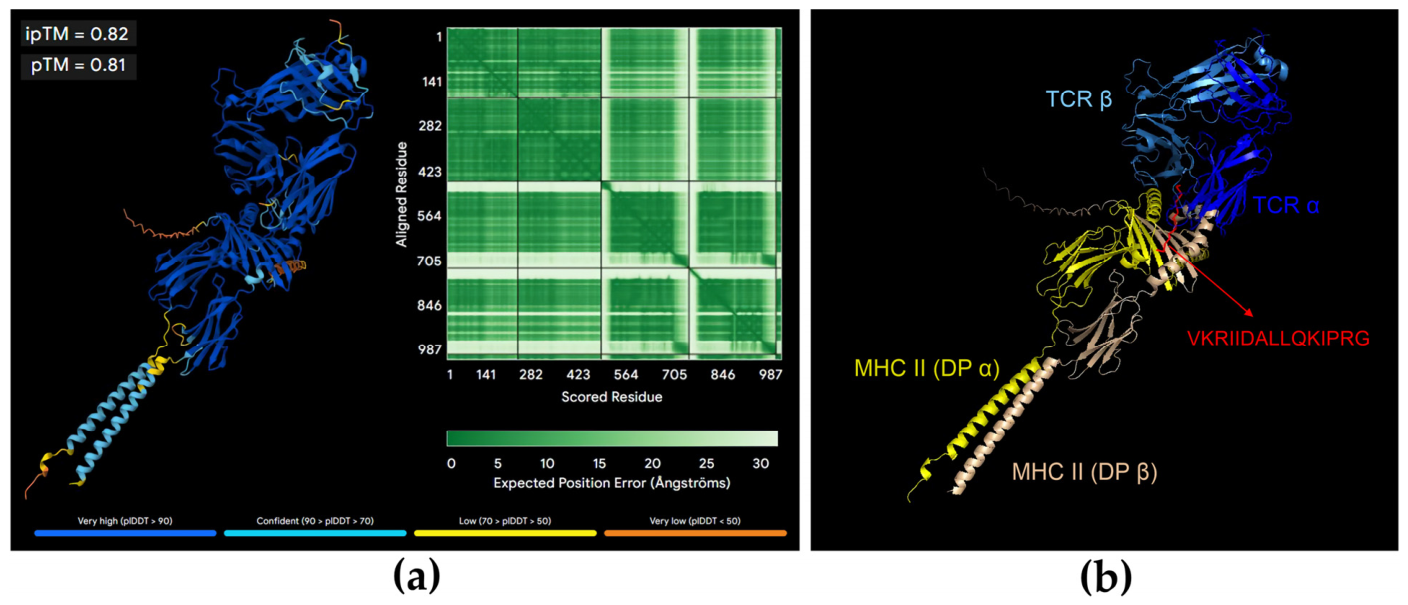
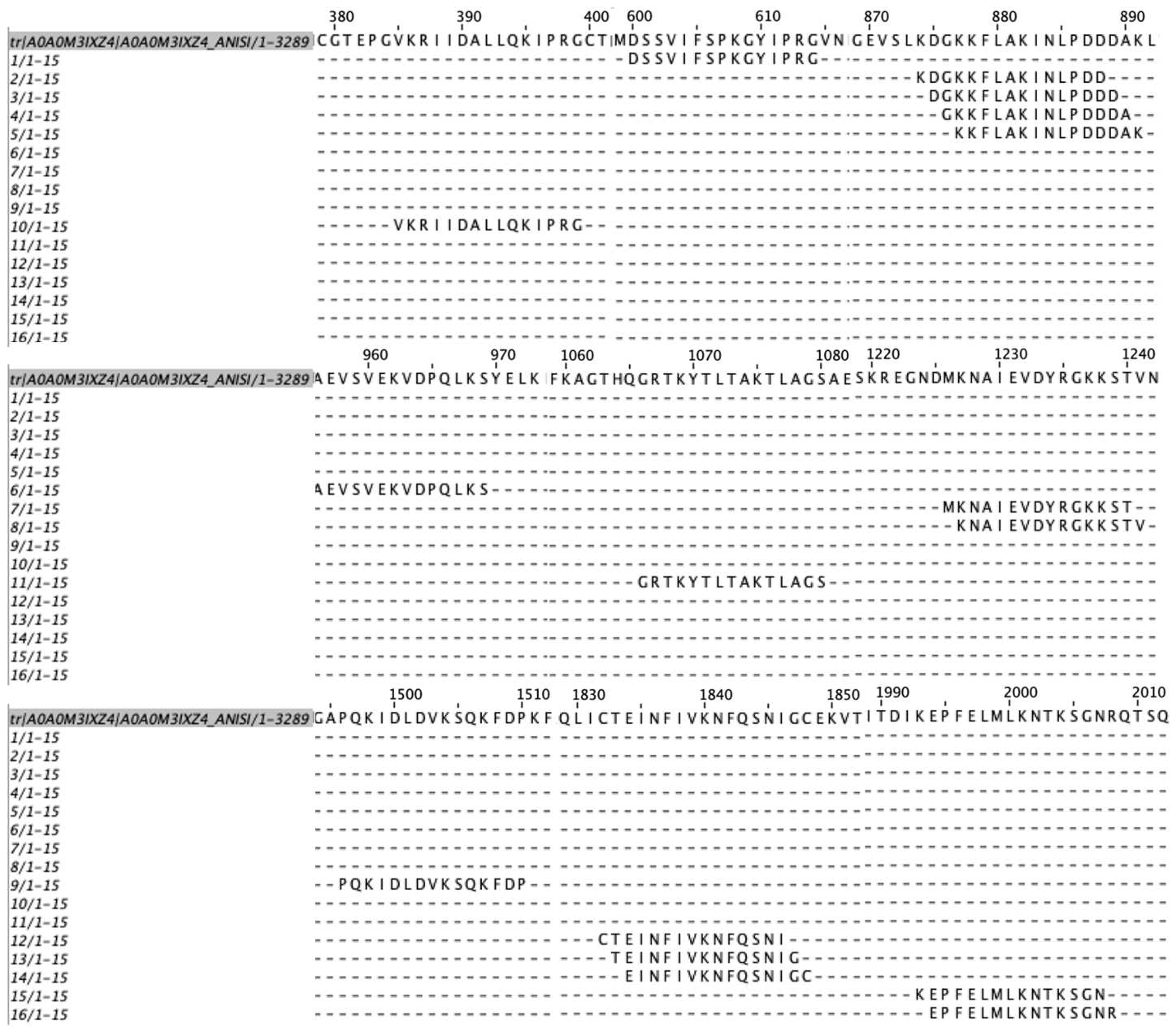
The peptides predicted as strong ligands were used in structural modeling with human MHC II BRD (α-β) and TCR (α-β) (Figure 3 and Table 6). The pTM (predicted template matching score) and ipTM (predicted interface template matching score) scores were evaluated, which represent the measurement of structure accuracy, with values for pTM > 0.5 indicating that the complex may be similar to the actual structure and values for ipTM > 0.8 being reliable (ipTM < 0.6 erroneous and in the range 0.6 to 0.8 probably reliable or not) [42,43]. Among the MHC-II-binding peptides, 5 were predicted to bind to the HLA-DQ, 11 to HLA-DR and 11 to HLA-DP isotypes. The best structural model of strong MHC-II ligands, considering the best prediction scores for the individual chain structure (pTM:0.81) and protein interface (ipTM:0.82), was the peptide VKRIIDALLQKIPRG from the Apolipophorins protein with the HLA-DP allele (Figure 4). All peptide sequences corresponding to apolipophorin were aligned with the protein to determine their location (Figure 5).

3. Discussion

Members of the Anisakidae family exhibit wide geographic distribution, occurring in fish from all the world’s oceans [15]. However, only certain members of this family possess a high zoonotic potential. Among them, the genus Anisakis, particularly the species Anisakis simplex and Anisakis pegreffii, are the most clinically relevant [15,27,44,45,46,47]. Nevertheless, the scientific literature also reports human infections caused by other species, including Pseudoterranova cattani [48,49] and Pseudoterranova decipiens [39,50,51].
To date, no studies have been conducted in northern Chile to determine the current situation regarding Anisakis spp. infection in commonly consumed fish. In the present study, we report by PCR that 51.6% of Chilean jack mackerel specimens were infected with A. simplex larvae. This finding is relevant because it contrasts with previously documented data from central southern Chile. Recently, Muñoz-Caro et al. [49] reported the presence of Anisakis in 180 commonly consumed fish belonging to three species collected along the central coast of Chile. Hake (Merluccius gayi) and snoek (Thyrsites atun) showed 100% infection rates, whereas redbanded seabream (Brama australis) exhibited a 35% infection rate [49]. However, in that study, the Anisakidae species identified were P. cattani and A. pegreffii, while A. simplex was not detected [49]. Conversely, another study evaluating 78 “ceviche” portions sold in restaurants in the cities of Valdivia and Niebla revealed that 21.8% of samples contained larvae of Pseudoterranova spp. and Anisakis spp. [52]. According to the available literature, A. simplex sensu stricto has been described mainly in fish captured in the northern hemisphere [15,29,52,53,54]. Meanwhile, a study conducted on fish captured along the central coast of Peru, including jack mackerel, mackerel, hake, and Chilean jack mackerel, showed that A. pegreffii was the predominant species, with a prevalence of 53.7% [55]. However, at least one earlier description reports the presence of A. simplex in Peruvian and Chilean coastal waters [56]. In that report, identification relied solely on morphological traits, and no molecular methods were employed.
In our study, the second most heavily parasitized fish species was Chub mackerel. This finding is consistent with reports from other authors describing high levels of Anisakis spp. infection in this species [57,58,59]. Notably, in our study, only a single Anisakis species was detected. This contrasts with findings from other researchers who have reported up to five Anisakis species infecting fish within a single geographic region [60]. These findings are epidemiologically relevant, as the high prevalence of A. simplex sensu stricto larvae in the musculature of these species represents a public health concern for the Chilean population, particularly considering the widespread consumption of raw or undercooked fish preparations. Likewise, the molecular identification of the infecting species is essential, since among all Anisakis species described to date, A. simplex has been reported as the primary etiological agent responsible for the majority of human anisakiasis cases [60]. In a previous study conducted on fish captured along the central coast of Chile, anisakid prevalences ranging from 16.7% to 41% were reported in Spanish rockfish (Sebastes capensis), Painted comber (Palabrax humeralis), and yellowtail amberjack (Seriola lalandii) [61]. In the present study, identification was conducted via PCR, owing to its high sensitivity, specificity, and capacity for diagnostic resolution at both the genus and species levels. The application of monoclonal antibodies targeting parasite-specific proteins or allergens may further serve as a complementary diagnostic tool. Conversely, the parasite detection using antibodies present in human sera demonstrates reduced specificity for identification purposes, as the literature documents cross-reactivity between Anisakis and other helminths, as well as between certain Dermatophagoides and Anisakis allergens. Given these constraints, it must also be noted that monoclonal or polyclonal antibodies capable of identifying the parasite or its immunogenic molecules were not available for this study [62,63,64,65].
Various proteomics studies have contributed to deepening our knowledge of the biology of Anisakidae family members [66,67,68]. On the other hand, Palomba et al. (2023) [69] reported the proteomic characterization of extracellular vesicles (EVs) released by third-stage larvae (L3) of A. pegreffii, contributing to the understanding of the molecular signals that facilitate interaction between the parasite and its natural and accidental hosts [69]. In our mass spectrometry analysis of A. simplex L3, the most represented categories within molecular function were catalytic activity (GO:0003824), accounting for 37.3%, and binding (GO:0005488), accounting for 35.2%. Binding and catalytic functions have also been reported as the primary molecular functions of A. simplex L3 by Stryiński et al. (2019) [67]. Among catalytic activities, the most relevant were hydrolase (GO: 0016787), mainly endopeptidases characterized as metalloendopeptidases and cysteine-type endopeptidases. In A. simplex, a metalloaminopeptidase was first described by Sakanari and McKerrow in 1990 [70]. Metallopeptidases are involved in the invasion of host tissues by the parasite, as they can degrade the extracellular matrix, and are also involved in the process of ecdysis and digestion of nutrients [71]. On the other hand, high levels of transcripts of a metallopeptidase (nas10) were found in the L3 infecting the muscle of the host fish Micromesistius potassou [72]. Several metalloproteases have been shown to be overexpressed in A. simplex L3 larvae [73]. Among metalloendopeptidases, we have found neprilysin, which has been associated with intestinal digestion, host invasion, and molting [74]. Metalloprotease and cysteine proteinase activities have also been detected in A. simplex and A. pregreffii [75,76]. Cysteine proteases in nematodes also appear to play a critical role in parasitic virulence, participating in various processes such as penetration of host tissues, evasion of the immune response, virulence, digestion, embryogenesis, molting, and intracellular digestion [71,77]. Metalloprotease and Cathepsin L activity have been detected in Anisakis [75,77].
On the other hand, among the binding expressed proteins, mRNA and actin filament binding were the most expressed. Little is known about the cytoskeleton of Anisakis. However, a study using a monoclonal antibody obtained against human intermediate filaments reported cross-reactivity with A. simplex [78]. In A. pegreffii, actin has been reported to be the main cytoskeletal component present in EVs [69]. A powerful and well-organized cytoskeleton is necessary to keep the parasite moving and penetrate the muscles of infected fish and intermediate hosts. In the same way, the increased synthesis of forms of myosin, actin and troponin has been observed in nematodes as a response to the host immune system and may be related to the attempted reorganization or repair of the cytoskeleton and/or muscle layer in the host, immune-initiated increased mucus production and smooth muscle activity within the intestinal environment [79]. In addition, the anthelmintic drugs currently in use act on the cytoskeleton, so knowledge of these proteins is essential in strategies for developing new drugs or improving those currently in use [80].
In eukaryotes, the mRNA life cycle comprises different stages: transcription, precursor mRNA processing, nuclear export, translation, and degradation. At each stage, mRNAs interact with a specific set of RNA-binding proteins to form mRNA–protein complexes [81,82]. These RNA-binding proteins regulate the activity, localization, and stability of mRNA and influence its transition to the next stage of the life cycle [83]. In A. simplex, gene expression patterns have been studied in the L3 and L4 larval stages through various transcriptomic studies [73,84,85], and expression of RNA-binding proteins has been observed in response to exposure to antiparasitic drugs [86,87]. However, the regulation of mRNA expression has not been investigated in depth.
Similarly, within biological processes, the most common categories were cellular processes (GO:0009987) with 41.6% and metabolic processes (GO:0008152) with 28.4%. Among cellular processes, the most highly expressed proteins were chaperone-mediated protein folding (GO:0061077) and chaperone cofactor-dependent protein refolding (GO:0051085). Heat shock proteins (HSPs) are synthesized in response to cellular stress and represent a common mechanism across organisms. They stabilize the conformational structure of newly synthesized polypeptides and facilitate the degradation of those that are misfolded [88]. HSPs have been found to be a common component of nematode EVs and are considered a marker of EVs [89,90]. The presence of HSPs in L3 EVs appears to support their role in mediating and/or buffering the thermal and osmotic stress experienced by the parasite during its life cycle, as it transitions from the marine environment to a natural or accidental homeothermic host. Here, we have reported the higher expression of a 10 kDa mitochondrial heat shock protein and a DNAJ homolog subfamily B member 4. Hsp10 (10 kDa heat shock protein, also known as chaperonin 10 or Cpn10) is a co-chaperone of Hsp60 in the protein folding process. Hsp10 is a homolog of the bacterial protein GroES, which acts as a co-chaperone for Hsp60 (GroEL) in protein folding and assembly processes [91,92]. In eukaryotic cells, Hsp10 homologs are found in organelles such as mitochondria and chloroplasts [93], which originated from bacteria through endosymbiotic processes [94], and play a role analogous to that of their bacterial homologs in the protein folding process [95,96]. On the other hand, members of the HSP40/DNAJ protein family are involved in regulating the molecular chaperone activity of various HSP70 proteins by stimulating their adenosine triphosphate (ATPase) activity [97,98,99].
Therefore, among the metabolic process category, the higher expressed proteins involved were proteolysis, including ubiquitin-dependent protein catabolic process (GO:0006511) and proteasome-mediated ubiquitin-dependent protein catabolic process (GO:0043161). The presence and biological function of the ubiquitin–proteasome pathway were first described in human pathogenic protozoa [100,101,102]. In human parasitic helminths, ubiquitin–proteasome-dependent proteolysis and its biological significance have also been reported [103,104,105]. However, in members of the Anisakidae family, ubiquitin–proteasome pathway components have already been identified in proteomics and transcriptomics studies [72,73,106], but the biological role of this pathway is not well understood.
Finally, regarding cellular components, 73.4% of proteins were associated with the category “cellular anatomical entity” (GO:0110165), whereas 26.6% were associated with “protein-containing complex” (GO:0032991) [69] (Figure 2). Cellular anatomical entity refers to the expression of both cytoplasmic and intracellular organelle proteins. The main cytosolic proteins were the large and small ribosomal subunits, while mitochondrial respiratory complexes were the most highly expressed proteins among “protein-containing complex”. Mitochondrial enzyme genes are highly expressed in Anisakis L3 and L4 larvae. It has been reported that L3 of A. simplex transcribes genes related to invasion, migration, molting, and survival [36]. Studies in A. pegreffii have shown that mitochondrial enzymes are the most highly expressed in L3 larvae [107]. Proteomics studies with A. simplex L3 and L4 exposed to glucose concentrations showed that 24 proteins associated with translation and ribosome formation were affected [108]. On the other hand, the large and small ribosomal subunits have previously been reported by Marzano et al. [109].
Using advanced bioinformatics techniques, we initially screened 422 proteins corresponding to allergens in other species along with 14 well-known A. simplex allergens, including Ani s 1, Ani s 2, Ani s 3, Ani s 4, Ani s 5, and Ani s 7 (Table 3). These proteins were filtered and categorized into three groups based on their phylogenetic relationship to known allergens in helminths, protozoa, and other organisms such as crustaceans or peanuts (Supplementary Table S5). Following the removal of duplicates, 92 proteins were analyzed using AlgPred v. 2.0. By applying a hybrid score threshold of 1.0, the study identified 12 proteins predicted to be highly allergenic. Localization analysis using DeepLoc v. 2.1 further determined that ten of these proteins were cytoplasmic and two were extracellular; however, all of them were categorized as soluble proteins (Table 5).
Our study was focused particularly on the interaction with the human immune system through MHC II ligand mapping. Among the 12 allergenic proteins, 27 strong MHC II-binding peptides were identified, primarily within the myosin tail domain, tubulin, and apolipophorins (Figure 3). The apolipophorin protein (A0A0M3IXZ4) appears to be of higher interest because it contains 16 of the 27 identified strong binding peptides. When AlphaFold was utilized to generate three-dimensional models of these strong ligands interacting with human MHC II and T-cell receptors (TCR), we observed that the most reliable model was found for the peptide VKRIIDALLQKIPRG from the apolipophorin protein (Figure 4). These findings allow for a more precise prediction of which parasite components could trigger the most severe allergic reactions in humans.
To date, around 14 allergens have been described, and most of them have been detected in parasitic somatic and excreted/secreted products, being recognized by serum antibodies in most individuals affected by allergic anisakiasis. Some of them, such as Ani s 1, Ani s 4, Ani s 5, Ani s 8, Ani s 9, Ani s 10, and Ani s 11, have shown heat stability [110,111,112], which may cause problems even when infected fish is eaten cooked. On the other hand, twenty-eight different allergenic proteins have been classified, and most of them were described for the first time as potential new allergens in Anisakis [106]. These researchers showed differences in the allergenic abilities of the A. simplex complex species and, using 2D gel electrophoresis, found that A. simplex s.s. showed a higher number of immunoreactive protein spots (34 spots) than A. pegreffii (11 spots) [106]. In the same way, an approach was used by Fæste et al. [113] in a study based on gel banding patterns and IgE-immunostaining using sera from A. simplex-sensitized patients and proteome data obtained by mass spectrometry. Results showed that A. simplex proteins were homologous to allergens already characterized in other nematodes, insects, and shellfish [114]. This proteomic investigation of A. simplex L3 identified 17 novel putative allergens, including structural proteins such as myosin-4 and enzymes such as enolase and endochitinase [113].
Allergic symptoms, including anaphylaxis, have been reported in approximately 11% of anisakiasis cases in Spain [115]. In Japan, allergy induced by Anisakis is a common trigger of acute allergic reactions in adults and represents the second leading cause of anaphylactic shock, accounting for 23% of reported cases [116]. Allergic manifestations have also been described following inhalation or direct contact during the handling of infected fish, leading to rhinoconjunctivitis, dermatitis, and asthma [2,117]. All these clinical responses are triggered by an IgE-mediated immune mechanism.
To date, fourteen A. simplex allergens have been identified, designated Ani s 1 through Ani s 14 [118], but the interaction between these allergens and human MHC class II allele key molecules responsible for antigen presentation has been only minimally explored. Previous studies have identified binding motifs from Ani s 2 and Ani s 3 (paramyosin and tropomyosin) associated with alleles such as DRB11502, DQB10601, and DRB1*0404 [119]. Moreover, in a cohort of fish-processing workers in Croatia, PCR-based HLA typing and serological analyses revealed associations involving HLA-DRB1, HLA-DQA1, and HLA-DQB1 [120]. Similarly, little is known about the identification of peptides that participate as T-cell epitopes, as they have only been studied in a few allergens such as Ani s 1, Ani s 5, Ani s 7, and Ani s 13 [121,122,123,124].
Our work suggests that there are proteins not yet identified as A. simplex allergens that may generate an MHC class II-mediated allergenic response (Table 5). In silico analysis showed that 27 peptide sequences could be considered potential new allergens because they may react with MHC II alleles, the most notable being the peptide VKRIIDALLQKIPRG (Figure 4), which belongs to apolipophorin and contains a total of 16 probable binding epitopes.
Apolipophorin is a protein consisting of 3289 amino acids found in the Anisakis genome. Figure 5 shows the location of the MHC-II-binding peptides within the protein, which helps us understand the position of relevant peptides in its primary structure, such as VKRIIDALLQKIPRG, which is located between amino acid residues 385–400 of the protein. It was initially described as an allergen in Dermatophagoides farinae, characterized as an apolipoprotein possessing Mag 1 and Mag 3 sequences that bind to IgE, showing homology with a high molecular weight allergen M-177. This protein is similar to lipid transport apolipoproteins found in insects [125] and was subsequently classified as belonging to group 14 of the main mite allergens [126]. Similarly, apolipophorins have been reported in other insects, such as Culex quinquefasciatus, where they represent the main component of lipophorins that mediate the transport of various lipids in the hemolymph and form lipoprotein particles that bind to other lipoproteins and lipids [127].
Research using mass spectrometry has demonstrated allergenic characteristics of apolipophorins in species such as Acheta domesticus and Hermetia illucens [128]. They have also been identified in other insects such as Bombyx mori and Tenebrio molitor in studies focused on allergen detection for food safety using proteomic and bioinformatic analysis [129]. In addition, apolipophorin has been identified as a component of venom in the brown widow spider Latrodectus geometricus [130]. Although the LLTP superfamily has been described in various species for its allergenic potential to bind IgE, in nematodes this has mainly been associated with vitellogenin, not apolipophorin. Therefore, the allergenic potential of apolipophorin in Anisakis is demonstrated here for the first time.
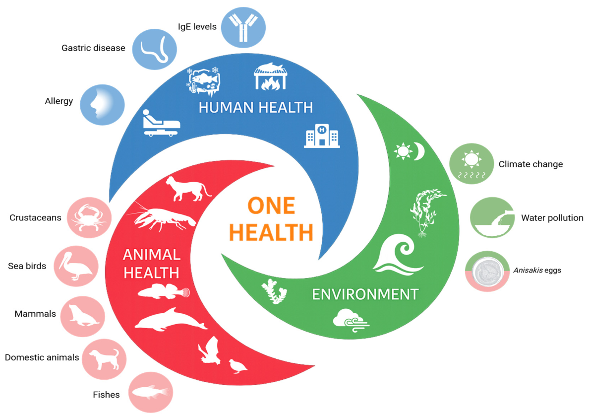
In this study, we show that a significant percentage of Chilean jack mackerel, a species widely consumed by humans, is parasitized by A. simplex. Our proteomics study showed that A. simplex L3 larvae possess several previously described allergens as well as potential new allergens suggested by our in silico analysis. This indicates potential health risks for both humans and animals and supports the need for a One Health approach [4,131]. At the human level, measures such as freezing fish or cooking it at temperatures above 70 °C are effective in preventing anisakiasis in its digestive forms. However, the presence of heat-stable allergens means that these measures do not prevent allergic reactions. Therefore, identifying and removing allergens from fish meat is highly necessary. Proteomics has proven effective in detecting proteins and allergens in fish meat [132], but methods to remove them are still lacking. Activated charcoal has been shown to absorb allergens [133,134,135], and modified charcoal nanoparticles that do not alter fish properties could be a potential solution. At the animal level, prevention is limited due to the parasite’s life cycle and host range. Infection in pets such as dogs and cats can be prevented by avoiding raw fish consumption. A major concern is environmental contamination: in Chile, fish waste is often discarded into the sea, feeding seabirds, fish, and sea lions, thus perpetuating the parasite’s life cycle. In the same way, a One Health approach aimed at preventing human and animal infection by Anisakidae while preserving the environment is illustrated in Figure 6.

4. Materials and Methods

4.1. Collection of Fish Specimens in the Antofagasta Region, Chile, and Extraction of Third-Stage Larvae (L3)

A total of 2968 specimens belonging to the most commercially important fish species sold in the seafood market of Antofagasta, Chile, and captured along the regional coastline were examined. Fish species marketed in the city but originating from other regions of the country were not included in this study. Each fish specimen was dissected to search for anisakid larvae in the abdominal cavity and viscera. Third-stage larvae (L3) were retrieved by carefully removing the visceral organs from the abdominal cavity. All recovered larvae were washed in saline solution (0.85% NaCl) and subsequently prepared for morphological identification under light microscopy.
For every specimen, records were kept including species, body length, and anisakid burden. All anisakid larvae were individually labeled, cataloged, and maintained at cold temperature for transport to the laboratory. For molecular and proteomic analyses, larvae were stored at −80 °C until processing.

4.2. Morphological Identification of Larvae of the Family Anisakidae

Morphological identification was based on the following characteristics: (a) position of the excretory pore, (b) tail shape, (c) length and shape of the ventricle, (d) presence, length, and position of the intestinal cecum, and (e) presence or absence of the ventricular appendix. Nematodes were cleared in graded glycerin solutions. Internal structures were examined using a light microscope equipped with a camera lucida. Nematodes belonging to the family Anisakidae were identified to the genus level [107,136].

4.3. DNA Extraction

Genomic DNA was isolated using the Wizard Genomic DNA Purification Kit (Promega, Madison, WI, USA) from whole individual larvae. Samples were homogenized using a tissue homogenizer (PT 1200 E, POLYTRON®, Kinematica AG, Malters, Switzerland) in 600 µL of lysis buffer (0.1 M Tris-HCl pH 7.6, 50 mM DTT, 2% SDS). The homogenate was incubated at 100 °C for 5 min, followed by the addition of 3 µL of RNase and incubation at 37 °C for 30 min. Subsequently, 200 µL of protein precipitation solution was added, the mixture was vortexed for 20 s, and incubated on ice for 5 min [133].
Samples were centrifuged at 16,000× g for 4 min, and the supernatant (600 µL) was transferred to a 1.5 mL microtube containing 600 µL of isopropanol. After centrifugation at 16,000× g for 1 min, the supernatant was discarded, and the pellet was washed with 600 µL of 70% ethanol, gently inverted several times, and centrifuged again at 16,000× g for 1 min. The pellet was air-dried for 10–15 min, resuspended in 50 µL of rehydration solution (10 mM Tris-HCl, 1 mM EDTA), and incubated at 65 °C for 1 h with occasional mixing. DNA quality was evaluated by absorbance at 260/280 nm using a Tecan Infinite M200 Pro spectrophotometer (Tecan Group Ltd., Männedorf, Switzerland) and by agarose gel electrophoresis. Purified DNA was stored at −80 °C until conventional PCR analysis.

Conventional PCR Identification of Anisakidae Larvae

PCR assays were conducted as previously described [137] using a final reaction volume of 20 µL on a T100 Thermal Cycler (Bio-Rad, Hercules, CA, USA). PCR products were visualized by agarose gel electrophoresis using SYBR™ Safe DNA Gel Stain (Invitrogen™, Thermo Fisher Scientific, Waltham, MA, USA). Species-specific primers targeting six anisakid species were used (Table 7). In all reactions, a universal type B reverse primer was included (5′-GCCGGATCCGAATCCTGGTTAGTTTCTTTTTCT-3′). Primers were synthesized by Integrated DNA Technologies (Coralville, IA, USA).
PCR products were resolved on 1% (w/v) agarose gels (UltraPure™ Agarose, Invitrogen, Thermo Fisher Scientific) and stained with SYBR™ Safe (Thermo Fisher Scientific). Genomic DNA from L3 larvae of Anisakis simplex, Anisakis pegreffii, Anisakis physeteris, and Pseudoterranova decipiens (provided by Dr. Rubén Mercado, Parasitology Unit, Faculty of Medicine, University of Chile) was used as positive control.

4.4. In Gel Digestion

Frozen larvae were homogenized using a Polytron PT 1200 E tissue homogenizer (Kinematica AG, Malters, Switzerland) in lysis buffer (0.1 M Tris–HCl pH 7.6, 50 mM DTT, 2% SDS). Lysates were boiled at 100 °C for 5 min, cooled to room temperature, and clarified by centrifugation at 16,000× g for 10 min [138]. Total protein concentration was determined using the Pierce™ 660 nm Protein Assay (Thermo Fisher Scientific).
Protein extracts were processed following an in-gel digestion workflow and digested with trypsin to generate peptides suitable for proteomic analysis [139]. Peptide quantification was performed using the Pierce™ Quantitative Peptide Assay (Thermo Fisher Scientific), and absorbance at 480 nm was measured in a Tecan Infinite 200 PRO microplate reader according to the manufacturer’s instructions.

4.5. Reverse Phase-Liquid Chromatography-Tandem Mass Spectrometry (RP-LC-MS/MS) Analysis

Peptide concentrations were measured on a NanoDrop One (Thermo Fisher Scientific) to load approximately 50 ng of peptides on a timsTOF Pro2 (Bruker Daltonics, Billerica, MA, USA) with a CaptiveSpray source coupled to nanoElute UHPLC (Bruker Daltonics) using a PepSep Ten column (10 cm × 75 µm inner diameter, 1.9 µm particle size with 20 µm emitter) heated to 50 °C. Buffer A consisted of 0.1% formic acid and 0.5% acetonitrile in water, and buffer B consisted of 0.1% formic acid and 99.4% acetonitrile in water. A standard 30 min gradient was used, increasing buffer B from 2% to 12% over 15 min and then to 33% over 15 min. The column was washed with 95% B over 8 min. The nanoElute thermostat temperature was set at 7 °C. The analysis was performed at a flow rate of 0.4 µL/min.
The timsTOF Pro2 was set to DIA-PASEF mode with positive polarity for the MS scan window from 100 to 1700 m/z. The capillary voltage was set to 1800 V, drying gas to 3 L/min, and drying temperature to 180 °C. The MS1 scan was followed by PASEF ramps containing 22 non-overlapping 35 m/z isolation windows covering the mass range 319.5–1089.5 Da. Ion mobility range (1/K0) was set to 0.70–1.35 V·s/cm2, with 100 ms ramp time, 100 ms accumulation time, 100% duty cycle, and 9.42 Hz ramp rate. Collision energy was ramped linearly as a function of mobility from 27 eV at 1/K0 = 0.70 V·s/cm2 to 55 eV at 1/K0 = 1.35 V·s/cm2.
The acquired data were searched with DIA-NN (PMID31768060) version 1.8.1 with a library-free search method, using UniProt’s Anisakis simplex protein sequences (UP000267096) and manually curated common contaminants (226 entries). Other search parameters include Trypsin/P digestion mode with 1 missed cleavage, 1 maximum number of variable modifications, N-terminal M excision, carbamidomethylation of C, and oxidation of M options enabled; peptide length ranged 7–30, precursor charge ranged 1–4, precursor m/z ranged 300–1800, and fragment ion m/z ranged 200–1800. Precursor FDR was set to 1%, with 0 Mass accuracy and MS1 accuracy (for the “auto” option of mass tolerance), enabling heuristic protein inference, use of isotopologues, match between run (MBR), and no shared spectra. Protein inference is set as “Protein name from FASTA”, Double-pass mode for neural network classifier, Robust LC (high precision) for Quantification Strategy, RT dependent mode for Cross-run normalization, and Smart profiling mode for Library generation.
The mass spectrometry proteomics data have been deposited to the ProteomeXchange Consortium via the PRIDE (PubMed ID: 34723319) partner repository with the dataset Identifier PXD044267.

4.6. Functional Analysis

To assess the biological and molecular functions of the identified proteins, the final list of non-redundant proteins obtained after global identification was classified into the three main Gene Ontology (GO) categories: molecular function, biological process, and cellular component. The Kyoto Encyclopedia of Genes and Genomes (KEGG) database was used to assign the identified proteins to specific biological pathways and cellular mechanisms, applying an adjusted p-value < 0.05 as the significance threshold. GO and KEGG enrichment analyses were performed using the PANTHER classification system (Protein ANalysis THrough Evolutionary Relationships, https://pantherdb.org/, accessed on 23 June 2025) [140] and DAVID (https://davidbioinformatics.nih.gov/, accessed on 26 June 2025) [141,142]. GO categorization applied the PANTHER overrepresentation test with Bonferroni correction to adjust the smallest p-values associated with enriched GO terms.

4.7. Bioinformatic Allergen Analysis

To identify putative novel allergens, the AllergenFP v1.1 platform (https://ddg-pharmfac.net/AllergenFP, accessed on 10 February 2024) [143] was used as an initial screening approach. Subsequently, allergen prediction and classification based on the obtained protein sequences were performed using AlgPred 2.0 (https://webs.iiitd.edu.in/raghava/algpred2/batch.html, accessed on 14 August 2025) [144]. In addition, AlphaFold 3 (https://deepmind.google/technologies/alphafold/, accessed on 15 August 2025) [145] was employed for structural modeling, incorporating MHC class II molecules and human T-cell receptors (TCR α and β). Structural references were based on the Protein Data Bank entry 6TRO [146]. Subcellular localization and membrane-association prediction were conducted using DeepLoc v2.1 (https://services.healthtech.dtu.dk/services/DeepLoc-2.1/, accessed on 2 September 2025) [147]. Finally, to obtain a deeper computational assessment of peptide-binding affinity to MHC class II alleles, the NetMHCIIpan 4.3 tool (https://services.healthtech.dtu.dk/ser-vices/NetMHCIIpan-4.3/, accessed on 11 November 2025) [148] was used.

5. Conclusions

This study provides robust molecular and proteomic evidence that significantly advances our understanding of A. simplex epidemiology and pathogenicity in northern Chile. The high prevalence of A. simplex sensu stricto detected in widely consumed fish species, particularly Chilean jack mackerel, represents a notable geographic and epidemiological shift compared to previous reports from central southern Chile and the southeastern Pacific. This finding underscores the need for updated surveillance strategies integrating molecular diagnostics to accurately identify zoonotic species with high clinical relevance.
From a mechanistic perspective, the proteomic characterization of L3 larvae reveals a complex biological system dominated by catalytic and binding activities, including metalloproteases and cytoskeletal-associated proteins, which are crucial for host invasion, immune evasion, and parasite survival. Furthermore, the identification of stress-response proteins and components of the ubiquitin–proteasome pathway highlights adaptive mechanisms that facilitate environmental transitions and host parasite interactions.
Importantly, the integration of bioinformatics and immunoinformatics approaches enabled the prediction of novel allergenic proteins and MHC-II binding epitopes, with apolipophorin emerging as a key candidate with previously unrecognized allergenic potential. These findings have profound implications for public health, as they expand the repertoire of clinically relevant allergens beyond currently characterized molecules.
Overall, this work emphasizes the necessity of a One Health framework, combining epidemiological monitoring, molecular research, and food safety interventions to mitigate both infectious and allergic risks associated with anisakiasis.

Supplementary Materials

The following supporting information can be downloaded at: https://www.mdpi.com/article/10.3390/ijms27135922/s1.

Author Contributions

Conceptualization, J.S.F. and J.G.; methodology, J.S.F., S.Z., A.Á., S.B., K.M., B.G., K.-M.M. and L.J.F.; software, J.S.F., K.M. and R.F.d.A.; validation, A.Á., R.F.d.A. and J.G.; formal analysis, J.S.F., R.F.d.A., L.J.F. and J.G.; investigation, J.S.F., A.Á., S.B., S.Z., K.M., N.V., S.A., C.A., F.C., G.P., B.G., K.-M.M., R.F.d.A. and J.G.; resources, K.-M.M., R.F.d.A., L.J.F. and J.G.; data curation, J.S.F., S.Z.; writing—original draft, J.S.F., R.F.d.A. and J.G.; writing—review and editing, J.S.F., R.F.d.A., L.J.F. and J.G.; visualization, J.G.; supervision, B.G. and J.G.; funding acquisition, L.J.F. and J.G. All authors have read and agreed to the published version of the manuscript.

Funding

Mass spectrometry infrastructure used here was supported by the Canada Foundation for Innovation, the BC Knowledge Development Fund, the UBC Life Sciences Institute, and Genome BC (274PRO).

Institutional Review Board Statement

The fish used in the research were sampled at the Antofagasta fish market. They were caught by artisanal fishermen and were dead at the time of sampling.

Informed Consent Statement

Not applicable.

Data Availability Statement

The original contributions presented in this study are included in the article/Supplementary Materials. Further inquiries can be directed to the corresponding authors.

Conflicts of Interest

The authors declare no conflicts of interest.

References

  1. Adroher, F.J.; Morales-Yuste, M.; Benítez, R. Anisakiasis and Anisakidae. Pathogens 2024, 13, 148. [Google Scholar]
  2. Nieuwenhuizen, N.E.; Lopata, A.L. Allergic reactions to Anisakis found in fish. Curr. Allergy Asthma Rep. 2014, 14, 455. [Google Scholar] [CrossRef]
  3. Aibinu, I.E.; Smooker, P.M.; Lopata, A.L. Anisakis nematodes in fish and shellfish- from infection to allergies. Int. J. Parasitol. Parasites Wildl. 2019, 9, 384–393. [Google Scholar] [CrossRef]
  4. Nonković, D.; Tešić, V.; Šimat, V.; Karabuva, S.; Medić, A.; Hrabar, J. Anisakidae and Anisakidosis: A public health perspective. Pathogens 2025, 14, 217. [Google Scholar] [CrossRef]
  5. Pravettoni, V.; Primavesi, L.; Piantanida, M. Anisakis simplex: Current knowledge. Eur. Ann. Allergy Clin. Immunol. 2012, 44, 150–156. [Google Scholar]
  6. OPRHANET. Available online: https://www.orpha.net/orphacom/cahiers/docs/GB/Prevalence_of_rare_diseases_by_alphabetical_list.pdf (accessed on 14 May 2026).
  7. EFSA Panel on Biological Hazards (BIOHAZ). Scientific Opinion on risk assessment of parasites in fishery products. EFSA J. 2010, 8, 1543. [Google Scholar] [CrossRef]
  8. Chai, J.Y.; Murrell, K.D.; Lymbery, A.J. Fish-borne parasitic zoonoses: Status and issues. Int. J. Parasitol. 2005, 35, 1233–1254. [Google Scholar] [CrossRef] [PubMed]
  9. Herrador, Z.; Daschner, Á.; Perteguer, M.J.; Benito, A. Epidemiological scenario of anisakidosis in Spain based on associated hospitalizations: The tip of the iceberg. Clin. Infect. Dis. 2019, 69, 69–76. [Google Scholar] [CrossRef] [PubMed]
  10. Del Rey Moreno, A.; Valero, A.; Mayorga, C.; Gómez, B.; Torres, M.J.; Hernández, J.; Ortiz, M.; Lozano Maldonado, J. Sensitization to Anisakis simplex s.l. in a healthy population. Acta Trop. 2006, 97, 265–269. [Google Scholar] [CrossRef]
  11. Purello-D’Ambrosio, F.; Pastorello, E.; Gangemi, S.; Lombardo, G.; Ricciardi, L.; Fogliani, O.; Merendino, R.A. Incidence of sensitivity to Anisakis simplex in a risk population of fishermen/fishmongers. Ann. Allergy Asthma Immunol. 2000, 84, 439–444. [Google Scholar] [PubMed]
  12. Rahmati, A.R.; Kiani, B.; Afshari, A.; Moghaddas, E.; Williams, M.; Shamsi, S. World-wide prevalence of Anisakis larvae in fish and its relationship to human allergic anisakiasis: A systematic review. Parasitol. Res. 2020, 119, 3585–3594, Correction in Parasitol. Res. 2021, 120, 1925–1926.. [Google Scholar] [CrossRef]
  13. Thielp, V. Anisakiasis. Acta Leiden. 1964, 33, 203. [Google Scholar]
  14. Adroher-Auroux, F.J.; Benítez-Rodríguez, R. Anisakiasis and Anisakis: An underdiagnosed emerging disease and its main etiological agents. Res. Vet. Sci. 2020, 132, 535–545. [Google Scholar] [CrossRef]
  15. Mattiucci, S.; Cipriani, P.; Levsen, A.; Paoletti, M.; Nascetti, G. Molecular epidemiology of Anisakis and Anisakiasis: An ecological and evolutionary road map. Adv. Parasitol. 2018, 99, 93–263. [Google Scholar] [PubMed]
  16. Hochberg, N.S.; Hamer, D.H. Anisakidosis: Perils of the deep. Clin. Infect. Dis. 2010, 51, 806–812. [Google Scholar] [CrossRef]
  17. Cong, W.; Elsheikha, H.M. Biology, epidemiology, clinical features, diagnosis, and treatment of selected fish-borne parasitic zoonoses. Yale J. Biol. Med. 2021, 94, 297–309. [Google Scholar]
  18. Takabe, K.; Ohki, S.; Kunihiro, O.; Sakashita, T.; Endo, I.; Ichikawa, Y.; Sekido, H.; Amano, T.; Nakatani, Y.; Suzuki, K.; et al. Anisakidosis: A cause of intestinal obstruction from eating sushi. Am. J. Gastroenterol. 1998, 93, 1172–1173. [Google Scholar] [CrossRef] [PubMed]
  19. Hiramatsu, K.; Kamiyamamoto, S.; Ogino, H.; Satomura, Y.; Konishi, K.; Miwa, A.; Demachi, H.; Noda, Y. A case of acute gastric anisakiasis presenting with malignant tumor-like features: A large gastric vanishing tumor accompanied by local lymph node swelling. Dig. Dis. Sci. 2004, 49, 965–969. [Google Scholar] [CrossRef]
  20. Kang, D.B.; Park, W.C.; Lee, J.K. Chronic gastric anisakiasis provoking a bleeding gastric ulcer. Ann. Surg. Treat. Res. 2014, 86, 270–273. [Google Scholar] [CrossRef] [PubMed]
  21. Yamada, M.; Murakoshi, F.; Ikoma, H.; Inamori, O.; Yanagisawa, A.; Konishi, E. A case of hepatic anisakidosis caused by Anisakis pegreffii mimicking liver cancer. Parasites Hosts Dis. 2023, 61, 292–297. [Google Scholar] [CrossRef]
  22. Kim, H.-J.; Park, C.; Cho, S.-Y. A case of extragastrointestinal anisakiasis involving a mesocolic lymph node. Korean J. Parasitol. 1997, 35, 63–66. [Google Scholar] [CrossRef] [PubMed]
  23. Shrestha, S.; Kisino, A.; Watanabe, M.; Itsukaichi, H.; Hamasuna, K.; Ohno, G.; Tsugu, A. Intestinal anisakiasis treated successfully with conservative therapy: Importance of clinical diagnosis. World J. Gastroenterol. 2014, 20, 598–602. [Google Scholar] [CrossRef] [PubMed]
  24. Pellegrini, M.; Occhini, R.; Tordini, G.; Vindigni, C.; Russo, S.; Marzocca, G. Acute abdomen due to small bowel anisakiasis. Dig. Liver Dis. 2005, 37, 65–67. [Google Scholar] [CrossRef]
  25. Cuéllar, C.; Daschner, A.; Valls, A.; DeFrutos, C.; Fernández-Fígares, V.; Anadón, A.M.; Rodríguez, E.; Gárate, T.; Rodero, M.; Ubeira, F.M. Ani s 1 and Ani s 7 recombinant allergens are able to differentiate distinct Anisakis simplex-associated allergic clinical disorders. Arch. Dermatol. Res. 2012, 304, 283–288. [Google Scholar] [CrossRef] [PubMed]
  26. Mladineo, I.; Popović, M.; Drmić-Hofman, I.; Poljak, V. A case report of Anisakis pegreffii (Nematoda, Anisakidae) identified from archival paraffin sections of a Croatian patient. BMC Infect. Dis. 2016, 16, 42. [Google Scholar]
  27. Mattiucci, S.; Paoletti, M.; Borrini, F.; Palumbo, M.; Palmieri, R.M.; Gomes, V.; Casati, A.; Nascetti, G. First molecular identification of the zoonotic parasite Anisakis pegreffii (Nematoda: Anisakidae) in a paraffin-embedded granuloma taken from a case of human intestinal anisakiasis in Italy. BMC Infect. Dis. 2011, 11, 82. [Google Scholar] [CrossRef]
  28. Najjari, M.; Sadjjadi, S.M.; Khodadadi, H.; Farzaneh, M.R.; Mattiucci, S. Anisakis spp, DNA detection in paraffin-embedded tissue biopsies recovered from patients with gastritis using real-time PCR in Bushehr, Persian Gulf, Iran. Mol. Biochem Parasitol. 2022, 251, 111494. [Google Scholar] [CrossRef]
  29. Roca-Geronès, X.; Alcover, M.M.; Godínez-González, C.; González-Moreno, O.; Masachs, M.; Fisa, R.; Montoliu, I. First Molecular Diagnosis of Clinical Cases of Gastric Anisakiosis in Spain. Genes 2020, 11, 452. [Google Scholar] [CrossRef]
  30. Rodero, M.; Cuéllar, C.; Chivato, T.; Mateos, J.M.; Laguna, R. Western blot antibody determination in sera from patients diagnosed with Anisakis sensitization with different antigenic fractions of Anisakis simplex purified by affinity chromatography. J. Helminthol. 2007, 81, 307–310. [Google Scholar] [CrossRef]
  31. Audicana, M.T.; de Corres, L.F.; Muñoz, D.; Fernández, E.; Navarro, J.A.; del Pozo, M.D. Recurrent anaphylaxis caused by Anisakis simplex parasitizing fish. J. Allergy Clin. Immunol. 1995, 96, 558–560. [Google Scholar] [CrossRef]
  32. Moneo, I.; Carballeda-Sangiao, N.; González-Muñoz, M. New perspectives on the diagnosis of allergy to Anisakis spp. Curr. Allergy Asthma Rep. 2017, 17, 27. [Google Scholar] [CrossRef]
  33. Audicana, M.T.; Ansotegui, I.J.; De Corres, L.F.; Kennedy, M.W. Anisakis simplex: Dangerous—Dead and alive? Trends Parasitol. 2002, 18, 20–25. [Google Scholar] [CrossRef]
  34. Kochanowski, M.; Dąbrowska, J.; Różycki, M.; Karamon, J.; Sroka, J.; Cencek, T. Proteomic profiling reveals new insights into the allergomes of Anisakis simplex, Pseudoterranova decipiens, and Contracaecum osculatum. J. Parasitol. 2020, 106, 572–588. [Google Scholar] [CrossRef]
  35. Saelens, G.; Planckaert, S.; Martínez-Sernández, V.; Ubeira, F.M.; Devreese, B.; Gabriël, S. Targeted proteomics and specific immunoassays reveal the presence of shared allergens between the zoonotic nematodes Anisakis simplex and Pseudoterranova decipiens. Sci. Rep. 2022, 12, 4127. [Google Scholar] [CrossRef]
  36. Baird, F.J.; Gasser, R.B.; Jabbar, A.; Lopata, A.L. Foodborne anisakiasis and allergy. Mol. Cell. Probes 2014, 28, 167–174. [Google Scholar] [CrossRef]
  37. EFSA Panel on Biological Hazards (BIOHAZ); Koutsoumanis, K.; Allende, A.; Alvarez-Ordóñez, A.; Bover-Cid, S.; Chemaly, M.; De Cesare, A.; Herman, L.; Hilbert, F.; Lindqvist, R.; et al. Re-evaluation of certain aspects of the EFSA Scientific Opinion of April 2010 on risk assessment of parasites in fishery products, based on new scientific data. Part 1: ToRs1–3. EFSA J. 2024, 22, e8719. [Google Scholar] [PubMed]
  38. Sapunar, J.; Doerr, E.; Letonja, T. Anisakiasis humana en Chile. Bol. Chil. Parasitol. 1976, 31, 79–83. [Google Scholar] [PubMed]
  39. Mercado, R.; Torres, P.; Muñoz, V.; Apt, W. Human infection by Pseudoterranova decipiens (Nematoda, Anisakidae) in Chile: Report of seven cases. Mem. Inst. Oswaldo Cruz 2001, 96, 653–655. [Google Scholar] [CrossRef] [PubMed]
  40. Mercado, R.; Torres, P.; Maira, J. Human case of gastric infection by a fourth larval stage of Pseudoterranova decipiens (Nematoda, Anisakidae). Rev. Saude Publica 1997, 31, 178–181. [Google Scholar] [CrossRef]
  41. World Health Organization. Available online: https://www.who.int/health-topics/one-health#tab=tab_1 (accessed on 14 May 2026).
  42. Zhang, Y.; Skolnick, J. Scoring function for automated assessment of protein structure template quality. Proteins 2004, 57, 702–710. [Google Scholar] [PubMed]
  43. Xu, J.; Zhang, Y. How significant is a protein structure similarity with TM-score = 0.5? Bioinformatics 2010, 26, 889–895. [Google Scholar] [CrossRef]
  44. D’Amelio, S.; Mathiopoulos, K.D.; Brandonisio, O.; Lucarelli, G.; Doronzo, F.; Paggi, L. Diagnosis of a case of gastric anisakidosis by PCR-based restriction fragment length polymorphism analysis. Parassitologia 1999, 41, 591–593. [Google Scholar]
  45. Mattiucci, S.; Fazii, P.; De Rosa, A.; Paoletti, M.; Megna, A.S.; Glielmo, A.; De Angelis, M.; Costa, A.; Meucci, C.; Calvaruso, V.; et al. Anisakiasis and gastroallergic reactions associated with Anisakis pegreffii infection, Italy. Emerg. Infect. Dis. 2013, 19, 496–499. [Google Scholar] [CrossRef]
  46. Mattiucci, S.; Paoletti, M.; Colantoni, A.; Carbone, A.; Gaeta, R.; Proietti, A.; Frattaroli, S.; Fazii, P.; Bruschi, F.; Nascetti, G. Invasive anisakiasis by the parasite Anisakis pegreffii (Nematoda: Anisakidae): Diagnosis by real-time PCR hydrolysis probe system and immunoblotting assay. BMC Infect. Dis. 2017, 17, 530. [Google Scholar] [CrossRef]
  47. De Benedetto, G.; Giannetto, A.; Riolo, K.; Iaria, C.; Brianti, E.; Gaglio, G. Anisakis pegreffii larvae in sphyraena viridensis and description of granulomatous lesions. Animals 2021, 11, 3449. [Google Scholar] [CrossRef]
  48. Weitzel, T.; Sugiyama, H.; Yamasaki, H.; Ramirez, C.; Rosas, R.; Mercado, R. Human infections with Pseudoterranova cattani nematodes, Chile. Emerg. Infect. Dis. 2015, 21, 1874–1875. [Google Scholar] [CrossRef] [PubMed]
  49. Muñoz-Caro, T.; Machuca, A.; Morales, P.; Verdugo, J.; Reyes, R.; García, M.; Rutaihwa, L.; Schindler, T.; Poppert, S.; Taubert, A.; et al. Prevalence and molecular identification of zoonotic Anisakis and Pseudoterranova species in fish destined to human consumption in Chile. Parasitol. Res. 2022, 121, 1295–1304. [Google Scholar] [CrossRef]
  50. Pinel, C.; Beaudevin, M.; Chermette, R.; Grillot, R.; Ambroise-Thomas, P. Gastric anisakidosis due to Pseudoterranova decipiens larva. Lancet 1996, 347, 1829. [Google Scholar] [CrossRef] [PubMed]
  51. Song, H.; Ryoo, S.; Jung, B.K.; Cho, J.; Chang, T.; Hong, S.; Shin, H.; Sohn, W.; Chai, J. Molecular diagnosis of Pseudoterranova decipiens sensu stricto infections, South Korea, 2002–2020. Emerg. Infect. Dis. 2022, 28, 1283–1285. [Google Scholar] [CrossRef]
  52. Torres-Frenzel, P.; Torres, P. Anisakid parasites in commercial hake ceviche in southern chile. J. Food Prot. 2014, 77, 1237–1240. [Google Scholar] [CrossRef] [PubMed]
  53. Mo, T.A.; Fossøy, F.; Poppe, T.T. Increasing intensities of Anisakis simplex (Rudolphi, 1809 det. Krabbe, 1878) larvae with weight and sea age in returning adult Atlantic salmon, Salmo salar L., of coastal waters of Norway. J. Fish. Dis. 2021, 44, 1075–1089. [Google Scholar] [CrossRef]
  54. Biary, A.; Berrouch, S.; Dehhani, O.; Maarouf, A.; Sasal, P.; Mimouni, B.; Hafid, J. Prevalence and identification of Anisakis nematodes in fish consumed in Marrakesh, Morocco. Mol. Biol. Rep. 2021, 48, 3417–3422. [Google Scholar] [CrossRef]
  55. Alburqueque, R.A.; Palomba, M.; Santoro, M.; Mattiucci, S. Molecular identification of zoonotic parasites of the genus anisakis (Nematoda: Anisakidae) from fish of the southeastern Pacific Ocean (off Peru coast). Pathogens 2020, 9, 910. [Google Scholar] [CrossRef]
  56. Oliva, M.E. Metazoan parasites of the Jack Mackerel Trachurus murphyi (Teleostei, Carangidae) in a latitudinal gradient from South America (Chile and Peru). Parasite 1999, 6, 223–230. [Google Scholar] [CrossRef]
  57. Cattan, P.E.; Videla, N.N. The presence of Anisakis sp. larvae in the Pacific jack mackerel, Trachurus murphyi, Nichols, 1920. (some comments on their relation with human eosinophilic granuloma) (author’s transl). Bol. Chil. Parasitol. 1976, 31, 71–74. [Google Scholar]
  58. Ohnishi, T.; Banzai, A.; Hara-Kudo, Y.; Sugiyama, H. Prevalence and abundance of Anisakis larvae in ready-to-eat mackerel products in Japan. Int. J. Food Microbiol. 2023, 395, 110181. [Google Scholar] [CrossRef]
  59. Kodo, Y.; Murata, R.; Suzuki, J.; Mori, K.; Sadamasu, K. Prevalence of Anisakis larvae in cultured mackerel Scomber japonicas in Japan and the relationship between the intensity of Anisakis infection in cultured mackerel and fish fatness. Int. J. Food Microbiol. 2023, 404, 110347. [Google Scholar] [CrossRef] [PubMed]
  60. Martin-Carrillo, N.; García-Livia, K.; Baz-González, E.; Abreu-Acosta, N.; Dorta-Guerra, R.; Valladares, B.; Foronda, P. Morphological and Molecular Identification of Anisakis spp. (Nematoda: Anisakidae) in Commercial Fish from the Canary Islands Coast (Spain): Epidemiological Data. Animals 2022, 12, 2634. [Google Scholar] [CrossRef] [PubMed]
  61. Jofré Morales, L.; Neira, O.P.; Noemí, H.I.; Cerva, C.J.L. Pseudoterranovosis and sushi. Rev. Chil. Infectol. 2008, 25, 200–205. [Google Scholar]
  62. Bernardini, R.; Mistrello, G.; Novembre, E.; Roncarolo, D.; Zanotta, S.; Lombardi, E.; Cianferoni, A.; Pucci, N.; De Martino, M.; Vierucci, A. Cross-reactivity between IgE-binding proteins from Anisakis simplex and Dermatophagoides pteronyssinus. Int. J. Immunopathol. Pharmacol. 2005, 18, 671–675. [Google Scholar] [PubMed]
  63. Lozano, M.J.; Martín, H.L.; Díaz, S.V.; Mañas, A.I.; Valero, L.A.; Campos, B.M. Cross-reactivity between antigens of Anisakis simplex s.l. and other ascarid nematodes. Parasite 2004, 11, 219–223. [Google Scholar]
  64. Perteguer, M.J.; Aguila, C.; Fenoy, S.; Guillén, J.L.; Cuéllar, C. Cross-reactivity induced by Anisakis simplex and Toxocara canis in mice. J. Helminthol. 2003, 77, 331–334. [Google Scholar] [CrossRef] [PubMed]
  65. Iglesias, R.; Leiro, J.; Ubeira, F.M.; Santamarina, M.T.; Navarrete, I.; Sanmartín, M.L. Antigenic cross-reactivity in mice between third-stage larvae of Anisakis simplex and other nematodes. Parasitol. Res. 1996, 82, 378–381. [Google Scholar] [PubMed]
  66. Fæste, C.K.; Moen, A.; Schniedewind, B.; Haug Anonsen, J.; Klawitter, J.; Christians, U. Development of liquid chromatography-tandem mass spectrometry methods for the quantitation of Anisakis simplex proteins in fish. J. Chromatogr. A 2016, 1432, 58–72. [Google Scholar] [CrossRef]
  67. Stryiński, R.; Mateos, J.; Pascual, S.; González, Á.F.; Gallardo, J.M.; Łopieńska-Biernat, E.; Medina, I.; Carrera, M. Proteome profiling of L3 and L4 Anisakis simplex development stages by TMT-based quantitative proteomics. J. Proteom. 2019, 201, 1–11. [Google Scholar] [CrossRef]
  68. Carrera, M.; Gallardo, J.M.; Pascual, S.; González, Á.F.; Medina, I. Protein biomarker discovery and fast monitoring for the identification and detection of Anisakids by parallel reaction monitoring (PRM) mass spectrometry. J. Proteom. 2016, 142, 130–137. [Google Scholar] [CrossRef]
  69. Palomba, M.; Rughetti, A.; Mignogna, G.; Castrignanò, T.; Rahimi, H.; Masuelli, L.; Napoletano, C.; Pinna, V.; Giorgi, A.; Santoro, M.; et al. Proteomic characterization of extracellular vesicles released by third stage larvae of the zoonotic parasite Anisakis pegreffii (Nematoda: Anisakidae). Front. Cell Infect. Microbiol. 2023, 13, 1079991. [Google Scholar]
  70. Sakanari, J.A.; McKerrow, J.H. Identification of the secreted neutral proteases from Anisakis simplex. J. Parasitol. 1990, 76, 625–630. [Google Scholar] [CrossRef] [PubMed]
  71. Malagón, D.; Benítez, R.; Kašný, M.; Adroher, F.J. Peptidases in parasitic nematodes: A review. In Parasites: Ecology, Diseases and Management; Erzinger, G.S., Ed.; Nova Science Publisher Inc.: Hauppauge, NY, USA, 2012. [Google Scholar]
  72. Palomba, M.; Cipriani, P.; Giulietti, L.; Levsen, A.; Nascetti, G.; Mattiucci, S. Differences in gene expression profiles of seven target proteins in third-stage larvae of Anisakis simplex (Sensu stricto) by sites of infection in blue whiting (micromesistius poutassou). Genes 2020, 11, 559. [Google Scholar] [CrossRef]
  73. Kim, J.H.; Kim, J.O.; Jeon, C.H.; Nam, U.H.; Subramaniyam, S.; Yoo, S.I.; Park, J.H. Comparative transcriptome analyses of the third and fourth stage larvae of Anisakis simplex (Nematoda: Anisakidae). Mol. Biochem. Parasitol. 2018, 226, 24–33. [Google Scholar] [CrossRef]
  74. Robinson, M.W.; Dalton, J.P. Cysteine Proteases of Pathogenics Organisms; Springer Nature: Berlin/Heidelberg, Germany, 2011. [Google Scholar]
  75. Torralbo-Ramírez, V.; Molina-Fernández, D.; Malagón, D.; Benítez, R.; Adroher, F.J. Differential cleaving of specific substrates for cathepsin-like activity shows cysteine and serine protease activities and a differential profile between Anisakis simplex s.s. and Anisakis pegreffii, sibling species major etiologic agents of Anisakiasis. Foodborne Pathog. Dis. 2019, 16, 744–751. [Google Scholar]
  76. Molina-Fernández, D.; Benítez, R.; Adroher, F.J.; Malagón, D. Differential proteolytic activity in Anisakis simplex s.s. and Anisakis pegreffii, two sibling species from the complex Anisakis simplex s.l., major etiological agents of Anisakiasis. Acta Trop. 2019, 195, 44–50. [Google Scholar]
  77. Dalton, J.P.; Clough, K.A.; Jones, M.K.; Brindley, P.J. Characterization of the cathepsin-like cysteine proteinases of Schistosoma mansoni. Infect. Immun. 1996, 64, 1328–1334. [Google Scholar] [CrossRef]
  78. Sato, H.; Kamiya, H. Immunofluorescent localization of intermediate filaments (IFs) in helminths using anti-mammalian Ifs monoclonal antibody. J. Parasitol. 2000, 86, 711–715. [Google Scholar]
  79. Morgan, C.; LaCourse, E.J.; Rushbrook, B.J.; Greetham, D.; Hamilton, J.V.; Barrett, J.; Bailey, K.; Brophy, P.M. Plasticity demonstrated in the proteome of a parasitic nematode within the intestine of different host strains. Proteomics 2006, 6, 4633–4645. [Google Scholar] [CrossRef]
  80. Chai, J.Y.; Jung, B.K.; Hong, S.J. Albendazole and mebendazole as anti-parasitic and anti-cancer agents: An update. Korean J. Parasitol. 2021, 59, 189–225. [Google Scholar]
  81. Singh, G.; Pratt, G.; Yeo, G.W.; Moore, M.J. The clothes make the mRNA: Past and present trends in mRNP fashion. Annu. Rev. Biochem. 2015, 84, 325–354. [Google Scholar]
  82. Mitchell, S.F.; Parker, R. Principles and properties of Eukaryotic mRNPs. Mol. Cell 2014, 54, 547–558. [Google Scholar] [CrossRef]
  83. Choi, Y.; Um, B.; Na, Y.; Kim, J.; Kim, J.S.; Kim, V.N. Time-resolved profiling of RNA binding proteins throughout the mRNA life cycle. Mol. Cell 2024, 84, 1764–1782. [Google Scholar] [CrossRef]
  84. Rios-Castro, R.; Abollo, E.; Pascual, S. Potential impact of marine heatwaves on the survival and transcriptomic signature of free-living third-stage larvae (L3) of Anisakis simplex (Nematoda: Anisakidae). Front. Vet. Sci. 2026, 13, 1758357. [Google Scholar] [CrossRef]
  85. Cavallero, S.; Lombardo, F.; Su, X.; Salvemini, M.; Cantacessi, C.; D’Amelio, S. Tissue-specific transcriptomes of Anisakis simplex (sensu stricto) and Anisakis pegreffii reveal potential molecular mechanisms involved in pathogenicity. Parasites Vectors 2018, 11, 31. [Google Scholar] [CrossRef]
  86. Maździarz, M.; Polak, I.; Paukszto, L.; Szczecińska, M.; Łopieńska-Biernat, E. Polymorphism-driven transcriptomic changes in anthelmintic metabolism pathways of Anisakis simplex s.s. L3 larvae. Parasites Vectors 2025, 18, 508. [Google Scholar] [CrossRef]
  87. Stryiński, R.; Polak, I.; Gawryluk, A.; Rosa, P.; Łopieńska-Biernat, E. The response of Anisakis simplex (s. s.) to anthelmintic Specific changes in xenobiotic metabolic processes. Exp. Parasitol. 2024, 261, 108751. [Google Scholar] [CrossRef]
  88. Pérez-Morales, D.; Espinoza, B. The role of small heat shock proteins in parasites. Cell Stress. Chaperones 2015, 20, 767–780. [Google Scholar] [PubMed]
  89. Eichenberger, R.M.; Sotillo, J.; Loukas, A. Immunobiology of parasitic worm extracellular vesicles. Immunol. Cell Biol. 2018, 96, 704–713. [Google Scholar] [CrossRef] [PubMed]
  90. Eichenberger, R.M.; Ryan, S.; Jones, L.; Buitrago, G.; Polster, R.; de Oca, M.M.; Zuvelek, J.; Giacomin, P.R.; Dent, L.A.; Engwerda, C.R.; et al. Hookworm secreted extracellular vesicles interact with host cells and prevent inducible colitis in mice. Front. Immunol. 2018, 9, 850. [Google Scholar] [CrossRef] [PubMed]
  91. Cheng, M.Y.; Hartl, F.U.; Martin, J.; Pollock, R.A.; Kalousek, F.; Neuper, W.; Hallberg, E.M.; Hallberg, R.L.; Horwich, A.L. Mitochondrial heat-shock protein hsp60 is essential for assembly of proteins imported into yeast mitochondria. Nature 1989, 337, 620–625. [Google Scholar] [CrossRef]
  92. Hartl, F.U.; Martin, J.; Neupert, W. Protein folding in the cell: The role of molecular chaperones Hsp70 and Hsp60. Annu. Rev. Biophys. Biomol. Struct. 1992, 21, 293–322. [Google Scholar] [CrossRef]
  93. Gupta, R.S. Evolution of the chaperonin families (HSP60, HSP 10 and TCP-1) of proteins and the origin of eukaryotic cells. Mol. Microbiol. 1995, 15, 1–11. [Google Scholar]
  94. Gray, M.W. Evolution of organellar genomes. Curr. Opin. Genet. Dev. 1999, 9, 678–687. [Google Scholar] [CrossRef]
  95. Georgopoulos, C.; Welch, W.J. Role of the major heat shock proteins as molecular chaperones. Annu. Rev. Cell Biol. 1993, 9, 601–634. [Google Scholar] [CrossRef]
  96. Ellis, R.J.; Hartl, F.U. Principles of protein folding in the cellular environment. Curr. Opin. Struct. Biol. 1999, 9, 102–110. [Google Scholar] [CrossRef]
  97. Hartl, F.U. Molecular chaperones in cellular protein folding. Nature 1996, 5, 92–102. [Google Scholar] [CrossRef] [PubMed]
  98. Cyr, D.M.; Langer, T.; Douglas, M.G. DnaJ-like proteins: Molecular chaperones and specific regulators of Hsp70. Trends Biochem. Sci. 1994, 19, 176–181. [Google Scholar] [CrossRef]
  99. Caplan, A.J.; Cyr, D.M.; Douglas, M.G. Eukaryotic homologues of Escherichia coli dnaJ: A diverse protein family that functions with HSP70 stress proteins. Mol. Biol. Cell 1993, 4, 555–563. [Google Scholar] [CrossRef] [PubMed]
  100. De Diego, J.L.; Katz, J.M.; Marshall, P.; Manning, B.; Gutiérrez, J.E.; Nussenzweig, V.; Gonzalez, J. The ubiquitin-proteasome pathwayplays an essential role in proteolysis during Trypanosoma cruzi remodeling. Biochemistry 2001, 40, 1053–1062. [Google Scholar] [CrossRef]
  101. Gonzalez, J.; Bai, G.; Frevert, U.; Corey, E.J.; Eichinger, D. Proteasome-dependent cyst formation and stage-specific ubiquitin mRNA accumulation in Entamoeba invadens. Eur. J. Biochem. 1999, 264, 897–904. [Google Scholar]
  102. González, J.; Ramalho-Pinto, F.J.; Frevert, U.; Ghiso, J.; Tomlinson, S.; Scharfstein, J.; Corey, E.J.; Nussenzweig, V. Proteasome activity is required for the stage-specific transformation of a protozoan parasite. J. Exp. Med. 1996, 184, 1909–1918. [Google Scholar] [CrossRef]
  103. de Paula, R.G.; de Magalhães Ornelas, A.M.; Morais, E.R.; de Souza Gomes, M.; de Paula Aguiar, D.; Magalhães, L.G.; Rodrigues, V. Proteasome stress responses in Schistosoma mansoni. Parasitol. Res. 2015, 114, 1747–1760. [Google Scholar] [CrossRef]
  104. De Paula, F.M.; Castro-Borges, W.; Júnior, O.S.P.; De Souza Gomes, M.; Ueta, M.T.; Rodrigues, V. The ubiquitin-proteasome system in Strongyloididae. Biochemical evidence for developmentally regulated proteolysis in Strongyloides venezuelensis. Parasitol. Res. 2009, 105, 567–576. [Google Scholar] [CrossRef]
  105. Guerra-Sá, R.; Castro-Borges, W.; Evangelista, E.A.; Kettelhut, I.C.; Rodrigues, V. Schistosoma mansoni: Functional proteasomes are required for development in the vertebrate host. Exp. Parasitol. 2005, 109, 228–236. [Google Scholar] [CrossRef] [PubMed]
  106. Arcos, S.C.; Ciordia, S.; Roberston, L.; Zapico, I.; Jiménez-Ruiz, Y.; Gonzalez-Muñoz, M.; Moneo, I.; Carballeda-Sangiao, N.; Rodriguez-Mahillo, A.; Albar, J.P.; et al. Proteomic profiling and characterization of differential allergens in the nematodes Anisakis simplex sensu stricto and A. pegreffii. Proteomics 2014, 14, 1547–1568. [Google Scholar] [CrossRef]
  107. Nam, U.H.; Kim, J.O.; Kim, J.H. De novo transcriptome sequencing and analysis of Anisakis pegreffii (Nematoda: Anisakidae) third-stage and fourth stage larvae. J. Nematol. 2020, 52, e2020-41. [Google Scholar] [CrossRef] [PubMed]
  108. Polak, I.; Stryiński, R.; Maździarz, M.; Paukszto, L.; Carrera, M.; Bogacka, I.; Łopieńska-Biernat, E. Coupled transcriptome and proteome analysis of L3 and L4 developmental stages of Anisakis simplex s. s.: Insights into target genes under glucose influence. BMC Genom. 2025, 26, 866. [Google Scholar] [CrossRef] [PubMed]
  109. Marzano, V.; Pane, S.; Foglietta, G.; Mortera, S.L.; Vernocchi, P.; Muda, A.O.; Putignani, L. Mass spectrometry based-proteomic analysis of Anisakis spp.: A preliminary study towards a new diagnostic tool. Genes 2020, 11, 693. [Google Scholar] [CrossRef] [PubMed]
  110. Carballeda-Sangiao, N.; Rodríguez-Mahillo, A.I.; Careche, M.; Navas, A.; Caballero, T.; Dominguez-Ortega, J.; Jurado-Palomo, J.; Gonzalez-Muñoz, M. Ani s 11-Like Protein Is a Pepsin- and Heat-Resistant Major Allergen of Anisakis spp. and a Valuable Tool for Anisakis Allergy Component-Resolved Diagnosis. Int. Arch. Allergy Immunol. 2016, 169, 108–112. [Google Scholar] [CrossRef]
  111. Carballeda-Sangiao, N.; Olivares, F.; Rodriguez-Mahillo, A.I.; Careche, M.; Tejada, M.; Moneo, I.; Gonzalez-Muñoz, M. Identification of autoclave-resistant Anisakis simplex allergens. J. Food Prot. 2014, 77, 605–609. [Google Scholar] [CrossRef]
  112. Rodriguez-Perez, R.; Moneo, I.; Rodriguez-Mahillo, A.; Caballero, M.L. Cloning and expression of Ani s 9, a new Anisakis simplex allergen. Mol. Biochem. Parasitol. 2008, 159, 92–97. [Google Scholar] [CrossRef]
  113. Fæste, C.K.; Jonscher, K.R.; Dooper, M.M.W.B.; Egge-Jacobsen, W.; Moen, A.; Daschner, A.; Egaas, E.; Christians, U. Characterisation of potential novel allergens in the fish parasite Anisakis simplex. EuPA Open Proteom. 2014, 4, 140–155. [Google Scholar] [CrossRef]
  114. International Helminth Genomes Consortium. Comparative genomics of the major parasitic worms International Helminth Genomes Consortium. Nat. Genet. 2019, 51, 163–174. [Google Scholar] [CrossRef]
  115. Audicana, M.T.; Kennedy, M.W. Anisakis simplex: From obscure infectious worm to inducer of immune hypersensitivity. Clin. Microbiol. Rev. 2008, 21, 360–379. [Google Scholar] [CrossRef]
  116. Nojo, M.; Suzuki, S.; Uno, T.; Miyata, Y.; Akihiko, T.; Sagara, H. Risk factors/cofactors for heightened anaphylaxis severity in Japanese adults: A 10-year single-center retrospective cohort study. World Allergy Organ. J. 2025, 18, 101062. [Google Scholar] [CrossRef]
  117. Nieuwenhuizen, N.; Lopata, A.L.; Jeebhay, M.F.; Herbert, D.R.; Robins, T.G.; Brombacher, F. Exposure to the fish parasite Anisakis causes allergic airway hyperreactivity and dermatitis. J. Allergy Clin. Immunol. 2006, 117, 1098–1105. [Google Scholar] [CrossRef]
  118. D’amelio, S.; Lombardo, F.; Pizzarelli, A.; Bellini, I.; Cavallero, S. Advances in omic studies drive discoveries in the biology of anisakid nematodes. Genes 2020, 11, 801. [Google Scholar] [CrossRef]
  119. Guarneri, F.; Guarneri, C.; Benvenga, S. Cross-reactivity of Anisakis simplex: Possible role of Ani s 2 and Ani s 3. Int. J. Dermatol. 2007, 46, 146–150. [Google Scholar] [CrossRef] [PubMed]
  120. Jerončić, A.; Nonković, D.; Vrbatović, A.; Hrabar, J.; Bušelić, I.; Martínez-Sernández, V.; Rocomonde, S.A.L.; Ubeira, F.M.; Jaman, S.; Jeličić, E.C. Anisakis sensitization in the Croatian fish processing workers: Behavioral instead of occupational risk factors? PLoS Negl. Trop. Dis. 2020, 14, e0008038. [Google Scholar] [CrossRef]
  121. González-Fernández, J.; Daschner, A.; Nieuwenhuizen, N.E.; Lopata, A.L.; De Frutos, C.; Valls, A.; Cuéllar, C. Haemoglobin, a new major allergen of Anisakis simplex. Int. J. Parasitol. 2015, 45, 399–407. [Google Scholar] [CrossRef]
  122. Rodríguez, E.; Anadón, A.M.; García-Bodas, E.; Romarís, F.; Iglesias, R.; Gárate, T.; Ubeira, F.M. Novel sequences and epitopes of diagnostic value derived from the Anisakis simplex Ani s 7 major allergen. Allergy 2008, 63, 219–225. [Google Scholar] [CrossRef]
  123. García-Mayoral, M.F.; Treviño, M.A.; Pérez-Piñar, T.; Caballero, M.L.; Knaute, T.; Umpierrez, A.; Briux, M.; Rodriguez-Perez, R. Relationships between IgE/IgG4 Epitopes, Structure and Function in Anisakis simplex Ani s 5, a Member of the SXP/RAL-2 Protein Family. PLoS Negl. Trop. Dis. 2014, 8, e2735. [Google Scholar] [CrossRef]
  124. Kobayashi, Y.; Ikeda, K.; Shiomi, K. Elucidation of IgE-binding epitopes of Ani s 1: The major Anisakis simplex allergen. Mol. Biochem. Parasitol. 2010, 174, 128–131. [Google Scholar] [CrossRef] [PubMed]
  125. Epton, M.J.; Dilworth, R.J.; Smith, W.; Hart, B.J.; Thomas, W.R. High-molecular-weight allergens of the house dust mite: An apolipophorin-like cDNA has sequence identity with the major M-177 allergen and the IgE-binding peptide fragments Mag1 and Mag3. Int. Arch. Allergy Immunol. 1999, 120, 185–191. [Google Scholar] [CrossRef] [PubMed]
  126. Kawamoto, S.; Aki, T.; Yamashita, M.; Tategaki, A.; Fujimura, T.; Tsuboi, S.; Katsuani, T.; Suzuki, O.; Shigeta, S.; Murooka, Y.; et al. Toward elucidating the full spectrum of mite allergens—State of the art. J. Biosci. Bioeng. 2002, 94, 285–298. [Google Scholar] [CrossRef]
  127. Zhao, L.; Becnel, J.J.; Clark, G.G.; Linthicum, K.J.; Chen, J.; Jin, X. Identification and expression profile of multiple genes in response to magnesium exposure in Culex quinquefasciatus larvae. J. Med. Entomol. 2010, 47, 1053–1061. [Google Scholar] [CrossRef]
  128. Karnaneedi, S.; Johnston, E.B.; Bose, U.; Juhász, A.; Broadbent, J.A.; Ruethers, T.; Jerry, E.M.; Kamath, S.D.; Limviphuvadh, V.; Stockell, S.; et al. The Allergen Profile of Two Edible Insect Species—Acheta domesticus and Hermetia illucens. Mol. Nutr. Food Res. 2024, 68, e2300811. [Google Scholar] [CrossRef]
  129. Barre, A.; Pichereaux, C.; Simplicien, M.; Burlet-Schiltz, O.; Benoist, H.; Rougé, P. A proteomic-and bioinformatic-based identification of specific allergens from edible insects: Probes for future detection as food ingredients. Foods 2021, 10, 280. [Google Scholar]
  130. Khamtorn, P.; Rungsa, P.; Jangpromma, N.; Klaynongsruang, S.; Daduang, J.; Tessiri, T.; Daduang, S. Partial proteomic analysis of brown widow spider (Latrodectus geometricus) venom to determine the biological activities. Toxicon X 2020, 8, 100062. [Google Scholar] [CrossRef]
  131. Boqvist, S.; Söderqvist, K.; Vågsholm, I. Food safety challenges and One Health within Europe. Acta Vet. Scand. 2018, 60, 1. [Google Scholar] [CrossRef]
  132. Fæste, C.K.; Rønning, H.T.; Christians, U.; Granum, P.E. Liquid chromatography and mass spectrometry in food allergen detection. J. Food Prot. 2011, 74, 316–345. [Google Scholar] [CrossRef]
  133. Duyan, M.; Vural, N. The utilization of activated charcoal in the management of anaphylaxis: A Case Series. Cureus 2022, 14, e31949. [Google Scholar] [CrossRef]
  134. Kopper, R.; Van, T.; Kim, A.; Helm, R. Release of soluble protein from peanut (Arachis hypogaea, Leguminosae) and its adsorption by activated charcoal. J. Agric. Food Chem. 2011, 59, 236–240. [Google Scholar] [CrossRef]
  135. Kopper, R.A.; Kim, A.; Van, T.; Helm, R.M. Adsorption of peanut (Arachis hypogaea, Leguminosae) proteins by activated charcoal. J. Agric. Food Chem. 2008, 56, 10619–10624. [Google Scholar] [CrossRef]
  136. Castellanos, J.A.; Tangua, A.R.; Salazar, L. Anisakidae nematodes isolated from the flathead grey mullet fish (Mugil cephalus) of Buenaventura, Colombia. Int. J. Parasitol. Parasites Wildl. 2017, 6, 265–270. [Google Scholar] [CrossRef]
  137. D’Amelio, S.; Mathiopoulos, K.D.; Santos, C.P.; Pugachev, O.N.; Webb, S.C.; Picanço, M.; Paggi, L. Genetic markers in ribosomal DNA for the identification of members of the genus Anisakis (Nematoda: Ascaridoidea) defined by polymerase-chain-reaction based restriction fragment length polymorphism. Int. J. Parasitol. 2000, 30, 223–226. [Google Scholar]
  138. Dias-Lopez, G.; Wiśniewski, J.R.; De Souza, N.P.; Vidal, V.E.; Padrón, G.; Britto, C.; Cuervo, P.; De Jesus, J.B. In-depth quantitative proteomic analysis of trophozoites and pseudocysts of Trichomonas vaginalis. J. Proteome Res. 2018, 17, 3704–3718. [Google Scholar] [CrossRef]
  139. San Francisco, J.; Astudillo, C.; Vega, J.L.; Catalán, A.; Gutiérrez, B.; Araya, J.E.; Zailberger, A.; Marina, A.; García, C.; Sanchez, N.; et al. Trypanosoma cruzi pathogenicity involves virulence factor expression and upregulation of bioenergetic and biosynthetic pathways. Virulence 2022, 13, 1827–1848. [Google Scholar] [CrossRef]
  140. Thomas, P.D.; Ebert, D.; Muruganujan, A.; Mushayahama, T.; Albou, L.P.; Mi, H. PANTHER: Making genome-scale phylogenetic accessible to all. Protein Sci. 2022, 31, 8–22. [Google Scholar] [CrossRef]
  141. Sherman, B.T.; Hao, M.; Qiu, J.; Jiao, X.; Baseler, M.W.; Lane, H.C.; Imamichi, T.; Chang, W. DAVID: A web server for functional enrichment analysis and functional annotation of gene lists (2021 update). Nucleic Acids Res. 2022, 50, W216–W221. [Google Scholar] [CrossRef]
  142. Huang, D.W.; Sherman, B.T.; Lempicki, R.A. Systematic and integrative analysis of large gene lists using DAVID bioinformatics resources. Nat. Protoc. 2009, 4, 44–57. [Google Scholar] [CrossRef]
  143. Dimitrov, I.; Naneva, L.; Doytchinova, I.; Bangov, I. AllergenFP: Allergenicity prediction by descriptor fingerprints. Bioinformatics 2014, 30, 846–851. [Google Scholar] [CrossRef]
  144. Sharma, N.; Patiyal, S.; Dhall, A.; Pande, A.; Arora, C.; Raghava, G.P.S. AlgPred 2.0: An improved method for predicting allergenic proteins and mapping of IgE epitopes. Brief. Bioinform. 2021, 22, bbaa294. [Google Scholar]
  145. Abramson, J.; Adler, J.; Dunger, J.; Evans, R.; Green, T.; Pritzel, A.; Ronneberger, O.; Willmore, L.; Ballard, A.J.; Bambrick, J.; et al. Accurate structure prediction of biomolecular interactions with AlphaFold 3. Nature 2024, 630, 493–500. [Google Scholar] [CrossRef]
  146. Coles, C.H.; McMurran, C.; Lloyd, A.; Hock, M.; Hibbert, L.; Raman, M.C.C.; Hayes, C.; Lupardus, P.; Cole, D.K.; Harper, S. T cell receptor interactions with human leukocyte antigen govern indirect peptide selectivity for the cancer testis antigen MAGE-A4. J. Biol. Chem. 2020, 295, 11486–11494. [Google Scholar] [CrossRef]
  147. Ødum, M.T.; Teufel, F.; Thumuluri, V.; Armenteros, J.J.A.; Johansen, A.R.; Winther, O.; Nielsen, H. DeepLoc 2.1: Multi-label membrane protein type prediction using protein language models. Nucleic Acids Res. 2024, 52, W215–W220. [Google Scholar]
  148. Nilsson, J.B.; Kaabinejadian, S.; Yari, H.; Kester, M.G.D.; van Balen, P.; Hildebrand, W.H.; Nielsen, M. Accurate prediction of HLA class II antigen presentation across all loci using tailored data acquisition and refined machine learning. Sci. Adv. 2023, 9, eadj6367. [Google Scholar] [CrossRef]
Figure 1. Polymerase chain reaction (PCR) for the identification of Anisakis simplex. Larvae collected from fish captured on the coast of Antofagasta. (L), molecular weight marker. Line 1, positive amplification control for A. simplex; line 2, negative amplification control for A. simplex; lines 3 to 8, amplification of A. simplex.
Ijms 27 05922 g001
Figure 2. Gene ontology analysis combined with quantitative data for expressed proteins in A. simplex. Expression proteins in Anisakis simplex associated with (a) molecular functions, (b) biological processes, and (c) cellular components. Only ontologies with q-value ≤ 0.05 are presented (Benjamini–Hochberg corrected).
Ijms 27 05922 g002
Figure 3. Structural predictions of multiple complexes between different candidate peptides and MHC class II interacting with the T-cell receptor (TCR). Each panel presents a distinct peptide predicted as strong ligands (%rank ≤ 0.02) that were used in structural modeling by AlphaFold in interaction with human MHC II alleles of gene loci DQ (a), DR (b), and DP (c). For each prediction, the three-dimensional structure is colored on the left according to the pLDDT confidence index (blue: high confidence; yellow/orange: low confidence), while the predicted position error (PAE) map is on the right, reflecting the relative reliability between different structural regions. The pTM (predicted template matching score) and ipTM (predicted interface template matching score) values indicate the overall quality of the complex prediction.
Ijms 27 05922 g003aIjms 27 05922 g003bIjms 27 05922 g003c
Figure 4. Predicted structural model of the interaction between Anisakis allergen peptide and the MHC II–TCR complex. On the left (a), structural model with pLDDT confidence coding indicating highly reliable regions (blue) and low-confidence regions (yellow to orange), along with the overall ipTM (0.82) and pTM (0.81) scores. The central predicted position error (PAE) map highlights the relative accuracy between different domains of the structure. On the right (b), MHC II–TCR complex is shown, highlighting the TCR α (dark blue) and TCR β (light blue) chains, as well as the α (yellow) and β (beige) chains of MHC II (DP). The peptide VKRIIDALLQKIPRG (in red) is in the MHC-II groove and in direct contact with the TCR, evidencing the molecular recognition interface.
Ijms 27 05922 g004
Figure 5. Alignment of identified peptide sequences with the Apolipophorin protein from A. simplex. The apolipophorin peptide sequences that bind to MHC-II were aligned to show their location within the protein; these sequences were aligned using Jalview.
Ijms 27 05922 g005
Figure 6. A one health vision for Anisakis spp. and anisakiasis.
Ijms 27 05922 g006
Table 1. Infestation by Anisakis simplex larvae in fish commonly consumed in northern Chile.
Common NameScientific NameNo. of FishInfectedPercentage
Chilean jack mackerelTrachurus murphyi1206251.6%
Chub mackerelScomber japonicus471021.3%
Cabinza gruntIsacia conceptionis4249.5%
Painted comberParalabrax humeralis8733.5%
Palm ruffSeriolella violacea14821.4%
BonitoSarda chiliensis chiliensis16710.6%
Chilean anchovyEngraulis ringens192600%
Chilean morwongCheilodactylus variegatus8300%
Yellowtail amberjackSeriola lalandi7400%
SilversideBasilichthys australis5600%
Chilean blennyLabrisomus philippii5300%
Cape rockfishSebastes capensis2800%
Chilean sanperchPinguipes chilensis2500%
Chilean gruntAnisotremus scapularis2400%
Bigeye hapukuHemilutjanus macrophthalmos1600%
Yellow corvinaCilus gilberti1600%
Golden kingklipGenypterus blacodes1400%
Pacific sardineSardinops sagax1400%
Atlantic menhadenBrevoortia maculata1100%
Chilean flounderParalichthys adspersus1000%
SnoekThyrsites atun400%
Black kingklipGenypterus maculatus200%
Painted sea bassAcanthistius pictus100%
Table 2. Analyses for DAVID. Bioinformatic analyses using the Database for Annotation, Visualization and Integrated Discovery platform to determine functional gene classifications and enrichment scores for specific groups of Anisakis simplex proteins.
Annotation cluster 1Enrichment Score: 17.25Countp-ValueBenjamini
GOTERM_MF_DIRECTStructural constituent of ribosome726.4 × 10−274.1 × 10⁻²⁴
GOTERM_BP_DIRECTTranslation576.9 × 10−233.9 × 10⁻²⁰
UP_KW_MOLECULAR_FUNCTIONRibosomal protein438.3 × 10−125.3 × 10⁻¹⁰
UP_KW_MOLECULAR_FUNCTIONRibonucleoprotein462.7 × 10−108.8 × 10⁻⁹
Annotation cluster 2Enrichment Score: 15.44Countp-ValueBenjamini
GOTERM_BP_DIRECTTranslation576.9 × 10−243.9 × 10⁻²⁰
GOTERM_CC_DIRECTRibonucleoprotein complex324.0 × 10−133.5 × 10⁻¹¹
GOTERM_CC_DIRECTRibosome291.7 × 10−121-1 × 10⁻¹⁰
Annotation cluster 3Enrichment Score: 7.06Countp-ValueBenjamini
UP_KW_CELLULAR_COMPONENTMitochondrion inner membrane258.0 × 10−119.4 × 10⁻¹⁰
UP_KW_BIOLOGICAL_PROCESSRespiratory chain143.2 × 10−72.0 × 10⁻⁵
UP_KW_BIOLOGICAL_PROCESSElectron transport132.5 × 10−52.7 × 10⁻⁴
Table 3. Anisakis simplex allergens detected by proteomic studies. Of the 1919 proteins identified by LC-MS/MS, 14 A. simplex allergens were detected using the AllergenFP tool. These are Ani s1, Ani s2, Ani s3, Ani s4, Ani s5, and Ani s7.
NoID ProteinName ProteinID Similar AllergenName ProteinAllergenOrganism
1A0A0M3JUR6Kunitz/Bovine pancreatic trypsin inhibitor domain proteinUniProtKB: Q7Z1K3Major allergen Ani s 1Ani s 1Anisakis simplex
2A0A0M3JKD1IF rod domain-containing proteinGenBank: AAF72796.1ParamyosinAni s 2Anisakis simplex
3A0A0M3K9P2Chloride intracellular channel exc-4 (inferred by orthology to a C. elegans protein)GenBank: AAF72796.1ParamyosinAni s 2Anisakis simplex
4A0A3P6NTN9Myosin tail domain-containing proteinGenBank: AAF72796.1ParamyosinAni s 2Anisakis simplex
5A0A158PP35Paramyosin (inferred by orthology to a C. elegans protein)GenBank: AAF72796.1ParamyosinAni s 2Anisakis simplex
6A0A3P6S783Myosin tail domain-containing proteinGenBank: AAF72796.1ParamyosinAni s 2Anisakis simplex
7A0A3P6PH85Myosin tail domain-containing proteinGenBank: AAF72796.1ParamyosinAni s 2Anisakis simplex
8A0A3P6PGP6Myosin tail domain-containing protein GenBank: AAF72796.1ParamyosinAni s 2Anisakis simplex
9A0A0M3J8T0HOOK domain-containing proteinGenBank: AAF72796.1Paramyosin Ani s 2Anisakis simplex
10A0A0M3KCE6Tropomyosin (inferred by orthology to a C. elegans protein)UniProtKB/Swiss-Prot: Q9NAS5.1Tropomyosin allergen Ani s 3Ani s 3Anisakis simplex
11A0A0M3JDX4CystatinGenBank: CAK50389.1Ani s 4 allergenAni s 4Anisakis simplex
12A0A3P6RUB2SXP/RAL-2 family protein Ani s 5-like cation-binding domain-containing proteinGenBank: BAF75707.1SXP/RAL-2 family protein 2 isoform 5Ani s 5Anisakis simplex
13A0A0M3JSH7Proteasome subunit alpha type-3UniProtKB: A1IKL2SXP/RAL-2 family protein Ani s 5Ani s 5Anisakis simplex
14A0A0M3JZS2Transmembrane proteinGenBank: ABL77410.1UA3-recognized allergen, partialAni s 7Anisakis simplex
Table 4. Allergens identified in other related organisms. The LC-MS/MS analysis identified proteins that are classified as allergens in other species. Therefore, the data were filtered using AlgPred v. 2.0 to predict which allergens in genetically related species might be potential allergens in Anisakis simplex. This was performed using bioinformatic analyses based on hybrid scores (ML Score, MERCI Score, BLAST Score) with a threshold value of 1.0 to predict highly allergenic proteins.
IDML ScoreMERCI ScoreBLAST ScoreHybrid ScorePredictionProtein NamesGene Names
A0A0M3J6930.640.50.51.64AllergenTropomyosin ASIM_LOCUS2926
A0A3P6NAG00.460.50.51.46AllergenMyosin tail domain-containing proteinASIM_LOCUS3260
A0A0M3K4R00.450.50.51.45AllergenTubulin alpha chainASIM_LOCUS15358
A0A0M3KB720.290.50.51.29AllergenTubulin_C domain-containing proteinASIM_LOCUS17621
A0A0M3K1E80.290.50.51.29Allergenarginine kinase (EC 2.7.3.3)ASIM_LOCUS14097
A0A3P6R6D80.160.50.51.16AllergenTubulin alpha chainASIM_LOCUS16522
A0A0M3KE550.6200.51.12AllergenCX9C domain-containing proteinASIM_LOCUS18653
A0A3P6N4420.60.501.1AllergenMyosin tail domain-containing proteinASIM_LOCUS2417
A0A158PPL00.080.50.51.08AllergenTubulin alpha chainASIM_LOCUS14373
A0A0M3IXZ40.070.50.51.07AllergenApolipophorins ASIM_LOCUS19
A0A3P6NTK30.5600.51.06Allergenprotein disulfide-isomerase (EC 5.3.4.1)ASIM_LOCUS4939
A0A3P6P7Q20.5600.51.06AllergenMyosin tail domain-containing proteinASIM_LOCUS6635
Table 5. Subcellular localization of allergens in Anisakis simplex. Using the DeepLoc v. 2.1 bioinformatics server, the localization of these putative allergens was predicted, showing that 10 of them are cytoplasmic and 2 are extracellular (A0A0M3KE55 and A0A0M3IXZ4).
IDProtein NamesLocalizationsSignalsMembrane Types
A0A0M3J693TropomyosinCytoplasm Soluble
A0A3P6NAG0Myosin tail domain-containing proteinCytoplasmNuclear localization signalSoluble
A0A0M3K4R0Tubulin alpha chainCytoplasmNuclear localization signalSoluble
A0A0M3KB72Tubulin_C domain-containing proteinCytoplasmNuclear export signalSoluble
A0A0M3K1E8arginine kinase (EC 2.7.3.3)Cytoplasm Soluble
A0A3P6R6D8Tubulin alpha chainCytoplasmNuclear export signalSoluble
A0A0M3KE55CX9C domain-containing proteinExtracellular|Endoplasmic reticulumSignal peptideSoluble
A0A3P6N442Myosin tail domain-containing proteinCytoplasmNuclear localization signalSoluble
A0A158PPL0Tubulin alpha chainCytoplasmPeroxisomal targeting signalSoluble
A0A0M3IXZ4ApolipophorinsExtracellularSignal peptideSoluble
A0A3P6NTK3protein disulfide-isomerase (EC 5.3.4.1)Cytoplasm Soluble
A0A3P6P7Q2Myosin tail domain-containing proteinCytoplasmNuclear localization signalSoluble
Table 6. In silico analysis of peptide binding to MHC-II ligands. Allergenic proteins from Anisakis simplex were mapped to human MHC-II ligands using the NetMHCIIpan-4.3 server. The analysis considered a percentile rank of 0.02, peptide lengths of 15 mer, and relevant human MHC-II alleles from the DR, DQ, and DP gene loci. The protein name and ID are from Uniprot. The pTM (predicted template matching score) and ipTM (predicted interface template matching score) are from AlphaFold analysis.
A. simplex Allergen
(Uniprot ID)
Protein Name
(Uniprot)
A. simplex Allergen Peptides
(NetMHCIIpan 4.3)
MHC II Alelle
(NetMHCIIpan 4.3)
% Rank
(NetMHCIIpan)
ipTM
(Alphafold)
pTM
(Alphafold)
A0A3P6N442Myosin tail domain-containing proteinNLHNEIAEWEDKYDSHLA-DQA10102-DQB105020.0135980.550.64
A0A3P6N442Myosin tail domain-containing proteinLHNEIAEWEDKYDSVHLA-DQA10102-DQB105020.0098490.540.63
A0A3P6N442Myosin tail domain-containing proteinHNEIAEWEDKYDSVNHLA-DQA10101-DQB10501 0.0181590.580.66
HLA-DQA10102-DQB105020.008021
HLA-DQA10105-DQB105010.018159
A0A3P6N442Myosin tail domain-containing proteinNEIAEWEDKYDSVNKHLA-DQA10102-DQB105020.0189270.550.63
A0A158PPL0Tubulin alpha chainKYVPRAIYVDLEPTVHLA-DQA10501-DQB102010.0190680.470.58
A0A3P6R6D8Tubulin alpha chainLEHGIQPDGKLPSDKDRB1_03010.0087030.720.74
A0A3P6R6D8Tubulin alpha chainEHGIQPDGKLPSDKQDRB1_03010.0059130.690.72
A0A0M3IXZ4Apolipophorins DSSVIFSPKGYIPRGDRB1_14010.0134560.680.72
A0A0M3IXZ4Apolipophorins KDGKKFLAKINLPDDDRB1_08010.0069860.710.73
A0A0M3IXZ4Apolipophorins DGKKFLAKINLPDDDDRB1_08010.0040910.660.71
A0A0M3IXZ4Apolipophorins GKKFLAKINLPDDDADRB1_08010.0036860.650.7
A0A0M3IXZ4Apolipophorins KKFLAKINLPDDDAKDRB1_08010.0095720.660.7
A0A0M3IXZ4Apolipophorins LAEVSVEKVDPQLKSDRB4_01010.0171420.760.78
A0A0M3IXZ4Apolipophorins MKNAIEVDYRGKKSTDRB1_03010.0110580.730.76
A0A0M3IXZ4Apolipophorins KNAIEVDYRGKKSTVDRB1_03010.008540.720.75
A0A0M3IXZ4Apolipophorins PQKIDLDVKSQKFDPDRB1_03010.0105190.70.73
A0A0M3K4R0Tubulin alpha chainPKDVNSAIATIKTKRHLA-DPA10201-DPB114010.015170.750.77
A0A0M3KB72Tubulin_C domain-containing proteinPKDVNAAIATIKTKRHLA-DPA10201-DPB103010.0092630.760.77
HLA-DPA10201-DPB114010.004646
HLA-DPA10202-DPB119010.019908
A0A0M3KB72Tubulin_C domain-containing proteinKDVNAAIATIKTKRSHLA-DPA10201-DPB114010.0079150.790.8
A0A158PPL0Tubulin alpha chainDVNAAIATIKTKRGIHLA-DPA10201-DPB114010.0199830.770.78
A0A0M3IXZ4Apolipophorins VKRIIDALLQKIPRGHLA-DPA10202-DPB102020.0113090.820.81
A0A0M3IXZ4Apolipophorins GRTKYTLTAKTLAGSHLA-DPA10201-DPB109010.0159790.790.8
A0A0M3IXZ4Apolipophorins CTEINFIVKNFQSNIHLA-DPA10103-DPB102010.0144650.790.79
A0A0M3IXZ4Apolipophorins TEINFIVKNFQSNIGHLA-DPA10103-DPB102010.004360.780.78
A0A0M3IXZ4Apolipophorins EINFIVKNFQSNIGCHLA-DPA10103-DPB102010.0079690.790.79
A0A0M3IXZ4Apolipophorins KEPFELMLKNTKSGNHLA-DPA10202-DPB119010.0063570.780.78
A0A0M3IXZ4Apolipophorins EPFELMLKNTKSGNRHLA-DPA10202-DPB119010.0128870.750.77
Table 7. Primers used to identify different members of the Anisakidae family.
SpeciesPrimers Sequences
Anisakis physeterisAPY (5′-GGCTGGTTGATGAACTGTTG-3′)
PseudoterranovaPD (5′-CGAGTACTTTTTATGGTCGTGAAGT-3′)
Anisakis simplex sensu strictoAC (5′-GACATTGTTATTTCATTGTATGTGTTGAAAATG-3′)
Contracaecum osculatumCOS (5′-TGATATGCTTGAAAGGCAGG-3′)
Hysterothylacium aduncumHAD (5′-GCCTTCCATATGCGCGTATA-3′)
Anisakis pegreffiiAPE1 (5′-GAGCAGCAGCTTAAGGCAGAGGC-3′) & APE2 (5′-GAGCAGCAGCTTAAGGCAGATGC-3′).
Disclaimer/Publisher’s Note: The statements, opinions and data contained in all publications are solely those of the individual author(s) and contributor(s) and not of MDPI and/or the editor(s). MDPI and/or the editor(s) disclaim responsibility for any injury to people or property resulting from any ideas, methods, instructions or products referred to in the content.