Radiolabeled Antimicrobials for Infection Imaging: A Scoping Review

Abstract
1. Introduction
1.1. Rationale
1.2. Objectives
2. Methods
2.1. Drug and Radioisotope Selection
2.2. Eligibility Criteria
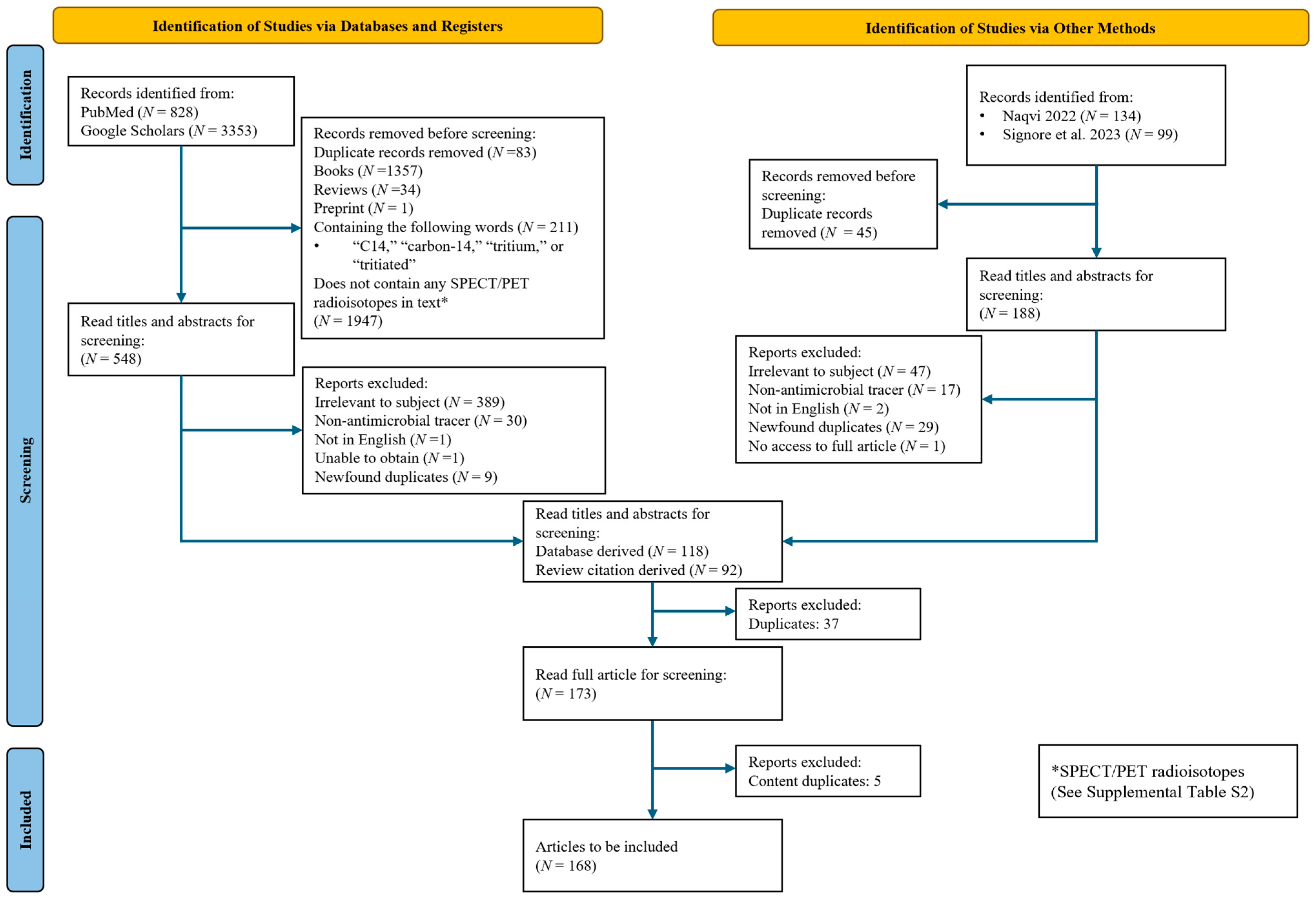
2.3. Search Strategy, Information Sources and Evidence Selection
2.4. Evidence Selection
2.5. Data Charting and Results Synthesis
T/NT Ratios by Mechanism
2.6. Graphics and Proofreading
3. Results
3.1. From Breakthrough to Setback: Case Study of 99mTc-Ciprofloxacin
3.2. Clinical Evaluation of Radiolabeled Antimicrobials
3.3. Evaluation of Antimicrobial Based Tracers
3.3.1. Radiolabeled Antimicrobials with High Translational Potential
Plazomicin
Caspofungin/Anidulafungin
Trimethoprim
3.4. Radiolabeled Antimicrobials with Moderate Translational Potential
3.5. Radiolabeled Antimicrobials with Low Translational Potential
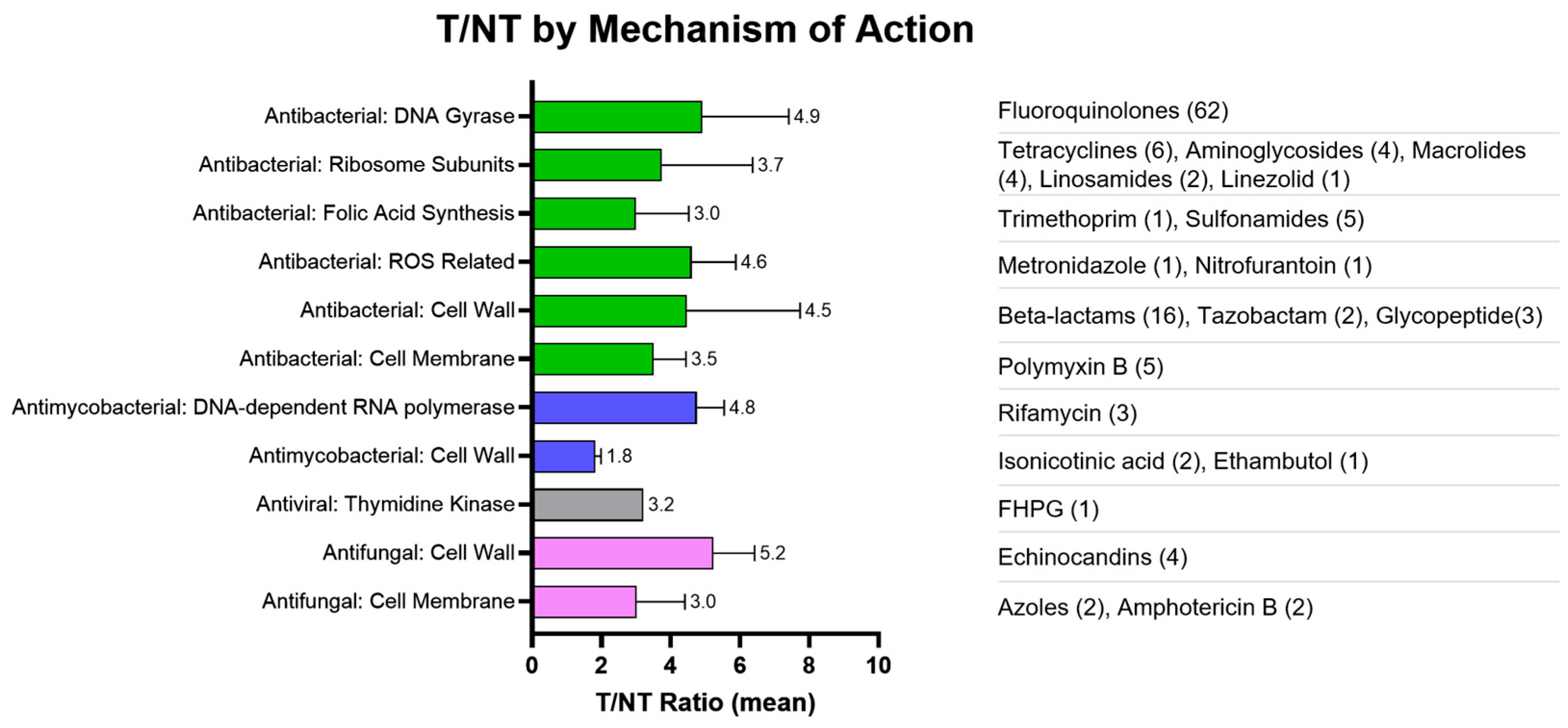
3.6. T/NT by Mechanism of Action
4. Discussion
4.1. Translational Gap
4.2. Predictive Framework
4.3. Potential Clinical Impact
4.4. Limitations
5. Conclusions and Future Directions
Supplementary Materials
Author Contributions
Funding
Institutional Review Board Statement
Informed Consent Statement
Data Availability Statement
Acknowledgments
Conflicts of Interest
References
- Naqvi, S.A.R. 99mTc-labeled antibiotics for infection diagnosis: Mechanism, action, and progress. Chem. Biol. Drug Des. 2022, 99, 56–74. [Google Scholar] [CrossRef]
- Signore, A.; Bentivoglio, V.; Varani, M.; Lauri, C. Current Status of SPECT Radiopharmaceuticals for Specific Bacteria Imaging. Semin. Nucl. Med. 2023, 53, 142–151. [Google Scholar] [CrossRef] [PubMed]
- Ordonez, A.A.; Jain, S.K. Pathogen-Specific Bacterial Imaging in Nuclear Medicine. Semin. Nucl. Med. 2018, 48, 182–194. [Google Scholar] [CrossRef] [PubMed]
- Northrup, J.D.; Mach, R.H.; Sellmyer, M.A. Radiochemical Approaches to Imaging Bacterial Infections: Intracellular versus Extracellular Targets. Int. J. Mol. Sci. 2019, 20, 5808. [Google Scholar] [CrossRef] [PubMed]
- Welling, M.M.; Hensbergen, A.W.; Bunschoten, A.; Velders, A.H.; Scheper, H.; Smits, W.K.; Roestenberg, M.; van Leeuwen, F.W.B. Fluorescent imaging of bacterial infections and recent advances made with multimodal radiopharmaceuticals. Clin. Transl. Imaging 2019, 7, 125–138. [Google Scholar] [CrossRef]
- Liu, S. Radiolabeled Antibiotics for Infection Imaging: A Scoping Review; OSF: Charlottesville, VA, USA, 2026. [Google Scholar] [CrossRef]
- Harzing, A.W. Publish or Perish; Tarma Software Research Ltd.: London, UK, 2007. [Google Scholar]
- Team, T.E. EndNote, version 21; Clarivate: Philadelphia, PA, USA, 2013.
- Ferro-Flores, G.; Avila-Rodríguez, M.A.; García-Pérez, F.O. Imaging of bacteria with radiolabeled ubiquicidin by SPECT and PET techniques. Clin. Transl. Imaging 2016, 4, 175–182. [Google Scholar] [CrossRef]
- Sachdeva, A.; Mitra, J.B.; Mukherjee, A. Ubiquicidin derived peptides for infection imaging. Nucl. Med. Biol. 2025, 146–147, 109049. [Google Scholar] [CrossRef]
- Vinjamuri, S.; Solanki, K.; Bomanji, J.; Siraj, Q.; Britton, K.; Hall, A.; O’Shaughnessy, E.; Das, S. Comparison of 99mTc infecton imaging with radiolabelled white-cell imaging in the evaluation of bacterial infection. Lancet 1996, 347, 233–235. [Google Scholar] [CrossRef]
- Britton, K.E.; Wareham, D.W.; Das, S.S.; Solanki, K.K.; Amaral, H.; Bhatnagar, A.; Katamihardja, A.H.S.; Malamitsi, J.; Moustafa, H.M.; Soroa, V.E.; et al. Imaging bacterial infection with 99mTc-ciprofloxacin (Infecton). J. Clin. Pathol. 2002, 55, 817–823. [Google Scholar] [CrossRef]
- Dumarey, N.; Blocklet, D.; Appelboom, T.; Tant, L.; Schoutens, A. Infecton is not specific for bacterial osteo-articular infective pathology. Eur. J. Nucl. Med. Mol. Imaging 2002, 29, 530–535. [Google Scholar] [CrossRef]
- Larikka, M.J.; Ahonen, A.K.; NiemelÄ, O.; Puronto, O.; Junila, J.A.; HÄMÄLÄInen, M.M.; Britton, K.; SyrjÄLÄ, H.P. 99mTc-ciprofloxacin (Infecton) imaging in the diagnosis of knee prosthesis infections. Nucl. Med. Commun. 2002, 23, 167–170. [Google Scholar] [CrossRef] [PubMed]
- Lee, M.; Yoon, M.; Hwang, K.H.; Choe, W. Tc-99m Ciprofloxacin SPECT of Pulmonary Tuberculosis. Nucl. Med. Mol. Imaging 2010, 44, 116–122. [Google Scholar] [CrossRef]
- Sarda, L.; Crémieux, A.-C.; Lebellec, Y.; Meulemans, A.; Lebtahi, R.; Hayem, G.; Génin, R.; Delahaye, N.; Huten, D.; Le Guludec, D. Inability of 99mTc-Ciprofloxacin Scintigraphy to Discriminate Between Septic and Sterile Osteoarticular Diseases. J. Nucl. Med. 2003, 44, 920–926. [Google Scholar]
- Yapar, Z.; Kibar, M.; Yapar, F.A.; Toğrul, E.; Kayaselçuk, U.; Sarpel, Y. The efficacy of technetium-99m ciprofloxacin (Infecton) imaging in suspected orthopaedic infection: A comparison with sequential bone/gallium imaging. Eur. J. Nucl. Med. 2001, 28, 822–830. [Google Scholar] [CrossRef]
- Malamitsi, J.; Giamarellou, H.; Kanellakopoulou, K.; Dounis, E.; Grecka, V.; Christakopoulos, J.; Koratzanis, G.; Antoniadou, A.; Panoutsopoulos, G.; Batsakis, C.; et al. Infecton: A 99mTc-ciprofloxacin radiopharmaceutical for the detection of bone infection. Clin. Microbiol. Infect. 2003, 9, 101–109. [Google Scholar] [CrossRef][Green Version]
- Langer, O.; Brunner, M.; Zeitlinger, M.; Ziegler, S.; Müller, U.; Dobrozemsky, G.; Lackner, E.; Joukhadar, C.; Mitterhauser, M.; Wadsak, W.; et al. In vitro and in vivo evaluation of [18F]ciprofloxacin for the imaging of bacterial infections with PET. Eur. J. Nucl. Med. Mol. Imaging 2005, 32, 143–150. [Google Scholar] [CrossRef]
- Brunner, M.; Langer, O.; Dobrozemsky, G.; Müller, U.; Zeitlinger, M.; Mitterhauser, M.; Wadsak, W.; Dudczak, R.; Kletter, K.; Müller, M. [18F]Ciprofloxacin, a new positron emission tomography tracer for noninvasive assessment of the tissue distribution and pharmacokinetics of ciprofloxacin in humans. Antimicrob. Agents Chemother. 2004, 48, 3850–3857. [Google Scholar] [CrossRef]
- Fischman, A.J.; Livni, E.; Babich, J.W.; Alpert, N.M.; Bonab, A.; Chodosh, S.; McGovern, F.; Kamitsuka, P.; Liu, Y.Y.; Cleeland, R.; et al. Pharmacokinetics of [18F]fleroxacin in patients with acute exacerbations of chronic bronchitis and complicated urinary tract infection studied by positron emission tomography. Antimicrob. Agents Chemother. 1996, 40, 659–664. [Google Scholar] [CrossRef] [PubMed]
- Ammar, A.; Fatima, S.; Mir, K.; Butt, S.T.; Batool, S.; Saeed, M.A.; Marwat, N.; Ahmed, N. Utility of Tc-99m-labeled levofloxacin as an infection-imaging agent in musculoskeletal infections. Pak. J. Nucl. Med. 2020, 10, 13–19. [Google Scholar] [CrossRef]
- Kaul, A.; Hazari, P.P.; Rawat, H.; Singh, B.; Kalawat, T.C.; Sharma, S.; Babbar, A.K.; Mishra, A.K. Preliminary evaluation of technetium-99m-labeled ceftriaxone: Infection imaging agent for the clinical diagnosis of orthopedic infection. Int. J. Infect. Dis. 2013, 17, e263–e270. [Google Scholar] [CrossRef] [PubMed]
- Ahmed, N.; Fatima, S.; Saeed, M.A.; Zia, M.; Irfan Ullah, J. 99mTc-Ceftizoxime: Synthesis, characterization and its use in diagnosis of diabetic foot osteomyelitis. J. Med. Imaging Radiat. Oncol. 2019, 63, 61–68. [Google Scholar] [CrossRef] [PubMed]
- Lee, I.K.; Jacome, D.A.; Cho, J.K.; Tu, V.; Young, A.J.; Dominguez, T.; Northrup, J.D.; Etersque, J.M.; Lee, H.S.; Ruff, A.; et al. Imaging sensitive and drug-resistant bacterial infection with [11C]-trimethoprim. J. Clin. Investig. 2023, 132, e156679. [Google Scholar] [CrossRef]
- Gordon, O.; Lee, D.E.; Liu, B.; Langevin, B.; Ordonez, A.A.; Dikeman, D.A.; Shafiq, B.; Thompson, J.M.; Sponseller, P.D.; Flavahan, K.; et al. Dynamic PET-facilitated modeling and high-dose rifampin regimens for Staphylococcus aureus orthopedic implant-associated infections. Sci. Transl. Med. 2021, 13, eabl6851. [Google Scholar] [CrossRef]
- Tucker, E.W.; Guglieri-Lopez, B.; Ordonez, A.A.; Ritchie, B.; Klunk, M.H.; Sharma, R.; Chang, Y.S.; Sanchez-Bautista, J.; Frey, S.; Lodge, M.A.; et al. Noninvasive 11C-rifampin positron emission tomography reveals drug biodistribution in tuberculous meningitis. Sci. Transl. Med. 2018, 10, eaau0965. [Google Scholar] [CrossRef]
- Singh, N.; Bhatnagar, A. Clinical Evaluation of Efficacy of 99mTc-Ethambutol in Tubercular Lesion Imaging. Tuberc. Res. Treat. 2010, 2010, 618051. [Google Scholar] [CrossRef][Green Version]
- Bhattacharya, B.; Damle, N.; Ranjan, P.; Arora, G.; Prakash, S.; Nischal, N.; Jorwal, P.; Kumar, A.; Tyagi, A.; Wig, N. 99mTc-Ethambutol Scintigraphy with Single-Photon Emission Computed Tomography/Computed Tomography in Lymph Node Tuberculosis: An Initial Experience. Indian J. Nucl. Med. 2022, 37, 323–328. [Google Scholar] [CrossRef]
- Kartamihardja, A.H.S.; Kurniawati, Y.; Gunawan, R. Diagnostic value of 99mTc-ethambutol scintigraphy in tuberculosis: Compared to microbiological and histopathological tests. Ann. Nucl. Med. 2018, 32, 60–68. [Google Scholar] [CrossRef]
- Tewson, T.J.; Yang, D.; Wong, G.; Macy, D.; DeJesus, O.J.; Nickles, R.J.; Perlman, S.B.; Taylor, M.; Frank, P. The synthesis of fluorine-18 lomefloxacin and its preliminary use in human studies. Nucl. Med. Biol. 1996, 23, 767–772. [Google Scholar] [CrossRef] [PubMed]
- Fischman, A.J.; Babich, J.W.; Bonab, A.A.; Alpert, N.M.; Vincent, J.; Callahan, R.J.; Correia, J.A.; Rubin, R.H. Pharmacokinetics of [18F]trovafloxacin in healthy human subjects studied with positron emission tomography. Antimicrob. Agents Chemother. 1998, 42, 2048–2054. [Google Scholar] [CrossRef] [PubMed][Green Version]
- Mota, F.; Ruiz-Bedoya, C.A.; Tucker, E.W.; Holt, D.P.; De Jesus, P.; Lodge, M.A.; Erice, C.; Chen, X.; Bahr, M.; Flavahan, K.; et al. Dynamic 18F-Pretomanid PET imaging in animal models of TB meningitis and human studies. Nat. Commun. 2022, 13, 7974. [Google Scholar] [CrossRef]
- Yaghoubi, S.; Barrio, J.R.; Dahlbom, M.; Iyer, M.; Namavari, M.; Satyamurthy, N.; Goldman, R.; Herschman, H.R.; Phelps, M.E.; Gambhir, S.S. Human Pharmacokinetic and Dosimetry Studies of [18F]FHBG: A Reporter Probe for Imaging Herpes Simplex Virus Type-1 Thymidine Kinase Reporter Gene Expression. J. Nucl. Med. 2001, 42, 1225–1234. [Google Scholar]
- Fischman, A.J.; Alpert, N.M.; Livni, E.; Ray, S.; Sinclair, I.; Callahan, R.J.; Correia, J.A.; Webb, D.; Strauss, H.W.; Rubin, R.H. Pharmacokinetics of 18F-labeled fluconazole in healthy human subjects by positron emission tomography. Antimicrob. Agents Chemother. 1993, 37, 1270–1277. [Google Scholar] [CrossRef]
- Solanki, K.K.; Bomanji, J.; Siraj, Q.; Small, M.; Britton, K.E. Tc-99m “Infecton”—A new class of radiopharmaceutical for imaging infection. J. Nucl. Med. 1993, 34, 119. [Google Scholar]
- Zhang, H.; Jiang, N.-y.; Zhu, L. Experimental studies on imaging of infected site with 99mTc-Iabeled ciprofloxacin in mice. Chin. Med. J. 2009, 122, 1907–1909. [Google Scholar]
- Sarda, L.; Saleh-Mghir, A.; Peker, C.; Meulemans, A.; Crémieux, A.-C.; Le Guludec, D. Evaluation of 99mTc-Ciprofloxacin Scintigraphy in a Rabbit Model of Staphylococcus aureus Prosthetic Joint Infection. J. Nucl. Med. 2002, 43, 239–245. [Google Scholar]
- Halder, K.K.; Nayak, D.K.; Baishya, R.; Sarkar, B.R.; Sinha, S.; Ganguly, S.; Debnath, M.C. 99mTc-labeling of ciprofloxacin and nitrofuryl thiosemicarbazone using fac-[99mTc(CO)3(H2O)3] core: Evaluation of their efficacy as infection imaging agents. Metallomics 2011, 3, 1041–1048. [Google Scholar] [CrossRef]
- Satpati, D.; Arjun, C.; Krishnamohan, R.; Samuel, G.; Banerjee, S. 68Ga-labeled Ciprofloxacin Conjugates as Radiotracers for Targeting Bacterial Infection. Chem. Biol. Drug Des. 2016, 87, 680–686. [Google Scholar] [CrossRef] [PubMed]
- Koźmiński, P.; Gawęda, W.; Rzewuska, M.; Kopatys, A.; Kujda, S.; Dudek, M.K.; Halik, P.K.; Królicki, L.; Gniazdowska, E. Physicochemical and Biological Study of 99mTc and 68Ga Radiolabelled Ciprofloxacin and Evaluation of [99mTc]Tc-CIP as Potential Diagnostic Radiopharmaceutical for Diabetic Foot Syndrome Imaging. Tomography 2021, 7, 829–842. [Google Scholar] [CrossRef]
- Langer, O.; Mitterhauser, M.; Brunner, M.; Zeitlinger, M.; Wadsak, W.; Mayer, B.X.; Kletter, K.; Muller, M. Synthesis of fluorine-18-labeled ciprofloxacin for PET studies in humans. Nucl. Med. Biol. 2003, 30, 285–291. [Google Scholar] [CrossRef] [PubMed]
- Zijlstra, S.; Gunawan, J.; Freytag, C.; Burchert, W. Synthesis and evaluation of fluorine-18 labelled compounds for imaging of bacterial infections with pet. Appl. Radiat. Isot. 2006, 64, 802–807. [Google Scholar] [CrossRef] [PubMed]
- Goethals, P.; Volkaert, A. Preparation of N′4-[11C]methyl-ciprofloxacin for positron emission tomography studies. J. Label. Compd. Radiopharm. 2002, 45, 213–216. [Google Scholar] [CrossRef]
- Sachin, K.; Kim, E.M.; Cheong, S.J.; Jeong, H.J.; Lim, S.T.; Sohn, M.H.; Kim, D.W. Synthesis of N4′-[18F]fluoroalkylated ciprofloxacin as a potential bacterial infection imaging agent for PET study. Bioconjug. Chem. 2010, 21, 2282–2288. [Google Scholar] [CrossRef]
- Fang, S.; Jiang, Y.; Gan, Q.; Ruan, Q.; Xiao, D.; Zhang, J. Design, Preparation, and Evaluation of a Novel 99mTcN Complex of Ciprofloxacin Xanthate as a Potential Bacterial Infection Imaging Agent. Molecules 2020, 25, 5837. [Google Scholar] [CrossRef]
- Papasavva, A.; Pirmettis, N.N.; Shegani, A.; Papadopoulou, E.; Kiritsis, C.; Georgoutsou-Spyridonos, M.; Mastellos, D.C.; Chiotellis, A.; Kyprianidou, P.; Pelecanou, M.; et al. Synthesis and Evaluation of 99mTc(CO)3 Complexes with Ciprofloxacin Dithiocarbamate for Infection Imaging. Pharmaceutics 2024, 16, 1210. [Google Scholar] [CrossRef]
- Rubin, R.H.; Livni, E.; Babich, J.; Alpert, N.M.; Liu, Y.-Y.; Tham, E.; Prosser, B.; Cleeland, R.; Callahan, R.J.; Correia, J.A.; et al. Pharmacokinetics of Fleroxacin as Studied by Positron Emission Tomography and [18F]Fleroxacin. Am. J. Med. 1993, 94, 31S–37S. [Google Scholar] [CrossRef]
- Ibrahim, I.T.; Motaleb, M.A.; Attalah, K.M. Synthesis and biological distribution of 99mTc-norfloxacin complex, a novel agent for detecting sites of infection. J. Radioanal. Nucl. Chem. 2010, 285, 431–436. [Google Scholar] [CrossRef]
- Nayak, D.; Baishya, R.; Halder, K.K.; Sen, T.; Sarkar, B.; Ganguly, S.; Das, M.K.; Debnath, M. Evaluation of 99mTc(i)-tricarbonyl complexes of fluoroquinolones for targeting bacterial infection. Metallomics 2012, 4, 1197–1208. [Google Scholar] [CrossRef]
- Zhang, S.; Zhang, W.; Wang, Y.; Jin, Z.; Wang, X.; Zhang, J.; Zhang, Y. Synthesis and biodistribution of a novel 99mTcN complex of norfloxacin dithiocarbamate as a potential agent for bacterial infection imaging. Bioconjug. Chem. 2011, 22, 369–375. [Google Scholar] [CrossRef] [PubMed]
- Motaleb, M.A. Radiochemical and biological characteristics of 99mTc-difloxacin and 99mTc-pefloxacin for detecting sites of infection. J. Label. Compd. Radiopharm. Off. J. Int. Isot. Soc. 2010, 53, 104–109. [Google Scholar] [CrossRef]
- El-Ghany, E.A.; El-Kolaly, M.T.; Amine, A.M.; El-Sayed, A.S.; Abdel-Gelil, F. Synthesis of 99mTc-pefloxacin: A new targeting agent for infectious foci. J. Radioanal. Nucl. Chem. 2005, 266, 131–139. [Google Scholar] [CrossRef]
- Motaleb, M.A. Preparation and biodistribution of 99mTc-lomefloxacin and 99mTc-ofloxacin complexes. J. Radioanal. Nucl. Chem. 2007, 272, 95–99. [Google Scholar] [CrossRef]
- Motaleb, M.A.; Ayoub, S.M. Preparation, quality control, and biodistribution of 99mTc-rufloxacin complex as a model for detecting sites of infection. Radiochemistry 2013, 55, 610–614. [Google Scholar] [CrossRef]
- Shah, S.Q.; Khan, M.R. Radiocharacterization of the 99mTc–rufloxacin complex and biological evaluation in Staphylococcus aureus infected rat model. J. Radioanal. Nucl. Chem. 2011, 288, 373–378. [Google Scholar] [CrossRef]
- Shahzad, S.; Qadir, M.A.; Rasheed, R.; Ahmad, A.; Shafiq, M.I.; Ahmed, M.; Noreen, S.; Ali, A.; Shahzadi, S.K.; Javed, M. A new method for synthesis of 99mTc-enorfloxacin: An infection imaging agent. Lat. Am. J. Pharm. 2016, 35, 259–264. [Google Scholar]
- Siaens, R.H.; Rennen, H.J.; Boerman, O.C.; Dierckx, R.; Slegers, G. Synthesis and comparison of 99mTc-enrofloxacin and 99mTc-ciprofloxacin. J. Nucl. Med. 2004, 45, 2088–2094. [Google Scholar] [PubMed]
- Naqvi, S.; Ishfaq, M.; Khan, Z.; Nagra, S.; Bukhari, I.; Hussain, A.; Mahmood, N.; Shahzad, S.; Haq, A.; Bokhari, T. 99mTc labeled levofloxacin as an infection imaging agent: A novel method for labeling levofloxacin using cysteine·HCl as co-ligand and in vivo study. Turk. J. Chem. 2012, 36, 267–277. [Google Scholar] [CrossRef]
- Shahzad, S.; Qadir, M.A.; Rasheed, R.; Anwar, S.; Ahmed, M. In vivo studies 99mTc-levofloxacin freeze dried kits in Salmonella typhi, Pseudoman aeruginosa, and Escherichia coli. Lat. Am. J. Pharm. 2015, 34, 760–765. [Google Scholar]
- Shah, S.Q.; Khan, M.R. Radiosynthesis and biodistribution of 99mTc-tricarbonyl complex of temafloxacin dithiocarbamate: A potential Streptococci pneumoniae infection radiotracer. J. Radioanal. Nucl. Chem. 2011, 288, 411–416. [Google Scholar] [CrossRef]
- Shah, S.Q.; Khan, M.R. Synthesis of 99mTc(CO)3-Pazufloxacin Dithiocarbamate Complex and Biodistribution in Experimentally Induced Infection in Female Nude Mice. Synth. React. Inorg. Met.-Org. Nano-Met. Chem. 2012, 42, 190–195. [Google Scholar] [CrossRef]
- Shah, S.Q.; Khan, M.R. Synthesis of 99mTc-Pazufloxacin dithiocarbamate complex and biological evaluation in Wister rats artificially infected with. J. Radioanal. Nucl. Chem. 2011, 288, 511–516. [Google Scholar] [CrossRef]
- Moustapha, M.E.; Motaleb, M.A.; Shweeta, H.; Farouk, M. Synthesis and biological evaluation of technetium-sarafloxacin complex for infection imaging. J. Radioanal. Nucl. Chem. 2016, 307, 699–705. [Google Scholar] [CrossRef]
- Shah, S.Q.; Khan, M.R. 99mTc(CO)3-tosufloxacin dithiocarbamate complexation and radiobiological evaluation in male Wister rat model. J. Radioanal. Nucl. Chem. 2011, 288, 485–490. [Google Scholar] [CrossRef]
- Singh, A.K.; Verma, J.; Bhatnagar, A.; Ali, A. Tc-99m labeled Sparfloxacin: A specific infection imaging agent. World J. Nucl. Med. 2003, 2, 103–109. [Google Scholar]
- Eid Moustapha, M.; Shweeta, H.A.; Motaleb, M.A. Technetium-labeled danofloxacin complex as a model for infection imaging. Arab. J. Chem. 2016, 9, S1928–S1934. [Google Scholar] [CrossRef]
- Shah, S.Q.; Khan, M.R. Radiolabeling of gemifloxacin with technetium-99m and biological evaluation in artificially Streptococcus pneumoniae infected rats. J. Radioanal. Nucl. Chem. 2011, 288, 307–312. [Google Scholar] [CrossRef]
- Shahzad, S.; Qadir, M.A.; Rasheed, R.; Ahmed, M. Synthesis of 99mTc-gemifloxacin freeze dried kits and their biodistribution in biodistribution in Salmonella typhi, Pseudomonas aeruginosa and Klebsiella Pneumonia. Arab. J. Chem. 2019, 12, 664–670. [Google Scholar] [CrossRef]
- Khoramrouz, S.J.; Erfani, M.; Athari Allaf, M. Technetium-99m Tricarbonyl Labeled a Broad-spectrum Quinolone as a Specific Imaging Agent in Infection Diseases. Iran. J. Pharm. Res. 2017, 16, 611–618. [Google Scholar]
- Motaleb, M.A.; El-Kolaly, M.T.; Ibrahim, A.B.; Abd El-Bary, A. Study on the preparation and biological evaluation of 99mTc-gatifloxacin and 99mTc-cefepime complexes. J. Radioanal. Nucl. Chem. 2011, 289, 57–65. [Google Scholar] [CrossRef]
- Shah, S.Q.; Khan, A.U.; Khan, M.R. Radiosynthesis and biological evaluation of 99mTcN-sitafloxacin dithiocarbamate as a potential radiotracer for Staphylococcus aureus infection. J. Radioanal. Nucl. Chem. 2011, 287, 827–832. [Google Scholar] [CrossRef]
- Shah, S.Q.; Khan, A.U.; Khan, M.R. Radiosynthesis and biological evolution of 99mTc(CO)3-sitafloxacin dithiocarbamate complex: A promising Staphylococcus aureus infection radiotracer. J. Radioanal. Nucl. Chem. 2011, 288, 131–136. [Google Scholar] [CrossRef]
- Chattopadhyay, S.; Saha Das, S.; Chandra, S.; De, K.; Mishra, M.; Ranjan Sarkar, B.; Sinha, S.; Ganguly, S. Synthesis and evaluation of 99mTc-moxifloxacin, a potential infection specific imaging agent. Appl. Radiat. Isot. 2010, 68, 314–316. [Google Scholar] [CrossRef]
- Shah, S.Q.; Khan, M.R. Radiosynthesis and biological evaluation of the 99mTc-tricarbonyl moxifloxacin dithiocarbamate complex as a potential Staphylococcus aureus infection radiotracer. Appl. Radiat. Isot. 2011, 69, 686–690. [Google Scholar] [CrossRef]
- Fischman, A.J.; Babich, J.W.; Alpert, N.M.; Vincent, J.; Wilkinson, R.A.; Callahan, R.J.; Correia, J.A.; Rubin, R.H. Pharmacokinetics of 18F-labeled trovafloxacin in normal and Escherichia coli-infected rats and rabbits studied with positron emission tomography. Clin. Microbiol. Infect. 1997, 3, 63–72. [Google Scholar] [CrossRef]
- Shah, S.Q.; Khan, M.R. Radiosynthesis and biodistribution of the 99mTc-trovafloxacin complex as a potential methicillin resistant Staphylococcus aureus infection radiotracer. J. Radioanal. Nucl. Chem. 2011, 288, 525–530. [Google Scholar] [CrossRef]
- Shah, S.Q.; Khan, M.R. Radiocomplexation and biological characterization of the 99mTcN-trovafloxacin dithiocarbamate: A novel methicillin-resistant Staphylococcus aureus infection imaging agent. J. Radioanal. Nucl. Chem. 2011, 288, 215–220. [Google Scholar] [CrossRef]
- Shah, S.Q.; Khan, M.R. Synthesis of the 99mTc(CO)3-trovafloxacin dithiocarbamate complex and biological characterization in artificially methicillin-resistant Staphylococcus aureus infected rats model. J. Radioanal. Nucl. Chem. 2011, 288, 297–302. [Google Scholar] [CrossRef]
- Shah, S.Q.; Khan, A.U.; Khan, M.R. Synthesis, biological evaluation and biodistribution of the 99mTc-Garenoxacin complex in artificially infected rats. J. Radioanal. Nucl. Chem. 2011, 288, 207–213. [Google Scholar] [CrossRef]
- Shah, S.Q.; Khan, A.U.; Khan, M.R. Radiosynthesis and biodistribution of 99mTcN-Garenoxacin dithiocarbamate complex a potential infection imaging agent. J. Radioanal. Nucl. Chem. 2011, 288, 59–64. [Google Scholar] [CrossRef]
- Shah, S.Q.; Khan, A.U.; Khan, M.R. 99mTc(CO)3–Garenoxacin dithiocarbamate synthesis and biological evolution in rats infected with multiresistant Staphylococcus aureus and penicillin-resistant Streptococci. J. Radioanal. Nucl. Chem. 2011, 288, 171–176. [Google Scholar] [CrossRef]
- Shah, S.Q.; Khan, M.R.; Ali, S.M. Radiosynthesis of 99mTc(CO)3-Clinafloxacin Dithiocarbamate and Its Biological Evaluation as a Potential Staphylococcus aureus Infection Radiotracer. Nucl. Med. Mol. Imaging 2011, 45, 248–254. [Google Scholar] [CrossRef][Green Version]
- Shah, S.Q.; Khan, M.R. Synthesis of 99mTcN-clinafloxacin Dithiocarbamate Complex and Comparative Radiobiological Evaluation in Staphylococcus aureus Infected Mice. World J. Nucl. Med. 2014, 13, 154–158. [Google Scholar] [CrossRef] [PubMed]
- Shah, S.; Khan, M. Synthesis of techentium-99m labeled clinafloxacin (99mTc–CNN) complex and biological evaluation as a potential Staphylococcus aureus infection imaging agent. J. Radioanal. Nucl. Chem. 2011, 288, 423–428. [Google Scholar] [CrossRef]
- Shah, S.Q.; Khan, A.U.; Khan, M.R. 99mTc-prulifloxacin in artificially infected animals. Radiosynthesis and biological evaluation. Nuklearmedizin 2011, 50, 134–140. [Google Scholar] [CrossRef] [PubMed]
- El-Kawy, O.A.; Farah, K. Radiocomplexation and biological evaluation of nemonoxacin in mice infected with multiresistant Staphylococcus aureus and penicillin-resistant Streptococci. J. Radioanal. Nucl. Chem. 2015, 306, 123–130. [Google Scholar] [CrossRef]
- Shahzad, M.A.; Naqvi, S.A.R.; Rasheed, R.; Yameen, M.; Anjum, F.; Ahmed, M.T.; Hussain, Z.; Gillani, S.J.H. Radiolabeling of benzylpenicillin with lutetium-177: Quality control and biodistribution study to develop theranostic infection imaging agent. Pak. J. Pharm. Sci. 2017, 30, 2349–2354. [Google Scholar]
- Shahzadi, S.K.; Qadir, M.A.; Shabnam, S.; Javed, M. 99mTc-amoxicillin: A novel radiopharmaceutical for infection imaging. Arab. J. Chem. 2019, 12, 2533–2539. [Google Scholar] [CrossRef]
- Durkan, K.; Tuncel, A.; Yurt, F. In vitro evaluation of 99mTc-sultamicillin for infection imaging. Biopharm. Drug Dispos. 2021, 42, 285–293. [Google Scholar] [CrossRef]
- El-Tawoosy, M. Preparation and biological distribution of 99mTc-cefazolin complex, a novel agent for detecting sites of infection. J. Radioanal. Nucl. Chem. 2013, 298, 1215–1220. [Google Scholar] [CrossRef]
- Sanad, M.H.; Eh, B. Performance characteristics of biodistribution of 99mTc-cefprozil for in vivo infection imaging. J. Anal. Sci. Technol. 2014, 5, 32. [Google Scholar] [CrossRef]
- Chattopadhyay, S.; Ghosh, M.; Sett, S.; Das, M.K.; Chandra, S.; De, K.; Mishra, M.; Sinha, S.; Ranjan Sarkar, B.; Ganguly, S. Preparation and evaluation of 99mTc-cefuroxime, a potential infection specific imaging agent: A reliable thin layer chromatographic system to delineate impurities from the 99mTc-antibiotic. Appl. Radiat. Isot. 2012, 70, 2384–2387. [Google Scholar] [CrossRef]
- Yurt Lambrecht, F.; Durkan, K.; Unak, P. Preparation, quality control and stability of 99mTc-cefuroxime axetil. J. Radioanal. Nucl. Chem. 2008, 275, 161–164. [Google Scholar] [CrossRef]
- Mostafa, M.; Motaleb, M.A.; Sakr, T.M. Labeling of ceftriaxone for infective inflammation imaging using 99mTc eluted from 99Mo/99mTc generator based on zirconium molybdate. Appl. Radiat. Isot. 2010, 68, 1959–1963. [Google Scholar] [CrossRef] [PubMed]
- Sohaib, M.; Khurshid, Z.; Roohi, S. Labelling of ceftriaxone with 99mTc and its bio-evaluation as an infection imaging agent. J. Label. Compd. Radiopharm. 2014, 57, 652–657. [Google Scholar] [CrossRef]
- Fazli, A.; Salouti, M.; Mazidi, M. 99mTc-ceftriaxone, as a targeting radiopharmaceutical for scintigraphic imaging of infectious foci due to Staphylococcus aureus in mouse model. J. Radioanal. Nucl. Chem. 2013, 298, 1227–1233. [Google Scholar] [CrossRef]
- Teixeira, L.E.M.; Soares, G.G.; Teixeira, H.C.; Takenaka, I.K.T.M.; Diniz, S.O.F.; de Andrade, M.A.P.; Cardoso, V.N.; de Araújo, I.D. Efficacy of 99mTc-Labeled Ceftizoxime in the Diagnosis of Subclinical Infections Associated with Titanium Implants in Rats. Surg. Infect. 2015, 16, 352–357. [Google Scholar] [CrossRef]
- Costa, P.H.; Diniz, S.O.; Cardoso, V.N.; Tarabal, B.; Takenaka, I.; Braga, O.; Vidigal, P.V.; Gelape, C.L.; Araujo, I.D. Scintigraphic imaging with technetium-99M-labelled ceftizoxime is a reliable technique for the diagnosis of deep sternal wound infection in rats. Acta Cir. Bras. 2015, 30, 632–638. [Google Scholar] [CrossRef]
- Mirshojaei, S.F.; Gandomkar, M.; Najafi, R.; Sadat Ebrahimi, S.E.; Babaei, M.H.; Shafiei, A.; Talebi, M.H. Radio labeling, quality control and biodistribution of 99mTc-cefotaxime as an infection imaging agent. J. Radioanal. Nucl. Chem. 2011, 287, 21–25. [Google Scholar] [CrossRef]
- Ilem-Ozdemir, D.; Asikoglu, M.; Ozkilic, H.; Yilmaz, F.; Hosgor-Limoncu, M.; Ayhan, S. Gamma scintigraphy and biodistribution of 99mTc-cefotaxime sodium in preclinical models of bacterial infection and sterile inflammation. J. Label. Compd. Radiopharm. 2016, 59, 109–116. [Google Scholar] [CrossRef]
- Mirshojaei, S.F.; Erfani, M.; Shafiei, M. Evaluation of 99mTc-ceftazidime as bacterial infection imaging agent. J. Radioanal. Nucl. Chem. 2013, 298, 19–24. [Google Scholar] [CrossRef]
- Motaleb, M.A. Preparation of 99mTc-cefoperazone complex, a novel agent for detecting sites of infection. J. Radioanal. Nucl. Chem. 2007, 272, 167–171. [Google Scholar] [CrossRef]
- Koźmiński, P.; Rzewuska, M.; Piądłowska, A.; Halik, P.; Gniazdowska, E. Synthesis, physicochemical and in vitro biological evaluation of 99mTc-cefepime radioconjugates, and development of DTPA-cefepime single vial kit formulation for labelling with technetium-99m. J. Radioanal. Nucl. Chem. 2022, 331, 2883–2894. [Google Scholar] [CrossRef]
- Sakr, T.M.; Motaleb, M.A.; Ibrahim, I.T. 99mTc–meropenem as a potential SPECT imaging probe for tumor hypoxia. J. Radioanal. Nucl. Chem. 2012, 292, 705–710. [Google Scholar] [CrossRef]
- Naqvi, S.A.R.; Jabbar, T.; Alharbi, M.A.; Noureen, A.; Alharbi, N.K.; Sherazi, T.A.; Shahzadi, A.; Ahmed, A.E.; Afzal, M.S.; Imran, M.B. Radiosynthesis, quality control, biodistribution, and infection-imaging study of a new 99mTc-labeled ertapenem radiopharmaceutical. Front. Chem. 2022, 10, 1020387. [Google Scholar] [CrossRef] [PubMed]
- Ozker, K.; Urgancioğlu, I. 99mTc-gentamicin: Chemical and biological evaluation. Eur. J. Nucl. Med. 1981, 6, 173–176. [Google Scholar] [CrossRef]
- Amina Watson, R.A.; Landon, J.; Edwards, C.R.W.; Shaw, E.J. Improved 125I-ligands for gentamicin radioimmunoassay. J. Antimicrob. Chemother. 1979, 5, 673–680. [Google Scholar] [CrossRef]
- Dhanani, J.A.; Goodman, S.; Ahern, B.; Cohen, J.; Fraser, J.F.; Barnett, A.; Diab, S.; Bhatt, M.; Roberts, J.A. Comparative lung distribution of radiolabeled tobramycin between nebulized and intravenous administration in a mechanically-ventilated ovine model, an observational study. Int. J. Antimicrob. Agents 2021, 57, 106232. [Google Scholar] [CrossRef] [PubMed]
- Van’t Veen, A.; Gommers, D.; Verbrugge, S.J.; Wollmer, P.; Mouton, J.W.; Kooij, P.P.; Lachmann, B. Lung clearance of intratracheally instilled 99mTc-tobramycin using pulmonary surfactant as vehicle. Br. J. Pharmacol. 1999, 126, 1091–1096. [Google Scholar] [CrossRef]
- Roohi, S.; Mushtaq, A.; Jehangir, M.; Malik, S.A. Synthesis, quality control and biodistribution of 99mTc-Kanamycin. J. Radioanal. Nucl. Chem. 2006, 267, 561–566. [Google Scholar] [CrossRef]
- Widyasari, E.; Halimah, I.; Sugiharti, R.J.; Sriyani, M.; Daruwati, I.; Iswahyudi, I.; Isabela, E.; Nuraeni, W. Biological Evaluation of 99mTc-Kanamycin for Infection Imaging. Indones. J. Phys. Nucl. Appl. 2017, 2, 34. [Google Scholar] [CrossRef][Green Version]
- Akbar, M.U.; Bokhari, T.H.; Khalid, M.; Ahmad, M.R.; Roohi, S.; Hina, S.; Mehmood, S.; Sohaib, M.; Jabbar, T. Radiolabeling, quality control, and biological characterization of 177Lu-labeled kanamycin. Chem. Biol. Drug Des. 2017, 90, 425–431. [Google Scholar] [CrossRef]
- El-Kawy, O.A.; Abdelaziz, G.; Abdel-Razek, A.S. Radiolabeling, characterization, and preclinical evaluation of plazomicin: A potential tracer for bacterial infection. Chem. Biol. Drug Des. 2022, 99, 688–702. [Google Scholar] [CrossRef]
- Ercan, M.T.; Aras, T.; Unsal, I.S. Evaluation of 99mTc-erythromycin and 99mTc-streptomycin sulphate for the visualization of inflammatory lesions. Int. J. Radiat. Appl. Instrum. B 1992, 19, 803–806. [Google Scholar] [CrossRef]
- Sanad, M.H. Labeling and biological evaluation of 99mTc-azithromycin for infective inflammation diagnosis. Radiochemistry 2013, 55, 539–544. [Google Scholar] [CrossRef]
- Abdel-Ghaney, I.Y.; Sanad, M.H. Synthesis of 99mTc-Erythromycin Complex as a Model for Infection Sites Imaging. Radiochemistry 2013, 55, 418–422. [Google Scholar] [CrossRef]
- Borai, E.H.; Sanad, M.H.; Fouzy, A.S.M. Optimized chromatographic separation and biological evaluation of 99mTc-clarithromycin for infective inflammation diagnosis. Radiochemistry 2016, 58, 84–91. [Google Scholar] [CrossRef]
- Rizvi, S.F.A.; Tariq, S.; Mehdi, M.; Hassan, A.J. Synthesis of 99mTc-roxithromycin: A novel diagnostic agent to discriminate between septic and aseptic inflammation. Chem. Biol. Drug Des. 2019, 93, 1166–1174. [Google Scholar] [CrossRef]
- İlem-Özdemir, D.; Asikoglu, M.; Ozkilic, H.; Yilmaz, F.; Hosgor-Limoncu, M.; Ayhan, S. 99mTc-Doxycycline hyclate: A new radiolabeled antibiotic for bacterial infection imaging. J. Label. Compd. Radiopharm. 2014, 57, 36–41. [Google Scholar] [CrossRef]
- Rizvi, S.F.A.; Jabbar, T.; Shahid, W.; Sanad, M.H.; Zhang, H. Facile One-Pot Strategy for Radiosynthesis of 99mTc-Doxycycline to Diagnose Staphylococcus aureus in Infectious Animal Models. Appl. Biochem. Biotechnol. 2022, 194, 2672–2683. [Google Scholar] [CrossRef]
- Milanović, Z.; Janković, D.; Vranješ-Đurić, S.; Radović, M.; Prijović, Ž.; Zavišić, G.; Perić, M.; Stanković, D.; Mirković, M. 177Lu-doxycycline as potential radiopharmaceutical: Electrochemical characterization, radiolabeling, and biodistribution in tumor-bearing mice. Int. J. Radiat. Biol. 2021, 97, 1687–1695. [Google Scholar] [CrossRef]
- Dewanjee, M.K.; Fliegel, C.; Treves, S.; Davis, M.A. 99mTc-tetracyclines: Preparation and biological evaluation. J. Nucl. Med. 1974, 15, 176–182. [Google Scholar] [PubMed]
- Philip, L.H.; Taylor, A.; Chauncey, D.M.; Schelbert, H. Comparison of 131I-tetracycline and 67Ga-citrate as abscess localizing agents. Nuklearmedizin 1977, 16, 76–78. [Google Scholar] [CrossRef]
- Saleem, S.M.; Jabbar, T.; Imran, M.B.; Noureen, A.; Sherazi, T.A.; Afzal, M.S.; Rab Nawaz, H.Z.; Ramadan, M.F.; Alkahtani, A.M.; Alsuwat, M.A.; et al. Radiosynthesis and Preclinical Evaluation of [99mTc]Tc-Tigecycline Radiopharmaceutical to Diagnose Bacterial Infections. Pharmaceuticals 2024, 17, 1283. [Google Scholar] [CrossRef]
- Essouissi, I.; Ghali, W.; Saied, N.M.; Saidi, M. Synthesis and evaluation of 99mTc-N-sulfanilamide ferrocene carboxamide as bacterial infections detector. Nucl. Med. Biol. 2010, 37, 821–829. [Google Scholar] [CrossRef] [PubMed]
- Ahmed, M.T.; Naqvi, S.A.R.; Rasheed, R.; Zahoor, A.F.; Usman, M.; Hussain, Z. Technetium-99m-Labeled Sulfadiazine: A Targeting Radiopharmaceutical for Scintigraphic Imaging of Infectious Foci Due To Escherichia coli in Mouse and Rabbit Models. Appl. Biochem. Biotechnol. 2017, 183, 374–384. [Google Scholar] [CrossRef] [PubMed]
- Essouissi, I.; Darghouth, F.; Saied, N.M.; Saidi, M.; Kanoun, A.; Saidi, M. Radiolabeling, Quality Control, and Biodistribution of 99mTc-Sulfadiazine as an Infection Imaging Agent. Radiochemistry 2015, 57, 307–311. [Google Scholar] [CrossRef]
- Ahmed, M.T.; Yameen, M.; Munir, B.; Asim, S.; Usman, M.; Naqvi, S.A.R.; Gillani, J.A.H.; Rasheed, R.; Shahzad, M.A. Evaluation of 99mTc-sulfadiazine as Bacillus microorganisms infection imaging agent using animal model. Pak. J. Pharm. Sci. 2018, 31, 1495–1499. [Google Scholar]
- Liu, F.; Zhao, B.; Xia, X.; Yan, J.; Yu, F.; Yan, G.; Hu, J.; Chen, S.; Wang, Y.; Liu, H.; et al. Al18F labeled sulfonamide-conjugated positron emission tomography tracer in vivo tumor-targeted imaging. J. Cell. Biochem. 2019, 120, 17006–17014. [Google Scholar] [CrossRef]
- Amin, A.M.; Ibrahim, I.T.; Attallah, K.M.Z.; Ali, S. 99mTc-sulfadimidine as a potential radioligand for differentiation between septic and aseptic inflammations. Radiochemistry 2014, 56, 72–75. [Google Scholar] [CrossRef]
- Hina, S.; Rajoka, M.I.; Roohi, S.; Haque, A.; Qasim, M. Preparation, Biodistribution, and Scintigraphic Evaluation of 99mTc-Clindamycin: An Infection Imaging Agent. Appl. Biochem. Biotechnol. 2014, 174, 1420–1433. [Google Scholar] [CrossRef]
- Bokhari, T.H.; Rizvi, S.F.A.; Roohi, S.; Hina, S.; Mushtaq, A.; Khalid, M.; Iqbal, M. Preparation, biodistribution and scintigraphic evaluation of 99mTc-lincomycin. Pak. J. Pharm. Sci. 2015, 28, 1965–1970. [Google Scholar] [PubMed]
- Sellmyer, M.A.; Lee, I.; Hou, C.; Lieberman, B.P.; Zeng, C.; Mankoff, D.A.; Mach, R.H. Quantitative PET Reporter Gene Imaging with [11C]Trimethoprim. Mol. Ther. 2017, 25, 120–126. [Google Scholar] [CrossRef] [PubMed]
- Sellmyer, M.A.; Lee, I.; Hou, C.; Weng, C.C.; Li, S.; Lieberman, B.P.; Zeng, C.; Mankoff, D.A.; Mach, R.H. Bacterial infection imaging with [18F]fluoropropyl-trimethoprim. Proc. Natl. Acad. Sci. USA 2017, 114, 8372–8377. [Google Scholar] [CrossRef]
- Iqbal, A.; Naqvi, S.A.R.; Rasheed, R.; Mansha, A.; Ahmad, M.; Zahoor, A.F. Radiosynthesis and Biodistribution of 99mTc-Metronidazole as an Escherichia coli Infection Imaging Radiopharmaceutical. Appl. Biochem. Biotechnol. 2018, 185, 127–139. [Google Scholar] [CrossRef]
- Kong, D.J.; Lu, J.; Ye, S.Z.; Wang, X.B. Synthesis and biological evaluation of a novel asymmetrical 99mTc-nitrido complex of metronidazole derivative. J. Label. Compd. Radiopharm. 2007, 50, 1137–1142. [Google Scholar] [CrossRef]
- Shah, S.Q.; Khan, A.U.; Khan, M.R. Radiosynthesis of 99mTc-nitrofurantoin a novel radiotracer for in vivo imaging of Escherichia coli infection. J. Radioanal. Nucl. Chem. 2011, 287, 417–422. [Google Scholar] [CrossRef]
- El-Azony, K.M.; El-Mohty, A.A.; Seddik, U.; Khater, S.I. Radioiodination and bioevaluation of nitrofurantoin for urinary tract imaging. J. Label. Compd. Radiopharm. 2012, 55, 315–319. [Google Scholar] [CrossRef]
- Auletta, S.; Galli, F.; Varani, M.; Campagna, G.; Conserva, M.; Martinelli, D.; Santino, I.; Signore, A. In vitro and In vivo Evaluation of 99mTc-Polymyxin B for Specific Targeting of Gram-Bacteria. Biomolecules 2021, 11, 232. [Google Scholar] [CrossRef]
- Kumar, P.; Shanbhag, N.C.; Chaudhari, P.; Mohanty, B.; Thakur, R.; Sasidharan, G.M. Radiolabeling and preclinical evaluation of technetium-99m labeled colistin. Appl. Radiat. Isot. 2024, 214, 111524. [Google Scholar] [CrossRef] [PubMed]
- Karpuz, M.; Özgenç, E.; Gündoğdu, E.; Burak, Z. Pre-study on radiolabeling of colistin with Lutetium-177 to develop theranostic infection agent. J. Res. Pharm. 2022, 26, 397–407. [Google Scholar] [CrossRef]
- Yurt Lambrecht, F.; Yilmaz, O.; Durkan, K.; Unak, P.; Bayrak, E. Preparation and biodistribution of [131I]linezolid in animal model infection and inflammation. J. Radioanal. Nucl. Chem. 2009, 281, 415–419. [Google Scholar] [CrossRef]
- Mota, F.; Jadhav, R.; Ruiz-Bedoya, C.A.; Ordonez, A.A.; Klunk, M.H.; Freundlich, J.S.; Jain, S.K. Radiosynthesis and Biodistribution of 18F-Linezolid in Mycobacterium tuberculosis-Infected Mice Using Positron Emission Tomography. ACS Infect. Dis. 2020, 6, 916–921. [Google Scholar] [CrossRef] [PubMed]
- Jalilian, A.; Hosseini, M.; Karimian, A.; Saddadi, F.; Sadeghi, M. Preparation and biodistribution of [201Tl](III)vancomycin complex in normal rats. Nukleonika 2006, 51, 203–208. [Google Scholar]
- Spoelstra, G.B.; Braams, L.M.; Ijpma, F.F.A.; van Oosten, M.; Feringa, B.L.; Szymanski, W.; Elsinga, P.H.; van Dijl, J.M. Bacteria-targeted imaging using vancomycin-based positron emission tomography tracers can distinguish infection from sterile inflammation. Eur. J. Nucl. Med. Mol. Imaging 2025, 52, 1878–1889. [Google Scholar] [CrossRef]
- Spoelstra, G.B.; Blok, S.N.; Reali Nazario, L.; Noord, L.; Fu, Y.; Simeth, N.A.; IJpma, F.F.A.; van Oosten, M.; van Dijl, J.M.; Feringa, B.L.; et al. Synthesis and preclinical evaluation of novel 18F-vancomycin-based tracers for the detection of bacterial infections using positron emission tomography. Eur. J. Nucl. Med. Mol. Imaging 2024, 51, 2583–2596. [Google Scholar] [CrossRef]
- Jalilian, A.R.; Yousef, Y.K.; Rowshanfarzad, P.; Sabet, M.; Kamali-Dehghan, M.; Majdabadi, A. Preparation and preliminary evaluation of [55Co](II) vancomycin. Nucl. Sci. Tech. 2008, 19, 347–353. [Google Scholar]
- Kış, T.; Köse, Ş.; Yılmaz, O.; Kış, M.; Yurt, F.; Acar, E.; Bekiş, R.; Yılmaz, C.; Barış, M.; Diniz, G.; et al. Evaluation of 99mTechnetium-Vancomycin Imaging Potential in Experimental Rat Model for the Diagnosis of Infective Endocarditis. Curr. Med. Imaging Rev. 2021, 17, 781–789. [Google Scholar] [CrossRef]
- Rasheed, R.; Naqvi, S.A.R.; Gillani, S.J.H.; Zahoor, A.F.; Jielani, A.; Saeed, N. 99mTc-tazobactam, a novel infection imaging agent: Radiosynthesis, quality control, biodistribution, and infection imaging studies. J. Label. Compd. Radiopharm. 2017, 60, 242–249. [Google Scholar] [CrossRef]
- Singh, A.K.; Verma, J.; Bhatnager, A.; Sen, S. Tc-99m Isoniazid: A specific agent for diagnosis of tuberculosis. World J. Nucl. Med. 2003, 2, 292–305. [Google Scholar]
- Weinstein, E.A.; Liu, L.; Ordonez, A.A.; Wang, H.; Hooker, J.M.; Tonge, P.J.; Jain, S.K. Noninvasive determination of 2-[18F]-fluoroisonicotinic acid hydrazide pharmacokinetics by positron emission tomography in Mycobacterium tuberculosis-infected mice. Antimicrob. Agents Chemother. 2012, 56, 6284–6290, Correction in Antimicrob. Agents Chemother. 2013, 57, 678. [Google Scholar] [CrossRef]
- Samad, A.; Sultana, Y.; Khar, R.K.; Aqil, M.; Kalam, M.A.; Chuttani, K.; Mishra, A.K. Radiolabeling and evaluation of alginate blend-isoniazid microspheres by 99mTc for the treatment of tuberculosis in rabbit model. J. Drug Target. 2008, 16, 509–515. [Google Scholar] [CrossRef]
- DeMarco, V.P.; Ordonez, A.A.; Klunk, M.; Prideaux, B.; Wang, H.; Zhuo, Z.; Tonge, P.J.; Dannals, R.F.; Holt, D.P.; Lee, C.K.K.; et al. Determination of [11C]Rifampin Pharmacokinetics within Mycobacterium tuberculosis-Infected Mice by Using Dynamic Positron Emission Tomography Bioimaging. Antimicrob. Agents Chemother. 2015, 59, 5768–5774, Erratum in Antimicrob. Agents Chemother. 2019, 63, e01368-19. [Google Scholar] [CrossRef]
- Shah, S.Q.; Khan, A.U.; Khan, M.R. Radiosynthesis and biodistribution of 99mTc-rifampicin: A novel radiotracer for in-vivo infection imaging. Appl. Radiat. Isot. 2010, 68, 2255–2260. [Google Scholar] [CrossRef]
- Shah, S.Q.; Alam, M. Synthesis of 99mTc-Rifabutin: A Potential Tuberculosis Radiodiagnostic Agent. Infect. Disord. Drug Targets 2017, 17, 185–191. [Google Scholar] [CrossRef]
- Syed, Q.S.; Saima, M. Synthesis of Labeled Rifabutin Dithiocarbamate: A Potential Mycobacterium Tuberculosis Imaging Agent. J. Glycom. Metab. 2017, 1, 12–23. [Google Scholar] [CrossRef][Green Version]
- Ordonez, A.A.; Carroll, L.S.; Abhishek, S.; Mota, F.; Ruiz-Bedoya, C.A.; Klunk, M.H.; Singh, A.K.; Freundlich, J.S.; Mease, R.C.; Jain, S.K. Radiosynthesis and PET Bioimaging of 76Br-Bedaquiline in a Murine Model of Tuberculosis. ACS Infect. Dis. 2019, 5, 1996–2002. [Google Scholar] [CrossRef] [PubMed]
- Zhang, Z.; Ordonez, A.A.; Smith-Jones, P.; Wang, H.; Gogarty, K.R.; Daryaee, F.; Bambarger, L.E.; Chang, Y.S.; Jain, S.K.; Tonge, P.J. The biodistribution of 5-[18F]fluoropyrazinamide in Mycobacterium tuberculosis-infected mice determined by positron emission tomography. PLoS ONE 2017, 12, e0170871. [Google Scholar] [CrossRef] [PubMed]
- Shah, S.Q.; Ullah, N. Preclinical Evaluation of 99mTc-Ethambutol, an Alternative Tuberculosis Diagnostic Tool. Radiochemistry 2019, 61, 233–237. [Google Scholar] [CrossRef]
- Buursma, A.R.; de Vries, E.F.; Garssen, J.; Kegler, D.; van Waarde, A.; Schirm, J.; Hospers, G.A.; Mulder, N.H.; Vaalburg, W.; Klein, H.C. [18F]FHPG positron emission tomography for detection of herpes simplex virus (HSV) in experimental HSV encephalitis. J. Virol. 2005, 79, 7721–7727. [Google Scholar] [CrossRef] [PubMed]
- Vries, E.F.d.; van Waarde, A.; Harmsen, M.C.; Mulder, N.H.; Vaalburg, W.; Hospers, G.A. [11C]FMAU and [18F]FHPG as PET tracers for herpes simplex virus thymidine kinase enzyme activity and human cytomegalovirus infections. Nucl. Med. Biol. 2000, 27, 113–119. [Google Scholar] [CrossRef]
- Muñoz-Álvarez, K.A.; Altomonte, J.; Laitinen, I.; Ziegler, S.; Steiger, K.; Esposito, I.; Schmid, R.M.; Ebert, O. PET imaging of oncolytic VSV expressing the mutant HSV-1 thymidine kinase transgene in a preclinical HCC rat model. Mol. Ther. 2015, 23, 728–736. [Google Scholar] [CrossRef]
- Gambhir, S.S.; Bauer, E.; Black, M.E.; Liang, Q.; Kokoris, M.S.; Barrio, J.R.; Iyer, M.; Namavari, M.; Phelps, M.E.; Herschman, H.R. A mutant herpes simplex virus type 1 thymidine kinase reporter gene shows improved sensitivity for imaging reporter gene expression with positron emission tomography. Proc. Natl. Acad. Sci. USA 2000, 97, 2785–2790. [Google Scholar] [CrossRef] [PubMed]
- Tisseraud, M.; Goutal, S.; Bonasera, T.; Goislard, M.; Desjardins, D.; Le Grand, R.; Parry, C.M.; Tournier, N.; Kuhnast, B.; Caillé, F. Isotopic Radiolabeling of the Antiretroviral Drug [18F]Dolutegravir for Pharmacokinetic PET Imaging. Pharmaceuticals 2022, 15, 587. [Google Scholar] [CrossRef]
- Di Mascio, M.; Srinivasula, S.; Bhattacharjee, A.; Cheng, L.; Martiniova, L.; Herscovitch, P.; Lertora, J.; Kiesewetter, D. Antiretroviral tissue kinetics: In vivo imaging using positron emission tomography. Antimicrob. Agents Chemother. 2009, 53, 4086–4095. [Google Scholar] [CrossRef]
- Seki, C.; Oh-Nishi, A.; Nagai, Y.; Minamimoto, T.; Obayashi, S.; Higuchi, M.; Takei, M.; Furutsuka, K.; Ito, T.; Zhang, M.R.; et al. Evaluation of [11C]oseltamivir uptake into the brain during immune activation by systemic polyinosine-polycytidylic acid injection: A quantitative PET study using juvenile monkey models of viral infection. EJNMMI Res. 2014, 4, 24. [Google Scholar] [CrossRef]
- Lupetti, A.; Welling, M.M.; Mazzi, U.; Nibbering, P.H.; Pauwels, E.K. Technetium-99m labelled fluconazole and antimicrobial peptides for imaging of Candida albicans and Aspergillus fumigatus infections. Eur. J. Nucl. Med. Mol. Imaging 2002, 29, 674–679. [Google Scholar] [CrossRef]
- de Assis, D.N.; Araújo, R.S.; Fuscaldi, L.L.; Fernandes, S.O.A.; Mosqueira, V.C.F.; Cardoso, V.N. Biodistribution of free and encapsulated 99mTc-fluconazole in an infection model induced by Candida albicans. Biomed. Pharmacother. 2018, 99, 438–444. [Google Scholar] [CrossRef]
- Fischman, A.J.; Alpert, N.M.; Livni, E.; Ray, S.; Sinclair, I.; Elmaleh, D.R.; Weiss, S.; Correia, J.A.; Webb, D.; Liss, R. Pharmacokinetics of 18F-labeled fluconazole in rabbits with candidal infections studied with positron emission tomography. J. Pharmacol. Exp. Ther. 1991, 259, 1351–1359. [Google Scholar] [CrossRef]
- Reyes, A.L.; Fernández, L.; Rey, A.; Terán, M. Development and evaluation of 99mTc-tricarbonyl-caspofungin as potential diagnostic agent of fungal infections. Curr. Radiopharm. 2014, 7, 144–150. [Google Scholar] [CrossRef] [PubMed]
- El-Kawy, O.A.; Sayed, M.S.; Abdel-Razek, A.S. Preparation and evaluation of 99mTc-anidulafungin: A potential radiotracer for fungal infection. J. Radioanal. Nucl. Chem. 2020, 325, 683–694. [Google Scholar] [CrossRef]
- Page, L.; Ullmann Andrew, J.; Schadt, F.; Wurster, S.; Samnick, S. In vitro Evaluation of Radiolabeled Amphotericin B for Molecular Imaging of Mold Infections. Antimicrob. Agents Chemother. 2020, 64, e02377-19. [Google Scholar] [CrossRef] [PubMed]
- Fernández, L.; Terán, M. Development and Evaluation of 99mTc-Amphotericin Complexes as Potential Diagnostic Agents in Nuclear Medicine. Int. J. Infect. 2017, 4, e62150. [Google Scholar] [CrossRef]
- Schottelius, M.; Simecek, J.; Hoffmann, F.; Willibald, M.; Schwaiger, M.; Wester, H.J. Twins in spirit—Episode I: Comparative preclinical evaluation of [68Ga]DOTATATE and [68Ga]HA-DOTATATE. EJNMMI Res. 2015, 5, 22. [Google Scholar] [CrossRef] [PubMed]
- Ruiz-Bedoya, C.A.; Mota, F.; Tucker, E.W.; Mahmud, F.J.; Reyes-Mantilla, M.I.; Erice, C.; Bahr, M.; Flavahan, K.; de Jesus, P.; Kim, J.; et al. High-dose rifampin improves bactericidal activity without increased intracerebral inflammation in animal models of tuberculous meningitis. J. Clin. Investig. 2022, 132, e155851. [Google Scholar] [CrossRef]
- Yaghoubi, S.S.; Gambhir, S.S. PET imaging of herpes simplex virus type 1 thymidine kinase (HSV1-tk) or mutant HSV1-sr39tk reporter gene expression in mice and humans using [18F]FHBG. Nat. Protoc. 2006, 1, 3069–3075. [Google Scholar] [CrossRef]
- Gajdács, M. The Concept of an Ideal Antibiotic: Implications for Drug Design. Molecules 2019, 24, 892. [Google Scholar] [CrossRef]
- Roberts, J.A.; Pea, F.; Lipman, J. The Clinical Relevance of Plasma Protein Binding Changes. Clin. Pharmacokinet. 2013, 52, 1–8. [Google Scholar] [CrossRef]
- Yu, W.; MacKerell, A.D., Jr. Computer-Aided Drug Design Methods. Methods Mol. Biol. 2017, 1520, 85–106. [Google Scholar] [CrossRef]
- Finazzi, S.; Luci, G.; Olivieri, C.; Langer, M.; Mandelli, G.; Corona, A.; Viaggi, B.; Di Paolo, A. Tissue Penetration of Antimicrobials in Intensive Care Unit Patients: A Systematic Review-Part I. Antibiotics 2022, 11, 1164. [Google Scholar] [CrossRef] [PubMed]
- Jager, N.G.L.; van Hest, R.M.; Lipman, J.; Roberts, J.A.; Cotta, M.O. Antibiotic exposure at the site of infection: Principles and assessment of tissue penetration. Expert Rev. Clin. Pharmacol. 2019, 12, 623–634. [Google Scholar] [CrossRef]
- Shah, S.; Lai, J.; Basuli, F.; Martinez-Orengo, N.; Patel, R.; Turner, M.L.; Wang, B.; Shi, Z.D.; Sourabh, S.; Peiravi, M.; et al. Development and preclinical validation of 2-deoxy 2-[18F]fluorocellobiose as an Aspergillus-specific PET tracer. Sci. Transl. Med. 2024, 16, eadl5934. [Google Scholar] [CrossRef] [PubMed]
- Sheng, J.; Zhang, T. Advancing drug development with “Fit-for-Purpose” modeling informed approaches. J. Pharmacokinet. Pharmacodyn. 2025, 52, 52. [Google Scholar] [CrossRef]
- El-Ghany, E.A.; Amin, A.M.; El-Kawy, O.A.; Amin, M. Technetium-99m labeling and freeze-dried kit formulation of levofloxacin (L-Flox): A novel agent for detecting sites of infection. J. Label. Compd. Radiopharm. 2007, 50, 25–31. [Google Scholar] [CrossRef]
- Bush, K.; Bradford, P.A. β-Lactams and β-Lactamase Inhibitors: An Overview. Cold Spring Harb. Perspect. Med. 2016, 6, a025247. [Google Scholar] [CrossRef] [PubMed]
- Bokhari, T.H.; Akbar, M.U.; Hina, S.; Usman, M.; Haq, A.; Roohi, S.; Saeed, S. Direct Labelling of Medically Interesting 99mTc-benzyl Penicillin. Oxid. Commun. 2016, 1, 187. [Google Scholar]
- Yurt Lambrecht, F.; Yilmaz, O.; Unak, P.; Seyitoglu, B.; Durkan, K.; Baskan, H. Evaluation of 99mTc-Cefuroxime axetil for imaging of inflammation. J. Radioanal. Nucl. Chem. 2008, 277, 491–494. [Google Scholar] [CrossRef]
- Bosnar, M.; Kelnerić, Z.; Munić, V.; Eraković, V.; Parnham, M.J. Cellular Uptake and Efflux of Azithromycin, Erythromycin, Clarithromycin, Telithromycin, and Cethromycin. Antimicrob. Agents Chemother. 2005, 49, 2372–2377. [Google Scholar] [CrossRef]
- Gabler, W.L. Fluxes and accumulation of tetracyclines by human blood cells. Res. Commun. Chem. Pathol. Pharmacol. 1991, 72, 39–51. [Google Scholar] [PubMed]
- Karpuz, M.; Atlihan-Gundogdu, E.; Demir, E.S.; Senyigit, Z. Radiolabeled Tedizolid Phosphate Liposomes for Topical Application: Design, Characterization, and Evaluation of Cellular Binding Capacity. AAPS PharmSciTech 2021, 22, 62. [Google Scholar] [CrossRef]
- Samuel, G.; Kothari, K.; Banerjee, S.; Das, T.; Subramanian, S.; Kameshwaran, M.; Pillai, M.R.A.; Venkatesh, M. On the 99mTc-labeling of isoniazid with different 99mTc cores. J. Label. Compd. Radiopharm. 2005, 48, 363–377. [Google Scholar] [CrossRef]
- Soghomonyan, S.; Hajitou, A.; Rangel, R.; Trepel, M.; Pasqualini, R.; Arap, W.; Gelovani, J.G.; Alauddin, M.M. Molecular PET imaging of HSV1-tk reporter gene expression using [18F]FEAU. Nat. Protoc. 2007, 2, 416–423. [Google Scholar] [CrossRef] [PubMed]
- Hackman, T.; Doubrovin, M.; Balatoni, J.; Beresten, T.; Ponomarev, V.; Beattie, B.; Finn, R.; Bornmann, W.; Blasberg, R.; Gelovani, J.G. Imaging expression of cytosine deaminase-herpes virus thymidine kinase fusion gene (CD/TK) expression with [124I]FIAU and PET. Mol. Imaging 2002, 1, 36–42. [Google Scholar] [CrossRef] [PubMed]
- Alauddin, M.M. Positron emission tomography (PET) imaging with (18)F-based radiotracers. Am. J. Nucl. Med. Mol. Imaging 2012, 2, 55–76. [Google Scholar] [PubMed]
- Lau, C.-Y.; Martinez-Orengo, N.; Lyndaker, A.; Flavahan, K.; Johnson, R.F.; Shah, S.; Hammoud, D.A. Advances and Challenges in Molecular Imaging of Viral Infections. J. Infect. Dis. 2023, 228, S270–S280. [Google Scholar] [CrossRef]
- Schäfer-Korting, M.; Korting, H.C.; Rittler, W.; Obermüller, W. Influence of serum protein binding on the in vitro activity of anti-fungal agents. Infection 1995, 23, 292–297. [Google Scholar] [CrossRef] [PubMed]





| Ligand | Study Size | Infection Type | Sensitivity (%) | Specificity (%) | Microbiological Data | Notes | Ref. |
|---|---|---|---|---|---|---|---|
| 99mTc-Ubiquicidin 29-41 | N = 622 | Wide range, predominantly musculoskeletal infection | 95.5 | 92.5 | - | Sensitivity and specificity were calculated from pooled values from 15 clinical studies. | [9] |
| 99mTc-CIP | N = 56 | Wide range including deep seated infections | 84 (4 h) | 96 (4 h) | Yes | An early study suggested 99mTc-CIP performed better than tagged white blood cells. Skeletal infection subgroup performed the worst in that study. | [11] |
| N = 879 | Wide range including deep seated infections | 85.4 (4 h) | 81.7 (4 h) | Yes (partial) | Multinational study evaluating multiple infection types including osteomyelitis, prosthesis, endocarditis, MTB, surgical infections. Appropriate diagnostic inclusion criteria. Specificity highest for surgical wound infections and endocarditis. Orthopedic prosthesis infection showed best performance—sensitivity 96% and specificity 92% | [12] | |
| N = 71 | OM/SA | 84.2 (4 h) | 54.5 (4 h) | Yes (partial) | Non-infectious inflammatory osteo-articular patient as control. Some patients exhibited thyroid uptake. Pediatric patient bone growth plate had high uptake, obscuring infection site. SA subgroup had higher sensitivity than OM subgroup. Hip and distal hand joints performed the worst. | [13] | |
| N = 16 | Prosthesis joint infection | 86 (4 h) | 20 (4 h) | Yes (partial) | At 24 h, sensitivity was 80% and specificity was 78%. One patient with false negative imaging had antibiotic treatment prior to scan. | [14] | |
| N = 21 | Pulmonary tuberculosis | 80.0 (3 h) | 90.9 (3 h) | Yes | Healthy or latent MTB patients as control group. Of active MTB patients, 50% of cases with post treatment scan had SPECT resolution, while other half had no change in uptake. | [15] | |
| N = 27 | OM/SA knee | 100 | 37.5 | Yes (partial) | T/NT ratios given for all time points with no significant changes in between 4 and 24 h. Very detailed recording of patient parameters. | [16] | |
| N = 22 | Prosthesis joint infection/OM/SA | 85 (4 h) | 92 (4 h) | Yes (partial) | Predominant prosthesis infections (n = 20). No significant changes between interpretation on SPECT imaging between 1 h and 4 h imaging. No final diagnosis given for non-infected patients for added context. | [17] | |
| N = 45 | OM | 97.2 (4 h) | 80.0 (4 h) | Yes | S. aureus and P. aeruginosa isolates predominate. False positive seen with high bone growth and bone tumor presence. | [18] | |
| 18F-CIP | N = 4 | Bacterial soft tissue infection | - | - | Yes | Patients are selected with known infections as proof of concept. Unknown underlying diagnosis of patients (chronic vs. acute infection). AUC between infected vs. not infected was significant across all patients. | [19] |
| N = 12 | PK in healthy volunteers | - | - | - | PK study equating PET imaging signal to PK parameters. | [20] | |
| 18F-fleroxacin | N = 10 | Bronchitis and UTI | - | - | Yes | Did not work for either infection. Patients with bronchitis showed decreased drug accumulation, thought to be secondary to fibrosis. | [21] |
| 99mTc-levofloxacin | N = 30 | Musculoskeletal infection | 93.8 (4 h) | 85.7 (4 h) | Yes | T/NT in infected patients hovered around 2.5 by 4 h. Individual patient diagnoses unclear. Culture isolates were only S. aureus or E. coli. | [22] |
| 99mTc-ceftriaxone | N = 36 | Orthopedic infection | 85.2 (1 h) | 77.8 (1 h) | Yes | Individual patient diagnoses unclear. Culture isolates not shown. | [23] |
| 99mTc-ceftizoxime | N = 5 | Diabetic foot OM | - | - | No | Confirmed patients with osteomyelitis diagnosis as proof of concept. True positives and negatives inappropriately based on 99mTc-Methylene Diphosphonate scan instead of histopathology or culture. | [24] |
| 11C-trimethoprim | N = 3 | Pneumonia/OM | - | - | Yes | Radiotracer not taken up by tumors controls. Bone marrow had higher uptake and did not clear like other organs. Further clinical trial results pending. | [25] |
| 11C-rifampin | N = 3 | OM (PK) | - | - | Yes | Confirmed S. aureus OM patients. Pharmacokinetic study to model bone penetration showed no difference between infected vs. noninfected bone uptake. However, pulmonary MTB patient data showed increased rifampin exposure when dose increased from 35 to 45 mg/kg in OM treatment. | [26] |
| N = 10 | Tuberculosis Meningitis (PK) | - | - | Yes | Confirmed MTB with microbiological diagnosis. PK study in humans with MTB meningitis that corroborates 11C-rifampin PK in rabbit MTB meningitis model. | [27] | |
| 99mTc-ethambutol | N = 16 | Pulmonary and OM tuberculosis | - | - | Yes | Two subjects were healthy volunteers. No quantitative measurements. No accompanying CT scan to correlate with SPECT images. | [28] |
| N = 19 | Tuberculosis lymphadenitis | - | - | Yes | No control group. All had confirmed MTB lymphadenitis; 42% with organ involvement. Cervical lymphadenitis detected at highest rate (63.6%). Mediastinal adenopathy detected at 28.5% and no abdominal involvement detected. Poor overall performance. | [29] | |
| N = 168 | Pulmonary and extra-pulmonary tuberculosis | 94.9 (4 h) | 83.3 (4 h) | Yes | Extra-pulmonary MTB patients with diverse infection locations. SPECT had 92.9% concordance with histopathological data. Pulmonary MTB subgroup had slightly better specificity than extra-pulmonary MTB patients. | [30] | |
| 18F-lemofloxacin | N = 2 | PK in healthy volunteers | - | - | - | Labeled for pharmacokinetic study. Lung uptake was half of liver uptake. | [31] |
| 18F-trovafloxacin | N = 16 | PK in healthy volunteers | - | - | - | Biodistribution and pharmacokinetics only. AUC of lung was third highest. Penetration in CNS exceeds MIC90 of many non-resistant pathogens. | [32] |
| 18F-pretomanid | N = 6 | PK in healthy volunteers | - | - | - | Brain parenchyma penetration was almost 2× plasma concentration. | [33] |
| 18F-FHBG | N = 10 | PK in healthy volunteers | - | - | - | High liver and kidney/bladder signal due to metabolism. Mild intestinal accumulation over time. | [34] |
| 18F-fluconazole | N = 9 | PK in healthy volunteers | - | - | - | Uniform brain penetration. Prostate and bowel exhibited higher penetration than the rest of the organs. | [35] |
| Antibiotic | Ligand | Peak In Vitro Binding | Model | Infection Model | T/NT | Notes | Ref. |
|---|---|---|---|---|---|---|---|
| Ciprofloxacin (CIP) | 99mTc-CIP | S. aureus: 58.5%, P. aeruginosa: 50.2%, E. coli: 43.9% | - | - | - | This was published as a Supplemental—no preclinical models done. | [36] |
| - | Mouse | S. aureus myositis | 4.2 at 12 h | Sterile inflammation T/NT at 1.2 by 6 and 12 h. Blood pool uptake was 2× as normal muscle by 12 h. Lung has high uptake. | [37] | ||
| Rabbit | S. aureus implant infection | 1.8 at 24 h | Highest T/NT ratio at 19 days post-surgery/infection. However, no significant difference between implanted knee vs. normal knee. | [38] | |||
| Human | - | - | See Table 1. | ||||
| 99mTc(CO)3-CIP | S. aureus: 3.62–4.23% | Rat | S. aureus myositis | 3.3 at 8 h | Sterile inflammation T/NT 1.4 at 8 h. Infected muscle uptake lower than blood pool and other organs. Scintigraphy showed high background signal. | [39] | |
| 99mTc(V)O-CIP | - | 2.1 at 8 h | Sterile inflammation T/NT 2.9 at 4 h. Infected muscle uptake lower than blood pool and other organs. Scintigraphy showed high background signal. | ||||
| 68Ga-CIP-PA-SCN-Bz-DOTA | S. aureus: 0.9–1.0% | Rat | S. aureus myositis | 3.0 at 2 h | Sterile inflammation T/NT 1.5. High blood pool residual. Higher organ background than for NOTA sister compound. Blood pool uptake higher than non-muscular organs (stomach, heart). | [40] | |
| 68Ga-CIP-PA-SCN-Bz-NOTA | S. aureus: 1.6–2.3% | Rat | S. aureus myositis | 6.7 at 2 h | Sterile inflammation T/NT 2.2. High blood pool residual. Blood pool uptake higher than non-muscular organs (stomach, heart). | ||
| 68Ga-DOTA-CIP | S. aureus: 1.1%, P. aeruginosa: 1.3% | - | - | - | Poor in vitro binding with S. aureus at 1.1% and 1.3% for P. aeruginosa. Not used in in vivo studies. | [41] | |
| 18F-CIP | - | - | - | - | MIC against S. aureus, E. faecalis, E. coli, P. aeruginosa did not differ between 18F-CIP and CIP. | [42] | |
| - | Human | - | - | See Table 1. | |||
| 18F-COPCA | - | - | - | - | No binding in vitro to S. aureus | [43] | |
| 11C-methyl-CIP | - | - | - | - | No further in vitro or in vivo studies done | [44] | |
| 18F-alkylate-CIP | - | - | - | - | No further in vitro or in vivo studies done | [45] | |
| 99mTcN-CP-F2XT | - | Mouse | S. aureus myositis | 4.8 at 4 h | Sterile inflammation T/NT 2.51. Tested organs such as lungs had higher uptake than infected muscle | [46] | |
| 99mTcN-CP-FXDTC | - | 1.5 at 4 h | No sterile inflammation model. No other biodistribution data available. Authors stated this radioligand more lipophilic. | ||||
| 99mTc(CO)3-CIP-DTC | - | Mouse | S. aureus myositis | 1 to 10 at 2 h | Very high organ background signal in all six complexes tested. | [47] | |
| Fleroxacin | 18F-fleroxacin | E. coli: 70% at 12 h | Rabbit | E. coli myositis | - | No sterile inflammation model. T/NT not calculated. Less fleroxacin seen in infected rabbit thigh than normal muscle. Low heart and lung uptake by 2 h; high bone uptake. | [48] |
| - | Human | - | - | See Table 1. | |||
| Norfloxacin | 99mTc-norfloxacin | S. aureus: ~50% at all times | Rat | S. aureus myositis | 6.1–6.9 at 2 h | Cannot distinguish sterile inflammation from infection. | [49] |
| 99mTc(CO)3-norfloxacin | S. aureus: 5.58–6.64% at 1 h | Rat | S. aureus myositis | - | No T/NT calculated via biodistribution or scintigraphy. Scintigraphy showed high background with infection comparable to lung, and much lower than other organs. | [50] | |
| 99mTcN-norfloxacin-DTC | - | Mouse | S. aureus myositis | 3.5 at 4 h | Sterile inflammation T/NT 1.2. Very high lung background, comparable to liver, a metabolizing organ. | [51] | |
| Difloxacin | 99mTc-difloxacin | S. aureus: ~50% at 1 h | Rat | S. aureus myositis | 3.7 at 4 h | Sterile inflammation T/NT 3.3. Cannot distinguish sterile inflammation from infection. | [52] |
| Pefloxacin | 99mTc-pefloxacin | S. aureus: ~50% at 1 h | Rat | S. aureus myositis | 4.0 at 4 h | Sterile inflammation T/NT 3. Cannot distinguish sterile inflammation from infection. | |
| - | Mouse | S. aureus myositis | 3.8 at 4 h | Sterile inflammation T/NT 1.3. Lung uptake was low but high stomach/intestinal uptake. Blood pool was 2× infected muscle. | [53] | ||
| Lomefloxacin | 99mTc-lemofloxacin | - | Rat | S. aureus myositis | 6.6 | No stated T/NT collection time. Blood pool uptake was ~3× higher than normal muscle. | [54] |
| 99mTc(CO)3-lemofloxacin | S. aureus: 6.7–8.18% at 1 h. | Rat | S. aureus myositis | - | No T/NT done via biodistribution or scintigraphy. Scintigraphy showed high background signal with infection comparable to lungs, but much lower than other organs. | [50] | |
| 18F-lemofloxacin | - | Human | - | - | See Table 1. | ||
| Ofloxacin | 99mTc-ofloxacin | - | Rat | S. aureus myositis | 4.3 | No stated T/NT collection time. Blood pool uptake was ~3× higher than normal muscle. | [54] |
| 99mTc(CO)3-ofloxacin | - | Mouse | S. aureus myositis | 2.0 at 4 h | High serum protein binding at 70%. High organ background signal. | ||
| Rufloxacin | 99mTc-rufloxacin | S. aureus: 77% at 11 h, E. coli: 70% at 12 h | Mouse | E. coli myositis | 10.0 at 3 h | Sterile inflammation T/NT 3 at 3 h; 1 at 12 h. Lung uptake low. Adequate organ washout by 3 h. Blood pool still higher than most organs by 12 h. High bone uptake. | [55] |
| S. aureus: ~72% at 2 h | Rat | S. aureus myositis | 4.4 at 2 h | Sterile inflammation T/NT of 1. No lung biodistribution. Blood pool uptake comparable to most organs tested at 2 h. | [56] | ||
| Enrofloxacin | 99mTc-enrofloxacin | - | Rabbit | Salmonella typhi myositis | Scintigraphy: 1.6 at 2 h | Serum protein binding of 59%. T/NT measured via scintigraphy, with best visualization at 1–2 h. Limited organ biodistribution data available besides renal and hepatic parameters. T/NT at 4 h was 1.2. | [57] |
| S. aureus: 3%; C. albicans: 4.5% | Rat | S. aureus myositis | 3.8 at 22 h | Cannot distinguish sterile inflammation. Higher uptake in all organs than infected thigh. Scintigraphy confirmed finding. | [58] | ||
| Levofloxacin | 99mTc-levofloxacin | S. aureus: 75%; E. coli: 45%. All at 1 h | Rat | S. aureus and E. coli myositis | S. aureus: 11.4; E. coli: 3.5. All at 4 h. | Sterile inflammation T/NT 3.83 at 4 h. Lung background is relatively low, but comparable to blood pool. Parameters comparable to 99mTc-CIP in same study. | [59] |
| - | Rabbit | E. coli, P. aeruginosa, S. typhi myositis | E coli: <2; P. aeruginosa: 5.4; S. typhi: 2.7. All at 4 h. | Serum protein binding 48.9%. Sterile inflammation T/NT ratio ~2. Limited organ biodistribution data besides renal and hepatic parameters. | [60] | ||
| - | Human | - | - | See Table 1. | |||
| 99mTc(CO)3-levofloxacin | S. aureus: 5.47–5.89% at 1 h | Rat | S. aureus myositis | - | No T/NT done via biodistribution or scintigraphy. Scintigraphy showed high background with infection comparable to lungs, but much lower than other organs. | [50] | |
| Temafloxacin | 99mTc(CO)3-temafloxacin-DTC | Streptococci pneumoniae: ~80% at 1.5 h. | Rat | S. pneumoniae myositis | 4.0 at 2 h. | Sterile inflammation T/NT 1.1. No lung biodistribution data available. Adequate organ washout by 2 h like 99mTc-rufloxacin. | [61] |
| Pazufloxacin | 99mTc(CO)3-pazufloxacin-DTC | E coli: 75% at 1.5 h | Mouse | E. coli myositis | 4.0 at 2 h | Sterile inflammation T/NT 1. No lung biodistribution data available. Adequate organ washout by 2 h, mildly better than 99mTc-rufloxacin. | [62] |
| 99mTcN-pazufloxacin-DTC | E coli: 75% at 1.5 h | Rat | E. coli myositis | 5.2 at 2 h | Sterile inflammation T/NT 1. No lung biodistribution available. Adequate organ washout by 2 h, mildly better than 99mTc-rufloxacin. | [63] | |
| Sarafloxacin | 99mTc-sarafloxacin | - | Mouse | S. aureus myositis | 4.2 at 2 h | T/NT for sterile inflammation 3.25 at 2 h, cannot distinguish sterile inflammation from infection. | [64] |
| Tosufloxacin | 99mTc(CO)3-tosufloxacin-DTC | Proteus mirabilis: 80% at 1.5 h | Rat | Proteus mirabilis myositis | 5.3 at 2 h | Model pathogen usually causes UTI; no further testing with Gram positive bacteria. Sterile inflammation T/NT 1.2, no lung biodistribution data available. Adequate organ washout by 2 h, mildly better than 99mTc-rufloxacin. Liver and other organs had similar uptake except for kidneys. | [65] |
| Sparfloxacin | 99mTc-sparfloxacin | - | Rabbit | S. aureus myositis | 4.0 at 2 h | Limited information available | [66] |
| Danofloxacin | 99mTc-danofloxacin | - | Mouse | S. aureus myositis | 6.2 at 4 h | Sterile inflammation T/NT 2.8 at 4 h. At 4 h, infected muscle uptake was significantly lower than blood pool. However, by 24 h, good washout was observed with low background signal in the lungs and other organs, while infected muscle had good uptake retention. | [67] |
| Gemifloxacin | 99mTc-gemifloxacin | S. pneumoniae: 63% at 1.5 h | Rat | S. pneumoniae myositis | 3.3 at 2 h | Limited biodistribution data reported. Sterile inflammation T/NT of 1. Serum protein binding of 55%. | [68] |
| Rabbit | Salmonella typhi, K. pneumonia, P. aeruginosa myositis | Salmonella typhi: 8; K. pneumonia: 8.8; P. aeruginosa: 16.51. All at 4 h | Limited biodistribution data reported, restricted to the spleen and kidneys/bladder. Scintigraphy in mice showed high signal from liver as well, but lung background looked reasonable. | [69] | |||
| 99mTc(CO)3-gemifloxacin | Mouse | S. aureus myositis | 9.7 at 4 h | Relatively high lung background uptake. No sterile inflammation model. Blood pool 8x higher than infected muscle uptake at 24 h. | [70] | ||
| Gatifloxacin | 99mTc-gatifloxacin | E. coli: 50% at 1 h | Rat | E. coli myositis | 4.5 at 3 h | Cannot distinguish sterile inflammation. | [71] |
| 99mTcN-gatifloxacin-DTC | S. pneumoniae: 75% at 1.5 h | Rat | S. pneumoniae myositis | 5.0 at 2 h | Sterile inflammation performance T/NT~1. Adequate organ washout by 2 h similar to 99mTc(CO)3-tosufloxacin-DTC. No lung biodistribution reported. | [68] | |
| Sitafloxacin | 99mTcN-sitafloxacin dithiocarbomate (SFDE) | S. aureus: 19% at 1.5 h | Rat | S. aureus myositis | 7.4 at 2 h | Sterile inflammation T/NT at 1.1. No lung distribution data noted. Scintigraphy showed good localization of infected thigh with acceptable organ background and good washout by 2 h. | [72] |
| Rabbit | - | One rabbit infected. Infection visually distinguishable, but no T/NT calculated. No thoracic region shown, | |||||
| 99mTc(CO)3-SFDE | - | Rat | S. aureus myositis | 5.8 at 1.5 h | Sterile inflammation T/NT at 1.2. No lung distribution data. Scintigraphy showed good localization in infected thigh with acceptable organ background with good washout by 2 h. | [73] | |
| Moxifloxacin | 99mTc-moxifloxacin | - | Rat | E. coli myositis | 6.8 at 1 h | Biodistribution showed higher blood pool value than inflamed muscle after 2 h. Relatively high lung background, no sterile inflammation tested. | [74] |
| - | Rabbit | E. coli myositis | 1.8 at 3 h | Scintigraphy shows localization in infected thigh but activity appeared lower than ex vivo biodistribution data. | |||
| 99mTc(CO)3-moxifloxacin dithiocarbamate (MXND) | S. aureus: 78% at 1.5 h | Rat | S. aureus myositis | 4.7 at 2 h | Sterile inflammation T/NT 1.2. No lung biodistribution tested. Good organ washout by 2 h, similar to 99mTc(CO)3-SFDE. | [75] | |
| 99mTcN-MXND | S. aureus: 73% at 1.5 h | 4.5 at 2 h | Sterile inflammation T/NT 1.2. No lung biodistribution tested. Good organ washout by 2 h, similar to 99mTc(CO)3-SFDE. | ||||
| Trovafloxacin (TVN) | 18F-TVN | - | Rat | E. coli myositis | 2.0 at 2 h | No sterile inflammation model. Intestine uptake increases overtime, testicular uptakes higher in infected animals. | [76] |
| - | Rabbit | E. coli myositis | - | Decreased ligand accumulation in infected muscle compared to normal muscle, No T/NT as Area under curve (AUC) was measured. Scintigraphy did not reveal infection well and had high abdominal uptake. | |||
| - | Human | - | - | See Table 1. | |||
| 99mTc-TVN | MRSA: 60% at 1.5 h | Rat | S. aureus myositis | 4.5 at 2 h | Sterile inflammation T/NT 1.2, No lung biodistribution reported. Adequate organ washout by 2 h, mildly better than 99mTc-rufloxacin. | [77] | |
| 99mTcN-TVN dithiocarbamate (TVND) | MRSA: 70% at 1.5 h | Rat | MRSA myositis | 5.0 at 2 h | Minimal difference compared to 99mTc-TVN, no lung biodistribution reported. Adequate organ washout by 2 h, mildly better than 99mTc-rufloxacin. | [78] | |
| 99mTc(CO)3-TVND | MRSA: 60% at 1.5 h | Rat | MRSA myositis | 4.6 at 2 h | Minimal difference compared to 99mTc-TVN, no lung biodistribution reported. Adequate organ washout by 2 h, mildly better than 99mTc-rufloxacin. | [79] | |
| Garenoxacin (GXN) | 99mTc-GXN | MRSA: 65%; S. pneumoniae: 67%. All at 1.5 h | Rat | MRSA and S. pneumoniae myositis | MRSA: 4.0; S. pneumoniae: 4.2 at 2 h | Sterile inflammation T/NT of 1.2. Adequate organ washout by 2 h similar to 99mTcN-gatifloxacin-DTC, but worse than 99mTcN-GXND and 99mTc(CO)3-GXND. No lung biodistribution reported. | [80] |
| 99mTcN-GXN dithiocarbamate (GXND) | MRSA: 15%; S. pneumoniae: 14%. All at 1.5 h | Rat | MRSA and S. pneumoniae myositis | MRSA: 5.4; S. pneumoniae: 5.5 at 2 h | Sterile inflammation T/NT of1.2. Good organ washout by 2 h, better than 99mTc-GXN, similar to 99mTc(CO)3-SFDE. No lung biodistribution reported. | [81] | |
| 99mTc(CO)3-GXND | MRSA: 60%; S. pneumoniae: 65%. All at 1.5 h | Rat | MRSA and S. pneumoniae myositis | MRSA: 4.4; S. pneumoniae: 5.3 at 2 h | Sterile inflammation T/NT of 1.2. Good organ washout by 2 h, better than 99mTc-GXN, similar to 99mTc(CO)3-SFDE. No lung biodistribution reported. | [82] | |
| Clinafloxacin (CNN) | 99mTc(CO)3-CNN dithiocarbamate (CNND) | MRSA: 60% at 2 h | Mouse | S. aureus myositis | 5 at 2 h | Sterile inflammation T/NT of 1.2. Organ washout similar to 99mTc-GXN. No lung biodistribution reported. | [83] |
| 99mTcN-CNND | MRSA: ~60% at 2 h | Mouse | S. aureus myositis | 4.05 at 2 h | Sterile inflammation T/NT of 1. Organ washout similar to 99mTc-GXN. No lung biodistribution reported. | [84] | |
| 99mTc-CNN | MRSA: 62% at 2 h | Rat | S. aureus myositis | 5 | Only abstract assessable. | [85] | |
| Prulifloxacin | 99mTc-prulifloxacin | S. aureus: 40% at 2 h | Rat | S. aureus myositis | 3.2 at 2 h | Sterile inflammation T/NT of 1. No lung biodistribution noted in study. Good organ washout by 2 h, similar to 99mTc(CO)3-SFDE. | [86] |
| Rabbit | S. aureus myositis | - | Adequate distinction of infection focus from background on scintigraphy. Thoracic area with notable background. High liver/gallbladder uptake not seen in rat biodistribution study. | ||||
| Nemonoxacin | 99mTc-nemonoxacin | MRSA: 70%, S. pneumoniae: ~62%. All at 2 h | Mouse | MRSA, S. pneumoniae myositis | MRSA: 5.2, S. pneumoniae: 5.4 at 2 h | Sterile inflammation T/NT at 1.1. No lung biodistribution done. Good organ washout by 2 h, similar to 99mTc(CO)3-SFDE. Scintigraphy demonstrated clear localization with higher signal of infection site but whole mouse image displayed no signals from other organs. | [87] |
| Antibiotic | Ligand | Peak In Vitro Binding | Animal Model | Infection Model | T/NT | Notes | Ref. |
|---|---|---|---|---|---|---|---|
| Benzylpenicillin | 177Lu-benzylpenicillin | - | Rabbit | - | - | Synthesized. PK in rabbits. | [88] |
| Amoxicillin | 99mTc-amoxicillin | S. pneumoniae: 60% at 2 h | Rabbit | S. pneumoniae myositis | 4.7 at 2 h | No sterile inflammation control. Higher serum protein binding of 76% compared to unlabeled compound. Scintigraphy of infected rabbit showed very high background signal at 2 h but better at 24 h. | [89] |
| Sultamicillin | 99mTc-sultamicillin | S. aureus: 75%, E. coli: 90%. All at 2 h | - | - | - | In vitro only. | [90] |
| Cefazolin | 99mTc-cefazolin | - | Mouse | S. aureus myositis | 4.7 at 2 h | Sterile inflammation T/NT 2 at 2 h. Blood pool uptake 6× than infected muscle at 2 h. Infected muscle uptake lower than most organs. | [91] |
| Cefprozil | 99mTc-cefprozil | S. aureus: ~75% at 4 h | Mouse | S. aureus myositis | 3.7 at 4 h | Sterile inflammation T/NT 2 at 2 h. All organs are at par or higher in uptake than infected muscle. | [92] |
| Cefuroxime | 99mTc-cefuroxime | - | Rat | S. aureus myositis | - | No biodistribution data, Scintigraphy showed high thoracic uptake. | [93] |
| 99mTc-cefuroxime axetil | - | Rat | S. aureus myositis | 2.5 at 4 h | All organs had higher uptake than infected muscle. | [94] | |
| Ceftriaxone (CRO) | 99mTc-CRO | S. aureus: 45%, E. coli: 70%. All at 4 h | Mouse | E. coli myositis | 5.6 at 4 h | Plasma protein binding 90%. High intestinal and lung uptake. Sterile inflammation T/NT 1.4, but uptake of normal muscle was 4× of uptake of normal muscle in infected mice. Lung uptake was just as high as infected muscle. | [95] |
| - | Rats | S. aureus and E. coli myositis | S. aureus: 2.4; E.coli: 12.7. All at 4 h | Sterile inflammation T/NT 1.4 at 4 h. Scintigraphy of E. coli myositis did not reflect ex vivo biodistribution; infection focus had a much lower signal, while there were high thoracic and even higher intestinal uptake. | [96] | ||
| S. aureus: 45% at 3 h | Mouse | S. aureus myositis | 2.4 at 4 h | Sterile inflammation T/NT ~1 at 4 h. Normal muscle uptake lower than that of most other organs. Infected thigh uptake comparable to lungs and heart. Scintigraphy done but visually difficult to distinguish infection versus normal muscle. | [97] | ||
| - | Human | - | - | See Table 1. | |||
| Ceftizoxime | 99mTc-ceftizoxime | - | Rat | S. aureus implant infection | 2.0 at 3.5 h | No biodistribution data available. Sternal implant infection not very well seen on scintigraphy. | [98] |
| - | S. aureus subcutaneous titanium implant infection | - | Abstract has limited information available. | [99] | |||
| - | Human | - | - | See Table 1. | |||
| Cefotaxime | 99mTc-cefotaxime | S. aureus: 35% at 1 h | Mouse | S. aureus myositis | 2.9 | T/NT ratio collection time unclear. Plasma protein binding 25%. As high or higher organ uptake than infected muscle. | [100] |
| - | Rats | E. coli myositis | 3.8 at 1 h | Sterile inflammation T/NT of 3.3. Cannot distinguish infection from sterile inflammation. | [101] | ||
| Ceftazidime | 99mTc-ceftazidime | S. aureus: 35% at 1 h | Mouse | S. aureus myositis | 1.1 at 2 h | Ceftazidime is mainly active against Gram negative bacteria, which might contribute to a low T/NT ratio for S. aureus. Blood pool uptake higher than infected thigh. Scintigraphy failed to visualize infections. | [102] |
| Cefoperazone | 99mTc-cefoperazone | - | Rat | S. aureus myositis | 4.7 at 2 h | No sterile inflammation model. Low overall organ uptake contributing to low background signal. | [103] |
| Cefepime | 99mTc-cefepime | E. coli: 75% at 12 h | Mouse | E. coli myositis | 10.0 at 3 h | Sterile inflammation T/NT 3.3, low lung background signal. Infected thigh had higher uptake than most organs except for intestine at 3 h. | [71] |
| 99mTc-DTPA-cefepime | S. aureus: 4% at 6 h | - | - | - | In vitro only | [104] | |
| Meropenem | 99mTc-meropenem | - | Mouse | E. coli myositis | Tumor 4.0 at 1 h and E. coli 1.0 at 4 h | Oncology-focused study, cannot differentiate infection from uninfected muscle. | [105] |
| Ertapenem | 99mTc-ertapenem | - | Rat | S. aureus, E. coli myositis | - | T/NT not calculated. No absolute values given. Biodistribution showed good washout by 4 h. Ex vivo biodistribution indicated E. coli seems to have higher uptake than S. aureus infected thighs. | [106] |
| Rabbit | S. aureus, E. coli myositis | S. aureus: 2.9; E.coli: 2.4. All at 4 h | Sterile inflammation T/NT of 1.3, scintigraphy showed promising results with E. coli performing better than its ex vivo biodistribution data. Lungs on scintigraphy had much lower signal than infection. |
| Antibiotic | Ligand | Peak In Vitro Binding | Animal Model | Infection Model | T/NT | Notes | Ref. |
|---|---|---|---|---|---|---|---|
| Gentamicin | 99mTc-gentamicin | - | Rat | - | - | Early study in the 1970s, biodistribution only. | [107] |
| 125I-gentamicin | - | - | - | - | Used for radioimmunoassay. | [108] | |
| Tobramycin | 99mTc-tobramycin | - | Sheep | - | - | Used to study PK of nebulized tobramycin. | [109] |
| - | Rat | - | - | Used to study PK of nebulized tobramycin with pulmonary surfactant as vehicle. | [110] | ||
| Kanamycin | 99mTc-kanamycin | S. aureus: 40% at 4 h | Rat | S. aureus myositis | 2.4 at 4 h | Normal muscle has comparable uptake to other organs only after 24 h, No sterile inflammation model. | [111] |
| Rabbit | S. aureus myositis | - | Similar to rat studies, high liver and kidney uptake at 2 h; however infection site was not well noted on image. | ||||
| S. aureus: 53%, E. coli: 37%. All at 1 h | Mouse | S. aureus, E. coli myositis | S. aureus: 1.8; E. coli: 1.8. All at 2 h | No sterile inflammation model used. Study evaluated parameters only up to 2 h post injection with infected muscle uptake lower than most organs. Scintigraphy of S. aureus myositis showed minimal elevation of infected muscle compared to normal, and high background signal in the lung/thoracic and abdominal regions of mouse. | [112] | ||
| 177Lu-kanamycin | - | Mouse | - | - | In addition to the kidneys, all other organs have similar low uptake by 24 h. | [113] | |
| Rabbit | - | - | Good background signal by 45 min post injection. | ||||
| Plazomicin | 99mTc-plazomicin | E. coli: 79% at 1 h | Mouse | S. aureus myositis | 7.0 at 4 h | Sterile inflammation T/NT 1.73 at 4 h. Background organ uptake comparable to normal muscle uptake. C. albicans and sterile inflammation models had low uptake. Scintigraphy performed showed low background signal except for kidneys and S. aureus infection. | [114] |
| C. albicans myositis | 1.9 at 4 h | Performed similarly to sterile inflammation. | |||||
| Streptomycin | 99mTc-streptomycin | - | Mouse | - | - | Sterile inflammation T/NT of 2.4 at 6 h. High lung uptake, and even higher spleen uptake. | [115] |
| Antibiotic | Ligand | Peak In Vitro Binding | Animal Model | Infection Model | T/NT | Notes | Ref. |
|---|---|---|---|---|---|---|---|
| Azithromycin | 99mTc-azithromycin | S. aureus: 65% at 4 h | Mouse | S. aureus myositis | 6.2 at 2 h | Higher background organ uptake than that in infected tissues. | [116] |
| Erythromycin | 99mTc-erythromycin | S. aureus: 50% | Mouse | S. aureus myositis | 3.5 at 4 h | Cannot distinguish between sterile inflammation and infection. Higher background organ uptake than that in infected tissues. | [117] |
| Clarithromycin | 99mTc-clarithromycin | S. aureus: 65% at 1 h | Mouse | S. aureus myositis | 7.4 at 2 h | Higher background organ uptake than that in infected tissues. | [118] |
| Roxithromycin | 99mTc-roxithromycin | - | Mouse | S. aureus myositis | 2.9 at 24 h | Very high serum protein binding at 91%. Higher background signal in most organs than in infected tissues. | [119] |
| Antibiotic | Ligand | Peak In Vitro Binding | Animal Model | Infection Model | T/NT | Notes | Ref. |
|---|---|---|---|---|---|---|---|
| Doxycycline | 99mTc-doxycycline hyclate | - | Rat | E. coli myositis | 2.2 at 5 h | No sterile inflammation model tested. Blood pool and myositis uptake were the same. | [120] |
| 99mTc-doxycycline | 90–99% at 24 h | Mouse | S. aureus myositis | 2.2 at 4 h | Cannot distinguish sterile inflammation from infection. | [121] | |
| Rabbit | S. aureus myositis | 3.5 at 4 h | T/NT done through scintigraphy, high thoracic up-take by 4 and 24 h, no sterile inflammation model tested. | ||||
| 177Lu-doxycycline | - | Mouse | - | - | Tracer was injected intraperitoneally. Utilized in oncology imaging. High uptake for intraabdominal organs after 3 h. | [122] | |
| Tetracycline | 99mTc-tetracycline | - | Rat | - | - | High kidney and intestinal uptake, but other organs have relatively low uptake by 24 h. | [123] |
| 99mTc-oxytetracycline | - | - | - | Lowest uptake in heart and lungs at 0.1% ID/organ compared to the other analogs | |||
| 99mTc-chlotetracycline | - | - | - | Highest background signal among other tetracycline-derived compounds. | |||
| 99mTc-demethylchlortetracycline | - | - | - | More liver accumulation than kidney at 24 h, opposite of oxytetracycline. Low heart and lung uptake. | |||
| 131I-tetracycline | - | Rat | S. aureus myositis | 2.4 at 24 h | No sterile inflammation model used. Very limited biodistribution data. | [124] | |
| Tigecycline | 99mTc-tigecycline | - | Rat | E. coli, S. aureus myositis | S. aureus: 2.9; E. coli: 2.4 | T/NT ratio timing unclear. Sterile inflammation model done but no T/NT ratio reported, only graphs were presented; looks like it cannot distinguish sterile inflammation from infection. High organ background signal. | [125] |
| Rabbit | - | - | Scintigraphy in normal uninfected rabbits. |
| Antibiotic | Ligand | Peak In Vitro Binding | Animal Model | Infection Model | T/NT | Notes | Ref. |
|---|---|---|---|---|---|---|---|
| Sulfanilamide | 99mTcN-sulfanilamide ferrocene carboxamide | S. aureus: 69%, E. coli: 62%. All at 1 h | Mouse | S. aureus myositis | S. aureus: 2.9 at 30 min | Infection uptake lower than background organ uptake. Blood pool uptake 30× higher; cannot distinguish infection from sterile inflammation | [126] |
| Sulfadiazine | 99mTc-sulfadiazine | - | Mouse | E. coli myositis | 5.9 at 4 h | No sterile inflammation model. Lung biodistribution was comparable to normal thigh muscle. | [127] |
| Rabbit | E. coli myositis | - | Scintigraphy showed very high thoracic background signal than infected thigh. | ||||
| - | Mouse | S. aureus myositis | 3.0 at 1 h | Background organ uptake much higher than infected muscle, with lungs and stomach increasing accumulation over time. | [128] | ||
| Rabbit | Bacillus myositis | 2.21 at 1 h | Abstract only available. | [129] | |||
| 18F-Al-NOTA-sulfadiazine | - | - | - | - | Used for tumor detection. Able to detect tumor well. | [130] | |
| Sulfadimidine | 99mTc-sulfadimidine | - | Mouse | E. coli myositis | 1.5 at 3 h | Sterile inflammation T/NT 1. Infection uptake lower than background organ uptake. | [131] |
| Clindamycin | 99mTc-clindamycin | S. aureus: 95–98% at 1 h | Rat | S. aureus myositis | S. aureus: 2.6 at 4 h | Cannot distinguish sterile inflammation (T/NT 2.02) from infection. High blood pool uptake comparable to infection even at 24 h. Scintigraphy shows low abdominal background. | [132] |
| Lincomycin | 99mTc-lincomycin | S. aureus: 99%, E. coli: 84%. All at 4 h | Rat | S. aureus myositis | S. aureus: 1.5 at 4 h | Sterile inflammation T/NT 1.2. While infected thigh has higher uptake than most organs, it is not by much. | [133] |
| Rabbit | S. aureus myositis | - | Authors presented joint scintigraphy in rabbits, with surrounding muscle demonstrating moderate background signal. | ||||
| Trimethoprim | 11C-trimethoprim | - | Mouse | - | - | Very short half-life. Used for tumor imaging but demonstrated high intestinal uptake. | [134] |
| - | Human | - | - | See Table 1 | |||
| 18F-fluoropropyl-trimethoprim | - | Mouse | E. coli, S. aureus, P. aeruginosa myositis | E. coli: 2.5; S. aureus: ~1, P. aeruginosa: 4.0. All at 2 h | T/NT ratio obtained via PET. Sterile inflammation T/NT 1. Tumor T/NT 1.3. Sterile inflammation from P. aeruginosa infected mice was ~3. High small intestinal uptake, but very low lung and other organ uptake. | [135] | |
| Rhesus monkeys | - | - | Not an infection model. Heart had increasing uptake over time, while lung uptake remained low. | ||||
| Metronidazole | 99mTc-metronidazole | - | Rat | E. coli myositis | 5.5 at 24 h | T/NT calculated using infected and sterile inflamed muscles, low organ background uptake. | [136] |
| - | Rabbit | E. coli myositis | - | Scintigraphy showed high thoracic background signal and no discernible difference between infection and sterile inflammation. | |||
| 99mTcN-PNP5-metronidazole-DTC | - | Mouse | - | - | Used for tumor hypoxia measurement but has adequate washout by 4 h; however lung background signal was as high as tumor signal. | [137] | |
| Nitrofurantoin | 99mTc-nitrofurantoin | E. coli: 50–65% at 1 h | Rat | E. coli myositis | 3.7 at 2 h | Blood pool uptake higher than in some organ. Sterile inflammation T/NT of 1. No lung biodistribution data. | [138] |
| Rabbit | E. coli myositis | - | Scintigraphy at 1 h showed good localization of E. coli infection, though thoracic and abdominal background higher than desired. | ||||
| 125I-nitrofurantoin | - | Mouse | - | - | Biodistribution showed lower lung uptake, but very high intestinal uptake. However, blood pool values remained high after 1.5 h. No infection model used. | [139] | |
| Polymyxin B | 99mTc-polymyxin B | E. coli: 36%; P. aeruginosa: 31.5%; A. baumanii: 37.4%; K. pneumoniae: 45%; S. aureus: 15.9%; E. faecalis: 18.5%. All at 1 h | Mouse | E. coli, P. aeruginosa, A. baumanii, S. aureus, E. faecalis myositis | E. coli: 4.5; P. aeruginosa: 4; A. baumanii: 4; S. aureus: 2.5; E. faecalis: 2.5. All at 6 h | All infection loci are reported as T/NT ratios. No sterile inflammation model. Organ background 2–5 times normal muscle uptake at 6 h. Blood pool uptake remains higher than most organs at 6 h. | [140] |
| Colistin | 99mTc-colistin | - | Mouse | - | - | Serum binding 30%, no infection model used. High background signal in the abdomen via scintigraphy, but low ex vivo intestinal uptake. | [141] |
| 177Lu-colistin | - | - | - | - | Synthesized. | [142] | |
| Linezolid | 131I-linezolid | - | Rat | S. aureus myositis | S. aureus: 11.1 at 1 h | Sterile inflammation T/NT 3. No uptake values for uninfected muscle compared to other organs. Lung had low uptake, while stomach had very high uptake. | [143] |
| 18F-linezolid | - | Rat | M. tuberculosis pneumonia | - | No T/NT ratio reported. Pharmacokinetic study. Good penetration into infected foci. | [144] | |
| Vancomycin | 201Tl-vancomycin | - | Rat | - | - | Biodistribution only | [145] |
| 18F-BODIPY-FL-vancomycin | E. faecalis, S. captis, S. aureus, S. epidermidis: ~37–62%; C. acnes: <20%. All at 30 min | Mouse | S. aureus, E. coli myositis | S. aureus: 3.0; E. coli: 2.7. All at 1 h | Sterile inflammation T/NT 1.91. Gram negative bacteria (four species) showed <5% in vitro binding. Blood pool uptake was higher than infection, high lung uptake. | [146,147] | |
| 18F-PQ-VE1-vancomycin | E. faecalis, S. captis, S. aureus, S. epidermidis: 37–65%; C. acnes: ~35%. All at 30 min | Mouse | S. aureus, E. coli myositis | S. aureus: 1.5; E. coli: 1.2. All at 1 h | Sterile inflammation T/NT 1.23. Gram negative bacteria (four species) showed <5% in vitro binding; cannot distinguish infection from sterile inflammation. Higher background signal than FDG, higher blood pool uptake than in infection site. | ||
| 18F-FB-vancomycin | - | - | - | - | Rapidly degraded in vitro and in vivo, only urine accumulation was observed. | ||
| 55Co(II)-Vancomycin | - | - | - | - | Synthesized. Abstract only. | [148] | |
| 99mTc-vancomycin | - | Rat | S. aureus endocarditis | - | Synthesized. Abstract only with no T/NT reported. | [149] | |
| Tazobactam | 99mTc-tazobactram | - | Rat | P. aeruginosa, S. enterica myositis | P. aeruginosa: 10.3; S. enterica: 7.6. All at 2 h | Sterile inflammation T/NT 1.3. Organ biodistribution improved after 24 h. Lung uptake similar to normal muscles. | [150] |
| Rabbit | P. aeruginosa, S. enterica myositis | - | Scintigraphy showed slightly higher P. aeruginosa signal than S. enterica. However, thoracic background was quite high at 2 h, visually similar to infection signal. |
| Antibiotic | Ligand | Peak In Vitro Binding | Animal Model | Infection Model | T/NT | Notes | Ref. |
|---|---|---|---|---|---|---|---|
| Isoniazid | 99mTc-isoniazid | - | Rabbit | M. tuberculosis myositis | 2.0 at 2 h | No sterile inflammation reported, and no organ distribution reported. | [151] |
| 99mT(CO)3-isoniazid | M. tuberculosis: No binding | - | - | - | No in vitro binding, not further pursued. | ||
| 99mTc-HYNIC-isoniazid | M. tuberculosis: No binding | - | - | - | No in vitro binding, not further pursued. | ||
| 18F-fluoroisonicotinic acid hydrazide | M. tuberculosis: 100% at 8 h | Mouse | M. tuberculosis pneumonia | 1.7 at 40 min | T/NT ratio obtained from PET imaging. No sterile inflammation model used. No direct correlation between uptake and MTB lesions. | [152] | |
| 99mTc-alginate-isoniazid | - | Rabbit | - | - | Drug biodistribution study. Blood pool still retained a significant amount of radiotracer after 24 h. | [153] | |
| Rifampin | 11C-rifampin | - | Mouse | M. tuberculosis pneumonia (PK) | - | Drug PK study. Necrotic lung M. tuberculosis lesions had significantly lower drug penetration compared to uninfected lung tissue (63%). | [154] |
| - | Rabbit | M. tuberculosis meningitis (PK) | - | Drug PK study. Rifampin penetration into infected brain lesions was limited and spatially heterogeneous. | [27] | ||
| - | Mouse | S. aureus bone implant (PK) | - | Drug PK study. Higher rifampin dose increased drug penetration, and 3 weeks of vancomycin and high rifampin dosing was non-inferior to 6 weeks of vancomycin and standard rifampin dosing in S. aureus bone implant infection murine model. | [26] | ||
| - | Human | - | - | See Table 1. | |||
| 99mTc-rifampin | - | Rat | MRSA myositis | 5.7 at 2 h | Sterile inflammation T/NT of 1. No lung biodistribution done. Biodistribution similar to 99mTc-rufloxacin. | [155] | |
| Rabbit | MRSA myositis | - | Distinguishes infection vs. inflammation well (not quantified), though high thoracic background signal was observed. | ||||
| Rifabutin | 99mTc-rifabutin | M. tuberculosis: ~60% at 1.5 h | Rat | M. tuberculosis myositis | 4.3 at 2 h | Sterile inflammation T/NT of 1.1. No lung biodistribution done. Biodistribution similar to 99mTc(CO)3-tosufloxacin-DTC. | [156] |
| 99mTc(CO)3-rifabutin-DTC | M. tuberculosis: ~50% at 1.5 h | Rat | M. tuberculosis myositis | 4.3 at 2 h | Sterile inflammation T/NT of 1. No lung biodistribution done. Biodistribution similar to 99mTc(CO)3-tosufloxacin-DTC. | [157] | |
| Rabbit | M. tuberculosis myositis | - | Scintigraphy showed high thoracic background, comparable to infected muscle. | ||||
| Pretomanid | 18F-pretomanid | M. tuberculosis: 75–83% at 3 h | Mouse | M. tuberculosis meningitis (PK) | - | Serum protein binding 74–83% at 3 h. Drug PK study. Brain MTB lesions had lower drug penetration than non-infectious lesions in brain parenchyma and lung. | [33] |
| Rabbit | M. tuberculosis meningitis (PK) | - | Very low penetration into infected brain lesions. | ||||
| - | Human | - | - | See Table 1. | |||
| Bedaquiline | 76Br-bedaquiline | - | Mouse | M. tuberculosis pneumonia (PK) | - | Poor penetration into infected lung lesions. | [158] |
| Pyrazinamide | 18F-pyrazinamide | - | Mouse | M. tuberculosis pneumonia | - | Findings suggest rapid defluorination in vivo, plateauing after 1 h at 40% defluorination. | [159] |
| Ethambutol | 99mTc-ethambutol | M. tuberculosis: ~70% at 1.5 h | Mouse | M. tuberculosis myositis | 1.8 at 4 h | No mention of lung biodistribution, and no sterile inflammation model. | [160] |
| Rabbit | M. tuberculosis myositis | - | Scintigraphy showed good localization of infected muscle, but thoracic background looked high at 2 h. | ||||
| - | Human | - | - | See Table 1. |
| Antiviral | Ligand | Animal Model | Infection Model | T/NT | Notes | Ref. |
|---|---|---|---|---|---|---|
| Ganciclovir | 18F-FHPG | Rat | HSV encephalitis | 3.2 at 55 min | Rats were infected via nostril inhalation. Olfactory region had the highest T/NT uptake of 3.2 at 55 min. No other biodistribution noted. No other viruses used for control. | [161] |
| - | - | - | In vitro study comparing non-infected versus CMV infected cells showed targeted accumulation in infected cells. | [162] | ||
| 18F-FHBG | Rat | - | - | Detection of transgenic tumors expressing HSV1-thymidine kinase that were implanted in rats. | [163] | |
| Human | - | - | See Table 1. | |||
| Penciclovir | 18F-FPCV | Mouse | - | - | Detection of transgenic tumors expressing HSV1-thymidine kinase that were implanted in mice. | [164] |
| Dolutegravir | 18F-dolutegravir | Rhesus macaques | - | - | Confirmed elimination route through the liver, gall bladder and kidneys. Also confirmed low CNS penetration in mouse studies. | [165] |
| Tenofovir | 18F-FPMPA | Rat | - | - | PK study largely mirroring rat 14C-PMPA distribution with exception of lungs and kidneys. | [166] |
| Oseltamivir | 11C-oseltamivir | Japanese macaque | - | - | Sterile inflammation model used to simulate a viral infection profile. All measured parameters—brain concentration, brain-to-plasma concentration ratio, and plasma-to-brain transfer rate—were unchanged from inflammation. Not used for infection diagnosis. | [167] |
| Antifungal | Ligand | Peak In Vitro Binding | Animal Model | Infection Model | T/NT | Notes | Ref. |
|---|---|---|---|---|---|---|---|
| Fluconazole | 99mTc-fluconazole | C. albicans: 38%; A. fumigatus: 18%, mammalian: 12%. All at 1 h | Mouse | C. albicans, A. fumigatus myositis | C. albicans: ~3.5; A. fumigatus: ~1.5. All at 2 h | Sterile inflammation T/NT ~1.5 at 2 h. Biodistribution only evaluated bladder and liver. Scintigraphy showed low uptake, and very high abdominal background signal. | [168] |
| 99mTc-Fluconazole-PLA-POLOX | - | Mouse | C. albicans myositis | AUC T/NT 1.6 after 4 h | ~4× Higher blood pool uptake at 4 h than 99mTc-fluconazole. ~2× higher liver uptake than parent compound. Low lung uptake. No sterile inflammation model. | [169] | |
| 99mTc-Fluconazole-PLA-PEG | - | AUC T/NT 1.5 after 4 h | ~5× Higher blood pool uptake at 4 h than 99mTc-fluconazole. ~2× higher liver uptake than parent compound. No sterile inflammation model. Low lung uptake. | ||||
| 18F-Fluconazole | - | Rabbit | C. albicans myositis | AUC T/NT was ~1.3 after 2 h | Liver, muscles, blood, and other organs assessed had similar uptake by 2 h. No sterile inflammation model. | [170] | |
| - | Human | - | - | See Table 1. | |||
| Caspofungin | 99mTc(CO)3-caspofungin | - | Mouse | C. albicans, A. niger myositis | Biodistribution: C. albicans: 5.1; A. niger: 3.6. All at 12 h. Scintigraphy: C. albicans: 9.5; A. niger: 13.4. All at 12 h | Plasma protein binding 78.7%. Sterile inflammation T/NT 1.1. Blood pool uptake was comparable to, if not higher than most organs except for liver/kidney even after 12 h. Scintigraphy appeared more promising than biodistribution data, especially for A. niger infection. | [171] |
| Anidulafungin | 99mTc(CO)3-anidulafungin | - | Mouse | S. aureus, C. albicans, A. fumigatus myositis | S. aureus: 1.6; C. albicans: 5.9; A. fumigatus: 6.3. All at 6 h | Plasma protein binding 77%. Sterile inflammation T/NT 1.4 at 6 h. Blood pool remains higher than in most organs at 6 h. Lung uptake was comparable to normal muscle uptake. Infection uptake was higher than all organ uptake by significant amount. No scintigraphy done. | [172] |
| Amphotericin B (AmB) | 99mTc-AmB | A. fumigatus: 1.1%; R. oryzae: 0.2%. All at 2 h | - | - | - | In vitro data only. The manuscript’s Supplementary Materials showed that a wide variety of mold/fungi had higher accumulation with 68Ga-AmB than 99mTc-AmB. | [173] |
| 68Ga-AmB | A. fumigatus and R. oryzae: ~1.1%. All at 2 h | ||||||
| 99mTc(CO)3-AmB | C. albicans: 39.9% at 1 h | Mouse | C. albicans, A. niger myositis | C. albicans: 4.7; A. niger: 2.4 | T/NT ratio collection time unclear (3 or 6 h). Plasma protein binding 77.6%, sterile inflammation T/NT 1.5. Blood pool had higher uptake than most organs tested, except for liver and bladder. No lung distribution tested. | [174] | |
| 99mTcN-AmB | C. albicans: 14.2% at 1 h | - | - | - | Plasma protein binding 53.0%. No in vivo studies done. |
| Criteria | Definition |
|---|---|
| 1. In vivo nuclear imaging | SPECT or PET imaging suggesting favorable performance with good target-to-non-target ratio. Preferably in multiple animals. |
| 2. Sterile inflammation model | Sterile inflammation control with T/NT ratio approaching 1 |
| 3. Non-target pathogen model | Infection model using a non-target pathogen demonstrates appropriately low uptake |
| 4. Target signal retention | Sustained or increasing target tissue uptake over time. Uptake rate = (last %ID/g − 1st %ID/g)/(last timepoint − 1st timepoint) |
| 5. Complete organ biodistribution | Favorable biodistribution data including vital organs |
| 6. Multiple target infection models | Tracer evaluated in more than one target infection model |
| Optional: Pharmacokinetic parameters | Tracer pharmacokinetic data considered when available |
| Drug Name | Plazomicin | Caspofungin | Anidulafungin | Trimethoprim |
| Radioligand | 99mTc-plazomicin | 99mTc(CO)3-caspofungin | 99mTc(CO)3-anidulafungin | 18F-fluoropropyl-trimethoprim |
| Reference | [114] | [171] | [172] | [135] |
| Injection Dose | 35 MBq | 0.37–1.1 MBq | 150 MBq | 370 MBq |
| Infection Model | S. aureus myositis | C. albicans, A. niger myositis | C. albicans, A. fumigatus myositis | E. coli, S. aureus, P. aeruginosa myositis |
| Nuclear Imaging Done | Planar SPECT | SPECT/CT | Planar SPECT | PET/CT |
| Imaging clearly showing area of uptake? | Yes | Yes | Yes | Yes |
| T/NT based on imaging | No | Yes C. albicans: 9.5 (12 h) A. niger: 13.4 (12 h) | No | Yes E. coli: 2.8 (2 h) S. aureus: 1 (2 h) |
| Target Signal %ID/g (hrs post injection) | 7.8 (4 h) | C. albicans: 5.6 (12 h) A. niger: 2.9 (12 h) | C. albicans: 5.8 (6 h) A. fumigatus: 5 (6 h) | - |
| Target Signal Retention (%ID/g per hr) | +0.25 | C. albicans: −0.01 A. niger: +0.12 | C. albicans: +0.05 A. fumigatus: −0.03 | - |
| Complete ex vivo biodistribution | Yes | Yes | Yes | Yes |
| T/NT based on biodistribution (hrs post injection) | 7 (4 h) | C. albicans: 6.9 (12 h) A. niger: 3.6 (12 h) | C. albicans: 6.4 (6 h) A. fumigatus: 6.25 (6 h) | - |
| Number of Target Inflammation model(s) used | 1, S. aureus | 2, C. albicans and A. niger | 2, C. albicans and A. fumigatus | 2, E. coli and S. aureus |
| Other non-target infectious inflammation model (name, T/NT, hrs post injection) | Yes (C. albicans, 1.9, 4 h) | No | Yes (S. aureus, 1.6, 6 h) | Yes (P. aeruginosa, 3, 2 h) |
| Sterile inflammation | Yes | Yes | Yes | Yes |
| Washout kinetics (blood) | Described. First order kinetic, 2.6 %ID/g 4 h post-injection | Not described. 5.7 %ID/g 12 h post injection | First order kinetic, 3.6 and 1.6 %ID/g at 4, 6 h post injection, respectively | - |
Disclaimer/Publisher’s Note: The statements, opinions and data contained in all publications are solely those of the individual author(s) and contributor(s) and not of MDPI and/or the editor(s). MDPI and/or the editor(s) disclaim responsibility for any injury to people or property resulting from any ideas, methods, instructions or products referred to in the content. |
© 2026 by the authors. Licensee MDPI, Basel, Switzerland. This article is an open access article distributed under the terms and conditions of the Creative Commons Attribution (CC BY) license.
Share and Cite
Liu, S.; Townley, J.; Lau, C.-Y. Radiolabeled Antimicrobials for Infection Imaging: A Scoping Review. Int. J. Mol. Sci. 2026, 27, 5313. https://doi.org/10.3390/ijms27125313
Liu S, Townley J, Lau C-Y. Radiolabeled Antimicrobials for Infection Imaging: A Scoping Review. International Journal of Molecular Sciences. 2026; 27(12):5313. https://doi.org/10.3390/ijms27125313
Chicago/Turabian StyleLiu, Sichen, James Townley, and Chuen-Yen Lau. 2026. "Radiolabeled Antimicrobials for Infection Imaging: A Scoping Review" International Journal of Molecular Sciences 27, no. 12: 5313. https://doi.org/10.3390/ijms27125313
APA StyleLiu, S., Townley, J., & Lau, C.-Y. (2026). Radiolabeled Antimicrobials for Infection Imaging: A Scoping Review. International Journal of Molecular Sciences, 27(12), 5313. https://doi.org/10.3390/ijms27125313