Airborne Particulate Matter as an Emerging Driver of Gastric Carcinogenesis: Molecular Pathways Linking Inflammation and Cancer

 , , , , , , , and
Abstract
1. Introduction
2. Global Overview of Gastric Cancer
3. Risk Factor of GC
3.1. Non-Modifiable GC Risk Factors
3.2. Traditional GC Risk Factors
4. Air Pollution, Another Risk Factor for GC
4.1. Types of Particulate Matter, Composition and Carcinogenicity (PM10, PM2.5, PM1-0.1)
4.2. Epidemiological Evidence of the Effect of PM on GC
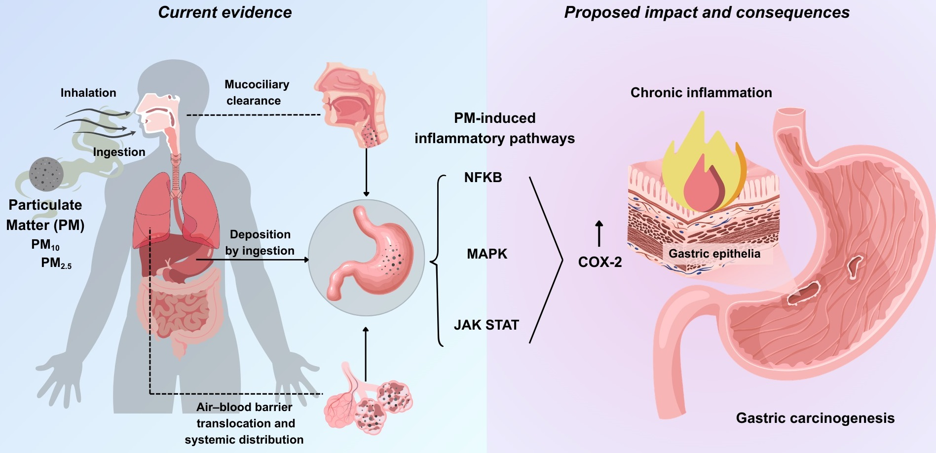
5. Mechanisms of PM Translocation to Stomach: Systemic Circulation, Ingestion, and Mucociliary Clearance
6. Overview of Inflammatory Pathways as a Central Axis in GC
6.1. NF-κB as a Master Regulator of Gastric Inflammation
6.2. MAPK Pathway: Is a Key Node in the Integration of Inflammatory Signals
6.3. JAK/STATs as the Central Axis of Inflammation and Oncogenesis
6.4. COX-2/PGE2 Pathway: A Crosstalk Between Multiple Inflammation Pathways and Tumor Promotion
7. Evidence of the Role of PM as a Modulator of Inflammatory Signaling Pathways
7.1. PM and NF-κB Activation
7.2. PM and MAPK Activation
7.3. PM and JAK/STAT Activation
7.4. PM and COX-2 Activation as a Common Target of Inflammatory Pathways
8. Inflammation as a Common Mechanism of Gastric Cancer Risk Factors: Potential Contribution of Particulate Matter
9. Discussion
10. Materials and Methods
11. Conclusions
Author Contributions
Funding
Data Availability Statement
Acknowledgments
Conflicts of Interest
Abbreviations
| PM | Particulate Matter |
| GC | Gastric Cancer |
| Hp | Helicobacter pylori |
References
- Li, D.; Morgan, D.R.; Corral, J.E.; Montgomery, E.A.; Riquelme, A.; Shah, S.C. Gastric Cancer Screening in the United States: A Review of Current Evidence, Challenges, and Future Perspectives. Am. J. Gastroenterol. 2025, 120, 765–777. [Google Scholar] [CrossRef]
- Wu, Z.; Wei, C.; Sun, J.; Qiu, B.; Kong, W.; Yang, Y.; Huang, Y.; Li, C.; Wu, L.; Liu, F.; et al. Acute air pollution exposure and gastrointestinal cancer mortality: A case-crossover study in coastal China. Front. Public Health 2025, 13, 1666928. [Google Scholar] [CrossRef]
- Bray, F.; Laversanne, M.; Sung, H.; Ferlay, J.; Siegel, R.L.; Soerjomataram, I.; Jemal, A. Global cancer statistics 2022: GLOBOCAN estimates of incidence and mortality worldwide for 36 cancers in 185 countries. CA Cancer J. Clin. 2024, 74, 229–263. [Google Scholar] [CrossRef]
- Kong, W.; Sun, Y.; Qin, X.; Zhu, G.; Zhu, X.; Kuang, Z.; Xiao, Z.; Li, J. The Burden and Risk Factors of Gastric Cancer in Eastern Asia From 1990 to 2021: Longitudinal Observational Study of the Global Burden of Disease Study 2021. JMIR Cancer 2025, 11, e75728. [Google Scholar] [CrossRef]
- Patel, A.K.; Sethi, N.S.; Park, H. Gastric Cancer: A Review. JAMA 2026, 335, 439–450. [Google Scholar] [PubMed]
- Lauren, P. The Two Histological Main Types of Gastric Carcinoma: Diffuse and So-Called Intestinal-Type Carcinoma. An Attempt at a Histo-Clinical Classification. Acta Pathol. Microbiol. Scand. 1965, 64, 31–49. [Google Scholar] [CrossRef]
- Lv, L.; Liang, X.; Wu, D.; Wang, F.; Zhang, Y.; Cang, H.; Deng, X.; Li, M. Is cardia cancer a special type of gastric cancer? A differential analysis of early cardia cancer and non-cardia cancer. J. Cancer 2021, 12, 2385–2394. [Google Scholar] [CrossRef] [PubMed]
- Yang, L.; Kartsonaki, C.; Yao, P.; de Martel, C.; Plummer, M.; Chapman, D.; Guo, Y.; Clark, S.; Walters, R.G.; Chen, Y.; et al. The relative and attributable risks of cardia and non-cardia gastric cancer associated with Helicobacter pylori infection in China: A case-cohort study. Lancet Public Health 2021, 6, e888–e896. [Google Scholar] [PubMed]
- Yao, P.; Kartsonaki, C.; Butt, J.; Jeske, R.; de Martel, C.; Plummer, M.; Guo, Y.; Clark, S.; Walters, R.G.; Chen, Y.; et al. Helicobacter pylori multiplex serology and risk of non-cardia and cardia gastric cancer: A case-cohort study and meta-analysis. Int. J. Epidemiol. 2023, 52, 1197–1208. [Google Scholar] [CrossRef]
- Arnold, M.; Park, J.Y.; Camargo, M.C.; Lunet, N.; Forman, D.; Soerjomataram, I. Is gastric cancer becoming a rare disease? A global assessment of predicted incidence trends to 2035. Gut 2020, 69, 823–829. [Google Scholar] [CrossRef]
- Jayakrishnan, T.; Ng, K. Early-Onset Gastrointestinal Cancers: A Review. JAMA 2025, 334, 1373–1385. [Google Scholar] [CrossRef]
- Boland, C.R.; Yurgelun, M.B. Historical Perspective on Familial Gastric Cancer. Cell Mol. Gastroenterol. Hepatol. 2017, 3, 192–200. [Google Scholar] [CrossRef]
- Shah, D.; Bentrem, D. Environmental and genetic risk factors for gastric cancer. J. Surg. Oncol. 2022, 125, 1096–1103. [Google Scholar] [CrossRef]
- Setia, N.; Clark, J.W.; Duda, D.G.; Hong, T.S.; Kwak, E.L.; Mullen, J.T.; Lauwers, G.Y. Familial Gastric Cancers. Oncologist 2015, 20, 1365–1377. [Google Scholar] [CrossRef] [PubMed]
- de Martel, C.; Georges, D.; Bray, F.; Ferlay, J.; Clifford, G.M. Global burden of cancer attributable to infections in 2018: A worldwide incidence analysis. Lancet Glob. Health 2020, 8, e180–e190. [Google Scholar] [CrossRef] [PubMed]
- Malfertheiner, P.; Camargo, M.C.; El-Omar, E.; Liou, J.M.; Peek, R.; Schulz, C.; Smith, S.I.; Suerbaum, S. Helicobacter pylori infection. Nat. Rev. Dis. Primers 2023, 9, 19. [Google Scholar] [CrossRef]
- Yang, J.; Liu, Z.; Zeng, B.; Hu, G.; Gan, R. Epstein-Barr virus-associated gastric cancer: A distinct subtype. Cancer Lett. 2020, 495, 191–199. [Google Scholar] [CrossRef]
- Tsugane, S.; Sasazuki, S. Diet and the risk of gastric cancer: Review of epidemiological evidence. Gastric Cancer 2007, 10, 75–83. [Google Scholar] [CrossRef]
- Morais, S.; Costa, A.; Albuquerque, G.; Araujo, N.; Pelucchi, C.; Rabkin, C.S.; Liao, L.M.; Sinha, R.; Zhang, Z.F.; Hu, J.; et al. Salt intake and gastric cancer: A pooled analysis within the Stomach cancer Pooling (StoP) Project. Cancer Causes Control 2022, 33, 779–791. [Google Scholar] [CrossRef]
- Ladeiras-Lopes, R.; Pereira, A.K.; Nogueira, A.; Pinheiro-Torres, T.; Pinto, I.; Santos-Pereira, R.; Lunet, N. Smoking and gastric cancer: Systematic review and meta-analysis of cohort studies. Cancer Causes Control 2008, 19, 689–701. [Google Scholar] [CrossRef] [PubMed]
- Lin, X.J.; Wang, C.P.; Liu, X.D.; Yan, K.K.; Li, S.; Bao, H.H.; Zhao, L.Y.; Liu, X. Body mass index and risk of gastric cancer: A meta-analysis. Jpn. J. Clin. Oncol. 2014, 44, 783–791. [Google Scholar] [CrossRef]
- Deng, W.; Jin, L.; Zhuo, H.; Vasiliou, V.; Zhang, Y. Alcohol consumption and risk of stomach cancer: A meta-analysis. Chem. Biol. Interact. 2021, 336, 109365. [Google Scholar] [CrossRef] [PubMed]
- Kreuzer, M.; Straif, K.; Marsh, J.W.; Dufey, F.; Grosche, B.; Nosske, D.; Sogl, M. Occupational dust and radiation exposure and mortality from stomach cancer among German uranium miners, 1946–2003. Occup. Environ. Med. 2012, 69, 217–223. [Google Scholar] [CrossRef] [PubMed]
- Reytor-Gonzalez, C.; Leyva Ricardo, S.E.; Sanchez Suarez, Y.; Burboa Charis, V.A.; Jimenez-Flores, E.; Cevallos-Fernandez, E.; Campuzano-Donoso, M.; Simancas-Racines, D. Heavy Metal-Contaminated Soils and Gastric Cancer Risk: Molecular Insights and the Relevance of a One Health Perspective. Int. J. Mol. Sci. 2025, 26, 11526. [Google Scholar] [CrossRef]
- Ke, M.; Jiang, Q.; Zhu, Y.; Zhang, B.; Li, D.; Ofosuhemaa, P.; Jin, G.; Zhu, Y.; Wang, W.; Zhang, M.; et al. Dietary metal(loid)s exposure and their health risks to patients with gastric precancerous lesions and gastric cancer in Anhui Province, Eastern China. J. Environ. Sci. 2026, 161, 812–821. [Google Scholar] [CrossRef] [PubMed]
- Yin, J.; Wu, X.; Li, S.; Li, C.; Guo, Z. Impact of environmental factors on gastric cancer: A review of the scientific evidence, human prevention and adaptation. J. Environ. Sci. 2020, 89, 65–79. [Google Scholar] [CrossRef]
- Kang, M.Y.; Jung, J.; Koo, J.W.; Kim, I.; Kim, H.R.; Myong, J.P. Increased risk of gastric cancer in workers with occupational dust exposure. Korean J. Intern. Med. 2021, 36, S18–S26. [Google Scholar] [CrossRef]
- Yu, P.; Xu, R.; Li, S.; Coelho, M.; Saldiva, P.H.N.; Sim, M.R.; Abramson, M.J.; Guo, Y. Associations between long-term exposure to PM2.5 and site-specific cancer mortality: A nationwide study in Brazil between 2010 and 2018. Environ. Pollut. 2022, 302, 119070. [Google Scholar] [CrossRef]
- Yang, C. Long-term fine particulate air pollution exposure and risk of gastric cancer mortality in Taiwan. J. Toxicol. Environ. Health A 2026, 89, 31–40. [Google Scholar] [CrossRef]
- Zhu, J.; Tao, H.; Kang, N.; Li, S.; Song, L.; Li, X.; Du, L. Long-term exposure to ambient air pollution and sequential carcinogenesis in the cardia gastric: A cross-sectional study. BMC Med. 2025, 23, 708. [Google Scholar] [CrossRef]
- GBD 2023 Disease and Injury and Risk Factor Collaborators. Burden of 375 diseases and injuries, risk-attributable burden of 88 risk factors, and healthy life expectancy in 204 countries and territories, including 660 subnational locations, 1990–2023: A systematic analysis for the Global Burden of Disease Study 2023. Lancet 2025, 406, 1873–1922.
- Institute for Health Metrics and Evaluation. State of Global Air 2025: A Report on Air Pollution and Its Role in the World’s Leading Causes of Death; Institute for Health Metrics and Evaluation: Boston, MA, USA, 2025. [Google Scholar]
- WHO. Ambient (Outdoor) Air Pollution; WHO: Geneva, Switzerland, 2022. [Google Scholar]
- Falcon-Rodriguez, C.I.; Osornio-Vargas, A.R.; Sada-Ovalle, I.; Segura-Medina, P. Aeroparticles, Composition, and Lung Diseases. Front. Immunol. 2016, 7, 3. [Google Scholar] [CrossRef]
- Santibanez-Andrade, M.; Quezada-Maldonado, E.M.; Quintana-Belmares, R.; Morales-Barcenas, R.; Rosas-Perez, I.; Amador-Munoz, O.; Miranda, J.; Sanchez-Perez, Y.; Garcia-Cuellar, C.M. Sampling, composition, and biological effects of Mexico City airborne particulate matter from multiple periods. Sci. Total Environ. 2024, 926, 171933. [Google Scholar] [CrossRef] [PubMed]
- Valavanidis, A.; Fiotakis, K.; Vlachogianni, T. Airborne particulate matter and human health: Toxicological assessment and importance of size and composition of particles for oxidative damage and carcinogenic mechanisms. J. Environ. Sci. Health Part C Environ. Carcinog. Ecotoxicol. Rev. 2008, 26, 339–362. [Google Scholar] [CrossRef]
- Parnell, C.B.a.B.W.S. Physical characteristics of particulate matter and health effects standards. In Proceedings of the 1999 Beltwide Cotton Conferences, Orlando, FL, USA, 3–7 January 1999; pp. 139–144. [Google Scholar]
- Loomis, D.; Huang, W.; Chen, G. The International Agency for Research on Cancer (IARC) evaluation of the carcinogenicity of outdoor air pollution: Focus on China. Chin. J. Cancer 2014, 33, 189–196. [Google Scholar] [CrossRef]
- IARC. Outdoor air pollution. In International Agency for Research on Cancer: Monographs on the Evaluation of Carcinogenic Risks to Humans; IARC: Lyon, France, 2015; Volume 109, pp. 1–448. [Google Scholar]
- Yu, P.; Guo, S.; Xu, R.; Ye, T.; Li, S.; Sim, M.R.; Abramson, M.J.; Guo, Y. Cohort studies of long-term exposure to outdoor particulate matter and risks of cancer: A systematic review and meta-analysis. Innovation 2021, 2, 100143. [Google Scholar] [CrossRef]
- Borgie, M.; Ledoux, F.; Verdin, A.; Cazier, F.; Greige, H.; Shirali, P.; Courcot, D.; Dagher, Z. Genotoxic and epigenotoxic effects of fine particulate matter from rural and urban sites in Lebanon on human bronchial epithelial cells. Environ. Res. 2015, 136, 352–362. [Google Scholar] [CrossRef]
- Li, R.; Zhao, C.; Zhang, Y.; Huang, W.; Wang, J.; Cao, G.; Cai, Z. PM2.5-induced DNA oxidative stress in A549 cells and regulating mechanisms by GST DNA methylation and Keap1/Nrf2 pathway. Toxicol. Mech. Methods 2024, 34, 517–526. [Google Scholar] [CrossRef] [PubMed]
- Meraz-Cruz, N.; Manzano-Leon, N.; Sandoval-Colin, D.E.; Garcia de Leon Mendez, M.D.C.; Quintana-Belmares, R.; Tapia, L.S.; Osornio-Vargas, A.R.; Buxton, M.A.; O’Neill, M.S.; Vadillo-Ortega, F. Effects of PM10 Airborne Particles from Different Regions of a Megacity on In Vitro Secretion of Cytokines by a Monocyte Line during Different Seasons. Toxics 2024, 12, 149. [Google Scholar] [CrossRef] [PubMed]
- Quezada-Maldonado, E.M.; Lozolla-Ortiz, J.I.; Santibanez-Andrade, M.; Morales-Barcenas, R.; Garcia-Cuellar, C.M.; Sanchez-Perez, Y. Airborne PM10 Decreases Ku80 Expression and Ku70-Ku80 Heterodimer Levels of the Non-Homologous End Joining Repair Pathway in Lung Epithelial Cells. Int. J. Mol. Sci. 2025, 26, 8936. [Google Scholar] [CrossRef]
- Shao, M.; Li, C.; Li, J.; Chen, R.; Wang, Y.; Su, J.; Tu, Y.; Zhang, F.; Zhang, X.; Ding, W. NSUN2 promotes PM2.5-induced epithelial-mesenchymal transition through methylating chitinase 3-like-1 mRNA. J. Hazard. Mater. 2025, 495, 138883. [Google Scholar] [CrossRef]
- Kim, D.H.; Lee, H.; Kim, M.Y.; Hwangbo, H.; Ji, S.Y.; Bang, E.; Hong, S.H.; Kim, G.Y.; Leem, S.H.; Ryu, D.; et al. Particulate matter 2.5 stimulates pyroptosis and necroptosis via the p38 MAPK/Akt/NF-kappaB signaling pathway in human corneal epithelial cells. Toxicology 2025, 515, 154138. [Google Scholar] [CrossRef] [PubMed]
- Winkelstein, W., Jr.; Kantor, S. Stomach cancer. Positive association with suspended particulate air pollution. Arch. Environ. Health 1969, 18, 544–547. [Google Scholar] [CrossRef] [PubMed]
- Chiu, H.F.; Tsai, S.S.; Chen, P.S.; Liao, Y.H.; Liou, S.H.; Wu, T.N.; Yang, C.Y. Traffic air pollution and risk of death from gastric cancer in Taiwan: Petrol station density as an indicator of air pollutant exposure. J. Toxicol. Environ. Health A 2011, 74, 1215–1224. [Google Scholar] [CrossRef]
- Ethan, C.J.; Mokoena, K.K.; Yu, Y.; Shale, K.; Fan, Y.; Rong, J.; Liu, F. Association between PM2.5 and mortality of stomach and colorectal cancer in Xi’an: A time-series study. Environ. Sci. Pollut. Res. Int. 2020, 27, 22353–22363. [Google Scholar] [CrossRef]
- Fan, Z.; Li, Y.; Wei, J.; Chen, G.; Wang, R.; Xu, R.; Liu, T.; Lv, Z.; Huang, S.; Sun, H.; et al. Long-term exposure to fine particulate matter and site-specific cancer mortality: A difference-in-differences analysis in Jiangsu province, China. Environ. Res. 2023, 222, 115405. [Google Scholar] [CrossRef]
- Coleman, N.C.; Burnett, R.T.; Higbee, J.D.; Lefler, J.S.; Merrill, R.M.; Ezzati, M.; Marshall, J.D.; Kim, S.Y.; Bechle, M.; Robinson, A.L.; et al. Cancer mortality risk, fine particulate air pollution, and smoking in a large, representative cohort of US adults. Cancer Causes Control 2020, 31, 767–776. [Google Scholar] [CrossRef]
- Yu, P.; Xu, R.; Wu, Y.; Huang, W.; Coelho, M.; Saldiva, P.H.N.; Ye, T.; Wen, B.; Liu, Y.; Yang, Z.; et al. Cancer mortality risk from short-term PM2.5 exposure and temporal variations in Brazil. J. Hazard. Mater. 2024, 473, 134606. [Google Scholar] [CrossRef]
- Li, Y.; He, Z.; Wei, J.; Xu, R.; Liu, T.; Zhong, Z.; Liu, L.; Liang, S.; Zheng, Y.; Chen, G.; et al. Long-term exposure to ambient fine particulate matter constituents and mortality from total and site-specific gastrointestinal cancer. Environ. Res. 2024, 244, 117927. [Google Scholar] [CrossRef] [PubMed]
- Weinmayr, G.; Pedersen, M.; Stafoggia, M.; Andersen, Z.J.; Galassi, C.; Munkenast, J.; Jaensch, A.; Oftedal, B.; Krog, N.H.; Aamodt, G.; et al. Particulate matter air pollution components and incidence of cancers of the stomach and the upper aerodigestive tract in the European Study of Cohorts of Air Pollution Effects (ESCAPE). Environ. Int. 2018, 120, 163–171. [Google Scholar] [CrossRef]
- Nagel, G.; Stafoggia, M.; Pedersen, M.; Andersen, Z.J.; Galassi, C.; Munkenast, J.; Jaensch, A.; Sommar, J.; Forsberg, B.; Olsson, D.; et al. Air pollution and incidence of cancers of the stomach and the upper aerodigestive tract in the European Study of Cohorts for Air Pollution Effects (ESCAPE). Int. J. Cancer 2018, 143, 1632–1643. [Google Scholar] [CrossRef]
- Huang, Y.J.; Lee, P.H.; Chen, L.C.; Lin, B.C.; Lin, C.; Chan, T.C. Relationships among green space, ambient fine particulate matter, and cancer incidence in Taiwan: A 16-year retrospective cohort study. Environ. Res. 2022, 212, 113416. [Google Scholar] [CrossRef]
- Liu, J.; Gan, T.; Hu, W.; Li, Y. Does ambient particulate matter 1 increase the risk of gastric cancer in the northwest of China? Int. J. Cancer 2025, 156, 104–113. [Google Scholar] [CrossRef]
- Nemmar, A.; Hoylaerts, M.F.; Hoet, P.H.; Nemery, B. Possible mechanisms of the cardiovascular effects of inhaled particles: Systemic translocation and prothrombotic effects. Toxicol. Lett. 2004, 149, 243–253. [Google Scholar] [CrossRef]
- Gupta, A.D.; Gupta, T. A review on potential approach for in silico toxicity analysis of respirable fraction of ambient particulate matter. Environ. Monit. Assess. 2023, 195, 1216. [Google Scholar] [CrossRef]
- Nakane, H. Translocation of particles deposited in the respiratory system: A systematic review and statistical analysis. Environ. Health Prev. Med. 2012, 17, 263–274. [Google Scholar] [CrossRef]
- Stur, R. Modelling the deposition of fine particulate matter (PM2.5) in the human respiratory trac. AME Med. J. 2020, 5, 14. [Google Scholar] [CrossRef]
- Nemmar, A.; Hoet, P.H.; Vanquickenborne, B.; Dinsdale, D.; Thomeer, M.; Hoylaerts, M.F.; Vanbilloen, H.; Mortelmans, L.; Nemery, B. Passage of inhaled particles into the blood circulation in humans. Circulation 2002, 105, 411–414. [Google Scholar] [CrossRef]
- Nemmar, A.; Vanbilloen, H.; Hoylaerts, M.F.; Hoet, P.H.M.; Verbruggen, A.; Nemery, B. Passage of Intratracheally Instilled Ultrafine Particles from the Lung into the Systemic Circulation in Hamster. Am. J. Respir. Crit. Care Med. 2001, 164, 1665–1668. [Google Scholar] [CrossRef]
- Liu, X.; Meng, Z. Effects of airborne fine particulate matter on antioxidant capacity and lipid peroxidation in multiple organs of rats. Inhal. Toxicol. 2005, 17, 467–473. [Google Scholar] [CrossRef]
- Wolff, R.K. Effects of airborne pollutants on mucociliary clearance. Environ. Health Perspect. 1986, 66, 223–237. [Google Scholar] [CrossRef] [PubMed]
- Wallenborn, J.G.; McGee, J.K.; Schladweiler, M.C.; Ledbetter, A.D.; Kodavanti, U.P. Systemic translocation of particulate matter-associated metals following a single intratracheal instillation in rats. Toxicol. Sci. 2007, 98, 231–239. [Google Scholar] [CrossRef]
- Sciarra, G.; Sartorelli, P.; Aprea, C.; Scancarello, G.; Strambi, F.; Palmi, S.; Scarselli, R. Solubilization of lead from crystal dust in protein solution (pseudointerstitial fluid) and gastric juice. Environ. Res. 1997, 74, 169–173. [Google Scholar] [CrossRef]
- Uzu, G.; Sauvain, J.J.; Baeza-Squiban, A.; Riediker, M.; Hohl, M.S.; Val, S.; Tack, K.; Denys, S.; Pradere, P.; Dumat, C. In vitro assessment of the pulmonary toxicity and gastric availability of lead-rich particles from a lead recycling plant. Environ. Sci. Technol. 2011, 45, 7888–7895. [Google Scholar] [CrossRef] [PubMed]
- Hettiarachchi, E.; Das, M.; Cadol, D.; Frey, B.A.; Rubasinghege, G. The fate of inhaled uranium-containing particles upon clearance to gastrointestinal tract. Environ. Sci. Process Impacts 2022, 24, 1257–1266. [Google Scholar] [CrossRef]
- Gabersek, M.; Gosar, M. Oral bioaccessibility of potentially toxic elements in various urban environmental media. Environ. Geochem. Health 2024, 46, 259. [Google Scholar] [CrossRef]
- Bourliva, A.; Papadopoulou, L.; da Silva, E.F.; Patinha, C. In vitro assessment of oral and respiratory bioaccesibility of trace elements of environmental concern in Greek fly ashes: Assessing health risk via ingestion and inhalation. Sci. Total Environ. 2020, 704, 135324. [Google Scholar] [CrossRef] [PubMed]
- Huang, M.; Wang, W.; Chan, C.Y.; Cheung, K.C.; Man, Y.B.; Wang, X.; Wong, M.H. Contamination and risk assessment (based on bioaccessibility via ingestion and inhalation) of metal(loid)s in outdoor and indoor particles from urban centers of Guangzhou, China. Sci. Total Environ. 2014, 479–480, 117–124. [Google Scholar] [CrossRef]
- Li, C.; Cui, X.Y.; Fan, Y.Y.; Teng, Y.; Nan, Z.R.; Ma, L.Q. Tenax as sorption sink for in vitro bioaccessibility measurement of polycyclic aromatic hydrocarbons in soils. Environ. Pollut. 2015, 196, 47–52. [Google Scholar] [CrossRef]
- Collins, C.D.; Mosquera-Vazquez, M.; Gomez-Eyles, J.L.; Mayer, P.; Gouliarmou, V.; Blum, F. Is there sufficient ‘sink’ in current bioaccessibility determinations of organic pollutants in soils? Environ. Pollut. 2013, 181, 128–132. [Google Scholar] [CrossRef]
- Castel, R.; Tassistro, V.; Lebarillier, S.; Dupuy, N.; Noack, Y.; Orsiere, T.; Malleret, L. Chemical and genotoxic characterization of bioaccessible fractions as a comprehensive in vitro tool in assessing the health risk due to dust-bound contaminant ingestion. Environ. Sci. Pollut. Res. Int. 2025, 32, 16680–16695. [Google Scholar] [CrossRef]
- Raffy, G.; Mercier, F.; Glorennec, P.; Mandin, C.; Le Bot, B. Oral bioaccessibility of semi-volatile organic compounds (SVOCs) in settled dust: A review of measurement methods, data and influencing factors. J. Hazard. Mater. 2018, 352, 215–227. [Google Scholar] [CrossRef] [PubMed]
- Singh, N.; Baby, D.; Rajguru, J.P.; Patil, P.B.; Thakkannavar, S.S.; Pujari, V.B. Inflammation and cancer. Ann. Afr. Med. 2019, 18, 121–126. [Google Scholar] [CrossRef] [PubMed]
- Cordon-Cardo, C.; Prives, C. At the crossroads of inflammation and tumorigenesis. J. Exp. Med. 1999, 190, 1367–1370. [Google Scholar] [CrossRef]
- Correa, P. Human gastric carcinogenesis: A multistep and multifactorial process--First American Cancer Society Award Lecture on Cancer Epidemiology and Prevention. Cancer Res. 1992, 52, 6735–6740. [Google Scholar] [PubMed]
- Correa, P.; Piazuelo, M.B.; Wilson, K.T. Pathology of gastric intestinal metaplasia: Clinical implications. Am. J. Gastroenterol. 2010, 105, 493–498. [Google Scholar] [CrossRef]
- Basseres, D.S.; Baldwin, A.S. Nuclear factor-kappaB and inhibitor of kappaB kinase pathways in oncogenic initiation and progression. Oncogene 2006, 25, 6817–6830. [Google Scholar] [CrossRef]
- Dolcet, X.; Llobet, D.; Pallares, J.; Matias-Guiu, X. NF-kB in development and progression of human cancer. Virchows Arch. 2005, 446, 475–482. [Google Scholar] [CrossRef]
- Peng, C.; Ouyang, Y.; Lu, N.; Li, N. The NF-kappaB Signaling Pathway, the Microbiota, and Gastrointestinal Tumorigenesis: Recent Advances. Front. Immunol. 2020, 11, 1387. [Google Scholar]
- Chaithongyot, S.; Jantaree, P.; Sokolova, O.; Naumann, M. NF-kappaB in Gastric Cancer Development and Therapy. Biomedicines 2021, 9, 870. [Google Scholar] [CrossRef]
- Sokolova, O.; Naumann, M. NF-kappaB Signaling in Gastric Cancer. Toxins 2017, 9, 119. [Google Scholar]
- Teng, H.; Xue, L.; Wang, Y.; Ding, X.; Li, J. Nuclear factor kappaB -inducing kinase is a diagnostic marker of gastric cancer. Medicine 2020, 99, e18864. [Google Scholar]
- Kwon, H.C.; Kim, S.H.; Oh, S.Y.; Lee, S.; Lee, J.H.; Jang, J.S.; Kim, M.C.; Kim, K.H.; Kim, S.J.; Kim, S.G.; et al. Clinicopathologic significance of expression of nuclear factor-kappaB RelA and its target gene products in gastric cancer patients. World J. Gastroenterol. 2012, 18, 4744–4750. [Google Scholar]
- Yuan, W.; Shi, Y.; Dai, S.; Deng, M.; Zhu, K.; Xu, Y.; Chen, Z.; Xu, Z.; Zhang, T.; Liang, S. The role of MAPK pathway in gastric cancer: Unveiling molecular crosstalk and therapeutic prospects. J. Transl. Med. 2024, 22, 1142. [Google Scholar] [CrossRef]
- Cargnello, M.; Roux, P.P. Activation and function of the MAPKs and their substrates, the MAPK-activated protein kinases. Microbiol. Mol. Biol. Rev. 2011, 75, 50–83. [Google Scholar] [CrossRef]
- Drosten, M.; Barbacid, M. Targeting the MAPK Pathway in KRAS-Driven Tumors. Cancer Cell 2020, 37, 543–550. [Google Scholar] [CrossRef] [PubMed]
- Yang, M.; Huang, C.Z. Mitogen-activated protein kinase signaling pathway and invasion and metastasis of gastric cancer. World J. Gastroenterol. 2015, 21, 11673–11679. [Google Scholar] [CrossRef] [PubMed]
- Morgos, D.T.; Stefani, C.; Miricescu, D.; Greabu, M.; Stanciu, S.; Nica, S.; Stanescu, S., II; Balan, D.G.; Balcangiu-Stroescu, A.E.; Coculescu, E.C.; et al. Targeting PI3K/AKT/mTOR and MAPK Signaling Pathways in Gastric Cancer. Int. J. Mol. Sci. 2024, 25, 1848. [Google Scholar] [PubMed]
- Atmaca, A.; Pauligk, C.; Steinmetz, K.; Altmannsberger, H.M.; Jager, E.; Al-Batran, S.E. Prognostic impact of phosphorylated mitogen-activated protein kinase expression in patients with metastatic gastric cancer. Oncology 2011, 80, 130–134. [Google Scholar] [CrossRef]
- Hu, X.; Li, J.; Fu, M.; Zhao, X.; Wang, W. The JAK/STAT signaling pathway: From bench to clinic. Signal Transduct. Target. Ther. 2021, 6, 402. [Google Scholar] [CrossRef]
- O’Shea, J.J.; Schwartz, D.M.; Villarino, A.V.; Gadina, M.; McInnes, I.B.; Laurence, A. The JAK-STAT pathway: Impact on human disease and therapeutic intervention. Annu. Rev. Med. 2015, 66, 311–328. [Google Scholar] [CrossRef] [PubMed]
- Chen, M.; Wang, T.; Tian, D.; Hai, C.; Qiu, Z. Induction, growth, drug resistance, and metastasis: A comprehensive summary of the relationship between STAT3 and gastric cancer. Heliyon 2024, 10, e37263. [Google Scholar] [CrossRef] [PubMed]
- Khanna, P.; Chua, P.J.; Bay, B.H.; Baeg, G.H. The JAK/STAT signaling cascade in gastric carcinoma (Review). Int. J. Oncol. 2015, 47, 1617–1626. [Google Scholar] [CrossRef]
- Judd, L.M.; Menheniott, T.R.; Ling, H.; Jackson, C.B.; Howlett, M.; Kalantzis, A.; Priebe, W.; Giraud, A.S. Inhibition of the JAK2/STAT3 pathway reduces gastric cancer growth in vitro and in vivo. PLoS ONE 2014, 9, e95993. [Google Scholar] [CrossRef] [PubMed]
- Yu, S.; Li, G.; Wang, Z.; Wang, Z.; Chen, C.; Cai, S.; He, Y. The prognostic value of pSTAT3 in gastric cancer: A meta-analysis. J. Cancer Res. Clin. Oncol. 2016, 142, 649–657. [Google Scholar] [CrossRef]
- Lim, J.W.; Kim, H.; Kim, K.H. Nuclear factor-kappaB regulates cyclooxygenase-2 expression and cell proliferation in human gastric cancer cells. Lab. Investig. 2001, 81, 349–360. [Google Scholar]
- Li, Q.; Liu, N.; Shen, B.; Zhou, L.; Wang, Y.; Wang, Y.; Sun, J.; Fan, Z.; Liu, R.H. Helicobacter pylori enhances cyclooxygenase 2 expression via p38MAPK/ATF-2 signaling pathway in MKN45 cells. Cancer Lett. 2009, 278, 97–103. [Google Scholar] [CrossRef]
- Lee, T.H.; Liu, P.S.; Tsai, M.M.; Chen, J.L.; Wang, S.J.; Hsieh, H.L. The COX-2-derived PGE(2) autocrine contributes to bradykinin-induced matrix metalloproteinase-9 expression and astrocytic migration via STAT3 signaling. Cell Commun. Signal 2020, 18, 185. [Google Scholar] [CrossRef]
- Thiel, A.; Mrena, J.; Ristimaki, A. Cyclooxygenase-2 and gastric cancer. Cancer Metastasis Rev. 2011, 30, 387–395. [Google Scholar] [CrossRef]
- Jiang, J.; Dingledine, R. Role of prostaglandin receptor EP2 in the regulations of cancer cell proliferation, invasion, and inflammation. J. Pharmacol. Exp. Ther. 2013, 344, 360–367. [Google Scholar] [CrossRef]
- Hirata, T.; Narumiya, S. Prostanoid receptors. Chem. Rev. 2011, 111, 6209–6230. [Google Scholar] [CrossRef]
- Ben Ayed-Guerfali, D.; Charfi, S.; Khabir, A.; Sellami-Boudawara, T.; Gargouri, A.; Mokdad-Gargouri, R. Clinical and prognosis relevance of COX-2 expression in Tunisian patients with primary gastric adenocarcinoma. Cancer Biomark. 2016, 17, 67–73. [Google Scholar] [CrossRef]
- Ji, X.K.; Madhurapantula, S.V.; He, G.; Wang, K.Y.; Song, C.H.; Zhang, J.Y.; Wang, K.J. Genetic variant of cyclooxygenase-2 in gastric cancer: More inflammation and susceptibility. World J. Gastroenterol. 2021, 27, 4653–4666. [Google Scholar] [CrossRef]
- Chen, X.; Deng, T.; Huo, T.; Dong, F.; Deng, J. MiR-140-5p/TLR4/NF-kappaB signaling pathway: Crucial role in inflammatory response in 16HBE cells induced by dust fall PM2.5. Ecotoxicol. Environ. Saf. 2021, 208, 111414. [Google Scholar] [CrossRef]
- Zhang, L.; Xu, F.; Yang, Y.; Yang, L.; Wu, Q.; Sun, H.; An, Z.; Li, J.; Wu, H.; Song, J.; et al. PM2.5 exposure upregulates pro-inflammatory protein expression in human microglial cells via oxidant stress and TLR4/NF-kappaB pathway. Ecotoxicol. Environ. Saf. 2024, 277, 116386. [Google Scholar] [CrossRef] [PubMed]
- Gui, J.; Liu, J.; Wang, L.; Luo, H.; Huang, D.; Yang, X.; Song, H.; Han, Z.; Ding, R.; Yang, J.; et al. TREM2 mitigates NLRP3-mediated neuroinflammation through the NF-kappaB and PI3k/Akt signaling pathways in juvenile rats exposed to ambient particulate matter. Environ. Sci. Pollut. Res. Int. 2023, 30, 119863–119878. [Google Scholar] [CrossRef] [PubMed]
- Bao, X.D.; Zu, Y.Y.; Wang, B.X.; Li, M.Y.; Jiang, F.S.; Qian, C.D.; Zhou, F.M.; Ding, Z.S. Coelonin protects against PM2.5-induced macrophage damage via suppressing TLR4/NF-kappaB/COX-2 signaling pathway and NLRP3 inflammasome activation in vitro. Environ. Toxicol. 2023, 38, 1196–1210. [Google Scholar] [CrossRef]
- Zeng, Y.; Zhu, G.; Zhu, M.; Song, J.; Cai, H.; Song, Y.; Wang, J.; Jin, M. Edaravone Attenuated Particulate Matter-Induced Lung Inflammation by Inhibiting ROS-NF-kappaB Signaling Pathway. Oxidative Med. Cell. Longev. 2022, 2022, 6908884. [Google Scholar] [CrossRef] [PubMed]
- Jiao, R.; Han, Z.; Ma, J.; Wu, S.; Wang, Z.; Zhou, G.; Liu, X.; Li, J.; Yan, X.; Meng, A. Irisin attenuates fine particulate matter induced acute lung injury by regulating Nod2/NF-kappaB signaling pathway. Immunobiology 2023, 228, 152358. [Google Scholar] [CrossRef]
- Li, X.; Geng, J.; Chen, Y.; Chen, F.; Liu, C.; Xu, Q.; Zhao, J.; Hu, J.; Xie, J.; Xu, B. Exposure to particulate matter induces cardiomyocytes apoptosis after myocardial infarction through NFkappaB activation. Biochem. Biophys. Res. Commun. 2017, 488, 224–231. [Google Scholar] [CrossRef]
- Liu, C.W.; Lee, T.L.; Chen, Y.C.; Liang, C.J.; Wang, S.H.; Lue, J.H.; Tsai, J.S.; Lee, S.W.; Chen, S.H.; Yang, Y.F.; et al. PM2.5-induced oxidative stress increases intercellular adhesion molecule-1 expression in lung epithelial cells through the IL-6/AKT/STAT3/NF-kappaB-dependent pathway. Part. Fibre Toxicol. 2018, 15, 4. [Google Scholar] [CrossRef] [PubMed]
- Ryu, Y.S.; Kang, K.A.; Piao, M.J.; Ahn, M.J.; Yi, J.M.; Hyun, Y.M.; Kim, S.H.; Ko, M.K.; Park, C.O.; Hyun, J.W. Particulate matter induces inflammatory cytokine production via activation of NFkappaB by TLR5-NOX4-ROS signaling in human skin keratinocyte and mouse skin. Redox Biol. 2019, 21, 101080. [Google Scholar] [CrossRef] [PubMed]
- Li, Q.; Li, S.; Xu, C.; Zhao, J.; Hou, L.; Jiang, F.; Zhu, Z.; Wang, Y.; Tian, L. microRNA-149-5p mediates the PM2.5-induced inflammatory response by targeting TAB2 via MAPK and NF-kappaB signaling pathways in vivo and in vitro. Cell Biol. Toxicol. 2023, 39, 703–717. [Google Scholar] [CrossRef]
- Liu, L.; Wan, C.; Zhang, W.; Guan, L.; Tian, G.; Zhang, F.; Ding, W. MiR-146a regulates PM1-induced inflammation via NF-kappaB signaling pathway in BEAS-2B cells. Environ. Toxicol. 2018, 33, 743–751. [Google Scholar]
- Wang, J.; Huang, J.; Wang, L.; Chen, C.; Yang, D.; Jin, M.; Bai, C.; Song, Y. Urban particulate matter triggers lung inflammation via the ROS-MAPK-NF-kappaB signaling pathway. J. Thorac. Dis. 2017, 9, 4398–4412. [Google Scholar] [CrossRef]
- Xu, C.; Shi, Q.; Zhang, L.; Zhao, H. High molecular weight hyaluronan attenuates fine particulate matter-induced acute lung injury through inhibition of ROS-ASK1-p38/JNK-mediated epithelial apoptosis. Environ. Toxicol. Pharmacol. 2018, 59, 190–198. [Google Scholar] [CrossRef] [PubMed]
- Chen, L.; Wu, L.; Cheng, X.; Huang, J.; Peng, J. Effects of PM2.5 on mucus hypersecretion in airway through miR-133b-5p/EGFR/Claudin1/MUC5AC axis. Aging 2024, 16, 8472–8483. [Google Scholar] [CrossRef]
- Ping, F.; Cao, Q.; Lin, H.; Han, S.Z. Antagonistic Effects of N-acetylcysteine on Mitogen-activated Protein Kinase Pathway Activation, Oxidative Stress and Inflammatory Responses in Rats with PM2.5 Induced Lung Injuries. Chin. Med. Sci. J. 2019, 34, 270–276. [Google Scholar]
- Xiao, X.; Yao, T.; Du, S.; Wang, J.; Yan, P.; Lei, Y.; Cao, L.; Shen, Z.; Cao, Y. Chronic real-time particulate matter exposure causes rat pulmonary arteriole hyperresponsiveness and remodeling: The role of ET(B)R-ERK1/2 signaling. Toxicol. Appl. Pharmacol. 2020, 403, 115154. [Google Scholar]
- Zhang, Y.; Zhang, L.; Chen, W.; Zhang, Y.; Wang, X.; Dong, Y.; Zhang, W.; Lin, X. Shp2 regulates PM2.5-induced airway epithelial barrier dysfunction by modulating ERK1/2 signaling pathway. Toxicol. Lett. 2021, 350, 62–70. [Google Scholar] [CrossRef]
- Xiao, X.; Lei, Y.; Yao, T.; Huang, T.; Yan, P.; Cao, L.; Cao, Y. PM10 exposure induces bronchial hyperresponsiveness by upreguating acetylcholine muscarinic 3 receptor. Toxicol. Appl. Pharmacol. 2024, 490, 117035. [Google Scholar] [CrossRef] [PubMed]
- Kim, D.H.; Lee, H.; Hwangbo, H.; Kim, S.Y.; Ji, S.Y.; Kim, M.Y.; Park, S.K.; Park, S.H.; Kim, M.Y.; Kim, G.Y.; et al. Particulate matter 2.5 promotes inflammation and cellular dysfunction via reactive oxygen species/p38 MAPK pathway in primary rat corneal epithelial cells. Cutan. Ocul. Toxicol. 2022, 41, 273–284. [Google Scholar] [CrossRef]
- Xiao, X.; Wang, R.; Cao, L.; Shen, Z.X.; Cao, Y.X. The Role of MAPK Pathways in Airborne Fine Particulate Matter-Induced Upregulation of Endothelin Receptors in Rat Basilar Arteries. Toxicol. Sci. 2016, 149, 213–226. [Google Scholar] [CrossRef]
- Kwon, K.; Park, S.H.; Han, B.S.; Oh, S.W.; Lee, S.E.; Yoo, J.A.; Park, S.J.; Kim, J.; Kim, J.W.; Cho, J.Y.; et al. Negative Cellular Effects of Urban Particulate Matter on Human Keratinocytes Are Mediated by P38 MAPK and NF-kappaB-dependent Expression of TRPV 1. Int. J. Mol. Sci. 2018, 19, 2660. [Google Scholar] [CrossRef]
- Yun, J.; Kim, J.E. Broccoli Sprout Extract Suppresses Particulate-Matter-Induced Matrix-Metalloproteinase (MMP)-1 and Cyclooxygenase (COX)-2 Expression in Human Keratinocytes by Direct Targeting of p38 MAP Kinase. Nutrients 2024, 16, 4156. [Google Scholar] [CrossRef] [PubMed]
- Chin, B.Y.; Choi, M.E.; Burdick, M.D.; Strieter, R.M.; Risby, T.H.; Choi, A.M. Induction of apoptosis by particulate matter: Role of TNF-alpha and MAPK. Am. J. Physiol. 1998, 275, L942–L949. [Google Scholar] [CrossRef]
- Zhang, Y.; Zheng, H.; Wang, S.; Wei, X.; Li, Q.; Wang, F. Coal-fired PM2.5 induces endothelial cell injury and the expression of atherosclerosis-related adhesion molecules: Involvement of the p38 and JNK signaling pathways. Hyg. Environ. Health Adv. 2024, 10, 100093. [Google Scholar] [CrossRef]
- Cao, J.; Qin, G.; Shi, R.; Bai, F.; Yang, G.; Zhang, M.; Lv, J. Overproduction of reactive oxygen species and activation of MAPKs are involved in apoptosis induced by PM2.5 in rat cardiac H9c2 cells. J. Appl. Toxicol. 2016, 36, 609–617. [Google Scholar] [CrossRef]
- Xie, Y.; Fan, D.; Wu, B.; Guo, J.; Wang, G.; Yu, L.; Zhang, C.; Zhao, J.; Zhang, S. PM2.5 promotes platelet activation and thrombosis via ROS/MAPKs pathway-mediated mitochondrial dysfunction. Environ. Res. 2025, 283, 122116. [Google Scholar] [CrossRef] [PubMed]
- Xu, X.; Xu, H.; Qimuge, A.; Liu, S.; Wang, H.; Hu, M.; Song, L. MAPK/AP-1 pathway activation mediates AT1R upregulation and vascular endothelial cells dysfunction under PM2.5 exposure. Ecotoxicol. Environ. Saf. 2019, 170, 188–194. [Google Scholar] [CrossRef]
- Wang, R.; Cao, L.; Shen, Z.X.; Cao, Y.X.; Yu, J. PM2.5 upregulates rat mesenteric arteries 5-HT(2A) receptor via inflammatory-mediated mitogen-activated protein kinases signaling pathway. Environ. Toxicol. 2019, 34, 1094–1104. [Google Scholar] [CrossRef]
- Rui, W.; Guan, L.; Zhang, F.; Zhang, W.; Ding, W. PM2.5-induced oxidative stress increases adhesion molecules expression in human endothelial cells through the ERK/AKT/NF-kappaB-dependent pathway. J. Appl. Toxicol. 2016, 36, 48–59. [Google Scholar] [CrossRef]
- Carmona, J.J.; Sofer, T.; Hutchinson, J.; Cantone, L.; Coull, B.; Maity, A.; Vokonas, P.; Lin, X.; Schwartz, J.; Baccarelli, A.A. Short-term airborne particulate matter exposure alters the epigenetic landscape of human genes associated with the mitogen-activated protein kinase network: A cross-sectional study. Environ. Health 2014, 13, 94. [Google Scholar] [CrossRef] [PubMed]
- Zeng, Y.; Bai, X.; Zhu, G.; Zhu, M.; Peng, W.; Song, J.; Cai, H.; Ye, L.; Chen, C.; Song, Y.; et al. m(6)A-mediated HDAC9 upregulation promotes particulate matter-induced airway inflammation via epigenetic control of DUSP9-MAPK axis and acts as an inhaled nanotherapeutic target. J. Hazard. Mater. 2024, 477, 135093. [Google Scholar]
- Wang, L.; Lin, Y.; Yang, Z.; Zhang, K.; Gong, H.; Zheng, Y.; Wang, B.; Zhang, X.; Sun, M. Microglia-derived extracellular vesicles mediate fine particulate matter-induced Alzheimer’s disease-like behaviors through the miR-34a-5p/DUSP10/p-p38 MAPK pathway. J. Hazard. Mater. 2025, 495, 138853. [Google Scholar] [CrossRef]
- Li, Y.; Tang, D.; Zhang, J.; Ou, W.; Sun, X.; Yang, Q.; Wu, J. LncRNA SPRY4-IT1 regulates 16HBE cell malignant transformation induced by particulate matter through DUSP6-ERK1/2-Chk1 signaling pathway. Chemosphere 2023, 344, 140358. [Google Scholar] [CrossRef]
- Reyes-Zarate, E.; Sanchez-Perez, Y.; Gutierrez-Ruiz, M.C.; Chirino, Y.I.; Osornio-Vargas, A.R.; Morales-Barcenas, R.; Souza-Arroyo, V.; Garcia-Cuellar, C.M. Atmospheric particulate matter (PM) exposure-induced cell cycle arrest and apoptosis evasion through STAT3 activation via PKCzeta and Src kinases in lung cells. Environ. Pollut. 2016, 214, 646–656. [Google Scholar] [CrossRef]
- Yue, W.; Chen, X.; He, S.; Li, N.; Zhang, L.; Chen, J. Exposure interval to ambient fine particulate matter (PM2.5) collected in Southwest China induced pulmonary damage through the Janus tyrosine protein kinase-2/signal transducer and activator of transcription-3 signaling pathway both in vivo and in vitro. J. Appl. Toxicol. 2021, 41, 2042–2054. [Google Scholar] [CrossRef] [PubMed]
- Yuan, Q.; Chen, Y.; Li, X.; Zhang, Z.; Chu, H. Ambient fine particulate matter (PM2.5) induces oxidative stress and pro-inflammatory response via up-regulating the expression of CYP1A1/1B1 in human bronchial epithelial cells in vitro. Mutat. Res. Genet. Toxicol. Environ. Mutagen. 2019, 839, 40–48. [Google Scholar] [CrossRef] [PubMed]
- Xu, Z.; Wu, H.; Zhang, H.; Bai, J.; Zhang, Z. Interleukins 6/8 and cyclooxygenase-2 release and expressions are regulated by oxidative stress-JAK2/STAT3 signaling pathway in human bronchial epithelial cells exposed to particulate matter ≤2.5 μm. J. Appl. Toxicol. 2020, 40, 1210–1218. [Google Scholar]
- Liu, H.; Nie, H.; Lai, W.; Shi, Y.; Liu, X.; Li, K.; Tian, L.; Xi, Z.; Lin, B. Different exposure modes of PM2.5 induces bronchial asthma and fibrosis in male rats through macrophage activation and immune imbalance induced by TIPE2 methylation. Ecotoxicol. Environ. Saf. 2022, 247, 114200. [Google Scholar] [CrossRef]
- Yang, Y.; Li, X.; An, X.; Zhang, L.; Li, X.; Wang, L.; Zhu, G. Continuous exposure of PM2.5 exacerbates ovalbumin-induced asthma in mouse lung via a JAK-STAT6 signaling pathway. Adv. Clin. Exp. Med. 2020, 29, 825–832. [Google Scholar] [CrossRef]
- Luo, C.M.; Feng, J.; Zhang, J.; Gao, C.; Cao, J.Y.; Zhou, G.L.; Jiang, Y.J.; Jin, X.Q.; Yang, M.S.; Pan, J.Y.; et al. 1,25-Vitamin D3 protects against cooking oil fumes-derived PM2.5-induced cell damage through its anti-inflammatory effects in cardiomyocytes. Ecotoxicol. Environ. Saf. 2019, 179, 249–256. [Google Scholar]
- Hu, H.; Wu, J.; Li, Q.; Asweto, C.; Feng, L.; Yang, X.; Duan, F.; Duan, J.; Sun, Z. Fine particulate matter induces vascular endothelial activation via IL-6 dependent JAK1/STAT3 signaling pathway. Toxicol. Res. 2016, 5, 946–953. [Google Scholar] [CrossRef]
- Li, T.; Zhao, J.; Ge, J.; Yang, J.; Song, X.; Wang, C.; Mao, J.; Zhang, Y.; Zou, Y.; Liu, Y.; et al. Particulate Matter Facilitates C6 Glioma Cells Activation and the Release of Inflammatory Factors Through MAPK and JAK2/STAT3 Pathways. Neurochem. Res. 2016, 41, 1969–1981. [Google Scholar] [CrossRef]
- Lu, Y.Y.; Lin, Y.; Ding, D.X.; Su, S.; Chi, Q.Q.; Zhang, Y.C.; Sun, J.; Zhang, X.; Zhu, H.M.; Huang, Q.S.; et al. MiR-26a functions as a tumor suppressor in ambient particulate matter-bound metal-triggered lung cancer cell metastasis by targeting LIN28B-IL6-STAT3 axis. Arch. Toxicol. 2018, 92, 1023–1035. [Google Scholar] [CrossRef]
- Tan, Y.; Wang, Y.; Zou, Y.; Zhou, C.; Yi, Y.; Ling, Y.; Liao, F.; Jiang, Y.; Peng, X. LncRNA LOC101927514 regulates PM2.5-driven inflammation in human bronchial epithelial cells through binding p-STAT3 protein. Toxicol. Lett. 2020, 319, 119–128. [Google Scholar] [CrossRef]
- Jia, Y.; Li, X.; Nan, A.; Zhang, N.; Chen, L.; Zhou, H.; Zhang, H.; Qiu, M.; Zhu, J.; Ling, Y.; et al. Circular RNA 406961 interacts with ILF2 to regulate PM2.5-induced inflammatory responses in human bronchial epithelial cells via activation of STAT3/JNK pathways. Environ. Int. 2020, 141, 105755. [Google Scholar] [CrossRef] [PubMed]
- Fu, H.; Liu, X.; Li, W.; Zu, Y.; Zhou, F.; Shou, Q.; Ding, Z. PM2.5 Exposure Induces Inflammatory Response in Macrophages via the TLR4/COX-2/NF-kappaB Pathway. Inflammation 2020, 43, 1948–1958. [Google Scholar] [CrossRef] [PubMed]
- Yin, J.; Xia, W.; Li, Y.; Guo, C.; Zhang, Y.; Huang, S.; Jia, Z.; Zhang, A. COX-2 mediates PM2.5-induced apoptosis and inflammation in vascular endothelial cells. Am. J. Transl. Res. 2017, 9, 3967–3976. [Google Scholar] [PubMed]
- Li, B.; Guo, L.; Ku, T.; Chen, M.; Li, G.; Sang, N. PM2.5 exposure stimulates COX-2-mediated excitatory synaptic transmission via ROS-NF-kappaB pathway. Chemosphere 2018, 190, 124–134. [Google Scholar] [CrossRef]
- Vo, T.T.T.; Wee, Y.; Chen, Y.L.; Cheng, H.C.; Tuan, V.P.; Lee, I.T. Surfactin attenuates particulate matter-induced COX-2-dependent PGE(2) production in human gingival fibroblasts by inhibiting TLR2 and TLR4/MyD88/NADPH oxidase/ROS/PI3K/Akt/NF-kappaB signaling pathway. J. Periodontal Res. 2021, 56, 1185–1199. [Google Scholar] [CrossRef] [PubMed]
- Tsai, M.H.; Hsu, L.F.; Lee, C.W.; Chiang, Y.C.; Lee, M.H.; How, J.M.; Wu, C.M.; Huang, C.L.; Lee, I.T. Resveratrol inhibits urban particulate matter-induced COX-2/PGE(2) release in human fibroblast-like synoviocytes via the inhibition of activation of NADPH oxidase/ROS/NF-kappaB. Int. J. Biochem. Cell Biol. 2017, 88, 113–123. [Google Scholar] [CrossRef]
- Song, C.; Liu, L.; Chen, J.; Hu, Y.; Li, J.; Wang, B.; Bellusci, S.; Chen, C.; Dong, N. Evidence for the critical role of the PI3K signaling pathway in particulate matter-induced dysregulation of the inflammatory mediators COX-2/PGE(2) and the associated epithelial barrier protein Filaggrin in the bronchial epithelium. Cell Biol. Toxicol. 2020, 36, 301–313. [Google Scholar] [CrossRef]
- Berkowitz, L.; Schultz, B.M.; Salazar, G.A.; Pardo-Roa, C.; Sebastian, V.P.; Alvarez-Lobos, M.M.; Bueno, S.M. Impact of Cigarette Smoking on the Gastrointestinal Tract Inflammation: Opposing Effects in Crohn’s Disease and Ulcerative Colitis. Front. Immunol. 2018, 9, 74. [Google Scholar] [CrossRef] [PubMed]
- Li, L.F.; Chan, R.L.; Lu, L.; Shen, J.; Zhang, L.; Wu, W.K.; Wang, L.; Hu, T.; Li, M.X.; Cho, C.H. Cigarette smoking and gastrointestinal diseases: The causal relationship and underlying molecular mechanisms (review). Int. J. Mol. Med. 2014, 34, 372–380. [Google Scholar] [CrossRef]
- van der Vaart, H.; Postma, D.S.; Timens, W.; ten Hacken, N.H. Acute effects of cigarette smoke on inflammation and oxidative stress: A review. Thorax 2004, 59, 713–721. [Google Scholar] [CrossRef]
- Kuo, C.H.; Wu, L.L.; Chen, H.P.; Yu, J.; Wu, C.Y. Direct effects of alcohol on gut-epithelial barrier: Unraveling the disruption of physical and chemical barrier of the gut-epithelial barrier that compromises the host-microbiota interface upon alcohol exposure. J. Gastroenterol. Hepatol. 2024, 39, 1247–1255. [Google Scholar] [CrossRef]
- Bode, C.; Bode, J.C. Alcohol’s role in gastrointestinal tract disorders. Alcohol. Health Res. World 1997, 21, 76–83. [Google Scholar] [PubMed]
- Lian, S.; Li, S.; Zhu, J.; Xia, Y.; Do Jung, Y. Nicotine stimulates IL-8 expression via ROS/NF-kappaB and ROS/MAPK/AP-1 axis in human gastric cancer cells. Toxicology 2022, 466, 153062. [Google Scholar] [CrossRef]
- Zhong, C.Y.; Zhou, Y.M.; Douglas, G.C.; Witschi, H.; Pinkerton, K.E. MAPK/AP-1 signal pathway in tobacco smoke-induced cell proliferation and squamous metaplasia in the lungs of rats. Carcinogenesis 2005, 26, 2187–2195. [Google Scholar] [PubMed]
- Nowak, A.J.; Relja, B. The Impact of Acute or Chronic Alcohol Intake on the NF-kappaB Signaling Pathway in Alcohol-Related Liver Disease. Int. J. Mol. Sci. 2020, 21, 9407. [Google Scholar] [CrossRef]
- Lee, H.T.; Kim, S.K.; Choi, M.R.; Park, J.H.; Jung, K.H.; Chai, Y.G. Effects of the activated mitogen-activated protein kinase pathway via the c-ros receptor tyrosine kinase on the T47D breast cancer cell line following alcohol exposure. Oncol. Rep. 2013, 29, 868–874. [Google Scholar] [CrossRef]
- Itskoviz, D.; Boltin, D.; Leibovitzh, H.; Tsadok Perets, T.; Comaneshter, D.; Cohen, A.; Niv, Y.; Levi, Z. Smoking increases the likelihood of Helicobacter pylori treatment failure. Dig. Liver Dis. 2017, 49, 764–768. [Google Scholar] [CrossRef]
- Hussein, R.A.; Al-Ouqaili Mushtak, T.S.; Majeed Yasin, H. Association between alcohol consumption, cigarette smoking, and Helicobacter pylori infection in Iraqi patients submitted to gastrointestinal endoscopy. J. Emerg. Med. Trauma. Acute Care 2022, 2022, 12. [Google Scholar] [CrossRef]
- Yu, J.; Yang, P.; Qin, X.; Li, C.; Lv, Y.; Wang, X. Impact of smoking on the eradication of Helicobacter pylori. Helicobacter 2022, 27, e12860. [Google Scholar] [CrossRef] [PubMed]
- Haley, K.P.; Gaddy, J.A. Nutrition and Helicobacter pylori: Host Diet and Nutritional Immunity Influence Bacterial Virulence and Disease Outcome. Gastroenterol. Res. Pr. 2016, 2016, 3019362. [Google Scholar] [CrossRef]
- Joossens, J.V.; Hill, M.J.; Elliott, P.; Stamler, R.; Lesaffre, E.; Dyer, A.; Nichols, R.; Kesteloot, H. Dietary salt, nitrate and stomach cancer mortality in 24 countries. European Cancer Prevention (ECP) and the INTERSALT Cooperative Research Group. Int. J. Epidemiol. 1996, 25, 494–504. [Google Scholar] [CrossRef] [PubMed]
- Rota, M.; Possenti, I.; Valsassina, V.; Santucci, C.; Bagnardi, V.; Corrao, G.; Bosetti, C.; Specchia, C.; Gallus, S.; Lugo, A. Dose-response association between cigarette smoking and gastric cancer risk: A systematic review and meta-analysis. Gastric Cancer 2024, 27, 197–209. [Google Scholar] [CrossRef]
- Lei, Z.N.; Teng, Q.X.; Tian, Q.; Chen, W.; Xie, Y.; Wu, K.; Zeng, Q.; Zeng, L.; Pan, Y.; Chen, Z.S.; et al. Signaling pathways and therapeutic interventions in gastric cancer. Signal Transduct. Target. Ther. 2022, 7, 358. [Google Scholar] [CrossRef]
- Jaroenlapnopparat, A.; Bhatia, K.; Coban, S. Inflammation and Gastric Cancer. Diseases 2022, 10, 35. [Google Scholar] [CrossRef]
- Echizen, K.; Hirose, O.; Maeda, Y.; Oshima, M. Inflammation in gastric cancer: Interplay of the COX-2/prostaglandin E2 and Toll-like receptor/MyD88 pathways. Cancer Sci. 2016, 107, 391–397, Correction in Cancer Sci. 2016, 107, 1059. [Google Scholar]
- Hanahan, D.; Weinberg, R.A. The hallmarks of cancer. Cell 2000, 100, 57–70. [Google Scholar] [CrossRef]
- Hanahan, D.; Weinberg, R.A. Hallmarks of cancer: The next generation. Cell 2011, 144, 646–674. [Google Scholar] [CrossRef]
- Panya, A.; Thongyim, S.; Sattayawat, P.; Inwongwan, S. Acute PM2.5 Exposure in Distinct NSCLC Cell Lines Reveals Strong Oxidative Stress and Therapy Resistance Signatures Through Transcriptomic Analysis. Toxics 2025, 13, 484. [Google Scholar] [CrossRef]
- Santibanez-Andrade, M.; Sanchez-Perez, Y.; Chirino, Y.I.; Morales-Barcenas, R.; Herrera, L.A.; Garcia-Cuellar, C.M. Airborne particulate matter induces mitotic slippage and chromosomal missegregation through disruption of the spindle assembly checkpoint (SAC). Chemosphere 2019, 235, 794–804. [Google Scholar] [CrossRef]
- Quezada-Maldonado, E.M.; Cerrato-Izaguirre, D.; Morales-Barcenas, R.; Bautista-Ocampo, Y.; Santibanez-Andrade, M.; Quintana-Belmares, R.; Chirino, Y.I.; Basurto-Lozada, P.; Robles-Espinoza, C.D.; Sanchez-Perez, Y.; et al. Mutational landscape induced by chronic exposure to environmental PM10 and PM2.5 in A549 lung epithelial cell. Chemosphere 2024, 368, 143766. [Google Scholar]
- Afthab, M.; Hambo, S.; Kim, H.; Alhamad, A.; Harb, H. Particulate matter-induced epigenetic modifications and lung complications. Eur. Respir. Rev. 2024, 33, 240129. [Google Scholar] [CrossRef]
- Zhou, W.; Tian, D.; He, J.; Wang, Y.; Zhang, L.; Cui, L.; Jia, L.; Zhang, L.; Li, L.; Shu, Y.; et al. Repeated PM2.5 exposure inhibits BEAS-2B cell P53 expression through ROS-Akt-DNMT3B pathway-mediated promoter hypermethylation. Oncotarget 2016, 7, 20691–20703. [Google Scholar]
- Ning, J.; Du, H.; Zhang, Y.; Liu, Q.; Jiang, T.; Pang, Y.; Tian, X.; Yan, L.; Niu, Y.; Zhang, R. N6-Methyladenosine Modification of CDH1 mRNA Promotes PM2.5-Induced Pulmonary Fibrosis via Mediating Epithelial Mesenchymal Transition. Toxicol. Sci. 2022, 185, 143–157. [Google Scholar] [CrossRef]
- Tian, L.; Li, Y.; Liu, H.L.; Lai, W.Q.; Shi, Y.; Liu, X.H.; Xi, Z.G.; Lin, B.C. Exposure to PM2.5 Enhances the PI3K/AKT Signaling and Malignancy of ERalpha Expression-dependent Non-small Cell Lung Carcinoma. Biomed. Environ. Sci. 2021, 34, 319–323. [Google Scholar]
- Liao, R.; Zhang, Q.; Lu, Y.; Huang, F.; Cao, W.; Li, M.; Zhou, L.; Li, Y. Fine Particulate Matter (PM2.5) Disrupts Intestinal Barrier Function by Inducing Oxidative Stress and PI3K/AKT-Mediated Inflammation in Caco-2 Cells. Int. J. Mol. Sci. 2025, 26, 8271. [Google Scholar] [CrossRef]
- Iram, S.; Fedele, R.; McKenzie, M.; Zare, A.; Jovanovic, M.; Stevanovic, S. Seasonal, compositional, and meteorological drivers of PM2.5 oxidative potential: Evidence from a year-long multi-assay study in Melbourne. Sci. Total Environ. 2026, 1011, 181047. [Google Scholar] [CrossRef] [PubMed]
- Ju, S.; Lim, L.; Ki, Y.J.; Choi, D.H.; Song, H. Oxidative stress generated by polycyclic aromatic hydrocarbons from ambient particulate matter enhance vascular smooth muscle cell migration through MMP upregulation and actin reorganization. Part. Fibre Toxicol. 2022, 19, 29. [Google Scholar] [CrossRef] [PubMed]
- Xia, T.; Kovochich, M.; Nel, A. The role of reactive oxygen species and oxidative stress in mediating particulate matter injury. Clin. Occup. Environ. Med. 2006, 5, 817–836. [Google Scholar] [CrossRef] [PubMed]

Disclaimer/Publisher’s Note: The statements, opinions and data contained in all publications are solely those of the individual author(s) and contributor(s) and not of MDPI and/or the editor(s). MDPI and/or the editor(s) disclaim responsibility for any injury to people or property resulting from any ideas, methods, instructions or products referred to in the content. |
© 2026 by the authors. Licensee MDPI, Basel, Switzerland. This article is an open access article distributed under the terms and conditions of the Creative Commons Attribution (CC BY) license.
Share and Cite
Sánchez-Pérez, Y.; Bautista-Ocampo, Y.; Moreno-Bautista, E.; Morales-Bárcenas, R.; Quintana-Belmares, R.; Herrera-Martínez, M.; Coronel-Hernández, J.; Cerrato-Izaguirre, D.; García-Cuellar, C.M.; Quezada-Maldonado, E.M. Airborne Particulate Matter as an Emerging Driver of Gastric Carcinogenesis: Molecular Pathways Linking Inflammation and Cancer. Int. J. Mol. Sci. 2026, 27, 5203. https://doi.org/10.3390/ijms27125203
Sánchez-Pérez Y, Bautista-Ocampo Y, Moreno-Bautista E, Morales-Bárcenas R, Quintana-Belmares R, Herrera-Martínez M, Coronel-Hernández J, Cerrato-Izaguirre D, García-Cuellar CM, Quezada-Maldonado EM. Airborne Particulate Matter as an Emerging Driver of Gastric Carcinogenesis: Molecular Pathways Linking Inflammation and Cancer. International Journal of Molecular Sciences. 2026; 27(12):5203. https://doi.org/10.3390/ijms27125203
Chicago/Turabian StyleSánchez-Pérez, Yesennia, Yanueh Bautista-Ocampo, Edith Moreno-Bautista, Rocío Morales-Bárcenas, Raúl Quintana-Belmares, Marytere Herrera-Martínez, Jossimar Coronel-Hernández, Dennis Cerrato-Izaguirre, Claudia M. García-Cuellar, and Ericka Marel Quezada-Maldonado. 2026. "Airborne Particulate Matter as an Emerging Driver of Gastric Carcinogenesis: Molecular Pathways Linking Inflammation and Cancer" International Journal of Molecular Sciences 27, no. 12: 5203. https://doi.org/10.3390/ijms27125203
APA StyleSánchez-Pérez, Y., Bautista-Ocampo, Y., Moreno-Bautista, E., Morales-Bárcenas, R., Quintana-Belmares, R., Herrera-Martínez, M., Coronel-Hernández, J., Cerrato-Izaguirre, D., García-Cuellar, C. M., & Quezada-Maldonado, E. M. (2026). Airborne Particulate Matter as an Emerging Driver of Gastric Carcinogenesis: Molecular Pathways Linking Inflammation and Cancer. International Journal of Molecular Sciences, 27(12), 5203. https://doi.org/10.3390/ijms27125203