Article

Exploratory FDG PET/CT Imaging in mCRPC Patients Treated with Sipuleucel-T ± IL-7: A Phase II Trial Subanalysis

1
Department of Medicine, Washington University School of Medicine, St. Louis, MO 63110, USA
2
Alvin J. Siteman Cancer Center, Washington University School of Medicine, St. Louis, MO 63110, USA
3
Department of Radiology, Washington University School of Medicine, St. Louis, MO 63110, USA
4
Department of Biomedical Engineering, Washington University, St. Louis, MO 63130-4899, USA
*
Author to whom correspondence should be addressed.
These authors contributed equally to this work.
Int. J. Mol. Sci. 2026, 27(11), 5129; https://doi.org/10.3390/ijms27115129
Submission received: 17 March 2026 / Revised: 27 May 2026 / Accepted: 28 May 2026 / Published: 5 June 2026

Abstract

Immunotherapy has shown limited efficacy in metastatic castration-resistant prostate cancer (mCRPC), highlighting the need for noninvasive tools to monitor treatment-related biological changes. This case series evaluated exploratory metabolic imaging correlates using [18F]-FDG PET/CT in mCRPC patients enrolled in a Cancer Immunotherapy Trials Network (CITN) trial of sipuleucel-T (sip-T) with or without recombinant interleukin-7 (IL-7). Among 54 enrolled patients (NCT01881867), three underwent serial [18F]-FDG PET/CT imaging at baseline and following sip-T therapy. Imaging assessed tumor metabolic activity and potential immune-related metabolic changes, based on the premise that immune processes may influence FDG uptake. Patient 1, treated with sip-T alone, demonstrated progressive sternal metastasis with stable SUVmax and marked increases in metabolic tumor volume (MTV: 10.1–34.5) and total lesion glycolysis (TLG: 43.4–145.8), consistent with poor prognosis. Patient 2 showed minimal tumor avidity but transient increases in splenic SUVmax and SUVmean following sip-T, which may reflect treatment-related or immune-associated metabolic activity. Patient 3, treated with sip-T plus IL-7, demonstrated a 33% reduction in SUVmax, stable disease on follow-up imaging, decreased PSA levels, and metabolic changes consistent with treatment response in this individual case. These findings were observed in a single patient and are considered exploratory and hypothesis-generating only. [18F]-FDG PET/CT imaging may provide exploratory insights into metabolic changes in tumors and immune-related organs in mCRPC. Metabolic changes in tumors and immune organs may provide insights into treatment-associated biological effects. Larger studies are warranted to validate these findings.

1. Introduction

Prostate cancer is the most common non-cutaneous malignancy in males globally and is the second leading cause of cancer mortality among men in the United States [1,2]. While most prostate cancers are detected at the localized stage and can be readily cured with radiation therapy or surgery, approximately 20–50% of patients will have recurrent disease within ten years [3]. When disease progression occurs with evidence of metastases, despite adequate androgen deprivation therapy (ADT), it is defined as metastatic castration-resistant prostate cancer (mCRPC) [4]. This stage represents the most advanced form of prostate cancer with the shortest overall survival, and is incurable [5]. As of October 2022, with the approval of Lutetium-177 (177Lu) vipivotide tetraxetant, there are currently nine Food and Drug Administration (FDA)-approved treatments for mCRPC that significantly improve overall survival in patients with mCRPC [6,7]. They can be broadly classified into androgen receptor antagonists, androgen biosynthesis inhibitors, taxane chemotherapeutic agents, poly (ADP-ribose) polymerase (PARP) inhibitors, radiopharmaceuticals, and a single immunotherapy (i.e., Sipuleucel-T; sip-T) [4,7,8,9,10,11].
Sip-T is an autologous cellular immunotherapeutic cancer vaccine, which is FDA approved for asymptomatic or minimally symptomatic mCRPC based in part on the 2010 phase III IMPACT trial showing a median survival benefit of 4.1 months over placebo [12]. Sip-T is produced from autologous peripheral blood mononuclear cells that are activated ex vivo with a recombinant fusion protein (PA2024), which consists of prostatic acid phosphatase (PAP) fused to a granulocyte-macrophage colony-stimulating factor (GM-CSF) to stimulate a host immune response [13]. T-cell proliferation responses to PA2024 were observed in 73% of treated patients, and a statistically significant correlation between overall survival and the humoral response was measured by antibody production against PA2024 [14]. Fong et al. performed a trial of neoadjuvant Sip-T in patients with localized prostate cancer who then underwent radical prostatectomy. Importantly, treatment with Sip-T resulted in a significant increase in T lymphocyte accumulation around sites of the tumor within the prostate when compared with a matched control cohort of patients who had not received Sip-T. This accumulation of T lymphocytes into tumor sites suggests that adoptively transferred Sip-T cells can traffic into tumor sites after circulating throughout the periphery [15]. Sip-T is unique in the prostate cancer space in that other immune therapy approaches, most notably immune checkpoint blockade antibodies, alone or in combination, have failed to significantly benefit prostate cancer patients, although there have been hints of longer term disease control [16,17,18].
In the past decade, advancements in Positron Emission Tomography (PET) radiotracers that target the prostate-specific membrane antigen (PSMA) have transformed prostate cancer imaging [19]. These novel radiotracers offer superior sensitivity and specificity for detecting prostate cancer compared to traditional imaging methods such as bone scans, CT, and MRI [5]. The latest guidelines from the National Comprehensive Cancer Network (NCCN) endorse PSMA PET as a preferred initial imaging tool for evaluation of metastatic disease in patients that are high risk and at risk of biochemical recurrence [20]. To date, no study has evaluated the tumor localization of Sip-T to metastatic tumor sites, and no clinical trial of Sip-T localization utilizing PET imaging has been performed. An ongoing clinical study at Washington University School of Medicine and University of Washington/FRCC (PIs: Yu and Pachynski) aims to address the former by analyzing pre- and post-treatment metastatic biopsies in mCRPC patients treated with Sip-T, utilizing conventional imaging. This current report begins to address systemic Sip-T localization using [18F]-FDG PET/CT imaging and represents the first study of its kind.

2. Results

2.1. Participant Characteristics

The study included a total of 54 patients with metastatic castration-resistant prostate cancer (mCRPC), divided into two cohorts: 26 in the observation group and 28 receiving rhIL-7 treatment. Demographically, the cohorts were well-matched in terms of age and ethnicity, although there was a slight variation in the number of African American/Black participants. The majority of patients in both groups were white (rhIL-7: 89.3%; observation: 80.8%) as detailed in Table 1. In terms of health status, seven patients in the observation group (26.9%) and four in the rhIL-7 group (14.3%) had an ECOG performance status of 1, while the remaining participants in both groups had a status of 0. All participants were confirmed to have mCRPC, with bone metastases present in 17 patients in the observation cohort (73.9%) and 17 patients in the rhIL-7 cohort (65.4%). Within this larger study, a focused exploratory imaging case series included three patients with mCRPC. These patients, aged 62 to 80 years, received Sip-T with or without rhIL-7 and were included for [18F]-FDG PET/CT imaging to explore treatment-associated metabolic changes.

2.2. Treatment Protocol and Imaging Follow-Up

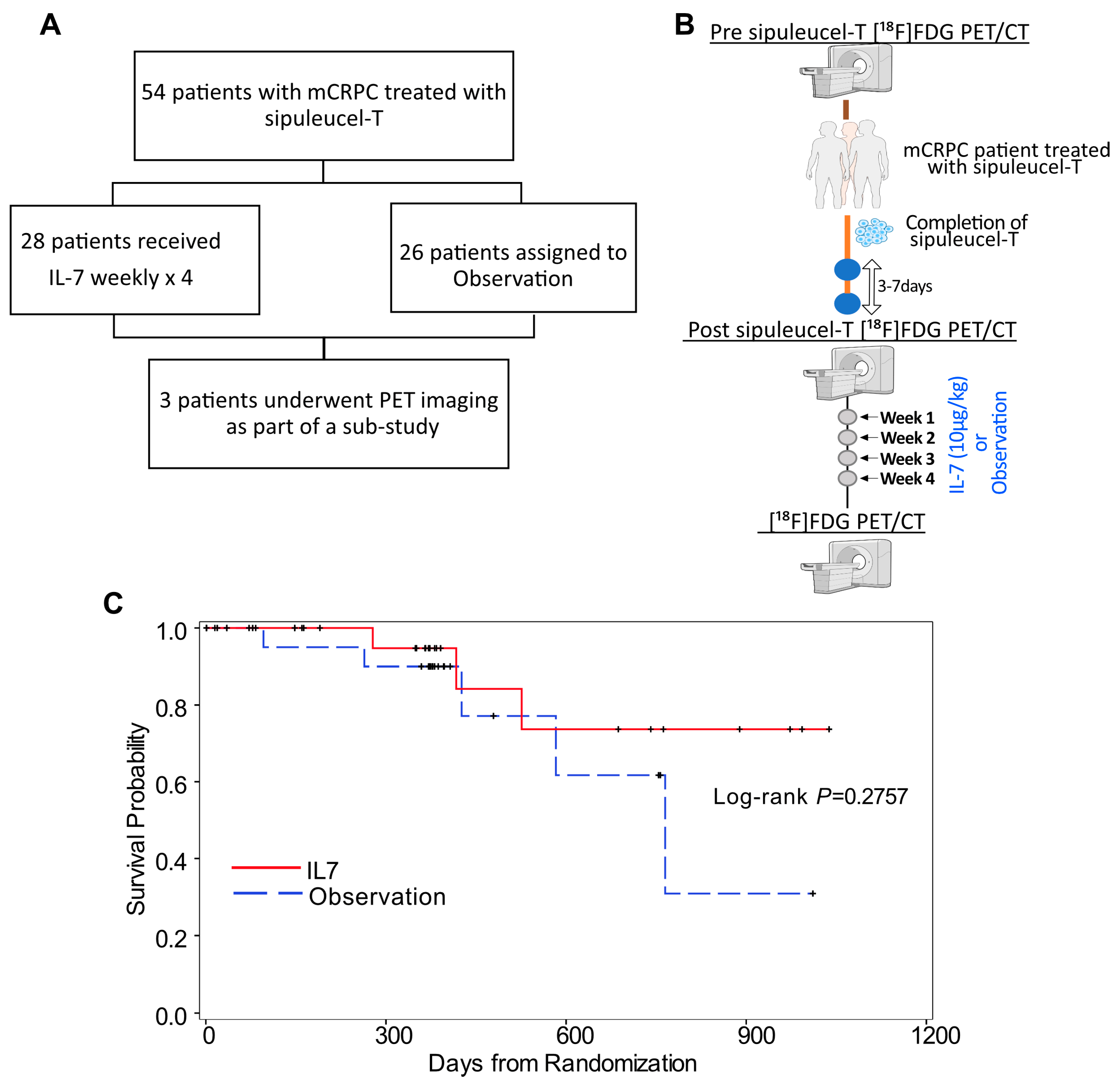
Following randomization, patients in both the observation and rhIL-7 (CYT107) cohorts underwent similar study procedures, except that week 2 and week 4 visits were optional for the observation group (Figure 1A). rhIL-7 was administered subcutaneously at 10 µg/kg beginning 3–7 days after completion of Sip-T and continued weekly for four doses (Figure 1B). The first six patients in the rhIL-7 cohort were monitored for an additional four weeks to assess safety and tolerability. No significant safety concerns were identified, allowing the study to expand to more patients. Patients in the rhIL-7 cohort completed one cycle of treatment (four doses total) unless they encountered unacceptable adverse events, illnesses preventing further administration, or disease progression.
Among patients with RECIST v1.1 measurable disease, the best overall response was determined as the highest level of response observed from the start of treatment until disease progression or recurrence, using the smallest post-treatment measurements as the reference for progressive disease.
Over a follow-up period of 5 years, data indicated variability in patient responses. While some patients showed stable disease or PSA responses, others exhibited progressive disease. The median rPFS, best overall responses, and overall survival (OS) were comparable and not significantly different between the two groups. However, the rhIL-7 group did show numerical improvement in median OS (median OS = not reached) compared to observation group (Figure 1A); however, the study was not statistically powered to detect differences in survival outcomes between the two groups. Thus, further studies are required to draw definitive conclusions.

2.3. Treatment Outcomes

The study included a secondary objective to examine clinical results. However, the study was not statistically powered to detect clinical differences between the two groups. We observed that there were no apparent differences in radiographic progression-free survival (rPFS) or overall survival (OS) between the study groups (Figure 1C). However, there were interesting trends in the rhIL-7 group, in which the survival curves extended longer and the median OS was reached. The final follow-up was completed on 31 December 2018, and these findings were previously published by our group [21]; thus, these results from our imaging substudy were largely exploratory and should be considered as generating hypotheses for further research. Sip-T treatment generally does not yield deep PSA responses, with only 2.6% of patients in the IMPACT trial achieving more than a 50% PSA reduction (PSA50) [21]. We analyzed PSA levels for potential treatment effects and found no significant differences in mean baseline PSA values between the groups. PSA values were assessed at pre-specified intervals (weeks 6, 11, 23, and 53), a decrease in PSA percentage change from baseline at week 6 (p = 0.042) was observed in the rhIL-7 group, but not at other time points. Additionally, PSA doubling times (PSADT) were then calculated and, similarly, PSADTs over 6 months was observed more frequently in the rhIL-7 group (31% vs. 14%) compared to observation. Given the limited sample size and exploratory nature of this analysis, these PSA-related findings should be interpreted descriptively rather than inferentially. Despite the study not being powered to detect PSA differences, numerically higher rates of PSA50 (4.3% vs. 0%) and any PSA decline (30.4% vs. 10.5%) were observed at week 6 in the rhIL-7 group compared to the observation group. Taken together, these exploratory findings suggest a possible signal of clinical activity with the addition of rhIL-7 to Sip-T; but require validation in larger, adequately powered studies.

2.4. Analysis of Tumor and Immune Responses in Patients Treated with Sipuleucel-T Using [18F]-FDG PET/CT Imaging

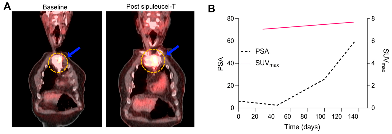
Table 2 summarizes the baseline demographic and clinical characteristics of the three patients enrolled in the clinical trial across the observation (Sip-T only) and rhIL-7 treatment arms (Sip-T + rhIL-7). Patient 1 received treatment with Sip-T only (observation arm). As shown in Figure 2B, the patient experienced progressive disease with a PSA doubling time of 1.1 months. The sternal metastasis was the primary site of [18F]-FDG avidity. While the sternal lesion’s maximum standardized uptake value (SUVmax) remained relatively stable, its metabolic tumor volume (MTV) increased from 10.1 at baseline to 34.5 after administration of Sip-T, as shown in Figure 2A. MTV measures the metabolically active tumor volume and has shown prognostic significance in various malignancies, including non-small cell lung cancer (NSCLC), peripheral T-cell lymphoma, and pancreatic cancer [22,23,24,25]. High MTV and total lesion glycolysis (TLG) obtained by multiplying the MTV by the mean standardized uptake value (SUVmean) have been associated with a high correlation with poor prognosis in a prospective study of 75 patients with NSCLC treated with immunotherapy [26]. Patient 1 had an increase in TLG from 43.4 at baseline to 145.8 on the PET scan obtained after administration of Sip-T.
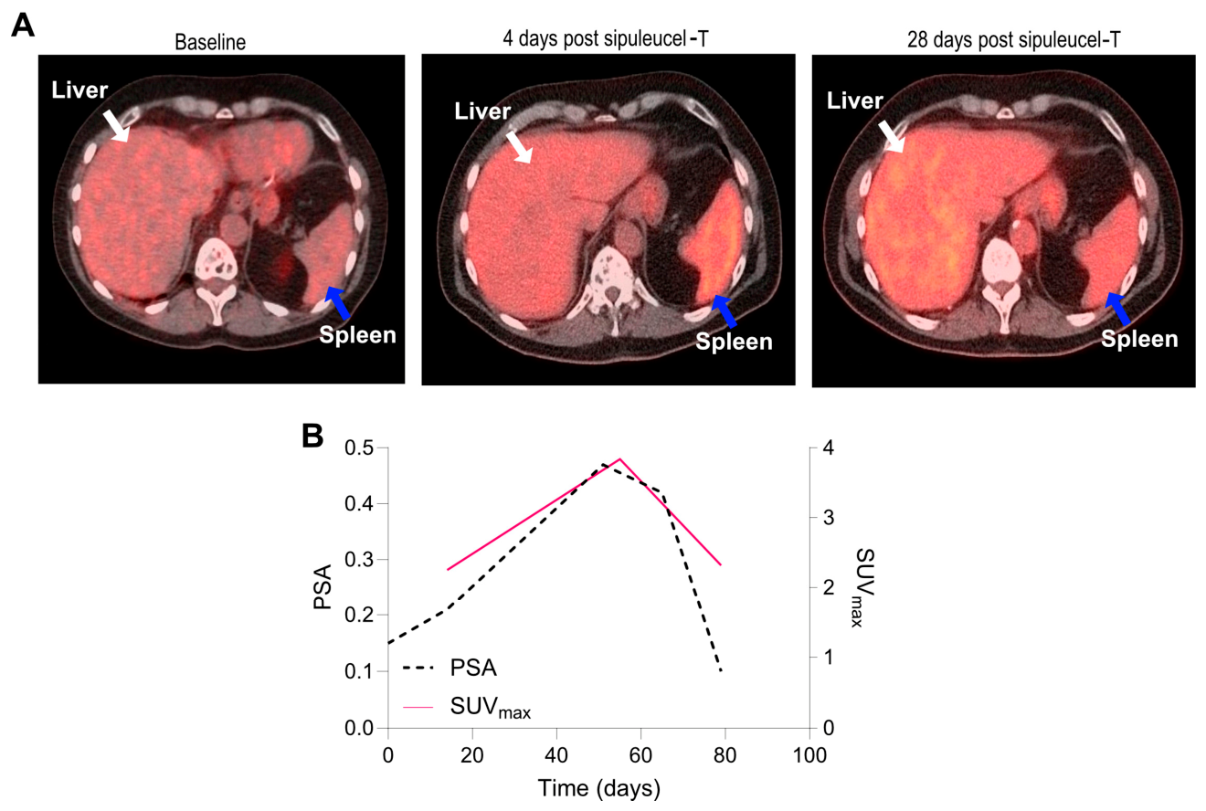
Patient 2 underwent treatment in the observation arm with Sip-T only, and although no sites of increased [18F]-FDG-avidity were observed aside from a sigmoid diverticulum, the spleen showed a notable 70% increase in SUVmax and a 96% increase in SUVmean 4 days after Sip-T infusion (Figure 3A). The SUVmax and SUVmean declined to near pretreatment levels 28 days after treatment with Sip-T, as shown in Figure 3A,B. Meanwhile, the liver’s SUVmax and SUVmean increased by only 10% and 18%, respectively. The spleen is the largest lymphoid organ and plays a critical role in mounting a complex adaptive immune response [27]. This relative increase in splenic SUV compared to the liver shortly after Sip-T administration may reflect immune cell activation related to the infusion of autologous CD54-positive dendritic cells and other cellular components of Sip-T [28]. However, [18F]-FDG uptake in lymphoid organs is nonspecific and may reflect multiple biological processes [29]; therefore, this interpretation remains exploratory and cannot be definitively attributed to immune activation in this small case series.
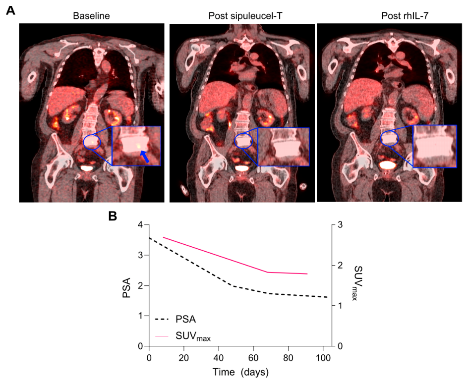
Finally, Patient 3 was a 75-year-old male enrolled in the Sip-T plus rhIL-7 arm of the trial. Serial [18F]-FDG PET/CT scans were obtained at baseline (prior to Sip-T), post-Sip-T completion, and four weeks following rhIL-7 administration (Figure 4A). At baseline, PET imaging identified metabolically active metastatic lesions at L5 (SUVmax 2.69; SUVmean 1.46), and bone marrow of the L5 vertebra (SUVmax 3.55; SUVmean 1.46). The spleen showed moderate [18F]-FDG uptake (SUVmax 2.24; SUVmean 1.45), consistent with baseline immune activity. PSA at this time was 3.57.
Post-Sip-T, metabolic activity decreased at these sites. The L5 lesion’s SUVmax reduced to 1.83 (32% reduction), with SUVmean slightly decreased to 1.43. Bone marrow SUVmax and SUVmean at L5 vertebra also decreased to 2.67 and 1.43, respectively. Spleen [18F]-FDG uptake decreased modestly (SUVmax 2.03; SUVmean 1.21), suggesting minimal splenic immune activation immediately post-Sip-T. PSA correspondingly declined to 1.99.
Following rhIL-7 therapy, additional reductions in tumor metabolic activity were observed. SUVmax at L5 lesion declined slightly to 1.79 (an additional 2% decrease), and SUVmean decreased to 1.23. The L2 lesion’s SUVmax increased modestly to 3.45, with SUVmean rising to 1.60. Bone marrow metabolic activity continued to decline (SUVmax 1.72; SUVmean 1.23). Interestingly, splenic [18F]-FDG uptake increased post-rhIL-7 (SUVmax 2.38; SUVmean 1.60), possibly reflecting rhIL-7–associated immune cell activation. PSA levels continued to decrease during this period ([1.62 post-rhIL-7 PSA]). Metabolic tumor volume (MTV) remained stable at 15.63 cm3 in the spleen throughout all time points, with no measurable changes in bone marrow or tumor sites. These serial PET measurements suggest decreasing tumor metabolic activity after Sip-T and rhIL-7 therapies, along with subtle splenic metabolic changes post-rhIL-7, potentially consistent with immune activation. The contrasting [18F]-FDG uptake patterns between tumor lesions and immune organs showed differing [18F]-FDG uptake patterns between tumor lesions and lymphoid tissues over time. Given that these observations are derived from a single patient, they should be interpreted as illustrative and hypothesis-generating rather than evidence of therapeutic efficacy.

3. Discussion

Immunotherapies using anticancer vaccines, checkpoint inhibiting antibodies, and engineered immune cells have been established in the preceding two decades as a pillar of cancer care. This exploratory study evaluated [18F]-FDG PET/CT imaging findings in patients with mCRPC treated with Sip-T with or without rhIL-7 [21].
Patient 1, who received Sip-T monotherapy, presented a clear case of progressive disease with a rapid PSA doubling time of 1.1 months, as reflected in Figure 2B. [18F]-FDG-PET imaging highlighted the sternal metastasis as the primary site of [18F]-FDG avidity. Despite a stable SUVmax for the sternal lesion, the metabolic tumor volume (MTV) significantly increased from 10.1 at baseline to 34.5 post-Sip-T administration (Figure 2A). This substantial rise in MTV, a measure of metabolically active tumor volume, has shown prognostic significance across various malignancies, including non-small cell lung cancer (NSCLC), peripheral T-cell lymphoma, and pancreatic cancer [23,24,25,30]. Furthermore, the increase in total lesion glycolysis (TLG) from 43.4 to 145.8 underscores the correlation between high MTV and poor prognosis, as noted in NSCLC patients treated with immunotherapy [26,31].
Patient 2, in contrast, did not exhibit significant [18F]-FDG avidity in metastatic sites apart from a sigmoid diverticulum. However, four days post-Sip-T infusion, [18F]-FDG-PET scans revealed notable changes in the spleen, showing a 70% increase in SUVmax and a 96% increase in SUVmean (Figure 3A,B). These values declined to near baseline levels 28 days post-treatment (Figure 3B), while the liver showed modest increases of 10% in SUVmax and 18% in SUVmean. This relative increase in splenic [18F]-FDG uptake compared to the liver following Sip-T administration suggests significant immune cell activation induced by the infusion of CD54-positive dendritic cells [27]. Several studies have shown both cellular and humoral systemic responses to Sip-T [32], which could be consistent with this finding of lymphoid organ hypermetabolism, although given this is the first study to utilize [18F]-FDG PET/CT in Sip-T patients, this remains to be proven definitively.
Patient 3, who received rhIL-7 following Sip-T, showed a reduction in SUVmax by approximately 33%, accompanied by a decrease in PSA level (Figure 4A,B). This temporal association may reflect changes in tumor metabolic activity; however, interpretation is limited by the single-patient nature of this observation. Prior studies have associated rhIL-7 with enhanced T-cell responses during immunotherapy [33,34,35]. Importantly, reductions in SUVmax during immunotherapy must be interpreted with caution, as 18F-FDG uptake may reflect both metabolism and immune-related activity [36,37,38]. While prior clinical data from the CITN12-03 trial by Pachynski et al. have demonstrated immunologic and PSA-related effects associated with rhIL-7 therapy, the current imaging substudy includes only a single rhIL-7-treated patient [21].
Currently, there is a paucity of data evaluating noninvasive methods for assessing immune responses to immune-modulating agents, emphasizing the novelty of our study. Although limited by small sample size and early study closure, this exploratory imaging substudy evaluated the potential utility of [18F]-FDG PET/CT imaging in assessing disease and/or immune responses to Sip-T +/- rhIL-7 therapy. The imaging substudy was limited by early study closure, which contributed to a small number of participants undergoing PET/CT imaging. Sip-T remains the only FDA-approved cellular immunotherapy for prostate cancer, and given the relative lack of PSA and conventional imaging responses in the setting of significantly improved overall survival additional advanced imaging modalities, such as PET/CT, could provide much needed monitoring for these patients and providers. Advanced imaging approaches may offer complementary information regarding tumor and immune-related metabolic activity in mCRPC [39]. Unlike PSMA PET, which is restricted to PSMA-expressing tumor lesions, [18F]-FDG PET/CT provides complementary functional information by capturing glycolytic tumor activity and immune-related metabolic changes in both tumor and lymphoid tissues, supporting further investigation in the context of immunotherapy monitoring [40].
The recent FDA approval of PSMA-targeted radiotracers (e.g., Ga-68 PSMA-11, F-18 DCFPyL) for patients with suspected prostate cancer metastases marks significant progress in this domain [40,41]. Looking ahead, the development of molecularly targeted tracers could further refine the evaluation of immune responses to tumors, providing improved characterization of treatment-related biological responses [42]. By leveraging advanced imaging technologies, this study provides preliminary observations regarding heterogeneous imaging findings following Sip-T and rhIL-7 therapy, supporting further evaluation in larger prospective studies.

4. Methods

4.1. Patient Population

The CITN12-03 trial (NCT01881867) was a phase II, open-label, multicenter, randomized study aimed at assessing the administration of CYT107 (recombinant human IL-7 (rhIL-7); RevImmune Inc) following the completion of Sip-T treatment. The study targeted patients with asymptomatic or minimally symptomatic mCRPC who had concluded their standard Sip-T therapy.
Between 2015 and 2017, 54 patients provided informed consent and were randomized in equal proportions to either the observation group (n = 26) or the rhIL-7 treatment group (n = 28). To maintain balance between the groups, stratification was done based on prior abiraterone treatment or neoadjuvant chemotherapy, with or without abiraterone. Patients with a history of chemotherapy for mCRPC or who had received investigational immunotherapy were excluded from participation.
Patients needed to have evaluable disease, as evidenced by a positive bone scan or measurable disease on a CT scan or MRI of the abdomen and pelvis. Key exclusion criteria for the study included ongoing systemic steroid treatment, any history of immunodeficiency disorders, clinically significant autoimmune conditions, or the use of immunosuppressive medications within 30 days prior to enrollment.

4.2. Trial Methods

After randomization, patients in the observation and IL-7 cohorts followed the same study procedures (Figure 1A and Supplemental Figure S1), except that week 2 and week 4 study visits were optional for patients assigned to observation. IL-7 (CYT107) was administered at 10 µg/ kg subcutaneously starting within 3–7 days of completion of Sip-T and continued weekly for four doses total. The first six patients randomized to receive CYT107 were followed for an additional 4 weeks prior to the study expanding past six patients, to ensure safety and tolerability of the selected CYT107 dose (Figure 1B). Patients in the IL-7 cohort were treated for one cycle (four doses total) or until unacceptable adverse event(s), intercurrent illness preventing further administration of treatment, or disease progression. Conventional (e.g., CT, MRI) imaging was performed at baseline and then every 12 weeks post therapy and at the time of removal from the study for progressive disease. Radiographic progression-free survival was based on RECIST V.1.1 and Prostate Cancer Working Group 2 criteria. For patients with RECIST V.1.1 measurable disease, the best overall response was the best response recorded from the start of the treatment until disease progression/recurrence (the smallest measurements recorded after treatment initiation were used as the reference for progressive disease). Patients were followed for 53 weeks or until death, whichever occurred first. [18F]-FDG PET/CT imaging was not mandated by the trial protocol. The parent trial was terminated earlier than planned due to drug availability and funding limitations. [18F]-FDG PET/CT imaging was not mandated by the trial protocol. Participation in PET/CT imaging was optional and offered to all eligible participants at the study site as part of an exploratory imaging substudy. The three patients included in this analysis represent all participants who elected to undergo PET/CT imaging during the study period. No formal selection criteria were applied for inclusion in the imaging substudy.

4.3. Imaging with [18F]-FDG

Patients enrolled in the CITN 12-03 clinical trial were potentially eligible to undergo [18F]-FDG PET/CT imaging on an IRB-approved imaging substudy protocol (PI: Wahl). After screening, interested patients signed consent and were enrolled. Three participants were selected and consented to undergo additional [18F]-FDG PET scans for exploratory purposes before their responses to therapy were known. Patients were administered a single intravenous dose of 155 MBq of [18F]-FDG (± 20% of dose). The PET/CT scans were then performed from the mid-thighs to the vertex approximately 60 ± 10 min (50–70 min) post-injection. Imaging was performed on a PET/CT scanner following standardized acquisition and reconstruction protocols consistent with the RSNA Quantitative Imaging Biomarkers Alliance (QIBA) [18F]-FDG PET/CT profile to ensure harmonization and quantitative reliability across scans [43]. Imaging was performed at baseline, early post-treatment, and post-IL-7 (where applicable), based on prior immunotherapy PET imaging protocols and our previous experience in immune response imaging studies [44]. Radiographic progression-free survival (rPFS) was evaluated based on RECIST v1.1 and Prostate Cancer Working Group 2 criteria. Imaging, performed at baseline and then every 12 weeks, continued until disease progression or removal from the study.
The [18F]-FDG PET/CT images were evaluated by three independent central readers, who were blinded to any clinical details. Discrepancies between readers were resolved by consensus review rather than formal statistical adjudication due to the limited sample size. Locations of [18F]-FDG-positive tumors and lymph nodes were identified by visualizing areas of focal uptake that exceeded the physiological background activity of the specific anatomical site or blood pool. For quantitative analysis, metabolic tumor volume (MTV) was obtained using semi-automated volume-of-interest (VOI) delineation with a standardized SUV-based threshold above local background. Total lesion glycolysis (TLG) was calculated as MTV × SUVmean. When multiple lesions were present, whole-body MTV and TLG were derived by summing individual lesion values. All segmentations and quantitative measurements were performed in consensus by three blinded readers. No partial volume correction was applied, consistent with the exploratory nature of the study.

5. Conclusions

This randomized trial evaluated Sip-T with or without rhIL-7 in patients with metastatic castration-resistant prostate cancer (mCRPC), with an exploratory imaging substudy using [18F]-FDG PET/CT. This exploratory study demonstrates the feasibility of serial [18F]-FDG PET/CT acquisition and suggests biological plausibility for detecting treatment-associated metabolic changes. In this small imaging cohort, we observed heterogeneous metabolic responses across patients, including changes in tumor burden, splenic uptake, and PSA kinetics. These findings are exploratory and hypothesis-generating and do not establish therapeutic efficacy. Further prospective studies with larger cohorts are needed to validate the role of Sip-T plus rhIL-7 and to determine the utility of [18F]-FDG PET/CT as a noninvasive biomarker of immune response.

6. Key Points

Question: Can serial [18F]-FDG PET/CT detect metabolic and immune responses in mCRPC patients treated with sipuleucel-T with or without recombinant IL-7?
Pertinent Findings: In this case series, serial [18F]-FDG PET/CT showed progressive metabolic tumor activity in patients receiving sipuleucel-T alone, while the patient receiving sipuleucel-T plus recombinant IL-7 demonstrated a 33% decline in tumor SUVmax and disease stability over follow-up in a single patient. Transient increases in splenic metabolic activity after sipuleucel-T were also observed, which may be consistent with systemic immune activation.
Implications for Patient Care: [18F]-FDG PET/CT may help identify metabolic and immune responses during immunotherapy in mCRPC, supporting more personalized treatment decisions.
Clinical Trail Registration: NCT01881867 https://clinicaltrials.gov/study/NCT01881867 (accessed on 2 May 2026).

Supplementary Materials

The following supporting information can be downloaded at: https://www.mdpi.com/article/10.3390/ijms27115129/s1.

Author Contributions

Conceptualization, S.S.P. and D.B.; methodology, R.W., R.K.P.; software, J.C. and R.W.; validation, J.P., J.E.I., H.R., D.L.J.T. and R.K.P.; formal analysis, S.S.P. and J.C.; investigation, R.K.P. and R.W.; resources, R.K.P., R.W. and D.L.J.T.; data curation, D.L.J.T. and J.C.; writing—original draft preparation, S.S.P. and R.K.P.; writing—review and editing, S.S.P. and R.K.P.; visualization, R.W., R.K.P. and D.L.J.T.; supervision, R.K.P. and R.W.; project administration, R.W. and R.K.P.; funding acquisition R.W. and R.K.P. All authors have read and agreed to the published version of the manuscript.

Funding

This work was supported by grants from the National Cancer Institute and additional funding from Dendreon (Seattle, Washington).

Institutional Review Board Statement

The study was conducted in accordance with the Declaration of Helsinki, and approved by the Institutional Review Board of Washington University School of Medicine (protocol code 201503108; date of approval 5 September 2017).

Informed Consent Statement

Informed consent was obtained from all individual participants included in the study.

Data Availability Statement

Data are contained within the article. Further inquiries can be directed to the corresponding author.

Acknowledgments

This work was supported by RevImmune (Paris, France).

Conflicts of Interest

RKP served in advisory/consulting roles for Dendreon.

References

  1. National Cancer Institute (Ed.) Cancer Stat Facts: Prostate Cancer; National Cancer Institute: Bethesda, MD, USA, 2024.
  2. American Cancer Society. Key Statistics for Prostate Cancer; American Cancer Society: Hagerstown, MD, USA, 2024. [Google Scholar]
  3. Kupelian, P.A.; Mahadevan, A.; Reddy, C.A.; Reuther, A.M.; Klein, E.A. Use of different definitions of biochemical failure after external beam radiotherapy changes conclusions about relative treatment efficacy for localized prostate cancer. Urology 2006, 68, 593–598. [Google Scholar] [CrossRef] [PubMed]
  4. James, L.M.; Emmanuel, S.A.; Andrew, J.A.; Anthony, V.D.A.; Brian, J.D.; Tanya, D.; James, A.E.; Charles, A.E.; Thomas, A.F.; Celestia, S.H.; et al. Prostate Cancer, Version 2.2019, NCCN Clinical Practice Guidelines in Oncology. J. Natl. Compr. Cancer Netw. 2019, 17, 479–505. [Google Scholar] [CrossRef] [PubMed]
  5. Cornford, P.; Bellmunt, J.; Bolla, M.; Briers, E.; De Santis, M.; Gross, T.; Henry, A.M.; Joniau, S.; Lam, T.B.; Mason, M.D.; et al. EAU-ESTRO-SIOG Guidelines on Prostate Cancer. Part II: Treatment of Relapsing, Metastatic, and Castration-Resistant Prostate Cancer. Eur. Urol. 2017, 71, 630–642. [Google Scholar] [CrossRef]
  6. Lee, J.; Sato, M.M.; Coel, M.N.; Lee, K.-H.; Kwee, S.A. Prediction of PSA progression in castrate-resistant prostate cancer based on treatment-associated change in tumor burden quantified by (18)F-fluorocholine PET/CT. J. Nucl. Med. Off. Publ. Soc. Nucl. Med. 2016, 57, 1058–1064. [Google Scholar]
  7. Sartor, O.; de Bono, J.; Chi, K.N.; Fizazi, K.; Herrmann, K.; Rahbar, K.; Tagawa, S.T.; Nordquist, L.T.; Vaishampayan, N.; El-Haddad, G.; et al. Lutetium-177–PSMA-617 for Metastatic Castration-Resistant Prostate Cancer. N. Engl. J. Med. 2021, 385, 1091–1103. [Google Scholar] [CrossRef]
  8. Sartor, O.; Coleman, R.; Nilsson, S.; Heinrich, D.; Helle, S.I.; O’Sullivan, J.M.; Fosså, S.D.; Chodacki, A.; Wiechno, P.; Logue, J.; et al. Effect of radium-223 dichloride on symptomatic skeletal events in patients with castration-resistant prostate cancer and bone metastases: Results from a phase 3, double-blind, randomised trial. Lancet Oncol. 2014, 15, 738–746. [Google Scholar] [CrossRef] [PubMed]
  9. Hussain, M.; Mateo, J.; Fizazi, K.; Saad, F.; Shore, N.; Sandhu, S.; Chi, K.N.; Sartor, O.; Agarwal, N.; Olmos, D.; et al. Survival with Olaparib in Metastatic Castration-Resistant Prostate Cancer. N. Engl. J. Med. 2020, 383, 2345–2357. [Google Scholar] [CrossRef]
  10. Higano, C.S.; Schellhammer, P.F.; Small, E.J.; Burch, P.A.; Nemunaitis, J.; Yuh, L.; Provost, N.; Frohlich, M.W. Integrated data from 2 randomized, double-blind, placebo-controlled, phase 3 trials of active cellular immunotherapy with sipuleucel-T in advanced prostate cancer. Cancer 2009, 115, 3670–3679. [Google Scholar] [CrossRef]
  11. Small, E.J.; Schellhammer, P.F.; Higano, C.S.; Redfern, C.H.; Nemunaitis, J.J.; Valone, F.H.; Verjee, S.S.; Jones, L.A.; Hershberg, R.M. Placebo-controlled phase III trial of immunologic therapy with sipuleucel-T (APC8015) in patients with metastatic, asymptomatic hormone refractory prostate cancer. J. Clin. Oncol. 2006, 24, 3089–3094. [Google Scholar] [CrossRef]
  12. Kantoff, P.W.; Higano, C.S.; Shore, N.D.; Berger, E.R.; Small, E.J.; Penson, D.F.; Redfern, C.H.; Ferrari, A.C.; Dreicer, R.; Sims, R.B.; et al. Sipuleucel-T Immunotherapy for Castration-Resistant Prostate Cancer. N. Engl. J. Med. 2010, 363, 411–422. [Google Scholar] [CrossRef]
  13. Madan, R.A.; Antonarakis, E.S.; Drake, C.G.; Fong, L.; Yu, E.Y.; McNeel, D.G.; Lin, D.W.; Chang, N.N.; Sheikh, N.A.; Gulley, J.L. Putting the Pieces Together: Completing the Mechanism of Action Jigsaw for Sipuleucel-T. JNCI J. Natl. Cancer Inst. 2020, 112, 562–573. [Google Scholar] [CrossRef]
  14. Sheikh, N.A.; Petrylak, D.; Kantoff, P.W.; dela Rosa, C.; Stewart, F.P.; Kuan, L.-Y.; Whitmore, J.B.; Trager, J.B.; Poehlein, C.H.; Frohlich, M.W.; et al. Immunotherapy, Sipuleucel-T immune parameters correlate with survival: An analysis of the randomized phase 3 clinical trials in men with castration-resistant prostate cancer. Cancer Immunol. Immunother. 2013, 62, 137–147. [Google Scholar] [CrossRef] [PubMed]
  15. Fong, L.; Carroll, P.; Weinberg, V.; Chan, S.; Lewis, J.; Corman, J.; Amling, C.L.; Stephenson, R.A.; Simko, J.; Sheikh, N.A.; et al. Activated lymphocyte recruitment into the tumor microenvironment following preoperative sipuleucel-T for localized prostate cancer. J. Natl. Cancer Inst. 2014, 106, dju268. [Google Scholar] [CrossRef] [PubMed]
  16. Petrylak, D.P.; Ratta, R.; Matsubara, N.; Korbenfeld, E.; Gafanov, R.; Mourey, L.; Todenhöfer, T.; Gurney, H.; Kramer, G.; Bergman, A.M.; et al. Pembrolizumab Plus Docetaxel Versus Docetaxel for Previously Treated Metastatic Castration-Resistant Prostate Cancer: The Randomized, Double-Blind, Phase III KEYNOTE-921 Trial. J. Clin. Oncol. 2025, 43, 1638–1649. [Google Scholar] [CrossRef]
  17. Fizazi, K.; Drake, C.G.; Beer, T.M.; Kwon, E.D.; Scher, H.I.; Gerritsen, W.R.; Bossi, A.; den Eertwegh, A.; Krainer, M.; Houede, N.; et al. Final Analysis of the Ipilimumab Versus Placebo Following Radiotherapy Phase III Trial in Postdocetaxel Metastatic Castration-resistant Prostate Cancer Identifies an Excess of Long-term Survivors. Eur. Urol. 2020, 78, 822–830. [Google Scholar] [CrossRef]
  18. Pachynski, R.K. A “Tail” of Immunotherapy in Metastatic Prostate Cancer. Eur. Urol. 2020, 78, 831–833. [Google Scholar] [CrossRef]
  19. Houshmand, S.; Lawhn-Heath, C.; Behr, S. PSMA PET imaging in the diagnosis and management of prostate cancer. Abdom. Radiol. 2023, 48, 3610–3623. [Google Scholar] [CrossRef] [PubMed]
  20. National Comprehensive Cancer Network. NCCN Clinical Practice Guidelines in Oncology. 2008. Available online: http://www.nccn.org/professionals/physician_gls/PDF/occult.pdf (accessed on 22 February 2025).
  21. Russell, K.P.; Chihiro, M.; Russell, S.; Lauren, H.; Leonard, A.; Paul, M.; Rhonda, L.B.; Omer, K.; Frederick, M.; John, D.S.; et al. IL-7 expands lymphocyte populations and enhances immune responses to sipuleucel-T in patients with metastatic castration-resistant prostate cancer (mCRPC). J. Immunother. Cancer 2021, 9, e002903. [Google Scholar]
  22. Satoh, Y.; Onishi, H.; Nambu, A.; Araki, T. Volume-based Parameters Measured by Using FDG PET/CT in Patients with Stage I NSCLC Treated with Stereotactic Body Radiation Therapy: Prognostic Value. Radiology 2014, 270, 275–281. [Google Scholar] [CrossRef]
  23. Kim, K.; Kim, S.J.; Kim, I.J.; Kim, Y.S.; Pak, K.; Kim, H. Prognostic value of volumetric parameters measured by F-18 FDG PET/CT in surgically resected non-small-cell lung cancer. Nucl. Med. Commun. 2012, 33, 613–620. [Google Scholar] [CrossRef]
  24. Jung, S.-H.; Ahn, J.-S.; Kim, Y.-K.; Kweon, S.-S.; Min, J.-J.; Bom, H.-S.; Kim, H.-J.; Chae, Y.S.; Moon, J.H.; Sohn, S.K.; et al. Prognostic significance of interim PET/CT based on visual, SUV-based, and MTV-based assessment in the treatment of peripheral T-cell lymphoma. BMC Cancer 2015, 15, 198. [Google Scholar] [CrossRef]
  25. Lee, J.W.; Kang, C.M.; Choi, H.J.; Lee, W.J.; Song, S.Y.; Lee, J.-H.; Lee, J.D. Prognostic Value of Metabolic Tumor Volume and Total Lesion Glycolysis on Preoperative 18F-FDG PET/CT in Patients with Pancreatic Cancer. J. Nucl. Med. 2014, 55, 898–904. [Google Scholar] [CrossRef]
  26. Chardin, D.; Paquet, M.; Schiappa, R.; Darcourt, J.; Bailleux, C.; Poudenx, M.; Sciazza, A.; Ilie, M.; Benzaquen, J.; Martin, N.; et al. Baseline metabolic tumor volume as a strong predictive and prognostic biomarker in patients with non-small cell lung cancer treated with PD1 inhibitors: A prospective study. J. Immunother. Cancer 2020, 8, e000645. [Google Scholar] [CrossRef]
  27. Mebius, R.E.; Kraal, G. Structure and function of the spleen. Nat. Rev. Immunol. 2005, 5, 606–616. [Google Scholar] [CrossRef] [PubMed]
  28. Saeed, M.A.; Peng, B.; Kim, K.; Rawat, K.; Kuehm, L.M.; Siegel, Z.R.; Borkowski, A.; Habib, N.; Van Tine, B.; Sheikh, N.; et al. High-Dimensional Analyses Reveal IL15 Enhances Activation of Sipuleucel-T Lymphocyte Subsets and Reverses Immunoresistance. Cancer Immunol. Res. 2024, 12, 559–574. [Google Scholar] [CrossRef] [PubMed]
  29. Athiraman, H.; Miller, K.; Guido, J.; Borghero, Y. Unusual Prostate-Specific Membrane Antigen (PSMA) Splenic Uptake in a Patient with Prostate Cancer. Cureus 2024, 16, e70751. [Google Scholar] [CrossRef]
  30. Takahashi, N.; Yamamoto, T.; Matsushita, H.; Sugawara, T.; Kubozono, M.; Umezawa, R.; Ishikawa, Y.; Kozumi, M.; Katagiri, Y.; Tasaka, S.; et al. Metabolic tumor volume on FDG-PET/CT is a possible prognostic factor for Stage I lung cancer patients treated with stereotactic body radiation therapy: A retrospective clinical study. J. Radiat. Res. 2016, 57, 655–661. [Google Scholar] [CrossRef] [PubMed]
  31. Im, H.-J.; Bradshaw, T.; Solaiyappan, M.; Cho, S.Y. Current Methods to Define Metabolic Tumor Volume in Positron Emission Tomography: Which One is Better? Nucl. Med. Mol. Imaging 2018, 52, 5–15. [Google Scholar] [CrossRef]
  32. Rawat, K.; Punia, V.; Mathews, P.; McCoy, S.; Song, W.; Saeed, M.A.; Pachynski, R.K. Synergistic potential of sipuleucel-T in enhancing immunotherapy for metastatic castration-resistant prostate cancer. J. Immunother. Cancer 2025, 13, e012690. [Google Scholar] [CrossRef]
  33. Tan, J.T.; Dudl, E.; LeRoy, E.; Murray, R.; Sprent, J.; Weinberg, K.I.; Surh, C.D. IL-7 is critical for homeostatic proliferation and survival of naïve T cells. Proc. Natl. Acad. Sci. USA 2001, 98, 8732–8737. [Google Scholar] [CrossRef]
  34. Rathmell, J.C.; Farkash, E.A.; Gao, W.; Thompson, C.B. IL-7 enhances the survival and maintains the size of naive T cells. J. Immunol. 2001, 167, 6869–6876. [Google Scholar] [CrossRef]
  35. Ding, Z.-C.; Habtetsion, T.; Cao, Y.; Li, T.; Liu, C.; Kuczma, M.; Chen, T.; Hao, Z.; Bryan, L.; Munn, D.H.; et al. Adjuvant IL-7 potentiates adoptive T cell therapy by amplifying and sustaining polyfunctional antitumor CD4+ T cells. Sci. Rep. 2017, 7, 12168. [Google Scholar] [CrossRef]
  36. Aide, N.; De Pontdeville, M.; Lopci, E. Evaluating response to immunotherapy with 18F-FDG PET/CT: Where do we stand? Eur. J. Nucl. Med. Mol. Imaging 2020, 47, 1019–1021. [Google Scholar] [CrossRef] [PubMed]
  37. Larimer, B.M. PET Imaging for Monitoring Cellular and Immunotherapy of Cancer. Cancer J. 2024, 30, 153–158. [Google Scholar] [CrossRef]
  38. Anderson, T.M.; Chang, B.H.; Huang, A.C.; Xu, X.; Yoon, D.; Shang, C.G.; Mick, R.; Schubert, E.; McGettigan, S.; Kreider, K.; et al. FDG PET/CT Imaging 1 Week after a Single Dose of Pembrolizumab Predicts Treatment Response in Patients with Advanced Melanoma. Clin. Cancer Res. 2024, 30, 1758–1767. [Google Scholar] [CrossRef]
  39. Wilky, B.A. Immune checkpoint inhibitors: The linchpins of modern immunotherapy. Immunol. Rev. 2019, 290, 6–23. [Google Scholar] [CrossRef]
  40. Hofman, M.S.; Lawrentschuk, N.; Francis, R.J.; Tang, C.; Vela, I.; Thomas, P.; Rutherford, N.; Martin, J.M.; Frydenberg, M.; Shakher, R.; et al. Prostate-specific membrane antigen PET-CT in patients with high-risk prostate cancer before curative-intent surgery or radiotherapy (proPSMA): A prospective, randomised, multicentre study. Lancet 2020, 395, 1208–1216. [Google Scholar] [CrossRef] [PubMed]
  41. Pienta, K.J.; Gorin, M.A.; Rowe, S.P.; Carroll, P.R.; Pouliot, F.; Probst, S.; Saperstein, L.; Preston, M.A.; Alva, A.S.; Patnaik, A.; et al. A Phase 2/3 Prospective Multicenter Study of the Diagnostic Accuracy of Prostate Specific Membrane Antigen PET/CT with (18)F-DCFPyL in Prostate Cancer Patients (OSPREY). J. Urol. 2021, 206, 52–61. [Google Scholar] [CrossRef]
  42. van de Donk, P.P.; Kist de Ruijter, L.; Lub-de Hooge, M.N.; Brouwers, A.H.; van der Wekken, A.J.; Oosting, S.F.; Fehrmann, R.S.; de Groot, D.J.A.; de Vries, E.G. Molecular imaging biomarkers for immune checkpoint inhibitor therapy. Theranostics 2020, 10, 1708–1718. [Google Scholar] [CrossRef] [PubMed]
  43. Kinahan, P.E.; Perlman, E.S.; Sunderland, J.J.; Subramaniam, R.; Wollenweber, S.D.; Turkington, T.G.; Lodge, M.A.; Boellaard, R.; Obuchowski, N.A.; Wahl, R.L. The QIBA Profile for FDG PET/CT as an Imaging Biomarker Measuring Response to Cancer Therapy. Radiology 2020, 294, 647–657. [Google Scholar] [CrossRef]
  44. Cho, S.Y.; Lipson, E.J.; Im, H.J.; Rowe, S.P.; Gonzalez, E.M.; Blackford, A.; Chirindel, A.; Pardoll, D.M.; Topalian, S.L.; Wahl, R.L. Prediction of Response to Immune Checkpoint Inhibitor Therapy Using Early-Time-Point (18)F-FDG PET/CT Imaging in Patients with Advanced Melanoma. J. Nucl. Med. 2017, 58, 1421–1428. [Google Scholar] [CrossRef] [PubMed]
Figure 1. (A) Treatment schedule showing Sip-T administration followed by CYT107 (IL-7), initiated 3–7 days after the third Sip-T dose. (B) Study schema of patients undergoing serial [18F]-FDG PET/CT imaging at baseline, post–Sip-T, and post–IL-7 (treatment arm). (C) Kaplan–Meier overall survival analysis comparing study groups (log-rank test) [21].
Ijms 27 05129 g001
Figure 2. Serial [18F]-FDG PET/CT imaging of Patient 1 at baseline and post–Sip-T (39 days). (A) Increased metabolic tumor volume (MTV) in a sternal metastasis after treatment. (B) Clinical progression with rising PSA despite minimal change in SUVmax.
Ijms 27 05129 g002
Figure 3. Serial [18F]-FDG PET/CT imaging of Patient 2 following Sip-T. (A) Increased splenic [18F]-FDG uptake after treatment with subsequent return toward baseline. (B) Corresponding temporal changes in splenic uptake across follow-up imaging.
Ijms 27 05129 g003
Figure 4. Serial [18F]-FDG PET/CT imaging of Patient 3 at baseline, post–Sip-T, and post–rhIL-7. (A) Baseline scan showing [18F]-FDG-avid disease at the L5 vertebral lesion. (B) Post– Sip-T imaging demonstrating reduced [18F]-FDG uptake at the L5 lesion with concurrent change in PSA levels.
Ijms 27 05129 g004
Table 1. Baseline characteristics of patients enrolled in clinical trial.
CharacteristicIL-7 (n = 28)Observation (n = 26)Total (n = 54)
Age, years68 (51–81)66.5 (56–81)67 (46–81)
Race
White25 (89.3%)21 (80.8%)46 (85.2%)
Black or African American2 (7.1%)4 (15.4%)6 (11.1%)
Not reported1 (3.6%)1 (3.8%)2 (3.7%)
Hemoglobin (g/dL)12.9 (10.9–14.4)12.2 (10.3–14.7)12.7 (10.3–14.7)
Alkaline phosphatase (U/L)70.5 (39–151)70 (29–1113)70 (29–1113)
PSA (ng/mL)5.8 (0.11–156.4)15.4 (0.47–202.7)10.6 (0.11–202.7)
ECOG 024 (85.7%)19 (73.1%)43 (79.6%)
ECOG 14 (14.3%)7 (26.9%)11 (20.4%)
Disease distribution
Bone only12 (46.2%)14 (60.9%)26 (53.1%)
Lymph node only6 (23.1%)4 (17.4%)10 (20.4%)
Mixed5 (19.2%)3 (13.0%)8 (16.3%)
Table 2. Baseline characteristics of patients enrolled in [18F]-FDG PET/CT imaging.
PATIENT 1PATIENT 2PATIENT 3
Observation (Sip-T only)Observation
(Sip-T only)
IL7 Arm
(IL7 + Sip-T)
AGE816375
RACEWhiteWhiteWhite
BASE HEMOGLOBIN10.812.811.5
BASELINE ALKALINE PHOSPHATASE733565
BASELINE PSA25.80.153.57
ECOG100
For baseline PSA data, IL-7 had n = 26, Obs had n = 21.
Disclaimer/Publisher’s Note: The statements, opinions and data contained in all publications are solely those of the individual author(s) and contributor(s) and not of MDPI and/or the editor(s). MDPI and/or the editor(s) disclaim responsibility for any injury to people or property resulting from any ideas, methods, instructions or products referred to in the content.