Review

The Hidden Link Between Intestinal Helminthiasis, Gut Microbiome Alterations, and Colorectal Cancer Risk: A Systematic Review

by
Dieketseng Palesa Shemfe
1,2,*,
Nontobeko Eunice Mvubu
1,3,*,
Pragalathan Naidoo
1,2,
Jennifer Giandhari
4,
Doratha Armen Byrd
5,
Sayed Shakeel Kader
6 and
Zilungile Lynette Mkhize-Kwitshana
1,2,7
1
Department of Medical Microbiology, School of Medicine, College of Health Sciences, Nelson R. Mandela School of Medicine, University of KwaZulu-Natal, Durban 4001, South Africa
2
Division of Research Capacity Development, South African Medical Research Council (SAMRC), Tygerberg, Cape Town 7505, South Africa
3
Department of Medical Biosciences, Faculty of Natural Sciences, University of the Western Cape, Cape Town 7535, South Africa
4
KwaZulu-Natal Research Innovation and Sequencing Platform, School of Medicine, College of Health Sciences, Nelson R. Mandela School of Medicine, University of KwaZulu-Natal, Durban 4001, South Africa
5
Department of Cancer Epidemiology, H. Lee Moffitt Cancer Center and Research Institute, Tampa, FL 33606, USA
6
Department of Surgery, University of KwaZulu-Natal, Durban 4001, South Africa
7
Biomedical Sciences Department, School of Life and Consumer Sciences, College of Agriculture and Environmental Sciences, University of South Africa, Florida Campus, Johannesburg 1710, South Africa
*
Authors to whom correspondence should be addressed.
Int. J. Mol. Sci. 2026, 27(11), 4957; https://doi.org/10.3390/ijms27114957
Submission received: 31 March 2026 / Revised: 24 May 2026 / Accepted: 28 May 2026 / Published: 29 May 2026

Abstract

Colorectal cancer (CRC) is an increasing health concern in low- and middle-income countries (LMICs), especially in Africa, driven by dietary shifts, urbanisation, infections, and limited treatment access. The gut microbiome plays a central role in CRC, while soil-transmitted helminths (STHs) exert complex effects that can promote or mitigate risk depending on species, infection intensity, and host context. This systematic review synthesised 17 human studies (2000–2026) examining helminth impacts on gut microbial diversity, revealing a dualistic pattern. Several studies reported that chronic or moderate helminth infections, such as Ascaris lumbricoides and Trichuris trichiura, were associated with increased bacterial richness and the expansion of beneficial taxa, including Paraprevotellaceae, Parabacteroides, Agathobacter, Ruminococcaceae, and Lactobacillus. These taxa are associated with the production of short-chain fatty acids (SCFAs), protection of the epithelial barrier, and regulation of the immune system, suggesting a potential buffering effect against inflammation-driven carcinogenesis. On the contrary, other studies demonstrated helminth-associated dysbiosis characterised by reduced diversity and enrichment of pro-inflammatory and oncogenic taxa. T. trichiura and Strongyloides stercoralis infections were associated with the expansion of Treponema succinifaciens, Streptococcus gallolyticus, Enterobacteriaceae, and Ruminococcus torques, which are linked to reduced gut microbiome diversity, pro-inflammatory states, and oncogenic processes. Furthermore, A. lumbricoides infections altered the host microbiome at the phylum level, with increased Proteobacteria and reduced Firmicutes and Bacteroidetes, alongside metabolome shifts in amino acid and lipid pathways that have been associated with tumourigenic processes. Collectively, the evidence shows that helminthiasis may either enrich potentially protective microbes or be associated with pro-tumourigenic dysbiosis, with outcomes shaped by species, infection intensity, and host context. Notably, none of the included studies directly assessed CRC, underscoring the fact that current evidence is indirect and mechanistic. Overall, helminths are associated with gut microbiome shifts in both potentially protective and potentially harmful directions. This systematic synthesis of human evidence provides an integrated understanding of how helminth-associated microbiome shifts may influence colorectal carcinogenesis and highlights the need for longitudinal mechanistic studies to clarify causality and inform biomarker discovery and prevention in endemic regions.

1. Introduction

Helminthiasis remains one of the most prevalent parasitic diseases globally, with an estimated 1.5 billion people worldwide infected with soil-transmitted helminths (STHs), primarily in tropical and subtropical regions [1]. The highest prevalence is reported in Sub-Saharan Africa, East Asia, and parts of South America, where inadequate sanitation, unsafe water, and overcrowding facilitate transmission [2]. Epidemiological studies have directly linked helminth infection rates to poor hygiene infrastructure; for instance, Pullan and Smith [3] found prevalence strongly correlating with inadequate sanitation and population density, while Freeman and Chard [4] showed significantly higher STH infection rates among Kenyan children lacking access to clean water. Beyond their transmission dynamics and health impacts, helminth infections are increasingly recognised for their complex interactions with host biology, particularly their ability to reshape the gut microbiome [5].
These alterations occur through direct mechanisms such as increased mucus secretion, antimicrobial peptide release, altered gut motility, and enhanced epithelial permeability [6,7,8,9]. Additionally, these infections release excretory-secretory products (ESPs), including bioactive antimicrobial molecules that selectively inhibit or promote specific bacterial taxa [10]. Indirectly, helminths provoke potent host immune modulation, typically skewing host responses toward T-helper type 2 (Th2) immunity and regulatory T-cell (Treg)-associated immune responses that influence microbial community structure and colorectal cancer (CRC)-relevant immune pathways [11,12]. In CRC, Treg accumulation can suppress anti-tumour immune surveillance by weakening effector T-cell function, particularly clusters of differentiation 8-positive (CD8+) T-cell-mediated anti-tumour activity, thereby enabling transformed or malignant epithelial cells to evade immune clearance and supporting tumour immune escape and progression [13,14]. This mechanism is supported by CRC-specific evidence showing that targeting tumour-infiltrating Tregs restores CD8+ T-cell function and induces anti-tumour immunity [15].
In parallel, persistent Th2-type signalling may favour alternatively activated, M2-like macrophage responses. Although these responses are normally involved in tissue repair and immune regulation, within the colorectal tumour microenvironment, they can become tumour-supportive by promoting immunosuppression, angiogenesis, tissue remodelling, invasion and metastatic potential [16,17,18]. Thus, helminth-induced Th2/Treg polarisation provides a plausible, context-dependent mechanistic link between infection-driven immune regulation, dysbiosis and CRC susceptibility by potentially weakening anti-tumour immune surveillance while favouring immune-regulatory, tissue-remodelling and stromal conditions that may become tumour-supportive within the colorectal tumour microenvironment. These shifts have been observed in helminth-endemic populations; for instance, Zaiss, Rapin [19] reported enrichment of short-chain fatty acid (SCFA)-producing bacteria that support immune regulation, while Ramanan, Bowcutt [20] observed the expansion of Clostridiales and depletion of Bacteroidales that reversed after deworming. Collectively, such helminth-driven microbiome changes may create a permissive intestinal niche for parasite survival, but they also raise concerns for long-term host health, including CRC.
CRC is the third most commonly diagnosed cancer and the second leading cause of cancer deaths worldwide, with approximately 1.93 million new cases and 0.94 million deaths in 2020 [21,22]. Its burden continues to rise by 2–4% annually in many low- and middle-income countries (LMICs), including regions where helminths remain endemic [23]. This trend reflects broader epidemiological transitions, driven by dietary shifts, urbanisation, and changing microbial exposures. The gut microbiome has emerged as a central factor in CRC pathogenesis, influencing disease development through the production of genotoxins, modulation of inflammation, and disruption of epithelial barrier integrity. However, gut microbial composition is not shaped by disease alone; dietary patterns, antibiotic exposure, geographic settings, and host genetic backgrounds are well-established independent determinants of microbiome structure that must be considered when interpreting helminth-associated microbial changes [24,25].
In recent years, numerous studies have shown that the gut microbiome of CRC patients differs markedly from that of healthy individuals [26,27,28]. Extensive case–control studies have further demonstrated that both young and old-onset CRC patients harbour similarly altered gut microbiome profiles, distinctly different from healthy controls and characterised by enrichment of known pathogenic taxa [29,30]. These tumour-associated microbial shifts collectively promote inflammation, genotoxicity, and immune evasion [31,32,33]. Large-scale metagenomic studies have confirmed these patterns. In a pooled analysis of 3741 stool metagenomes from 18 case–control studies, Piccinno and Thompson [34] demonstrated that gut microbiome profiles could discriminate CRC patients from healthy controls with high accuracy (area under the ROC curve = 0.85). Piccinno and Thompson [34] further showed that enrichment of Fusobacterium nucleatum is a consistent CRC feature, contributing substantially to the predictive microbiome signature. Similarly, the Zepeda-Rivera and Minot [35] study, comparing 627 CRC patients and 619 healthy controls, identified a clade of F. nucleatum (subspecies animalis clade C2) that was significantly overrepresented in both tumour tissues and stool samples.
Beyond F. nucleatum, multiple other pathogens have been implicated in CRC. Enterotoxigenic Bacteroides fragilis (ETBF) secretes B. fragilis toxin (BFT), inducing DNA damage and colonic inflammation [36]. Adherent-invasive Escherichia coli carrying the pks island produce colibactin, a genotoxin that causes DNA double-strand breaks [37]. Enterococcus faecalis generates extracellular reactive oxygen species that impair DNA repair and promote genomic instability [38]. Peptostreptococcus has likewise been associated with CRC, as it is enriched in tumours and stools and linked to pro-inflammatory dysbiosis [39]. Collectively, these pathogens reduce protective commensals, fostering a pro-carcinogenic microbiome. Consistent with this, large-scale studies have reported reduced bacterial α-diversity (a measure of species richness and evenness within individual samples) in CRC patients alongside enrichment of pro-inflammatory taxa [40,41,42]. From a prognostic perspective [39], this demonstrates that higher faecal microbial α-diversity is associated with significantly improved disease-free survival among stage I–III CRC patients (hazard ratio [HR] = 0.40; 95% confidence interval [CI] 0.19–0.87; p = 0.02), while at the taxa level, Peptostreptococcus predicts worse outcomes (HR = 1.62; p = 0.01) and Clostridiales are associated with better survival (HR = 0.62; p = 0.01). Despite these advances, the role of helminths in shaping CRC-relevant microbiome alterations remains poorly understood.
Human studies provide early evidence of this link. Hookworm-infected adults in Ghana exhibited higher bacterial richness and restructuring of gut communities compared to uninfected peers [43]. Ethiopian children harbouring Trichuris trichiura displayed lower α-diversity and distinct compositional shifts [44]. In Uganda, Schistosoma mansoni infection was associated with elevated faecal calprotectin and occult blood, which are markers of intestinal inflammation [45]. Similarly, Amaruddin and Koopman [46] reported increased intestinal permeability among Indonesian children with STHs, as indicated by higher lactulose:mannitol ratios and elevated plasma intestinal fatty-acid binding protein (I-FABP) levels, which serve as a marker for intestinal injury or inflammation. Yet, the direct mechanistic contribution of helminth-driven dysbiosis to CRC remains insufficiently characterised. Reference [47] proposed immune pathways linking helminth infection to CRC progression, while [48] described overlaps between parasitic infection and microbiota profiles in Malaysian CRC patients. However, neither study directly investigated microbiome alterations as a causal mechanism of CRC.
This review addresses this critical gap through two related but distinct aims: first, the aim is to systematically synthesise human evidence on helminth-associated alterations in gut microbiome composition, diversity, and functional capacity across endemic and non-endemic populations; the second aim is to evaluate the biological plausibility of mechanistic pathways through which such microbiome shifts may influence colorectal carcinogenesis. Given that none of the included studies directly assessed CRC endpoints, the second aim is necessarily inferential, drawing on established microbiome–cancer mechanisms to contextualise the helminth-associated microbial patterns identified in the included studies. Clarifying this helminth–microbiome–cancer relationship is essential for identifying biomarkers and informing prevention strategies, particularly in helminth-endemic LMICs where chronic infections coincide with a rising CRC burden.

2. Methods

2.1. Literature Search Strategy

A systematic review of the current literature examining the association between alterations in the gut microbiome during intestinal helminth infections and the potential risk of colorectal cancer was conducted. Searches were performed in the databases ScienceDirect, Google Scholar, MEDLINE, PubMed, and the Institute for Scientific Information (ISI) Web of Knowledge.
A comprehensive search strategy was developed using Boolean operators combining three domains: (i) helminth-related terms, (ii) gut microbiome-related terms, and (iii) colorectal cancer-related terms. The sample search string used in PubMed was: (“helminth infection” OR “soil-transmitted helminths” OR “intestinal parasites”) AND (“gut microbiome” OR “gut microbiota” OR “bacterial diversity”) AND (“colorectal cancer” OR “colon cancer” OR “tumorigenesis”). The search strategy was adapted across databases.
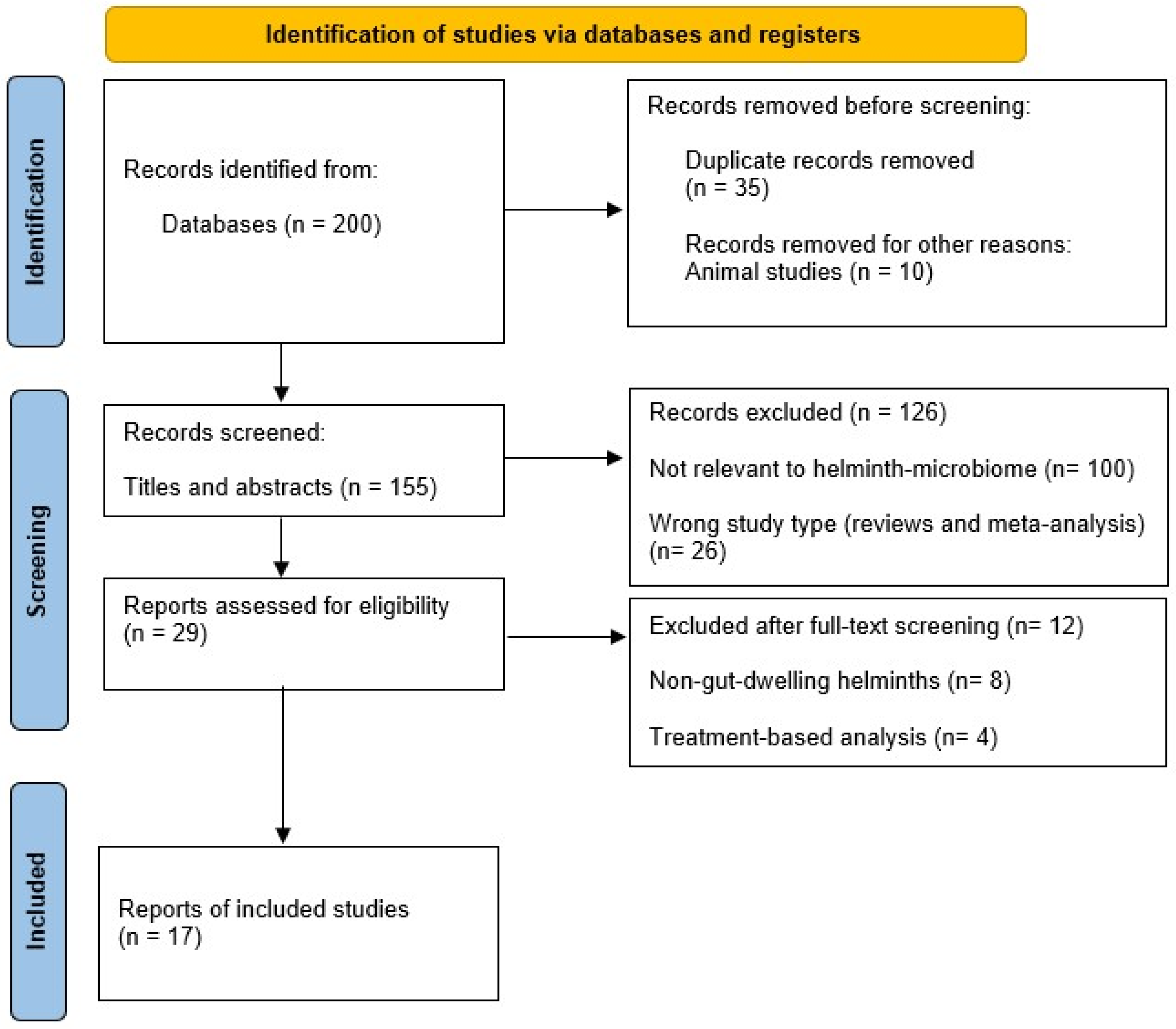
Keywords used included terms related to the gut microbiome (“gut microbiota,” “bacterial diversity,” “microbial dysbiosis”), helminth infections (“helminth infection,” “soil-transmitted helminths,” “intestinal parasites,” “worms”), and colorectal cancer (“colorectal cancer,” “colon cancer,” “tumorigenesis,” “intestinal carcinogenesis”). To maximise sensitivity, these were expanded to include additional helminth-specific terms (e.g., hookworm, Ascariasis, Trichuriasis, Schistosomiasis, Strongyloidiasis), microbiome-related terms (e.g., “microbial community,” “16S rRNA sequencing,” metagenomics, qPCR), and cancer-related synonyms (e.g., “colorectal neoplasm,” “bowel cancer,” CRC). Relevant articles identified through this comprehensive search were evaluated and documented following PRISMA guidelines [49], as illustrated in Figure 1.

2.2. Study Selection, Quality of Studies, and Data Extraction

The primary author (D.P.S.) initially screened the literature by reviewing titles, abstracts, and full texts according to predefined inclusion and exclusion criteria. The six co-authors (N.E.M., P.N., J.G., D.A.B., S.S.K. and Z.L.M.K.) conducted an additional assessment of the selected studies’ eligibility for inclusion in this systematic review, identifying discrepancies and eliminating duplicates. The extracted data quality was classified into four tiers: high, moderate, poor, and very low-quality (Group, 2004). This classification was evaluated utilising the GRADE (Grading of Recommendations, Assessment, Development, and Evaluations) methodology [50]. The quality assessments informed the interpretation of findings presented in the Results and Discussion sections.
Inclusion criteria:
  • Studies in the literature addressing helminth infections, the diversity of the gut microbiome, and CRC:
Peer-reviewed publications investigating the correlation between helminth infections and gut microbiome diversity in human subjects. Investigations assessing the impact of intestinal helminth species on gut microbiome, including alterations in microbial composition and diversity, and the potential elevation in CRC risk.
  • Language: Only articles published in English were considered.
  • Population: Research encompassing human participants of all age demographics.
  • Study Design: Observational studies, clinical trials, cohort studies, cross-sectional studies, and case–control studies.
  • Publication Period: January 2000 to March 2026.
Exclusion criteria:
  • Publication Date: Studies in the literature published before January 2000.
  • Language: Literature not published in English.
  • Study Design: Systematic reviews, meta-analyses, editorials, conference abstracts, and animal studies.
  • Relevance: Studies that do not specifically address the association between helminth infection, gut microbiome diversity, and CRC risk.

3. Results

3.1. Study Selection

A total of seventeen original research articles met the predefined inclusion criteria and were included in this systematic review, following PRISMA-guided screening and eligibility assessment.

3.2. Study Characteristics

The included studies (n = 17) represented a broad geographical distribution, encompassing populations from Southeast Asia (Thailand, Malaysia, and Indonesia), sub-Saharan Africa (Tanzania and Ethiopia), South America (Ecuador), and non-endemic Western settings (Netherlands, United Kingdom, and Italy). Sample sizes varied substantially across studies, ranging from 10 to 650 participants, reflecting heterogeneity in study scale and design. Across the included studies, the majority focused on soil-transmitted helminths, particularly Ascaris lumbricoides, T. trichiura, and Necator americanus. Mixed helminth infections were reported in 10 of the 17 studies, whereas fewer studies investigated single-species infections, including Strongyloides stercoralis (n = 3) and T. trichiura (n = 1), highlighting variability in infection profiles across populations.
Regarding study design, cross-sectional studies were the most common (n = 9), followed by longitudinal designs (n = 4), randomised controlled trials (n = 3), and experimental infection studies (n = 2). This distribution indicates a predominance of observational evidence, with relatively limited longitudinal and experimental data to support causal inference. Experimental studies involving controlled N. americanus infection provided insight into microbiome responses under defined exposure conditions. In terms of microbiome profiling, 16S rRNA gene sequencing was employed in the majority of studies (n = 14), while shotgun metagenomics was used in two studies, and one study utilised both 16S rRNA sequencing and shotgun metagenomics. Across all included studies (n = 17), stool samples constituted the primary biological material analysed. However, a subset of studies incorporated additional sample types, including helminth gut material (n = 2) and saliva (n = 1), to complement microbial analyses.
Overall, the methodological quality of included studies ranged from moderate to high, although many were limited by small sample sizes and cross-sectional designs, which constrained causal interpretation. A detailed summary of study characteristics is presented in Table 1, while gut microbiome outcomes, including diversity, taxonomic composition, and clinical associations, are summarised in Table 2.

4. Discussion

This systematic review evaluated how helminth infections influence the gut microbiome and explored their potential implications for CRC. Across 17 studies, the evidence demonstrates that helminths profoundly alter microbial richness, composition, and metabolic function. However, these effects are highly variable, with some patterns suggesting protective roles against carcinogenesis, including enrichment of SCFA-producing taxa and microbial diversity [51,53,55,61,65] and others pointing to pro-inflammatory and pro-tumourigenic consequences, such as expansion of S. gallalyticus, Enterobacteriaceae, and mucin-degrading species [44,59,60,61]. Overall, the evidence base is characterised by heterogeneity in study design, population, and analytical approaches, which should be considered when interpreting these findings.

4.1. Protective Impacts of Helminths on the Microbiome and CRC Risk

Several of the included studies reported that helminth infections enhance bacterial diversity and enrich taxa with potential protective roles. T. trichiura colonisation among rural Malaysians was associated with increased microbial richness and enrichment of pathways related to genetic information processing, specifically DNA repair, replication, and transcriptional regulation [55]. These functions are essential for maintaining microbial genome stability and metabolic adaptability, which may support ecological resilience within the gut. Such resilience is thought to buffer against dysbiosis and chronic inflammation, both of which are key hallmarks implicated in colorectal carcinogenesis [66,67]. In a controlled trial, N. americanus infection was associated with bacterial expansion during the chronic phase of infection, suggesting the establishment of a stable host–microbiota equilibrium [51]. Similarly, multiple sclerosis patients with hookworm maintained stable microbial diversity with expansion of Parabacteroides and Tenericutes, which are taxa linked to immune modulation through enhanced Treg activity and suppression of pro-inflammatory cytokines such as IL-6 and TNF-α [53]. These immunomodulatory effects may be associated with reduced chronic intestinal inflammation and restoration of mucosal immune tolerance, potentially lowering the risk of inflammation-driven colorectal carcinogenesis. Importantly, these findings suggest that helminth-induced increases in microbial diversity and functional capacity contribute to ecological stability within the gut, a factor consistently associated with resistance to dysbiosis and inflammation-driven carcinogenesis.
For instance, Agathobacter, a known butyrate producer, was enriched in Ethiopian children with T. trichiura [44]. Because butyrate supports epithelial integrity and exerts anti-inflammatory effects, its presence is generally interpreted as beneficial [68,69,70]. However, whether this enrichment reflects a protective response or simply a feature of helminth-associated dysbiosis remains unclear. At the same time, the study reported reduced diversity and enrichment of other taxa with pro-carcinogenic associations (see Section 4.2). Collectively, these findings indicate that helminth-driven microbiome shifts are not uniformly beneficial or harmful, but instead reflect a complex, context-dependent reorganisation of the microbial community. Helminth-positive Malaysian villages exhibited Firmicutes-rich microbiomes, which is a finding of potential significance because many Firmicutes genera (such as Faecalibacterium and Roseburia) are established butyrate producers, and their enrichment has been associated with SCFA-mediated protection against CRC [71,72]. Studies on S. stercoralis demonstrated an increase in Paraprevotellaceae and Ruminococcaceae [61]. While Ruminococcaceae are well-recognised SCFA producers linked to barrier reinforcement and immune regulation [70,71,72], the role of Paraprevotellaceae in CRC protection is less clear, highlighting the heterogeneity of helminth-induced microbial shifts.
Complementing these findings, Nguyen and Hongsrichan [73] reported that individuals with S. stercoralis from Thailand exhibited decreased serum acetic acid despite increased microbial richness, suggesting that helminth infection may alter SCFA metabolism in a context-dependent manner. Together, these findings indicate that helminth colonisation can foster bacterial communities capable of producing SCFAs such as butyrate and propionate. These metabolites play critical anti-carcinogenic roles by supporting epithelial integrity, suppressing pro-inflammatory cytokines, and inducing apoptosis in colonocytes, functions that have been well-characterised in human studies [70]. Collectively, these data suggest that in certain contexts, helminths may buffer against dysbiosis and reduce CRC initiation risk by reinforcing microbial stability and anti-inflammatory function.
Most studies included in this systematic review reported that helminthiasis was also associated with an enrichment of SCFA-related lineages, indicating not only an enhanced microbial fermentation capacity but also potential protective effects against colorectal carcinogenesis. SCFAs, particularly butyrate and propionate, are bioactive metabolites that regulate gut homeostasis through multiple tumour-suppressive mechanisms. First, they reinforce the intestinal epithelial barrier by upregulating tight junction proteins such as claudin-1 (a transmembrane protein regulating paracellular permeability, often dysregulated in CRC) and zonula occludens-1 (ZO-1), a scaffolding protein that stabilises tight junctions, the loss of expression of which is associated with barrier dysfunction and CRC progression. In support of this, Yan and Ajuwon [74] demonstrated that exposure to butyrate in intestinal epithelial cells significantly increased the expression of claudin-1 and ZO-1, while enhancing transepithelial electrical resistance (TEER) as a functional indicator of barrier integrity.
In addition, SCFAs enhance mucin 2 (MUC2) production (a major component of the intestinal mucus layer, the reduction of which compromises barrier function and predisposes to tumourigenesis) [70,75]. Secondly, SCFAs exert potent anti-inflammatory effects by inhibiting nuclear factor kappa-light-chain-enhancers of activated B cells (NF-κB) (a transcription factor central to inflammatory signalling) and reducing pro-inflammatory cytokines such as interleukin-6 (IL-6) and tumour necrosis factor-alpha (TNF-α), while simultaneously promoting the expansion of Tregs(a T-cell subset that produces IL-10 and maintains immune tolerance) [24,71]. Thirdly, butyrate induces apoptosis in transformed colonocytes by acting as a histone deacetylase (HDAC) inhibitor (enzymes with inhibition that lead to chromatin relaxation, cell cycle arrest, and programmed cell death of cancer cells) [24]. Finally, SCFAs modulate key oncogenic pathways, including Wnt/β-catenin (a signalling cascade that regulates intestinal stem cell renewal and proliferation, and is frequently hyperactivated in CRC) and phosphoinositide 3-kinase/protein kinase B (PI3K/Akt) (a survival pathway regulating growth and apoptosis resistance, often upregulated in CRC), thereby restraining tumour-promoting signals [74,76]. Collectively, these findings suggest that helminth-driven enrichment of SCFA-producing taxa could counterbalance oncogenic pressures by strengthening epithelial integrity, dampening inflammation, and activating tumour-suppressive signalling cascades. However, despite these well-established anti-carcinogenic mechanisms, direct evidence linking helminth-associated enrichment of SCFA-producing taxa to reduced CRC risk in humans remains limited, as none of the included studies directly assessed cancer outcomes. Furthermore, the assumption that enrichment of SCFA-producing taxa is uniformly protective warrants nuance. Microbial functional redundancy means that taxonomic presence does not reliably predict metabolic output, and 16S rRNA amplicon sequencing, the predominant method used across the included studies, cannot confirm in situ SCFA production or distinguish functionally active from dormant populations. Helminth-associated enrichment of putative SCFA producers should therefore be interpreted as indicative of potential rather than confirmed anti-carcinogenic activity.

4.2. Harmful and Pro-Carcinogenic Impacts of Helminths

In contrast, several included studies demonstrated that helminth infections may foster microbial profiles associated with inflammation and carcinogenesis. In Ethiopian schoolchildren, T. trichiura infection was linked to reduced microbial diversity, marked by loss of beneficial Prevotella spp. and enrichment of taxa such as Agathobacter, T. succinifaciens, and Streptococcus gallolyticus [44]. While Agathobacter is typically described in the literature as a butyrate-producing genus associated with gut health, its enrichment here occurred in a setting of reduced diversity and co-expansion of pro-carcinogenic taxa such as S. gallolyticus. This suggests that its function in helminth-associated dysbiosis may be context-dependent and should not be assumed to confer protection in this scenario.
Similarly, Tanzanian women with T. trichiura showed depletion of probiotic genera (Weissella and Leuconostoc) and enrichment of pathogenic pathways, including increased S. gallolyticus, a species strongly associated with human colorectal neoplasia [59]. Clinical evidence demonstrates that S. gallolyticus bacteremia and endocarditis are strongly associated with undiagnosed CRC or advanced adenomas, as suggested by Boleij and van Gelder [77], while functional studies suggest that S. gallolyticus can directly promote colon cancer cell proliferation and tumour growth via β-catenin activation, as suggested by Kumar and Herold [78], underlining its carcinogenic relevance. Rosa and Supali [60] reported the enrichment of Enterococcus, Flavonifractor, and Olsenella in individuals heavily infected in Liberia and Indonesia; these taxa were associated with genotoxic metabolite production and chronic mucosal inflammation. Tran and Chaidee [61] further showed that chronic S. stercoralis infection selectively enriched Ruminococcus torques, a mucin-degrading species linked to barrier erosion and CRC, while reducing carbohydrate metabolism post-treatment, reflecting functional instability. A. lumbricoides infections also carry risk. Muslim and Aazmi [58] observed altered host microbial profiles in A. lumbricoides-infected individuals, while Klomkliew and Sawaswong [54] found that severe Ascaris cases were characterised by elevated amino acids, lipids, and nucleotide precursors in parasite metabolomes, potentially creating nutrient-rich, pro-growth environments in the gut.
Epidemiological evidence supports a connection between chronic schistosomiasis and CRC risk. Reviews of human cases with Schistosoma japonicum highlight frequent co-occurrence with CRC, as reported by Salim and Hamid [79], and a retrospective analysis found schistosomiasis to be an independent predictor of poor overall survival in CRC patients, especially in advanced stages [80]. Additionally, systematic human-focused syntheses highlight the broader impact of schistosomiasis-induced inflammation, which has been associated with microbial and carcinogenic shifts in the colon [81]. Collectively, these findings suggest that helminth-induced dysbiosis, particularly under conditions of high burden or chronicity, may be associated with oncogenic processes through sustained inflammation, epithelial damage, metabolic reprogramming, and impaired immune surveillance.

4.3. Context Dependence of Helminth–Microbiome Interactions

The divergent findings across studies emphasise that the effects of helminths on the microbiome and CRC risk are highly context-dependent. Beyond immune polarisation, ESPs may directly modify mucosal conditions, including epithelial permeability, mucus glycoprotein composition, and antimicrobial peptide output, that govern selective pressure on luminal microbial communities, providing a helminth-specific mechanistic basis for the compositional shifts documented across the included studies. Martin and Kaisar [57] reported that helminth infection altered the normal relationship between microbial composition and cytokine responses. In participants negative for helminth, higher bacterial diversity correlated with stronger IFN-γ responses to phytohaemagglutinin (PHA) stimulation, while higher proportions of Bacteroidetes were associated with lower IL-10 responses to LPS stimulation; both relationships were weakened in helminth-positive individuals. Lee and Tang [25] demonstrated that helminth infection exerted greater effects on microbial composition than either diet or blood biochemistry in indigenous Malaysian individuals. The infections were associated with changes in systemic zinc and iron, both of which are critical for immune and redox balance, and with taxonomic shifts involving Clostridiales and Bacteroidales.
Notably, not all helminths produced disruption of the gut microbial community diversity or composition. In Ecuadorian children, single infections with T. trichiura were not associated with significant faecal microbiota differences compared to uninfected peers. By contrast, Ascaris–Trichuris co-infections were linked to reduced overall diversity, together with depletion of Clostridium sensu stricto and clostridial cluster IX taxa [63]. While the reduction in diversity is typically considered detrimental, the loss of these clostridial groups is often associated with heightened CRC risk, which may paradoxically reflect a protective shift. This underlines the dual and context-dependent nature of helminth–microbiome interactions, where the same infection can simultaneously generate potentially harmful and protective microbial outcomes.
Gobert and Atkinson [52] reported that O. viverrini infection in Thai participants led to only modest shifts in gut taxa (Bacteroides and Clostridium spp.) but significantly increased salivary α-diversity, with higher abundance of Desulfovibrio and Dialister. Both genera have been implicated in CRC development through their pro-inflammatory and genotoxic potential, underscoring the fact that parasite species and tissue niche (the gut versus oral cavity) can differentially influence cancer risk. Jenkins and Rathnayaka [62] found no significant changes in α-diversity among Sri Lankan participants with helminth infection, but β-diversity (a measure of compositional differences in microbial communities between samples) differed significantly, with enrichment of Enterobacteriaceae. This family has been consistently associated with intestinal inflammation and CRC progression, suggesting that even in the absence of diversity shifts, helminth infections may be associated with tumour-relevant microbial signatures. Similarly, in volunteers from non-endemic settings, S. stercoralis infection left α-diversity stable but shifted β-diversity and metabolomic pathways, including increased amino acid metabolites and reduced carbohydrate metabolism. Such changes have been associated with reduced SCFA production—metabolites critical for epithelial integrity and anti-carcinogenic protection that may contribute to a more permissive metabolic environment to CRC development [64,73].
Environmental and community-level factors also shaped outcomes. Tee and Er [65] reported that helminth-positive indigenous Malaysian individuals had Firmicutes-rich microbiomes, marked by overrepresentation of Agathobacter rectalis and Blautia wexlerae, but the effects varied significantly between villages, suggesting strong local modulation. In contrast, Nguélé and Mozzicafreddo [59] found that infected adults exhibited an increased Firmicutes/Bacteroidetes ratio, a pattern often associated with inflammatory bowel disorders (IBDs) including Crohn’s disease, which are conditions that may heighten CRC risk. By comparison, Taye and Mekonnen [44] observed enrichment of Agathobacter, a butyrate-producing genus within the Lachnospiraceae family, in Ethiopian children with T. trichiura infection. Given the anti-inflammatory and anti-carcinogenic roles of butyrate, this may represent a potentially protective microbial shift. Importantly, Martin and Djuardi [56] showed that albendazole treatment influenced microbiome composition in both infected and uninfected participants, with shifts in Actinobacteria and Bacteroidetes, highlighting the potential for drug effects to confound helminth–microbiome associations. Beyond treatment effects, dietary fibre intake and antibiotic history represent additional uncontrolled variables that were not systematically documented across the included studies. Fibre-rich diets independently enrich SCFA-producing Firmicutes and Ruminococcaceae, the very taxa most frequently attributed to helminth colonisation in this review, while antibiotic exposure can deplete commensal diversity in ways that may obscure or mimic helminth-associated dysbiosis. Geographic heterogeneity across studies spanning Southeast Asia, sub-Saharan Africa, South America, and non-endemic Western settings further introduces variation in baseline microbiome composition and co-infection burden that was not systematically accounted for in between-study comparisons. These observations illustrate that parasite species, infection burden, chronicity, host immunity, and environmental exposures collectively shape whether helminth-associated microbiome changes are protective or deleterious.

4.4. Synthesis and Implications for CRC

The relationship between helminths, the gut microbiome, and CRC cannot be reduced to a simple directional pathway. Instead, it operates through an integrated immune–microbiome–tumour axis in which helminth-driven immune polarisation, characterised by Th2/regulatory skewing, IL-10, TGF-β, and expanded Treg populations, simultaneously remodels microbial community structure, alters luminal metabolite production, and modulates epithelial barrier integrity. At the molecular level, epithelial barrier disruption may involve altered expression or localisation of tight junction proteins, including claudin-1, occludin and ZO-1, together with reduced MUC2, thereby increasing epithelial permeability, microbial translocation and CRC-relevant inflammatory signalling.
These interconnected shifts engage tumour-relevant processes, including genotoxin exposure, impaired immune surveillance, and chronic inflammatory signalling. Critically, the net outcome across this axis is not fixed: the same infection may enrich SCFA-producing commensals with anti-carcinogenic properties while concurrently permitting expansion of established CRC-associated taxa, depending on parasite species, infection intensity, chronicity, and host context. The evidence synthesised below maps this dualism across both protective and pro-tumourigenic trajectories.
On the one hand, they may enhance diversity, enrich SCFA-producing taxa, and promote immune homeostasis. For instance, Parabacteroides and Tenericutes expanded in patients with hookworm infection [53], Agathobacter increased in Ethiopian children [44], Firmicutes-rich profiles marked by A. rectalis and B. wexlerae were more common in helminth-positive Malaysian villages [65], and Paraprevotellaceae and Ruminococcaceae expanded in S. stercoralis infections [61,64]. These taxa are strongly associated with butyrate and propionate production: metabolites that reinforce epithelial integrity, suppress pro-inflammatory cytokines, and induce apoptosis in colonocytes, providing potential anti-carcinogenic benefits [70].
On the other hand, several studies showed enrichment of pathogenic and pro-inflammatory bacteria. S. gallolyticus, detected in Tanzanian women and Ethiopian children with T. trichiura infection, is a well-established CRC-associated bacterium [44,59]. Taken together, these two studies reveal the paradoxical role of T. trichiura in shaping microbial communities. On the one hand, Taye and Mekonnen [44] documented concurrent enrichment of Agathobacter, a butyrate-producing genus associated with epithelial protection and anti-inflammatory activity. On the other hand, Nguélé and Mozzicafreddo [59] highlighted the presence of S. gallolyticus, a species consistently linked to colorectal tumourigenesis, through its ability to adhere to colonic epithelial cells, promote chronic inflammation, and induce DNA damage that facilitates malignant transformation [78,82,83]. This contrast illustrates that T. trichiura infection has been associated with both protective shifts that support gut homeostasis and detrimental alterations potentially linked to CRC risk. The divergent outcomes most likely reflect differences in host immunity, infection intensity, and local environmental factors, reinforcing the need to view Trichuris–microbiome interactions as context-dependent rather than uniformly beneficial or harmful.
Enterococcus, Flavonifractor, and Olsenella were also enriched in individuals heavily infected in Liberia and Indonesia [60]. All three genera have been linked to CRC. Enterococcus is associated with DNA-damaging reactive oxygen species [84,85,86], while Flavonifractor has been connected to pro-inflammatory flavonoid metabolism. Emerging human evidence shows that the genus Olsenella may be enriched in CRC. For instance, a large prospective study of the oral microbiome reported that the presence of Olsenella was significantly associated with increased distal CRC risk (a hazard ratio of around 2.16; 95% CI 1.59–2.95) [87]. While mechanistic pathways remain under investigation, one proposed route involves bile-salt hydrolase activity in the family Atopobiaceae, which includes Olsenella. This activity may alter bile acid pools in favour of secondary bile acids that exert pro-tumourigenic effects, leading to epithelial stress and inflammatory signalling [88,89].
Beyond these taxa, other helminth-associated microbial shifts have also been linked to pathways relevant for colorectal carcinogenesis. Ruminococcus torques, a mucin-degrading species linked to barrier disruption, expanded in chronic S. stercoralis infections [61]. The loss of mucosal integrity associated with this expansion may contribute to microbial translocation and inflammation, which have been linked to tumour development [90,91]. At the molecular level, this barrier dysfunction may involve depletion or degradation of MUC2, a major structural component of the colonic mucus layer, together with impaired epithelial tight-junction integrity involving occludin, claudins and ZO-1 [92,93]. Disruption of these barrier components has been associated with increased paracellular permeability and exposure to microbial products, which may sustain CRC-relevant inflammatory signalling [93,94].
A. lumbricoides infections were associated with phylum-level shifts toward Firmicutes and Proteobacteria [58]. In severe cases, parasite metabolomes were enriched in amino acids, lipids, and nucleotide precursors [54], potentially creating nutrient-rich conditions favourable to tumourigenesis.
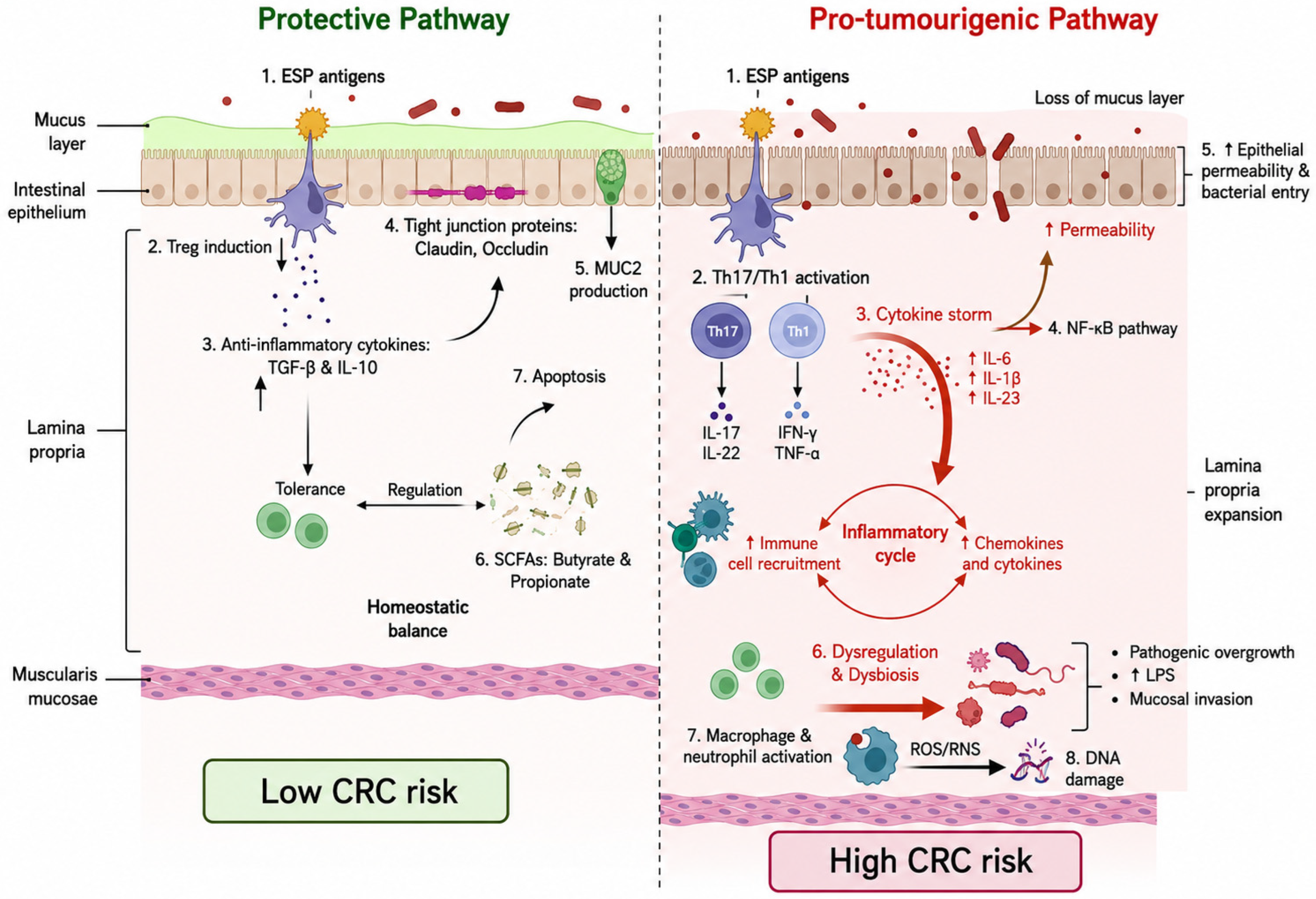
In endemic, low-sanitation settings, many individuals harbour mild or moderate helminth infections, which are often associated with increased microbial diversity and enrichment of fermentative taxa that may stabilise the gut ecosystem [55,65]. In the same environments, however, subgroups with heavy or chronic infections exhibit microbial dysbiosis characterised by loss of beneficial commensals and enrichment of CRC-associated bacteria [59,60,61]. This spectrum helps reconcile why studies in endemic regions sometimes report protective effects, while others document pro-carcinogenic microbial shifts. The outcome is strongly dependent on infection intensity, chronicity, and host context. To provide a visual synthesis of these contrasting effects, Figure 2 depicts the proposed pathways through which helminth infections can exert either protective or pro-tumourigenic influences on the gut microbiome and CRC risk.
Protective pathway (left): (1) Helminth ESPs act on dendritic cells to promote (2) Treg induction. (3) Tregs release transforming growth factor-β (TGF-β) and IL-10, enhancing tolerance and regulation. (4) Tight junction proteins (claudin and occludin) and (5) MUC2 secretion maintain barrier integrity. (6) Commensals generate SCFAs, particularly butyrate and propionate, which fuel colonocytes and drive (7) apoptosis of abnormal cells. Together, these events sustain homeostasis and reduce CRC risk.
Pro-tumourigenic pathway (right): (1) ESPs stimulate dendritic cells, driving (2) Th17/Th1 activation. Th17 produces IL-17 and IL-22; Th1 secretes interferon-γ (IFN-γ) and tumour necrosis factor-α (TNF-α), leading to (3) a cytokine storm (↑ IL-6, IL-1β, and IL-23). (4) NF-κB activation disrupts (5) epithelial barrier integrity, increasing permeability and bacterial entry. (6) Dysbiosis follows, marked by loss of commensals and pathogenic overgrowth (S. gallolyticus and Enterobacteriaceae), increased lipopolysaccharide (LPS), and mucosal invasion. (7) Macrophage and neutrophil activation drive reactive oxygen and nitrogen species (ROS/RNS), causing (8) DNA damage.
Chronic inflammation and genomic instability promote tumour initiation and progression, resulting in high CRC risk.
This dualism is not a contradiction but rather the central insight of this review, showing that helminth–microbiome interactions are not uniformly protective or harmful, but exist along a context-dependent continuum. For instance, O. viverrini infection in Thai participants led to modest shifts but increased salivary α-diversity [52], while albendazole treatment in Sri Lanka and Malaysia altered Actinobacteria and Bacteroidetes, even in uninfected participants [56,65], showing that treatment itself may act as a confounder. The immunosuppressive milieu generated by helminths, while protective in autoimmune settings, may simultaneously impair tumour immunosurveillance [57]. This aligns with high-impact human CRC metagenomic studies showing reproducible disease signatures such as F. nucleatum, Enterobacteriaceae, and S. gallolyticus across populations [40,77,78,95]. This overlap strengthens the argument that helminth-driven microbial shifts can converge mechanistically with established CRC-associated dysbiosis.
Beyond SCFAs, helminth-associated microbiome shifts may influence CRC-relevant biology through additional metabolite classes. Bile-salt hydrolase activity in Olsenella and related Atopobiaceae may skew luminal bile acid pools toward deoxycholic acid and lithocholic acid, which activate Wnt/β-catenin and NF-κB signalling and promote epithelial oxidative stress. Proteolytic fermentation products such as ammonia, phenols, and hydrogen sulphide can impair colonocyte respiration and induce genotoxic injury, while the nutrient-enriched parasite metabolome documented in severe Ascaris infections [54] may further favour pro-growth luminal conditions. Host genetic variation in Toll-like receptor 2 (TLR2), Toll-like receptor 4 (TLR4), and nucleotide-binding oligomerisation domain-containing protein 2 (NOD2) can modulate epithelial responses to these helminth-conditioned microbial signals, and chronic dysbiosis may leave epigenetic imprints, including promoter hypermethylation of tumour suppressor genes and butyrate-depletion-mediated loss of HDAC inhibition that compounds carcinogenic risk. Although none of the included studies assessed these mechanisms directly, they represent priority axes for future research and underscore that fully resolving the helminth–microbiome–CRC relationship will require longitudinal, multi-omics study designs that integrate parasitological, immunological, microbial, and host genomic data within the same cohort.

5. Limitations

A prominent limitation across most studies included in this systematic review is the small sample size, which reduces statistical power and the ability to detect subtle changes. For instance, Muslim and Aazmi [58] incorporated only four mature Ascaris worms and eight human faecal samples. Similarly, the recent Zanzibar cohort studied by Nguélé, Mozzicafreddo [59], only comprised a dozen women, with the author calling for larger follow-up cohorts, while Tran and Chaidee [61] compared 21 chronic Strongyloides-positive and 21 matched negative controls. Jenkins and Rathnayaka [62] also had a modest sample of 76 volunteers, limiting the generalisability of the observed changes in microbial beta diversity. The limited sample sizes underscore a significant deficiency in thoroughly evaluating helminth-induced gut microbiome diversity and CRC risk.
Another significant limitation is the restricted depth of microbiome analysis. Several studies relied on 16S rRNA sequencing or even older platforms (454 pyrosequencing). Nine of the seventeen studies used in this systematic review employed 16S rRNA gene sequencing [44,54,55,57,58,60,61]. These studies recognised the role of the 16S rRNA constraint in elucidating intricate microbial roles, advocating for metagenomic shotgun sequencing as an adjunctive method. Martin and Djuardi [56] encountered difficulties using obsolete sequencing systems, such as 454 pyrosequencing, which restricted the discovery of uncommon species in comparison to more advanced technologies like Illumina. Although Nguélé and Mozzicafreddo [59] used shotgun sequencing, a large number of reads, approximately 50%, remain unclassified due to incomplete reference databases. Similarly, Jenkins and Rathnayaka [62] and Jenkins and Pritchard [53] employed 16S-based approaches, which may not fully capture functional gene shifts or rare microbial taxa potentially linked to CRC development. These technical limitations highlight the necessity of utilising modern approaches to comprehensively assess microbiome makeup and function. Such approaches detect broad taxonomic changes but cannot assess microbial functional potential or rare taxa as comprehensively as shotgun metagenomics. Consequently, functional inferences drawn from taxonomic data, including SCFA production capacity, genotoxin output, and bile acid metabolism, represent plausible mechanistic hypotheses rather than confirmed biological activity, and should be interpreted with appropriate caution.
Study design issues also emerged, particularly regarding control of confounding variables and insufficient quality of methodologies; thus, there was minimal control for diet, antibiotic use, host immunity, or other environmental exposures that shape the gut microbiome. Klomkliew and Sawaswong [54] emphasised the deficiency of data on participants’ dietary habits, antibiotic consumption, and bacterial illness history, all of which may affect gut microbiota composition. Their inability to confirm findings using quantitative PCR, owing to sample limitations, constrained their conclusions. On the other hand, Muslim and Aazmi [58] only utilised microscopic analysis to detect parasite infections, lacking the sensitivity and specificity of molecular techniques. Lee and Tang [55] compared rural Malaysians with helminth infection to urban U.S. residents processed differently; differences in sample preservation and host demographics (unmatched ages and environments) could contribute to the observed microbial differences. Additionally, Lee and Tang [55] reported on the different storage protocols: rural Malaysian stools were stored in potassium dichromate at 4 °C for days, versus immediate freezing of U.S. samples. Lee and Tang [25] further illustrate the difficulty in disentangling dietary effects from helminth-associated changes in microbiota; despite identifying infection as the stronger determinant, the overlapping variables still complicate causal inferences. Jenkins and Pritchard [53] also faced challenges isolating infection effects from MS disease variability, given the limited samples and presence of comorbidities. Gobert and Atkinson [52] underscored how host-specific variables and microbial baseline differences across individuals and populations confound the direct attribution of gut changes to Opisthorchis infection alone. More broadly, the geographic spread of the included studies, spanning four continents with markedly different sanitation infrastructure, dietary traditions, co-infection profiles, study designs, helminth species, infection intensities, sequencing platforms and reported microbiome endpoints, introduces substantial between-study heterogeneity that limits direct cross-study comparisons, reduces the generalisability of pooled conclusions and precludes meta-analysis.
The complexity of studying interactions between multiple factors, such as co-infections or ecological variables, also posed challenges. Rosa and Supali [60] proposed augmenting sample sizes and integrating phylogenetic data into machine learning models to enhance prediction efficacy in multifactorial scenarios. Ducarmon and Hoogerwerf [51] in a controlled experimental study, found only temporary shifts in gut microbiota following N. americanus infection, suggesting that gut microbiome changes may be transient or masked by other dynamic gut processes.
Lastly, most of these studies were performed outside Africa, despite the increasing incidence of helminth infections in the region, mostly due to ongoing sanitation issues. However, while a recent Zanzibar study provides an African perspective, this is just one isolated context that cannot capture the continent’s epidemiological diversity.

6. Conclusions

This systematic review highlights the complex relationship between helminth infections and gut microbiome diversity, with significant implications for CRC risk. The findings reveal that helminths can profoundly reshape the composition and functional capacity of the gut microbiome, with outcomes ranging from enhanced stability and enrichment of SCFA-producing taxa to dysbiosis characterised by loss of protective commensals and expansion of pro-carcinogenic bacteria. This duality highlights that helminth–microbiome interactions exist along a spectrum, where mild or moderate infections may enhance microbial resilience, whereas heavy, chronic, or mixed infections foster inflammatory and oncogenic profiles.
Several studies reviewed, including those by [54,58,62], demonstrated shifts in dominant microbial taxa associated with helminth infection. These shifts included increases in families such as Verrucomicrobiaceae, Enterobacteriaceae, and Lachnospiraceae, enrichment of S. gallolyticus and Ruminococcus torques, and depletion of protective genera such as Prevotella, Weissella, and Leuconostoc. These microbial changes, particularly under high worm burdens or mixed infections, resemble those associated with early colorectal tumourigenesis, such as enriched DNA-damaging strains, depletion of protective SCFA producers, and increased microbial translocation across a compromised gut barrier [33,96,97,98]. The relationship between helminths and the gut microbiome is influenced by parasite species, infection intensity, co-infection status, and host immunity [54,55,60,62]. Context-dependent findings were also evident in studies such as those by Cooper and Walker [63] and Gobert and Atkinson [52], which reported minimal or site-specific microbial changes, highlighting that the effects of helminths cannot be generalised. While anthelmintic treatments can induce transient changes in the microbiome structure, evidence suggests that helminth-associated signatures may persist post-treatment. For instance, Lee, Tang [25] and Ducarmon and Hoogerwerf [51] showed that microbiome alterations linked to helminth exposure may re-establish after clearance, while [56,57] and Rosa and Supali [60] reported distinct microbial or immune configurations in individuals who remained infected. This effect suggests that helminth infections may leave long-term microbial and immunological imprints that sustain chronic low-grade inflammation, a known driver of CRC.
Additionally, helminths may indirectly modulate CRC risk through immune regulation. By potentially dampening tumour immunosurveillance or contributing to pro-inflammatory responses, helminths may be associated with microenvironments favourable to neoplastic transformation [53,57]. Given the reproducible overlap between helminth-associated taxa and CRC signatures identified in large-scale meta-analyses (e.g., S. gallolyticus, Enterobacteriaceae, and F. nucleatum) [40,77,78,95]. These findings strengthen the hypothesis that helminth-driven dysbiosis converges mechanistically with established pathways of colorectal carcinogenesis.
Future research should integrate functional metagenomics, metabolomics, and host–microbe immunological studies to unravel causal pathways linking helminth-induced microbial shifts to CRC. Longitudinal cohort studies in endemic populations will also be critical to determine whether infection history predicts long-term CRC outcomes. Importantly, this emerging body of evidence highlights the need for integrated public health approaches in helminth-endemic regions, where parasite control, nutritional interventions, and microbiome-targeted strategies could act synergistically to reduce the cancer burden.

Author Contributions

D.P.S.: Acquisition of data, literature screening, data analysis and interpretation, manuscript drafting, incorporation of revisions and final preparation of the manuscript; N.E.M.: Conception, reviewing, editing, final approval, and supervision; P.N.: Conception, reviewing, editing and final approval; J.G.: Reviewing and editing, and final approval; D.A.B.: Reviewing and editing and final approval; S.S.K.: Reviewing and editing and final approval; Z.L.M.-K.: Conception, reviewing and editing, funding, supervision, and final approval. All authors have read and agreed to the published version of the manuscript.

Funding

The research was supported by the South African Medical Research Council (SAMRC) (ZLMK MSC grant number: HDID5149/KR/202) through its Division of Research Capacity Development under the Research Capacity Development Initiative, with funding received from the South African National Treasury. Additionally, this work was funded by the National Research Foundation (NRF) (NRF reference number: PMDS22070734338). The content and findings reported/illustrated are the sole deductions, views, and responsibility of the researchers and do not reflect the official position and sentiments of the SAMRC or NRF.

Institutional Review Board Statement

This manuscript is a systematic review of previously published studies. No primary data were collected, and no human participants or animals were directly involved. Therefore, ethics approval and consent to participate were not required.

Informed Consent Statement

Not applicable. This manuscript does not contain personal data in any form.

Data Availability Statement

All data analysed in this systematic review are derived from publicly available published studies. No new datasets were generated, and no additional files accompany this manuscript.

Acknowledgments

The authors sincerely thank the South African Medical Research Council (SAMRC) and National Research Foundation (NRF) for funding this study.

Conflicts of Interest

The authors declare no conflicts of interest.

Abbreviations

The following abbreviations are used in this manuscript:
16S rRNA16S ribosomal ribonucleic acid
ADONISPermutational multivariate analysis of variance based on distance matrices
AktProtein kinase B
ANOSIMAnalysis of similarity
AUCArea under the curve
BFTBacteroides fragilis toxin
CD4+Cluster of differentiation 4-positive
CD8+Cluster of differentiation 8-positive
CIConfidence interval
CNSCentral nervous system
CRCColorectal cancer
DNADeoxyribonucleic acid
ESPsExcretory-secretory products
ETBFEnterotoxigenic Bacteroides fragilis
FDRFalse discovery rate
GIGastrointestinal
GRADEGrading of Recommendations, Assessment, Development and Evaluations
H−Helminth-negative individuals
H+Individuals infected helminth
HDACHistone deacetylase
HRHazard ratio
HtIndividuals treated for helminth individuals
IARCInternational Agency for Research on Cancer
IBDInflammatory bowel disease
I-FABPIntestinal fatty acid-binding protein
IFN-γInterferon-gamma
IL-10Interleukin-10
IL-17Interleukin-17
IL-1βInterleukin-1 beta
IL20RBInterleukin-20 receptor subunit beta
IL-22Interleukin-22
IL-23Interleukin-23
IL-5Interleukin-5
IL-6Interleukin-6
KEGGKyoto Encyclopaedia of Genes and Genomes
KOsKEGG orthology terms
LMICsLow- and middle-income countries
LPSLipopolysaccharide
M2-like macrophagesAlternatively activated macrophage-like phenotype
MSMultiple sclerosis
MUC2Mucin 2
N+Necator americanus-infected individuals
NF-κBNuclear factor kappa-light-chain-enhancer of activated B cells
NKNatural killer
NOD2Nucleotide-binding oligomerisation domain-containing protein 2
NTDsNeglected tropical diseases
OROdds ratio
OTUsOperational taxonomic units
PBOPlacebo
PCRPolymerase chain reaction
PERMANOVAPermutational multivariate analysis of variance
PHAPhytohaemagglutinin
PI3KPhosphoinositide 3-kinase
pksPolyketide synthase
PRISMAPreferred Reporting Items for Systematic Reviews and Meta-Analyses
qPCRQuantitative polymerase chain reaction
RCTRandomised controlled trial
RMSRelapsing multiple sclerosis
RNSReactive nitrogen species
ROCReceiver operating characteristic
ROSReactive oxygen species
S+Individuals with Strongyloides stercoralis
SCFAsShort-chain fatty acids
STHsSoil-transmitted helminths
TEERTransepithelial electrical resistance
TGF-βTransforming growth factor-beta
Th1T-helper type 1
Th17T-helper type 17
Th2T-helper type 2
TLR2Toll-like receptor 2
TLR4Toll-like receptor 4
TNF-αTumour necrosis factor-alpha
TregRegulatory T cell
UniFracUnique fraction metric
ZO-1Zonula occludens-1
α-diversityAlpha diversity; a measure of species richness and evenness within individual samples
β-diversityBeta diversity; a measure of compositional differences in microbial communities between samples

References

  1. WHO. Soil-Transmitted Helminth Infections; WHO: Geneva, Switzerland, 2023. [Google Scholar]
  2. Chen, J.; Gong, Y.; Chen, Q.; Li, S.; Zhou, Y. Global burden of soil-transmitted helminth infections, 1990–2021. Infect. Dis. Poverty 2024, 13, 68–77. [Google Scholar] [CrossRef]
  3. Pullan, R.L.; Smith, J.L.; Jasrasaria, R.; Brooker, S.J. Global numbers of infection and disease burden of soil transmitted helminth infections in 2010. Parasites Vectors 2014, 7, 37. [Google Scholar] [CrossRef]
  4. Freeman, M.; Chard, A.; Nikolay, B.; Garn, J.; Okoyo, C.; Kihara, J.; Njenga, S.; Pullan, R.; Brooker, S.; Mwandawiro, C. Associations between school-and household-level water, sanitation and hygiene conditions and soil-transmitted helminth infection among Kenyan school children. Parasites Vectors 2015, 8, 412. [Google Scholar] [CrossRef]
  5. Walusimbi, B.; Lawson, M.A.; Nassuuna, J.; Kateete, D.P.; Webb, E.L.; Grencis, R.K.; Elliott, A.M. The effects of helminth infections on the human gut microbiome: A systematic review and meta-analysis. Front. Microbiomes 2023, 2, 1174034. [Google Scholar] [CrossRef]
  6. McDermott, J.R.; Bartram, R.E.; Knight, P.A.; Miller, H.R.; Garrod, D.R.; Grencis, R.K. Mast cells disrupt epithelial barrier function during enteric nematode infection. Proc. Natl. Acad. Sci. USA 2003, 100, 7761–7766. [Google Scholar] [CrossRef] [PubMed]
  7. McKay, D.M.; Shute, A.; Lopes, F. Helminths and intestinal barrier function. Tissue Barriers 2017, 5, e1283385. [Google Scholar] [CrossRef] [PubMed]
  8. Su, C.-w.; Cao, Y.; Kaplan, J.; Zhang, M.; Li, W.; Conroy, M.; Walker, W.A.; Shi, H.N. Duodenal helminth infection alters barrier function of the colonic epithelium via adaptive immune activation. Infect. Immun. 2011, 79, 2285–2294. [Google Scholar] [CrossRef] [PubMed]
  9. Zaiss, D.M.; Yang, L.; Shah, P.R.; Kobie, J.J.; Urban, J.F.; Mosmann, T.R. Amphiregulin, a TH2 cytokine enhancing resistance to nematodes. Science 2006, 314, 1746. [Google Scholar] [CrossRef]
  10. Llinás-Caballero, K.; Caraballo, L. Helminths and bacterial microbiota: The interactions of two of humans’ “old friends”. Int. J. Mol. Sci. 2022, 23, 13358. [Google Scholar] [CrossRef]
  11. Maizels, R.M.; McSorley, H.J. Regulation of the host immune system by helminth parasites. J. Allergy Clin. Immunol. 2016, 138, 666–675. [Google Scholar] [CrossRef]
  12. Su, C.; Su, L.; Li, Y.; Long, S.; Chang, J.; Zhang, W.; Walker, W.; Xavier, R.; Cherayil, B.; Shi, H. Helminth-induced alterations of the gut microbiota exacerbate bacterial colitis. Mucosal Immunol. 2018, 11, 144–157. [Google Scholar] [CrossRef]
  13. Yu, H.; Yang, R.; Li, M.; Li, D.; Xu, Y. The role of Treg cells in colorectal cancer and the immunotherapy targeting Treg cells. Front. Immunol. 2025, 16, 1574327. [Google Scholar] [CrossRef]
  14. Zhang, A.; Fan, T.; Liu, Y.; Yu, G.; Li, C.; Jiang, Z. Regulatory T cells in immune checkpoint blockade antitumor therapy. Mol. Cancer 2024, 23, 251. [Google Scholar] [CrossRef]
  15. Chen, Q.; Shen, M.; Yan, M.; Han, X.; Mu, S.; Li, Y.; Li, L.; Wang, Y.; Li, S.; Li, T. Targeting tumor-infiltrating CCR8+ regulatory T cells induces antitumor immunity through functional restoration of CD4+ Tconvs and CD8+ T cells in colorectal cancer. J. Transl. Med. 2024, 22, 709. [Google Scholar] [CrossRef]
  16. Hou, S.; Zhao, Y.; Chen, J.; Lin, Y.; Qi, X. Tumor-associated macrophages in colorectal cancer metastasis: Molecular insights and translational perspectives. J. Transl. Med. 2024, 22, 62. [Google Scholar] [CrossRef]
  17. Kim, H.-D.; Kim, S.Y.; Kim, J.; Kim, J.E.; Hong, Y.S.; Han, B.; Tak, E.; Ryu, Y.-M.; Kim, S.-Y.; Kim, T.W. Dynamic increase of M2 macrophages is associated with disease progression of colorectal cancers following cetuximab-based treatment. Sci. Rep. 2022, 12, 1678. [Google Scholar] [CrossRef]
  18. Zhang, W.; Wang, M.; Ji, C.; Liu, X.; Gu, B.; Dong, T. Macrophage polarization in the tumor microenvironment: Emerging roles and therapeutic potentials. Biomed. Pharmacother. 2024, 177, 116930. [Google Scholar] [CrossRef] [PubMed]
  19. Zaiss, M.M.; Rapin, A.; Lebon, L.; Dubey, L.K.; Mosconi, I.; Sarter, K.; Piersigilli, A.; Menin, L.; Walker, A.W.; Rougemont, J. The intestinal microbiota contributes to the ability of helminths to modulate allergic inflammation. Immunity 2015, 43, 998–1010. [Google Scholar] [CrossRef] [PubMed]
  20. Ramanan, D.; Bowcutt, R.; Lee, S.C.; Tang, M.S.; Kurtz, Z.D.; Ding, Y.; Honda, K.; Gause, W.C.; Blaser, M.J.; Bonneau, R.A. Helminth infection promotes colonization resistance via type 2 immunity. Science 2016, 352, 608–612. [Google Scholar] [CrossRef]
  21. IARC. IARC Marks Colorectal Cancer Awareness Month 2025. 2025. Available online: https://www.iarc.who.int/news-events/iarc-marks-colorectal-cancer-awareness-month-2025/ (accessed on 4 August 2025).
  22. WHO. Global Cancer Burden Growing, Amidst Mounting Need for Services. 2024. Available online: https://www.who.int/news/item/01-02-2024-global-cancer-burden-growing--amidst-mounting-need-for-services (accessed on 4 August 2025).
  23. Arnold, M.; Sierra, M.S.; Laversanne, M.; Soerjomataram, I.; Jemal, A.; Bray, F. Global patterns and trends in colorectal cancer incidence and mortality. Gut 2017, 66, 683–691. [Google Scholar] [CrossRef] [PubMed]
  24. Bultman, S.J. Interplay between diet, gut microbiota, epigenetic events, and colorectal cancer. Mol. Nutr. Food Res. 2017, 61, 1500902. [Google Scholar] [CrossRef]
  25. Lee, S.C.; Tang, M.S.; Easton, A.V.; Devlin, J.C.; Chua, L.L.; Cho, I.; Moy, F.M.; Khang, T.F.; Lim, Y.A.; Loke, P.n. Linking the effects of helminth infection, diet and the gut microbiota with human whole-blood signatures. PLoS Pathog. 2019, 15, e1008066. [Google Scholar] [CrossRef]
  26. Shi, J.; Shen, H.; Huang, H.; Zhan, L.; Chen, W.; Zhou, Z.; Lv, Y.; Xiong, K.; Jiang, Z.; Chen, Q. Gut microbiota characteristics of colorectal cancer patients in Hubei, China, and differences with cohorts from other Chinese regions. Front. Microbiol. 2024, 15, 1395514. [Google Scholar] [CrossRef]
  27. Wachsmannová, L.; Dulanská, S.; Karhánek, M.; Števurková, V.; Čierniková, S.; Repiská, V.; Styk, J.; Šoltys, K.; Trnka, M. Characterization of Gut Microbiota Composition in Colorectal Adenoma and Carcinoma Patients Compared to Healthy Controls. Bratisl. Med. J. 2025, 126, 1428–1437. [Google Scholar] [CrossRef]
  28. Wang, H.; Zhu, W.; Lei, J.; Liu, Z.; Cai, Y.; Wang, S.; Li, A. Gut microbiome differences and disease risk in colorectal cancer relatives and healthy individuals. Front. Cell. Infect. Microbiol. 2025, 15, 1573216. [Google Scholar] [CrossRef] [PubMed]
  29. Qin, Y.; Tong, X.; Mei, W.-J.; Cheng, Y.; Zou, Y.; Han, K.; Yu, J.; Jie, Z.; Zhang, T.; Zhu, S. Consistent signatures in the human gut microbiome of old-and young-onset colorectal cancer. Nat. Commun. 2024, 15, 3396. [Google Scholar] [CrossRef]
  30. Yang, Y.; Du, L.; Shi, D.; Kong, C.; Liu, J.; Liu, G.; Li, X.; Ma, Y. Dysbiosis of human gut microbiome in young-onset colorectal cancer. Nat. Commun. 2021, 12, 6757. [Google Scholar] [CrossRef] [PubMed]
  31. Clay, S.L.; Fonseca-Pereira, D.; Garrett, W.S. Colorectal cancer: The facts in the case of the microbiota. J. Clin. Investig. 2022, 132, e155101. [Google Scholar] [CrossRef] [PubMed]
  32. Sears, C.L.; Garrett, W.S. Microbes, microbiota, and colon cancer. Cell Host Microbe 2014, 15, 317–328. [Google Scholar] [CrossRef]
  33. Wong, S.H.; Yu, J. Gut microbiota in colorectal cancer: Mechanisms of action and clinical applications. Nat. Rev. Gastroenterol. Hepatol. 2019, 16, 690–704. [Google Scholar] [CrossRef]
  34. Piccinno, G.; Thompson, K.N.; Manghi, P.; Ghazi, A.R.; Thomas, A.M.; Blanco-Míguez, A.; Asnicar, F.; Mladenovic, K.; Pinto, F.; Armanini, F.; et al. Pooled analysis of 3,741 stool metagenomes from 18 cohorts for cross-stage and strain-level reproducible microbial biomarkers of colorectal cancer. Nat. Med. 2025, 31, 2416–2429. [Google Scholar] [CrossRef]
  35. Zepeda-Rivera, M.; Minot, S.S.; Bouzek, H.; Wu, H.; Blanco-Míguez, A.; Manghi, P.; Jones, D.S.; LaCourse, K.D.; Wu, Y.; McMahon, E.F. A distinct Fusobacterium nucleatum clade dominates the colorectal cancer niche. Nature 2024, 628, 424–432. [Google Scholar] [CrossRef]
  36. Jasemi, S.; Emaneini, M.; Fazeli, M.S.; Ahmadinejad, Z.; Nomanpour, B.; Sadeghpour Heravi, F.; Sechi, L.A.; Feizabadi, M.M. Toxigenic and non-toxigenic patterns I, II and III and biofilm-forming ability in Bacteroides fragilis strains isolated from patients diagnosed with colorectal cancer. Gut Pathog. 2020, 12, 28. [Google Scholar] [CrossRef]
  37. Pleguezuelos-Manzano, C.; Puschhof, J.; Rosendahl Huber, A.; van Hoeck, A.; Wood, H.M.; Nomburg, J.; Gurjao, C.; Manders, F.; Dalmasso, G.; Stege, P.B. Mutational signature in colorectal cancer caused by genotoxic pks+ E. coli. Nature 2020, 580, 269–273. [Google Scholar] [CrossRef]
  38. Zhang, L.; Deng, M.; Liu, J.; Zhang, J.; Wang, F.; Yu, W. The pathogenicity of vancomycin-resistant Enterococcus faecalis to colon cancer cells. BMC Infect. Dis. 2024, 24, 230. [Google Scholar] [CrossRef]
  39. Byrd, D.A.; Damerell, V.; Gomez Morales, M.F.; Hogue, S.R.; Lin, T.; Ose, J.; Himbert, C.; Ilozumba, M.N.; Kahlert, C.; Shibata, D. The gut microbiome is associated with disease-free survival in stage I–III colorectal cancer patients. Int. J. Cancer 2025, 157, 64–73. [Google Scholar] [CrossRef] [PubMed]
  40. Thomas, A.M.; Manghi, P.; Asnicar, F.; Pasolli, E.; Armanini, F.; Zolfo, M.; Beghini, F.; Manara, S.; Karcher, N.; Pozzi, C. Metagenomic analysis of colorectal cancer datasets identifies cross-cohort microbial diagnostic signatures and a link with choline degradation. Nat. Med. 2019, 25, 667–678, Correction in Nat. Med. 2019, 25, 1948. [Google Scholar] [CrossRef] [PubMed]
  41. Yu, J.; Feng, Q.; Wong, S.H.; Zhang, D.; yi Liang, Q.; Qin, Y.; Tang, L.; Zhao, H.; Stenvang, J.; Li, Y. Metagenomic analysis of faecal microbiome as a tool towards targeted non-invasive biomarkers for colorectal cancer. Gut 2017, 66, 70–78. [Google Scholar] [CrossRef] [PubMed]
  42. Zeller, G.; Tap, J.; Voigt, A.Y.; Sunagawa, S.; Kultima, J.R.; Costea, P.I.; Amiot, A.; Böhm, J.; Brunetti, F.; Habermann, N. Potential of fecal microbiota for early-stage detection of colorectal cancer. Mol. Syst. Biol. 2014, 10, 766. [Google Scholar] [CrossRef]
  43. Appiah-Twum, F.; Akorli, J.; Okyere, L.; Sagoe, K.; Osabutey, D.; Cappello, M.; Wilson, M.D. The effect of single dose albendazole (400 mg) treatment on the human gut microbiome of hookworm-infected Ghanaian individuals. Sci. Rep. 2023, 13, 11302. [Google Scholar] [CrossRef]
  44. Taye, B.; Mekonnen, Z.; Belanger, K.D.; Davenport, E.R. Gut-microbiome profiles among Soil-transmitted helminths (STHs) infected Ethiopian children enrolled in the school-based mass deworming program. PLoS Neglected Trop. Dis. 2024, 18, e0012485. [Google Scholar] [CrossRef]
  45. Nassuuna, J.; Sterk, J.; Walusimbi, B.; Natukunda, A.; Nkangi, R.; Amongin, R.; Zirimenya, L.; Webb, E.L.; Elliott, A.M.; Nkurunungi, G. Helminth driven gut inflammation and microbial translocation associate with altered vaccine responses in rural Uganda. npj Vaccines 2025, 10, 56. [Google Scholar] [CrossRef]
  46. Amaruddin, A.I.; Koopman, J.P.R.; Muhammad, M.; Lenaerts, K.; van Eijk, H.M.; Brienen, E.A.; Geelen, A.R.; van Lieshout, L.; Wahyuni, S.; Kuijper, E.J. Intestinal permeability before and after albendazole treatment in low and high socioeconomic status schoolchildren in Makassar, Indonesia. Sci. Rep. 2022, 12, 3394. [Google Scholar] [CrossRef]
  47. Li, H.; Shan, C.; Zhu, Y.; Yao, X.; Lin, L.; Zhang, X.; Qian, Y.; Wang, Y.; Xu, J.; Zhang, Y. Helminth-induced immune modulation in colorectal cancer: Exploring therapeutic applications. Front. Immunol. 2025, 16, 1484686. [Google Scholar] [CrossRef]
  48. Siti Farah Norasyikeen, S.O.; Ngui, R.; Syaza Zafirah, A.R.; Md Zoqratt, M.Z.H.; Eng, W.W.H.; Ayub, Q.; Amin Nordin, S.; Narcisse Mary Sither Joseph, V.; Musa, S.; Lim, Y.A.L. Study on intestinal parasitic infections and gut microbiota in cancer patients at a tertiary teaching hospital in Malaysia. Sci. Rep. 2024, 14, 13650. [Google Scholar] [CrossRef]
  49. Page, M.J.; McKenzie, J.E.; Bossuyt, P.M.; Boutron, I.; Hoffmann, T.C.; Mulrow, C.D.; Shamseer, L.; Tetzlaff, J.M.; Akl, E.A.; Brennan, S.E. The PRISMA 2020 statement: An updated guideline for reporting systematic reviews. BMJ 2021, 372, n71. [Google Scholar] [CrossRef] [PubMed]
  50. Guyatt, G.H.; Oxman, A.D.; Vist, G.E.; Kunz, R.; Falck-Ytter, Y.; Alonso-Coello, P.; Schünemann, H.J. GRADE: An emerging consensus on rating quality of evidence and strength of recommendations. BMJ 2008, 336, 924–926. [Google Scholar] [CrossRef]
  51. Ducarmon, Q.; Hoogerwerf, M.; Janse, J.; Geelen, A.; Koopman, J.; Zwittink, R.; Goeman, J.; Kuijper, E.; Roestenberg, M. Dynamics of the bacterial gut microbiota during controlled human infection with Necator americanus larvae. Gut Microbes 2020, 12, 1840764. [Google Scholar] [CrossRef] [PubMed]
  52. Gobert, G.N.; Atkinson, L.E.; Lokko, A.; Yoonuan, T.; Phuphisut, O.; Poodeepiyasawat, A.; Homsuwan, N.; Mousley, A.; Adisakwattana, P. Clinical helminth infections alter host gut and saliva microbiota. PLoS Neglected Trop. Dis. 2022, 16, e0010491. [Google Scholar] [CrossRef] [PubMed]
  53. Jenkins, T.P.; Pritchard, D.I.; Tanasescu, R.; Telford, G.; Papaiakovou, M.; Scotti, R.; Cortés, A.; Constantinescu, C.S.; Cantacessi, C. Experimental infection with the hookworm, Necator americanus, is associated with stable gut microbial diversity in human volunteers with relapsing multiple sclerosis. BMC Biol. 2021, 19, 74. [Google Scholar] [CrossRef]
  54. Klomkliew, P.; Sawaswong, V.; Chanchaem, P.; Nimsamer, P.; Adisakwattana, P.; Phuphisut, O.; Tipthara, P.; Tarning, J.; Payungporn, S.; Reamtong, O. Gut bacteriome and metabolome of Ascaris lumbricoides in patients. Sci. Rep. 2022, 12, 19524. [Google Scholar] [CrossRef] [PubMed]
  55. Lee, S.C.; Tang, M.S.; Lim, Y.A.; Choy, S.H.; Kurtz, Z.D.; Cox, L.M.; Gundra, U.M.; Cho, I.; Bonneau, R.; Blaser, M.J. Helminth colonization is associated with increased diversity of the gut microbiota. PLoS Neglected Trop. Dis. 2014, 8, e2880, Correction in PLoS Neglected Trop. Dis. 2021, 15, e0009325. [Google Scholar] [CrossRef]
  56. Martin, I.; Djuardi, Y.; Sartono, E.; Rosa, B.A.; Supali, T.; Mitreva, M.; Houwing-Duistermaat, J.J.; Yazdanbakhsh, M. Dynamic changes in human-gut microbiome in relation to a placebo-controlled anthelminthic trial in Indonesia. PLoS Neglected Trop. Dis. 2018, 12, e0006620. [Google Scholar] [CrossRef]
  57. Martin, I.; Kaisar, M.M.; Wiria, A.E.; Hamid, F.; Djuardi, Y.; Sartono, E.; Rosa, B.A.; Mitreva, M.; Supali, T.; Houwing-Duistermaat, J.J. The effect of gut microbiome composition on human immune responses: An exploration of interference by helminth infections. Front. Genet. 2019, 10, 1028. [Google Scholar] [CrossRef]
  58. Muslim, A.; Aazmi, S.; Er, Y.X.; Shahrizal, S.; Lim, Y.A.L. Ascaris lumbricoides harbors a distinct gut microbiota profile from its human host: Preliminary insights. Food Waterborne Parasitol. 2024, 34, e00223. [Google Scholar] [CrossRef]
  59. Nguélé, A.T.; Mozzicafreddo, M.; Chen, H.; Piersanti, A.; Salum, S.S.; Ali, S.M.; Zhang, J.; Miceli, C. Trichuris trichiura infection is associated with changes in gut microbiome composition and function among women of reproductive age from Pemba, Tanzania. Front. Trop. Dis. 2024, 5, 1276210. [Google Scholar] [CrossRef]
  60. Rosa, B.A.; Supali, T.; Gankpala, L.; Djuardi, Y.; Sartono, E.; Zhou, Y.; Fischer, K.; Martin, J.; Tyagi, R.; Bolay, F.K. Differential human gut microbiome assemblages during soil-transmitted helminth infections in Indonesia and Liberia. Microbiome 2018, 6, 33. [Google Scholar] [CrossRef]
  61. Tran, N.T.; Chaidee, A.; Surapinit, A.; Yingklang, M.; Roytrakul, S.; Charoenlappanit, S.; Pinlaor, P.; Hongsrichan, N.; Anutrakulchai, S.; Cha’on, U. Chronic Strongyloides stercoralis infection increases presence of the Ruminococcus torques group in the gut and alters the microbial proteome. Sci. Rep. 2023, 13, 4216. [Google Scholar] [CrossRef] [PubMed]
  62. Jenkins, T.P.; Rathnayaka, Y.; Perera, P.K.; Peachey, L.E.; Nolan, M.J.; Krause, L.; Rajakaruna, R.S.; Cantacessi, C. Infections by human gastrointestinal helminths are associated with changes in faecal microbiota diversity and composition. PLoS ONE 2017, 12, e0184719. [Google Scholar] [CrossRef] [PubMed]
  63. Cooper, P.; Walker, A.W.; Reyes, J.; Chico, M.; Salter, S.J.; Vaca, M.; Parkhill, J. Patent human infections with the whipworm, Trichuris trichiura, are not associated with alterations in the faecal microbiota. PLoS ONE 2013, 8, e76573. [Google Scholar] [CrossRef]
  64. Jenkins, T.P.; Formenti, F.; Castro, C.; Piubelli, C.; Perandin, F.; Buonfrate, D.; Otranto, D.; Griffin, J.L.; Krause, L.; Bisoffi, Z. A comprehensive analysis of the faecal microbiome and metabolome of Strongyloides stercoralis infected volunteers from a non-endemic area. Sci. Rep. 2018, 8, 15651, Correction in Sci. Rep. 2019, 9, 8571. [Google Scholar] [CrossRef]
  65. Tee, M.Z.; Er, Y.X.; Easton, A.V.; Yap, N.J.; Lee, I.L.; Devlin, J.; Chen, Z.; Ng, K.S.; Subramanian, P.; Angelova, A. Gut microbiome of helminth-infected indigenous Malaysians is context dependent. Microbiome 2022, 10, 214. [Google Scholar] [CrossRef] [PubMed]
  66. Dogra, S.K.; Doré, J.; Damak, S. Gut microbiota resilience: Definition, link to health and strategies for intervention. Front. Microbiol. 2020, 11, 572921. [Google Scholar] [CrossRef] [PubMed]
  67. Marchesi, J.R.; Dutilh, B.E.; Hall, N.; Peters, W.H.; Roelofs, R.; Boleij, A.; Tjalsma, H. Towards the human colorectal cancer microbiome. PLoS ONE 2011, 6, e20447. [Google Scholar] [CrossRef] [PubMed]
  68. Kang, J.; Sun, M.; Chang, Y.; Chen, H.; Zhang, J.; Liang, X.; Xiao, T. Butyrate ameliorates colorectal cancer through regulating intestinal microecological disorders. Anti-Cancer Drugs 2023, 34, 227–237. [Google Scholar]
  69. Kaźmierczak-Siedlecka, K.; Marano, L.; Merola, E.; Roviello, F.; Połom, K. Sodium butyrate in both prevention and supportive treatment of colorectal cancer. Front. Cell. Infect. Microbiol. 2022, 12, 1023806. [Google Scholar] [CrossRef]
  70. Parada Venegas, D.; De la Fuente, M.K.; Landskron, G.; González, M.J.; Quera, R.; Dijkstra, G.; Harmsen, H.J.; Faber, K.N.; Hermoso, M.A. Short chain fatty acids (SCFAs)-mediated gut epithelial and immune regulation and its relevance for inflammatory bowel diseases. Front. Immunol. 2019, 10, 277, Erratum in Front. Immunol. 2019, 10, 1486. [Google Scholar]
  71. Louis, P.; Flint, H.J. Formation of propionate and butyrate by the human colonic microbiota. Environ. Microbiol. 2017, 19, 29–41. [Google Scholar] [CrossRef]
  72. Singh, V.; Lee, G.; Son, H.; Koh, H.; Kim, E.S.; Unno, T.; Shin, J.-H. Butyrate producers, “The Sentinel of Gut”: Their intestinal significance with and beyond butyrate, and prospective use as microbial therapeutics. Front. Microbiol. 2023, 13, 1103836. [Google Scholar] [CrossRef]
  73. Nguyen, H.T.; Hongsrichan, N.; Intuyod, K.; Pinlaor, P.; Yingklang, M.; Chaidee, A.; Sengthong, C.; Pongking, T.; Dangtakot, R.; Banjong, D. Investigation of gut microbiota and short-chain fatty acids in Strongyloides stercoralis-infected patients in a rural community. Biosci. Microbiota Food Health 2022, 41, 121–129. [Google Scholar] [CrossRef]
  74. Yan, H.; Ajuwon, K.M. Butyrate modifies intestinal barrier function in IPEC-J2 cells through a selective upregulation of tight junction proteins and activation of the Akt signaling pathway. PLoS ONE 2017, 12, e0179586. [Google Scholar] [CrossRef]
  75. Hatayama, H.; Iwashita, J.; Kuwajima, A.; Abe, T. The short chain fatty acid, butyrate, stimulates MUC2 mucin production in the human colon cancer cell line, LS174T. Biochem. Biophys. Res. Commun. 2007, 356, 599–603. [Google Scholar] [CrossRef]
  76. Kaiko, G.E.; Ryu, S.H.; Koues, O.I.; Collins, P.L.; Solnica-Krezel, L.; Pearce, E.J.; Pearce, E.L.; Oltz, E.M.; Stappenbeck, T.S. The colonic crypt protects stem cells from microbiota-derived metabolites. Cell 2016, 165, 1708–1720. [Google Scholar] [CrossRef]
  77. Boleij, A.; van Gelder, M.M.; Swinkels, D.W.; Tjalsma, H. Clinical Importance of Streptococcus gallolyticus infection among colorectal cancer patients: Systematic review and meta-analysis. Clin. Infect. Dis. 2011, 53, 870–878. [Google Scholar] [CrossRef]
  78. Kumar, R.; Herold, J.L.; Schady, D.; Davis, J.; Kopetz, S.; Martinez-Moczygemba, M.; Murray, B.E.; Han, F.; Li, Y.; Callaway, E. Streptococcus gallolyticus subsp. gallolyticus promotes colorectal tumor development. PLoS Pathog. 2017, 13, e1006440. [Google Scholar] [CrossRef] [PubMed]
  79. Salim, O.E.; Hamid, H.K.; Mekki, S.O.; Suleiman, S.H.; Ibrahim, S.Z. Colorectal carcinoma associated with schistosomiasis: A possible causal relationship. World J. Surg. Oncol. 2010, 8, 68. [Google Scholar] [CrossRef] [PubMed]
  80. Wang, Z.; Du, Z.; Liu, Y.; Wang, W.; Liang, M.; Zhang, A.; Yang, J. Comparison of the clinicopathological features and prognoses of patients with schistosomal and nonschistosomal colorectal cancer. Oncol. Lett. 2020, 19, 2375–2383. [Google Scholar] [CrossRef] [PubMed]
  81. Afful, P.; Abotsi, G.K.; Adu-Gyamfi, C.O.; Benyem, G.; Katawa, G.; Kyei, S.; Arndts, K.; Ritter, M.; Asare, K.K. Schistosomiasis–Microbiota Interactions: A Systematic Review and Meta-Analysis. Pathogens 2024, 13, 906. [Google Scholar] [CrossRef]
  82. Abdulamir, A.S.; Hafidh, R.R.; Bakar, F.A. The association of Streptococcus bovis/gallolyticus with colorectal tumors: The nature and the underlying mechanisms of its etiological role. J. Exp. Clin. Cancer Res. 2011, 30, 11. [Google Scholar] [CrossRef]
  83. Kumar, R.; Taylor, J.C.; Jain, A.; Jung, S.Y.; Garza, V.; Xu, Y. Modulation of the extracellular matrix by Streptococcus gallolyticus subsp. gallolyticus and importance in cell proliferation. PLoS Pathog. 2022, 18, e1010894. [Google Scholar] [CrossRef]
  84. Huycke, M.M.; Abrams, V.; Moore, D.R. Enterococcus faecalis produces extracellular superoxide and hydrogen peroxide that damages colonic epithelial cell DNA. Carcinogenesis 2002, 23, 529–536. [Google Scholar] [CrossRef]
  85. Strickertsson, J.A.; Desler, C.; Martin-Bertelsen, T.; Machado, A.M.D.; Wadstrøm, T.; Winther, O.; Rasmussen, L.J.; Friis-Hansen, L. Enterococcus faecalis infection causes inflammation, intracellular oxphos-independent ROS production, and DNA damage in human gastric cancer cells. PLoS ONE 2013, 8, e63147. [Google Scholar] [CrossRef] [PubMed]
  86. Williamson, A.J.; Jacobson, R.; Van Praagh, J.; Gaines, S.; Koo, H.Y.; Lee, B.; Chan, W.-C.; Weichselbaum, R.; Alverdy, J.C.; Zaborina, O. Enterococcus faecalis promotes a migratory and invasive phenotype in colon cancer cells. Neoplasia 2022, 27, 100787. [Google Scholar] [CrossRef]
  87. Vogtmann, E.; Yano, Y.; Zouiouich, S.; Hua, X.; Wan, Y.; Purandare, V.; Li, S.; Dagnall, C.L.; Jones, K.; Hicks, B.D. The human oral microbiome and risk of colorectal cancer within three prospective cohort studies in the United States. Cancer 2025, 131, e35802. [Google Scholar] [CrossRef]
  88. Devlin, A.S.; Fischbach, M.A. A biosynthetic pathway for a prominent class of microbiota-derived bile acids. Nat. Chem. Biol. 2015, 11, 685–690. [Google Scholar] [CrossRef]
  89. Ridlon, J.M.; Kang, D.-J.; Hylemon, P.B. Bile salt biotransformations by human intestinal bacteria. J. Lipid Res. 2006, 47, 241–259. [Google Scholar] [CrossRef]
  90. Herath, M.; Hosie, S.; Bornstein, J.C.; Franks, A.E.; Hill-Yardin, E.L. The role of the gastrointestinal mucus system in intestinal homeostasis: Implications for neurological disorders. Front. Cell. Infect. Microbiol. 2020, 10, 248. [Google Scholar] [CrossRef]
  91. Png, C.W.; Lindén, S.K.; Gilshenan, K.S.; Zoetendal, E.G.; McSweeney, C.S.; Sly, L.I.; McGuckin, M.A.; Florin, T.H. Mucolytic bacteria with increased prevalence in IBD mucosa augmentin vitroutilization of mucin by other bacteria. Am. J. Gastroenterol. 2010, 105, 2420–2428. [Google Scholar] [CrossRef]
  92. Capaldo, C.T.; Powell, D.N.; Kalman, D. Layered defense: How mucus and tight junctions seal the intestinal barrier. J. Mol. Med. 2017, 95, 927–934. [Google Scholar] [CrossRef] [PubMed]
  93. Chelakkot, C.; Ghim, J.; Ryu, S.H. Mechanisms regulating intestinal barrier integrity and its pathological implications. Exp. Mol. Med. 2018, 50, 1–9. [Google Scholar] [CrossRef] [PubMed]
  94. Stolfi, C.; Maresca, C.; Monteleone, G.; Laudisi, F. Implication of intestinal barrier dysfunction in gut dysbiosis and diseases. Biomedicines 2022, 10, 289. [Google Scholar] [CrossRef]
  95. Wirbel, J.; Pyl, P.T.; Kartal, E.; Zych, K.; Kashani, A.; Milanese, A.; Fleck, J.S.; Voigt, A.Y.; Palleja, A.; Ponnudurai, R. Meta-analysis of fecal metagenomes reveals global microbial signatures that are specific for colorectal cancer. Nat. Med. 2019, 25, 679–689. [Google Scholar] [CrossRef] [PubMed]
  96. Louis, P.; Hold, G.L.; Flint, H.J. The gut microbiota, bacterial metabolites and colorectal cancer. Nat. Rev. Microbiol. 2014, 12, 661–672. [Google Scholar] [CrossRef] [PubMed]
  97. O’keefe, S.J. Diet, microorganisms and their metabolites, and colon cancer. Nat. Rev. Gastroenterol. Hepatol. 2016, 13, 691–706. [Google Scholar] [CrossRef]
  98. Ternes, D.; Karta, J.; Tsenkova, M.; Wilmes, P.; Haan, S.; Letellier, E. Microbiome in colorectal cancer: How to get from meta-omics to mechanism? Trends Microbiol. 2020, 28, 401–423. [Google Scholar] [CrossRef] [PubMed]
Figure 1. PRISMA 2020 flow diagram showing the study selection process for the systematic review titled “The Impact of Helminth Infections on the Gut Microbiome and Its Potential Implications for Colorectal Cancer: A Systematic Review.” A total of 29 full-text articles were assessed for eligibility after initial screening from databases including PubMed, ScienceDirect, Google Scholar, MEDLINE, and ISI Web of Knowledge. Following the inclusion and exclusion criteria, 17 original research articles were included in the final synthesis. Reasons for exclusion at each stage are detailed in the diagram.
Ijms 27 04957 g001
Figure 2. Dual effects of helminth infections on gut microbiome composition and colorectal cancer (CRC) development. The diagram depicts the contrasting protective (left) and tumour-promoting (right) mechanisms through which helminths influence host–microbiome interactions. The image was created in BioRender.com (accessed 27 October 2025).
Ijms 27 04957 g002
Table 1. Characteristics of included studies examining helminth–gut microbiome interactions in human populations (n = 17).
StudyCountry
(Study Setting)
Study DesignPopulation/
Study Group
Sample Size
(n)
Sample TypeHelminth SpeciesMicrobiome Profiling Method
[51]NetherlandsExperimentalHealthy adults, experimental Necator americanus infection20StoolNecator americanus16S rRNA sequencing
[52]ThailandCross-sectionalAdults with natural helminth infection90Stool, SalivaMixed helminths16S rRNA sequencing
[53]United Kingdom ExperimentalRMS 1 patients, experimental Necator americanus infection71StoolNecator americanus16S rRNA sequencing
[54]ThailandCross-sectionalChildren and young adults with natural Ascaris lumbricoides infection49Stool and helminth gutAscaris lumbricoides16S rRNA sequencing
[55]MalaysiaCross-sectionalAdults with natural helminth infection70StoolMixed helminths16S rRNA sequencing
[56]IndonesiaRandomised
controlled trial
Adults with natural helminth infection150StoolMixed helminths16S rRNA sequencing
[57]IndonesiaRandomised controlled trialAdults with natural helminth infection66StoolMixed helminths16S rRNA sequencing
[58]MalaysiaCross-sectionalChildren with natural Ascaris lumbricoides infection12Stool and helminth gutMixed helminths16S rRNA sequencing
[59]TanzaniaCross-sectionalWomen of reproductive age with Trichuris trichiura infection10StoolTrichuris trichiurashotgun metagenomics
[60]Liberia, IndonesiaRandomised controlled trial Adults with natural helminth infection402StoolMixed helminths16S rRNA sequencing and
Shotgun metagenomics
[44]EthiopiaCross-sectionalChildren with natural helminth infection 138StoolMixed helminths16S rRNA sequencing
[61]ThailandLongitudinalAdults with natural Strongyloides stercoralis infection42StoolStrongyloides stercoralis16S rRNA sequencing
[62]Sri LankaCross-sectionalAdults with natural helminth infection76StoolMixed helminths16S rRNA sequencing
[25]MalaysiaLongitudinalAdults with natural helminth infection67StoolMixed helminths16S rRNA sequencing
[63]EcuadorCross-sectionalChildren with natural Trichuris trichiura infection97StoolMixed helminths16S rRNA sequencing
[64]ItalyLongitudinalAdults with natural Strongyloides stercoralis infection31StoolStrongyloides stercoralis16S rRNA sequencing
[65]MalaysiaLongitudinalAdults with natural helminth infection650StoolMixed helminthsShotgun metagenomics
1 RMS, relapsing multiple sclerosis.
Table 2. Summary of gut microbiome alterations and related outcomes in human studies of helminth infection. Statistical measures reported reflect those used in the original studies and include p-values, odds ratios, and β coefficients; direct cross-study comparison is limited by methodological heterogeneity across the included studies.
StudyStudy Aim (s)Key Microbiome and Clinical Findings
[51]To investigate temporal changes in the gut microbiota in response to different dosages (ranging from 50 to 150L3) of Necator americanus larvae in healthy young volunteers.
To investigate temporal differences in the gut microbiota between healthy volunteers experiencing different numbers of clinical symptoms.
Alpha diversity:
Increased bacterial richness during established infection (Chao1; p = 0.0174; weeks 8–20)
Relative abundance:
Barnesiella is more abundant in volunteers with higher GI 2 symptoms (p < 0.05)
Microbiota stability/timepoints:
Instability observed during early infection (weeks 0–8; p = 0.036) with recovery by week 20 (p = 0.004)
Clinical outcomes:
GI symptoms associated with microbial instability in early infection, with recovery by week 20
[52]To determine if microbial changes are associated with parasite infection by profiling faecal and saliva microbiota of participants with helminth infection.No significant changes in stool. Significant changes observed in saliva.
Alpha diversity:
Increase in saliva (Chao1; p = 0.042 at genus level; p = 0.026 at phylum level)
Relative abundance:
Increase: Desulfovibrio (p = 0.039), Oxalobacter (p = 0.022), Oxalobacteraceae (p = 0.022), Dialister (p = 0.027), and Abiotrophia (p = 0.031)
Decrease: Haemophilus
Specific to Opisthorchis viverrini: Odoribacter (p = 0.0066), Phascolarctobacterium (p = 0.031), and Succinivibrio (p = 0.05)
[53]To investigate qualitative and quantitative changes in faecal bacterial composition in RMS 3 patients before and after experimental infection with Necator americanus, and to assess the therapeutic efficacy of live hookworm infective larvae in RMS patients.Alpha diversity:
Decrease in PBO 4 subjects at T9 compared to T pre (p < 0.05)
Beta diversity:
Higher in N+ (Necator americanus infected) subjects at T9 compared to PBO (p = 0.048)
Relative abundance:
Parabacteroides significantly expanded in N+ individuals with no relapses
Clinical outcomes:
51% of N+ subjects showed no new CNS 5 lesions vs. 28% of PBO subjects
[54]To investigate the gut bacteriomes of Ascaris lumbricoides helminths and stool samples of patients with different infection intensities, as well as to characterise the metabolomes of Ascaris lumbricoides in heavy and light ascariasis cases.Alpha diversity:
Chao1 richness is higher in heavily infected patients (p < 0.05)
Beta diversity:
Bray–Curtis distance did not distinguish between infected and uninfected individuals (PERMANOVA 6 p < 0.001)
Relative abundance:
Prevotella is more abundant in ascariasis patients (p < 0.05); Streptococcus is more abundant in heavily infected patients; Lactococcus is more abundant in lightly infected patients
Clinical outcomes:
Increased levels of essential biomolecules in heavily infected patients
[55]To compare the gut microbiota of individuals with helminth infection and individuals who were not colonised by helminths, and to develop new approaches to examine the interactions of microbes with each other and their hosts.Alpha diversity:
Increase in helminth-colonised individuals (p = 0.04)
Beta diversity:
ANOSIM 7 (R = 0.18; p = 0.04)
Relative abundance:
Increase in Paraprevotellaceae, Mollicutes, Bacteroidales, and Alphaproteobacteria in helminth-colonised individuals; increase in Bifidobacterium in helminth-negative individuals
[56]To characterise the joint effects of several predictors, including helminth infection and treatment, on each bacterial category.Relative abundance:
Actinobacteria increased (OR 8 = 1.57; 95% CI 9: 1.05–2.35) and Bacteroidetes decreased (OR = 0.35; 95% CI: 0.18–0.70) in infected subjects receiving albendazole vs. PBO
Alpha diversity:
No significant differences in Shannon diversity or richness between infected and uninfected groups (pre- and post-treatment)
Beta diversity:
Bray–Curtis dissimilarity remained stable (61%) between pre- and post-treatment samples
Clinical outcomes:
STH 10 prevalence reduced in the albendazole group (21.7% vs. 54.3%; p < 0.001) at 21 months
[57]To characterise the association between gut microbiome composition and immune responses, and to examine the effect of helminth infections on this relationship.Alpha diversity:
Higher diversity associated with increased IFN-γ 11 response (β = 0.95; 95% CI: 0.15–1.75; p = 0.056)
Relative abundance/effect sizes:
Bacteroidetes negatively associated with IL-10 12 response (β = −1.96; 95% CI: −3.05 to −0.87; p = 0.002); Actinobacteria associated with decreased TNF-α 13 (β = −1.55; 95% CI: −2.87 to −0.22; p = 0.024); Firmicutes associated with IL-5 14 response post-treatment (β = −1.52; p = 0.024)
Clinical outcomes:
Deworming did not significantly alter microbiome–cytokine associations
[58]To characterise the gut microbiota of the SHT parasite Ascaris lumbricoides and compare it to the gut microbiota of their human hosts.Alpha diversity:
Lower in Ascaris (p = 0.006)
Beta diversity:
Significant separation between Ascaris and human gut microbiota (Bray–Curtis p = 0.002; Jaccard p = 0.004)
Relative abundance:
Firmicutes higher in Ascaris (84.2%; p = 0.03); Clostridium significantly higher in Ascaris (p = 0.001)
Timepoints:
Samples obtained 3 days post-albendazole treatment
[59]To investigate the effects of Trichuris trichiura infection on the gut microbiome composition and function in women of reproductive age from Pemba, Tanzania.Relative abundance:
Prevotella is higher in healthy participants; Weissella cibaria, Leuconostoc citreum, and Leuconostoc lactis are lower in infected individuals; Proteobacteria are higher in infected samples; Treponema succinifaciens and Streptococcus gallolyticus are higher in infected samples; overall fungal abundance is higher in infected participants
Functional analysis:
Cholesterol metabolism and pathogenic infection pathways are higher in infected samples (p < 0.05)
Firmicutes/Bacteroidetes ratio:
Higher in infected participants
[60]To enhance understanding of human gut microbiome interactions with STHs in Liberia and Indonesia, despite varying gut microbiome structures among individuals.Alpha diversity:
Higher richness and evenness (89.5 vs. 82.1, p = 5.8 × 10⁻¹⁰; 2.8 vs. 2.4, p = 0.0008)
Beta diversity:
Higher in infected individuals (0.52 vs. 0.43, p < 10−5); lower with self-clearing over time (2010: 0.54 vs. 0.60, p = 0.008)
Relative abundance:
Prevotella dominant (32%)
Functional analysis:
Arachidonic acid metabolism and K00560 were enriched; 11 Kos 15 were depleted
Clinical outcomes:
Self-clearing individuals were older (51.1 vs. 21.4 years, p = 0.012)
[44]To identify and explain differences in microbial communities between STH-infected and non-infected Ethiopian school children.Alpha diversity:
Lower in Trichuris trichiura-infected children (p < 0.05)
Beta diversity:
Differences by infection status (PERMANOVA p < 0.01)
Relative abundance/correlation:
Agathobacter was higher in infected children (adjusted p = 0.001) and positively correlated with egg counts (p < 0.05)
[61]To explore the impact of chronic Strongyloides stercoralis infection on the gut microbiome and microbial activity in a longitudinal study.Alpha diversity:
No significant difference
Beta diversity:
Difference in unweighted UniFrac (R2 = 0.037; p = 0.028)
Relative abundance:
Ruminococcus torques group is over-represented in chronic infection
Timepoints:
T0 (baseline), T1 (1 year), and T2 (4 months post-treatment)
Clinical outcomes:
Reduction in Ruminococcus torques group after treatment; increased carbohydrate metabolism
[62]To explore the qualitative and quantitative differences in microbial community profiles between individuals with patent infections by parasitic nematodes, uninfected individuals, and those who have received regular anthelmintic treatment.Alpha diversity:
No significant difference (Shannon; p = 0.65)
Beta diversity:
Higher in individuals with (H+) and treated for helminth (Ht) (p = 0.04)
Relative abundance:
Verrucomicrobiaceae and Enterobacteriaceae are higher in H+; Leuconostocaceae are higher in helminth-negative (H−); Bacteroidaceae are higher in Ht; and Akkermansia muciniphila is higher in H+ individuals
[25]To identify the important factors influencing the gut microbiota and to determine the relative impact of helminth infection on host responses and the microbiota.Microbiome:
Helminth infection influenced gut microbiota more than diet; OTUs 16 associated with fibre intake and Trichuris trichiura burden
Clinical/immune:
Zinc was higher in Orang Asli (p = 1.67 × 10⁻⁹); iron was higher in urban (p = 0.002); CD8+ was higher and naïve CD4+ was lower; and natural killer cells were reduced post-treatment
Gene expression:
Deworming altered 654 genes (FDR 17 < 10%) and reduced the Trichuris trichiura burden associated with increased IL20RB 18
[63]To investigate the effects of Trichuris trichiura infections on faecal microbiota and to determine if anthelmintic treatment reverses these changes towards a microbiota composition similar to that of uninfected individuals.Alpha diversity:
Lower in mixed infections (Shannon; p = 0.004)
Relative abundance:
Clostridium sensu stricto was higher in uninfected individuals (4.1% vs. 1.5%; p = 0.013); uncharacterised clostridial cluster IX bacteria were higher in uninfected individuals (2.3% vs. 0.6%; p = 0.026)
Clinical outcomes:
100% cure of STH infections at 7 and 21 days post-treatment
[64]To determine the effects of chronic, monospecific infections by Strongyloides stercoralis on the faecal microbiome and metabolome of human volunteers from a non-endemic area and to establish whether these effects are reversed by anthelmintic treatment.Alpha diversity:
Higher in Strongyloides stercoralis positive (S+) subjects (Simpson’s index: F = 5; p = 0.03; evenness: F = 4.2; p = 0.05)
Beta diversity:
Lower in S+ subjects (R = 0.11; p = 0.04)
Relative abundance:
Leuconostocaceae, Ruminococcaceae, Paraprevotellaceae, and Peptococcus are higher in S+; Bacteroides is lower in S+
Metabolites:
Alanine, formate, lysine, and leucine are higher in S+ (F ≈ 4.2–4.5; p = 0.03–0.05)
[65]To investigate the interactions between helminth infections and the human gut microbiome in indigenous Malaysians using shotgun metagenomics.Alpha diversity:
Higher richness in infected subjects (p = 2.5 × 10⁻⁵); no significant change post-treatment
Beta diversity:
Small effect size between pre- and post-treatment (ADONIS 19: R2 = 0.014; p = 0.001; ANOSIM: R = 0.072; p = 0.001)
Relative abundance:
Specific replicating taxa associated with helminth infection
Timepoints:
Longitudinal changes assessed post-albendazole treatment
2 GI, gastrointestinal; 3 RMS, relapsing multiple sclerosis; 4 PBO, placebo; 5 CNS, central nervous system; 6 PERMANOVA, permutational multivariate analysis of variance; 7 ANOSIM, analysis of similarity; 8 OR, odds ratio; 9 CI, confidence interval; 10 STH, soil-transmitted helminth; 11 IFN-γ, interferon-gamma; 12 IL-10, interleukin-10; 13 TNF-α, tumour necrosis factor-alpha; 14 IL-5, interleukin 5; 15 KOs, KEGG orthology terms; 16 OTUs, operational taxonomic units; 17 FDR, false discovery rate; 18 IL20RB, interleukin-20 receptor subunit beta; 19 ADONIS, permutational multivariate analysis of variance based on distance matrices.
Disclaimer/Publisher’s Note: The statements, opinions and data contained in all publications are solely those of the individual author(s) and contributor(s) and not of MDPI and/or the editor(s). MDPI and/or the editor(s) disclaim responsibility for any injury to people or property resulting from any ideas, methods, instructions or products referred to in the content.