Acute Cytokine Responses to High-Intensity Intermittent Exercise in Humans: A Systematic Review

 , ,
, ,
Abstract
1. Introduction
2. Materials and Methods
2.1. Eligibility Criteria
2.2. Information Sources
2.3. Search Strategy
2.4. Selection Process
2.5. Data Collection Process
2.6. Data Items
2.7. Study Risk-of-Bias Assessment
2.8. Synthesis Methods
2.9. Certainty of Evidence
3. Results and Discussion
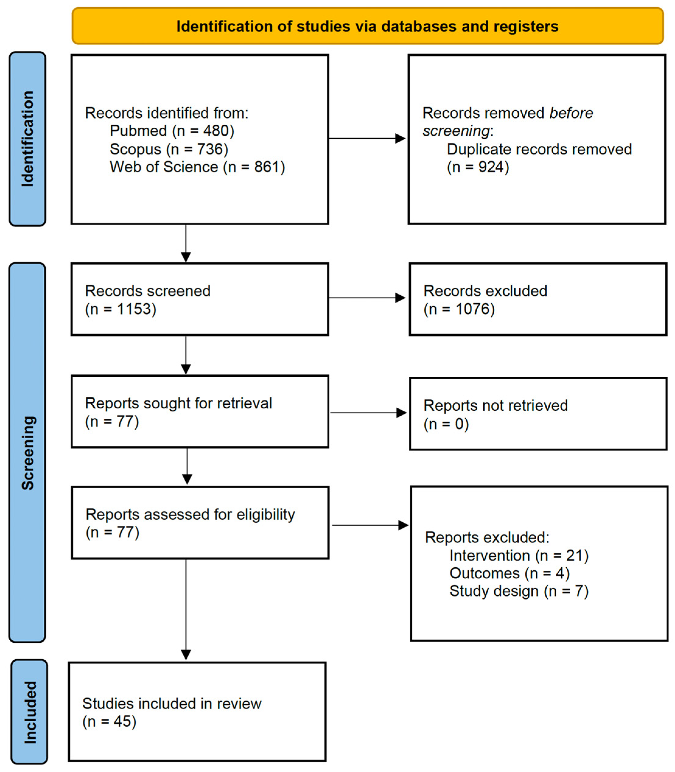
3.1. Study Selection
3.2. Study Characteristics
3.3. Risk-of-Bias Assessment
3.4. Results of Individual Studies
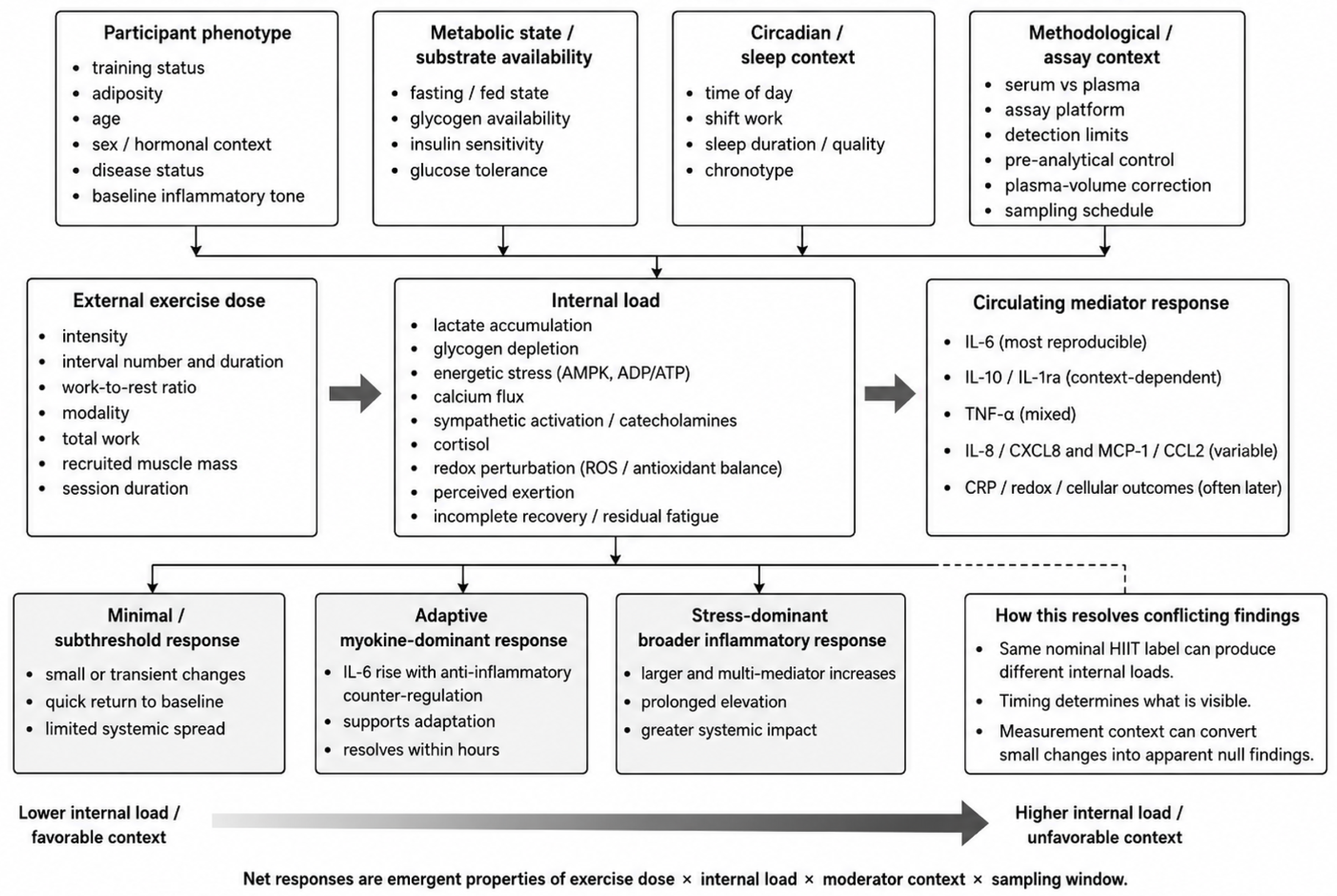
3.5. Synthesis of Response Patterns and Moderators
3.6. Certainty of Evidence
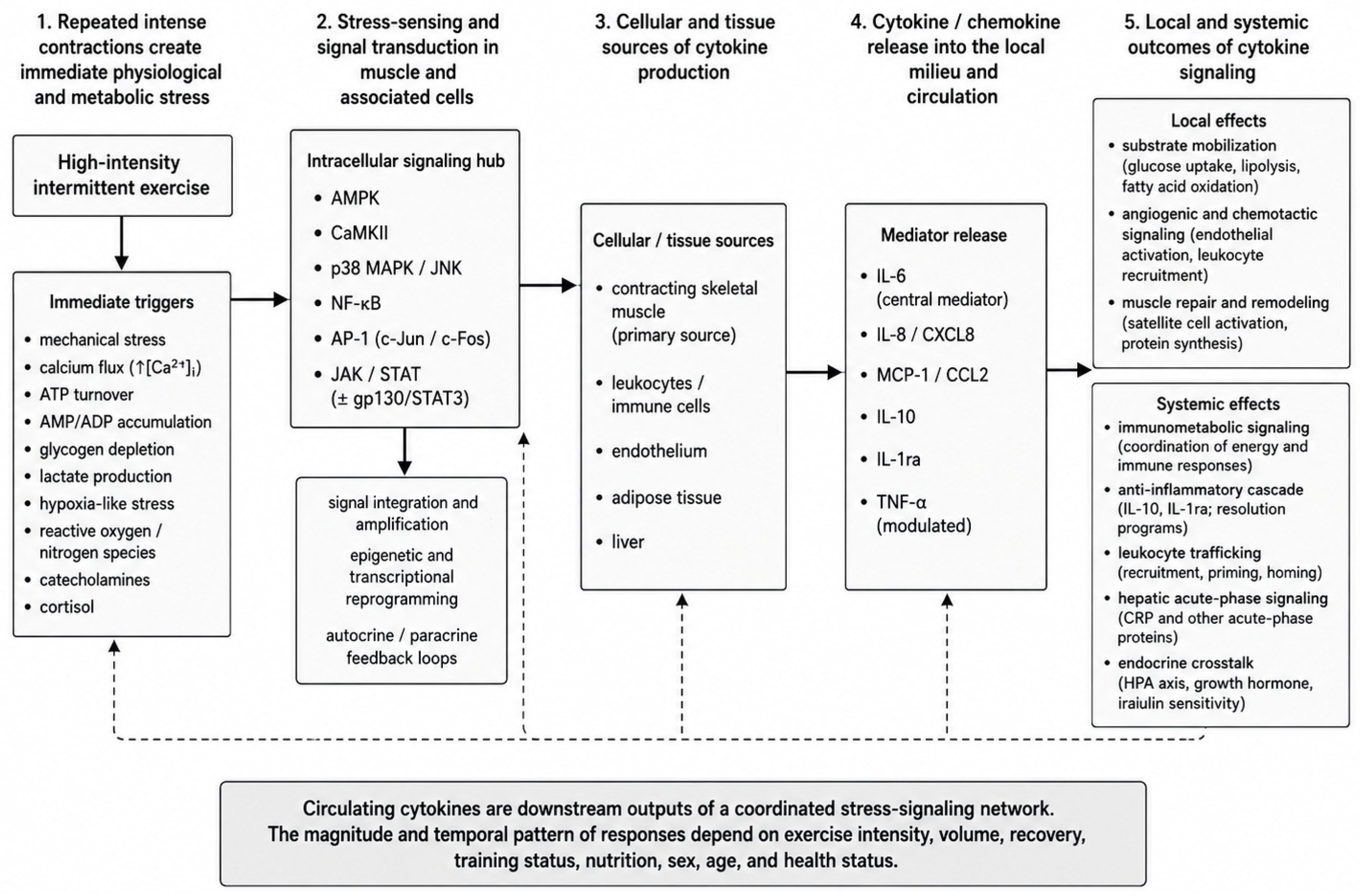
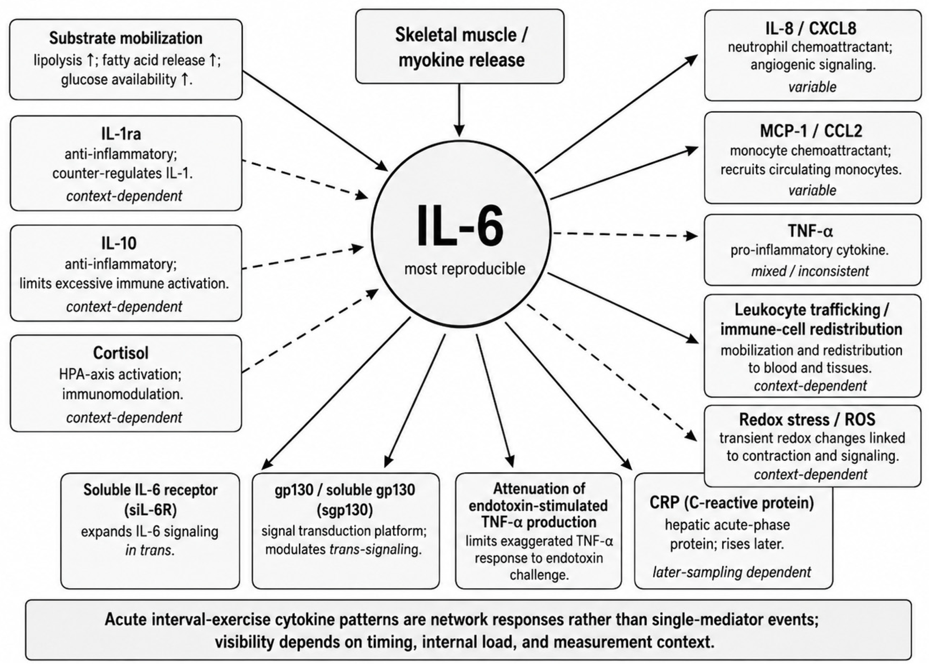
3.7. Mechanistic Framework: Cellular-Stress Signaling, IL-6, and Hormetic Dose Response
3.8. Metabolic State as an Immunometabolic Moderator
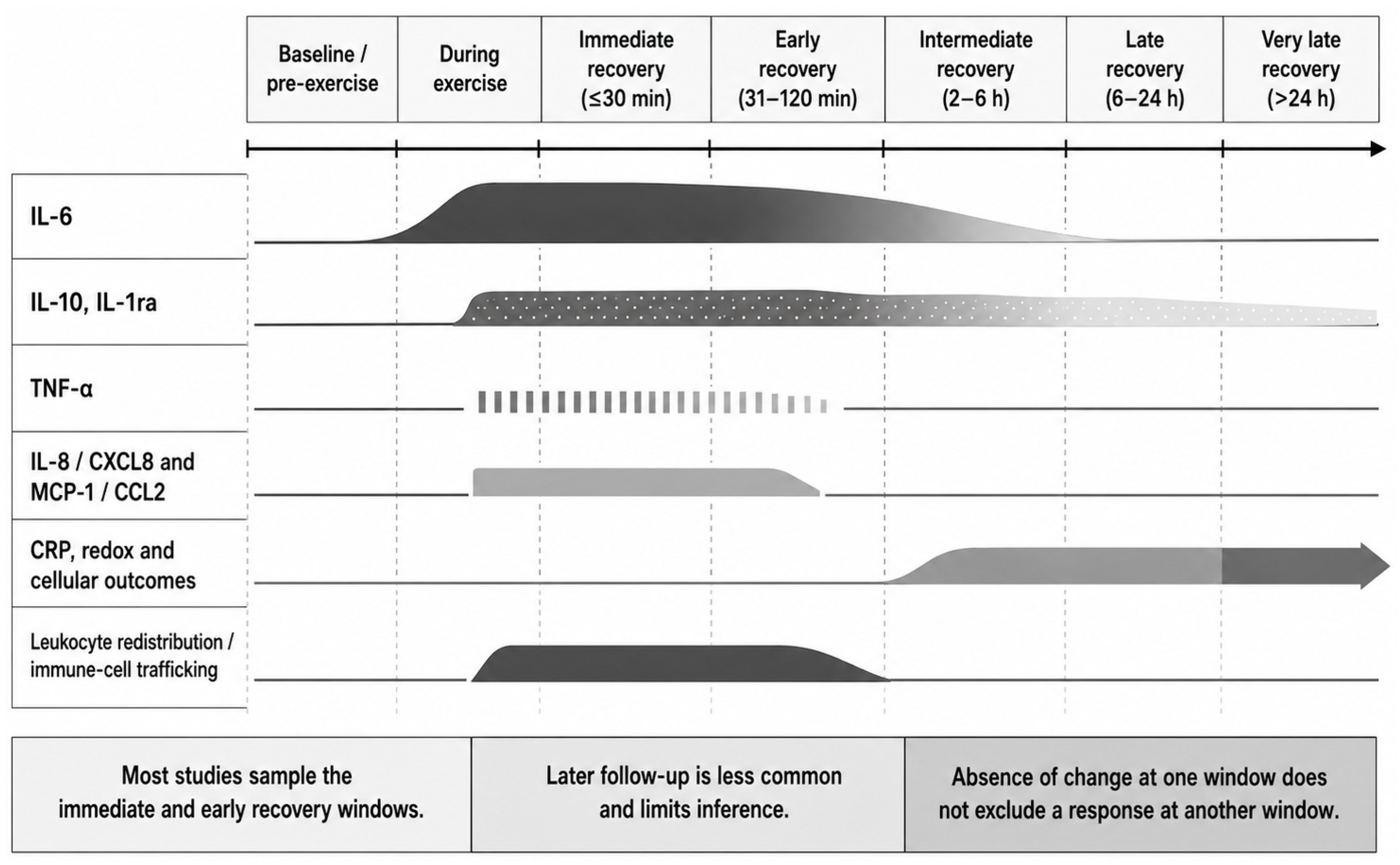
3.9. Acute Inflammatory Cytokine Responses and the Role of Time Windows
3.10. Oxidative Stress and Redox Biology
3.11. Cellular and Functional Immune Outcomes
3.12. Beyond Inflammation: Cardiometabolic, Vascular, Appetite, and Neurobiological Outcomes
3.13. Training Interventions
3.14. Circadian and Temporal Biological Regulation of Cytokine Responses
3.15. Limitations of the Evidence Base and Implications for Practice and Research
4. Conclusions
Author Contributions
Funding
Institutional Review Board Statement
Informed Consent Statement
Data Availability Statement
Conflicts of Interest
Abbreviations
| HIIT | High-intensity interval training |
| CRP | C-reactive protein |
References
- Petersen, A.M.W.; Pedersen, B.K. The Anti-Inflammatory Effect of Exercise. J. Appl. Physiol. 2005, 98, 1154–1162. [Google Scholar] [CrossRef] [Scilit]
- Petersen, A.M.W.; Pedersen, B.K. The Role of IL-6 in Mediating the Anti-Inflammatory Effects of Exercise. J. Physiol. Pharmacol. 2006, 57, 43–51. [Google Scholar]
- Nieman, D.C.; Wentz, L.M. The Compelling Link between Physical Activity and the Body’s Defense System. J. Sport Health Sci. 2019, 8, 201–217. [Google Scholar] [CrossRef] [Scilit]
- Brown, W.M.C.; Davison, G.W.; McClean, C.M.; Murphy, M.H. A Systematic Review of the Acute Effects of Exercise on Immune and Inflammatory Indices in Untrained Adults. Sports Med. Open 2015, 1, 35. [Google Scholar] [CrossRef] [Scilit]
- MacInnis, M.J.; Gibala, M.J. Physiological Adaptations to Interval Training and the Role of Exercise Intensity. J. Physiol. 2017, 595, 2915–2930. [Google Scholar] [CrossRef] [Scilit]
- Ploeger, H.E.; Takken, T.; de Greef, M.H.G.; Timmons, B.W. The Effects of Acute and Chronic Exercise on Inflammatory Markers in Children and Adults with a Chronic Inflammatory Disease: A Systematic Review. Exerc. Immunol. Rev. 2009, 15, 6–41. [Google Scholar] [PubMed]
- Kramer, H.F.; Goodyear, L.J. Exercise, MAPK, and NF-ΚB Signaling in Skeletal Muscle. J. Appl. Physiol. 2007, 103, 388–395. [Google Scholar] [CrossRef] [Scilit] [PubMed]
- Gibala, M.J.; McGee, S.L.; Garnham, A.P.; Howlett, K.F.; Snow, R.J.; Hargreaves, M. Brief Intense Interval Exercise Activates AMPK and P38 MAPK Signaling and Increases the Expression of PGC-1α in Human Skeletal Muscle. J. Appl. Physiol. 2009, 106, 929–934. [Google Scholar] [CrossRef] [Scilit]
- Ji, L.L.; Kang, C.; Zhang, Y. Exercise-Induced Hormesis and Skeletal Muscle Health. Free Radic. Biol. Med. 2016, 98, 113–122. [Google Scholar] [CrossRef] [Scilit] [PubMed]
- Radak, Z.; Ishihara, K.; Tekus, E.; Varga, C.; Posa, A.; Balogh, L.; Boldogh, I.; Koltai, E. Exercise, Oxidants, and Antioxidants Change the Shape of the Bell-Shaped Hormesis Curve. Redox Biol. 2017, 12, 285–290. [Google Scholar] [CrossRef] [Scilit]
- Pedersen, B.K.; Steensberg, A.; Fischer, C.; Keller, C.; Ostrowski, K.; Schjerling, P. Exercise and Cytokines with Particular Focus on Muscle-Derived IL-6. Exerc. Immunol. Rev. 2001, 7, 18–31. [Google Scholar] [PubMed]
- Steensberg, A.; Febbraio, M.A.; Osada, T.; Schjerling, P.; van Hall, G.; Saltin, B.; Pedersen, B.K. Interleukin-6 Production in Contracting Human Skeletal Muscle Is Influenced by Pre-exercise Muscle Glycogen Content. J. Physiol. 2001, 537, 633–639. [Google Scholar] [CrossRef] [Scilit]
- Croft, L.; Bartlett, J.D.; MacLaren, D.P.M.; Reilly, T.; Evans, L.; Mattey, D.L.; Nixon, N.B.; Drust, B.; Morton, J.P. High-Intensity Interval Training Attenuates the Exercise-Induced Increase in Plasma IL-6 in Response to Acute Exercise. Appl. Physiol. Nutr. Metab. 2009, 34, 1098–1107. [Google Scholar] [CrossRef] [Scilit] [PubMed]
- Ferrandi, P.J.; Fico, B.G.; Whitehurst, M.; Zourdos, M.C.; Bao, F.; Dodge, K.M.; Rodriguez, A.L.; Pena, G.; Huang, C.-J. Acute High-Intensity Interval Exercise Induces Comparable Levels of Circulating Cell-Free DNA and Interleukin-6 in Obese and Normal-Weight Individuals. Life Sci. 2018, 202, 161–166. [Google Scholar] [CrossRef] [Scilit]
- Pedersen, B.K.; Febbraio, M.A. Muscles, Exercise and Obesity: Skeletal Muscle as a Secretory Organ. Nat. Rev. Endocrinol. 2012, 8, 457–465. [Google Scholar] [CrossRef] [Scilit] [PubMed]
- Pedersen, B.K.; Edward, F. Adolph Distinguished Lecture: Muscle as an Endocrine Organ: IL-6 and Other Myokines. J. Appl. Physiol. 2009, 107, 1006–1014. [Google Scholar] [CrossRef] [Scilit]
- Steensberg, A.; van Hall, G.; Osada, T.; Sacchetti, M.; Saltin, B.; Pedersen, B.K. Production of Interleukin-6 in Contracting Human Skeletal Muscles Can Account for the Exercise-induced Increase in Plasma Interleukin-6. J. Physiol. 2000, 529, 237–242. [Google Scholar] [CrossRef] [Scilit]
- Pedersen, B.K.; Febbraio, M.A. Muscle as an Endocrine Organ: Focus on Muscle-Derived Interleukin-6. Physiol. Rev. 2008, 88, 1379–1406. [Google Scholar] [CrossRef] [Scilit]
- Steensberg, A.; Fischer, C.P.; Keller, C.; Møller, K.; Pedersen, B.K. IL-6 Enhances Plasma IL-1ra, IL-10, and Cortisol in Humans. Am. J. Physiol. Endocrinol. Metab. 2003, 285, E433–E437. [Google Scholar] [CrossRef] [Scilit]
- Starkie, R.; Ostrowski, S.R.; Jauffred, S.; Febbraio, M.; Pedersen, B.K. Exercise and IL-6 Infusion Inhibit Endotoxin-induced TNF-α Production in Humans. FASEB J. 2003, 17, 1–10. [Google Scholar] [CrossRef] [Scilit]
- Nielsen, A.R.; Pedersen, B.K. The Biological Roles of Exercise-Induced Cytokines: IL-6, IL-8, and IL-15. Appl. Physiol. Nutr. Metab. 2007, 32, 833–839. [Google Scholar] [CrossRef] [Scilit]
- Leggate, M.; Nowell, M.A.; Jones, S.A.; Nimmo, M.A. The Response of Interleukin-6 and Soluble Interleukin-6 Receptor Isoforms Following Intermittent High Intensity and Continuous Moderate Intensity Cycling. Cell Stress Chaperones 2010, 15, 827–833. [Google Scholar] [CrossRef] [Scilit] [PubMed]
- Benschop, R.J.; Rodriguez-Feuerhahn, M.; Schedlowski, M. Catecholamine-Induced Leukocytosis: Early Observations, Current Research, and Future Directions. Brain Behav. Immun. 1996, 10, 77–91. [Google Scholar] [CrossRef] [Scilit]
- Gibala, M.J.; Jones, A.M. Physiological and Performance Adaptations to High-Intensity Interval Training; Nestle Nutrition Institute Workshop Series; S. Karger AG: Basel, Switzerland, 2013; pp. 51–60. [Google Scholar]
- Tschakert, G.; Hofmann, P. High-Intensity Intermittent Exercise: Methodological and Physiological Aspects. Int. J. Sports Physiol. Perform. 2013, 8, 600–610. [Google Scholar] [CrossRef] [Scilit]
- Andreato, L.V. High-Intensity Interval Training: Methodological Considerations for Interpreting Results and Conducting Research. Trends Endocrinol. Metab. 2020, 31, 812–817. [Google Scholar] [CrossRef] [Scilit]
- Cullen, T.; Thomas, A.W.; Webb, R.; Hughes, M.G. Interleukin-6 and Associated Cytokine Responses to an Acute Bout of High-Intensity Interval Exercise: The Effect of Exercise Intensity and Volume. Appl. Physiol. Nutr. Metab. 2016, 41, 803–808. [Google Scholar] [CrossRef] [Scilit]
- Meckel, Y.; Eliakim, A.; Seraev, M.; Zaldivar, F.; Cooper, D.M.; Sagiv, M.; Nemet, D. The Effect of a Brief Sprint Interval Exercise on Growth Factors and Inflammatory Mediators. J. Strength Cond. Res. 2009, 23, 225–230. [Google Scholar] [CrossRef] [Scilit]
- Hoekstra, S.P.; Bishop, N.C.; Leicht, C.A. Can Intervals Enhance the Inflammatory Response and Enjoyment in Upper-Body Exercise? Eur. J. Appl. Physiol. 2017, 117, 1155–1163. [Google Scholar] [CrossRef] [Scilit] [PubMed]
- Docherty, S.; Harley, R.; McAuley, J.J.; Crowe, L.A.N.; Pedret, C.; Kirwan, P.D.; Siebert, S.; Millar, N.L. The Effect of Exercise on Cytokines: Implications for Musculoskeletal Health: A Narrative Review. BMC Sports Sci. Med. Rehabil. 2022, 14, 5. [Google Scholar] [CrossRef] [Scilit] [PubMed]
- Khalafi, M.; Symonds, M.E. The Impact of High-intensity Interval Training on Inflammatory Markers in Metabolic Disorders: A Meta-analysis. Scand. J. Med. Sci. Sports 2020, 30, 2020–2036. [Google Scholar] [CrossRef] [Scilit]
- Martland, R.; Mondelli, V.; Gaughran, F.; Stubbs, B. Can High-Intensity Interval Training Improve Physical and Mental Health Outcomes? A Meta-Review of 33 Systematic Reviews across the Lifespan. J. Sports Sci. 2020, 38, 430–469. [Google Scholar] [CrossRef] [Scilit]
- Souza, D.; Vale, A.F.; Silva, A.; Araújo, M.A.S.; de Paula Júnior, C.A.; de Lira, C.A.B.; Ramirez-Campillo, R.; Martins, W.; Gentil, P. Acute and Chronic Effects of Interval Training on the Immune System: A Systematic Review with Meta-Analysis. Biology 2021, 10, 868. [Google Scholar] [CrossRef] [Scilit]
- Page, M.J.; McKenzie, J.E.; Bossuyt, P.M.; Boutron, I.; Hoffmann, T.C.; Mulrow, C.D.; Shamseer, L.; Tetzlaff, J.M.; Akl, E.A.; Brennan, S.E.; et al. The PRISMA 2020 Statement: An Updated Guideline for Reporting Systematic Reviews. Int. J. Surg. 2021, 88, 105906. [Google Scholar] [CrossRef] [PubMed]
- Abedelmalek, S.; Souissi, N.; Chtourou, H.; Denguezli, M.; Aouichaoui, C.; Ajina, M.; Aloui, A.; Dogui, M.; Haddouk, S.; Tabka, Z. Effects of Partial Sleep Deprivation on Proinflammatory Cytokines, Growth Hormone, and Steroid Hormone Concentrations During Repeated Brief Sprint Interval Exercise. Chronobiol. Int. 2013, 30, 502–509. [Google Scholar] [CrossRef] [Scilit] [PubMed]
- Afzalpour, M.; Nayebifar, S.; Kazemi, T.; Abtahi-Eivary, S.-H.; Mogharnasi, M. Determination of Atherosclerosis Markers Changes after HIIT and Ginger Consumption in Response to Acute Exercise in Overweight Women. J. Appl. Pharm. Sci. 2016, 6, 078–084. [Google Scholar] [CrossRef] [Scilit]
- Andersson, Å.; Olsson, M.C.; Torell, A.; Mogard, E.; Haglund, E. Effects on Serum Protein Levels from One Bout of High Intensity Interval Training in Individuals with Axial Spondyloarthritis and Controls. Immun. Inflamm. Dis. 2025, 13, e70305. [Google Scholar] [CrossRef] [Scilit]
- Brown, M.; McClean, C.M.; Davison, G.W.; Brown, J.C.W.; Murphy, M.H. The Acute Effects of Walking Exercise Intensity on Systemic Cytokines and Oxidative Stress. Eur. J. Appl. Physiol. 2018, 118, 2111–2120. [Google Scholar] [CrossRef] [Scilit]
- Brzezińska, P.; Kochanowicz, A.; Borkowska, A.; Moskal, B.; Grześkiewicz, K.; Stankiewicz, B.; Durzyńska, A.; Kochanowicz, M.; Hofman, J.; Antosiewicz, J.; et al. Ischemic Preconditioning Modulates Iron Metabolism, Acute Inflammatory and Neurotrophic Responses to Anaerobic Exercise in Untrained Individuals: A Randomized Controlled Trial. Sci. Rep. 2026, 16, 7258. [Google Scholar] [CrossRef] [Scilit]
- Cabral-Santos, C.; Gerosa-Neto, J.; Inoue, D.S.; Panissa, V.L.G.; Gobbo, L.A.; Zagatto, A.M.; Campos, E.Z.; Lira, F.S. Similar Anti-Inflammatory Acute Responses from Moderate-Intensity Continuous and High-Intensity Intermittent Exercise. J. Sports Sci. Med. 2015, 14, 849–856. [Google Scholar] [PubMed]
- Cabral-Santos, C.; Castrillón, C.I.M.; Miranda, R.A.T.; Monteiro, P.A.; Inoue, D.S.; Campos, E.Z.; Hofmann, P.; Lira, F.S. Inflammatory Cytokines and BDNF Response to High-Intensity Intermittent Exercise: Effect the Exercise Volume. Front. Physiol. 2016, 7, 509, Erratum in Front. Physiol. 2017, 7, 662. [Google Scholar] [CrossRef] [Scilit]
- Casuso, R.A.; Aragon-Vela, J.; Huertas, J.R.; Ruiz-Ariza, A.; Martínez-Lopez, E.J. Comparison of the Inflammatory and Stress Response between Sprint Interval Swimming and Running. Scand. J. Med. Sci. Sports 2018, 28, 1371–1378. [Google Scholar] [CrossRef] [Scilit]
- Collins, B.E.G.; Hartmann, T.E.; Marino, F.E.; Skein, M. A Comparison of Acute High- and Moderate-Intensity Exercise on Cardio- Metabolic Function and Sleep Among Shift Workers. J. Sci. Sport Exerc. 2024, 6, 35–43. [Google Scholar] [CrossRef] [Scilit]
- Cruz, R.; Koch, S.; Matsuda, M.; Marquezini, M.; Sforça, M.L.; Lima-Silva, A.E.; Saldiva, P.; Koehle, M.; Bertuzzi, R. Air Pollution and High-Intensity Interval Exercise: Implications to Anti-Inflammatory Balance, Metabolome and Cardiovascular Responses. Sci. Total Environ. 2022, 809, 151094. [Google Scholar] [CrossRef] [Scilit]
- Dorneles, G.P.; Haddad, D.O.; Fagundes, V.O.; Vargas, B.K.; Kloecker, A.; Romão, P.R.T.; Peres, A. High Intensity Interval Exercise Decreases IL-8 and Enhances the Immunomodulatory Cytokine Interleukin-10 in Lean and Overweight–Obese Individuals. Cytokine 2016, 77, 1–9. [Google Scholar] [CrossRef] [Scilit] [PubMed]
- Durrer, C.; Francois, M.; Neudorf, H.; Little, J.P. Acute High-Intensity Interval Exercise Reduces Human Monocyte Toll-like Receptor 2 Expression in Type 2 Diabetes. Am. J. Physiol. Regul. Integr. Comp. Physiol. 2017, 312, R529–R538. [Google Scholar] [CrossRef] [Scilit] [PubMed]
- Dzik, K.P.; Grzywacz, T.; Łuszczyk, M.; Kujach, S.; Flis, D.J.; Kaczor, J.J. Single Bout of Exercise Triggers the Increase of Vitamin D Blood Concentration in Adolescent Trained Boys: A Pilot Study. Sci. Rep. 2022, 12, 1825. [Google Scholar] [CrossRef] [Scilit] [PubMed]
- Gassner, M.; Balaskovits, P.; Melchior, P.; Wagner, M.; Wagner, K.-H.; König, D. Reactive Oxygen Species and IL-6 Responses to Resistance-Based HIIT: A Time-Dependent Analysis. Free Radic. Biol. Med. 2026, 242, 250–257. [Google Scholar] [CrossRef] [Scilit]
- Gerosa-Neto, J.; Monteiro, P.A.; Inoue, D.S.; Antunes, B.M.; Batatinha, H.; Dorneles, G.P.; Peres, A.; Rosa-Neto, J.C.; Lira, F.S. High- and Moderate-Intensity Training Modify LPS-Induced Ex-Vivo Interleukin-10 Production in Obese Men in Response to an Acute Exercise Bout. Cytokine 2020, 136, 155249. [Google Scholar] [CrossRef] [Scilit]
- Gökbel, H.; Okudan, N.; Gül, İ.; Belviranli, M.; Gergerlioğlu, H.S.; BaŞaral, M.K. Effects of Repeated Bouts of Supramaximal Exercise on Plasma Adiponectin, Interleukin-6, and Tumor Necrosis Factor-α Levels in Sedentary Men. J. Strength Cond. Res. 2012, 26, 1675–1679. [Google Scholar] [CrossRef] [Scilit]
- Hall, B.; Żebrowska, A.; Sikora, M.; Siatkowski, S.; Robins, A. The Effect of High-Intensity Interval Exercise on Short-Term Glycaemic Control, Serum Level of Key Mediator in Hypoxia and Pro-Inflammatory Cytokines in Patients with Type 1 Diabetes—An Exploratory Case Study. Nutrients 2023, 15, 3749. [Google Scholar] [CrossRef] [Scilit]
- Herranz-López, M.; Agustín, R.M.-S.; Olivares-Vicente, M.; Pardines-Oltra, L.; Del Pino, A.C.; Bondia, J.; Rossetti, P.; Ampudia-Blasco, F.J.; Roche, E. Patterns of Circulating Cytokines after 2 Types of Exercise in Active Type-1 Diabetic Patients. J. Physiol. Biochem. 2025, 81, 1335–1344. [Google Scholar] [CrossRef] [Scilit]
- Henke, E.; Oliveira, V.S.; da Silva, I.M.; Schipper, L.; Dorneles, G.; Elsner, V.R.; de Oliveira, M.R.; Romão, P.R.T.; Peres, A. Acute and Chronic Effects of High Intensity Interval Training on Inflammatory and Oxidative Stress Markers of Postmenopausal Obese Women. Transl. Sports Med. 2018, 1, 257–264. [Google Scholar] [CrossRef] [Scilit]
- Ellyas, I.S.; Sugiyanto, S.; Hidayatullah, M.F.; Doewes, M.; Doewes, R.I.; Umar, F. The Effects of High-Intensity Interval Training on Cardiorespiratory Fitness and IL-6 in Adolescents. Int. J. Hum. Mov. Sports Sci. 2021, 9, 568–576. [Google Scholar] [CrossRef] [Scilit]
- Jakobsson, J.; De Brandt, J.; Hedlund, M.; Rullander, A.-C.; Sandström, T.; Nyberg, A. Feasibility and Acute Physiological Responses to Supramaximal High-Intensity Interval Training in COPD: A Randomised Crossover Trial. ERJ Open Res. 2025, 11, 01321–02024. [Google Scholar] [CrossRef] [Scilit]
- Kaspar, F.; Jelinek, H.F.; Perkins, S.; Al-Aubaidy, H.A.; deJong, B.; Butkowski, E. Acute-Phase Inflammatory Response to Single-Bout HIIT and Endurance Training: A Comparative Study. Mediat. Inflamm. 2016, 2016, 5474837. [Google Scholar] [CrossRef] [Scilit]
- Kon, M.; Ebi, Y.; Nakagaki, K. Effects of a Single Bout of High-Intensity Interval Exercise on C1q/TNF-Related Proteins. Appl. Physiol. Nutr. Metab. 2019, 44, 47–51. [Google Scholar] [CrossRef] [Scilit]
- Kon, M.; Taniguchi, K.; Ebi, Y.; Nakagaki, K. Effects of High-Intensity Interval Exercise under Hyperoxia on HSP27 and Oxidative Stress Responses. Respir. Physiol. Neurobiol. 2021, 283, 103544. [Google Scholar] [CrossRef] [Scilit]
- Lira, F.S.; dos Santos, T.; Caldeira, R.S.; Inoue, D.S.; Panissa, V.L.G.; Cabral-Santos, C.; Campos, E.Z.; Rodrigues, B.; Monteiro, P.A. Short-Term High- and Moderate-Intensity Training Modifies Inflammatory and Metabolic Factors in Response to Acute Exercise. Front. Physiol. 2017, 8, 856. [Google Scholar] [CrossRef] [Scilit]
- Meyer, T.; Gabriel, H.H.; Rätz, M.; Müller, H.J.; Kindermann, W. Anaerobic Exercise Induces Moderate Acute Phase Response. Med. Sci. Sports Exerc. 2001, 33, 549–555. [Google Scholar] [CrossRef] [Scilit] [PubMed]
- Minuzzi, L.G.; Lira, F.S.; de Poli, R.A.B.; Fialho Lopes, V.H.; Zagatto, A.M.; Suzuki, K.; Antunes, B.M. High-Intensity Intermittent Exercise Induces a Potential Anti-Inflammatory Response in Healthy Women across the Menstrual Cycle. Cytokine 2022, 154, 155872. [Google Scholar] [CrossRef] [Scilit]
- Monteiro, P.A.; Freitas Junior, I.F.; Zagatto, A.M.; Ribeiro, J.P.J.; Cabral-Santos, C.; Inoue, D.S.; Gerosa-Neto, J.; Lira, F.S. Acute Effect of High-Intensity Interval Training on Metabolic and Inflammatory Markers in Obese and Overweight Adolescents: Pilot Study. Eur. J. Inflamm. 2019, 17, 2058739219877710. [Google Scholar] [CrossRef] [Scilit]
- Narciso, P.H.; von Ah Morano, A.E.; Agostinete, R.R.; Werneck, A.O.; Giannopoulos, A.J.; Bell, M.; Antunes, B.M.; Lira, F.S.; Fernandes, R.A.; Klentrou, P. Cytokine and Adipokine Response Following High-Intensity Interval Running and Cycling in Female Adolescents. Eur. J. Appl. Physiol. 2025, 125, 3543–3552. [Google Scholar] [CrossRef] [Scilit]
- de Oliveira Ottone, V.; Costa, K.; Tossige-Gomes, R.; de Matos, M.; Brito-Melo, G.; Magalhaes, F.; Esteves, E.; Amorim, F.; Rocha-Vieira, E. Late Neutrophil Priming Following a Single Session of High-Intensity Interval Exercise. Int. J. Sports Med. 2019, 40, 171–179. [Google Scholar] [CrossRef] [Scilit]
- Panissa, V.L.G.; Julio, U.F.; St-Pierre, D.H.; Tavares da Silva Gomes, A.; Caldeira, R.S.; Lira, F.S.; Takito, M.Y.; Franchini, E. Timing of High-Intensity Intermittent Exercise Affects Ad Libitum Energy Intake in Overweight Inactive Men. Appetite 2019, 143, 104443. [Google Scholar] [CrossRef] [Scilit]
- Ren, Y.; Liu, Z.; Wang, M.; Wang, Y.; Wang, Y. Acute Immunometabolic Changes in First-Presentation Graves’ Hyperthyroidism Patients Undergoing Strenuous Physical Activity. Front. Endocrinol. 2025, 16, 1685496. [Google Scholar] [CrossRef] [Scilit] [PubMed]
- Rohnejad, B.; Monazzami, A. Effects of High-Intensity Intermittent Training on Some Inflammatory and Muscle Damage Indices in Overweight Middle-Aged Men. Apunt. Sports Med. 2023, 58, 100404. [Google Scholar] [CrossRef] [Scilit]
- Ruegsegger, G.N.; Ekholm, E.R.; Monroe, C.E.; Rappaport, C.I.; Huppert, R.D.; Anton, C.R.; Ferguson, M.J. Glucose Tolerance Status Associates with Improvements in Cognitive Function Following High-Intensity Exercise in Adults with Obesity. Physiol. Behav. 2023, 272, 114389. [Google Scholar] [CrossRef] [Scilit]
- Rusdiawan, A.; Widodo, A.; Lani, A.; Özman, C.; Putra, A.Y.; Susanto, N.; Munandar, R.A.; Kafrawi, M.F.; Irmawati, F. The Impact of Ice Compression and Massage Techniques on Lactate Removal and Interleukin-6 Concentrations During Halftime Recovery in Intermittent Sports. Phys. Educ. Theory Methodol. 2025, 25, 1033–1041. [Google Scholar] [CrossRef] [Scilit]
- Sasimontonkul, S.; Sirivarasai, J. The 40-Min HIIT Acutely Induced Bone Formation Which Was Likely through the Increases in Muscle Derived Interleukin 6 and Adiponectin Activation: The 16 Weeks of HIIT Intervention, Longitudinal Randomized Controlled Trial. Bone 2024, 184, 117105. [Google Scholar] [CrossRef] [Scilit]
- Siasos, G.; Athanasiou, D.; Terzis, G.; Stasinaki, A.; Oikonomou, E.; Tsitkanou, S.; Dimitropoulos, S.; Kolokytha, T.; Tzirogiannis, K.; Giannaki, A.; et al. The Acute Impact of Different Types of Aerobic Exercise on Arterial Wave Reflections and Inflammation. Cardiology 2016, 135, 81–86. [Google Scholar] [CrossRef] [Scilit] [PubMed]
- Sim, M.; Dawson, B.; Landers, G.; Swinkels, D.W.; Tjalsma, H.; Trinder, D.; Peeling, P. Effect of Exercise Modality and Intensity on Postexercise Interleukin-6 and Hepcidin Levels. Int. J. Sport Nutr. Exerc. Metab. 2013, 23, 178–186. [Google Scholar] [CrossRef] [Scilit]
- Uchida, M.; Kojima, C.; Hashimoto, T.; Watanabe, K.; Nishikawa, T.; Isaka, T.; Iemitsu, M. A Pilot Study: Salivary Human Herpesvirus-6 and Human Herpesvirus-7 Responses to Different Types of Acute Exercise in Healthy Young Men. Physiol. Rep. 2025, 13, e70697. [Google Scholar] [CrossRef] [Scilit]
- Vardar, S.A.; Doğanlar, Z.B.; Kaya, O.; Tayfur, P.; Sut, N.; Doğanlar, O. Different Responses of Apoptotic, Inflammatory and Heat Shock Protein Gene Expression to a Single Bout of High-Intensity Interval Exercise between Physically Active and Inactive Men. Appl. Physiol. Nutr. Metab. 2021, 46, 743–752. [Google Scholar] [CrossRef] [Scilit]
- Verbickas, V.; Baranauskiene, N.; Eimantas, N.; Kamandulis, S.; Rutkauskas, S.; Satkunskiene, D.; Sadauskas, S.; Brazaitis, M.; Skurvydas, A. Effect of Sprint Cycling and Stretch-Shortening Cycle Exercises on the Neuromuscular, Immune and Stress Indicators in Young Men. J. Physiol. Pharmacol. 2017, 68, 125–132. [Google Scholar]
- Wadley, A.J.; Keane, G.; Cullen, T.; James, L.; Vautrinot, J.; Davies, M.; Hussey, B.; Hunter, D.J.; Mastana, S.; Holliday, A.; et al. Characterization of Extracellular Redox Enzyme Concentrations in Response to Exercise in Humans. J. Appl. Physiol. 2019, 127, 858–866. [Google Scholar] [CrossRef] [Scilit]
- Windsor, M.T.; Bailey, T.G.; Perissiou, M.; Meital, L.; Golledge, J.; Russell, F.D.; Askew, C.D. Cytokine Responses to Acute Exercise in Healthy Older Adults: The Effect of Cardiorespiratory Fitness. Front. Physiol. 2018, 9, 203. [Google Scholar] [CrossRef] [Scilit]
- Zwetsloot, K.; John, C.; Lawrence, M.; Battista, R.; Shanely, R.A. High-Intensity Interval Training Induces a Modest Systemic Inflammatory Response in Active, Young Men. J. Inflamm. Res. 2014, 7, 9–17. [Google Scholar] [CrossRef] [Scilit]
- Dimitrov, S.; Benedict, C.; Heutling, D.; Westermann, J.; Born, J.; Lange, T. Cortisol and Epinephrine Control Opposing Circadian Rhythms in T Cell Subsets. Blood 2009, 113, 5134–5143. [Google Scholar] [CrossRef] [Scilit]
- Nilsonne, G.; Lekander, M.; Åkerstedt, T.; Axelsson, J.; Ingre, M. Diurnal Variation of Circulating Interleukin-6 in Humans: A Meta-Analysis. PLoS ONE 2016, 11, e0165799. [Google Scholar] [CrossRef] [Scilit]
- Kim, H.-K.; Konishi, M.; Takahashi, M.; Tabata, H.; Endo, N.; Numao, S.; Lee, S.-K.; Kim, Y.-H.; Suzuki, K.; Sakamoto, S. Effects of Acute Endurance Exercise Performed in the Morning and Evening on Inflammatory Cytokine and Metabolic Hormone Responses. PLoS ONE 2015, 10, e0137567. [Google Scholar] [CrossRef] [Scilit]
- Pledge, D.; Grosset, J.-F.; Onambélé-Pearson, G.L. Is There a Morning-to-Evening Difference in the Acute IL-6 and Cortisol Responses to Resistance Exercise? Cytokine 2011, 55, 318–323. [Google Scholar] [CrossRef] [Scilit]
- Powers, S.K.; Duarte, J.; Kavazis, A.N.; Talbert, E.E. Reactive Oxygen Species Are Signalling Molecules for Skeletal Muscle Adaptation. Exp. Physiol. 2010, 95, 1–9. [Google Scholar] [CrossRef] [Scilit]
- Bartlett, J.D.; Hwa Joo, C.; Jeong, T.-S.; Louhelainen, J.; Cochran, A.J.; Gibala, M.J.; Gregson, W.; Close, G.L.; Drust, B.; Morton, J.P. Matched Work High-Intensity Interval and Continuous Running Induce Similar Increases in PGC-1α MRNA, AMPK, P38, and P53 Phosphorylation in Human Skeletal Muscle. J. Appl. Physiol. 2012, 112, 1135–1143. [Google Scholar] [CrossRef] [Scilit]
- Keller, C.; Steensberg, A.; Pilegaard, H.; Osada, T.; Saltin, B.; Pedersen, B.K.; Neufer, P.D. Transcriptional Activation of the IL-6 Gene in Human Contracting Skeletal Muscle: Influence of Muscle Glycogen Content. FASEB J. 2001, 15, 1–15. [Google Scholar] [CrossRef] [Scilit]
- MacDonald, C.; Wojtaszewski, J.F.P.; Pedersen, B.K.; Kiens, B.; Richter, E.A. Interleukin-6 Release from Human Skeletal Muscle during Exercise: Relation to AMPK Activity. J. Appl. Physiol. 2003, 95, 2273–2277. [Google Scholar] [CrossRef] [Scilit]
- Whitham, M.; Chan, M.H.S.; Pal, M.; Matthews, V.B.; Prelovsek, O.; Lunke, S.; El-Osta, A.; Broenneke, H.; Alber, J.; Brüning, J.C.; et al. Contraction-Induced Interleukin-6 Gene Transcription in Skeletal Muscle Is Regulated by c-Jun Terminal Kinase/Activator Protein-1. J. Biol. Chem. 2012, 287, 10771–10779. [Google Scholar] [CrossRef] [Scilit]
- van Hall, G.; Steensberg, A.; Sacchetti, M.; Fischer, C.; Keller, C.; Schjerling, P.; Hiscock, N.; Møller, K.; Saltin, B.; Febbraio, M.A.; et al. Interleukin-6 Stimulates Lipolysis and Fat Oxidation in Humans. J. Clin. Endocrinol. Metab. 2003, 88, 3005–3010. [Google Scholar] [CrossRef] [Scilit]
- Carey, A.L.; Steinberg, G.R.; Macaulay, S.L.; Thomas, W.G.; Holmes, A.G.; Ramm, G.; Prelovsek, O.; Hohnen-Behrens, C.; Watt, M.J.; James, D.E.; et al. Interleukin-6 Increases Insulin-Stimulated Glucose Disposal in Humans and Glucose Uptake and Fatty Acid Oxidation In Vitro via AMP-Activated Protein Kinase. Diabetes 2006, 55, 2688–2697. [Google Scholar] [CrossRef] [Scilit]
- O’Byrne, N.A.; Yuen, F.; Butt, W.Z.; Liu, P.Y. Sleep and Circadian Regulation of Cortisol: A Short Review. Curr. Opin. Endocr. Metab. Res. 2021, 18, 178–186. [Google Scholar] [CrossRef] [Scilit]



| Study | Participants | Design and Comparison | Cytokine/Mediator Scope and Prioritization | Pre-Analytical Control | Blood Matrix and Assay Reporting | Sampling Schedule and Recovery Window Coverage |
|---|---|---|---|---|---|---|
| Abedelmalek et al., [35] | 13 male football players; age 21.1 years (19–24); body mass index 22.6 kg per square meter (18.47–24.46) | Within-participant crossover; baseline sleep vs. partial sleep deprivation; morning session at 08:00 | Targeted outcomes: Interleukin 6; tumor necrosis factor alpha | Control level: Stronger standardization Feeding: Before morning testing, only one glass of water was allowed; no food or stimulant intake was allowed after early awakening in the partial sleep deprivation condition Time: Testing performed at 08:00 Recent/stimulants: Participants were nonsmokers and did not consume caffeine or alcoholic beverages; standard eating times were required before the study Sleep/circadian: Participants slept in the laboratory for four consecutive nights before assessment; sleep was monitored by wrist actigraphy | Matrix ambiguity; Detection/CV incomplete Matrix: Matrix reporting is inconsistent: cytokine results are described as plasma concentrations, while the methods describe serum tubes; Assay: Commercial enzyme-linked immunosorbent assay kits for interleukin 6 and tumor necrosis factor alpha Detection/precision: Intra-assay coefficients of variation: interleukin 6, 1.6 to 6.8 percent; tumor necrosis factor alpha, 1.6 to 10 percent. Inter-assay coefficients of variation: interleukin 6, 7. Plasma-volume correction: Corrected using the method of Costill and Fink | Coverage: Immediate/early only Schedule: Before exercise after 15 min of rest; after the first run; after the fourth run; 60 min after exercise Mapped windows: During exercise; immediate recovery; early recovery |
| Afzalpour et al. [36] | 24 sedentary overweight women; age 20–30 years; body mass index greater than 25 kg per square meter; 3 groups (8 per group) | Randomized placebo-controlled parallel groups; acute exercise tested before training and 72 h after last training session | Targeted outcomes: Intercellular adhesion molecule 1; monocyte chemoattractant protein 1; interleukin 10 | Control level: Limited/unclear standardization Feeding: Participants arrived after 10 to 12 h of fasting Time: Training sessions were performed at 08:00; acute challenge timing was not explicitly stated Recent/stimulants: Exclusion criteria included smoking, medication use, supplementation with ginger, antioxidant, or multivitamin products; Sex/hormonal: Samples were collected while participants were in the follicular phase of the menstrual cycle | Matrix specified; Detection/CV incomplete Matrix: Serum Assay: Commercial assay kits; the article does not explicitly name the analytical platform Detection/precision: Intra-assay coefficients of variation: intercellular adhesion molecule 1, less than 8 percent; monocyte chemoattractant protein 1, less than 8 percent; interleukin 10, equal to 3. Plasma-volume correction: Corrected using the method of Dill and Costill | Coverage: Immediate only Schedule: Immediately before and immediately after the acute exercise challenge; repeated before the intervention and 72 h after the final training session Mapped windows: Immediate recovery |
| Andersson et al. [37] | 21 adults (10 with axial spondyloarthritis, 11 healthy controls); 10 women; age 18–50 years, mean 40 years; most patients used anti-inflammatory medication | Controlled pre–post pilot study; axial spondyloarthritis versus age- and sex-matched healthy controls; single acute session | Broad/exploratory panel: Interleukin 6; interleukin 17; interleukin 18; tumor necrosis factor alpha; C-X-C motif chemokine ligand 10; vascular endothelial growth factor A; C-reactive protein | Control level: Partial standardization Feeding: Participants were asked to avoid food and drinks for two hours before arrival Time: Time of day was not explicitly reported Recent/stimulants: Participants were asked to avoid tobacco for 30 min before arrival and high-intensity exercise the evening before testing Sex/hormonal: Not reported | Matrix specified; Detection/CV reported Matrix: Serum Assay: Luminex MAGPIX for cytokine, chemokine, myokine, and bone-panel analytes; Detection/precision: Lowest detection limits reported for all analytes: interleukin 6, 0.18 picograms per milliliter; interleukin 17, 0.89 picograms per milliliter; interleukin 18, 0. Plasma-volume correction: Not reported | Coverage: Early only Schedule: Before exercise and one hour after the high-intensity interval bout ended Mapped windows: Early recovery |
| Brown et al. [38] | 17 healthy recreationally active men; age 22.6 ± 4.6 years; maximal oxygen uptake 53.7 ± 7.1 milliliters per kilogram per minute; nonsmokers; no medication | Randomized crossover; high-intensity intermittent walking versus continuous moderate walking; 7-day washout | Targeted outcomes: Interleukin 6; tumor necrosis factor alpha | Control level: Partial standardization Feeding: Participants completed testing after a standard ten-hour overnight fast and replicated dietary intake before the second trial Time: Time of day was not explicitly reported Recent/stimulants: Participants refrained from exercise and alcohol consumption for 24 h before each trial | Matrix specified; Detection/CV incomplete Matrix: Plasma for interleukin 6 and tumor necrosis factor alpha; serum for endothelin 1, lipid hydroperoxides, and hydrogen peroxide; plasma for ascorbyl radical and lipid-soluble antioxidants Assay: Enzyme-linked immunosorbent assay for interleukin 6, tumor necrosis factor alpha, and endothelin 1; ferrous iron xylenol orange assay for lipid hydroperoxides Detection/precision: Assay detection limits and coefficients of variation were not clearly reported for the inflammatory mediators in the extracted text Plasma-volume correction: Not reported | Coverage: Includes very late (>24 h) Schedule: Before exercise; immediately after exercise; 2 h, 4 h, 24 h, and 48 h after exercise Mapped windows: Immediate recovery; early recovery; intermediate recovery; late recovery; very late recovery |
| Brzezinska et al. [39] | 28 healthy untrained young men analyzed; mean age 20.3 years; randomized to ischemic preconditioning (15) or sham (13) | Single-blind randomized controlled parallel groups; ischemic preconditioning versus sham; acute exercise tested before and after 14-day intervention | Broad/exploratory panel: Interleukin 6; interleukin 10; interleukin 15; leukemia inhibitory factor; growth differentiation factor 15; follistatin-like 1 | Control level: Partial standardization Feeding: Participants followed a standardized eating pattern before the study; intervention sessions were performed before any meal Time: Testing was described as early morning; cuff intervention sessions were performed between 08:00 and 10:00 Recent/stimulants: Participants abstained for one month from alcohol and substances that could influence performance, including caffeine, guarana, theine, and chocolate | Matrix specified; Detection/CV incomplete. Matrix: Serum Assay: MAGPIX fluorescence-based detection system with Luminex assay kits for inflammatory and neurotrophic markers Detection/precision: Assay detection limits and coefficients of variation were not clearly reported for the inflammatory and neurotrophic markers in the extracted text Plasma-volume correction: Not reported | Coverage: Immediate/early only Schedule: Immediately before, directly after within 5 min, and 2 h after the double Wingate anaerobic test; repeated before and after the fourteen-day intervention Mapped windows: Immediate recovery; early recovery |
| Cabral-Santos et al. [40] | 8 physically active men; age 24.6 years; body mass index 24.3 kg per square meter; peak oxygen uptake 59.9 milliliters per kilogram per minute | Randomized crossover; high-intensity intermittent exercise versus continuous moderate exercise; sessions separated by at least 72 h | Focused multi-mediator set: Interleukin 6; interleukin 10; tumor necrosis factor alpha; interleukin 10 to tumor necrosis factor alpha ratio | Control level: Stronger standardization Feeding: Participants were asked to abstain from eating or drinking for two hours before testing and to maintain their nutritional and hydration routines Time: All tests took place at the same time of day for each participant Recent/stimulants: Participants were instructed to abstain from strenuous exercise for at least 24 h before each testing session | Mixed matrices; Detection/CV incomplete Matrix: Plasma and serum were collected; cytokine matrix was not explicitly specified Assay: Enzyme-linked immunosorbent assay commercial kits for interleukin 6, interleukin 10, and tumor necrosis factor alpha Detection/precision: Assay precision and detection limits were not clearly reported Plasma-volume correction: Not reported | Coverage: Immediate/early only Schedule: At rest; immediately after exercise; 30 min after exercise; 60 min after exercise Mapped windows: Immediate recovery; early recovery |
| Cabral-Santos et al. [41] | 10 physically active men; age 25.22 ± 1.74 years; body mass index 24.85 kg per square meter; peak oxygen uptake 59.94 ± 9.38 milliliters per kilogram per minute | Randomized crossover; two high-intensity intermittent exercise volumes (1.25 km vs. 2.5 km); sessions separated by at least 72 h | Targeted outcomes: Interleukin 6; interleukin 10; monocyte chemoattractant protein 1 | Control level: Stronger standardization Feeding: Diet was not standardized, but participants were required to eat three hours before testing Time: Tests took place at the same time of day for each participant, between 10:00 and 12:00 Recent/stimulants: Participants were instructed to abstain from strenuous exercise for at least 24 h and avoid stimulants or alcoholic beverages before testing | Mixed matrices; Detection/CV incomplete Matrix: Serum in abstract; plasma and serum were collected and cytokine-specific matrix was not fully explicit Assay: Commercial enzyme-linked immunosorbent assay kits from R and D Systems for interleukin 6, interleukin 10, brain-derived neurotrophic factor, and monocyte chemo… Detection/precision: Assay precision and detection limits were not clearly reported in the extracted text Plasma-volume correction: Not reported | Coverage: Immediate/early only Schedule: At rest, immediately after exercise, and 60 min after exercise Mapped windows: Immediate recovery; early recovery |
| Casuso et al. [42] | 18 trained adults involved in swimming and running for at least 2 years; age 23 ± 4.2 years; maximal running oxygen uptake 67 ± 8.2 milliliters per kilogram per minute | Randomized crossover; sprint interval swimming versus sprint interval running; sessions separated by 7 to 14 days | Broad/exploratory panel: Interleukin 6; interleukin 10; tumor necrosis factor alpha | Control level: Stronger standardization. Feeding: Participants consumed a standardized breakfast at least one hour before arriving; no food was allowed during the two-hour recovery period Time: All procedures were performed in the morning Recent/stimulants: No strenuous exercise was permitted for 72 h before each trial Sex/hormonal: Not reported | Matrix specified; Detection/CV incomplete Matrix: Serum Assay: Milliplex MAP Human Cytokine/Chemokine Magnetic Bead Panel analyzed on a Luminex 200 system; all samples and standards analyzed in duplicate Detection/precision: Intra-assay coefficients of variation: interleukin 6, 5.5 percent; interleukin 10, 7.2 percent; tumor necrosis factor alpha, 6.1 percent. Plasma-volume correction: Not reported | Coverage: Immediate/early only Schedule: Before exercise after ten minutes of rest; approximately three minutes after exercise; two hours after exercise Mapped windows: Immediate recovery; early recovery |
| Collins et al. [43] | 26 sedentary male rotational shift workers; age 38 ± 8 years; body mass index 32.2 ± 6.0 kg per square meter; cytokine analyses n = 15 | Randomized parallel groups; high-intensity interval exercise versus continuous moderate exercise; morning trial after day off or day shift | Targeted outcomes: Interleukin 1 receptor antagonist; interleukin 6; tumor necrosis factor alpha | Control level: Stronger standardization Feeding: Fasted venous blood sampling was performed; participants reported for laboratory testing between 06:00 and 09:00 Time: Testing performed between 06:00 and 09:00 after a day off or day shift Recent/stimulants: Participants were sedentary, nonsmokers, and free from known cardiometabolic or sleep disorders; recent-exercise restrictions were not detailed in the extracted text Sleep/circadian: Participants wore actigraphy for approximately seven days before testing | Matrix specified; Detection/CV reported Matrix: Plasma for interleukin 6 and interleukin 1 receptor antagonist; serum for tumor necrosis factor alpha Assay: Commercial enzyme-linked immunosorbent assays Detection/precision: Minimum detectable cytokine levels: interleukin 6 less than 5 picograms per milliliter; tumor necrosis factor alpha 1.7 picograms per milliliter Plasma-volume correction: Not reported | Coverage: Immediate/early only Schedule: Before exercise; immediately after exercise; 30 min after exercise; 60 min after exercise Mapped windows: Immediate recovery; early recovery |
| Cullen et al. [27] | 10 healthy active adults; 5 men and 5 women; age 24 ± 4 years; maximal oxygen uptake 49 ± 5 milliliters per kilogram per minute | Counterbalanced within-participant repeated-measures study; LOW versus MOD versus HIGH; 3 sessions within 2 weeks, minimum 3 days apart | Targeted outcomes: Interleukin 6; interleukin 10; soluble interleukin 6 receptor | Control level: Stronger standardization Feeding: Participants refrained from eating or drinking other than water for two hours before testing Time: Participants arrived at the same time of day before each test Recent/stimulants: Participants maintained similar diet and activity; refrained from alcohol, caffeine, and strenuous exercise during the previous twenty-four hours Sleep/circadian: Not specifically a sleep or circadian intervention Sex/hormonal: Not reported beyond inclusion of both sexes; no menstrual-cycle control reported | Matrix specified; Detection/CV reporte. Matrix: Plasma for interleukin 6, interleukin 10, and soluble interleukin 6 receptor; whole blood for messenger ribonucleic acid expression Assay: High-sensitivity enzyme-linked immunosorbent assays for interleukin 6 and interleukin 10 Detection/precision: Interleukin 6 detection limit 0.039 picograms per milliliter; interleukin 6 intra-assay coefficient of variation 3.8 percent plus or minus 2.9 percent Plasma-volume correction: Not reported | Coverage: Immediate only Schedule: Immediately before and immediately after exercise Mapped windows: Baseline; immediate recovery |
| Cruz et al. [44] | 15 healthy physically active men; age 28.0 ± 6.0 years; body mass index 23.1 ± 1.7 kg per square meter; cytokine analyses n = 13 | Randomized single-blind repeated measures crossover; filtered air versus traffic-related air pollution; counterbalanced order | Broad/exploratory panel: Interleukin 6; interleukin 10; tumor necrosis factor alpha; vascular endothelial growth factor; interleukin 10 to interleukin 6 ratio; interleukin 10 to tumor necrosis factor alpha ratio | Control level: Stronger standardization Feeding: Participants replicated dietary intake for the 24 h before each trial and fasted for two hours before each session Time: Each participant performed the high-intensity interval exercise at the same time and day of week, separated by one week Recent/stimulants: Participants were asked to refrain from vigorous physical activity, caffeine, and alcohol for 48 h before every visit | Matrix specified; Detection/CV reported Matrix: Serum Assay: Custom 13-cytokine Milliplex MAP Human Cytokine/Chemokine Magnetic Bead Panel analyzed with Magpix xMAP technology Detection/precision: Standard-curve fit used for mean fluorescence intensity versus picograms per milliliter; coefficients of variation and detection limits were not specified in the extracted text Plasma-volume correction: Not reported | Coverage: Immediate/early only Schedule: Baseline; 10 min after exercise; 1 h after exercise Mapped windows: Immediate recovery; early recovery |
| Dorneles et al. [45] | 22 untrained healthy men; 10 lean and 12 overweight–obese; age 20–40 years; nonsmokers; no exercise training in prior 6 months | Randomized crossover; high-intensity interval exercise versus moderate-intensity interval exercise; analyzed by lean and overweight–obese subgroup; sessions at least 1 week apart | Broad/exploratory panel: Interleukin 1 receptor antagonist; interleukin 6; interleukin 8; interleukin 10; interleukin 17A; monocyte chemoattractant protein 1 | Control level: Stronger standardization Feeding: Participants arrived after at least one hour of fasting Time: All sessions performed between 09:30 and 10:30 Recent/stimulants: Participants refrained from alcohol, coffee, and vigorous physical activity for twenty-four hours; diet was recorded and repeated across exercise trials Sleep/circadian: Not specifically a sleep or circadian intervention | Matrix specified; Detection/CV incomplete Matrix: Serum for cytokines and muscle damage markers; whole blood for leukocyte counts Assay: Commercial enzyme-linked immunosorbent assays for interleukin 1 receptor antagonist, interleukin 6, interleukin 8, interleukin 10, interleukin 17A, and monocyte Detection/precision: Intra-assay coefficient of variation was less than 7.5 percent for cytokine assays; detection limits were not reported Plasma-volume correction: Adjusted using the Dill and Costill approach from hematocrit and hemoglobin | Coverage: Immediate only Schedule: Before exercise; immediately after exercise; thirty minutes after exercise Mapped windows: Baseline; immediate recovery |
| Durrer et al. [46] | 10 adults with type 2 diabetes (5 men, 5 women) and 9 age-matched healthy controls (4 men, 5 women) | Two-group time-series study; type 2 diabetes versus healthy controls; single acute high-intensity interval exercise session | Broad/exploratory panel: Tumor necrosis factor alpha; lipopolysaccharide-stimulated tumor necrosis factor alpha; toll-like receptor 2; toll-like receptor 4 | Control level: Stronger standardization Feeding: Exercise was performed four hours postprandial; water was provided ad libitum Time: Exercise began at either 11:00 or 16:00 Recent/stimulants: Participants refrained from exercise for forty-eight hours before the acute exercise trial; type 2 diabetes participants maintained their usual medication schedule Sleep/circadian: No sleep intervention; time of testing was reported Sex/hormonal: Mixed-sex study; no menstrual-cycle control reported | Matrix specified; Detection/CV incomplete Matrix: Plasma for circulating tumor necrosis factor alpha; diluted whole blood culture for lipopolysaccharide-stimulated tumor necrosis factor alpha; whole blood for flow cytometry Assay: MagPIX assays for plasma tumor necrosis factor alpha and whole-blood culture supernatant tumor necrosis factor alpha Detection/precision: Assay panels were specified; coefficients of variation and detection limits were not reported in the extracted text Plasma-volume correction: Not reported | Coverage: Immediate/early only Schedule: Before exercise; immediately after exercise; one hour after exercise Mapped windows: Baseline; immediate recovery; early recovery |
| Dzik et al. [47] | 26 trained adolescent boys (soccer players); age 13.8 ± 0.7 years; randomized to maximal oxygen uptake test (n = 12) or repeated Wingate test (n = 14) | Randomized parallel groups; maximal oxygen uptake test until exhaustion versus repeated Wingate anaerobic test | Targeted outcomes: Interleukin 6 | Control level: Stronger standardization Feeding: Participants performed testing at least three hours after a light breakfast Time: Tests performed at similar times in the morning Recent/stimulants: Participants withdrew from high-intensity workouts for at least forty-eight hours before testing Sleep/circadian: No sleep intervention; morning testing reduced time-of-day variability | Matrix specified; Detection/CV incomplete Matrix: Plasma for interleukin 6, parathyroid hormone, non-esterified fatty acids, and glycerol; serum for 25-hydroxyvitamin D3 Assay: Commercial enzyme-linked immunosorbent assays for 25-hydroxyvitamin D3, interleukin 6, and parathyroid hormone Detection/precision: Interleukin 6 measured with high-sensitivity R&D Systems kit HS600; values reported as mean plus or minus standard error of the mean in Table 2 Plasma-volume correction: Not reported | Coverage: Immediate/early only Schedule: Before exercise; 15 min after exercise; one hour after exercise; additional lactate samples after the first and second Wingate tests Mapped windows: Baseline; immediate recovery; early recovery |
| Gassner et al. [48] | 30 healthy sedentary young women; age 25.0 ± 4.0 years; body mass index 21.4 ± 1.8 kg per square meter; no regular resistance training in prior 6 months | Descriptive repeated-measures pilot study; uncontrolled single-arm; 2 h seated rest before acute exercise | Targeted outcomes: Reactive oxygen species; interleukin 6 | Control level: Stronger standardization Feeding: Participants completed 12 h overnight fasting; consumed 0.5 L tap water one hour before first blood analysis and again immediately after first blood analysis Time: Procedures started at 08:30 Recent/stimulants: Participants abstained from physical activity and alcohol for 48 h before the experimental day Sleep/circadian: No sleep intervention; participants fasted overnight; hormonal contraceptive users were excluded after confounder analysis Sex/hormonal: Female-only sample; hormonal contraceptive users were excluded from final analysis | Matrix specified; Detection/CV reported Matrix: Capillary blood for reactive oxygen species; plasma for interleukin 6 Assay: Electron paramagnetic resonance spectroscopy for reactive oxygen species; high-sensitivity enzyme-linked immunosorbent assay for interleukin 6 Detection/precision: Reactive oxygen species calibration and limit-of-detection procedures reported; interleukin 6 kit detectable mean reported as 6.4 picograms per milliliter Plasma-volume correction: Not reported | Coverage: Immediate only Schedule: Baseline after overnight fasting; after two-hour seated rest immediately before exercise; immediately after exercise; 15 min after exercise Mapped windows: Immediate recovery |
| Gerosa-Neto et al. [49] | 22 inactive obese men; 11 per group; mean age about 29 years; body mass index about 34 kg per square meter | Randomized parallel groups; high-intensity interval training versus moderate-intensity continuous training; acute responses assessed in first and last training sessions across 6 weeks | Broad/exploratory panel: Macrophage inflammatory protein 1 alpha; interleukin 6; interleukin 10; tumor necrosis factor alpha; cytokine ratios; lipopolysaccharide-stimulated interleukin 10 and tumor necrosis factor alpha | Control level: Partial standardization Feeding: Fasting sample after 12 h; pre-exercise sample collected at rest 90 min after standardized breakfast Time: Not clearly specified for acute sessions Recent/stimulants: Habitual dietary habits and daily physical activity were maintained during the training period Sleep/circadian: No sleep or circadian manipulation reported | Matrix specified; Detection/CV not specifie. Matrix: Serum for peripheral inflammatory mediators; plasma from lipopolysaccharide-stimulated whole blood culture for ex vivo cytokine release Assay: Peripheral cytokines measured using Quantikine enzyme-linked immunosorbent assay kits Detection/precision: Peripheral assay sensitivities: tumor necrosis factor alpha, 15.6 to 1000 picograms per milliliter; interleukin 6, 3.13 to 300 picograms per milliliter; interleukin 10, 7 Plasma-volume correction: Not reported | Coverage: Immediate/early only Schedule: Fasting; pre-exercise; immediately post-exercise; 30 min post-exercise; 60 min post-exercise during first and last training sessions Mapped windows: Immediate recovery; early recovery |
| Gokbel et al. [50] | 14 healthy nonsmoking sedentary men; age 19.9 ± 0.9 years | Single-arm repeated-measures time series; repeated supramaximal cycling bouts | Targeted outcomes: Adiponectin; interleukin 6; tumor necrosis factor alpha | Control level: Stronger standardization Feeding: Two hours after carbohydrate-rich light breakfast Time: 09:00 to 10:00 Recent/stimulants: No vitamins, minerals, or medications affecting adiponectin or cytokines for at least three months Sleep/circadian: No sleep control reported | Matrix specified; Detection/CV incomplete Matrix: Plasma for adiponectin, interleukin 6, and tumor necrosis factor alpha; serum for myoglobin Assay: Enzyme-linked immunosorbent assay for adiponectin, interleukin 6, and tumor necrosis factor alpha; chemiluminescence for myoglobin Detection/precision: Detection limits and coefficients of variation not reported in extracted text Plasma-volume correction: Not corrected | Coverage: Immediate/early only Schedule: Preexercise; immediately after within one minute; 15 min; 60 min Mapped windows: Baseline; immediate recovery; early recovery |
| Hall et al. [51] | 9 adults with type 1 diabetes (2 women; duration about 12 years) and 9 healthy controls (3 women) | Exploratory case study with control group; type 1 diabetes versus healthy controls; single acute high-intensity interval exercise; follow-up 24 h | Targeted outcomes: Hypoxia-inducible factor 1 alpha; tumor necrosis factor alpha; vascular endothelial growth factor | Control level: Partial standardization Feeding: At least two hours after breakfast; diet, glycemia, and insulin dosage monitored Time: Exact time of day not reported in extracted article section Recent/stimulants: No exercise, alcohol, or caffeinated drinks for 24 h Sleep/circadian: No sleep control reported Sex/hormonal: Not reported; mixed-sex sample | Matrix specified; Detection/CV reported Matrix: Serum for hypoxia-inducible factor 1 alpha, tumor necrosis factor alpha, and vascular endothelial growth factor; capillary blood for blood glucose Assay: Immunoassay for tumor necrosis factor alpha; enzyme-linked immunosorbent assay for hypoxia-inducible factor 1 alpha and vascular endothelial growth factor Detection/precision: Tumor necrosis factor alpha coefficients of variation 6.3 and 3.3 percent; hypoxia-inducible factor 1 alpha and vascular endothelial growth factor coefficients of variation Plasma-volume correction: Not reported | Coverage: Extends to late (6–24 h); no very late Schedule: Rest; immediately after; 24 h after exercise Mapped windows: Baseline; immediate recovery; late recovery |
| Herranz-Lopez et al. [52] | 26 active men with type 1 diabetes; age 29.3 ± 6.3 years; body mass index 25.1 kg per square meter; moderate habitual activity n = 12, intense habitual activity n = 14 | Randomized within-participant crossover; aerobic exercise versus high-intensity interval exercise; sessions separated by at least 72 h | Broad/exploratory panel: Interleukin 1 beta; interleukin 2; interleukin 4; interleukin 6; interleukin 7; interleukin 8; interleukin 10; interleukin 17A; interleukin 22; tumor necrosis factor alpha; interferon gamma | Control level: Partial standardization Feeding: Fasting status for cytokine sampling was not described for the acute sessions Time: Not clearly reported Recent/stimulants: Single sessions separated by at least seventy-two hours; glucose was monitored during exercise sessions | Matrix specified; Detection/CV incomplete Matrix: Plasma Assay: Procartaplex Mix and Match high-sensitivity multiplex immunoassay measured using the Luminex MAGPIX system Detection/precision: Assay precision and detection limits were not reported in the extracted table section; not-detected results were explicitly reported for some cytokines Plasma-volume correction: Not reported | Coverage: Immediate only Schedule: Twenty minutes before and twenty minutes after each exercise session; optional fasting sample after twelve weeks of training Mapped windows: Baseline; immediate recovery; training endpoint |
| Henke et al. [53] | 10 sedentary postmenopausal obese women; age 58.3 ± 3.1 years; body mass index 31.7 ± 1.3 kg per square meter | Single-group quasi-experimental repeated measures; acute responses compared in first and eighth high-intensity interval sessions across 4 weeks of training | Focused multi-mediator set: Interleukin 1 beta; interleukin 1 receptor antagonist; monocyte chemoattractant protein 1; interleukin 6; interleukin 10 | Control level: Limited/unclear standardization Feeding: Fasting status was not clearly reported Time: Not clearly reported Recent/stimulants: Participants were sedentary at baseline; training sessions supervised; no recent stimulant-control details were reported Sleep/circadian: No sleep or circadian manipulation reported Sex/hormonal: Postmenopausal status confirmed by estrone and estradiol concentrations; hormone-replacement therapy excluded | Matrix specified; Detection/CV reported Matrix: Blood-derived matrix not explicitly assigned by outcome; heparinized and non-anticoagulant tubes were collected Assay: Commercial enzyme-linked immunosorbent assay kits for cytokines; spectrofluorometric thiobarbituric acid-reactive substances; Griess-method nitrite Detection/precision: Cytokine intra-assay coefficient of variation was less than 7.5 percent Plasma-volume correction: Not reported | Coverage: Immediate only Schedule: Before and immediately after the first and eighth high-intensity interval exercise sessions Mapped windows: Baseline; immediate recovery |
| Intan et al. [54] | 60 healthy untrained adolescents (30 male, 30 female); age 16.1 ± 0.4 years | Parallel-group pre–post training comparison; high-intensity interval training versus moderate-intensity continuous training; 6 weeks, 3 sessions weekly; acute blood sampling in first and last sessions | Targeted outcomes: Plasma interleukin 6 | Control level: Stronger standardization Feeding: Exercises were performed after overnight fasting; no breakfast was consumed Time: Training sessions began at 05:00 Recent/stimulants: Participants were untrained and did not take vitamin supplements or anti-inflammatory drugs; school dormitory context standardized daily activity and diet Sleep/circadian: Dormitory schedule standardized rest and daily activities, but no formal sleep manipulation was reported Sex/hormonal: Not reported | Matrix specified; Detection/CV incomplete. Matrix: Plasma Assay: Commercial enzyme-linked immunosorbent assay kit for human interleukin 6 Detection/precision: Assay precision and detection information not reported Plasma-volume correction: Not reported | Coverage: Immediate only Schedule: Before and immediately after exercise in the first training session and at the end of week six Mapped windows: Immediate recovery |
| Jakobsson et al. [55] | 32 older adults (16 chronic obstructive pulmonary disease, 16 healthy controls); mean age about 74 years; 50% women | Randomized crossover trial; supramaximal high-intensity interval training versus moderate-intensity continuous training; additional higher-intensity interval session; visits separated by at least 48 h and completed within 2 weeks | Broad/exploratory panel: Plasma brain-derived neurotrophic factor; clusterin; hepatocyte growth factor; interleukin 6; lactate | Control level: Stronger standardization Feeding: Participants were instructed to avoid caffeine for six hours before visits; ordinary medication routines were maintained Time: Visits two to four were scheduled at the same time of day, either 09:00 or 13:00 Recent/stimulants: Participants were instructed to refrain from vigorous physical activity for forty-eight hours before each visit and to avoid smoking for eight hours before each visit Sleep/circadian: Time of day was controlled across visits to reduce circadian fluctuation | Matrix specified; Detection/CV not specified Matrix: Plasma Assay: Singleplex or multiplex fluorescent bead-based immunoassays and enzyme-linked immunosorbent assays, depending on marker Plasma-volume correction: Changes in plasma volume were considered in selected analyses | Coverage: Immediate only Schedule: Pre-exercise baseline; iso-time during exercise; immediately post-exercise; 30 min post-exercise Mapped windows: During exercise; immediate recovery |
| Kaspar et al. [56] | 7 healthy untrained adults; age 20.9 ± 0.9 years; 1 male, 6 female | Repeated-measures crossover study; single-bout endurance training versus high-intensity interval training; sessions in random order and at least 7 days apart | Broad/exploratory panel: C-reactive protein; interleukin 1 beta; interleukin 6; interleukin 10; monocyte chemoattractant protein 1; interleukin 6 to interleukin 10 ratio | Control level: Partial standardization Feeding: Fasting state was not clearly reported Time: Exercise sessions performed in the morning between 08:00 and 12:00 Recent/stimulants: Participants abstained from exercise for 48 h before and after the interventions; alcohol and caffeine were prohibited during the study Sex/hormonal: Not reported | Matrix specified; Detection/CV reported Matrix: Plasma for cytokines and insulin-like growth factor 1; serum for C-reactive protein Assay: Commercial sandwich enzyme-linked immunosorbent assay kits for interleukin 1 beta, interleukin 6, interleukin 10, monocyte chemoattractant protein 1, and insulin Detection/precision: Lower limit of quantification: interleukin 1 beta less than 1 picogram per milliliter Plasma-volume correction: Not reported | Coverage: Includes very late (>24 h) Schedule: Before exercise; 30 min after exercise; 2 days after exercise Mapped windows: Baseline; immediate recovery; very late recovery |
| Kon et al. [57] | 8 healthy men; age 23.4 ± 1.1 years; body mass index 22.9 ± 0.5 kg/m2 | Single-arm pre–post study; single bout of high-intensity interval training | Focused multi-mediator set: C1q/tumor necrosis factor-related protein 1; C1q/tumor necrosis factor-related protein 9; high-molecular-weight adiponectin; tumor necrosis factor alpha | Control level: Partial standardization Feeding: Overnight fasting; participants did not eat until the final blood sample Time: Trials conducted between 07:30 and 11:30 Recent/stimulants: Participants were nonsmokers and were not taking medications; recent exercise control was not clearly reported | Mixed matrices; Detection/CV reported Matrix: Serum and plasma Assay: Enzyme-linked immunosorbent assay kits for C1q/tumor necrosis factor-related protein 1, C1q/tumor necrosis factor-related protein 9, high-molecular-weight adiponectin Detection/precision: Reported coefficients of variability: C1q/tumor necrosis factor-related protein 1, 2.6 percent; C1q/tumor necrosis factor-related protein 9, 2.8 percent Plasma-volume correction: Not reported | Coverage: Immediate/early only Schedule: Before exercise; immediately after exercise; 15 min after exercise; 30 min after exercise; 120 min after exercise Mapped windows: Baseline; immediate recovery; early recovery |
| Kon et al. [58] | 8 healthy men; age 23.6 ± 1.1 years; body mass index 22.9 ± 0.6 kg per square meter | Single-blind crossover; normoxic versus hyperoxic high-intensity interval exercise; trials randomized and at least 1 week apart | Broad/exploratory panel: Derivatives of reactive oxygen metabolites; lipid peroxide; heat shock protein 27; biological antioxidant potential; interleukin 6; tumor necrosis factor alpha | Control level: Stronger standardization Feeding: Overnight fasting; no food until final blood sampling Time: Trials performed between 07:30 and 12:30 Recent/stimulants: Nonsmokers; no medication use | Matrix specified; Detection/CV not specified Matrix: Serum for oxidative stress markers, heat shock protein 27, interleukin 6, and tumor necrosis factor alpha; blood lactate measured separately Assay: FREE Carrio Duo; thiobarbituric acid method; enzyme-linked immunosorbent assay; automatic lactate analyzer Detection/precision: Mean plus or minus standard error; assay precision not fully reported Plasma-volume correction: Not reported | Coverage: Immediate/early only Schedule: Before exercise; immediately after exercise; 60 min; 180 min Mapped windows: Baseline; immediate recovery; early recovery |
| Lira et al. [59] | 30 physically active non-obese men; age 26.4 ± 4.2 years; body mass index 25 kg per square meter or lower; 10 per group | Randomized parallel groups; high-intensity intermittent training versus steady-state training versus non-exercising control; acute exercise assessed in first and last training sessions over 5 weeks | Targeted outcomes: Interleukin 6; interleukin 10; tumor necrosis factor alpha | Control level: Limited/unclear standardization Feeding: Overnight fasting followed by standardized breakfast Time: Fasting blood collection after 8 to 12 h; exact clock time not reported Recent/stimulants: Not clearly reported | Mixed matrices; Detection/CV reported Matrix: Matrix assignment not fully explicit; plasma and serum were prepared Assay: Commercial enzyme-linked immunosorbent assay kits for cytokines; commercial glucose kit; colorimetric kit for non-esterified fatty acids Detection/precision: Interleukin 6 sensitivity 0.7 picograms per milliliter; interleukin 10 sensitivity 3.9; tumor necrosis factor alpha sensitivity 5.5 Plasma-volume correction: Not reported | Coverage: Immediate/early only Schedule: Fasting; pre-exercise; immediately after; 30 min for interleukin 10; 60 min Mapped windows: Baseline; immediate recovery; early recovery |
| Meyer et al. [60] | 12 male physical education students; age 26.9 ± 1.5 years; 6–8 h per week recreational sports; not specifically endurance trained | Randomized crossover; control day, single maximal test, and anaerobic training session performed in random order within 5–8 days | Broad/exploratory panel: Interleukin 6; interleukin 8; C-reactive protein; cortisol; neutrophils; CD16-positive monocytes | Control level: Stronger standardization Feeding: Participants arrived at 08:00 after an overnight fast Time: All study visits were morning appointments; first venous sample at 08:30 and exercise timing standardized Recent/stimulants: Participants were instructed to avoid strenuous exercise during the preceding two days Sleep/circadian: No explicit sleep manipulation; morning timing was standardized | Matrix specified; Detection/CV reported Matrix: Plasma for interleukin 6 and interleukin 8; serum for cortisol and C-reactive protein; whole blood for flow-cytometric cell counts Assay: Enzyme immunoassay for interleukin 6 and interleukin 8; enzyme immunoassay for cortisol; turbidimetric C-reactive protein assay Detection/precision: Interleukin 6 assay sensitivity 3 picograms per milliliter; interleukin 8 assay sensitivity 10 picograms per milliliter; cytokine intra-assay coefficients of variation 1.3 to 6 Plasma-volume correction: Corrected for plasma-volume changes | Coverage: Extends to late (6–24 h); no very late Schedule: Rest; fifteen minutes after exercise; two hours after exercise; twenty-four hours after exercise Mapped windows: Baseline; immediate recovery; intermediate recovery; late recovery |
| Minuzzi et al. [61] | 14 physically active women; age 24 ± 2 years; body mass index 22.8 ± 1.9 kg per square meter; regular menstrual cycles; no hormonal contraceptive use in prior 3 months | Within-participant repeated-measures study; same session tested in follicular and luteal menstrual phases identified by self-report cycle tracking | Focused multi-mediator set: Tumor necrosis factor alpha; interleukin 6; interleukin 10; interleukin 17A; cytokine ratios | Control level: Stronger standardization Feeding: Standardized breakfast provided on the day of high-intensity intermittent exercise Time: Not fully specified in the article; testing sessions were scheduled according to menstrual-cycle phase Recent/stimulants: Participants refrained from strenuous effort and ergogenic or alcoholic drinks for at least seventy-two hours before experimental sessions Sleep/circadian: Menstrual-cycle phase was tracked for three months before the study and during the evaluation month Sex/hormonal: Female-only study; follicular and luteal phases evaluated; no hormonal contraceptive use in the prior three months | Matrix specified; Detection/CV reported Matrix: Serum Assay: Enzyme-linked immunosorbent assay for tumor necrosis factor alpha, interleukin 6, interleukin 10, and interleukin 17A Detection/precision: Tumor necrosis factor alpha assay range 15.6 to 1000 picograms per milliliter; interleukin 6 range 3.13 to 100 picograms per milliliter; interleukin 10 range 7 Plasma-volume correction: Not reported | Coverage: Immediate/early only Schedule: Before exercise; immediately after exercise; one hour after exercise Mapped windows: Baseline; immediate recovery; early recovery |
| Monteiro et al. [62] | 19 obese and overweight adolescents (12 boys, 7 girls); age 11–17 years; sedentary | Single-group acute pilot study with sex subgroup comparison; treadmill maximal test, then high-intensity interval exercise 72 h later | Targeted outcomes: Interleukin 6; interleukin 10; tumor necrosis factor alpha | Control level: Stronger standardization Feeding: Participants were instructed to fast for three hours before exercise Time: Afternoon testing under controlled temperature Recent/stimulants: Participants were instructed not to perform physical exercise for 48 h before testing and not to undertake any diet Sex/hormonal: Not reported; mixed-sex adolescent sample | Mixed matrices; Detection/CV incomplete Matrix: Serum reported for inflammatory markers; plasma and serum were both collected Assay: Commercial kits for total cholesterol, triacylglycerol, glucose, non-esterified fatty acids, cytokines, plasminogen activator inhibitor 1, and cortisol Detection/precision: Detection limits and coefficients of variation were not reported in the article Plasma-volume correction: Not reported | Coverage: Immediate only Schedule: At rest before exercise and immediately after the exercise protocol Mapped windows: Baseline; immediate recovery |
| Narciso et al. [63] | 11 untrained adolescent females; age 17.4 ± 0.9 years; post-peak height velocity; body mass index 24 ± 5.1 | Randomized crossover study; high-intensity interval running versus high-intensity interval cycling; four laboratory visits over 2 weeks | Focused multi-mediator set: Interleukin 6; interleukin 10; tumor necrosis factor alpha; interleukin 6 to interleukin 10 ratio; tumor necrosis factor alpha to interleukin 10 ratio | Control level: Partial standardization Feeding: Standardized breakfast provided on the morning of each trial; pre-exercise sample collected 1.5 h post-prandial Time: Morning testing between 07:30 and 10:30 Recent/stimulants: Not explicitly stated in the extracted main text beyond exercise-testing scheduling Sex/hormonal: Menstrual-cycle phase was not controlled; authors report this as a limitation | Matrix specified; Detection/CV reported Matrix: Serum Assay: Automated enzyme-linked immunosorbent assay platform for interleukin 6, interleukin 10, and tumor necrosis factor alpha Detection/precision: Inter-assay coefficients of variation: interleukin 6, 9.9 percent; interleukin 10, 6.6 percent; tumor necrosis factor alpha, 8.7 percent Plasma-volume correction: Not reported | Coverage: Immediate/early only Schedule: Pre-exercise, 5 min after exercise, and 60 min after exercise Mapped windows: Baseline; immediate recovery; early recovery |
| Ottone et al. [64] | 12 sedentary men; young adults; nonsmokers; no anti-inflammatory medications or antioxidant supplements | Single-group acute study; high-intensity interval exercise performed 48 h after a maximal cycle test | Focused multi-mediator set: Neutrophil phagocytic capacity; neutrophil reactive oxygen species generation; interleukin 8; neutrophil redox markers | Control level: Stronger standardization Feeding: Standardized breakfast containing 170 g of carbohydrates; 500 milliliters water recommended 2 h before testing Time: Exercise session performed between 07:30 and 08:30 Recent/stimulants: No strenuous physical activity or alcohol for 24 h; no caffeinated beverages, aspirin, or topical corticosteroid for 48 h Sleep/circadian: Participants were advised to sleep 8 h the night before testing | Matrix specified; Detection/CV reported Matrix: Heparinized whole blood for neutrophil isolation; plasma for cytokines and muscle-damage markers Assay: Flow cytometry for neutrophil functional assays; BD Cytometric Bead Array for plasma cytokines Detection/precision: Mean coefficient of variation for each cytokine analyte was less than 10 percent; precision tested at 80, 625, and 2500 picograms per milliliter Plasma-volume correction: Not reported | Coverage: Extends to late (6–24 h); no very late Schedule: Before exercise; 30 min after exercise; 24 h after exercise Mapped windows: Baseline; immediate recovery; late recovery |
| Panissa et al. [65] | 14 inactive overweight men; age 30.2 ± 4.1 years; peak oxygen consumption 33.8 ± 6.6 milliliters per kilogram per minute | Randomized crossover (Williams square); 5 sessions: high-intensity intermittent exercise and steady-state exercise performed 1 h or 2.5 h after standardized breakfast, plus no-exercise control; 72 h to 1 week between sessions | Targeted outcomes: Interleukin 6; insulin; blood lactate | Control level: Stronger standardization Feeding: Participants arrived after at least 10 h of fasting and consumed a standardized breakfast; breakfast provided 20 percent of estimated daily energy needs Time: Experimental protocols began around 08:00; exercise timing was experimentally varied at 1 h or 2.5 h after breakfast Recent/stimulants: Participants maintained usual hydration and diet; same preceding food intake replicated across sessions as far as possible | Mixed matrices; Detection/CV not specified Matrix: Plasma and serum for interleukin 6 and insulin; capillary blood for lactate Assay: Commercial enzyme-linked immunosorbent assay kits for interleukin 6 and insulin; lactate analyzer for capillary blood lactate Detection/precision: All samples were analyzed in identical runs; intra-assay variance was less than 7 percent Plasma-volume correction: Not reported | Coverage: Immediate/early only Schedule: Interleukin 6 and insulin at 1.0, 1.75, 2.5, and 3.25 h after breakfast; lactate before and after exercise; ad libitum meal at 3.5 h after breakfast Mapped windows: Baseline relative to exercise condition; immediate recovery; early recovery |
| Ren et al. [66] | 50 women; 25 newly diagnosed Graves’ hyperthyroidism and 25 healthy controls; mean age about 40 years; body mass index about 23 kg per square meter | Controlled acute study; newly diagnosed Graves’ hyperthyroidism versus healthy controls; single repeated high-intensity intermittent exercise session | Targeted outcomes: Interleukin 6; interleukin 15; tumor necrosis factor alpha | Control level: Limited/unclear standardization Feeding: Dietary restrictions: no high-sugar or high-fat diets or snacks during standardized lifestyle week; fasting not reported Time: Not reported Recent/stimulants: Avoided strenuous exercise for 24 h before the test; standardized lifestyle for one week Sleep/circadian: Sleep from 23:00 to 7:00 during standardized lifestyle week Sex/hormonal: Not reported | Matrix specified; Detection/CV not specified Matrix: Serum; capillary blood for glucose and lactate Assay: Enzyme-linked immunosorbent assay for cytokines, leptin, irisin, and creatine kinase; Accutrend Plus for lactate; ACCU-CHEK for glucose Detection/precision: Commercial kit identifiers reported for interleukin 6, interleukin 15, tumor necrosis factor alpha, leptin, irisin, and creatine kinase Plasma-volume correction: Not reported | Coverage: Immediate only Schedule: Before exercise; lactate 1 min after each Wingate test; serum and glucose immediately after third Wingate test Mapped windows: During session; immediate recovery |
| Rohnejad et al. [67] | 22 overweight middle-aged men; 12 high-intensity intermittent training and 10 control; age 40–60 years; body mass index 25–30 kg per square meter | Randomized controlled parallel groups; high-intensity intermittent training versus no-exercise control; single acute training session | Targeted outcomes: Cortisol; interleukin 6; C-reactive protein | Control level: Partial standardization Feeding: Participants asked not to change eating style; specific fasting state not reported Time: Training sessions held from 3:00 to 4:00 p.m./matched control time points Recent/stimulants: Participants asked not to participate in additional exercise program or use supplements/medications Sleep/circadian: Not reported | Matrix specified; Detection/CV incomplete Matrix: Serum, although collection tube description creates matrix ambiguity Assay: Luminescence method for cortisol and interleukin 6; enzymatic kits for aspartate aminotransferase, alanine aminotransferase, creatine kinase, lactate dehydrogenase Detection/precision: Kit suppliers reported; detection limits not reported Plasma-volume correction: Not reported | Coverage: Includes very late (>24 h) Schedule: Before exercise; 1 h, 24 h, and 48 h after exercise/Before exercise; 1 h, 24 h, and 48 h after matched control exposure Mapped windows: Early, late, and very late recovery |
| Ruegsegger et al. [68] | 49 adults (15 lean, 18 obese with normal glucose tolerance, 16 obese with impaired glucose tolerance); age 18–65 years; men and women; obese groups matched by age and body mass index | Two-visit acute study; single session high-intensity interval exercise with group comparisons by glucose tolerance status; fasting at both visits | Focused multi-mediator set: Interleukin 1 beta; interleukin 6; tumor necrosis factor alpha; C-reactive protein | Control level: Partial standardization Feeding: Both study visits were performed after a twelve-hour fast Time: Exercise visit time of day not explicitly reported Recent/stimulants: Exclusion criteria included smoking, structured exercise more than twice weekly, medications affecting energy metabolism or insulin sensitivity, active coronary artery disease Sleep/circadian: Sleep control not reported Sex/hormonal: Not reported; both men and women were included | Matrix specified; Detection/CV reported Matrix: Plasma for glucose and insulin; serum for interleukin 1 beta, interleukin 6, tumor necrosis factor alpha, and C-reactive protein Assay: Glucose and insulin assays as previously described by authors; commercially available enzyme-linked immunosorbent assays from R and D Systems for interleukin 1 Detection/precision: Intra-sample coefficients of variation: glucose, 3.7 percent; insulin, 4.2 percent; interleukin 6, 5.1 percent; interleukin 1 beta, 5.6 percent; tumor necrosis factor alpha, 4 Plasma-volume correction: Not reported | Coverage: Immediate/early only Schedule: Blood biomarker subset sampled 15 min before exercise, immediately after exercise, and 60 min after exercise; cognitive tests 30 min before and 60 min after exercise Mapped windows: Baseline; immediate recovery; early recovery |
| Rusdiawan et al. [69] | 36 male soccer players; age 18–24 years; at least 4 years of competitive experience; training ≥4 sessions per week; randomized to ice compression, sports massage, or passive recovery (12 per group) | Randomized controlled parallel-group study; intermittent exercise, then 15 min halftime recovery intervention (ice compression versus sports massage versus passive recovery) | Targeted outcomes: Interleukin 6 | Control level: Stronger standardization Feeding: Participants fasted the night before testing; water was allowed as needed Time: Participants attended health check at 06:00 in the morning Recent/stimulants: No injuries or anti-inflammatory drug use in the preceding month; cardiovascular or metabolic disorders were exclusion criteria Sleep/circadian: Sleep control not reported beyond overnight fasting | Matrix specified; Detection/CV incomplete Matrix: Serum for interleukin 6; capillary blood for lactate Assay: Human interleukin 6 Quantikine enzyme-linked immunosorbent assay; Roche Cobas Accutrend Plus GCTL meter for lactate Detection/precision: Assay precision not reported Plasma-volume correction: Not reported | Coverage: Immediate only Schedule: Before intermittent exercise, immediately after intermittent exercise, and after 15 min recovery intervention Mapped windows: Baseline; immediate recovery |
| Sasimontonkul et al. [70] | 22 overweight premenopausal women; age 30–55 years; body mass index greater than 23 kg per square meter; randomized 11 exercise and 11 control | Longitudinal randomized controlled study; acute 40 min high-intensity interval training session assessed on first and last training days within 16-week training program | Focused multi-mediator set: Interleukin 6; adiponectin; leptin; high-sensitivity C-reactive protein | Control level: Stronger standardization Feeding: Overnight fasting for eight hours before blood sampling Time: Blood collected between 07:00 and 07:30 Recent/stimulants: No explicit stimulant restriction reported; participants were asked to maintain usual dietary intake Sex/hormonal: Premenopausal women with regular menstrual cycle were included; specific menstrual-cycle phase timing was not reported | Matrix specified; Detection/CV reported Matrix: Serum for adiponectin, leptin, interleukin 6, procollagen type 1 N-terminal propeptide, and C-terminal telopeptide of type 1 collagen; plasma for high-sensitivity C-reactive protein Assay: Automated electrochemiluminescence immunoassay for procollagen type 1 N-terminal propeptide and C-terminal telopeptide of type 1 collagen Detection/precision: Intra-assay coefficients of variation: C-terminal telopeptide of type 1 collagen, 1.37 percent; procollagen type 1 N-terminal propeptide, 1.20 percent; adiponectin, 4.7 percent Plasma-volume correction: Not reported | Coverage: Immediate only Schedule: Before and immediately after 40 min high-intensity interval session on day 1 and on final day of 16-week program; resting pre- and post-intervention samples also collected Mapped windows: Immediate recovery; training endpoint |
| Siasos et al. [71] | 20 healthy men; mean age 22.6 ± 3.3 years; crossover design; fasted; avoided intense activity for 48 h | Randomized crossover acute study; continuous moderate-intensity aerobic exercise versus high-intensity interval aerobic exercise on a cycle ergometer, separated by 1 week | Targeted outcomes: Interleukin 17 | Control level: Stronger standardization Feeding: Participants attended in a fasted state Time: All exercise sessions and measurements took place at 09:00 Recent/stimulants: Participants avoided intense physical activity for 48 h and caffeine and alcohol for 24 h before each session | Matrix specified; Detection/CV incomplete Matrix: Serum Assay: Commercial enzyme-linked immunosorbent assay kits for interleukin 17 from R&D Systems Detection/precision: Assay precision and detection-limit information not reported in extracted article sections Plasma-volume correction: Not reported | Coverage: Immediate only Schedule: Before and ten minutes after each exercise session Mapped windows: Immediate recovery |
| Sim et al. [72] | 10 trained male triathletes; mean age 23 years; healthy iron status; no iron supplementation | Randomized counterbalanced crossover; four exercise trials (running and cycling at low and high-intensity), each separated by at least 7 days; laboratory-based testing | Focused multi-mediator set: Serum interleukin 6; serum hepcidin; serum iron; serum ferritin. | Control level: Stronger standardization Feeding: Food intake recorded and replicated; iron-free standardized meal provided one hour after exercise Time: Participants arrived at 07:00 Recent/stimulants: No manual labor or structured exercise during the twenty-four hours before each testing session | Matrix specified; Detection/CV reported Matrix: Serum Assay: Serum iron and ferritin by Architect analyzer; interleukin 6 by Quantikine enzyme-linked immunosorbent assay Detection/precision: Reported coefficients of variation: iron, 1.73% and 0.61%; ferritin, 4.58%, 4.46%, and 4.36%; interleukin 6, 5.46% and 8.95%; hepcidin, 2.7% intrarun and 6.5% inter-run Plasma-volume correction: Water provided to minimize hemoconcentration effects | Coverage: Immediate/early only Schedule: Baseline, immediately post-exercise, and 3 h post-exercise Mapped windows: Baseline; immediate recovery; early recovery |
| Uchida et al. [73] | 11 healthy young men without regular exercise habits; mean age 22 years; randomized crossover with at least 3 days washout | Randomized crossover acute study; continuous cycling versus interval cycling; saliva and blood collected pre, immediately post, 30 min post, and 24 h post | Focused multi-mediator set: Salivary human herpesvirus 6 DNA; salivary human herpesvirus 7 DNA; whole-blood lactate; serum interleukin 6 change. | Control level: Stronger standardization Feeding: Participants refrained from eating after 22:00 the night before; no drinking until sixty minutes after exercise Time: Measurements conducted from 08:00 to 12:00 after 45 min seated rest Recent/stimulants: No excessive exercise or alcohol for twenty-four hours before maximum oxygen uptake testing and each exercise trial | Matrix specified; Detection/CV not specified Matrix: Saliva; serum; whole blood; not blood-derived for maximum voluntary contraction and subjective fatigue Assay: Salivary human herpesvirus 6 and human herpesvirus 7 DNA by real-time polymerase chain reaction; lactate by Lactate Pro2 Detection/precision: Primer/probe sequences and polymerase-chain-reaction cycling reported; interleukin 6 assay kit reported; assay precision not fully tabulated Plasma-volume correction: Plasma-volume correction not reported | Coverage: Extends to late (6–24 h); no very late Schedule: Before exercise, immediately after exercise, 30 min after exercise, and 24 h after exercise Mapped windows: Baseline; immediate recovery; early recovery; late recovery |
| Vardar et al. [74] | 18 healthy men; 9 physically active and 9 physically inactive; ages 28.7 ± 6.3 and 30.2 ± 4.5 years | Parallel-group acute comparison; physically active versus physically inactive men; same high-intensity interval exercise session | Targeted outcomes: Interleukin 6 messenger RNA expression; tumor necrosis factor alpha messenger RNA expression. | Control level: Stronger standardization Feeding: Not extractable from uploaded abstract page Time: Not extractable from uploaded abstract page Recent/stimulants: Not extractable from uploaded abstract page Sleep/circadian: Not extractable from uploaded abstract page | Matrix specified; Detection/CV reported Matrix: Blood; messenger RNA expression Assay: Quantitative real-time polymerase chain reaction analysis for messenger RNA expression Detection/precision: Assay precision and detection information not extractable from uploaded abstract page Plasma-volume correction: Not extractable from uploaded abstract page | Coverage: Window coverage unclear Schedule: Before exercise, five minutes after high-intensity interval exercise, and twenty-four hours after high-intensity interval exercise Mapped windows: Baseline; 5 min after exercise; 24 h after exercise |
| Verbickas et al. [75] | 20 healthy physically active young men; randomized 10 sprint interval cycling exercise and 10 stretch–shortening cycle exercise | Randomized parallel-group acute comparison; sprint interval cycling exercise versus stretch–shortening cycle exercise | Targeted outcomes: Interleukin 6; interleukin 10. | Control level: Stronger standardization Feeding: Participants refrained from food before baseline measurements Time: Experiment performed between 8:00 and 11:00 in the morning Recent/stimulants: Participants refrained from physical exercise, caffeine, and alcohol for at least twenty-four hours before testing Sleep/circadian: Participants were asked to sleep at least eight hours the night before the experiment | Matrix specified; Detection/CV incomplete Matrix: Serum for brain-derived neurotrophic factor, cortisol, interleukin 6, and interleukin 10; plasma for norepinephrine Assay: Gemini immunoassay enzyme-linked immunosorbent assay analyzer for norepinephrine, brain-derived neurotrophic factor, interleukin 6, and interleukin 10 Detection/precision: Reliability information reported for neuromuscular tests; assay precision values for blood markers not reported in the article text Plasma-volume correction: Not reported | Coverage: Immediate/early only Schedule: Before exercise and two minutes, one hour, twelve hours, and twenty-four hours after exercise/before exercise and two minutes, one hour, twelve hours, and twenty-four hours after exercise Mapped windows: Baseline; immediate recovery; early recovery; 12 h after exercise; 24 h after exercise |
| Wadley et al. [76] | 9 healthy untrained participants analyzed; mean age 29 years; body mass index 24.2 kg per square meter | Randomized crossover acute trial (study 1 only from a two-study paper); moderate-intensity continuous cycling versus high-intensity interval exercise; at least 1 week washout | Broad/exploratory panel: Peroxiredoxin 2; peroxiredoxin 4; superoxide dismutase 3; thioredoxin 1; thioredoxin reductase; interleukin 6 | Control level: Stronger standardization Feeding: Participants completed trials after at least a 10 h fast/Participants completed testing after at least a 10 h fast Time: Morning testing between 07:00 and 08:00 Recent/stimulants: Refrained from strenuous physical activity, alcoholic beverages, and caffeine for two days before experimental sessions | Matrix specified; Detection/CV incomplete Matrix: Plasma/Serum Assay: In-house enzyme-linked immunosorbent assays for peroxiredoxin 2, peroxiredoxin 4, thioredoxin 1, thioredoxin reductase, and superoxide dismutase 3 Detection/precision: No cross-reactivity detected in enzyme-linked immunosorbent assay validation; detailed coefficients of variation not reported for redox assays in the article Plasma-volume correction: Adjusted for plasma-volume changes/adjusted for plasma-volume changes where applicable | Coverage: Includes very late (>24 h) Schedule: Before exercise after rest; immediately after exercise; 30 min after exercise; 60 min after exercise/Before exercise after rest; immediately after exercise; 30 min after exercise; 3 h after exercise Mapped windows: Baseline; immediate recovery; early recovery/baseline; immediate recovery; early recovery; very late recovery |
| Windsor et al. [77] | 30 healthy older adults (26 men, 4 women); age 60–86 years; 16 lower-fit and 14 higher-fit | Randomized crossover; non-exercise control, moderate-intensity continuous cycling, and high-intensity interval cycling; stratified by cardiorespiratory fitness group | Targeted outcomes: Interleukin 6; interleukin 10; tumor necrosis factor alpha | Control level: Stronger standardization Feeding: Participants were fasted for 3 h after a standardized snack Time: Testing performed at the same time of day under consistent laboratory conditions Recent/stimulants: Participants refrained from alcohol and caffeine for 12 h and from pro re nata anti-inflammatory medication for 72 h before each visit Sex/hormonal: Not clearly controlled; mixed-sex older adult sample | Matrix specified; Detection/CV reported Matrix: Plasma Assay: Commercial sandwich enzyme-linked immunosorbent assays for interleukin 6, interleukin 10, and tumor necrosis factor alpha Detection/precision: Within-assay coefficients of variation: interleukin 6, 4.7 ± 5.0 percent; interleukin 10, 4.6 ± 5.3 percent; tumor necrosis factor alpha, 4.3 ± 4.3 percent Plasma-volume correction: Not corrected for plasma-volume changes | Coverage: Immediate/early only Schedule: Before protocol; immediately after protocol; 20 min after protocol for interleukin 6 and interleukin 10; 90 min after protocol Mapped windows: Baseline; immediate recovery; early recovery |
| Zwetsloot et al. [78] | 7 healthy recreationally active young men analyzed; 8 completed; age 22 ± 2 years; self-reported physical activity 4 ± 2 days weekly | Single-group repeated-measures training study; acute high-intensity interval training response compared between first and sixth training sessions after 2 weeks | Broad/exploratory panel: Interleukin 6; interleukin 8; interleukin 10; tumor necrosis factor alpha; monocyte chemoattractant protein 1; interferon gamma; granulocyte macrophage-colony stimulating factor; interleukin 1 beta | Control level: Stronger standardization Feeding: Participants reported being well hydrated and were instructed to consume the same diet before both exercise testing visits Time: Testing and training sessions at approximately the same time of day Recent/stimulants: Participants refrained from non-steroidal anti-inflammatory drugs, anti-inflammatory or antioxidant supplements, and other aerobic activities during the study | Matrix specified; Detection/CV reported Matrix: Serum Assay: Bead-based multiplex assay using MAGPIX and xPONENT software Detection/precision: Inter-assay coefficients of variation: granulocyte macrophage-colony stimulating factor, 13.9 percent; interferon gamma, 5.9 percent; interleukin 10, 8.7 percent Plasma-volume correction: Not reported | Coverage: Immediate/early only Schedule: Before exercise; immediately after exercise; 15 min after exercise; 30 min after exercise; 45 min after exercise Mapped windows: Baseline; immediate recovery; early recovery |
| Study | Arm or Condition | Mode and Context | Work Intervals | Recovery Between Intervals | Dose and Schedule |
|---|---|---|---|---|---|
| Abedelmalek et al., [35] | Baseline sleep | Treadmill; morning (08:00) | 4 × 250 m running at 80% personal maximal speed | 3 min rest between runs | Single session; blood sampling during and after exercise |
| Abedelmalek et al., [35] | Partial sleep deprivation | Treadmill; morning (08:00); sleep restriction before session | 4 × 250 m running at 80% personal maximal speed | 3 min rest between runs | Single session; identical exercise dose to baseline sleep |
| Afzalpour et al. [36] | High-intensity interval training plus ginger | Training: Shuttle running; morning (08:00). Acute test: cycle ergometer | Training: 30 s maximal shuttle run; 4 to 8 repeats (progressed every 2 weeks). Acute test: 4 × 30 s all-out cycling at 0.075 kg per kilogram body mass | Training: 30 s active rest. Acute test: 4 min active rest | Training: 3 sessions weekly for 10 weeks; warm-up and cool-down 5 to 10 min. Acute test performed before training and 72 h after last session |
| Afzalpour et al. [36] | High-intensity interval training plus placebo | Training: Shuttle running; morning (08:00). Acute test: cycle ergometer | Training: 30 s maximal shuttle run; 4 to 8 repeats (progressed every 2 weeks). Acute test: 4 × 30 s all-out cycling at 0.075 kg per kilogram body mass | Training: 30 s active rest. Acute test: 4 min active rest | Training: 3 sessions weekly for 10 weeks; warm-up and cool-down 5 to 10 min. Acute test performed before training and 72 h after last session |
| Andersson et al. [37] | Axial spondyloarthritis | Cycle ergometer | 4 × 4 min; target above 90% estimated maximum heart rate; rating of perceived exertion above 17 | 3 min active rest at about 70% maximum heart rate | Single session; same protocol as healthy controls; blood sampled before and 1 h after exercise |
| Andersson et al. [37] | Healthy controls | Cycle ergometer | 4 × 4 min; target above 90% estimated maximum heart rate; rating of perceived exertion above 17 | 3 min active rest at about 70% maximum heart rate | Single session; same protocol as axial spondyloarthritis group; blood sampled before and 1 h after exercise |
| Brown et al. [38] | High-intensity intermittent walking | Treadmill walking | 3 × 5 min at 80% maximal oxygen uptake | 3 × 5 min walking at 30% maximal oxygen uptake | Single session in randomized crossover design; comparator session separated by 7 days |
| Brzezinska et al. [39] | Ischemic preconditioning | Bilateral thigh cuffs; morning (08:00–10:00), before meal; acute test on cycle ergometer | Intervention: 4 × 5 min occlusion at 220 mm of mercury. Acute test: double Wingate Anaerobic Test; 30 s all-out at 75 g per kilogram body mass | Intervention: 5 min reperfusion between cuff inflations. Acute test: not clearly reported between Wingate bouts | Intervention daily for 14 consecutive days; participants supine; same acute test performed before and after intervention |
| Brzezinska et al. [39] | Sham control | Bilateral thigh cuffs; morning (08:00–10:00), before meal; acute test on cycle ergometer | Intervention: 4 × 5 min cuff inflation at 20 mm of mercury. Acute test: double Wingate Anaerobic Test; 30 s all-out at 75 g per kilogram body mass | Intervention: 5 min reperfusion between cuff inflations. Acute test: not clearly reported between Wingate bouts | Intervention daily for 14 consecutive days; participants supine; same acute test performed before and after intervention |
| Cabral-Santos et al. [40] | High-intensity intermittent exercise | Treadmill running | 5 km run performed as repeated 1 min bouts at speed associated with peak oxygen uptake | 1 min passive recovery between bouts | Single volume-matched session in randomized crossover design; sessions separated by at least 72 h; warm-up 5 min at 50% speed associated with peak oxygen uptake |
| Cabral-Santos et al. [41] | High-intensity intermittent exercise 1.25 km | Treadmill running | Repeated 1 min bouts at 100% speed associated with peak oxygen uptake; run until 1.25 km completed | 1 min passive recovery between bouts | Single session in randomized crossover design; warm-up 5 min at 50% speed associated with peak oxygen uptake; sessions separated by at least 72 h |
| Cabral-Santos et al. [41] | High-intensity intermittent exercise 2.5 km | Treadmill running | Repeated 1 min bouts at 100% speed associated with peak oxygen uptake; run until 2.5 km completed | 1 min passive recovery between bouts | Single session in randomized crossover design; warm-up 5 min at 50% speed associated with peak oxygen uptake; sessions separated by at least 72 h |
| Casuso et al. [42] | Sprint interval swimming | 25 m indoor swimming pool | 8 × 30 s all-out swimming | 3 min 30 s rest between bouts | Single session in randomized crossover design; performed in the morning; comparator running session separated by 7 to 14 days |
| Casuso et al. [42] | Sprint interval running | 400 m running track | 8 × 30 s all-out running | 3 min 30 s rest between bouts | Single session in randomized crossover design; performed in the morning; comparator swimming session separated by 7 to 14 days |
| Collins et al. [43] | High-intensity interval exercise | Cycle ergometer; morning (06:00–09:00); after day off or day shift | 30 min as 60 s at 100% peak oxygen uptake | 240 s at 50% peak oxygen uptake | Single session in randomized parallel design; workload matched across arms; blood sampled pre-session, immediately post-session, and 30 min and 60 min after |
| Cullen et al. [27] | Low | Cycle ergometer | 35 min continuous cycling at 50% maximal oxygen uptake | Not applicable | Single comparator session within counterbalanced repeated-measures design; blood sampled pre- and immediately post-session |
| Cullen et al. [27] | Mod | Cycle ergometer | 5 × 5 min at 50% maximal oxygen uptake interspersed with 5 × 2 min at 80% maximal oxygen uptake | Alternating low-intensity cycling blocks served as recovery | Single interval comparator session; total duration 35 min; blood sampled pre- and immediately post-session |
| Cullen et al. [27] | High | Cycle ergometer | 5 × 4 min at 80% maximal oxygen uptake | 3 min at 50% maximal oxygen uptake between intervals | Single high-intensity interval exercise session; total duration 35 min; blood sampled pre- and immediately post-session |
| Cruz et al. [44] | Filtered air | Cycle ergometer in environmental exposure chamber | 10 × 1 min cycling at 100% maximal oxygen uptake | 10 × 1 min at 40% maximal oxygen uptake | Single session after 5 min warm-up at 30 watts; approximately 95 min chamber exposure; randomized counterbalanced crossover |
| Cruz et al. [44] | Traffic-related air pollution | Cycle ergometer in environmental exposure chamber | 10 × 1 min cycling at 100% maximal oxygen uptake | 10 × 1 min at 40% maximal oxygen uptake | Single session with identical exercise dose under non-filtered chamber air; randomized counterbalanced crossover |
| Dorneles et al. [45] | High-intensity interval exercise | Treadmill; morning (09:30–10:30) | 10 × 60 s running at 85–90% maximal power output | 10 × 75 s at 50% maximal power output | Single session after 5 min warm-up at 60% and 5 min cool-down at 50%; randomized crossover with at least 1 week washout |
| Dorneles et al. [45] | Moderate-intensity interval exercise | Treadmill; morning (09:30–10:30) | 10 × 60 s running at 70–75% maximal power output | 10 × 60 s at 50% maximal power output | Single comparator session with identical warm-up and cool-down; randomized crossover with at least 1 week washout |
| Durrer et al. [46] | High-intensity interval exercise | Cycle ergometer; performed in adults with type 2 diabetes and healthy controls | 7 × 1 min cycling at 85% peak power output | 1 min cycling at 15% peak power output between intervals | Single session after 4 min warm-up at 30 watts and 3 min cool-down; blood sampled pre-, immediately post-, and 1 h post-session |
| Dzik et al. [47] | Repeated Wingate anaerobic test | Cycle ergometer; morning testing at least 3 h after light breakfast | 3 × 30 s all-out cycling; resistance 0.075 kg per kilogram body mass | 5 min rest between bouts | Single session; blood sampled pre-, 15 min post-, and 1 h post-session; lactate also sampled 3 min after first and second bout |
| Gassner et al. [48] | Resistance-circuit high-intensity interval training | Resistance exercise machines; laboratory; morning testing | 3 circuits of rowing, chest press, leg curl, lat pull, and leg press; 40 s per exercise at 50% 1-repetition maximum | 10 s to change stations; 1 min between circuits | Single session after 5-repetition maximum testing; target 20 repetitions per exercise with 1 s concentric and 1 s eccentric phases |
| Gerosa-Neto et al. [49] | High-intensity interval training | Treadmill running | 10 × 1 min at 100% maximal aerobic velocity | 1 min passive rest | Approximately 300 kilocalories per session; mean duration 19.0 min; 3 sessions weekly for 6 weeks; acute responses assessed in first and last sessions |
| Gokbel et al. [50] | Repeated Wingate tests | Cycle ergometer; morning testing (09:00–10:00); 2 h after light breakfast | 5 × 30 s all-out cycling; resistance 75 g per kilogram body weight | 2 min rest between bouts | Single session; blood sampled pre-, immediately post- (within 1 min after fifth test), and 15 min and 60 min post-session |
| Hall et al. [51] | High-intensity interval exercise | Cycle ergometer; laboratory; type 1 diabetes and healthy control groups | 4 × 5 min cycling at 120% lactate threshold | 5 min rest between intervals | Single session; capillary and venous blood sampled at rest, immediately post-exercise, and 24 h post-exercise |
| Herranz-Lopez et al. [52] | High-intensity interval exercise | Elastic resistance bands plus cycle ergometer warm-up | 2 × 4 min of 20 s maximal elastic-band exercise with 10 s rest across 8 upper and lower limb exercises; rating of perceived exertion at least 8 | 10 s between bouts; 3 min rest between series | Single session in randomized crossover design; aerobic session separated by at least 72 h; blood sampled 20 min pre- and post-session |
| Henke et al. [53] | High-intensity interval training | Cycle ergometer | 60 s at 85–90% maximal heart rate | 75 s at 40% maximal heart rate between bouts | Warm-up and cool-down 5 min at 50% maximal heart rate; 10 bouts in week 1, 12 bouts in weeks 2–3, 14 bouts in week 4; 2 sessions weekly for 4 weeks; blood sampled before and after first and eighth sessions |
| Intan et al. [54] | High-intensity interval training | Interval running; supervised; morning fasted | Two sets of 4–6 × 30 s running at 80–95% maximum heart rate | 1:3 work-to-recovery at 60–70% maximum heart rate | Warm-up and cool-down 5 min; 3 sessions weekly for 6 weeks; progression from 4 repetitions (weeks 1–2) to 5 (weeks 3–4) to 6 (weeks 5–6); blood sampled before and immediately after first and last sessions |
| Jakobsson et al. [55] | Supramaximal high-intensity interval training at 60% of maximum mean power output for 6 s | Cycle ergometer; warm-up and cool-down at 30% maximum aerobic power | 10 × 6 s at 80–90 revolutions per minute (about 150% maximum aerobic power) | 54 s recovery: 24 s passive rest and 30 s active recovery at 30% maximum aerobic power | Single session within randomized crossover; visits separated by at least 48 h and completed within 2 weeks; blood sampled before, iso-time, after, and 30 min after |
| Jakobsson et al. [55] | Supramaximal high-intensity interval training at 80% of maximum mean power output for 6 s | Cycle ergometer; warm-up and cool-down at 30% maximum aerobic power | 10 × 6 s at 80–90 revolutions per minute (about 200% maximum aerobic power) | 54 s recovery: 24 s passive rest and 30 s active recovery at 30% maximum aerobic power | Single session within randomized crossover; visits separated by at least 48 h and completed within 2 weeks; blood sampled before, iso-time, after, and 30 min after |
| Kaspar et al. [56] | High-intensity interval training | Cycle ergometer; supervised; morning (08:00–12:00) | 6 × 30 s all-out cycling after 2 min warm-up (<50 watts) | 4 min recovery: rest or cycling <30 watts | Single session; randomized order versus endurance training; sessions separated by at least 7 days; blood sampled pre-, 30 min post-, and 2 days post-session |
| Kon et al. [57] | High-intensity interval training | Cycle ergometer; overnight fasted; morning (07:30–11:30) | 4 × 30 s maximal cycling; resistance 7.5% of body weight | 4 min passive rest on the ergometer between bouts | Single session; rested 30 min before baseline sample; no food until final sample; blood sampled pre-, immediately post-, and 15 min, 30 min, and 120 min post-session |
| Kon et al. [58] | Normoxic high-intensity interval exercise | Cycle ergometer; overnight fasted; face mask with room air | 4 × 30 s all-out cycling; resistance 7.5% of body weight | 4 min passive rest between bouts | Single session within single-blind crossover; trials randomized and at least 1 week apart; blood sampled pre-, immediately post-, and 60 min and 180 min post-session |
| Kon et al. [58] | Hyperoxic high-intensity interval exercise | Cycle ergometer; overnight fasted; face mask with 60% oxygen | 4 × 30 s all-out cycling; resistance 7.5% of body weight | 4 min passive rest between bouts | Single session within single-blind crossover; hyperoxia from 10 min pre-exercise until immediately after last bout; trials at least 1 week apart; blood sampled pre-, immediately post-, and 60 min and 180 min post-session |
| Lira et al. [59] | High-intensity intermittent training | Treadmill running | Repeated 1 min bouts at 100% maximal aerobic speed until 5 km completed | 1 min passive recovery between bouts | Warm-up 5 min at 50% maximal aerobic speed; 3 sessions weekly for 5 weeks; acute blood sampled fasting, pre- and immediately, 30 min, and 60 min post-session for first and last training sessions |
| Meyer et al. [60] | Single maximal test | Cycle ergometer; fasted | 1 × 60 s all-out cycling | Not applicable | Single session within randomized crossover against anaerobic training session and control day; no strenuous exercise for 2 days before testing; blood sampled at rest and 15 min, 2 h, and 24 h post-session |
| Meyer et al. [60] | Anaerobic training session | Cycle ergometer; fasted | 1 × 60 s all-out cycling, then 8 × 10 s all-out cycling | 10 min after first bout, then 4 min 50 s between 10 s bouts | Single session within randomized crossover against single maximal test and control day; blood sampled at rest and 15 min, 2 h, and 24 h post-session |
| Minuzzi et al. [61] | High-intensity intermittent exercise | Treadmill running; motorized treadmill at 1% gradient; standardized breakfast | 10 × 1 min at 90% maximum aerobic velocity after 5 min warm-up at 40% | 1 min passive recovery between bouts | Single session performed once in follicular phase and once in luteal phase, 72 h after phase-specific graded test; blood sampled pre-, immediately post-, and 1 h post-session |
| Monteiro et al. [62] | High-intensity interval exercise | Treadmill running; afternoon testing | 10 × 2 min at 95% peak speed after 5 min warm-up at 5 km per hour | 1 min passive rest between bouts | Single session performed 72 h after maximal treadmill test; total 34 min and about 3.3 km; blood sampled at rest and immediately post-exercise |
| Narciso et al. [63] | High-intensity interval running | Treadmill running; morning; standardized breakfast 1.5 h before exercise | 8 × 1 min at 90–100% maximal workload | 1 min passive recovery between bouts | Single session within randomized crossover; performed 48–72 h after mode-specific incremental test; blood sampled pre- and 5 min and 60 min post-exercise |
| Narciso et al. [63] | High-intensity interval cycling | Cycle ergometer; morning; standardized breakfast 1.5 h before exercise | 8 × 1 min at 90–100% maximal workload (may drop to 90% if unable to complete at 100%) | 1 min passive recovery between bouts | Single session within randomized crossover; performed 48–72 h after mode-specific incremental test; blood sampled pre- and 5 min and 60 min post-exercise |
| Ottone et al. [64] | High-intensity interval exercise | Cycle ergometer; morning (07:30–08:30); environmental chamber | 8 × 60 s at 90% of peak power output | 75 s active recovery at 30 watts between bouts | 2 min at 30 watts before and after; single session 48 h after maximal test; blood sampled pre- and 30 min and 24 h post-exercise |
| Panissa et al. [65] | High-intensity intermittent exercise (1 h after breakfast) | Cycle ergometer; standardized breakfast; exercise started 1 h after breakfast | 30 s at maximal aerobic power, repeated for 30 min | 30 s passive recovery between repetitions | Warm-up 5 min at 40% maximal aerobic power and rest 2 min; session duration 30 min; blood at 1.0, 1.75, 2.5 and 3.25 h after breakfast; lactate 1, 3 and 5 min post-exercise |
| Panissa et al. [65] | High-intensity intermittent exercise (2.5 h after breakfast) | Cycle ergometer; standardized breakfast; exercise started 2.5 h after breakfast | 30 s at maximal aerobic power, repeated for 30 min | 30 s passive recovery between repetitions | Warm-up 5 min at 40% maximal aerobic power and rest 2 min; session duration 30 min; blood at 1.0, 1.75, 2.5 and 3.25 h after breakfast; lactate 1, 3 and 5 min post-exercise |
| Panissa et al. [65] | Steady-state exercise (1 h after breakfast) | Cycle ergometer; standardized breakfast; exercise started 1 h after breakfast | 30 min at 50% maximal aerobic power (70–80 revolutions per minute) | Not applicable | Warm-up 5 min at 40% maximal aerobic power and rest 2 min; session duration 30 min; blood at 1.0, 1.75, 2.5 and 3.25 h after breakfast; lactate 1, 3 and 5 min post-exercise |
| Panissa et al. [65] | Steady-state exercise (2.5 h after breakfast) | Cycle ergometer; standardized breakfast; exercise started 2.5 h after breakfast | 30 min at 50% maximal aerobic power (70–80 revolutions per minute) | Not applicable | Warm-up 5 min at 40% maximal aerobic power and rest 2 min; session duration 30 min; blood at 1.0, 1.75, 2.5 and 3.25 h after breakfast; lactate 1, 3 and 5 min post-exercise |
| Ren et al. [66] | Graves’ hyperthyroidism | Cycle ergometer Wingate test | 3 × 30 s all-out cycling at 75 g per kilogram body mass | 5 min rest between tests | Single session after 15 min warm-up; blood sampled pre- and immediately post-third test; lactate sampled 1 min after each test |
| Ren et al. [66] | Healthy controls | Cycle ergometer Wingate test | 3 × 30 s all-out cycling at 75 g per kilogram body mass | 5 min rest between tests | Single session with identical protocol to Graves’ hyperthyroidism group; blood sampled pre- and immediately post-third test; lactate sampled 1 min after each test |
| Rohnejad et al. [67] | High-intensity intermittent training | Treadmill running | 4 sets × 4 × 30 s at 100% maximal aerobic velocity | 30 s active recovery at 50% maximal aerobic velocity; 5 min passive rest between sets | Single acute session after 15 min warm-up; blood sampled before and 1 h, 24 h, and 48 h after exercise |
| Ruegsegger et al. [68] | Lean normal glucose tolerance | Cycle ergometer; 12 h fast | 4 × 4 min at 75% maximal watts after 10 min warm-up | 3 min rest at no load between intervals | 5 min cool-down; single session; blood sampled 15 min pre-, immediately post-, and 60 min post-exercise (subset) |
| Ruegsegger et al. [68] | Obese with normal glucose tolerance | Cycle ergometer; 12 h fast | 4 × 4 min at 75% maximal watts after 10 min warm-up | 3 min rest at no load between intervals | 5 min cool-down; single session; blood sampled 15 min pre-, immediately post-, and 60 min post-exercise (subset) |
| Ruegsegger et al. [68] | Obese with impaired glucose tolerance | Cycle ergometer; 12 h fast | 4 × 4 min at 75% maximal watts after 10 min warm-up | 3 min rest at no load between intervals | 5 min cool-down; single session; blood sampled 15 min pre-, immediately post-, and 60 min post-exercise (subset) |
| Rusdiawan et al. [69] | Ice compression | Intermittent soccer-simulation; morning field testing | 15 s sprinting at 90–95% maximal heart rate (repeated across 7 cycles; 42 min total) | 30 s jogging at 60–70% maximal heart rate and 15 s walking at 30–40% maximal heart rate (work-rest ratio 2:1) | 15 min localized ice compression (12–15 °C gel packs) applied to quadriceps, hamstrings, and calves; blood sampled at T0 pre-, T1 post-, and T2 post-intervention |
| Rusdiawan et al. [69] | Sports massage | Intermittent soccer-simulation; morning field testing | 15 s sprinting at 90–95% maximal heart rate (repeated across 7 cycles; 42 min total) | 30 s jogging at 60–70% maximal heart rate and 15 s walking at 30–40% maximal heart rate (work-rest ratio 2:1) | 15 min sports massage by certified therapist using effleurage, petrissage, shaking, tapotement, and walking techniques; moderate pressure 2–3 kg-force per square centimeter; blood sampled T0 pre-, T1 post-, T2 post-intervention |
| Rusdiawan et al. [69] | Passive recovery | Intermittent soccer-simulation; morning field testing | 15 s sprinting at 90–95% maximal heart rate (repeated across 7 cycles; 42 min total) | 30 s jogging at 60–70% maximal heart rate and 15 s walking at 30–40% maximal heart rate (work-rest ratio 2:1) | 15 min seated passive recovery; blood sampled T0 pre-, T1 post-, T2 post-intervention |
| Sasimontonkul et al. [70] | High-intensity interval training | Treadmill; supervised laboratory sessions | 1 min running at 80–90% heart rate reserve, repeated for 40 min | 2 min walking at 50–60% heart rate reserve between running bouts | 3 sessions per week for 16 weeks; acute blood sampled pre- and immediately post-session on first and last training days; control group maintained usual activities |
| Siasos et al. [71] | High-intensity interval aerobic exercise | Cycle ergometer; fasted morning testing (09:00) | 30 × 30 s at 100% maximal aerobic work after 3 min warm-up | 30 s rest between intervals | Single session within randomized crossover; blood sampled pre- and 10 min post-session; sessions separated by 1 week |
| Sim et al. [72] | High-intensity interval running | Treadmill running; laboratory conditions | 8 × 3 min at 85% peak running velocity (after 8 min warm-up at 50% peak) | 1.5 min rest between intervals (2:1 work-rest ratio); 8 min cool-down at 40% peak | 40 min total; single session within randomized crossover; trials separated by at least 7 days; blood sampled at baseline and immediately post- and 3 h post-session |
| Sim et al. [72] | High-intensity interval cycling | Cycle ergometer; laboratory conditions | 8 × 3 min at 85% peak cycling power output (after 8 min warm-up at 50% peak) | 1.5 min rest between intervals (2:1 work-rest ratio); 8 min cool-down at 40% peak | 40 min total; single session within randomized crossover; trials separated by at least 7 days; blood sampled at baseline and immediately post- and 3 h post-session |
| Uchida et al. [73] | Interval exercise | Cycle ergometer; fasted morning testing | Five sets of 2 min at 90% maximal oxygen uptake (20 min total) | 2 min at 50% maximal oxygen uptake between high-intensity bouts | Single session within randomized crossover; washout at least 3 days; saliva and blood sampled pre-, immediately post-, 30 min post-, and 24 h post-session |
| Vardar et al. [74] | Physically active men | Cycle ergometer | 4 repeats of a Wingate test at load 0.050 kg per kilogram body weight | Not reported | Single session; blood sampled pre-exercise, 5 min post-exercise, and 24 h post-exercise |
| Vardar et al. [74] | Physically inactive men | Cycle ergometer | 4 repeats of a Wingate test at load 0.050 kg per kilogram body weight | Not reported | Single session; blood sampled pre-exercise, 5 min post-exercise, and 24 h post-exercise |
| Verbickas et al. [75] | Sprint interval cycling exercise | Cycle ergometer; morning laboratory testing | 12 × 5 s all-out stationary cycling sprints | 3 min rest between sprints | Single session; blood sampled pre-exercise and 2 min, 1 h, 12 h, and 24 h post-exercise |
| Verbickas et al. [75] | Stretch–shortening cycle exercise | Drop jumps from 0.5 m height; morning laboratory testing | 200 intermittent drop jumps | 30 s between jumps | Single session; blood sampled pre-exercise and 2 min, 1 h, 12 h, and 24 h post-exercise |
| Wadley et al. [76] | High-intensity interval exercise | Cycle ergometer; morning fasted laboratory testing | 10 × 4 min cycling at 85% maximal oxygen uptake | 2 min rest between intervals | Single session within randomized crossover; 58 min total; energy-matched to continuous trial; washout at least 1 week; blood sampled pre- and immediately, 30 min, and 60 min post-exercise |
| Windsor et al. [77] | High-intensity interval cycling | Cycle ergometer; older adults | 12 × 1 min at 70% peak power output | 1 min active recovery at 10% peak power output | Single session within randomized crossover; 24 min total; work-matched to continuous trial; blood sampled pre- and immediately, 20 min, and 90 min post-exercise |
| Zwetsloot et al. [78] | High-intensity interval training | Cycle ergometer | 60 s cycling at workload equivalent to 100% peak oxygen uptake | 75 s active recovery at 50 watts | 3 sessions weekly for 2 weeks; sessions 1–2: 8 intervals, sessions 3–4: 10 intervals, sessions 5–6: 12 intervals; 3 min warm-up and cool-down at 50 watts; blood sampled in sessions 1 and 6 |
| Study | D1 Randomization Process | D1b Period/Carryover Effects | D2 Deviations from Intended Interventions | D3 Missing Outcome Data | D4 Measurement of the Outcome | D5 Selection of the Reported Result | RoB 2 Overall |
|---|---|---|---|---|---|---|---|
| Brown et al. [38] | Some concerns | Low | Low | Low | Some concerns | Some concerns | Some concerns |
| Cabral-Santos et al. [40] | Some concerns | Low | Low | Low | Some concerns | Some concerns | Some concerns |
| Cabral-Santos et al. [41] | Some concerns | Low | Low | Low | Some concerns | Some concerns | Some concerns |
| Casuso et al. [42] | Some concerns | Low | Low | Low | Low | Some concerns | Some concerns |
| Cruz et al. [44] | Some concerns | Low | Low | Some concerns | Low | Some concerns | Some concerns |
| Cullen et al. [27] | Some concerns | Low | Low | Low | Low | Some concerns | Some concerns |
| Dorneles et al. [45] | Some concerns | Low | Low | Low | Low | Some concerns | Some concerns |
| H Herranz-Lopez et al. [52] | Some concerns | Low | Low | Low | Low | Some concerns | Some concerns |
| Jakobsson et al. [55] | Some concerns | Some concerns | Low | Low | Low | Some concerns | Some concerns |
| Kaspar et al. [56] | Some concerns | Low | Low | High | Low | Some concerns | High |
| Kon et al. [58] | Some concerns | Low | Low | Low | Low | Some concerns | Some concerns |
| Meyer et al. [60] | Some concerns | Low | Low | Low | Low | Some concerns | Some concerns |
| Narciso et al. [63] | Some concerns | Low | Low | Some concerns | Low | Some concerns | Some concerns |
| Panissa et al. [65] | Some concerns | Low | Low | Low | Low | Some concerns | Some concerns |
| Siasos et al. [71] | Some concerns | Low | Low | Low | Low | Some concerns | Some concerns |
| Sim et al. [72] | Some concerns | Low | Low | Low | Low | Some concerns | Some concerns |
| Uchida et al. [73] | Some concerns | Low | Low | Low | Low | Some concerns | Some concerns |
| Wadley et al. [76] | Some concerns | Low | Low | Low | Low | Some concerns | Some concerns |
| W Windsor et al. [77] | Some concerns | Low | Low | Low | Low | Some concerns | Some concerns |
| Study | D1 Randomization Process | D2 Deviations from Intended Interventions | D3 Missing Outcome Data | D4 Measurement of the Outcome | D5 Selection of the Reported Result | RoB 2 Overall |
|---|---|---|---|---|---|---|
| Collins et al. [43] | Some concerns | Low | Low | Low | Some concerns | Some concerns |
| D Dzik et al. [47] | Some concerns | Low | Low | Low | Some concerns | Some concerns |
| Verbickas et al. [75] | Some concerns | Low | Low | Low | Some concerns | Some concerns |
| Study | Domain: Temporal Precedence | Domain: Selection/Control | Domain: Outcome Measurement | Domain: Retention | Domain: Statistical Conclusion Validity |
|---|---|---|---|---|---|
| Abedelmalek et al., [35] | Low concern | High concern | Some concerns | Some concerns | Low concern |
| Afzalpour et al. [36] | Low concern | High concern | Low concern | Some concerns | Low concern |
| Andersson et al. [37] | Low concern | High concern | Some concerns | Some concerns | Low concern |
| Brzezinska et al. [39] | Low concern | High concern | Some concerns | High concern | Low concern |
| Durrer et al. [46] | Low concern | High concern | Low concern | Low concern | Low concern |
| Gassner et al. [48] | Low concern | High concern | Low concern | High concern | Low concern |
| Gerosa-Neto et al. [49] | Low concern | High concern | Low concern | Low concern | Low concern |
| Gokbel et al. [50] | Low concern | High concern | Low concern | Low concern | Low concern |
| Hall et al. [51] | Low concern | High concern | Low concern | Low concern | Low concern |
| Henke et al. [53] | Low concern | High concern | Low concern | Low concern | Low concern |
| Intan et al. [54] | Low concern | High concern | Low concern | Low concern | Low concern |
| Kon et al. [57] | Low concern | High concern | Low concern | Low concern | Low concern |
| Lira et al. [59] | Low concern | High concern | Some concerns | Low concern | Low concern |
| Minuzzi et al. [61] | Low concern | High concern | Low concern | Low concern | Low concern |
| Monteiro et al. [62] | Low concern | High concern | Low concern | High concern | Low concern |
| Ottone et al. [64] | Low concern | High concern | Low concern | Low concern | Low concern |
| Ren et al. [66] | Low concern | High concern | Low concern | Low concern | Low concern |
| Ruegsegger et al. [68] | Low concern | High concern | Low concern | Some concerns | Low concern |
| Rusdiawan et al. [69] | Low concern | High concern | Low concern | Low concern | Low concern |
| Sasimontonkul et al. [70] | Low concern | High concern | Low concern | Some concerns | Low concern |
| Vardar et al. [74] | Low concern | High concern | Low concern | Low concern | Low concern |
| Zwetsloot et al. [78] | Low concern | High concern | Low concern | High concern | Low concern |
| Study | Comparison | Main Outcomes | Sampling Time Points (Time Windows) | Main Statistical Result and Main Finding |
|---|---|---|---|---|
| Abedelmalek et al., [35] | Baseline night versus partial sleep deprivation night (within-participant); repeated brief sprint interval running | Interleukin 6; tumor necrosis factor alpha; growth hormone; cortisol; testosterone | Before exercise; immediately after first 250 m run (immediate); immediately after fourth 250 m run (immediate); 60 min after exercise (early) | Compared with baseline night, partial sleep deprivation night produced higher growth hormone and testosterone 60 min after exercise (p value less than 0.05). Interleukin 6 and tumor necrosis factor alpha were higher during exercise (after the first and fourth run) and at 60 min after exercise (p value less than 0.05). Cortisol showed no statistically significant difference between sleep conditions. |
| Andersson et al. [37] | Axial spondyloarthritis group versus healthy control group; before versus one hour after one bout (between-group and within-participant) | Interleukin 6; vascular endothelial growth factor A; bone morphogenetic protein 7; C-reactive protein; serum protein panel | Before exercise; one hour after exercise (early) | Main effect of time for interleukin 6 from baseline to one hour after exercise (p value 0.03); post hoc increase in healthy controls (p value 0.04) but not in axial spondyloarthritis (p value 0.23). Group difference for vascular endothelial growth factor A (p value 0.03), lower in axial spondyloarthritis at both time points with no time effect. Main effect of time for bone morphogenetic protein 7 (p value less than 0.001), increased in both groups (axial spondyloarthritis p value 0.004; healthy controls p value 0.02). No statistically significant changes for the remaining proteins. |
| Cabral-Santos et al. [40] | High-intensity intermittent 5 km treadmill run versus steady-state 5 km treadmill run (crossover; volume matched) | Interleukin 6; tumor necrosis factor alpha; interleukin 10; cortisol; lactate; non-esterified fatty acids | At rest; immediately after exercise (immediate); 30 min after exercise (immediate); 60 min after exercise (early) | Main effect of time for cortisol (p value less than 0.001), with higher concentrations immediately, 30 min, and 60 min after exercise than at rest. Interleukin 6 was higher after high-intensity intermittent exercise than after steady-state exercise (condition effect p value 0.012) and increased immediately after exercise (time effect p value less than 0.001). Tumor necrosis factor alpha was lower after high-intensity intermittent exercise than after steady-state exercise (condition effect p value 0.012) and increased immediately after exercise (time effect p value 0.050). Interleukin 10 increased over time (p value 0.002) and the interleukin 10 to tumor necrosis factor alpha ratio increased (time effect p value 0.015) with a condition-by-time interaction (p value 0.002). Non-esterified fatty acids were higher immediately after steady-state exercise than after high-intensity intermittent exercise (interaction p value 0.044). |
| Cabral-Santos et al. [41] | Shorter versus longer high-intensity intermittent treadmill session (1.25 km versus 2.5 km; crossover) | Interleukin 6; interleukin 10; brain-derived neurotrophic factor; monocyte chemoattractant protein 1; glucose; lactate | At rest; immediately after exercise (immediate); 60 min after exercise (early) | Both volumes increased lactate and interleukin 6 immediately after exercise (lactate p value less than 0.0001). Interleukin 6 percent increase from rest to immediately after exercise was larger after the 2.5 km session than after the 1.25 km session (p value 0.014), and interleukin 6 remained higher at 60 min after the 2.5 km session (p value 0.019). Only the 2.5 km session increased interleukin 10 (p value 0.023), and interleukin 10 percent increase from immediately after to 60 min after exercise was larger after the 2.5 km session than after the 1.25 km session (p value 0.012). Glucose was higher at 60 min after the 1.25 km session than at rest (p value 0.007). Brain-derived neurotrophic factor increased immediately after both sessions. Monocyte chemoattractant protein 1 did not change. |
| Casuso et al. [42] | Sprint interval swimming versus sprint interval running (crossover) | Interleukin 6; interleukin 10; tumor necrosis factor alpha; cortisol; sodium; potassium; lactate dehydrogenase; creatine kinase myocardial band isoform | Before exercise; 3 min after exercise (immediate); 2 h after exercise (intermediate) | Main effect of time for interleukin 6 (p value less than 0.001) with a time-by-condition interaction (p value 0.012): both protocols increased interleukin 6 3 min after exercise, but only running remained elevated at 2 h; interleukin 6 was lower after swimming than after running at 2 h (p value 0.032). Main effect of time for interleukin 10 (p value 0.033) and tumor necrosis factor alpha (p value less than 0.001), both increased 3 min after exercise. Main effect of time for cortisol (p value less than 0.001). Potassium showed a time-by-condition interaction (p value less than 0.001) with lower potassium 3 min after swimming than after running (p value less than 0.001). |
| Brown et al. [38] | High-intensity intermittent walking versus continuous moderate walking (randomized crossover) | Interleukin 6; tumor necrosis factor alpha; endothelin 1; lipid hydroperoxides; hydrogen peroxide; ascorbyl radical; alpha-tocopherol; lycopene | Before exercise; immediately after exercise (immediate); 2 h after exercise (intermediate); 4 h after exercise (intermediate); 24 h after exercise (late); 48 h after exercise (very late) | Main effect of time for interleukin 6 (p value less than 0.05), increased immediately, 2 h, and 4 h after both conditions and returned to baseline by 24 h; no time-by-condition interaction. Main effect of time for tumor necrosis factor alpha (p value less than 0.05), increased from baseline during follow-up with no time-by-condition interaction. Oxidative stress markers (lipid hydroperoxides, hydrogen peroxide, and ascorbyl radical) showed no statistically significant change. Alpha-tocopherol increased at 2 and 4 h (p value less than 0.05). Lycopene showed a time-by-condition interaction (p value less than 0.05), decreasing at 2 h only after high-intensity intermittent walking. |
| Collins et al. [43] | High-intensity interval cycling versus moderate-intensity continuous cycling (randomized parallel groups) | Interleukin 1 receptor antagonist; interleukin 6; tumor necrosis factor alpha; glucose; insulin; sleep fragmentation (wake after sleep onset) | Before exercise; immediately after exercise (immediate); 30 min after exercise (immediate); 60 min after exercise (early); sleep assessed the following night | Both exercise conditions increased interleukin 1 receptor antagonist immediately and 30 min after exercise (p value less than 0.016). No statistically significant changes were observed for interleukin 6 or tumor necrosis factor alpha (p value greater than 0.05), and no statistically significant changes were observed for insulin sensitivity indices (p value greater than 0.05). Wake after sleep onset decreased after moderate-intensity continuous cycling (p value less than 0.05) but not after high-intensity interval cycling. |
| Cruz et al. [44] | High-intensity interval cycling in filtered air versus high-intensity interval cycling in traffic-related air pollution (randomized crossover) | Interleukin 6; interleukin 10; tumor necrosis factor alpha; interleukin 10 to tumor necrosis factor alpha ratio; serum metabolome; systolic blood pressure; diastolic blood pressure | Baseline before exercise; 10 min after exercise (immediate); 60 min after exercise (early) | In filtered air, interleukin 10 increased and the interleukin 10 to tumor necrosis factor alpha ratio increased at 60 min after exercise (p value less than 0.01). In traffic-related air pollution, interleukin 6 increased 60 min after exercise (p value less than 0.01) and there was no increase in the interleukin 10 to tumor necrosis factor alpha ratio. Metabolomics enrichment showed incomplete fatty acid metabolism 10 min after exercise (p value less than 0.05) and increased ketone body metabolism 10 min and 60 min after exercise (p value less than 0.05) in traffic-related air pollution. The exercise-induced reduction in systolic blood pressure observed in filtered air was not observed 10 min after exercise and was attenuated 60 min after exercise in traffic-related air pollution (p value less than 0.05). |
| Cullen et al. [27] | Three cycling sessions with fixed duration and different intensity and volume (within-participant): low-intensity, moderate-intensity, and high-intensity interval exercise | Interleukin 6 (plasma); interleukin 10 (plasma); interleukin 6 gene expression; interleukin 10 gene expression; interleukin 4 receptor gene expression | Before exercise; immediately after exercise (immediate) | Plasma interleukin 6 increased after exercise, with a larger fold change in high-intensity interval exercise than in low-intensity exercise (p value 0.04). The plasma interleukin 6 response was positively correlated with mean exercise intensity (correlation p value less than 0.01). No statistically significant changes were observed for plasma interleukin 10, or for interleukin 10 and interleukin 4 receptor gene expression, indicating no detectable systemic anti-inflammatory response after these short sessions. |
| Dorneles et al. [45] | High-intensity interval running versus moderate-intensity interval running (randomized crossover within-participant; analyzed separately in lean and overweight–obese groups) | Interleukin 1 receptor antagonist; interleukin 6; interleukin 8; interleukin 10; interleukin 17A; C-C motif chemokine ligand 2; leukocyte counts | Before exercise; immediately after exercise (immediate); 30 min after exercise (immediate) | Moderate-intensity interval exercise produced no statistically significant cytokine changes in either group. High-intensity interval exercise decreased interleukin 8 at 30 min after exercise in both lean and overweight–obese men and increased interleukin 10 immediately and 30 min after exercise in both groups (p value less than 0.05). In overweight–obese men, interleukin 6 increased immediately and 30 min after high-intensity interval exercise, whereas in lean men interleukin 6 increased only at 30 min. Leukocyte counts increased after exercise, with patterns differing by group and protocol. |
| Durrer et al. [46] | Before versus after one bout of high-intensity interval cycling (within-participant), compared between type 2 diabetes and healthy control groups | Toll-like receptor 2 expression on monocyte subsets; lipopolysaccharide-stimulated tumor necrosis factor alpha in whole blood; plasma tumor necrosis factor alpha | Before exercise; immediately after exercise (immediate); one hour after exercise (early) | Toll-like receptor 2 surface expression on classical and CD16-positive monocytes decreased immediately after exercise and one hour after exercise compared with before exercise (p value less than 0.05). Lipopolysaccharide-stimulated tumor necrosis factor alpha release decreased one hour after exercise (p value less than 0.05), and plasma tumor necrosis factor alpha decreased one hour after exercise (p value less than 0.05). A group-by-time interaction was reported for plasma tumor necrosis factor alpha, with a larger decrease in healthy controls than in adults with type 2 diabetes (p value less than 0.05). |
| Gassner et al. [48] | Before versus after one resistance-circuit high-intensity interval training session (single group); includes two-hour seated rest control period | Reactive oxygen species; interleukin 6 | Baseline after overnight fasting; after two-hour seated rest (before exercise); immediately after exercise (immediate); 15 min after exercise (immediate) | During the two-hour seated rest, reactive oxygen species decreased by 8.5% (p value less than 0.001) and interleukin 6 decreased by 12.3% (p value less than 0.05). From before exercise to immediately after exercise, reactive oxygen species increased by 12.3% (p value less than 0.001) and interleukin 6 increased by 48.1% (p value less than 0.05). From immediately after to 15 min after exercise, reactive oxygen species decreased by 6.9% (p value less than 0.05) and interleukin 6 decreased by 20.4% (p value less than 0.05), indicating partial recovery within 15 min. |
| Gokbel et al. [50] | Before versus after five repeated supramaximal cycling tests (single group) | Adiponectin; interleukin 6; tumor necrosis factor alpha; myoglobin | Before exercise; immediately after the fifth cycling test (immediate); 15 min after exercise (immediate); 60 min after exercise (early) | Adiponectin decreased 60 min after exercise compared with before exercise and immediately after exercise (p value less than 0.05). Interleukin 6 increased immediately, 15 min, and 60 min after exercise compared with before exercise (p value less than 0.05), and increased further at 60 min compared with 15 min (p value less than 0.05). Tumor necrosis factor alpha showed no statistically significant change (p value greater than 0.05). Myoglobin showed no statistically significant change (p value greater than 0.05). |
| Hall et al. [51] | Before versus after one high-intensity interval exercise session (within-participant), compared between type 1 diabetes and healthy control groups | Blood glucose; hypoxia-inducible factor 1 alpha; vascular endothelial growth factor; tumor necrosis factor alpha | At rest before exercise; immediately after exercise (immediate); 24 h after exercise (late) | Glycemia showed a group effect (p value 0.01), with higher resting blood glucose in type 1 diabetes than in healthy controls (p value 0.04). In type 1 diabetes, blood glucose decreased from before to immediately after exercise (p value 0.03), with no statistically significant difference at 24 h compared with before exercise (p value 0.59). Hypoxia-inducible factor 1 alpha showed a group effect (p value 0.01) and a group-by-time interaction (p value 0.01); resting concentrations were higher in type 1 diabetes than in healthy controls (p value 0.01), with no statistically significant within-group changes after exercise (p value greater than 0.05). Tumor necrosis factor alpha showed a group-by-time interaction (p value 0.04) but no statistically significant post hoc differences. Vascular endothelial growth factor showed a group effect (p value 0.02) and a group-by-time interaction (p value 0.01); concentrations immediately after exercise were lower in type 1 diabetes than in healthy controls (p value 0.02), with no statistically significant within-group changes after exercise. |
| Herranz-Lopez et al. [52] | Aerobic cycling session versus high-intensity interval training session (randomized order crossover); pre- versus 20 min post-exercise for each session | Interleukin 1 beta; interleukin 2; interleukin 4; interleukin 6; interleukin 7; interleukin 8; interleukin 10; interleukin 17 A; interleukin 22; tumor necrosis factor alpha; interferon gamma; protein carbonyls | 20 min before exercise; 20 min after exercise (immediate) | After the aerobic session, interleukin 2 increased in participants with high habitual physical activity (p value less than 0.05), with no statistically significant pre-to-post changes in the remaining cytokines or protein carbonyls (p value greater than 0.05). After the high-intensity interval training session, interleukin 2 increased in participants with high habitual physical activity (p value less than 0.05), with no statistically significant pre-to-post changes in the remaining cytokines or protein carbonyls (p value greater than 0.05). Interleukin 22 was not detected before or after the single sessions. |
| Dzik et al. [47] | Maximal oxygen uptake test to exhaustion versus three repeated 30 s Wingate anaerobic tests (between-participant randomized) | 25-hydroxyvitamin D3; parathyroid hormone; interleukin 6; lactate; non-esterified fatty acids; glycerol | Before exercise; 15 min after exercise (immediate); one hour after exercise (early). Additional lactate sampling: 3 min after first and second Wingate anaerobic tests | 25-hydroxyvitamin D3 increased in all boys 15 min after exercise (p value 0.016) and one hour after exercise (p value 0.011). In pubertal boys performing repeated Wingate anaerobic tests, 25-hydroxyvitamin D3 increased 15 min after exercise (p value 0.032). Interleukin 6 increased 15 min after both exercise tests (p value 0.003) and remained elevated one hour after exercise only after repeated Wingate anaerobic tests (p value 0.015). Parathyroid hormone changed over time in pubertal boys after repeated Wingate anaerobic tests, with a decrease one hour after exercise compared with 15 min after exercise (p value 0.030). Glycerol increased 15 min after repeated Wingate anaerobic tests (p value 0.00001) and decreased by one hour after exercise (p value 0.00008). |
| Jakobsson et al. [55] | Supramaximal interval cycling versus moderate-intensity continuous cycling (randomized crossover for supramaximal interval cycling at 60% of maximum mean power output for 6 s and moderate-intensity continuous cycling); additional supramaximal interval cycling at 80% of maximum mean power output for 6 s | Plasma brain-derived neurotrophic factor; clusterin; hepatocyte growth factor; interleukin 6; lactate; dyspnoea | Before exercise; during exercise at iso-time (end of supramaximal interval cycling compared with 10 min of moderate-intensity continuous cycling); immediately after exercise (immediate); 30 min after exercise (immediate) | Plasma brain-derived neurotrophic factor increased during supramaximal interval cycling and moderate-intensity continuous cycling in both groups, with no statistically significant difference between exercise modalities and no statistically significant difference between groups. The mean relative increase was 59% (range 30% to 87%). When normalized per minute of exercise at target power, supramaximal interval cycling produced a 5-fold to 10-fold greater increase than moderate-intensity continuous cycling, but this difference was not consistently statistically significant. Responses of clusterin, hepatocyte growth factor, lactate, and interleukin 6 varied across sessions, with no consistent evidence of superior responses in one exercise modality. |
| Kon et al. [57] | Before versus after one all-out high-intensity interval cycling session (single group) | C1q and tumor necrosis factor-related protein 1; C1q and tumor necrosis factor-related protein 9; high-molecular-weight adiponectin; tumor necrosis factor alpha; glucose; free fatty acids | Before exercise; immediately after exercise (immediate); 15 min after exercise (immediate); 30 min after exercise (immediate); 120 min after exercise (early) | C1q and tumor necrosis factor-related protein 9 increased immediately after exercise (p value less than 0.05). C1q and tumor necrosis factor-related protein 1 increased 120 min after exercise compared with before exercise (p value less than 0.01). High-molecular-weight adiponectin showed no statistically significant change. Tumor necrosis factor alpha increased immediately after exercise and 15 min after exercise (p value less than 0.01). Glucose increased immediately after exercise and 15 min after exercise (p value less than 0.01). Free fatty acids decreased 30 min after exercise and increased 120 min after exercise compared with before exercise (p value less than 0.01). |
| Kon et al. [58] | All-out high-intensity interval cycling under 60% oxygen versus room air (single-blind crossover) | Derivatives of reactive oxygen metabolites; lipid peroxide; biological antioxidant potential; heat shock protein 27; interleukin 6; tumor necrosis factor alpha | Before exercise; immediately after exercise (immediate); 60 min after exercise (early); 180 min after exercise (intermediate) | Derivatives of reactive oxygen metabolites and lipid peroxide increased immediately after exercise in both trials (p value less than 0.01), with smaller percentage increases under 60% oxygen than under room air (p value less than 0.05). Heat shock protein 27 increased immediately after exercise in both trials (p value less than 0.05) and the exercise-induced increase was smaller under 60% oxygen than under room air (p value less than 0.05). Biological antioxidant potential increased immediately after exercise in both trials (p value less than 0.01) with no statistically significant difference between oxygen conditions. Interleukin 6 increased 60 min and 180 min after exercise (p value less than 0.01) and tumor necrosis factor alpha increased immediately after exercise (p value less than 0.01), with no statistically significant differences between oxygen conditions for these cytokines. |
| Kaspar et al. [56] | Endurance cycling session versus high-intensity interval cycling session (randomized order crossover; at least 7 days apart) | C-reactive protein; interleukin 1 beta; interleukin 6; interleukin 10; monocyte chemoattractant protein 1; insulin-like growth factor 1; interleukin 6 to interleukin 10 ratio | Before exercise; 30 min after exercise (immediate); 2 days after exercise (very late) | Wilcoxon signed-rank tests showed no statistically significant changes in C-reactive protein, interleukin 1 beta, interleukin 6, interleukin 10, or insulin-like growth factor 1 after either session (p value greater than 0.05). After endurance cycling, the interleukin 6 to interleukin 10 ratio decreased at 30 min after exercise compared with before exercise (p value 0.047), and monocyte chemoattractant protein 1 decreased at 2 days after exercise compared with before exercise (p value 0.03). Between-session comparisons at 30 min showed only trends: C-reactive protein was higher after endurance cycling than after high-intensity interval cycling (p value 0.1), and interleukin 6 was lower after endurance cycling than after high-intensity interval cycling (p value 0.09). |
| Meyer et al. [60] | Single 60 s all-out cycling test versus anaerobic training session with one 60 s all-out cycling test plus eight 10 s all-out cycling tests; compared with a no-exercise control day (randomized order crossover) | Neutrophils; CD16-positive monocytes (premacrophages); interleukin 6; interleukin 8; C-reactive protein; cortisol | At rest; 15 min after exercise (immediate); 2 h after exercise (intermediate); 24 h after exercise (late) | Wilcoxon tests showed a larger neutrophil increase 2 h after the anaerobic training session than after the single 60 s test (p value less than 0.01). CD16-positive monocytes increased earlier after the single 60 s test (15 min after exercise; p value less than 0.01 versus control day) and later after the anaerobic training session (2 h after exercise; p value less than 0.05 versus control day). Interleukin 6 increased markedly 15 min after the anaerobic training session (p value 0.002 versus single 60 s test and control day) and remained elevated 2 h after the anaerobic training session (p value 0.004 versus control day; p value 0.009 versus single 60 s test); interleukin 8 showed no statistically significant change. C-reactive protein increased 24 h after the anaerobic training session (p value 0.02) with no such increase after the single 60 s test. Cortisol increased after the anaerobic training session (p value 0.003 versus rest; p value 0.002 versus control day) and showed only a small increase after the single 60 s test versus control day (p value 0.002). |
| Minuzzi et al. [61] | High-intensity intermittent treadmill exercise performed in the follicular phase versus luteal phase (within-participant repeated sessions); before versus after one bout in each phase | Tumor necrosis factor alpha; interleukin 6; interleukin 10; interleukin 17; tumor necrosis factor alpha to interleukin 10 ratio; interleukin 10 to tumor necrosis factor alpha ratio | Before exercise; immediately after exercise (immediate); one hour after exercise (early) | Baseline tumor necrosis factor alpha and interleukin 10 were higher in the luteal phase than in the follicular phase (p value less than 0.01). Mixed-model analysis showed a main effect of time for interleukin 6 (p value less than 0.0001), with lower concentrations one hour after exercise than before and immediately after exercise in both phases. Tumor necrosis factor alpha showed a main effect of time (p value less than 0.001), with a decrease one hour after exercise versus before exercise in the luteal phase (p value less than 0.05) and no phase-by-time interaction. Interleukin 10 showed a phase-by-time interaction (p value less than 0.01): interleukin 10 decreased from before to immediately after exercise and from before to one hour after exercise in the luteal phase (p value less than 0.01), and interleukin 10 was lower one hour after exercise than immediately after exercise in the follicular phase (p value less than 0.05). Interleukin 17 showed no statistically significant change. |
| Monteiro et al. [62] | Before versus immediately after one treadmill high-intensity interval exercise session (single group), compared between girls and boys | Interleukin 6; interleukin 10; tumor necrosis factor alpha; cortisol; triacylglycerol; glucose; non-esterified fatty acids; plasminogen activator inhibitor 1; lipoprotein profile | At rest; immediately after exercise (immediate) | Two-way analysis of variance showed a main effect of time for cortisol (F 9.018; p value 0.008), triacylglycerol (F 25.189; p value less than 0.0001), and interleukin 6 (F 6.543; p value 0.020). Interleukin 6 increased from rest to immediately after exercise in girls (p value 0.040) but not in boys (p value 0.615). No statistically significant time, group, or interaction effects were observed for interleukin 10, tumor necrosis factor alpha, cholesterol, glucose, non-esterified fatty acids, or plasminogen activator inhibitor 1 (p value greater than 0.05). |
| Narciso et al. [63] | High-intensity interval running versus high-intensity interval cycling (randomized order crossover) | Interleukin 6; interleukin 10; tumor necrosis factor alpha; tumor necrosis factor alpha to interleukin 10 ratio; leptin; adiponectin | Before exercise; 5 min after exercise (immediate); 60 min after exercise (early) | Time effect for interleukin 6, with higher concentrations at 5 min (p value 0.032) and 60 min after exercise (p value 0.001) than before exercise in both exercise modes. Time effect for interleukin 10, with higher concentrations at 5 min (p value 0.025) and 60 min after exercise (p value less than 0.001) than before exercise in both exercise modes. Tumor necrosis factor alpha showed an exercise mode by time interaction (p value less than 0.001), with a peak at 5 min after running but not after cycling. Tumor necrosis factor alpha-to-interleukin 10 ratio decreased 60 min after exercise (p value 0.001). Leptin decreased 60 min after exercise (p value 0.004) and adiponectin showed no statistically significant change after adjustment for multiple comparisons (adjusted p value 0.082). |
| Ottone et al. [64] | Before versus after one high-intensity interval cycling session (single group) | Neutrophil phagocytic capacity; neutrophil reactive oxygen species generation; cluster of differentiation 11b; cluster of differentiation 16; reduced glutathione in neutrophils; superoxide dismutase activity in neutrophils; plasma interleukin 8; plasma lactate dehydrogenase | Before exercise; 30 min after exercise (immediate); 24 h after exercise (late) | Neutrophil reactive oxygen species generation in response to yeast increased 24 h after exercise (p value 0.03), and neutrophil phagocytic capacity increased 24 h after exercise (p value 0.006). Cluster of differentiation 11b and cluster of differentiation 16 expression showed no statistically significant change (p value 0.8 and p value 0.2, respectively). Reduced glutathione in neutrophils decreased 24 h after exercise (p value 0.02) and superoxide dismutase activity increased 24 h after exercise (p value 0.002). Plasma interleukin 8 increased 24 h after exercise (p value 0.01). Other measured plasma cytokines showed no statistically significant change. |
| Panissa et al. [65] | High-intensity intermittent exercise and steady-state exercise performed at two post-breakfast timings (1 h or 2.5 h) versus no-exercise control (randomized crossover) | Absolute energy intake; relative energy intake; hunger; insulin; interleukin 6; blood lactate | Blood at 1.0, 1.75, 2.5, and 3.25 h after breakfast; exercise started at 1.0 or 2.5 h after breakfast and lasted about 37 min. Post-exercise sampling occurred about 8 min after exercise (immediate) and up to about 98 min after exercise (early). Ad libitum meal at 3.5 h after breakfast | Effect of condition for absolute energy intake (F4, 49.3 = 3.90; p value 0.007), with higher energy intake in the no-exercise control condition than after high-intensity intermittent exercise performed 2.5 h after breakfast (p value 0.008). Effect of condition for relative energy intake (F4, 49.3 = 10.84; p value less than 0.001), with higher relative energy intake in the no-exercise control condition than after steady-state exercise performed 2.5 h after breakfast (p value 0.048), high-intensity intermittent exercise performed 1 h after breakfast (p value 0.014), and high-intensity intermittent exercise performed 2.5 h after breakfast (p value less than 0.001). Effect of condition for hunger (F4, 39.4 = 3.76; p value 0.005), with higher hunger in the no-exercise control condition than after high-intensity intermittent exercise performed 2.5 h after breakfast (p value 0.007). Condition-by-time interaction for insulin (F12, 247 = 3.37; p value less than 0.001) and for interleukin 6 (F12, 57.1 = 2.53; p value 0.009). Blood lactate increased after exercise in all exercise conditions (p value less than 0.001) and was higher after high-intensity intermittent exercise than after steady-state exercise (p value less than 0.001). |
| Ren et al. [66] | Graves’ hyperthyroidism group versus healthy control group; before versus after three repeated 30 s Wingate tests (between-group and within-participant) | Peak power; mean power; blood lactate; blood glucose; leptin; irisin; creatine kinase; interleukin 6; interleukin 15; tumor necrosis factor alpha | Before exercise; 1 min after Wingate test 1, test 2, and test 3 for blood lactate (immediate); immediately after the third Wingate test for blood glucose, leptin, irisin, creatine kinase, interleukin 6, interleukin 15, and tumor necrosis factor alpha (immediate) | Peak power decreased across the three tests (time effect: F = 410.698; p value less than 0.001) with no group effect (p value 0.212) and no time-by-group interaction (p value 0.278). Mean power decreased across the three tests (time effect: F = 381.635; p value less than 0.001) with a time-by-group interaction (F = 15.024; p value less than 0.001), indicating different fatigue patterns between groups. Blood lactate increased across tests (time effect: F = 923.837; p value less than 0.001) with no time-by-group interaction (p value 0.171). Blood glucose increased from before to after exercise (time effect: F = 129.664; p value less than 0.001) with no time-by-group interaction (p value = 0.416). Leptin increased from before to after exercise (time effect: F = 145.293; p value less than 0.001) with a time-by-group interaction (F = 6.181; p value 0.016). Irisin increased from before to after exercise (time effect: F = 57.247; p value less than 0.001) and creatine kinase increased from before to after exercise (time effect: F = 18.729; p value less than 0.001), with no time-by-group interaction for either outcome (p value 0.797 and p value 0.496, respectively). Interleukin 6 increased from before to after exercise (time effect: F = 44.448; p value less than 0.001) with a group effect (F = 21.435; p value less than 0.001) and a time-by-group interaction (F = 6.155; p value 0.017). Interleukin 15 and tumor necrosis factor alpha showed group effects (F = 6.600; p value 0.013 and F = 11.808; p value 0.001) but no statistically significant time effects (p value 0.054 and p value 0.977, respectively) and no time-by-group interactions (p value 0.166 and p value 0.184, respectively). |
| Rohnejad et al. [67] | Training group versus control group; before versus one hour, 24 h, and 48 h after one high-intensity intermittent treadmill session (parallel groups) | Cortisol; interleukin 6; C-reactive protein; creatine phosphokinase; lactate dehydrogenase; alanine aminotransferase; aspartate aminotransferase | Before exercise; one hour after exercise (early); 24 h after exercise (late); 48 h after exercise (very late) | Group-by-time interaction was statistically significant for interleukin 6 (p value 0.01), C-reactive protein (p value 0.015), creatine phosphokinase (p value 0.001), lactate dehydrogenase (p value 0.020), alanine aminotransferase (p value 0.002), and aspartate aminotransferase (p value 0.004), but not for cortisol (p value 0.26). Within the training group, cortisol (p value 0.001), interleukin 6 (p value 0.001), creatine phosphokinase (p value 0.005), lactate dehydrogenase (p value 0.01), alanine aminotransferase (p value 0.003), and aspartate aminotransferase (p value 0.001) increased at one hour after exercise; C-reactive protein did not increase (p value 0.2). At 24 h after exercise, interleukin 6 (p value 0.001) and creatine phosphokinase (p value 0.001) remained elevated; at 48 h after exercise, only creatine phosphokinase remained elevated (p value 0.001). Between groups, interleukin 6, creatine phosphokinase, alanine aminotransferase, and aspartate aminotransferase were higher in the training group than the control group at one hour after exercise (p value 0.01, 0.01, 0.04, and 0.04, respectively). |
| Ruegsegger et al. [68] | Lean adults with normal glucose tolerance versus obese adults with impaired glucose tolerance; before versus after one high-intensity interval cycling session | Composite cognitive function score; interleukin 6; C-reactive protein; insulin; glucose | Cognitive testing: 30 min before exercise and 60 min after exercise (early). Blood biomarkers (subset): 15 min before exercise; immediately after exercise (immediate); 60 min after exercise (early) | Executive function and working memory improved after exercise in lean adults and obese adults with normal glucose tolerance (p value less than 0.05) but not in obese adults with impaired glucose tolerance. The change in composite cognitive score differed by group, with larger improvements in lean adults and obese adults with normal glucose tolerance than in obese adults with impaired glucose tolerance (p value less than 0.01). After adjustment for body size and body composition, two-hour glucose during an oral glucose tolerance test was negatively associated with the change in composite cognitive score (partial correlation rp equals minus 0.398; p value 0.007). Interleukin 6 and C-reactive protein increased immediately after exercise in all groups, then returned to pre-exercise levels at 60 min after exercise in lean adults and obese adults with normal glucose tolerance but remained elevated at 60 min after exercise in obese adults with impaired glucose tolerance; 60 min changes in interleukin 6 and C-reactive protein were greater in obese adults with impaired glucose tolerance than in the other groups (p value less than 0.05). Insulin decreased immediately after exercise in all groups and did not differ from pre-exercise at 60 min; insulin at 60 min after exercise was negatively associated with the change in composite cognitive score (correlation r equals minus 0.60; p value less than 0.01). |
| Rusdiawan et al. [69] | Ice compression versus sports massage versus passive recovery during a 15 min recovery period after standardized intermittent exercise (parallel groups) | Blood lactate; interleukin 6 | Before intermittent exercise; immediately after intermittent exercise (immediate); 15 min after intermittent exercise following the recovery intervention (immediate) | Mixed-model analysis of variance showed a statistically significant effect of intervention for blood lactate and interleukin 6 (p value less than 0.001). Post-intervention comparisons: interleukin 6 was lower after ice compression than after passive recovery (p value 0.001) and lower after sports massage than after passive recovery (p value less than 0.001), with no statistically significant difference between ice compression and sports massage (p value 0.898). Blood lactate was lower after ice compression than after passive recovery (p value less than 0.001) and lower after sports massage than after passive recovery (p value 0.001), and blood lactate was lower after ice compression than after sports massage (p value 0.023). |
| Siasos et al. [71] | Continuous moderate-intensity aerobic cycling versus high-intensity interval aerobic cycling (randomized order crossover) | Augmentation index of the aortic pressure waveform; interleukin 17 | Before exercise; 10 min after exercise (immediate) | Augmentation index improved after continuous moderate-intensity aerobic exercise (p value 0.04) but did not change after high-intensity interval aerobic exercise (p value 0.65). Interleukin 17 increased after continuous moderate-intensity aerobic exercise (p value 0.042) but did not change after high-intensity interval aerobic exercise (p value 0.47). The increase in interleukin 17 was inversely associated with the improvement in augmentation index after continuous moderate-intensity aerobic exercise (p value 0.05). |
| Sim et al. [72] | Low-intensity continuous running versus low-intensity continuous cycling versus high-intensity interval running versus high-intensity interval cycling (randomized crossover) | Interleukin 6; hepcidin; serum iron; serum ferritin | Baseline before exercise; immediately after exercise (immediate); 3 h after exercise (intermediate) | Main effect of time for serum interleukin 6, with increases immediately after exercise in all trials (p value less than 0.05). Post-exercise interleukin 6 was higher after high-intensity interval running than after low-intensity continuous running (p value less than 0.05). Main effect of time for hepcidin, with increases 3 h after exercise in all trials (p value less than 0.05) and no trial effect (p value greater than 0.05). Serum iron increased immediately and 3 h after exercise in all trials except low-intensity continuous cycling (p value less than 0.05). Serum ferritin increased immediately and 3 h after exercise in low-intensity continuous running and high-intensity interval running (p value less than 0.05). |
| Uchida et al. [73] | Continuous cycling at 70% maximal oxygen uptake versus interval cycling with alternating 50% and 90% maximal oxygen uptake (randomized crossover) | Salivary human herpesvirus 6 deoxyribonucleic acid expression; salivary human herpesvirus 7 deoxyribonucleic acid expression; serum interleukin 6; blood lactate; maximal voluntary contraction | Before exercise; immediately after exercise (immediate); 30 min after exercise (immediate); 24 h after exercise (late) | Salivary human herpesvirus 6 increased immediately after exercise (p value less than 0.001) and 30 min after exercise (p value 0.002) after interval cycling, and was higher after interval cycling than after continuous cycling at both time points (p value 0.002 and p value 0.048). Salivary human herpesvirus 7 showed no statistically significant between-trial change. Changes in serum interleukin 6 and blood lactate were higher after interval cycling than after continuous cycling immediately after exercise (p value less than 0.001). The sum of time-dependent changes in maximal voluntary contraction from before exercise to 24 h after exercise was lower after interval cycling than after continuous cycling (p value 0.016). The change in salivary human herpesvirus 6 from before exercise to 24 h after exercise was negatively correlated with the change in maximal voluntary contraction (Spearman correlation coefficient minus 0.349; p value 0.047). |
| Vardar et al. [74] | Physically active group versus inactive group; before versus after one high-intensity interval exercise session (between-group and within-participant) | Interleukin 6 messenger ribonucleic acid; tumor necrosis factor alpha messenger ribonucleic acid; heat shock protein 60 messenger ribonucleic acid; heat shock protein 70 messenger ribonucleic acid; B cell lymphoma 2 messenger ribonucleic acid; Bcl-2-associated X protein messenger ribonucleic acid | Before exercise; 5 min after exercise (immediate); 24 h after exercise (late) | At 5 min after exercise, interleukin 6 messenger ribonucleic acid and tumor necrosis factor alpha messenger ribonucleic acid were higher in inactive men than in physically active men (p value 0.003 and p value 0.007). Heat shock protein 60 messenger ribonucleic acid was higher in inactive men than in physically active men 5 min after exercise (p value 0.027). Heat shock protein 70 messenger ribonucleic acid increased only in physically active men (p value 0.024). The increases in B cell lymphoma 2 and Bcl-2-associated X protein messenger ribonucleic acid were higher in inactive men than in physically active men 5 min after exercise (p value 0.047 and p value 0.024). |
| Verbickas et al. [75] | Sprint interval cycling versus stretch–shortening cycle exercise (parallel groups) | Brain-derived neurotrophic factor; cortisol; norepinephrine; interleukin 6; interleukin 10; maximal voluntary contraction torque | Before exercise; 2 min after exercise (immediate); one hour after exercise (early); 12 h after exercise (late); 24 h after exercise (late) | Brain-derived neurotrophic factor, cortisol, norepinephrine, and interleukin 6 increased more at 2 min after sprint interval cycling than after stretch–shortening cycle exercise (p value less than 0.05). Brain-derived neurotrophic factor and cortisol decreased at 24 h after both protocols (p value less than 0.05), with a larger decrease after stretch–shortening cycle exercise than after sprint interval cycling (p value less than 0.05). Interleukin 6 increased at 2 min after stretch–shortening cycle exercise and one hour after sprint interval cycling (p value less than 0.05), and remained higher at 12 h after both protocols (p value less than 0.05). Interleukin 10 showed no statistically significant change after either protocol. |
| Wadley et al. [76] | Moderate-intensity continuous cycling versus high-intensity interval cycling (energy and time matched; randomized crossover) | Peroxiredoxin 2; peroxiredoxin 4; superoxide dismutase 3; thioredoxin 1; thioredoxin reductase; interleukin 6 | Before exercise; immediately after exercise (immediate); 30 min after exercise (immediate); 60 min after exercise (early) | Trial-by-time interaction for superoxide dismutase 3 (F(3,1) = 5.3; p value 0.028): superoxide dismutase 3 increased after high-intensity interval exercise, peaking immediately and 30 min after exercise, and was higher than after moderate-intensity exercise at 30 min after exercise (p value less than 0.05). Peroxiredoxin 4 increased after high-intensity interval exercise at 30 min (p value 0.015) and 60 min after exercise (p value 0.008), and was higher than after moderate-intensity exercise at all post-exercise time points (p value 0.038). Thioredoxin reductase decreased after high-intensity interval exercise immediately after exercise (p value 0.021) and decreased after moderate-intensity exercise at 60 min after exercise (p value 0.038). No statistically significant time effects were observed for peroxiredoxin 2 or thioredoxin 1. Interleukin 6 increased after both trials (time effect: F(3) = 15.5; p value 0.0001) and the increase was larger after high-intensity interval exercise than after moderate-intensity exercise (trial-by-time interaction: F(3) = 7.0; p value 0.001). |
| Windsor et al. [77] | Moderate-intensity continuous cycling versus higher-intensity interval cycling versus seated control (randomized crossover); compared between higher-fit and lower-fit groups | Interleukin 6; interleukin 10; tumor necrosis factor alpha | Before protocol; immediately after protocol (immediate); 20 min after protocol (immediate); 90 min after protocol (early) | Baseline interleukin 6 was higher in fitter participants than in less fit participants (group effect: p value 0.02). Interleukin 6 increased immediately after all protocols (time effect: p value 0.02) and interleukin 10 increased immediately after all protocols (time effect: p value less than 0.01), with no statistically significant protocol-by-time or group-by-time interactions for either cytokine (p value greater than 0.05), indicating no measurable exercise-specific cytokine response beyond seated control. Tumor necrosis factor alpha showed no statistically significant changes over time and no statistically significant protocol effects (p value greater than 0.05). |
| Study | Training Arms | Main Outcomes | Assessment Points | Main Statistical Result and Main Finding |
|---|---|---|---|---|
| Afzalpour et al. [36] | High-intensity interval training plus ginger (3 g daily, 10 weeks, 3 sessions per week); high-intensity interval training plus placebo (10 weeks, 3 sessions per week); ginger only (3 g daily, no training) | Maximum oxygen consumption; percent body fat; serum intercellular adhesion molecule 1; serum monocyte chemotactic protein 1; serum interleukin 10 | Pre-training and post-training: venous blood immediately before and immediately after an acute high-intensity interval cycling protocol (4 × 30 s all-out cycling with 4 min active rest). Body composition and maximum oxygen consumption assessed pre-training and post-training | Maximum oxygen consumption increased after training in both training groups (within-group p value 0.002 for high-intensity interval training plus ginger; p value less than 0.001 for high-intensity interval training plus placebo), with a between-group difference in change (one-way analysis of variance p value less than 0.001). Percent body fat decreased only in the high-intensity interval training-plus-ginger group (within-group p value 0.04), with a between-group difference (one-way analysis of variance p value 0.005). For inflammatory markers, the acute exercise-related increase in intercellular adhesion molecule 1 after training was higher in the high-intensity interval training-plus-placebo group than in the high-intensity interval training-plus-ginger group (between-group p value 0.02), consistent with modest attenuation by ginger with training. Monocyte chemotactic protein 1 and interleukin 10 responses showed no statistically significant between-group differences (p value greater than 0.05). |
| Brzezinska et al. [39] | Ischemic preconditioning (14 consecutive days; 4 cycles of 5 min thigh occlusion at 220 mm of mercury with 5 min reperfusion, daily) versus sham-controlled (same schedule; 20 mm of mercury) | Ferritin; hepcidin; erythroferrone; serum iron; growth differentiation factor 15; interleukin 15; follistatin-like protein 1; insulin-like growth factor 1; soluble amyloid precursor protein alpha; brain-derived neurotrophic factor; anaerobic performance | Pre-intervention and post-intervention: blood at rest (before exercise), immediately after a double Wingate anaerobic test, and 2 h after exercise. Resting biomarkers also compared pre-intervention versus post-intervention | Compared with sham-controlled group, ischemic preconditioning increased resting ferritin (about 9%; p value less than 0.05), hepcidin (about 12%; p value less than 0.05), and erythroferrone (about 10%; p value less than 0.05), with significant group-by-time interactions for these markers. Anaerobic performance showed no statistically significant change (p value greater than 0.05). After intervention, ischemic preconditioning altered acute post-exercise responses: greater immediate increases in growth differentiation factor 15 and interleukin 15 (p value less than 0.05 versus sham-controlled), faster normalization of follistatin-like protein 1 by 2 h (p value less than 0.05), higher post-exercise insulin-like growth factor 1 release (about 8%; p value 0.03) and higher soluble amyloid precursor protein alpha release (about 10%; p value 0.04). Brain-derived neurotrophic factor was lower 2 h after exercise in ischemic preconditioning group than in sham-controlled group (p value less than 0.05). |
| Gerosa-Neto et al. [49] | High-intensity interval training (6 weeks, 3 sessions per week; 10 × 1 min at 100% maximal aerobic velocity with 1 min passive recovery; about 300 kilocalories per session) versus moderate-intensity continuous training (6 weeks, 3 sessions per week; 65% maximal aerobic velocity for energy-matched duration) | Serum interleukin 6; serum interleukin 10; serum tumor necrosis factor alpha; serum macrophage inflammatory protein 1 alpha; glucose; insulin; homeostatic model assessment of insulin resistance; oral glucose tolerance test; lipopolysaccharide-stimulated whole blood interleukin 10 and tumor necrosis factor alpha | Pre-training and post-training fasting blood. Acute sessions in the first and last training sessions: fasting; pre-exercise (90 min after standardized breakfast); immediately after exercise; 30 min after exercise; 60 min after exercise. Lipopolysaccharide-stimulated whole blood assayed at rest, immediately after exercise, 30 min after exercise, and 60 min after exercise | Fasting metabolic and inflammatory variables showed no statistically significant pre-training-to-post-training change (p value greater than 0.05). During acute sessions, repeated-measures analysis showed a main effect of time for serum interleukin 6 (F = 9.300; p value less than 0.001) and serum interleukin 10 (F = 6.231; p value 0.001), with interleukin 6 remaining elevated up to 60 min and interleukin 10 higher at 30 min and 60 min than at rest (p value 0.001). Area under the curve for serum interleukin 10 differed by training arm (F = 6.112; p value 0.023), with higher values after moderate-intensity continuous training than after high-intensity interval training. Lipopolysaccharide-stimulated whole-blood interleukin 10 showed a time effect with higher secretion immediately after exercise (F = 6.229; p value 0.003), with no condition effects for tumor necrosis factor alpha. Homeostatic model assessment of insulin resistance decreased only after exclusion of two least responsive participants (p value 0.020). |
| Henke et al. [53] | High-intensity interval training on a cycle ergometer (4 weeks, 2 sessions per week; 60 s at 85% to 90% maximal heart rate with 75 s at 40% maximal heart rate; progressed from 10 to 14 work bouts) | Interleukin 1 beta; interleukin 1 receptor antagonist; interleukin 6; interleukin 10; monocyte chemoattractant protein 1; thiobarbituric acid reactive substances; nitrites; advanced oxidation protein products; peak oxygen consumption | Pre-training and post-training resting blood (before exercise). Acute responses assessed before and immediately after the first and eighth training sessions. Peak oxygen consumption assessed pre-training and post-training | After 4 weeks of training, resting interleukin 6 was lower (p value less than 0.001) and resting interleukin 10 and interleukin 1 receptor antagonist were higher (p value less than 0.001 and p value 0.03, respectively). In the first session, monocyte chemoattractant protein 1, interleukin 6, and interleukin 10 increased immediately after exercise (p value less than 0.001), with no statistically significant acute change in interleukin 1 beta or interleukin 1 receptor antagonist (p value greater than 0.05). In the eighth session, interleukin 1 receptor antagonist, interleukin 6, and interleukin 10 increased immediately after exercise (p value 0.02, p value 0.01, and p value 0.001, respectively), with no statistically significant acute change in monocyte chemoattractant protein 1 or interleukin 1 beta (p value greater than 0.05). For oxidative stress markers, thiobarbituric acid reactive substances and advanced oxidation protein products increased after the first session (p value 0.009 and p value 0.03) but after 4 weeks only advanced oxidation protein products increased after exercise (p value 0.042) and no statistically significant resting changes were observed. |
| Intan et al. [54] | High-intensity interval training (6 weeks, 3 sessions per week; interval running with two sets of 4 to 6 × 30 s at 80% to 95% maximum heart rate with recovery at 60% to 70% maximum heart rate; total 20 to 25 min) versus moderate-intensity continuous training (6 weeks, 3 sessions per week; continuous running 40 to 60 min at 60% to 75% maximum heart rate) | Maximum oxygen consumption (estimated); plasma interleukin 6 response to exercise | Maximum oxygen consumption measured pre-training and post-training (two days after the last training session). Venous blood collected before and immediately after exercise in the first training session and in the last training session (end of week 6) | Maximum oxygen consumption increased after training in both groups (paired-comparison p value less than 0.001 for both), with no statistically significant between-group difference in change (p value 0.292). The acute pre-to-post-exercise change in plasma interleukin 6 was not statistically significant in the first training session (high-intensity interval training p value 0.845; moderate-intensity continuous training p value 0.846; between-group p value 0.912) and was not statistically significant in the last training session (high-intensity interval training p value 0.178; moderate-intensity continuous training p value 0.704; between-group p value 0.255). Overall, training improved maximum oxygen consumption without clear evidence of a change in the acute interleukin 6 response. |
| Lira et al. [59] | High-intensity intermittent training (5 weeks, 3 sessions per week; 5 km treadmill running as 1 min at 100% maximal aerobic speed with 1 min passive recovery) versus steady-state training (5 weeks, 3 sessions per week; 5 km treadmill running at 70% maximal aerobic speed) versus control (no training intervention) | Glucose; non-esterified fatty acids; interleukin 6; interleukin 10; tumor necrosis factor alpha | Fasting blood pre-training and post-training. Acute sessions in the first and last training sessions: overnight fasting; pre-exercise after standardized breakfast; immediately after exercise; 30 min after exercise (interleukin 10 only); 60 min after exercise | For fasting values, a group effect was reported for glucose (F = 5.29; p value 0.012), with higher concentrations in the control group than in the two training groups. In acute sessions, tumor necrosis factor alpha and glucose were higher immediately after exercise than pre-exercise (time effect), independent of training period and exercise intensity. Interleukin 6 showed a group-by-time interaction: increases occurred immediately after and 60 min after high-intensity intermittent exercise, whereas in steady-state exercise the increase was observed only at 60 min, independent of training period. Interleukin 10 showed a training-period-by-time interaction, with a different recovery profile after training and higher immediate post-exercise interleukin 10 after training than before training, independent of exercise intensity, indicating that 5 weeks of training modified the acute interleukin 10 response. |
| Sasimontonkul et al. [70] | High-intensity interval training (16 weeks, 3 sessions per week; 40 min treadmill alternation of 1 min running at 80% to 90% heart rate reserve with 2 min walking at 50% to 60% heart rate reserve) versus control (routine daily activities) | Adiponectin; leptin; interleukin 6; N-terminal propeptide of type 1 procollagen; cross-linked C-terminal telopeptide of type I collagen; bone mineral density (tibia, femur neck, lumbar spine); high-sensitivity C-reactive protein | Resting assessments pre-intervention and post-intervention: fasting blood and bone mineral density. Acute assessments: first and last training-day bouts with blood collected before and immediately after the 40 min session; adiponectin, leptin, and interleukin 6 assessed only on the last day | Acute effects in the exercise group: N-terminal propeptide of type 1 procollagen increased immediately after the first bout (p value 0.001) and the last bout (p value 0.039), while cross-linked C-terminal telopeptide of type I collagen did not change (p value 0.137 and p value 0.598). On the last day, interleukin 6 increased immediately after exercise (p value less than 0.001), while adiponectin and leptin did not change (p value 0.824 and p value 0.423). Training effects: Resting adiponectin increased in the exercise group (p value 0.048) and bone mineral density did not change in the exercise group (p value greater than 0.05). In the control group, tibial bone mineral density decreased (p value 0.020) with an increase in resting cross-linked C-terminal telopeptide of type I collagen (p value 0.010). |
| Zwetsloot et al. [78] | High-intensity interval training (2 weeks, 3 sessions per week; 60 s cycling intervals at workload equivalent to 100% maximal oxygen uptake with 75 s active recovery at 50 watts; progressed from 8 to 12 intervals per session) | Interleukin 6; interleukin 8; interleukin 10; tumor necrosis factor alpha; monocyte chemotactic protein 1; interleukin 1 beta; interferon gamma; granulocyte macrophage colony-stimulating factor; maximal oxygen uptake; peak cycling power | Pre-training and post-training maximal graded exercise tests. Acute inflammatory response assessed during training session 1 and training session 6 with serum collected at rest and immediately, 15, 30, and 45 min after exercise | Acute effects: Interleukin 6, interleukin 8, tumor necrosis factor alpha, monocyte chemotactic protein 1, and interleukin 10 increased after high-intensity interval training sessions compared with rest (time effects p value less than or equal to 0.002), while interleukin 1 beta, interferon gamma, and granulocyte macrophage colony-stimulating factor did not change. Training did not alter the acute inflammatory response (training effects p value 0.694 to 0.833; interaction effects p value 0.614 to 0.880). After 2 weeks, peak power increased by 4.6% (p value 0.007), while maximal oxygen uptake did not change (p value 0.481). Heart rate and rating of perceived exertion during the eighth interval were lower in session 6 than in session 1 (p value 0.014 and p value 0.028). |
| Moderator Domain | Dominance in Current Synthesis | How It Resolves Conflicting Findings | Main Limitation |
|---|---|---|---|
| Sampling window | Very high | Immediate and early sampling most often detects IL-6 responses; later inflammatory, redox, acute-phase, and cellular-functional responses may be missed when studies stop at ≤30–60 min. | Intermediate, late, and very late windows were sparsely sampled. |
| Exercise dose and internal load | Very high | Differences in interval intensity, total high-intensity work, work-to-rest ratio, recruited muscle mass, lactate accumulation, glycogen depletion, sympathetic activation, and recovery status can produce subthreshold, adaptive, or stress-dominant cytokine patterns. | Protocols were too heterogeneous for formal nonlinear meta-regression. |
| Metabolic phenotype and substrate availability | High | Fasting/fed state, carbohydrate availability, glycogen status, adiposity, insulin sensitivity, diabetes status, glucose tolerance, lactate, insulin, glucose, and non-esterified fatty acids can change both baseline inflammatory tone and the interpretation of IL-6 as an immunometabolic mediator rather than a purely pro-inflammatory cytokine. Exercise-induced IL-6 production is linked to contracting skeletal muscle and is amplified under low-glycogen conditions. | Direct muscle-glycogen and substrate-kinetic measures were rare. |
| Training status and baseline inflammatory burden | High | Trained participants may require a larger relative or novel stimulus to generate a measurable systemic cytokine signal, whereas sedentary, obese, older, or clinical cohorts may show different baseline inflammation, immune-cell responsiveness, and recovery kinetics. | Most studies were underpowered for interaction testing. |
| Sex and hormonal context | Moderate | Female-only studies and menstrual-cycle-controlled studies were sparse, limiting interpretation of sex-specific or ovarian-hormone-dependent cytokine kinetics. | Male-only cohorts predominated, and menstrual-cycle or contraceptive status was often incompletely reported. |
| Circadian, sleep, and time-of-day context | Moderate to high | Clock time, sleep duration, chronotype, shift-work context, cortisol rhythm, catecholamine state, and leukocyte trafficking can modify baseline cytokine concentrations and post-exercise responsiveness. Human studies show diurnal variation in circulating IL-6 and circadian neuroendocrine regulation of leukocyte subsets. | Few studies directly compared morning versus evening exercise or measured circadian phase. |
| Blood matrix, assay platform, and plasma-volume correction | High for small effects | Serum/plasma differences, multiplex versus ELISA platforms, detection limits, handling of non-detects, and plasma-volume correction can determine whether small cytokine changes are classified as increase, decrease, or no clear change. | Assay precision, detection limits, and matrix assignment were inconsistently reported. |
| Cellular source, receptor context, and immune-cell activation state | Moderate to high mechanistic relevance | Circulating cytokine abundance cannot determine whether the signal originates predominantly from skeletal muscle, leukocytes, endothelium, adipose tissue, or hepatic sources, nor whether target cells activate classical IL-6 signaling, trans-signaling, STAT3, MAPK/ERK, NF-κB, or other pathways. IL-6 biology depends on membrane IL-6 receptor, soluble IL-6 receptor, gp130, and soluble gp130 context. | Paired receptor, phospho-signaling, single-cell, and functional immune assays were uncommon. |
| Outcome | Studies Contributing to the Evidence | Tendency of the Finding | Main Reasons for Rating | Certainty |
|---|---|---|---|---|
| Interleukin 6 | 38 | Most consistent mediator-level signal; increases were concentrated in immediate and early recovery windows, although not universal across clinical and older cohorts. | Risk of bias and imprecision downgraded; inconsistency not serious enough to negate direction of effect. | Moderate |
| Tumor necrosis factor alpha | 20 | Responses were heterogeneous: increases occurred in some sprint or high-intensity protocols, but many studies reported no clear change. | Downgraded for inconsistency, risk of bias, and imprecision. | Low |
| Interleukin 10 | 19 | Responses were variable and appeared more dependent on protocol volume, timing, and context than on interleukin 6. | Downgraded for inconsistency and imprecision; indirectness minor because outcomes were directly measured. | Low |
| Interleukin 8 | 4 | Rapid increases were observed in selected protocols, but the evidence base was sparse. | Downgraded for imprecision and limited consistency assessment. | Very low |
| Monocyte chemoattractant protein 1 | 6 | No consistent acute directional pattern across populations and protocols. | Downgraded for inconsistency and imprecision. | Very low |
| Interleukin 1 beta and interleukin 1 receptor antagonist | 5 and 3 | Evidence suggested context-dependent or absent acute responses; later sampling was limited. | Downgraded for sparse data and inconsistency. | Very low |
| C-reactive protein | 5 | Most informative when sampling extended beyond immediate recovery, but late windows were uncommon. | Downgraded for indirectness of immediate sampling and imprecision. | Very low |
| Redox and oxidative-stress markers | 4 | Marker-dependent findings; high-intensity cycling increased selected extracellular redox enzymes in one study, while other markers were inconsistent. | Downgraded for inconsistency, sparse outcome-specific replication, and figure-only values in some studies. | Very low |
Disclaimer/Publisher’s Note: The statements, opinions and data contained in all publications are solely those of the individual author(s) and contributor(s) and not of MDPI and/or the editor(s). MDPI and/or the editor(s) disclaim responsibility for any injury to people or property resulting from any ideas, methods, instructions or products referred to in the content. |
© 2026 by the authors. Licensee MDPI, Basel, Switzerland. This article is an open access article distributed under the terms and conditions of the Creative Commons Attribution (CC BY) license.
Share and Cite
Trybulski, R.; Bjelica, D.; Çitozi, R.; Kisilewicz, A.; Smoter, M.; Urban, J. Acute Cytokine Responses to High-Intensity Intermittent Exercise in Humans: A Systematic Review. Int. J. Mol. Sci. 2026, 27, 4950. https://doi.org/10.3390/ijms27114950
Trybulski R, Bjelica D, Çitozi R, Kisilewicz A, Smoter M, Urban J. Acute Cytokine Responses to High-Intensity Intermittent Exercise in Humans: A Systematic Review. International Journal of Molecular Sciences. 2026; 27(11):4950. https://doi.org/10.3390/ijms27114950
Chicago/Turabian StyleTrybulski, Robert, Dusko Bjelica, Robert Çitozi, Aleksandra Kisilewicz, Małgorzata Smoter, and Joanna Urban. 2026. "Acute Cytokine Responses to High-Intensity Intermittent Exercise in Humans: A Systematic Review" International Journal of Molecular Sciences 27, no. 11: 4950. https://doi.org/10.3390/ijms27114950
APA StyleTrybulski, R., Bjelica, D., Çitozi, R., Kisilewicz, A., Smoter, M., & Urban, J. (2026). Acute Cytokine Responses to High-Intensity Intermittent Exercise in Humans: A Systematic Review. International Journal of Molecular Sciences, 27(11), 4950. https://doi.org/10.3390/ijms27114950