


Abstract
Pseudogenes are barely transcribed normally, but some of them are transcribed as long non-coding RNAs during tumorigenesis. The pathological features of pseudogenes in hepatocellular carcinoma (HCC) have not been illustrated clearly. Here, we engaged the pseudogene DUXAP8 into a feedback loop affecting the HCC indicator Piezo1 and concerning the HCC tumor microenvironment (TME). As we discovered, the DUXAP8 transcript was detectable in HCC. The transcriptional activity of DUXAP8 in HCC is associated with dismal HCC clinicopathological features. By depleting DUXAP8, the HCC cells presented the inhibition of cell proliferation along with significant cell apoptosis in vitro and potently suppressed the HCC tumorigenesis ability in vivo. Combined with the ChIP assay, the direct interaction of either the DUXAP8 transcript/miR-214-3p or miR-214-3p/KLF13 mRNA was verified, and the promoting effect of KLF13 on DUXAP8 transcription was also validated, which further illustrates a positive feedback loop of DUXAP8/miR-214-3p/KLF13. Moreover, KLF13 was found to facilitate Piezo1 transcription. As concluded, our findings suggest that the pseudogene DUXAP8 promotes HCC tumorigenesis through the feedback loop of DUXAP8/miR-214-3p/KLF13 and participates in HCC TME modulation by impacting Piezo1.
1. Introduction
Hepatocellular carcinoma (HCC) engages in tumor development, invasion, and metastasis, which are difficult to control [1]. Poor outcomes and high mortality occur in HCC patients due to the potent tumor heterogeneity and aggressive cell biological behavior [2]. Recently, based on HCC treatment developments, including targeted therapy and tumor immunotherapy, definitive results in HCC control have been achieved, especially for those patients in the later stages, via the introduction of targeted therapy and tumor immunotherapy. However, the overall survival (OS) of HCC patients is far from satisfactory [3]. The discovery of innovative and practical targets for HCC prevention and treatment is imperative.
The tumor microenvironment (TME) of HCC is a complex internal condition with hallmarks of irregular angiogenesis, uncontrolled chronic inflammation, and the abnormal remodeling of the extracellular matrix (ECM) [4]. The indispensable pathophysiological background provided by TME spans from cancer initiation to distant metastasis. And the elusive states of TME contribute to the diverse mechanisms of drug resistance, immune escape, and treatment failure by interacting with tumor cells [5,6]. Notably, TME involves diverse oncogenic signaling pathways in the development of tumors. As such, it can promote intercellular communication by releasing paracrine signals from cytokines, chemokines, growth factors, and proteases. This suggests that TME holds promise for discovering innovative targets and therapeutic means by elucidating potential molecular mechanisms.
The competing endogenous RNA (ceRNA) effect is an indirect regulatory mechanism of gene expression, where competing RNA molecules bind to the same microRNA (miRNA) and dampen the suppression effect. Currently, accumulating evidence has found that ceRNA affects various TMEs. Our studies have demonstrated that HCC is particularly involved with this pseudogene-related regulation [7]. Recently, Ren et.al. reported that Piezo-type mechanosensitive ion channel component 1 (Piezo1), a broadly expressed membrane protein that triggers biological signals, induced by mechanically activated ion channels, was upregulated in HCC and plays a pivotal role in a feedback loop with integrin β1, HIF-1α, and VEGF, which indicates a poor prognosis for HCC patients, concerning the matrix-stiffness-related TME. However, a comprehensive understanding of how Piezo1 regulates the HCC TME is still lacking.
In this study, we noticed the irregular transcription of the pseudogene DUXAP8 and elucidated a feedback loop interplaying with microRNA-214-3p (miR-214-3p, a suppressor of HCC) and KLF13 (KLF Transcription Factor 13, a transcription factor promoting Piezo1 expression in HCC). To our discovery, DUXAP8 is a tumor-promoter, undetectable in non-cancerous liver tissues, while extremely ascending in HCC. Based on the suppressive effects on tumor growth by knocking down DUXAP8 in HCC cells, we suggest that the feedback loop of DUXAP8/miR-214-3p/KLF13 is a potential indicator of HCC TME through enhancing Piezol transcription. The members of the feedback loop could be promising targets for HCC treatment.
2. Results
2.1. DUXAP8 Transcript Was Potently Upregulated in HCC Cell Lines and Tissues
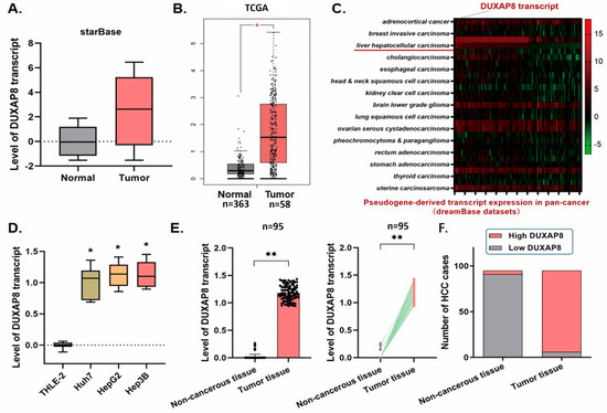
The analysis of the dreamBase, LCLE databases, and TCGA liver cancer datasets shows that the DUXAP8 transcript is detectable at a high level in the HCC tumor tissues, but is barely transcribed in normal liver tissues (Figure 1A–C). In the recruited HCC cell lines (Huh7, HepG2, and Hep3B), the DUXAP8 transcript was detected at a relatively high level in general, compared with the control THLE-2 cells, which showed an almost undetectable expression of DUXAP8 (Figure 1D,E).
Figure 1.
The pseudogene transcript level of DUXAP8 in HCC. (A) The transcript of the pseudogene DUXAP8 in HCC was detected by analyzing the relevant HCC dataset from the starBase database. A high level of DUXAP8 transcript in the HCC tissues was illustrated (p < 0.001). (B) The DUXAP8 transcript in HCC was detected by analyzing the TCGA database (n = 421, * p < 0.05). Transcription of DUXAP8 was activated and highly expressed in the HCC tissues (p < 0.0001). (C) The signature genes in HCC with expression changes were detected by analyzing the dreamBase dataset (expressions in HCC outlined in red), and the heatmap was generated. The DUXAP8 transcript was detected at a higher level in HCC tissues than in normal tissues. (D) RT-qPCR assay demonstrated that DUXAP8 was activated and significantly highly expressed in HCC cell lines in comparison with the control THLE-2 cells (* p < 0.05). (E) RT-qPCR assay was conducted on the 95 real patients’ specimens. The DUXAP8 transcript was highly expressed in the tumor tissues, and only a few non-cancerous tissues presented detectable DUXAP8 transcript at a lower level (** p < 0.01). (F) Statistic of the number of cases concerning the DUXAP8 transcription in HCC specimens. A significant increase in DUXAP8 transcript was detected in most HCC tissues (89/95, 93.68%), whereas, in non-cancerous tissues, only a small proportion (4/95, 4.21%) showed a low level of detectable DUXAP8 transcript.
The detection of the real patients’ specimens collected from our medical center demonstrated that DUXAP8 was transcribed in all the HCC tissues, with a portion of 93.68% (89/95) specimens that presented a significantly high DUXAP8 transcript level, and only a small portion (6.32%, 6/95) of a relatively lower level. In contrast, the DUXAP8 transcript was detectable in only 4.21% (4/95) of the adjacent non-cancerous liver tissues at an extremely low level (Figure 1F).
2.2. DUXAP8 Transcript Is Correlated with the Dismal Clinicopathologic Features in HCC Patients
The correlation between DUXAP8 transcription and the clinicopathologic features of the 95 HCC patients was statistically analyzed. Table 1 demonstrates that no significant correlation between the DUXAP8 transcript and the patient’s age, gender, tumor size, and virus control status was observed, while the highly transcribed DUXAP8 shows a positive relationship with the serum Alpha-fetoprotein (AFP) levels (p < 0.05), more advanced TNM stages (p < 0.05), tumor microsatellite formation (p < 0.05), and venous invasion (p < 0.05). These findings indicate that the DUXAP8 transcript plays a promoting role in HCC.
Table 1.
Correlation between DUXAP8 transcript and the clinicopathological features in 95 HCC specimens. The level of DUXAP8 transcript associated with clinicopathologic features in 95 HCC patients, including age, gender, tumor size, tumor stage (AJCC), tumor encapsulation, tumor microsatellite formation, vein invasion, HBsAg status, AFP level, and liver cirrhosis. Statistically, significance was assessed by Fisher’s exact test.
2.3. Knockdown of DUXAP8 Transcript Impairs HCC Cell Proliferation and Induces Cell Apoptosis
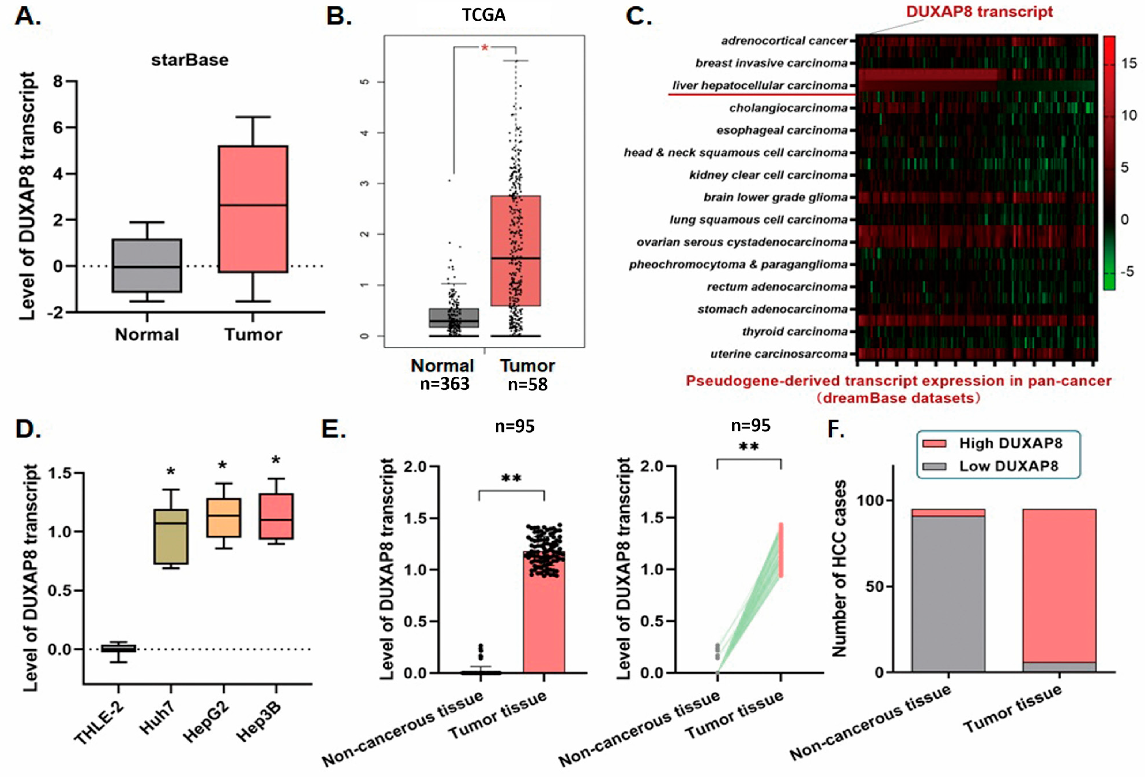
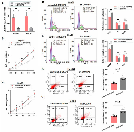
HepG2 and Hep3B demonstrated the highest DUXAP8 expression in our previous test and were selected for further tests. The DUXAP8 transcript was knocked down using the shRNA tools in both cell lines, and was validated through an RT-qPCR assay (Figure 2A, Table S3). The following in vitro experiments indicated that the cell proliferation ability in HCC cells was remarkably repressed in those two cell lines when the DUXAP8 transcript was knocked down (* p < 0.05; ** p < 0.01) (Figure 2B,C). A significant cell cycle arrest at the G0/G1 phases in the HCC cells was detected by the flow cytometric analysis in the cells with the DUXAP8 transcript knocking down (Figure 2D,E). The percentage of the HepG2 and Hep3B cells in the G0/G1 phase increased, respectively, from 49.1% to 63.8% (p < 0.01) and from 50.2% to 64.2% (p < 0.01). The percentage of the cells in the S phase declined (HepG2: from 25.9% to 17.0%, p < 0.05; Hep3B: from 25.1% to 19.2%, p < 0.01), and, also, the G2/M phase (HepG2: from 23.9% to 16.7%, p < 0.01; Hep3B: from 25.8% to 19.1%, p < 0.01).
Figure 2.
Knockdown of DUXAP8 impaired HCC cell proliferation and induced cell apoptosis. (A) Knockdown of DUXAP8 in HepG2 and Hep3B cells was conducted and validated by using the RT-qPCR assay (** p < 0.01). (B) The CCK8 assay was applied. Cell proliferation in HepG2 cells was significantly blocked after DUXAP8 knockdown (* p < 0.05, ** p < 0.01). (C) Similar to the HepG2 cells, the cell proliferation of Hep3B cells was significantly blocked after DUXAP8 knocking down (*p < 0.05, ** p < 0.01). (D) The representative histograms describing the cell cycle profiles of HepG2 cells by using flow cytometry are presented. For all histograms, the black and red lines indicate the raw data and the sum fit, respectively; the purple peak represents G0/G1 cells, while the green peak represents G2/M cells. The cell cycle of HepG2 cells was arrested in the G0/G1 phase by knockdown DUXAP8. The results are means of three independent experiments ± SD. (** p < 0.01). (E) The cell cycle of Hep3B cells was arrested in the G0/G1 phase after DUXAP8 knockdown. The results are means of three independent experiments ± SD. (** p < 0.01). (F) Cell apoptosis rate was detected by using flow cytometry. The representative histograms show that the cell apoptosis rate of HCC cells was significantly increased from 13.25% to 25.54% for HepG2 cells. The results are means of three independent experiments ± SD. (** p < 0.01). (G) The cell apoptosis rate of HCC cells was significantly increased from 13.39% to 21.56% for Hep3B cells. The results are means of all independent experiments ± SD. (** p < 0.01).
The calculated apoptotic HCC cells were increased by the flow cytometric analysis in both HepG2 and Hep3B cells (HepG2: from 13.25% to 25.54%, p < 0.01; Hep3B: from 13.39% to 21.56%, p < 0.01), sequentially induced by knocking down the DUXAP8 transcript with significance. The above findings illustrated that the DUXAP8 transcript exerts a potential promoting function by influencing cell growth and maintenance in HCC (Figure 2F,G).
2.4. Knockdown of DUXAP8 in HCC Cells Suppresses Tumor Growth and Lung Metastasis in the Orthotopic Transplantation Mouse Model
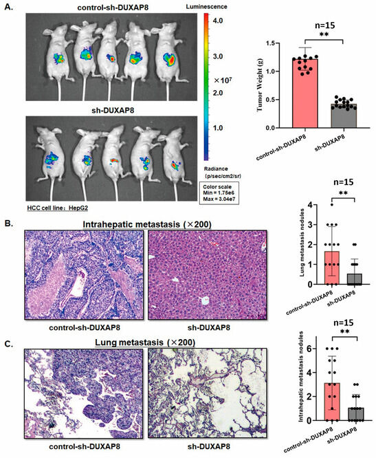
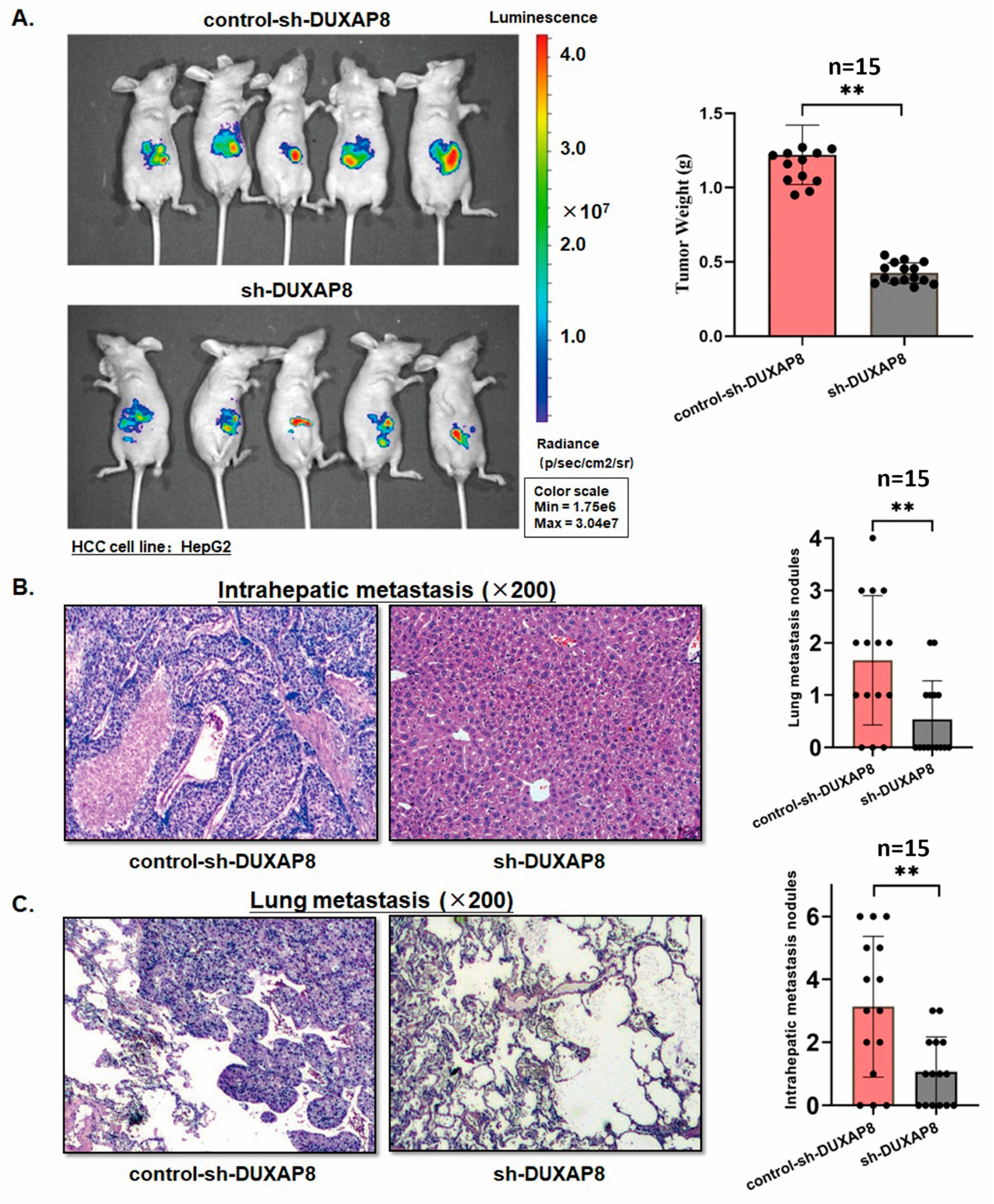
Xenograft mouse models were established by injecting the treated HepG2 cells. The orthotopically transplanted mass was quantified in livers six weeks post-orthotopic transplantation. The knocking down of DUXAP8 in HepG2 cells led to significantly smaller tumor masses in mouse livers compared with the control ones (Figure 3A). Additionally, the graphics from the HE staining examination demonstrated that either intrahepatic metastasis or lung metastasis lesions were generated less in the mice models with DUXAP8 knocking down than those in the control ones (Figure 3B,C).
Figure 3.
Knockdown of DUXAP8 suppresses tumor formation and lung metastasis in vivo. (A) Mouse liver orthotopic transplantation model was constructed. The tumor growth at week 6 was significantly suppressed in the mouse models by DUXAP8 knockdown in HepG2 cells. DUXAP8 knockdown induced a significant decrease in the xenograft tumor weight in vivo (** p < 0.01). (B) The xenograft tumor specimens were collected at week 6 and examined under HE staining examination. Knocking down of DUXAP8 led to fewer intrahepatic metastasis lesions in the mouse models than in the control ones (** p < 0.01). (C) Knocking down of DUXAP8 resulted in fewer metastasis lesions in the lungs of the mouse models than in the control ones (** p < 0.01).
The in vivo findings above support and complement our in vitro observation, and indicate that pseudogene DUXAP8 transcription exerts important effects on tumor development and invasiveness. This pseudogene is a potential target for intensive investigation.
2.5. DUXAP8 Transcript Sponges miR-214-3p in HCC Cells
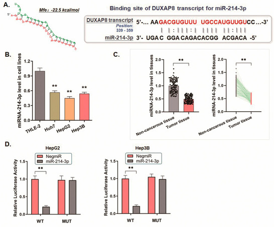
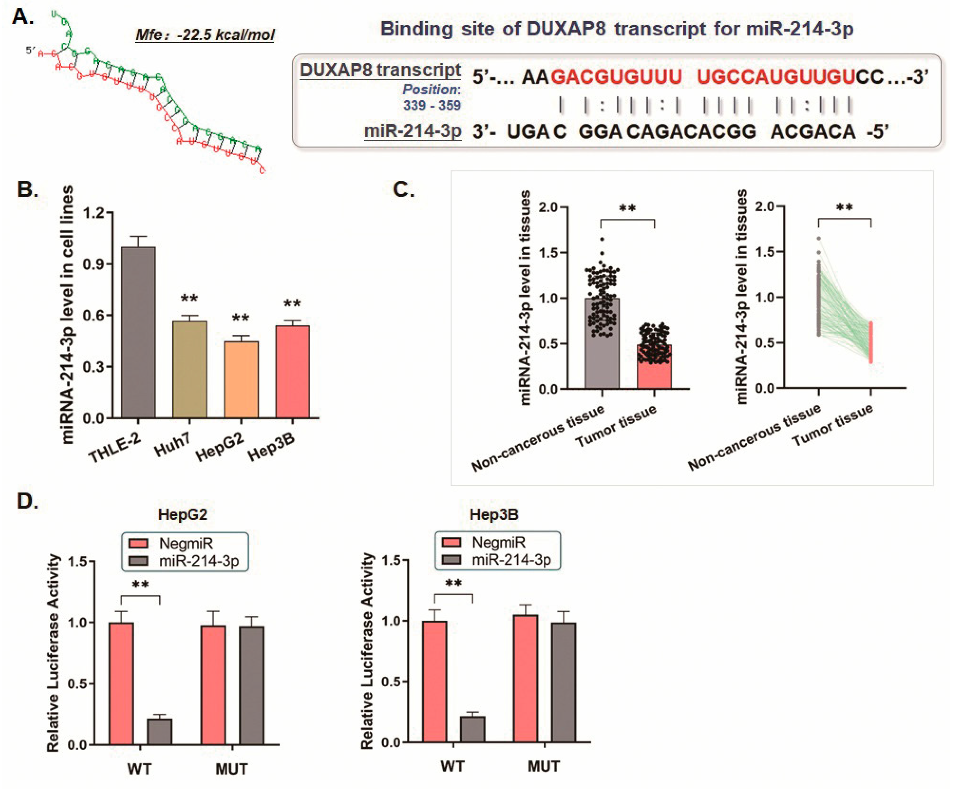
By analyzing the sequence of the DUXAP8 transcript and using the online microcosm prediction software, a short sequence from 339 bp to 359 bp to the 3′ end of DUXAP8 transcript is predicted to match the seed region of miR-214-3p (the minimum free energy, Mfe: −22.5 kcal/mol), which prompts the use of the DUXAP8 transcript as a probable competing endogenous RNA (ceRNA) targeting miR-214-3p (Figure 4A). Combined with the exploration of the HCC cell lines, we noticed that miR-214-3p, a reported inhibitor of HCC, was significantly downregulated in HCC (Figure 4B,C).
Figure 4.
DUXAP8 transcript sponges miR-214-3p in HCC cells. (A) The online microcosm software predicted that miR-214-3p could bind to the 3′-untranslated region (3′-UTR) of the DUXAP8 transcript (the minimum free energy, Mfe: −22.5 kcal/mol). (B) The RT-qPCR assay indicated a significant decrease of miR-214-3p in HCC cell lines (** p < 0.01). (C) The expression of miR-214-3p was significantly downregulated in HCC tissues compared with the non-cancerous tissues (** p < 0.01). (D) The dual-luciferase reporter assay demonstrated that the signal suppressive effect induced by miR-214-3p was significantly impaired in the HCC cells transfected with a mutated binding site of DUXAP8 transcript (** p < 0.01). This result indicates the sponging effect of the DUXAP8 transcript on miR-214-3p.
Based on the prediction, we constructed a mutated binding site in the DUXAP8 transcript for the dual-luciferase reporter assay to validate the potential interaction between the DUXAP8 transcript and miR-214-3p. The luciferase signal in either HepG2 or Hep3B cells transfected with miR-214-3p mimics was significantly decreased, based on the DUXAP8/pMIR/WT vector transfection, in comparison with the control ones. Conversely, the DUXAP8/pMIR/MUT vector transfection induced no significant signal change (Figure 4D, Table S4). All these findings above are consistent with the proposed ceRNA mechanism of the DUXAP8 transcript in sponging miR-214-3p.
2.6. DUXAP8 Engages a Feedback Loop via Transcriptional Activation by miR-214-3p Targeted KLF13
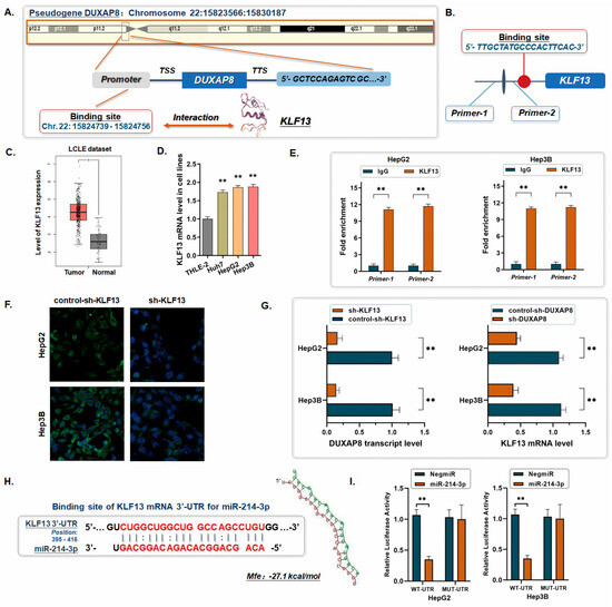
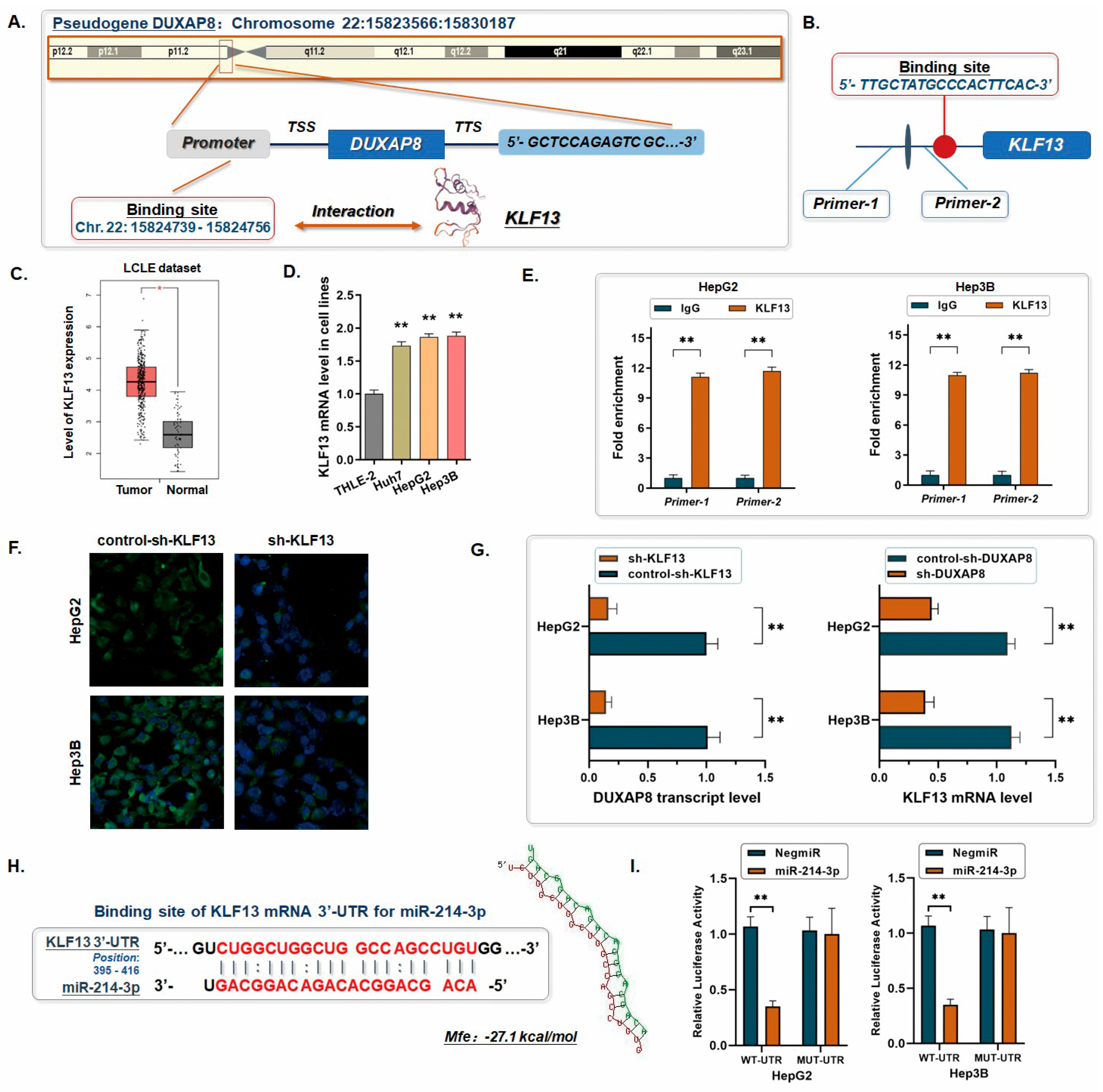
A 3000 bp fragment upstream of the first ATG of the pseudogene DUXAP8, regarded as the promoter region of DUXAP8, was intercepted for predicting the binding site of the potential transcription factor in tumorigenesis. Intriguingly, by evaluating through the Database of Human Transcription Factor Targets (https://guolab.wchscu.cn/hTFtarget/#!/, accessed on 13 October 2025), KLF13 was noticed as the probable transcription factor binding to the related region of the DUXAP8 gene (5′-TTGCTATGCCCACTTCAC-3′, Chr. 22: 15,824,739-15,824,756) (Figure 5A,B). Accordingly, KLF13 was highly expressed in HCC according to the TCGA liver cancer database and the HCC cell line exploration (Figure 5C,D). Thus, we conducted the ChIP assay to interrogate the interaction between these two genes and finally verified the predicted specific binding site for KLF13 on the promoter region of DUXAP8 (Figure 5E, Table S5).
Figure 5.
The feedback loop of DUXAP8/miR-214-3p/KLF13. (A) The 3000 bp fragment upstream of the pseudogene DUXAP8, which was regarded as its promoter region, was analyzed using the Database of Human Transcription Factor Targets. KLF13 was predicted as a probable transcription factor binding to the upstream sequence of pseudogene DUXAP8. (B) The binding site of the upstreaming region of DUXAP8 (5 TTGCTATGCCCACTTCAC-3′) is presented in the histogram, and two primers (Primer 1 and Primer 2) were synthesized. (C) The expression of KLF13 is significantly upregulated in HCC according to the TCGA datasets (* p < 0.05). (D) The level of KLF13 mRNA in the HCC cell lines was potently upregulated compared with the THLE-2 cells (** p < 0.01). (E) The ChIP assay was carried out to investigate the direct interaction between KLF13 and the equivalent region of the promoter upstreaming pseudogene DUXAP8 (** p < 0.01). IgG was used as the negative control. (F) Lentiviral vectors containing shRNA were used to deplete KLF13 in Hep3G and Hep3B cells. The effect was validated by the immunofluorescence detection at 40× magnification, as shown. (G) Either knocking down DUXAP8 or KLF13 in the HCC cells sequentially induced a significant decrease in the expression of the other side (** p < 0.01). (H) miR-214-3p was predicted to bind to the 3′-untranslated region (3′-UTR) of KLF13 mRNA (the minimum free energy, Mfe: −27.1 kcal/mol). (I) The dual-luciferase reporter assay was conducted. The signal suppressive effect induced by miR-214-3p was significantly impaired in the HCC cells transfected with the mutated binding site of the 3′-UTR of KLF13 mRNA (** p < 0.01).
Notably, we observed similar trends of expression changes for either the DUXAP8 transcript or KLF13 mRNA by selectively knocking down the expression of the other side through in the HCC cells, and the knocking down of KLF13 induced similar phenotypes in vitro to the knocking down of DUXAP8 (Figure 5F,G, Figures S1 and S2). A post-transcriptional regulation between miR-214-3p and KLF13 mRNA in the typical way was indicated. In brief, we used the Microcosm online software (https://www.microcosm.com/) and targeted miR-214-3p as an upstream regulator of KLF13, by potentially binding to the 3′-UTR of KLF13 mRNA (Figure 5H). Then, the vectors containing the fragment of 3′-UTR from KLF13 mRNA (WT-UTR) and the control luciferase vectors containing the mutated sequence (MUT-UTR) were constructed for the dual-luciferase reporter assay. The ectopic expression of miR-214-3p in HCC cells (HepG2/miR-214 and Hep3B/miR-214) significantly decreased the luciferase signal of KLF13/pMIR/WT, compared with the negative control (HepG2/NigmiR and Hep3B/NigmiR) (Figure 5I). The signal suppression induced by miR-214-3p was defective in the HCC cells transfected with a mutated binding sequence. The results above indicated a possible direct binding between miR-214-3p and KLF13 mRNA, which suggested a feedback loop of DUXAP8/miR-214-3p/KLF13.
2.7. Feedback Loop of DUXAP8/miR-214-3p/KLF13 Impacts the Transcription of Piezo1
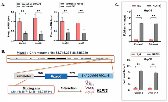
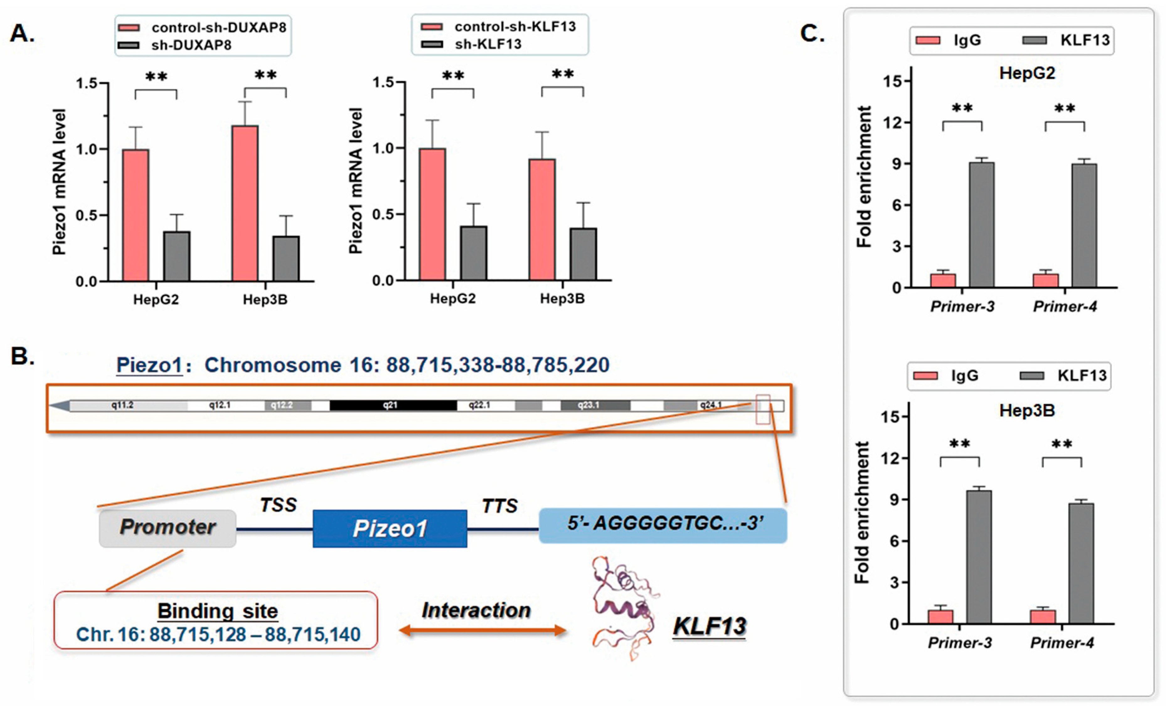
Piezo1 is a sensitive indicator of liver cancer, whose expression level changes along with the TME status and is commonly upregulated in HCC. In this study, we observed that knocking down either DUXAP8 or KLF13 in HCC cells induced the consequential decrease in Piezo1 mRNA levels (Figure 6A, Table S6). Here, we detected the promoter region of Piezo1, and predicted a potential binding site for KLF13 (5′-CTGCGGGAGGGGA-3′, Chr. 16: 88,715,128–88,715,140). According to the ChIP assay results, we further validated the direct interaction between the transcription factor KLF13 and the specific sequence included in the Piezo1 promoter region (Figure 6B,C, Table S7).
Figure 6.
Feedback loop of DUXAP8/miR-214-3p/KLF13 impacts the transcription of Piezo1. (A) The mRNA level of Piezo1 in HCC cell lines was significantly decreased by knocking down either DUXAP8 or KLF13 (** p < 0.01). (B) The promoter region of Piezo1 predicted as a potential binding site for KLF13 (5′-CTGCGGGAGGGGA-3′). (C) Two primers (Primer 3 and Primer 4) were synthesized for the ChIP assay. As shown in the ChIP assay histograms, the equivalent region of the promoter upstream Piezo1 gene was directly bound by KLF13. IgG was used as the negative control (** p < 0.01).
3. Discussion
As one of the major worldwide health challenges, HCC presents aggressive biological characteristics, leading to strong tumor growth, invasiveness, and motility phenotypes of the tumor cells [8]. The current progress in the treatment has promoted the outcomes of HCC patients to a certain extent, but rapid tumor progression and a high ratio of tumor recurrence after surgery largely limit the improvement of the OS and RFS. Hence, we are eager to understand more intensive intracellular mechanisms facilitating HCC aggression to explore better approaches to prevent and control this tumorous problem.
The current evidence has revealed the important roles of pseudogenes in human malignancies [9]. As described, the pathological transcript products of pseudogenes perform various functions throughout the whole process of tumor development, either promoting or suppressing tumorigenesis and progression [10]. The ceRNA effect is the most acknowledged function among the aberrant transcripts of pseudogenes verified in different human cancers by sponging miRNAs with a high affinity [11,12]. However, the complex functions of pseudogenes in HCC remain ambiguous. On the one hand, INTS6P1 (a pseudogene of Integrator complex subunit 6, INTS6) is activated to transcribe and inhibits HCC cell proliferation by sponging tumor-related miR-17-5p [13]. On the contrary, PCNAP1, the pseudogene of Proliferating Cell Nuclear Antigen (PCNA), plays an opposite role in promoting HCC initiation in HBV-infected patients, which is induced by suppressing miR-154 via a similar ceRNA mode [14].
Our recent research has revealed several signature pseudogenes that participate in HCC initiation and process through the application of the LCLE tools aforementioned. Innovatively, AKR1B10P1, UBE2MP1, and SNRPFP1 were reported to be anomalously transcribed and function as a set of lncRNAs promoting HCC progression either in tumor growth or metastasis via individual mechanisms [15,16,17]. As noticed, these pseudogene transcripts might compose a cross-talk network to exert the ceRNA effects on different microRNAs and stabilize a series of coding RNAs impacting the HCC development. For further intensive study, we screened out DUXAP8 as the co-expression pseudogene transcript for interrogation.
DUXAP8 is a pseudogene of the Double Homeobox Protein family member DUXA, whose translational products are involved in early embryonic development [18]. The current literature has introduced several DUXA-derived pseudogenes, like DUXAP9 and DUXAP10, which were sporadically reported as oncogenes in several human malignancies, including osteosarcoma, leukemia, and non-small-cell lung cancer [19,20,21]. As for the pseudogene DUXAP8, its length is 2307 bp, and the transcript is regarded as an lncRNA, which has a nucleotide composition over 200nt [22]. However, the exact bio-information of the parental gene DUXA is quite limited and provides no valuable details about its daughter pseudogene DUXAP8.
Currently, the abnormal overexpression of the DUXAP8 has been discovered in some human cancers (e.g., pancreatic cancer, lung adenocarcinoma, and gastric cancer), potentially associated with oncogenic events in most of them by affecting the proliferation, motility, and anti-autophagy of tumor cells [23,24,25]. The expression of DUXAP8 in HCC has been noticed and is generally thought to promote HCC by different bio-processes, including sponging microRNAs (miR-490 and miR-422) and enhancing cell proliferation [26,27]. According to our observations comparing HCC tumor tissues with paracancerous liver tissues, DUXAP8 was potently activated in tumors, along with the detectable transcript in tumor cells. A more highly expressed DUXAP8 was correlated with dismal clinicopathologic features, such as a higher AFP level and advanced tumor stage characteristics (microsatellite formation, venous invasion, and liver cirrhosis). Simultaneously, the loss of function of DUXAP8 in our study demonstrated that knocking down DUXAP8 in HCC cells remarkably impairs the cell proliferation ability and facilitates the cell apoptosis process. We hypothesize that DUXAP8 overexpression contributes to HCC progression. According to the in vivo examination, the HCC tumor formation treated by DUXAP8 knockdown was significantly suppressed in both the liver orthotopic transplantation and the lung metastasis lesions. These findings further elucidate DUXAP8 as a promising HCC oncogene.
The exploration in our study on the potential ceRNA role of DUXAP8 screened miR-214-3p as one of the targets, which shares the binding sites directly matched with the specific sequence of the DUXAP8 transcript. In the literature, miR-214-3p presents an aberrant profile of the expression in multiple malignancies, including breast cancer, colorectal cancer, and liver cancer, and mostly plays the role of a tumor suppressor [28,29,30]. For HCC, miR-214-3p is commonly downregulated in tumor tissues, and its ectopic expression through mimics induced the obvious inhibition of either cell proliferation or migration by degrading the mRNAs involved in the MAPK1 or HOTAIR signaling pathway [31,32]. Herein, we validated the direct interaction between the DUXAP8 transcript and miR-214-3p and observed a significant downregulating effect of DUXAP8 knockdown on the degradation of miR-214-3p.
Sequentially, we interrogated miR-214-3p by the luciferase reporter assay and verified it as an upstream regulator of KLF13, which is the transcription factor involved in various cancers. As acknowledged, KLF13 is a pivotal member of the Krüppel-like factor (KLF) family, containing conserved zinc-ester domains for regulating transcriptional activity. KLF13 has been reported to be highly expressed in oral cancer cells and to significantly promote cell proliferation [33]. Recently, researchers have described KLF13 as a overexpressed promoter in HCC, which mediates and enhances HMGCS1-related cholesterol biosynthesis [34]. However, the exact mechanism underlying KLF13 expression in HCC remains unclear. In this study, our findings demonstrated that the knocking down of DUXAP8 in the HCC cell lines induced a significant decrease in KLF13. Thus, we hypothesize that the abnormal transcription of DUXAP8 could exert the function of stabilization and a high expression status of KLF13 via the molecular sponging effect.
Importantly, we evaluated the probable downstream effectors of KLF13 in HCC to better understand the underlying mechanism. Interestingly, the pseudogene DUXAP8 was occasionally predicted as one of the KLF13 targets, sharing the potential binding site upstream of the DUXAP8 sequence. The ChIP assay showed that KLF13 directly binds to the promoter region of DUXAP8. Hence, we suggested that DUXAP8 and KLF13 consist of a positive feedback loop through the ceRNA effect on miR-214-3p, which affects the HCC process, probably in an amplification mode in the TME.
Does this feedback loop promote HCC progress by modulating some critical effectors? Based on this point of view, the TME-related Piezo1 was selected as another predicted candidate downstream of KLF13, and this gene may be a pivotal effector affected by the DUXAP8/miR-214-3p/KLF13 feedback loop. In brief, Piezo1 is widely distributed in various human organs and tissues, like the cardiovascular, lung, urinary, and immune systems [35]. Structurally, Piezo1 plays a specific role in mechanosensitive ion channels and functions in the activation of cell signaling pathways concerning mechanical stimulation [36].
Mechanical stress caused by tissue stiffening is the predominant characteristic in the composition of the liver TME [37]. Liver fibrosis and related cirrhosis are the most important causes of HCC tumorigenesis based on different inducements, like acute or chronic liver injury, and persistent hepatitis. Moreover, the TME of HCC involves complex intrinsic and extrinsic factors with a high heterogeneity that contribute to liver fibrosis, sclerosis, and permeability transition. Therefore, we believe that the expression change in Piezo1 is an innovative indicator of the liver TME status in the context of liver inflammation, fibrosis, or cirrhosis. Similarly, accumulating evidence has demonstrated that Piezo1 was upregulated in the hepatic fibrosis process and contributed to HCC progression. Although we did not further investigate the downstream effectors of Piezo1, the literature suggests that its activation can induce a range of cellular changes, including hypoxia tolerance, the inhibition of apoptosis, and the epithelial–mesenchymal transition. One piece of convincing evidence is that highly expressed Piezo1 activates the MAPK pathway in the YAP cascade by inducing the phosphorylation of JNK, p38, and ERK, and promotes HCC growth in vivo [38].
In our study, the modulation of the DUXAP8/miR-214-3p/KLF13 feedback loop could significantly affect Piezo1 transcription and HCC growth. This finding is consistent with the positive role of Piezo1 in HCC progression. We propose that DUXAP8, along with the members of this feedback loop, participate in the regulation of Piezo1 as a series of practicable indicators for liver TME and HCC progression. However, it is worth noticing that, though our results strongly indicate ceRNA regulation in the feedback loop, we cannot fully rule out parallel regulatory mechanisms with the current evidence. Further study would be needed to explore other possible pathways in this loop.
Last but not least, in the following research, we will focus on the intensive modulation mechanism induced by DUXAP8 via Piezo1, which has to be interrogated intensively, and the exact effects of Piezo1 on HCC TME will be further detected under the background of the DUXAP8/miR-214-3p/KLF13 feedback loop as well.
In summary, this study demonstrates a featured feedback loop in HCC progress predominantly triggered by the abnormally transcribed pseudogene DUXAP8, and the members of the loop not only stabilized the expression of DUXAP8 through a ceRNA way but also positively modulated the expression of the Piezo1. Our work probably provides a set of innovative indicators and therapeutic targets in HCC prevention.
4. Materials and Methods
4.1. Cell Culture and Preparation
Three typical HCC cell lines (Huh7, HepG2, and Hep3B) were cultured along with the control transformed human liver epithelial-2 (THLE-2) (Shanghai Institutes for Biological Sciences, Chinese Academy of Science, Shanghai, China). Briefly, the cell lines were respectively cultured in RPMI 1640, supplemented with 10% heat-inactivated fetal bovine serum (FBS), incubated at 37 °C, with 100 μg/mL streptomycin, and 100 U/mL Penicillin in a humidified atmosphere of 5% CO2. Cells were authenticated by short tandem repeat (STR) profiling and routinely tested for mycoplasma contamination using PCR and colorimetric assays, and only mycoplasma-free cells were used for experiments. Specifically, for the transfected cells, a medium containing G418 (Santa Cruz Biotechnology, Inc., Dallas, TX, USA; 400 μg/mL) was used for selection. The selection process was maintained until non-transfected control cells were completely eliminated, lasting for 5–7 days.
4.2. Clinical Specimens
Ninety-five pairs of HCC-related specimens containing the tumor tissues and the paracancerous liver tissues at a 1 cm distance from the tumor margin were collected. All patients underwent R0 radical resection without any pre- or post-operative treatment at the Department of General Surgery, Ruijin Hospital, Shanghai Jiao Tong University School of Medicine (2016 to 2020). The clinicopathologic features of those patients were organized, including gender, age, tumor size, number of lesions, grades, et al. The study was approved by the Ethics Committee of Ruijin Hospital, Shanghai Jiaotong University School of Medicine (No. 2021-421), and informed consent was obtained.
4.3. Preparation and Application of the Datasets
The differential gene expression for 369 liver tumors and 50 normal samples from the UCSC Xena database, combined with an additional 110 normal liver samples data from the GTEx and TCGA, were intensively explored by using the random-walk-based multi-graphic (RWMG) model algorithm developed by our team [39]. The relative information of the pseudogene-derived transcripts was analyzed and presented in the aforementioned LCLE tools. The starBase datasets (https://starbase.sysu.edu.cn/, accessed on 13 October 2025) and the dreamBase (https://ngdc.cncb.ac.cn/databasecommons/database/id/3746, accessed on 13 October 2025) datasets were introduced to provide much more supplementary information on the expression and relationship of the candidate genes.
4.4. RT-qPCR Assay and Immunohistochemistry Assay
RNA isolation from tissues or cells is performed according to the instructions of the TRIzol reagent (Invitrogen, Waltham, MA, USA). The first-strand cDNA was synthesized via a High-Capacity cDNA Reverse Transcription Kit (ABI, Los Angeles, CA, USA). All the primers were synthesized by Jike Biotech Company (Shanghai, China) (Table S1). Real-time quantitative polymerase chain reaction (RT-qPCR) was performed following the TaqMan Gene Expression Assays protocol (ABI, USA). The relative quantification of RNA in cell lines was normalized using GAPDH by the 2 − ΔCt method. The relative quantification of miR-214-3p in tissue specimens and cell lines was measured by using the mirVANATM miRNA Isolation Kit (ABI, USA). The PCR program was set as follows: 95 °C for 10 min, followed by 35 cycles of 95 °C for 15 s, 60 °C for 30 s, and 72 °C for 45 s.
Antibodies against KLF13 were purchased from Abcam, Waltham, MA, USA. The immunohistochemistry assay (IHC) complied with our previously described methods [40]. The protein expression levels detected by IHC were independently assigned to two experienced pathologists for blind examination and were separated into two groups based on staining intensity grade: no to low staining (0~1+) and moderate to high staining (2+~3+).
4.5. Plasmid Preparation and Cell Transfection
The lentiviral vectors pLKO.1 containing shRNA were transfected into cultured HepG2 and Hep3B cells at the exponential phase (JIKE Biochemistry, Shanghai, China) to suppress the expression of the DUXAP8 transcript, and the control vectors were used. The transfected cells were selected using a medium containing G418 (Santa Cruz Biotechnology, Inc.; 400 μg/mL). The mimic was used to transfect HCC cells for ectopically introducing miR-214-3p (HepG2/miR-214; Hep3B/miR-214), along with a set of negative controls (HepG2/NigmiR; Hep3B/NigmiR). The lentiviral vector pLV (Addgene, Cambridge, MA, USA) was applied for ectopically expressing KLF13 (pLV-KLF13) for the rescue experiments, and the pLV-Null was set for control.
4.6. Cell Proliferation and Cell Cycle Detection
The HCC cells (1 × 10⁶) were cultured in 96-well microtiter plates, triplicated, and incubated at an atmosphere of 5% CO2 and 37 °C for 5 days. Microplate computer software version 1.30 (Bio-Rad Laboratories, Inc., Hercules, CA, USA) was used to measure the OD following the Cell Counting Kit-8 (CCK-8) assay kit protocol (Dojindo, Tokyo, Japan), and the cell proliferation curves were plotted. The cells were fixed with ethanol, and then treated with RNase A and stained using propidium iodide. Flow cytometry detection was carried out using FACSCalibur (Becton-Dickinson, Franklin Lakes, NJ, USA), and ModFit software version 5.0 (Becton–Dickinson, Franklin Lakes, NJ, USA) was used for quantifying cell populations at the G0/G1, S, and G2/M phases (the debris and fixation artifacts of the cells were excluded).
4.7. Cell Apoptosis Analysis
Cell apoptosis rate was calculated using PE-Annexin V Apoptosis Detection Kit I (BD Pharmingen, Franklin Lakes, NJ, USA) according to the instructions. Transfected cells were resuspended in a concentration of 1 × 10⁶ cells/mL in the 1 × Binding Buffer. Then, 5 μL of FITC and 5 μL of PI were added into 100 μL of the cell suspension, followed by a 15-min incubation in darkness, with 400 μL × Binding Buffer added. The apoptosis rate was determined by flow cytometry (Becton Dickinson, Franklin Lakes, NJ, USA). FSC/SSC gating was used to exclude debris and FSC/SSC-B gating was used to exclude doublets. Cells were classified as follows: Annexin V−/PI− viable cells, Annexin V+/PI− early apoptotic cells, and Annexin V+/PI+ late apoptotic cells. Both early and late apoptotic cells were used to calculate the apoptosis rate. Appropriate unstained controls and single-stained controls were used for compensation.
4.8. The Mouse Liver Orthotopic Transplantation and Tumorigenicity Assay
The function of DUXAP8 in HCC development was assessed by the tumorigenicity assay. Five-week-old male BALB/c nude mice (Institute of Zoology, Chinese Academy of Sciences, Beijing, China) were purchased and housed in a pathogen-free environment and were randomized into the treatment and control groups (fifteen mice for each group). All protocols were performed in accordance with the guidelines of the Shanghai Medical Experimental Animal Care Commission, including the randomization and blinding statement. A total of 1 × 10⁶ HCC cells were suspended within the 25 µL serum-free DMEM, which was mixed with 25 µL Matrigel (1:1, v/v). Cells were orthotopically injected into the left hepatic lobes of the mice. The mice were monitored regularly for general condition and sacrificed at 6 weeks after injection. Investigators were blinded to group allocation during outcome assessment. The weight of all the xenografted livers was measured, and the mice’s livers and lungs were stained with hematoxylin and eosin (HE) for pathological examination. All animal experiments were approved by the IACUC of Ruijin Hospital, Shanghai Jiao Tong University School of Medicine (No. 2020-148), and were performed in accordance with relevant guidelines and regulations.
4.9. Dual-Luciferase Reporter Assay
MiR-214-3p was predicted to be a potential regulator of KLF13 using the Microcosm online tool (https://www.targetscan.org/vert_80/). We selected a 202 bp sequence containing the putative miR-214-3p binding site at the 3′-UTR of KLF13 mRNA and designed the mutant sequence (Table S2). The sequences were respectively cloned into the pMIR-Report luciferase vector, which contains firefly luciferase, and the pRL-TK vector luciferase was used as a control (Promega, Madison, WI, USA). These two sets of vectors were co-transfected into the HCC cells, introducing miR-214-3p or the controls. The luciferase activity was measured via the Dual-Glo Luciferase assay system (Promega) 48 h after the transfection. Simultaneously, we constructed the sequence and the relative mutated sequences, containing the putative binding site of miR-214-3p of DUXAP8 transcripts (Table S2).
4.10. Chromatin Immunoprecipitation Assay
The chromatin immunoprecipitation (ChIP) assay was performed to verify the interaction between the transcription factor and the promoter region of targeted genes (KL13 to DUXAP8 or Piezo1). A total of 5 × 10⁶ cells were cultured in each 10 cm dish and subjected to ChIP assay following ChIP-ITTM Kit’s protocol (Active Motif, Carlsbad, CA, USA). Chromatin was immunoprecipitated with 2 μg of either a transcription factor antibody (Abcam, USA) or IgG as the negative control. The extracted DNA was analyzed through RT-qPCR by using the relative primers (Table S1).
4.11. Statistical Analysis
Statistical analysis was conducted by using SPSS 20.0. Each experiment was repeated three times independently, and the statistical analysis was conducted according to the mean of these results. p-values were calculated following an unpaired Student’s t-test and Fisher’s exact test. Spearman and Logistic analysis were used to describe the association between the various parameters and the risk of DUXAP8 status. Differences were considered statistically significant at p-values < 0.05 in this study.
Supplementary Materials
The following supporting information can be downloaded at: https://www.mdpi.com/article/10.3390/ijms27114873/s1.
Author Contributions
Y.Z., X.L. and J.W. wrote the article and participated in revision; Y.L. and F.H. contributed to the data analysis and biomolecular experiments; X.F. was in charge of the pathological experiments and data mining; Y.Z. worked on the collection of clinicopathological features; and Y.C. and J.W. designed and directed the study. All authors have read and agreed to the published version of the manuscript.
Funding
This study was kindly supported by grants from the following: National Natural Science Foundation of China (No. 82372603; No. 82172900); Shanghai Leading Talent Program of Eastern Talent Plan (No. BJJY2024068); Chinese Society of Clinical Oncology (CSCO)-Chaoyang Oncology Research Foundation (No. Y-Young2021-0015); and Research Physician Project from Shanghai Jiao Tong University School of Medicine (No. 20191901).
Institutional Review Board Statement
Informed consent was obtained and the study was approved by the Ethics Committee of Ruijin Hospital, Shanghai Jiaotong University School of Medicine, following the Declaration of Helsinki (ethical approval number: 2021-421, date of approval: 24 September 2021) for humans. All animal experiments were approved by the IACUC of Ruijin Hospital, Shanghai Jiao Tong University School of Medicine (Ethical approval number: 2020-148, date of approval: 17 May 2020), and were performed in accordance with the relevant guidelines and regulations.
Informed Consent Statement
The informed written consent for participation in the study has been obtained.
Data Availability Statement
Data from this study is not made public due to privacy regulations but is available upon request from the authors via sending a message to Junqing Wang (e-mail: wangjunqingmd@hotmail.com).
Acknowledgments
The authors thank Shen Chen, Jiajun Ren, Xiaoyong Gong, and Simin Guo for providing valuable technical support and assistance.
Conflicts of Interest
The authors declare no conflict of interest.
Abbreviations
Hepatocellular carcinoma (HCC); overall survival (OS); tumor microenvironment (TME); extracellular matrix (ECM); Piezo-type mechanosensitive ion channel component 1 (Piezo1); KLF Transcription Factor 13 (KLF13); the chromatin immunoprecipitation assay (ChIP assay); 3′-untranslated region (3′-UTR); and competing endogenous RNA (ceRNA).
References
- Dhanasekaran, R. Deciphering Tumor Heterogeneity in Hepatocellular Carcinoma (HCC)—Multi-Omic and Singulomic Approaches. Semin. Liver Dis. 2021, 41, 9–18. [Google Scholar] [CrossRef] [Scilit] [PubMed]
- Zhang, Q.; Lou, Y.; Yang, J.; Wang, J.; Feng, J.; Zhao, Y.; Wang, L.; Huang, X.; Fu, Q.; Ye, M.; et al. Integrated multiomic analysis reveals comprehensive tumour heterogeneity and novel immunophenotypic classification in hepatocellular carcinomas. Gut 2019, 68, 2019–2031. [Google Scholar] [CrossRef] [Scilit] [PubMed]
- Siegel, R.L.; Giaquinto, A.N.; Jemal, A. Cancer statistics, 2024. CA Cancer J. Clin. 2024, 74, 12–49, Erratum in CA Cancer J. Clin. 2024, 74, 203. https://doi.org/10.3322/caac.21830.. [Google Scholar] [CrossRef] [Scilit]
- Schreiber, R.D.; Old, L.J.; Smyth, M.J. Cancer immunoediting: Integrating immunity’s roles in cancer suppression and promotion. Science 2011, 331, 1565–1570. [Google Scholar] [CrossRef] [Scilit]
- Kumari, S.; Advani, D.; Sharma, S.; Ambasta, R.K.; Kumar, P. Combinatorial therapy in tumor microenvironment: Where do we stand? Biochim. Biophys. Acta (BBA)-Rev. Cancer 2021, 1876, 188585. [Google Scholar] [CrossRef] [Scilit]
- Anderson, N.M.; Simon, M.C. The tumor microenvironment. Curr. Biol. 2020, 30, R921–R925. [Google Scholar] [CrossRef] [Scilit]
- Luo, X.-Y.; Lu, Y.-Q.; Zhang, Y.-F.; Wang, N.; Hao, F.-J.; Fei, X.-C.; Chen, Y.-J.; Wang, J.-Q. USP32 facilitates tumor development and is correlated with poor clinical outcomes in hepatocellular carcinoma patients and is modulated by the E2F7/miR-218-5p axis. LabMed Discov. 2025, 2, 100102. [Google Scholar] [CrossRef] [Scilit]
- Huang, D.Q.; El-Serag, H.B.; Loomba, R. Global epidemiology of NAFLD-related HCC: Trends, predictions, risk factors and prevention. Nat. Rev. Gastroenterol. Hepatol. 2021, 18, 223–238. [Google Scholar] [CrossRef] [Scilit]
- Chan, J.J.; Tay, Y. Noncoding RNA:RNA Regulatory Networks in Cancer. Int. J. Mol. Sci. 2018, 19, 1310. [Google Scholar] [CrossRef] [Scilit] [PubMed]
- Singh, R.K.; Singh, D.; Yadava, A.; Srivastava, A.K. Molecular fossils “pseudogenes” as functional signature in biological system. Genes Genom. 2020, 42, 619–630. [Google Scholar] [CrossRef] [Scilit]
- Tay, Y.; Rinn, J.; Pandolfi, P.P. The multilayered complexity of ceRNA crosstalk and competition. Nature 2014, 505, 344–352. [Google Scholar] [CrossRef] [Scilit] [PubMed]
- Karreth, F.A.; Pandolfi, P.P. ceRNA cross-talk in cancer: When ce-bling rivalries go awry. Cancer Discov. 2013, 3, 1113–1121. [Google Scholar] [CrossRef] [Scilit]
- Peng, H.; Ishida, M.; Li, L.; Saito, A.; Kamiya, A.; Hamilton, J.P.; Fu, R.; Olaru, A.V.; An, F.; Popescu, I.; et al. Pseudogene INTS6P1 regulates its cognate gene INTS6 through competitive binding of miR-17-5p in hepatocellular carcinoma. Oncotarget 2015, 6, 5666–5677. [Google Scholar] [CrossRef] [Scilit]
- Feng, J.; Yang, G.; Liu, Y.; Gao, Y.; Zhao, M.; Bu, Y.; Yuan, H.; Yuan, Y.; Yun, H.; Sun, M.; et al. LncRNA PCNAP1 modulates hepatitis B virus replication and enhances tumor growth of liver cancer. Theranostics 2019, 9, 5227–5245. [Google Scholar] [CrossRef] [Scilit]
- Hao, F.; Fei, X.; Ren, X.; Xiao, J.X.; Chen, Y.; Wang, J. Pseudogene AKR1B10P1 enhances tumorigenicity and regulates epithelial-mesenchymal transition in hepatocellular carcinoma via stabilizing SOX4. J. Cell. Mol. Med. 2020, 24, 11779–11790. [Google Scholar] [CrossRef] [Scilit] [PubMed]
- Hao, F.; Wang, N.; Gui, H.; Zhang, Y.; Wu, Z.; Wang, J. Pseudogene UBE2MP1 derived transcript enhances in vitro cell proliferation and apoptosis resistance of hepatocellular carcinoma cells through miR-145-5p/RGS3 axis. Aging 2022, 14, 7906–7925. [Google Scholar] [CrossRef] [Scilit]
- Wang, N.; Guo, S.; Hao, F.; Zhang, Y.; Chen, Y.; Fei, X.; Wang, J. Pseudogene SNRPFP1 derived long non-coding RNA facilitates hepatocellular carcinoma progress in vitro by sponging tumor-suppressive miR-126-5p. Sci. Rep. 2022, 12, 21867. [Google Scholar] [CrossRef] [Scilit]
- Leidenroth, A.; Hewitt, J.E. A family history of DUX4: Phylogenetic analysis of DUXA, B, C and Duxbl reveals the ancestral DUX gene. BMC Evol. Biol. 2010, 10, 364. [Google Scholar] [CrossRef] [Scilit] [PubMed]
- Wang, G.; Zhang, Q.; Wang, Q.; Wang, J.; Chen, L.; Sun, Q.; Miao, D. Long non-coding RNA DUXAP10 exerts oncogenic properties in osteosarcoma by recruiting HuR to enhance SOX18 mRNA stability. Hum. Cell 2022, 35, 1939–1951, Correction in Hum. Cell 2025, 38, 156. [Google Scholar] [CrossRef] [Scilit]
- Yao, R.; Feng, W.-T.; Xu, L.-J.; Zhong, X.-M.; Liu, H.; Sun, Y.; Zhou, L.-L. DUXAP10 regulates proliferation and apoptosis of chronic myeloid leukemia via PTEN pathway. Eur. Rev. Med. Pharmacol. Sci. 2018, 22, 4934–4940. [Google Scholar]
- Zhu, T.; An, S.; Choy, M.; Zhou, J.; Wu, S.; Liu, S.; Liu, B.; Yao, Z.; Zhu, X.; Wu, J.; et al. LncRNA DUXAP9-206 directly binds with Cbl-b to augment EGFR signaling and promotes non-small cell lung cancer progression. J. Cell. Mol. Med. 2019, 23, 1852–1864, Correction in J. Cell. Mol. Med. 2022, 26, 2137. [Google Scholar] [CrossRef] [Scilit]
- Wang, B.; Xu, W.; Cai, Y.; Chen, J.; Guo, C.; Zhou, G.; Yuan, C. DUXAP8: A Promising lncRNA with Carcinogenic Potential in Cancer. Curr. Med. Chem. 2022, 29, 1677–1686. [Google Scholar] [CrossRef] [Scilit]
- Lian, Y.; Yang, J.; Lian, Y.; Xiao, C.; Hu, X.; Xu, H. DUXAP8, a pseudogene derived lncRNA, promotes growth of pancreatic carcinoma cells by epigenetically silencing CDKN1A and KLF2. Cancer Commun. 2018, 38, 64. [Google Scholar] [CrossRef] [Scilit]
- Chen, M.; Fan, M.; Yang, J.; Lang, J. Identification of Potential Oncogenic Long Non-Coding RNA Set as a Biomarker Associated with Colon Cancer Prognosis. J. Environ. Pathol. Toxicol. Oncol. 2020, 39, 39–49. [Google Scholar] [CrossRef] [Scilit]
- Ma, H.-W.; Xie, M.; Sun, M.; Chen, T.-Y.; Jin, R.-R.; Ma, T.-S.; Chen, Q.-N.; Zhang, E.-B.; He, X.-Z.; De, W.; et al. The pseudogene derived long noncoding RNA DUXAP8 promotes gastric cancer cell proliferation and migration via epigenetically silencing PLEKHO1 expression. Oncotarget 2016, 8, 52211–52224. [Google Scholar] [CrossRef] [Scilit] [PubMed]
- Zhang, H.; Chu, K.; Zheng, C.; Ren, L.; Tian, R. Pseudogene DUXAP8 Promotes Cell Proliferation and Migration of Hepatocellular Carcinoma by Sponging MiR-490-5p to Induce BUB1 Expression. Front. Genet. 2020, 11, 666. [Google Scholar] [CrossRef] [Scilit] [PubMed]
- Wang, X.-K.; Liao, X.-W.; Huang, R.; Huang, J.-L.; Chen, Z.-J.; Zhou, X.; Yang, C.-K.; Han, C.-Y.; Zhu, G.-Z.; Peng, T. Clinical significance of long non-coding RNA DUXAP8 and its protein coding genes in hepatocellular carcinoma. J. Cancer 2020, 11, 6140–6156. [Google Scholar] [CrossRef] [Scilit]
- Han, L.-C.; Wang, H.; Niu, F.-L.; Yan, J.-Y.; Cai, H.-F. Effect miR-214-3p on proliferation and apoptosis of breast cancer cells by targeting survivin protein. Eur. Rev. Med. Pharmacol. Sci. 2019, 23, 7469–7474. [Google Scholar] [CrossRef] [Scilit]
- Chen, C.; Zhang, Q.; Wang, B.; Song, Y.; Feng, Z.; Ren, S. SPTBN2 regulated by miR-214-3p inhibits the proliferation and migration of colorectal cancer cells. Cell. Mol. Biol. 2023, 69, 126–131. [Google Scholar] [CrossRef] [Scilit]
- Li, H.; Gai, L.; Wu, Z.; Li, F. Maternal embryonic leucine zipper kinase serves as a potential prognostic marker and leads to sorafenib chemoresistance modified by miR-142-5p in hepatocellular carcinoma. Mol. Biol. Rep. 2022, 49, 3015–3024. [Google Scholar] [CrossRef] [Scilit] [PubMed]
- Wan, H.; Tian, Y.; Zhao, J.; Su, X. LINC00665 Targets miR-214-3p/MAPK1 Axis to Accelerate Hepatocellular Carcinoma Growth and Warburg Effect. J. Oncol. 2021, 2021, 9046798. [Google Scholar] [CrossRef] [Scilit]
- Liu, C.; Shang, Z.; Ma, Y.; Ma, J.; Song, J. HOTAIR/miR-214-3p/FLOT1 axis plays an essential role in the proliferation, migration, and invasion of hepatocellular carcinoma. Int. J. Clin. Exp. Pathol. 2019, 12, 50–63. [Google Scholar]
- Henson, B.; Gollin, S. Overexpression of KLF13 and FGFR3 in oral cancer cells. Cytogenet. Genome Res. 2010, 128, 192–198. [Google Scholar] [CrossRef] [Scilit] [PubMed]
- Chen, C.-C.; Xie, X.-M.; Zhao, X.-K.; Zuo, S.; Li, H.-Y. Krüppel-like Factor 13 Promotes HCC Progression by Transcriptional Regulation of HMGCS1-mediated Cholesterol Synthesis. J. Clin. Transl. Hepatol. 2022, 10, 1125–1137. [Google Scholar] [CrossRef] [Scilit]
- Wu, J.; Lewis, A.H.; Grandl, J. Touch, Tension, and Transduction—The Function and Regulation of Piezo Ion Channels. Trends Biochem. Sci. 2017, 42, 57–71. [Google Scholar] [CrossRef] [Scilit]
- Lai, A.; Cox, C.D.; Sekar, N.C.; Thurgood, P.; Jaworowski, A.; Peter, K.; Baratchi, S. Mechanosensing by Piezo1 and its implications for physiology and various pathologies. Biol. Rev. 2022, 97, 604–614. [Google Scholar] [CrossRef] [Scilit] [PubMed]
- Xie, C.; Singal, A.K. Global burden of cirrhosis and liver cancer due to alcohol: The past, present, and the future. Hepatol. Int. 2023, 17, 830–832. [Google Scholar] [CrossRef] [Scilit] [PubMed]
- Liu, S.; Xu, X.; Fang, Z.; Ning, Y.; Deng, B.; Pan, X.; He, Y.; Yang, Z.; Huang, K.; Li, J. Piezo1 impairs hepatocellular tumor growth via deregulation of the MAPK-mediated YAP signaling pathway. Cell Calcium 2021, 95, 102367. [Google Scholar] [CrossRef] [Scilit]
- Wang, J.; Wang, X.; Bhat, A.; Chen, Y.; Xu, K.; Mo, Y.-Y.; Yi, S.S.; Zhou, Y. Comprehensive Network Analysis Reveals Alternative Splicing-Related lncRNAs in Hepatocellular Carcinoma. Front. Genet. 2020, 11, 659. [Google Scholar] [CrossRef] [Scilit]
- Wang, N.; Hao, F.; Ren, J.; Fei, X.; Chen, Y.; Xu, W.; Wang, J. Positive feedback loop of AKR1B10P1/miR-138/SOX4 promotes cell growth in hepatocellular carcinoma cells. Am. J. Transl. Res. 2020, 12, 5465–5480. [Google Scholar]

Disclaimer/Publisher’s Note: The statements, opinions and data contained in all publications are solely those of the individual author(s) and contributor(s) and not of MDPI and/or the editor(s). MDPI and/or the editor(s) disclaim responsibility for any injury to people or property resulting from any ideas, methods, instructions or products referred to in the content. |
© 2026 by the authors. Licensee MDPI, Basel, Switzerland. This article is an open access article distributed under the terms and conditions of the Creative Commons Attribution (CC BY) license.