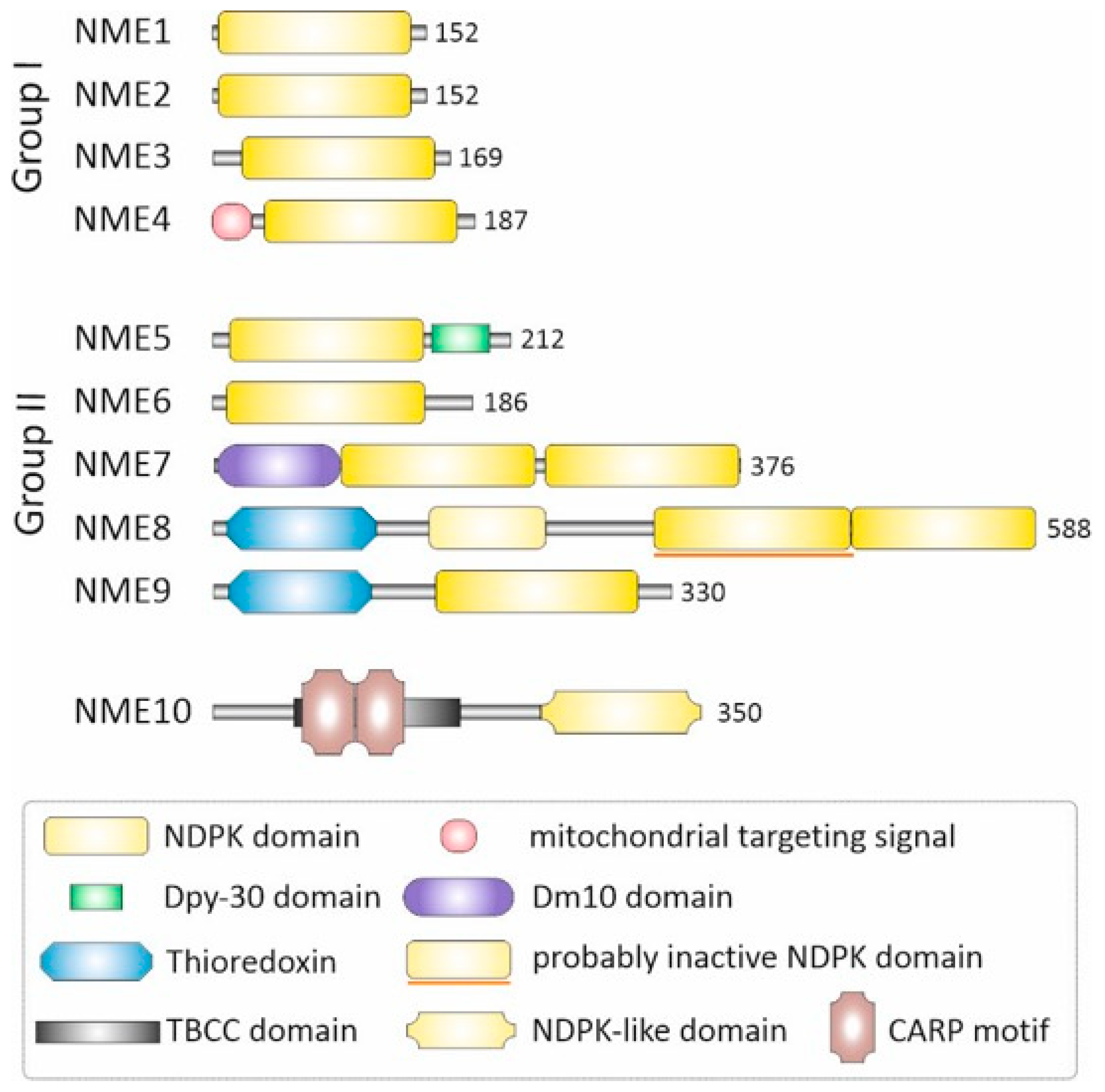
Functional Diversity and Emerging Roles of Human NME/NDPK Group II Proteins

 , ,
, ,  , and
, and
Abstract
1. Introduction
- The Group II NME/NDPK proteins
- NME5
- NME6
- NME7
- NME8 and NME9
2. Conclusions
Author Contributions
Funding
Data Availability Statement
Conflicts of Interest
Abbreviations
| ASS | Acephalic spermatozoa syndrome |
| ATP | Adenosine triphosphate |
| CDP | Cytidine diphosphate |
| CMP | Cytidine monophosphate |
| CTP | Cytidine triphosphate |
| DMT | Doublet microtubules |
| GFP | Green fluorescent protein |
| GPX-5 | Glutathione peroxidase 5 |
| GTP | Guanosine triphosphate |
| HCC | Hepatocellular carcinoma |
| JAK/STAT | Janus kinase/signal transducer and activator of transcription |
| KO | Knockout |
| mt-RNA | Mitochondrial RNA |
| NDP | Nucleoside diphosphate |
| NDPK | Nucleoside diphosphate kinase |
| NME | NME protein family |
| NME6-KI | NME6 knock-in cells |
| fNTP | Nucleoside triphosphate |
| PCD | Primary ciliary dyskinesia |
| RCC1L | Regulator of chromosome condensation 1-like protein |
| Sptrx-2 | Spermatid-specific thioredoxin 2 |
| TBCC | Tubulin-binding cofactor C |
| TXL2 | Thioredoxin-like 2 |
| TXNDC3 | Thioredoxin domain-containing protein 3 |
| TXNDC6 | Thioredoxin domain-containing protein 6 |
| UDP | Uridine diphosphate |
| UMP | Uridine monophosphate |
| UTP | Uridine triphosphate |
| γTuRC | γ-Tubulin ring complex |
References
- Lacombe, M.-L.L.; Munier, A.; Mehus, J.G.; Lambeth, D.O. The Human Nm23/Nucleoside Diphosphate Kinases. J. Bioenerg. Biomembr. 2000, 32, 247–258. [Google Scholar] [CrossRef]
- Lascu, I.; Gonin, P. The Catalytic Mechanism of Nucleoside Diphosphate Kinases. J. Bioenerg. Biomembr. 2000, 32, 237–246. [Google Scholar] [CrossRef]
- Boissan, M.; Montagnac, G.; Shen, Q.; Griparic, L.; Guitton, J.; Romao, M.; Sauvonnet, N.; Lagache, T.; Lascu, I.; Raposo, G.; et al. Nucleoside Diphosphate Kinases Fuel Dynamin Superfamily Proteins with GTP for Membrane Remodeling. Science 2014, 344, 1510–1515. [Google Scholar] [CrossRef]
- Boissan, M.; Schlattner, U.; Lacombe, M.-L. The NDPK/NME Superfamily: State of the Art. Lab. Investig. 2018, 98, 164–174. [Google Scholar] [CrossRef]
- Desvignes, T.; Pontarotti, P.; Bobe, J. Nme Gene Family Evolutionary History Reveals Pre-Metazoan Origins and High Con-servation between Humans and the Sea Anemone, Nematostella Vectensis. PLoS ONE 2010, 5, e15506, Correction in PLoS ONE. 2010, 5. https://doi.org/10.1371/annotation/ad419d55-e0bd-4a2e-a79e-5265f44ae545.. [Google Scholar] [CrossRef][Green Version]
- Leone, A.; Flatow, U.; King, C.R.; Sandeen, M.A.; Margulies, I.M.K.; Liotta, L.A.; Steeg, P.S. Reduced Tumor Incidence, Metastatic Potential, and Cytokine Responsiveness of Nm23-Transfected Melanoma Cells. Cell 1991, 65, 25–35. [Google Scholar] [CrossRef]
- Hartsough, M.T.; Steeg, P.S. Nm23/Nucleoside Diphosphate Kinase in Human Cancers. J. Bioenerg. Biomembr. 2000, 32, 301–308. [Google Scholar] [CrossRef]
- Boissan, M.; Dabernat, S.; Peuchant, E.; Schlattner, U.; Lascu, I.; Lacombe, M.-L. The Mammalian Nm23/NDPK Family: From Metastasis Control to Cilia Movement. Mol. Cell. Biochem. 2009, 329, 51–62. [Google Scholar] [CrossRef]
- Abu-Taha, I.H.; Heijman, J.; Feng, Y.; Vettel, C.; Dobrev, D.; Wieland, T. Regulation of Heterotrimeric G-Protein Signaling by NDPK/NME Proteins and Caveolins: An Update. Lab. Investig. 2018, 98, 190–197. [Google Scholar] [CrossRef]
- Bosnar, M.H.; Bago, R.; Ćetković, H. Subcellular Localization of Nm23/NDPK A and B Isoforms: A Reflection of Their Biological Function? Mol. Cell. Biochem. 2009, 329, 63–71. [Google Scholar] [CrossRef]
- Bosnar, M.H.; de Gunzburg, J.; Bago, R.; Brečević, L.; Weber, I.; Pavelić, J. Subcellular Localization of A and B Nm23/NDPK Subunits. Exp. Cell Res. 2004, 298, 275–284. [Google Scholar] [CrossRef]
- Romani, P.; Ignesti, M.; Gargiulo, G.; Hsu, T.; Cavaliere, V. Extracellular NME Proteins: A Player or a Bystander? Lab. Investig. 2018, 98, 248–257. [Google Scholar] [CrossRef]
- Karlsson, A.; Mesnildrey, S.; Xu, Y.; Moréra, S.; Janin, J.; Véron, M. Nucleoside Diphosphate Kinase. J. Biol. Chem. 1996, 271, 19928–19934. [Google Scholar] [CrossRef]
- Vieira, P.S.; de Giuseppe, P.O.; de Oliveira, A.H.C.; Murakami, M.T. The Role of the C-Terminus and Kpn Loop in the Quaternary Structure Stability of Nucleoside Diphosphate Kinase from Leishmania Parasites. J. Struct. Biol. 2015, 192, 336–341. [Google Scholar] [CrossRef]
- Mesnildrey, S.; Agou, F.; Karlsson, A.; Bonne, D.D.; Véron, M. Coupling between Catalysis and Oligomeric Structure in Nucleoside Diphosphate Kinase. J. Biol. Chem. 1998, 273, 4436–4442. [Google Scholar] [CrossRef]
- Dumas, C.; Lascu, I.; Moréra, S.; Glaser, P.; Fourme, R.; Wallet, V.; Lacombe, M.L.; Véron, M.; Janin, J. X-Ray Structure of Nucleoside Diphosphate Kinase. EMBO J. 1992, 11, 3203–3208. [Google Scholar] [CrossRef]
- Lascu, I.; Deville-Bonne, D.; Glaser, P.; Véron, M. Equilibrium Dissociation and Unfolding of Nucleoside Diphosphate Kinase from Dictyostelium Discoideum. Role of Proline 100 in the Stability of the Hexameric Enzyme. J. Biol. Chem. 1993, 268, 20268–20275. [Google Scholar] [CrossRef]
- Chiadmi, M.; Moréra, S.; Lascu, I.; Dumas, C.; Le Bras, G.; Véron, M.; Janin, J. Crystal Structure of the Awd Nucleotide Diphosphate Kinase from Drosophila. Structure 1993, 1, 283–293. [Google Scholar] [CrossRef]
- Sedmak, J.; Ramaley, R. Purification and Properties of Bacillus Subtilis Nucleoside Diphosphokinase. J. Biol. Chem. 1971, 246, 5365–5372. [Google Scholar] [CrossRef]
- Moynié, L.; Giraud, M.-F.; Georgescauld, F.; Lascu, I.; Dautant, A. The Structure of the Escherichia coli Nucleoside Diphosphate Kinase Reveals a New Quaternary Architecture for This Enzyme Family. PROTEINS Struct. Funct. Bioinform. 2007, 67, 755–765. [Google Scholar] [CrossRef]
- Williams, R.L.; Oren, D.A.; Muñoz-Dorado, J.; Inouye, S.; Inouye, M.; Arnold, E. Crystal Structure of Myxococcus xanthus Nucleoside Diphosphate Kinase and Its Interaction with a Nucleotide Substrate at 2·0 Å Resolution. J. Mol. Biol. 1993, 234, 1230–1247. [Google Scholar] [CrossRef]
- Ginther, C.L.; Ingraham, J.L. Nucleoside Diphosphokinase of Salmonella typhimurium. J. Biol. Chem. 1974, 249, 3406–3411. [Google Scholar] [CrossRef]
- Muñoz-Dorado, J.; Inouye, S.; Inouye, M. Nucleoside Diphosphate Kinase from Myxococcus xanthus. II. Biochemical Characterization. J. Biol. Chem. 1990, 265, 2707–2712. [Google Scholar] [CrossRef]
- Georgescauld, F.; Song, Y.; Dautant, A. Structure, Folding and Stability of Nucleoside Diphosphate Kinases. Int. J. Mol. Sci. 2020, 21, 6779. [Google Scholar] [CrossRef]
- Desvignes, T.; Pontarotti, P.; Fauvel, C.; Bobe, J. Nme Protein Family Evolutionary History, a Vertebrate Perspective. BMC Evol. Biol. 2009, 9, 256. [Google Scholar] [CrossRef]
- Gilles, A.M.; Presecan, E.; Vonica, A.; Lascu, I. Nucleoside Diphosphate Kinase from Human Erythrocytes. Structural Characterization of the Two Polypeptide Chains Responsible for Heterogeneity of the Hexameric Enzyme. J. Biol. Chem. 1991, 266, 8784–8789. [Google Scholar] [CrossRef]
- Chen, C.-W.; Wang, H.-L.; Huang, C.-W.; Huang, C.-Y.; Lim, W.K.; Tu, I.-C.; Koorapati, A.; Hsieh, S.-T.; Kan, H.-W.; Tzeng, S.-R.; et al. Two Separate Functions of NME3 Critical for Cell Survival Underlie a Neurodegenerative Disorder. Proc. Natl. Acad. Sci. USA 2019, 116, 566–574. [Google Scholar] [CrossRef]
- Chen, C.-W.; Tsao, N.; Zhang, W.; Chang, Z.-F. NME3 Regulates Mitochondria to Reduce ROS-Mediated Genome Instability. Int. J. Mol. Sci. 2020, 21, 5048. [Google Scholar] [CrossRef]
- Lacombe, M.-L.; Lamarche, F.; De Wever, O.; Padilla-Benavides, T.; Carlson, A.; Khan, I.; Huna, A.; Vacher, S.; Calmel, C.; Desbourdes, C.; et al. The Mitochondrially-Localized Nucleoside Diphosphate Kinase D (NME4) Is a Novel Metastasis Suppressor. BMC Biol. 2021, 19, 228. [Google Scholar] [CrossRef]
- Proust, B.; Radić, M.; Vidaček, N.Š.; Cottet, C.; Attia, S.; Lamarche, F.; Ačkar, L.; Mikulčić, V.G.; Tokarska-Schlattner, M.; Ćetković, H.; et al. NME6 Is a Phosphotransfer-Inactive, Monomeric NME/NDPK Family Member and Functions in Complexes at the Interface of Mitochondrial Inner Membrane and Matrix. Cell Biosci. 2021, 11, 195. [Google Scholar] [CrossRef]
- Tokarska-Schlattner, M.; Boissan, M.; Munier, A.; Borot, C.; Mailleau, C.; Speer, O.; Schlattner, U.; Lacombe, M.-L. The Nucleoside Diphosphate Kinase D (NM23-H4) Binds the Inner Mitochondrial Membrane with High Affinity to Cardiolipin and Couples Nucleotide Transfer with Respiration. J. Biol. Chem. 2008, 283, 26198–26207. [Google Scholar] [CrossRef]
- Herak Bosnar, M.; Radić, M.; Ćetković, H. A Young Researcher’s Guide to NME/Nm23/NDP Kinase. Period. Biol. 2018, 120, 3–9. [Google Scholar] [CrossRef]
- Ćetković, H.; Perina, D.; Harcet, M.; Mikoč, A.; Herak Bosnar, M. Nme Family of Proteins—Clues from Simple Animals. Naunyn-Schmiedeberg’s Arch. Pharmacol. 2015, 388, 133–142. [Google Scholar] [CrossRef]
- Munier, A.; Feral, C.; Milon, L.; Pinon, V.P.-B.; Gyapay, G.; Capeau, J.; Guellaen, G.; Lacombe, M.-L. A New Human Nm23 Homologue (Nm23-H5) Specifically Expressed in Testis Germinal Cells. FEBS Lett. 1998, 434, 289–294. [Google Scholar] [CrossRef]
- Hu, T.; Tang, X.; Ruan, T.; Long, S.; Liu, G.; Ma, J.; Li, X.; Zhang, R.; Huang, G.; Shen, Y.; et al. IQUB Mutation Induces Radial Spoke 1 Deficiency Causing Asthenozoospermia with Normal Sperm Morphology in Humans and Mice. Cell Commun. Signal. 2025, 23, 41. [Google Scholar] [CrossRef]
- Hwang, K.-C.; Ok, D.-W.; Hong, J.-C.; Kim, M.-O.; Kim, J.-H. Cloning, Sequencing, and Characterization of the Murine Nm23-M5 Gene during Mouse Spermatogenesis and Spermiogenesis. Biochem. Biophys. Res. Commun. 2003, 306, 198–207. [Google Scholar] [CrossRef]
- Choi, Y.-J.; Cho, S.-K.; Hwang, K.-C.; Park, C.; Kim, J.-H.; Park, S.-B.; Hwang, S.; Kim, J.-H. Nm23-M5 Mediates Round and Elongated Spermatid Survival by Regulating GPX-5 Levels. FEBS Lett. 2009, 583, 1292–1298. [Google Scholar] [CrossRef]
- Sarkar, H.; Arya, S.; Rai, U.; Majumdar, S.S. A Study of Differential Expression of Testicular Genes in Various Reproductive Phases of Hemidactylus flaviviridis (Wall Lizard) to Derive Their Association with Onset of Spermatogenesis and Its Relevance to Mammals. PLoS ONE 2016, 11, e0151150. [Google Scholar] [CrossRef]
- Tian, Y.; Zhang, Y.; Tang, X.; Zhao, C.; Jiang, X.; Shen, G.; Wang, X.; Jiang, C.; Ruan, T.; Ma, J.; et al. Discovery of a Pathogenic NME5 Variant Underlying Acephalic Spermatozoa Syndrome: Unraveling a Novel Genotype-Phenotype Association in Male Infertility. Clin. Genet. 2026, 109, 368–373. [Google Scholar] [CrossRef]
- Proust, B.; Herak Bosnar, M.; Ćetković, H.; Tokarska-Schlattner, M.; Schlattner, U. Mitochondrial NME6: A Paradigm Change within the NME/NDP Kinase Protein Family? Cells 2024, 13, 1278. [Google Scholar] [CrossRef]
- Li, F.; Hu, G.; Jiang, Z.; Guo, J.; Wang, K.; Ouyang, K.; Wen, D.; Zhu, M.; Liang, J.; Qin, X.; et al. Identification of NME5 as a Contributor to Innate Resistance to Gemcitabine in Pancreatic Cancer Cells. FEBS J. 2012, 279, 1261–1273. [Google Scholar] [CrossRef] [PubMed]
- Goc, A.; Kochuparambil, S.T.; Al-Husein, B.; Al-Azayzih, A.; Mohammad, S.; Somanath, P.R. Simultaneous Modulation of the Intrinsic and Extrinsic Pathways by Simvastatin in Mediating Prostate Cancer Cell Apoptosis. BMC Cancer 2012, 12, 409. [Google Scholar] [CrossRef] [PubMed]
- Ostrowski, L.E.; Blackburn, K.; Radde, K.M.; Moyer, M.B.; Schlatzer, D.M.; Moseley, A.; Boucher, R.C. A Proteomic Analysis of Human Cilia: Identification of Novel Components. Mol. Cell. Proteom. 2002, 1, 451–465. [Google Scholar] [CrossRef] [PubMed]
- Anderegg, L.; Im Hof Gut, M.; Hetzel, U.; Howerth, E.W.; Leuthard, F.; Kyöstilä, K.; Lohi, H.; Pettitt, L.; Mellersh, C.; Minor, K.M.; et al. NME5 Frameshift Variant in Alaskan Malamutes with Primary Ciliary Dyskinesia. PLoS Genet. 2019, 15, e1008378. [Google Scholar] [CrossRef]
- Sahabian, A.; von Schlehdorn, L.; Drick, N.; Pink, I.; Dahlmann, J.; Haase, A.; Göhring, G.; Welte, T.; Martin, U.; Ringshausen, F.C.; et al. Generation of Two HiPSC Clones (MHHi019-A, MHHi019-B) from a Primary Ciliary Dyskinesia Patient Carrying a Homozygous Deletion in the NME5 Gene (c.415delA (p.Ile139Tyrfs*8)). Stem Cell Res. 2020, 48, 101988. [Google Scholar] [CrossRef]
- Cho, E.H.; Huh, H.J.; Jeong, I.; Lee, N.Y.; Koh, W.; Park, H.; Ki, C. A Nonsense Variant in NME5 Causes Human Primary Ciliary Dyskinesia with Radial Spoke Defects. Clin. Genet. 2020, 98, 64–68. [Google Scholar] [CrossRef]
- Perina, D.; Korolija, M.; Mikoč, A.; Halasz, M.; Herak Bosnar, M.; Ćetković, H. Characterization of Nme5-Like Gene/Protein from the Red Alga Chondrus crispus. Mar. Drugs 2019, 18, 13. [Google Scholar] [CrossRef]
- Yoon, J.-H.; Singh, P.; Lee, D.-H.; Qiu, J.; Cai, S.; O’Connor, T.R.; Chen, Y.; Shen, B.; Pfeifer, G.P. Characterization of the 3′ → 5′ Exonuclease Activity Found in Human Nucleoside Diphosphate Kinase 1 (NDK1) and Several of Its Homologues. Biochemistry 2005, 44, 15774–15786. [Google Scholar] [CrossRef]
- Gopal, R.; Foster, K.W.; Yang, P. The DPY-30 Domain and Its Flanking Sequence Mediate the Assembly and Modulation of Flagellar Radial Spoke Complexes. Mol. Cell. Biol. 2012, 32, 4012–4024. [Google Scholar] [CrossRef]
- Hsu, D.R.; Chuang, P.T.; Meyer, B.J. DPY-30, a Nuclear Protein Essential Early in Embryogenesis for Caenorhabditis elegans Dosage Compensation. Development 1995, 121, 3323–3334. [Google Scholar] [CrossRef]
- Jiang, H.; Shukla, A.; Wang, X.; Chen, W.Y.; Bernstein, B.E.; Roeder, R.G. Role for Dpy-30 in ES Cell-Fate Specification by Regulation of H3K4 Methylation within Bivalent Domains. Cell 2011, 144, 513–525, Erratum in Cell 2011, 144, 825.. [Google Scholar] [CrossRef]
- Perina, D.; Herak Bosnar, M.; Mikoč, A.; Müller, W.E.G.; Ćetković, H. Characterization of Nme6-like Gene/Protein from Marine Sponge Suberites domuncula. Naunyn-Schmiedeberg’s Arch. Pharmacol. 2011, 384, 451–460. [Google Scholar] [CrossRef]
- Mehus, J.G.; Deloukas, P.; Lambeth, D.O. NME6: A New Member of the Nm23 /Nucleoside Diphosphate Kinase Gene Family Located on Human Chromosome 3p21.3. Hum. Genet. 1999, 104, 454–459. [Google Scholar] [CrossRef]
- Tsuiki, H.; Nitta, M.; Furuya, A.; Hanai, N.; Fujiwara, T.; Inagaki, M.; Kochi, M.; Ushio, Y.; Saya, H.; Nakamura, H. A Novel Human Nucleoside Diphosphate (NDP) Kinase, Nm23-H6, Localizes in Mitochondria and Affects Cytokinesis. J. Cell. Biochem. 2000, 76, 254–269. [Google Scholar] [CrossRef]
- Pagliarini, D.J.; Calvo, S.E.; Chang, B.; Sheth, S.A.; Vafai, S.B.; Ong, S.-E.; Walford, G.A.; Sugiana, C.; Boneh, A.; Chen, W.K.; et al. A Mitochondrial Protein Compendium Elucidates Complex I Disease Biology. Cell 2008, 134, 112–123. [Google Scholar] [CrossRef]
- Antonicka, H.; Lin, Z.-Y.; Janer, A.; Aaltonen, M.J.; Weraarpachai, W.; Gingras, A.-C.; Shoubridge, E.A. A High-Density Human Mitochondrial Proximity Interaction Network. Cell Metab. 2020, 32, 479–497. [Google Scholar] [CrossRef]
- Rhee, H.-W.; Zou, P.; Udeshi, N.D.; Martell, J.D.; Mootha, V.K.; Carr, S.A.; Ting, A.Y. Proteomic Mapping of Mitochondria in Living Cells via Spatially Restricted Enzymatic Tagging. Science 2013, 339, 1328–1331. [Google Scholar] [CrossRef]
- International Mouse Phenotyping Consortium (IMPC)_NME6. Available online: https://www.mousephenotype.org/data/genes/MGI:1861676 (accessed on 5 December 2025).
- Ernst, O.; Sun, J.; Lin, B.; Banoth, B.; Dorrington, M.G.; Liang, J.; Schwarz, B.; Stromberg, K.A.; Katz, S.; Vayttaden, S.J.; et al. A Genome-Wide Screen Uncovers Multiple Roles for Mitochondrial Nucleoside Diphosphate Kinase D in Inflammasome Activation. Sci. Signal. 2021, 14, eabe0387. [Google Scholar] [CrossRef]
- Seifert, M.; Welter, C.; Mehraein, Y.; Seitz, G. Expression of the Nm23 Homologues Nm23-H4, Nm23-H6, and Nm23-H7 in Human Gastric and Colon Cancer. J. Pathol. 2005, 205, 623–632. [Google Scholar] [CrossRef]
- Ke, J.; Lou, J.; Zhong, R.; Chen, X.; Li, J.; Liu, C.; Gong, Y.; Yang, Y.; Zhu, Y.; Zhang, Y.; et al. Identification of a Potential Regulatory Variant for Colorectal Cancer Risk Mapping to 3p21.31 in Chinese Population. Sci. Rep. 2016, 6, 25194. [Google Scholar] [CrossRef]
- Luo, L.; Li, Y.; Zhang, L.; Yang, L. NME6 as a Potential Biomarker and Therapeutic Target Involved in Immune Infiltration for Lung Adenocarcinoma. Technol. Health Care 2024, 32, 2277–2291. [Google Scholar] [CrossRef]
- Kim, H.S.; Choi, J.Y.; Baek, G.O.; Yoon, M.G.; Jang, S.H.; Han, J.E.; Kim, S.S.; Cheong, J.Y.; Jeong, J.Y.; Eun, J.W. A Novel Liver-Specific Pseudogene Biomarker, BMS1P8, for Diagnosis and Prognosis in Hepatocellular Carcinoma. Front. Biosci. 2025, 30, 41684. [Google Scholar] [CrossRef]
- Kim, S.; Kang, M.; Jeong, S.; Kim, J.; Kim, K.O.; Lee, W.S.; Baek, J.H.; Kim, J.H.; Nam, S. Elucidating Prognostic Significance of Purine Metabolism in Colorectal Cancer through Integrating Data from Transcriptomic, Immunohistochemical, and Single-cell RNA Sequencing Analysis. Mol. Oncol. 2025, 19, 2310. [Google Scholar] [CrossRef]
- Liu, Y.; Geng, R.; Zhao, S.; Yang, J.; Liu, J.; Zheng, Y.; Tang, W. Single-Cell and Spatial Transcriptomics Explore Purine Metabolism–Related Prognostic Risk Model and Tumor Immune Microenvironment Modulation in Ovarian Cancer. Hum. Mutat. 2025, 2025, 5530325. [Google Scholar] [CrossRef]
- Lin, L.; Liao, Z.H.; Li, C.Q. Insight into the Role of Mitochondrion-Related Gene Anchor Signature in Mitochondrial Dysfunction of Neutrophilic Asthma. J. Asthma 2024, 61, 912–929. [Google Scholar] [CrossRef]
- Wang, C.-H.H.; Ma, N.; Lin, Y.-T.T.; Wu, C.-C.C.; Hsiao, M.; Lu, F.L.; Yu, C.-C.C.; Chen, S.-Y.Y.; Lu, J. A ShRNA Functional Screen Reveals Nme6 and Nme7 Are Crucial for Embryonic Stem Cell Renewal. Stem Cells 2012, 30, 2199–2211. [Google Scholar] [CrossRef]
- Zhang, H.; Li, X.; Liu, Y.; Liang, Z.; Li, R.; Gao, W.; Wang, B.; Zhang, Y.; Tian, Y.; Hu, X.; et al. Identification of Potential Causal-Genes-Relevant Blood Pressure: A Mitochondria-Related Genome-Wide Mendelian Randomization Study. J. Hum. Hypertens. 2026, 40, 45–52. [Google Scholar] [CrossRef]
- Arroyo, J.D.; Jourdain, A.A.; Calvo, S.E.; Ballarano, C.A.; Doench, J.G.; Root, D.E.; Mootha, V.K. A Genome-Wide CRISPR Death Screen Identifies Genes Essential for Oxidative Phosphorylation. Cell Metab. 2016, 24, 875–885. [Google Scholar] [CrossRef]
- Floyd, B.J.; Wilkerson, E.M.; Veling, M.T.; Minogue, C.E.; Xia, C.; Beebe, E.T.; Wrobel, R.L.; Cho, H.; Kremer, L.S.; Alston, C.L.; et al. Mitochondrial Protein Interaction Mapping Identifies Regulators of Respiratory Chain Function. Mol. Cell 2016, 63, 621–632. [Google Scholar] [CrossRef]
- Huttlin, E.L.; Bruckner, R.J.; Navarrete-Perea, J.; Cannon, J.R.; Baltier, K.; Gebreab, F.; Gygi, M.P.; Thornock, A.; Zarraga, G.; Tam, S.; et al. Dual Proteome-Scale Networks Reveal Cell-Specific Remodeling of the Human Interactome. Cell 2021, 184, 3022–3040. [Google Scholar] [CrossRef]
- Wang, J.; Huo, K.; Ma, L.; Tang, L.; Li, D.; Huang, X.; Yuan, Y.; Li, C.; Wang, W.; Guan, W.; et al. Toward an Understanding of the Protein Interaction Network of the Human Liver. Mol. Syst. Biol. 2011, 7, 536, Correction in Mol. Syst. Biol. 2011, 13, 965.. [Google Scholar] [CrossRef]
- Antonicka, H.; Choquet, K.; Lin, Z.; Gingras, A.; Kleinman, C.L.; Shoubridge, E.A. A Pseudouridine Synthase Module Is Essential for Mitochondrial Protein Synthesis and Cell Viability. EMBO Rep. 2017, 18, 28–38. [Google Scholar] [CrossRef]
- Koyama, M.; Sasaki, T.; Sasaki, N.; Matsuura, Y. Crystal Structure of Human WBSCR16, an RCC1-like Protein in Mitochondria. Protein Sci. 2017, 26, 1870–1877. [Google Scholar] [CrossRef] [PubMed]
- Lloyd, R.E.; McGeehan, J.E. Structural Analysis of Mitochondrial Mutations Reveals a Role for Bigenomic Protein Interactions in Human Disease. PLoS ONE 2013, 8, e69003. [Google Scholar] [CrossRef]
- Reyes, A.; Favia, P.; Vidoni, S.; Petruzzella, V.; Zeviani, M. RCC1L (WBSCR16) Isoforms Coordinate Mitochondrial Ribosome Assembly through Their Interaction with GTPases. PLoS Genet. 2020, 16, e1008923. [Google Scholar] [CrossRef]
- Huang, G.; Massoudi, D.; Muir, A.M.; Joshi, D.C.; Zhang, C.-L.; Chiu, S.Y.; Greenspan, D.S. WBSCR16 Is a Guanine Nucleotide Exchange Factor Important for Mitochondrial Fusion. Cell Rep. 2017, 20, 923–934. [Google Scholar] [CrossRef]
- Grotehans, N.; McGarry, L.; Nolte, H.; Xavier, V.; Kroker, M.; Narbona-Pérez, Á.J.; Deshwal, S.; Giavalisco, P.; Langer, T.; MacVicar, T. Ribonucleotide Synthesis by NME6 Fuels Mitochondrial Gene Expression. EMBO J. 2023, 42, EMBJ2022113256. [Google Scholar] [CrossRef]
- Kramer, N.J.; Prakash, G.; Isaac, R.S.; Choquet, K.; Soto, I.; Petrova, B.; Merens, H.E.; Kanarek, N.; Churchman, L.S. Regulators of Mitonuclear Balance Link Mitochondrial Metabolism to MtDNA Expression. Nat. Cell Biol. 2023, 25, 1575–1589. [Google Scholar] [CrossRef] [PubMed]
- Janin, J.; Dumas, C.; Moréra, S.; Xu, Y.; Meyer, P.; Chiadmi, M.; Cherfils, J. Three-Dimensional Structure of Nucleoside Diphosphate Kinase. J. Bioenerg. Biomembr. 2000, 32, 215–225. [Google Scholar] [CrossRef]
- Proust, B.; Horvat, A.; Tadijan, A.; Vlašić, I.; Herak Bosnar, M. Mitochondrial NME6 Influences Basic Cellular Processes in Tumor Cells In Vitro. Int. J. Mol. Sci. 2024, 25, 9580. [Google Scholar] [CrossRef] [PubMed]
- Wanrooij, P.H.; Chabes, A. NME6: Ribonucleotide Salvage Sustains Mitochondrial Transcription. EMBO J. 2023, 42, EMBJ2023114990. [Google Scholar] [CrossRef]
- MacVicar, T. High Tide or Low Tide: The Transport and Metabolism of Mitochondrial Nucleotides. Biochem. J. 2025, 482, 1105. [Google Scholar] [CrossRef]
- Reish, O.; Aspit, L.; Zouella, A.; Roth, Y.; Polak-Charcon, S.; Baboushkin, T.; Benyamini, L.; Scheetz, T.E.; Mussaffi, H.; Sheffield, V.C.; et al. A Homozygous Nme7 Mutation Is Associated with Situs Inversus Totalis. Hum. Mutat. 2016, 37, 727–731. [Google Scholar] [CrossRef]
- Hutchins, J.R.A.; Toyoda, Y.; Hegemann, B.; Poser, I.; Hériché, J.-K.; Sykora, M.M.; Augsburg, M.; Hudecz, O.; Buschhorn, B.A.; Bulkescher, J.; et al. Systematic Analysis of Human Protein Complexes Identifies Chromosome Segregation Proteins. Science 2010, 328, 593–599. [Google Scholar] [CrossRef]
- Liu, P.; Choi, Y.-K.; Qi, R.Z. NME7 Is a Functional Component of the γ-Tubulin Ring Complex. Mol. Biol. Cell 2014, 25, 2017–2025. [Google Scholar] [CrossRef] [PubMed]
- Ji, M.; Cui, W.; Feng, Q.; Qi, J.; Wang, X.; Zhu, H.; Zhang, W.; Fu, W. NME7 Maintains Primary Cilium Assembly, Ciliary Microtubule Stability, and Hedgehog Signaling. Life Sci. Alliance 2025, 8, e202402933. [Google Scholar] [CrossRef]
- Ikeda, T. NDP Kinase 7 Is a Conserved Microtubule-Binding Protein Preferentially Expressed in Ciliated Cells. Cell Struct. Funct. 2010, 35, 23–30. [Google Scholar] [CrossRef]
- Ma, M.; Stoyanova, M.; Rademacher, G.; Dutcher, S.K.; Brown, A.; Zhang, R. Structure of the Decorated Ciliary Doublet Microtubule. Cell 2019, 179, 909–922.e12. [Google Scholar] [CrossRef]
- Leung, M.R.; Zeng, J.; Wang, X.; Roelofs, M.C.; Huang, W.; Zenezini Chiozzi, R.; Hevler, J.F.; Heck, A.J.R.; Dutcher, S.K.; Brown, A.; et al. Structural Specializations of the Sperm Tail. Cell 2023, 186, 2880–2896.e17. [Google Scholar] [CrossRef] [PubMed]
- Gui, M.; Farley, H.; Anujan, P.; Anderson, J.R.; Maxwell, D.W.; Whitchurch, J.B.; Botsch, J.J.; Qiu, T.; Meleppattu, S.; Singh, S.K.; et al. De Novo Identification of Mammalian Ciliary Motility Proteins Using Cryo-EM. Cell 2021, 184, 5791–5806.e19. [Google Scholar] [CrossRef] [PubMed]
- Gui, M.; Croft, J.T.; Zabeo, D.; Acharya, V.; Kollman, J.M.; Burgoyne, T.; Höög, J.L.; Brown, A. SPACA9 Is a Lumenal Protein of Human Ciliary Singlet and Doublet Microtubules. Proc. Natl. Acad. Sci. USA 2022, 119, e2207605119. [Google Scholar] [CrossRef]
- Zhou, L.; Liu, H.; Liu, S.; Yang, X.; Dong, Y.; Pan, Y.; Xiao, Z.; Zheng, B.; Sun, Y.; Huang, P.; et al. Structures of Sperm Flagellar Doublet Microtubules Expand the Genetic Spectrum of Male Infertility. Cell 2023, 186, 2897–2910.e19. [Google Scholar] [CrossRef]
- Tai, L.; Yin, G.; Huang, X.; Sun, F.; Zhu, Y. In-Cell Structural Insight into the Stability of Sperm Microtubule Doublet. Cell Discov. 2023, 91, 116. [Google Scholar] [CrossRef] [PubMed]
- Walton, T.; Gui, M.; Velkova, S.; Fassad, M.R.; Hirst, R.A.; Haarman, E.; O’Callaghan, C.; Bottier, M.; Burgoyne, T.; Mitchison, H.M.; et al. Axonemal Structures Reveal Mechanoregulatory and Disease Mechanisms. Nature 2023, 618, 625–633. [Google Scholar] [CrossRef]
- Chen, Z.; Shiozaki, M.; Haas, K.M.; Skinner, W.M.; Zhao, S.; Guo, C.; Polacco, B.J.; Yu, Z.; Krogan, N.J.; Lishko, P.V.; et al. De Novo Protein Identification in Mammalian Sperm Using in Situ Cryoelectron Tomography and AlphaFold2 Docking. Cell 2023, 186, 5041–5053.e19. [Google Scholar] [CrossRef] [PubMed]
- Leung, M.R.; Sun, C.; Zeng, J.; Anderson, J.R.; Niu, Q.; Huang, W.; Noteborn, W.E.M.; Brown, A.; Zeev-Ben-Mordehai, T.; Zhang, R. Structural Diversity of Axonemes across Mammalian Motile Cilia. Nature 2025, 637, 1170–1177. [Google Scholar] [CrossRef]
- Andersen, J.S.; Vijayakumaran, A.; Godbehere, C.; Lorentzen, E.; Mennella, V.; Schou, K.B. Uncovering Structural Themes across Cilia Microtubule Inner Proteins with Implications for Human Cilia Function. Nat. Commun. 2024, 151, 2687. [Google Scholar] [CrossRef]
- Vogel, P.; Read, R.; Hansen, G.M.; Freay, L.C.; Zambrowicz, B.P.; Sands, A.T. Situs Inversus in Dpcd/Poll−/−, Nme7−/−, and Pkd1l1−/− Mice. Vet. Pathol. 2010, 47, 120–131. [Google Scholar] [CrossRef]
- Vogel, P.; Read, R.W.; Hansen, G.M.; Payne, B.J.; Small, D.; Sands, A.T.; Zambrowicz, B.P. Congenital Hydrocephalus in Genetically Engineered Mice. Vet. Pathol. 2012, 49, 166–181. [Google Scholar] [CrossRef]
- Šedová, L.; Buková, I.; Bažantová, P.; Petrezsélyová, S.; Prochazka, J.; Školníková, E.; Zudová, D.; Včelák, J.; Makovický, P.; Bendlová, B.; et al. Semi-Lethal Primary Ciliary Dyskinesia in Rats Lacking the Nme7 Gene. Int. J. Mol. Sci. 2021, 22, 3810. [Google Scholar] [CrossRef] [PubMed]
- Carter, M.G.; Smagghe, B.J.; Stewart, A.K.; Rapley, J.A.; Lynch, E.; Bernier, K.J.; Keating, K.W.; Hatziioannou, V.M.; Hartman, E.J.; Bamdad, C.C. A Primitive Growth Factor, NME7AB, Is Sufficient to Induce Stable Naïve State Human Pluripotency; Reprogramming in This Novel Growth Factor Confers Superior Differentiation. Stem Cells 2016, 34, 847–859. [Google Scholar] [CrossRef] [PubMed]
- Vejražková, D.; Včelák, J.; Vaňková, M.; Lukášová, P.; Svojtková, M.; Grimmichová, T.; Kvasničková, H.; Tura, A.; Šedová, L.; Šeda, O.; et al. The NME7 Gene Is Involved in the Kinetics of Glucose Processing. Int. J. Mol. Sci. 2025, 26, 9821. [Google Scholar] [CrossRef]
- Ren, X.; Rong, Z.; Liu, X.; Gao, J.; Xu, X.; Zi, Y.; Mu, Y.; Guan, Y.; Cao, Z.; Zhang, Y.; et al. The Protein Kinase Activity of NME7 Activates Wnt/β-Catenin Signaling to Promote One-Carbon Metabolism in Hepatocellular Carcinoma. Cancer Res. 2022, 82, 60–74. [Google Scholar] [CrossRef]
- Bamdad, C.C.; Smagghe, B.J.; Carter, M.G.; Grant, T.J.; Page, D.M.; Reale, L.M.; Nash, M.J.; Nash, J.-L.S.; Stewart, A.K. Novel Anti-NME7antibody Inhibits Metastasis of Solid Tumor Cancers. Cancer Res. 2022, 82, 3458. [Google Scholar] [CrossRef]
- Sadek, C.M.; Damdimopoulos, A.E.; Pelto-Huikko, M.; Gustafsson, J.Å.; Spyrou, G.; Miranda-Vizuete, A. Sptrx-2, a Fusion Protein Composed of One Thioredoxin and Three Tandemly Repeated NDP-Kinase Domains Is Expressed in Human Testis Germ Cells. Genes Cells 2001, 6, 1077–1090. [Google Scholar] [CrossRef]
- Duriez, B.; Duquesnoy, P.; Escudier, E.; Bridoux, A.-M.; Escalier, D.; Rayet, I.; Marcos, E.; Vojtek, A.-M.; Bercher, J.-F.; Amselem, S. A Common Variant in Combination with a Nonsense Mutation in a Member of the Thioredoxin Family Causes Primary Ciliary Dyskinesia. Proc. Natl. Acad. Sci. USA 2007, 104, 3336–3341, Erratum in Proc. Natl. Acad. Sci USA 2007, 104, 6490.. [Google Scholar] [CrossRef] [PubMed]
- Sadek, C.M.; Jiménez, A.; Damdimopoulos, A.E.; Kieselbach, T.; Nord, M.; Gustafsson, J.-A.; Spyrou, G.; Davis, E.C.; Oko, R.; van der Hoorn, F.A.; et al. Characterization of Human Thioredoxin-like 2: A Novel Microtubule-Binding Thioredoxin Expressed Predominantly in the Cilia of Lung Airway Epithelium and Spermatid Manchette and Axoneme. J. Biol. Chem. 2003, 278, 13133–13142. [Google Scholar] [CrossRef] [PubMed]
- Zhu, H.; Wang, H.; Wang, D.; Liu, S.; Sun, X.; Qu, Z.; Zhang, A.; Ye, C.; Li, R.; Wu, B.; et al. Nme8 Is Essential for Protection against Chemotherapy Drug Cisplatin-Induced Male Reproductive Toxicity in Mice. Cell Death Dis. 2024, 15, 730. [Google Scholar] [CrossRef]
- Smith, T.B.; Baker, M.A.; Connaughton, H.S.; Habenicht, U.; Aitken, R.J. Functional Deletion of Txndc2 and Txndc3 Increases the Susceptibility of Spermatozoa to Age-Related Oxidative Stress. Free Radic. Biol. Med. 2013, 65, 872–881. [Google Scholar] [CrossRef]
- Puts, G.S.; Leonard, M.K.; Pamidimukkala, N.V.; Snyder, D.E.; Kaetzel, D.M. Nuclear Functions of NME Proteins. Lab. Investig. 2018, 98, 211–218. [Google Scholar] [CrossRef]
- Ma, Y.; Yang, L.; Zhan, F. Bioinformatics and Experimental Approaches Identify NME8 as a Promoter of the Metastasis of Renal Cell Carcinoma by Activating the JAK/STAT Signaling Pathway. Clin. Exp. Med. 2026, 26, 68. [Google Scholar] [CrossRef]
- Zhong, F.; Yao, F.; Xu, S.; Zhang, J.; Liu, J.; Wang, X. Identification and Validation of Hub Genes and Molecular Classifications Associated with Chronic Myeloid Leukemia. Front. Immunol. 2024, 14, 1297886. [Google Scholar] [CrossRef] [PubMed]
- Wu, W.; Liu, S.; Tian, L.; Li, C.; Jiang, Y.; Wang, J.; Lv, Y.; Guo, J.; Xing, D.; Zhai, Y.; et al. Identification of Microtu-bule-Associated Biomarkers in Diffuse Large B-Cell Lymphoma and Prognosis Prediction. Front. Genet. 2023, 13, 1092678, Erratum in Front. Genet. 2023, 14, 1180076.. [Google Scholar] [CrossRef]
- Chan, M.C.; Savela, J.; Ollikainen, R.K.; Teppo, H.-R.; Miinalainen, I.; Pirinen, R.; Kari, E.J.M.; Kuitunen, H.; Turpeenniemi-Hujanen, T.; Kuittinen, O.; et al. Testis-Specific Thioredoxins TXNDC2, TXNDC3, and TXNDC6 Are Expressed in Both Testicular and Systemic DLBCL and Correlate with Clinical Disease Presentation. Oxidative Med. Cell. Longev. 2021, 2021, 8026941. [Google Scholar] [CrossRef]
- Shahi, R.B.; De Brakeleer, S.; Caljon, B.; Pauwels, I.; Bonduelle, M.; Joris, S.; Fontaine, C.; Vanhoeij, M.; Van Dooren, S.; Teugels, E.; et al. Identification of Candidate Cancer Predisposing Variants by Performing Whole-Exome Sequencing on Index Patients from BRCA1 and BRCA2-Negative Breast Cancer Families. BMC Cancer 2019, 19, 313. [Google Scholar] [CrossRef]
- Shimizu, S.; Momozawa, Y.; Takahashi, A.; Nagasawa, T.; Ashikawa, K.; Terada, Y.; Izumi, Y.; Kobayashi, H.; Tsuji, M.; Kubo, M.; et al. A Genome-Wide Association Study of Periodontitis in a Japanese Population. J. Dent. Res. 2015, 94, 555–561. [Google Scholar] [CrossRef]
- Liu, A.; Liu, Y.; Su, K.J.; Greenbaum, J.; Bai, Y.; Tian, Q.; Zhao, L.J.; Deng, H.W.; Shen, H. A Transcriptome-Wide Association Study to Detect Novel Genes for Volumetric Bone Mineral Density. Bone 2021, 153, 116106. [Google Scholar] [CrossRef] [PubMed]
- Shi, D.; Nakamura, T.; Nakajima, M.; Dai, J.; Qin, J.; Ni, H.; Xu, Y.; Yao, C.; Wei, J.; Liu, B.; et al. Association of Single-Nucleotide Polymorphisms in RHOB and TXNDC3 with Knee Osteoarthritis Susceptibility: Two Case-Control Studies in East Asian Populations and a Meta-Analysis. Arthritis Res. Ther. 2008, 10, R54. [Google Scholar] [CrossRef] [PubMed]
- Liu, Y.; Yu, J.-T.; Wang, H.-F.; Hao, X.-K.; Yang, Y.-F.; Jiang, T.; Zhu, X.-C.; Cao, L.; Zhang, D.-Q.; Tan, L. Association between NME8 Locus Polymorphism and Cognitive Decline, Cerebrospinal Fluid and Neuroimaging Biomarkers in Alzheimer’s Disease. PLoS ONE 2014, 9, e114777. [Google Scholar] [CrossRef]
- Liu, S.-L.; Wang, X.-C.; Tan, M.-S.; Wang, H.-F.; Zhang, W.; Wang, Z.-X.; Yu, J.-T.; Tan, L. NME8 Rs2718058 Polymorphism with Alzheimer’s Disease Risk: A Replication and Meta-Analysis. Oncotarget 2016, 7, 36014–36020. [Google Scholar] [CrossRef][Green Version]
- Karch, C.M.; Goate, A.M. Alzheimer’s Disease Risk Genes and Mechanisms of Disease Pathogenesis. Biol. Psychiatry 2015, 77, 43–51. [Google Scholar] [CrossRef]
- Rosenthal, S.L.; Kamboh, M.I. Late-Onset Alzheimer’s Disease Genes and the Potentially Implicated Pathways. Curr. Genet. Med. Rep. 2014, 2, 85–101. [Google Scholar] [CrossRef]
- Chan, G.; White, C.C.; Winn, P.A.; Cimpean, M.; Replogle, J.M.; Glick, L.R.; Cuerdon, N.E.; Ryan, K.J.; Johnson, K.A.; Schneider, J.A.; et al. CD33 Modulates TREM2: Convergence of Alzheimer Loci. Nat. Neurosci. 2015, 18, 1556–1558. [Google Scholar] [CrossRef] [PubMed]
- Huovinen, J.; Helisalmi, S.; Paananen, J.; Laiterä, T.; Kojoukhova, M.; Sutela, A.; Vanninen, R.; Laitinen, M.; Rauramaa, T.; Koivisto, A.M.; et al. Alzheimer’s Disease-Related Polymorphisms in Shunt-Responsive Idiopathic Normal Pressure Hydrocephalus. J. Alzheimer’s Dis. 2017, 60, 1077–1085. [Google Scholar] [CrossRef]
- Bertilsson, F.; Hikmet, F.; Hansen, J.N.; Uhlén, M.; Méar, L.; Lindskog, C. A High-Resolution Subcellular Map of Proteins in Cells with Motile Cilia. J. Proteome Res. 2026, 25, 231–243. [Google Scholar] [CrossRef]
- Kristensson, M.A. The Game of Tubulins. Cells 2021, 10, 745. [Google Scholar] [CrossRef] [PubMed]
- Schlattner, U.; Tokarska-Schlattner, M.; Ramirez, S.; Tyurina, Y.Y.; Amoscato, A.A.; Mohammadyani, D.; Huang, Z.; Jiang, J.; Yanamala, N.; Seffouh, A.; et al. Dual Function of Mitochondrial Nm23-H4 Protein in Phosphotransfer and Intermembrane Lipid Transfer. J. Biol. Chem. 2013, 288, 111–121. [Google Scholar] [CrossRef] [PubMed]


| Protein | Synonyms | Gene Locus (Chr:Pos) | Proven Enzymatic Activity | Subcellular Location | Cilia/Axoneme/ Microtubule Role | Associated Diseases | Cancer Associations | Other Key Functions |
|---|---|---|---|---|---|---|---|---|
| NME5 | NM23-H5; RSPH23; NDK5 UniProt P56597 OMIM gene 603575 | 5q31.2 | • DISPLAY 3′ → 5′ exonuclease activity [48] • LACK NDPK activity [48] | • Sperm flagellum [34,35] • Respiratory Cilia [46] • RS1 stalk/head-neck complex of axonemal radial spoke [35,92,97] | • Lost of NME5 = Ciliary defects [35,39,44,46] | • Primary ciliary dyskinesia (PCD) [44,46] • Male infertility (connected to PCD) [35,39] • Acephalic Spermatozoa syndrome [39] • Respiratory tract Infection (connected to PCD) [44,46] | • NO clear link | • ROS protection during spermatogenesis (GPX5 pathway) [37] |
| NME6 | NM23-H6; Nm23-M6; NDK6; IPIA-ALPHA UniProt O75414 OMIM gene 608294 | 3p21.3 | • LACK NDPK activity as monomer [30] • GAINS NDPK activity upon oligomerization with RCC1L [78] | • Mitochondrial matrix/inner membrane (MIM) [30] • Co-localizes with mt-DNA nucleoids and mt-RNA granules [78] | • No cilia/axoneme role reported • Sole Group II NME exclusively mitochondrial [40] | • NME6-KO = Early lethality in mice [58] • NME6 KO → OXPHOS destabilization, ETC disruption, mt-RNA depletion [30,78] • Inflammation [59] | • NME6 High expression in colon cancer, lung cancer and hepatic cancer = Poor prognosis [60,61,62,63] • NME6 low expression in colon cancer = Poor prognosis [63] • NME6 high expression in Ovarian cancer = Better prognosis [65] | • Essential for ESC renewal [67] |
| NME7 | NM23-H7; NDK7; CFAP67; MN23H7 UniProt Q9Y5B8 OMIM gene 613465 | 1q24.2 | • DISPLAY Protein kinase activity: phosphorylates GSK3β-Ser9 (Wnt activation) [104] • DISPLAY 3′ → 5′ exonuclease activity [48] • LACK NDPK activity [25,48] | • Cilia (Respiratory tract, fallopian tube, sperm flagella) [90,91,92,93,96] • Within the doublet of Microtubule [90,91,92,93,96] • Centrosome (γ-TuRC component) [85,87] | • γ-TuRC component [84] • Required for primary cilium assembly and ciliary Microtubules stability [87] | • Situs inversus totalis [84,99] • Glucose metabolism [103] • Semi-lethal PCD in Nme7 KO rats: situs inversus, immotile cilia, sterility, respiratory tract infection [101] | • Oncogenic driver in Hepatocellular carcinoma = Poor prognosis [104] | • Essential for ESC renewal [67] |
| NME8 | TXNDC3; SPTRX2; NM23-H8; CILD6; DNAI8; HEL-S-99 UniProt Q8N427 OMIM gene 607421 | 7p14.1 | • LACK NDPK activity [48,106] • DISPLAY Autophosphorylation [48] • LACK thioredoxin reductase activity [106] • DISPLAY 3′ → 5′ exonuclease [48] | • Cilia (Respiratory tract, sperm flagella) [106,107] | • Outer dynein arm component [107] | • Primary ciliary dyskinesia (PCD) [107] • Male infertility (connected to PCD) [107] • Respiratory tract Infection (connected to PCD) • Risk of Alzheimer’s disease [120,121] | • NME8 High expression in renal cancer, Chronic myeloid leukemia = Poor prognosis [112,113] • NME8 low expression in lymphoma [114,115] | • ROS protection: Maintain redox balance during oxidative stress [109,110] |
| NME9 | TXNDC6; TXL-2; TXL2; NM23-H9; NXL2 UniProt Q86XW9 OMIM gene 618584 | 3q22.3 | • LACK thioredoxin reductase activity [108] • LACK NDPK activity [108] | • Ciliae (Respiratory tract, nasopharynx, bronchus, fallopian tube, endometrium, cervix) [108,126] • PRESENCE of NME9 in Spermatid (early stage of spermatozoid, without flagella) [108] • ABSENCE of NME9 in sperm-motile Flagella [97] | • Potential role in Sperm Flagella FORMATION ONLY [97,108] | • CANDIDATE for Primary ciliary dyskinesia (PCD) [107] | • NME9 cytoplasmic expression in lymphoma = Poor prognosis [115] | • ROS protection: Maintain redox balance during oxidative stress [109,110] |
Disclaimer/Publisher’s Note: The statements, opinions and data contained in all publications are solely those of the individual author(s) and contributor(s) and not of MDPI and/or the editor(s). MDPI and/or the editor(s) disclaim responsibility for any injury to people or property resulting from any ideas, methods, instructions or products referred to in the content. |
© 2026 by the authors. Licensee MDPI, Basel, Switzerland. This article is an open access article distributed under the terms and conditions of the Creative Commons Attribution (CC BY) license.
Share and Cite
Proust, B.; Ćetković, H.; Jazvinšćak Jembrek, M.; Šutić, M.; Vrbančić, L.; Bosnar, M.H. Functional Diversity and Emerging Roles of Human NME/NDPK Group II Proteins. Int. J. Mol. Sci. 2026, 27, 4871. https://doi.org/10.3390/ijms27114871
Proust B, Ćetković H, Jazvinšćak Jembrek M, Šutić M, Vrbančić L, Bosnar MH. Functional Diversity and Emerging Roles of Human NME/NDPK Group II Proteins. International Journal of Molecular Sciences. 2026; 27(11):4871. https://doi.org/10.3390/ijms27114871
Chicago/Turabian StyleProust, Bastien, Helena Ćetković, Maja Jazvinšćak Jembrek, Maja Šutić, Lea Vrbančić, and Maja Herak Bosnar. 2026. "Functional Diversity and Emerging Roles of Human NME/NDPK Group II Proteins" International Journal of Molecular Sciences 27, no. 11: 4871. https://doi.org/10.3390/ijms27114871
APA StyleProust, B., Ćetković, H., Jazvinšćak Jembrek, M., Šutić, M., Vrbančić, L., & Bosnar, M. H. (2026). Functional Diversity and Emerging Roles of Human NME/NDPK Group II Proteins. International Journal of Molecular Sciences, 27(11), 4871. https://doi.org/10.3390/ijms27114871