1. Introduction
Structural maintenance of chromosomes (SMC) complexes are conserved ATPases that shape higher-order chromosome architecture and, in doing so, safeguard genome integrity [
1,
2]. Among these, the SMC-5/6 complex is particularly tightly coupled to replication-associated genome maintenance [
3,
4,
5]. It has been implicated in double-strand break repair [
6], stabilization and restart of perturbed replication forks, and the processing of recombination intermediates [
4,
6], thereby preventing deleterious rearrangements and replication collapse under genotoxic stress [
4,
5]. Consistent with these roles, compromised SMC-5/6 activity leads to genome instability and, in many organisms, to pronounced developmental and germline defects [
7], underscoring a conserved requirement for SMC-5/6 in safeguarding reproductive genome integrity.
Within this complex, SMC-6 is a core ATPase subunit that, together with SMC-5, forms the structural backbone of the holocomplex [
8]. Through its ATPase head and hinge regions, SMC-6 is thought to contribute to the conformational transitions, chromatin engagement, and DNA repair-associated transactions required for SMC-5/6 activity [
6,
9]. However, the in vivo consequences of distinct
smc-6 lesions remains insufficiently defined, particularly in multicellular organisms.
Caenorhabditis elegans (
C. elegans) provides a suitable model for addressing this question because of its short life cycle, tractable genetics, and well-defined germline make it possible to examine genome-maintenance defects at the reproductive, developmental, and molecular levels [
10]. Previous studies have already connected the SMC-5/6 pathway to replication-associated genome stability and meiotic DNA repair in
C. elegans, including genetic analysis of
smc-5 and
brc-1 interactions [
11], as well as functional studies of NSE-4 [
12], the NSE-3 homolog MAGE-1 [
13], NSE-1 [
14], and BRC-1/BRCA1 [
15] in meiosis DNA repair. These findings provide a foundation for examining how different classes of
smc-6 mutations affect genome maintenance in vivo.
Because SMC-6 functions within the context of the SMC-5/6 holocomplex [
8,
9], analysis of
smc-6 in vivo benefits from a readout that reflects complex integrity and activity rather than only downstream phenotypic outcomes. Among the non-SMC elements, NSE-1 offers such an entry point [
14,
16,
17]. NSE-1 was originally identified as an integral non-SMC subunit of the SMC-5/6 complex [
16], and subsequent studies showed that it is not merely structural but also has important regulatory activity, including ubiquitin-ligase-associated function [
17]. In
C. elegans, NSE-1 has been shown to play a crucial role in meiosis and DNA repair [
14]. In addition, the availability of an
nse-1::gfp reporter enables direct visualization of NSE-1 nuclear localization in living animals. Using NSE-1 as a phenotypic readout therefore provides a practical means to identify mutations in
smc-6 that perturb the assembly, localization, or functional state of the SMC-5/6 complex.
This approach is valuable not only for defining the effects of
smc-6 mutations themselves, but also for understanding how perturbing a core SMC subunit influences the behavior of the entire SMC-5/6 complex in vivo [
18,
19]. Recent allele-based analysis of
smc-5 showed that lesions affecting different regions of the complex can produce separable genome-maintenance defects [
18]. A comparable analysis of
smc-6 is therefore expected to help clarify how distinct alterations in a core subunit are translated into complex-level phenotypes. At the same time, different classes of
smc-6 mutations are unlikely to be mechanistically equivalent: early nonsense alleles may behave as severe loss-of-function variants, whereas missense substitutions may retain partial activity and reveal more specific structural or regulatory defects [
18]. Systematic comparison of independently isolated
smc-6 alleles is thus needed to define how different SMC-6 lesions influence fertility, developmental progression, and responses to genotoxic stress [
19,
20], and in turn to refine our understanding of SMC-5/6 function in vivo.
In this study, we performed an ethyl methanesulfonate (EMS)-based forward genetic screen in C. elegans using nse-1::gfp as a visual reporter and isolated mutants with altered NSE-1 nuclear localization. we identified three new smc-6 alleles combining SNP mapping and whole-genome sequencing. We then compared their effects on brood size, progeny viability, sensitivity to methyl methanesulfonate (MMS), hydroxyurea (HU), and cisplatin, developmental progression, and DNA damage-associated transcriptional responses. These analyses provide new genetic tools for investigating smc-6 function and offer insight into how distinct smc-6 lesions affect the in vivo activity of the SMC-5/6 complex in C. elegans.
3. Discussion
In this study, we established a visualization-based forward genetic screening platform in
C. elegans, using nuclear enrichment of NSE-1::GFP in the germline as an in vivo readout of SMC-5/6 holocomplex integrity and functional state. Screening ~26,600 EMS-mutagenized genomes yielded multiple stable mutant lines with reproducible NSE-1::GFP mislocalization, and three independent isolates were ultimately mapped to
smc-6, thereby generating an
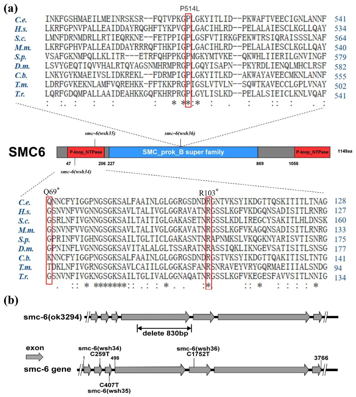
smc-6 allelic series suitable for structure–function analysis. By integrating SNP mapping, whole-genome resequencing, and targeted validation, we identified two nonsense lesions predicted to disrupt the ATPase head (
wsh34 and
wsh35) and one missense substitution in the hinge region (
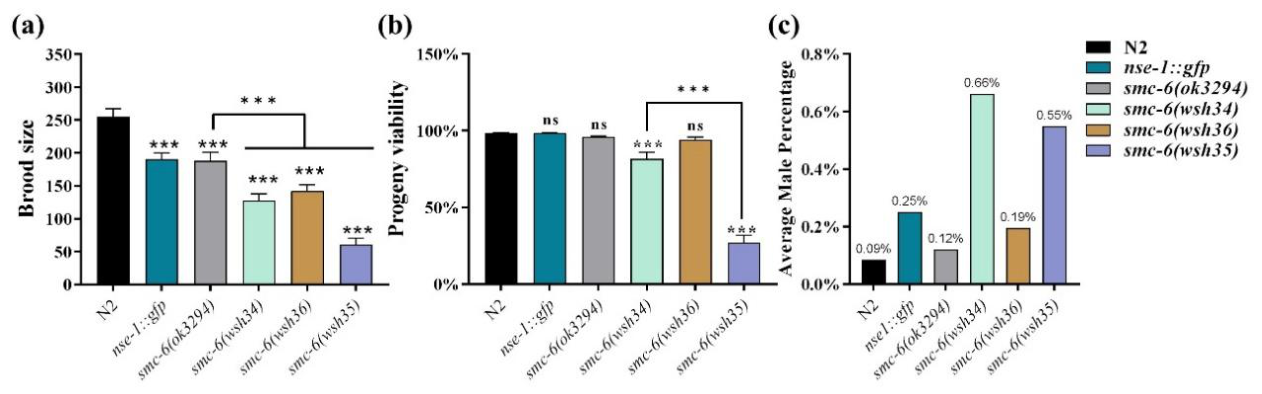
wsh36). These mutations affect conserved residues critical for SMC architecture and produced consistent, graded phenotypic differences across fertility, progeny viability, genotoxin sensitivity, developmental progression, and apoptosis-related outputs. In
C. elegans, previous studies on
smc-5, NSE-1, NSE-4, MAGE-1/NSE-3, and BRC-1/SMC-5/6 interactions have already established important roles for this pathway in genome stability, DNA repair, and meiosis [
11,
12,
13,
14,
15,
18]. Together, the data support a domain hierarchy model in which the N-terminal ATPase head of SMC-6 is indispensable for robust genome maintenance, whereas the hinge primarily contributes to structural flexibility and fine-tuning of complex function.
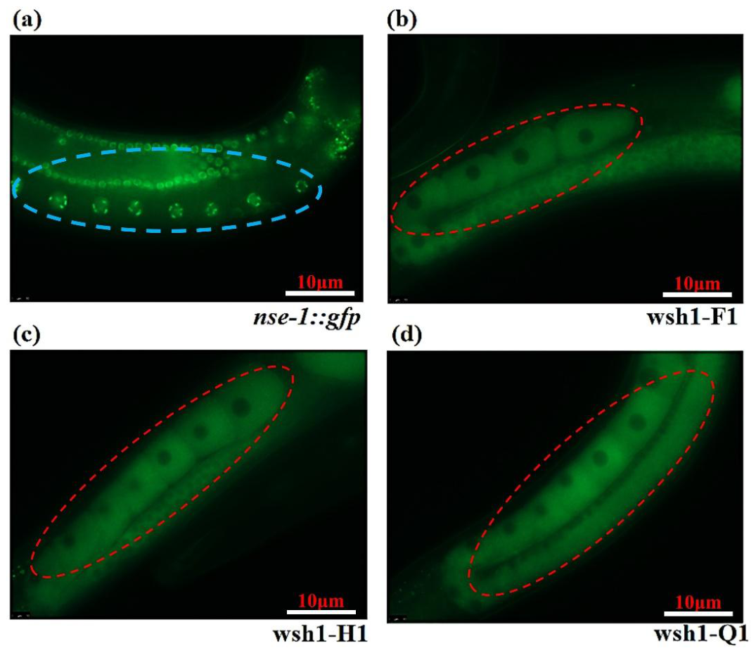
A major advance of this work is methodological. NSE-1 localization provides an information-rich proxy for SMC-5/6 functional state that can be read directly in vivo. In the parental reporter strain, NSE-1::GFP is stably enriched in germline nuclei, establishing a reliable baseline for scoring mislocalization under standardized imaging conditions. In budding yeast, Nse1 has been characterized as an integral non-SMC subunit of the SMC-5/6 complex and as part of the Nse1–Nse3–Nse4 subcomplex, with important structural and regulatory functions [
16,
17,
23]. In
C. elegans, NSE-1 has been shown to play a crucial role in meiosis and DNA repair [
14]. This strategy is therefore consistent with evidence from both yeast and nematode systems that perturbation of core SMC-5/6 architecture can be reflected in NSE-1 behavior. Notably, recent allele-series analyses of
smc-5 in
C. elegans suggested that subtle disruption at the head–kleisin connection can propagate to NSE-1 mislocalization, implicating defects in the chromosomal assembly of the holocomplex [
18]. Our findings extend this framework to SMC-6: monitoring NSE-1::GFP localization effectively captures lesions in both the ATPase and hinge modules, highlighting its sensitivity and breadth as an entry point for genetic discovery and functional stratification.
The molecular nature of the three
smc-6 alleles provides a direct explanation for phenotypic stratification.
wsh34 and
wsh35 introduce premature termination within the N-terminal ATPase head, and are expected to compromise the Walker motif framework required for ATP binding/hydrolysis as well as the coordinated head engagement mechanism. Such truncations are likely to block the ATPase-driven conformational cycle [
24], consistent with severe fertility defects, pronounced hypersensitivity to genotoxic agents, and strong developmental delay. In contrast,
wsh36 carries a P514L substitution at a highly conserved hinge residue. Given the strong evolutionary conservation of this position, it is more likely to support hinge geometry and opening–closing dynamics rather than directly abolish catalytic activity. Accordingly,
wsh36 displays intermediate phenotypes and partial stress sensitivity, consistent with a hypomorphic allele: core assembly and basal function may be retained, while efficiency or regulatory precision is reduced.
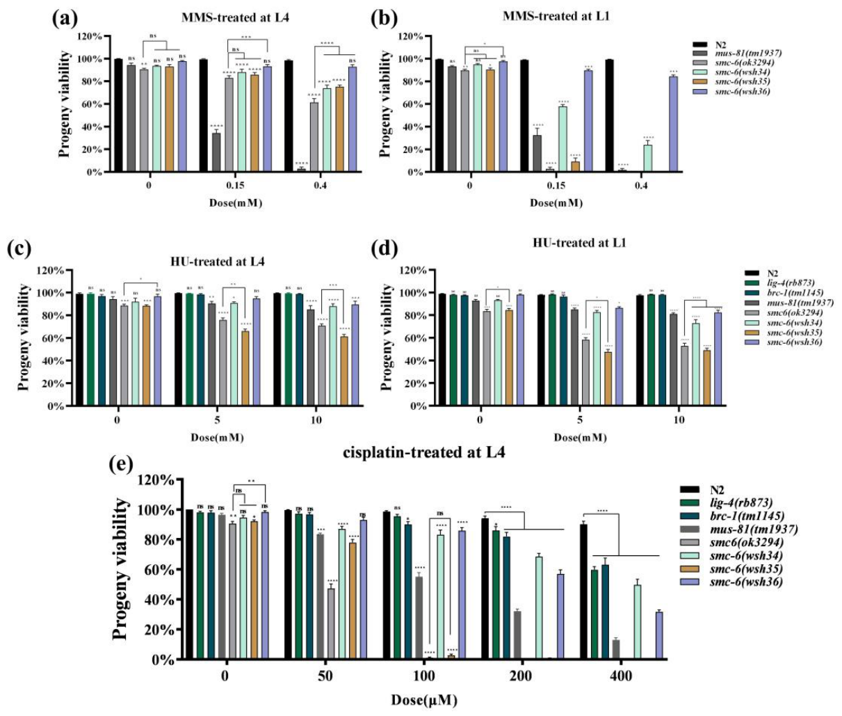
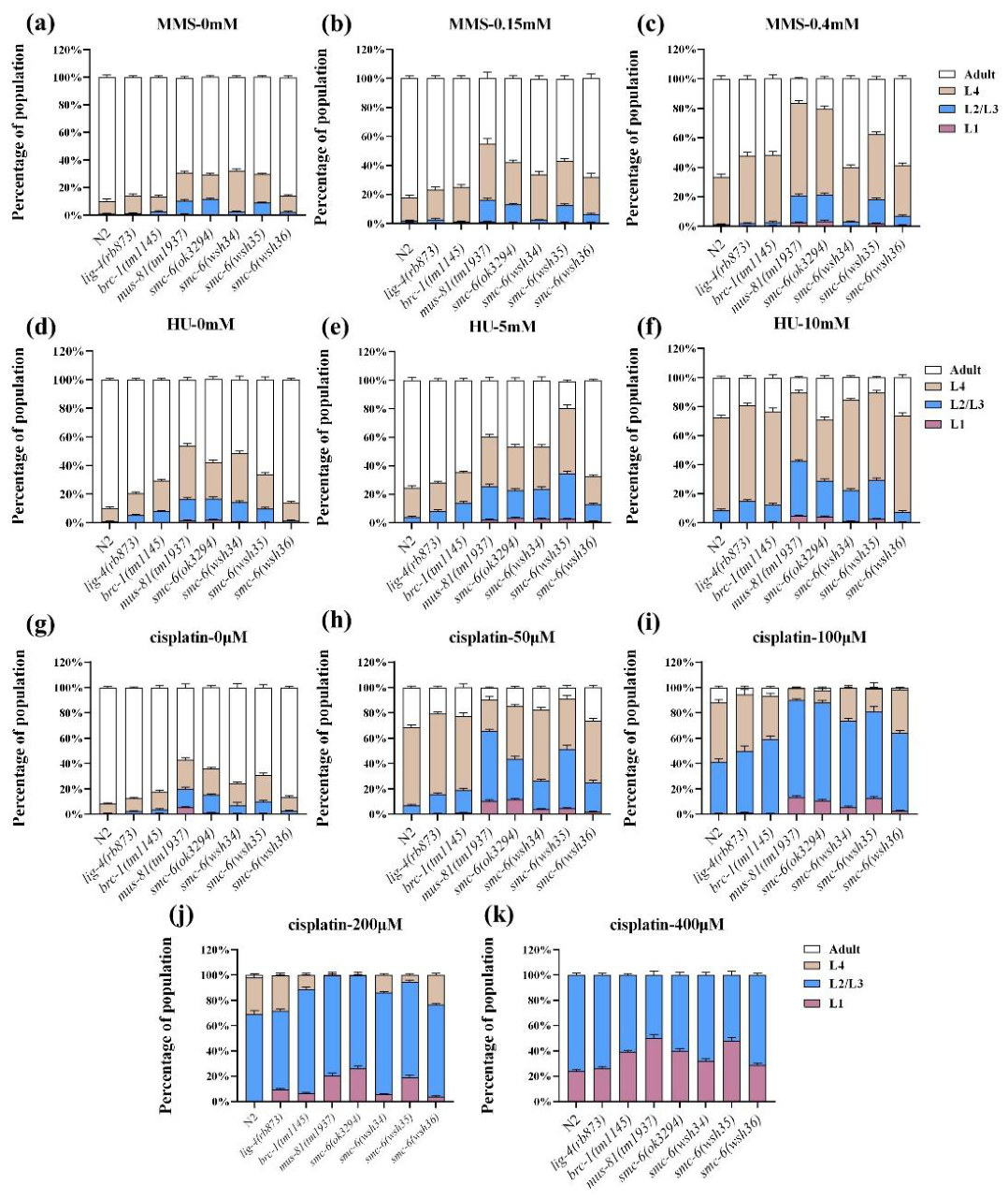
Across lesion types, we observed a clear functional hierarchy between ATPase-head and hinge mutations. Cisplatin (interstrand crosslinks) most strongly separated allele severity: the severe ATPase allele
wsh35 closely resembled the strong loss-of-function background
ok3294 and exhibited dose–response profiles overlapping those of the replication intermediate processing mutant
mus-81, whereas
wsh36 became overtly sensitive only at higher cisplatin doses. These patterns suggest that, under highly toxic replication-blocking lesions, ATPase-driven conformational switching of SMC-6 may be essential for stabilizing, remodeling, or processing stalled replication intermediates [
3,
5,
25]. Consistently, after MMS, HU, or cisplatin exposure of synchronized L1 larvae,
smc-6 mutants accumulated at larval stages in a dose-dependent manner, with the most pronounced arrest in ATPase-compromised alleles, indicating that genome maintenance defects under replication stress can be amplified into organismal developmental fragility [
20].
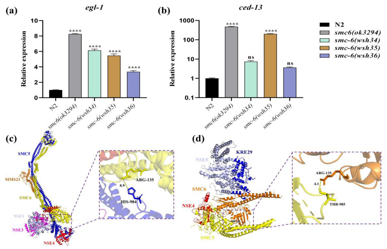
We further linked genome instability to reproductive defects through apoptosis-associated molecular readouts. RT–qPCR revealed robust induction of the CEP-1/p53 target genes
egl-1 and
ced-13 in severe backgrounds (
ok3294 and
wsh35), whereas induction in the hinge allele
wsh36 was weak or not significant. Notably,
wsh34 exhibited a mixed pattern (
egl-1 induced,
ced-13 unchanged), suggesting that distinct alleles may engage different branches or activation thresholds within the germline damage response. Together with reduced fertility and hatching rates, these data support a model in which persistent DNA damage elevates checkpoint and apoptotic pressure [
26], reducing the pool of functional germ cells and compromising embryo viability. In this sense, the allelic series helps distinguish catastrophic loss of repair capacity (ATPase truncations) from structural or regulatory perturbations (hinge missense), the latter permitting viability but lowering robustness under stress.
Structural modeling provides a physical framework for these domain-dependent outcomes. Homology modeling guided by the budding-yeast SMC-5/6 cryo-EM architecture places the residue corresponding to
wsh35 at or near the SMC5–SMC6 head dimer interface [
24], a key node that couples ATP binding/hydrolysis to head engagement and higher-order conformational organization of the holocomplex [
27]. Thus, truncation of the head module is expected to destabilize interface integrity and/or impair proper assembly and productive chromatin-bound conformations. In contrast, P514L is more plausibly modeled as a subtle mechanical perturbation of hinge properties [
28,
29]—altering local backbone constraints and steric environment—thereby affecting conformational flexibility without necessarily abolishing global folding or head function. Within this structural framework, the yeast-based model provides a useful reference for interpreting the positions of the corresponding
C. elegans lesions and their associated in vivo phenotypes. Integrating localization, genetics, and structural information, we propose a working model in which ATPase-head integrity is required to maintain or transition into effective chromatin-engaged repair states, and its disruption leads to broad replication-associated sensitivity and strong apoptotic activation; hinge perturbation primarily reduces the efficiency of conformational transitions and/or chromatin interactions, producing a hypomorphic phenotypic spectrum.
Several limitations remain and motivate future work. First, although mapping and sequencing strongly implicate smc-6, definitive causality can be further established by transgenic rescue or precise CRISPR recreation of each allele in a clean background. Second, our inference of holocomplex integrity largely derives from localization and organism-level phenotypes; direct measurements of complex assembly/stability, chromatin-binding dynamics, and ATPase-dependent conformational cycling (e.g., co-immunoprecipitation, fractionation, live-imaging recovery assays, or ChIP occupancy) will be necessary to test the proposed model. Third, positioning SMC-6 within repair pathway networks will require systematic genetic interaction analyses with brc-1, lig-4, mus-81 and related backgrounds to resolve upstream/downstream or parallel relationships under distinct lesion contexts. Finally, continued identification of the remaining non–smc-6 mutants recovered from the NSE-1::GFP screen may uncover additional regulatory nodes (e.g., SUMO pathway factors or kleisin/interface components), expanding our understanding of SMC-5/6 assembly and regulation in vivo.
In summary, we establish NSE-1::GFP nuclear localization as a practical in vivo indicator of SMC-5/6 integrity in
C. elegans and generate a new
smc-6 allelic series that reveals a clear division of labor and hierarchy between ATPase-head–mediated core functions and hinge-mediated structural regulation. Together with previous
C. elegans studies on the SMC-5/6 pathway [
11,
12,
13,
14,
15,
18], this genetic toolkit and the domain hierarchy model provide a foundation for dissecting how SMC-5/6 coordinates replication stress responses, lesion processing, and germline genome protection in the nematode.
4. Materials and Methods
4.1. Reagents and Instruments
NGM plates and M9 buffer were prepared according to standard
C. elegans procedures [
10]. EMS, MMS, HU, and cisplatin were purchased from Sigma-Aldrich; levamisole was obtained from Solarbio. Molecular biology reagents were purchased from GenStar, Takara (Taq DNA polymerase and PrimeSTAR Max DNA polymerase, Beijing Kangrun Chengye Biotechnology Co., Ltd., Beijing, China), TIANGEN (RNA extraction kit Takara Biomedical Technology (Beijing) Co., Ltd., Beijing, China,), and Vazyme (HiScript
® III RT SuperMix and 2 × ChamQ SYBR Master Mix, Vazyme Biotech Co., Ltd., Nanjing, China). Key equipment included a Motic SMZ-168 stereomicroscope (Motic China Group Co., Ltd., Xiamen, China), a Leica DM6B fluorescence microscope (Leica Microsystems CMS GmbH, Wetzlar, Germany), a GeneExplorer thermal cycler (Hangzhou Bioer Technology Co., Ltd., Hangzhou, China), a Roche LightCycler
® 96 real-time PCR system (Roche Diagnostics GmbH, Mannheim, Germany), a DYY-6D electrophoresis unit (Beijing Liuyi Biotechnology Co., Ltd., Beijing, China), an H3-20KR refrigerated centrifuge (Hunan Kecheng Instrument Equipment Co., Ltd., Changsha, China), and a −80 °C ultra-low freezer (Qingdao Haier Biomedical Co., Ltd., Qingdao, China). The wild-type strain was N2 (Bristol); CB4856 (Hawaiian) was used for SNP mapping. The
nse-1::gfp reporter was generated in-house by microinjection in the N2 strain. DNA repair pathway control mutants (
mus-81,
brc-1, and
lig-4) were maintained under the same standard conditions as other worm strains, namely at 20 °C on NGM agar plates seeded with
Escherichia coli OP50, and M9 buffer was used for washing, transfer, and treatments.
4.2. Worm Culture and Maintenance
A transgenic
nse-1::gfp reporter strain, previously generated in our laboratory, was used throughout this study [
12]. The reporter expresses an NSE-1–GFP fusion protein in an otherwise wild-type background to enable visualization of NSE-1 subcellular localization; unless otherwise stated, all mutant strains were generated and analyzed in the
nse-1::gfp background. Worms were maintained at 20 °C on NGM agar plates seeded with
Escherichia coli OP50, and M9 buffer was used for washing, transfer, and treatments [
30]. When synchronization was required, embryos were obtained either by alkaline hypochlorite bleaching or by egg laying from gravid adults, and were hatched overnight in M9 without food to obtain synchronized L1 larvae. Developmental stages were assigned based on body size and morphological criteria, and stage-matched animals were used in all assays to minimize the confounding effects of developmental variation.
4.3. nse-1::gfp Reporter and Localization Scoring
In the nse-1::gfp reporter strain, NSE-1::GFP is predominantly nuclear under normal conditions and displays a relatively stable intranuclear distribution. Because NSE-1 is an essential subunit of the SMC-5/6 complex and its localization depends on complex integrity and functional status, NSE-1::GFP nuclear localization was used as a phenotypic readout to establish the screening platform for identifying SMC-5/6 dysfunction. For screening and subsequent phenotypic analyses, stage-matched animals were selected. After mounting, images of gonadal cells were acquired by fluorescence microscopy, using the stable nuclear enrichment pattern observed in the nse-1::gfp control as the baseline for normal localization. All samples were imaged using identical objectives and acquisition settings to ensure comparability. Aberrant localization was scored when nuclear enrichment was markedly reduced and accompanied by a diffuse signal.
4.4. EMS-Based Forward Screen and Mutant Line Establishment
An EMS-based forward genetic screen was performed in the nse-1::gfp reporter background to identify genetic factors affecting NSE-1 subcellular localization. The workflow comprised chemical mutagenesis, phenotypic screening, and outcrossing to reduce background mutations. Synchronized L1 larvae were cultured on OP50-seeded NGM plates at 20 °C until the L4 stage, washed from plates with M9 buffer, and collected by gentle centrifugation. Worms were resuspended in 2 mL M9, EMS was added to a final concentration of 50 mM, and animals were incubated at 20 °C for 4 h. All EMS handling was performed in a chemical fume hood with appropriate personal protective equipment, and EMS-contaminated waste was collected and disposed of in accordance with institutional safety guidelines. Mutagenesis was terminated by washing twice with M9 to remove residual EMS, followed by recovery on fresh OP50-seeded NGM plates.
P0 animals were allowed to self-fertilize to generate F1 progeny, and F1 animals were selfed to obtain F2. Individual F2 animals were singled to establish independent lines; when the progeny reached the F3 adult stage, NSE-1::GFP localization in gonadal cells was examined under standardized imaging conditions to identify candidate mutants showing reproducible mislocalization.
Each candidate line was outcrossed to males of the parental nse-1::gfp strain to reduce EMS-induced background mutations. Young adult hermaphrodites were used as mothers and were mated with males for 12 h, after which individual animals were singled to plates. Successful crosses were inferred from an increased proportion of males among the progeny. In each subsequent generation, lines were selected that retained the NSE-1::GFP mislocalization phenotype while maintaining the reporter background. Outcrossing was repeated for four generations to obtain stable homozygous mutant strains for genetic mapping and phenotypic analyses.
4.5. SNP Mapping and Whole-Genome Sequencing
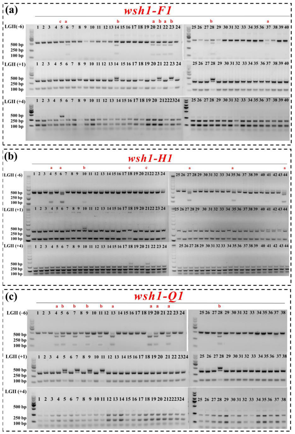
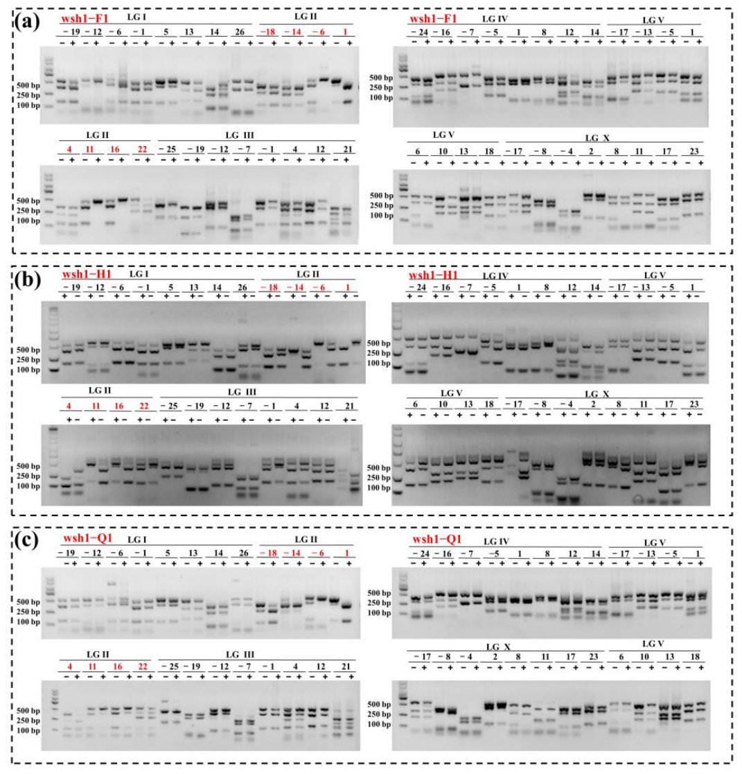
Stable homozygous mutant strains were subjected to SNP-based chromosomal mapping and interval mapping, followed by whole-genome sequencing (WGS) to systematically pinpoint the causal lesion. Initial chromosomal assignment was performed using the 48-marker SNP mapping strategy described by Davis et al. [
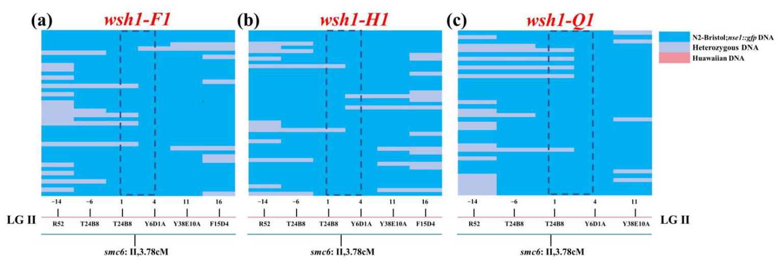
31], which distinguishes the N2 (Bristol) strain from the CB4856 (Hawaiian) isolate. Homozygous mutants were crossed with CB4856, and 20 F1 L4 animals were singled from plates with ~50% (indicative of successful crosses). After F1 self-fertilization, approximately 400 F2 L4 animals were randomly selected and propagated. When their progeny reached the F3 generation, animals were scored based on the NSE-1::GFP phenotype. For each mutant, 40–50 F3 individuals exhibiting the mutant phenotype and an equivalent number of phenotypically normal controls were collected. Animals were lysed, and samples with the same phenotypic class were pooled to generate PCR templates (phenotype-based pooling). SNP markers on each chromosome were genotyped by PCR amplification followed by DraI restriction digestion and agarose gel electrophoresis (PCR–RFLP). Recombination patterns were compared between the mutant-phenotype pool and the control pool. Enrichment of the N2 allele at markers within a chromosomal region in the mutant-phenotype pool was taken as evidence of linkage, thereby assigning the mutation to the corresponding chromosome. After chromosomal assignment, interval mapping was performed to refine the locus. Forty to fifty F3 plates showing either the mutant phenotype or segregation of the phenotype in the CB4856 background were selected. From each plate, five worms were lysed to prepare an individual PCR template (one plate per tube). Multiple SNP markers spanning the candidate chromosome were then genotyped by PCR–RFLP (PCR amplification followed by DraI digestion and gel electrophoresis). By comparing recombination events across markers, the linked interval was progressively narrowed to fine-map the mutation. Following genetic mapping, mutant strains and the parental
nse-1::gfp control were expanded in parallel, and ~3 × 10
4 worms were collected per sample.
Worms were washed repeatedly with M9 buffer to minimize OP50 bacterial contamination, pelleted, and snap-frozen in liquid nitrogen. Samples were submitted to Shanghai Institute for Biomedical and Pharmaceutical Technologies for whole-genome sequencing (WGS). Sequencing libraries were prepared using a standard Illumina paired-end DNA library protocol with an average insert size of approximately 350 bp and sequenced on an Illumina HiSeq X10 platform in paired-end 150 bp (PE150) mode, with a target coverage depth of at least 30× per sample. Raw reads were filtered to remove adapter-contaminated reads, reads containing more than 10% ambiguous bases (N), and low-quality reads in which more than 50% of bases had a Phred quality score ≤20. Clean reads were aligned to the nse-1::gfp reference genome using BWA-MEM. PCR duplicates were marked using Picard, and alignment files were processed with SAMtools. SNPs and small indels were identified using a standard variant-calling pipeline based on GATK HaplotypeCaller. Variants detected in each mutant were compared against those in the parental nse-1::gfp strain to identify mutant-specific variants. Candidate causal mutations were prioritized by integrating WGS calls with the chromosomal and interval mapping results, ultimately identifying the genes responsible for NSE-1::GFP mislocalization.
4.6. Fertility and Genotoxin Sensitivity Assays
Reproductive traits and DNA damage sensitivity were examined in mutant and control animals to assess phenotypic changes in reproductive development and DNA damage responses. All assays were performed using stage-matched worms and were repeated at least three times. OP50 was diluted in M9 buffer to facilitate accurate egg counting. For each strain, 30 L4-stage worms were singled onto plates seeded with diluted OP50 and maintained at 20 °C. Animals were transferred to fresh plates every 12 h, and the total number of eggs laid on the previous plate was counted immediately after transfer until egg laying ceased. Unhatched embryos were counted the following day, and progeny viability (hatching rate) was calculated as the number of hatched larvae divided by the total number of eggs laid. Male progeny were scored 3 days later, and the male frequency was calculated as the percentage of males among surviving animals. To apply distinct genotoxic stress conditions, cisplatin, methyl methanesulfonate (MMS), and hydroxyurea (HU) were diluted in M9 buffer and mixed with 5% concentrated OP50. L1- and L4-stage animals were exposed to the indicated agents under these conditions. Cisplatin and MMS treatments were performed for 16 h, whereas HU treatment was performed for 20 h. After drug exposure, animals were washed to remove residual compounds, and progeny hatching rates and the proportions of animals at each developmental stage were quantified.
4.7. Structural Modeling and Analysis
A homology model of C. elegans SMC-6 was built using SWISS-MODEL with the cryo-EM structure of the Saccharomyces cerevisiae SMC-5/6 complex as a template (PDB: 7Y8L). Mutations (Q69*, R103*, and P514L) were introduced and visualized in PyMOL v2.5. Structural alignments and analyses of domain–domain interactions were performed using ChimeraX v1.6. Differences in hydrogen-bonding networks and electrostatic interactions between the wild-type and mutant models were evaluated.
4.8. Statistical Analysis
All experiments were independently repeated at least three times. Data are shown as mean ± SEM. Statistical analyses were performed using GraphPad Prism 10. One-way ANOVA with Tukey’s multiple-comparisons test was used for analyses involving more than two groups, whereas two-tailed Student’s t tests were used for pairwise comparisons. Statistical significance was defined as p < 0.05.