


Abstract
Alzheimer’s disease (AD) is a progressive neurodegenerative disorder characterized by amyloid-β (Aβ) plaque deposition, neurofibrillary tau tangles, synaptic dysfunction, and progressive cognitive decline. AD is increasingly recognized as a condition in which chronic neuroinflammation actively shifts amyloid precursor protein (APP) processing toward the amyloidogenic pathway, driving Aβ production and accumulation rather than merely accompanying amyloid deposition. In this review, we examine the molecular cross-talk between inflammatory signalling and secretase regulation, highlighting how pro-inflammatory mediators promote amyloidogenic processing and contribute to downstream synaptic dysfunction. We discuss the major pathways linking glial activation to aberrant APP cleavage, including STAT3-dependent BACE1 upregulation, immune-mediated modulation of γ-secretase through IFITM3, and activation of the C/EBPβ/δ-secretase axis, which connects inflammatory stress to both amyloid and tau pathology. We further address the contribution of epigenetic mechanisms, particularly microRNA-mediated derepression of BACE1 and suppression of ADAM10, as well as SIRT3-related impairment of Aβ clearance. These interconnected processes establish a feed-forward pathogenic network in which neuroinflammation amplifies secretase imbalance, amyloidogenesis, and synaptic vulnerability. Finally, we discuss emerging multi-target therapeutic strategies aimed at modulating inflammatory signalling, restoring non-amyloidogenic APP processing, and preserving proteostatic and synaptic resilience. Collectively, this framework supports the view that targeting the inflammatory control of secretase activity may represent a biologically relevant strategy for disease modification in AD.
1. Introduction: Neuroinflammation, Secretase Dysregulation, and Synaptic Vulnerability in Alzheimer’s Disease
Alzheimer’s disease (AD) is a progressive neurodegenerative disorder characterized by worsening cognitive impairment, including memory decline, reduced reasoning abilities, and behavioral disturbances, ultimately leading to loss of functional independence [1]. Its global prevalence continues to rise in parallel with population ageing, with dementia currently affecting nearly 50 million individuals worldwide and projected to increase markedly in the coming decades [2]. Neuropathologically, AD is defined by the accumulation of amyloid-beta (Aβ) plaques and neurofibrillary tangles in brain regions subserving cognition [3].
Although Aβ deposition and tau pathology remain the defining histopathological hallmarks of AD, increasing evidence supports a more complex pathological landscape in which aberrant protein accumulation, impaired proteostasis, and chronic glial activation converge to promote amyloidogenic processing and progressive neuronal injury [4,5,6].
Notably, within this framework, early functional alterations at the synaptic level have been observed and may contribute to cognitive decline, although they are likely the consequence of a broader cascade of events rather than a primary pathogenic driver of AD [4,5].
While transgenic models have been instrumental in clarifying familial AD mechanisms, the pathogenesis of sporadic AD remains incompletely understood and is thought to involve a multifactorial interplay among genetic susceptibility, vascular and metabolic comorbidities, and environmental stressors [7].
In this context, increasing attention has focused on the reciprocal interaction between neuroinflammation and secretase dysregulation as a mechanistic axis linking extracellular inflammatory cues to amyloidogenic APP processing and, ultimately, to synaptic vulnerability. This review therefore examines how neuroinflammatory signalling regulates secretase activity and APP processing in AD, with particular emphasis on the molecular pathways that sustain amyloidogenesis and propagate towards synaptic dysfunction.
1.1. The Bidirectional Cross-Talk: The Pro-Inflammatory Cytokine Background
The pathogenesis of AD is strictly correlated to a bidirectional cross-talk between Aβ accumulation and a chronic pro-inflammatory cytokine environment [8]. The deposition of Aβ actively stimulates local microglia and astrocytes, enhancing the continuous secretion of pro-inflammatory cytokines such as interleukin-1β (IL-1β), interleukin-6 (IL-6), and tumor necrosis factor-alpha (TNF-α) [9]. Exposure to these Aβ aggregates results in chronic microglial activation, which directly causes the generation of reactive oxygen species and neurotoxic inflammatory mediators [9].
Neuroinflammation in AD should not be regarded solely as a secondary response to amyloid deposition. Instead, inflammatory signalling actively reshapes APP processing and contributes to the maintenance of a feed-forward pathogenic loop in which Aβ accumulation promotes cytokine release, while inflammatory mediators further shift APP metabolism towards the amyloidogenic pathway [8,9,10]. In this framework, cytokine signalling becomes a permissive molecular environment for sustained amyloidogenesis rather than a passive by-product of plaque formation [11].
A major component of this inflammatory transduction is the STAT3 pathway. Systemic inflammatory stimuli, including lipopolysaccharide (LPS)-induced immune activation, promote STAT3 phosphorylation in the hippocampus, where phosphorylated STAT3 translocates to the nucleus and acts as a positive transcriptional regulator of BACE1, increasing BACE1 expression and enzymatic activity. BACE1 is the β-secretase responsible for the rate-limiting step of amyloidogenic APP cleavage [11]. In parallel, persistent STAT3 activation is associated with increased IL-6, IL-1β, and TNF-α signalling, thereby reinforcing chronic glial reactivity and sustaining a neurotoxic inflammatory state [11,12,13].
Defective Aβ clearance may further intensify this inflammatory–secretase axis. In this regard, downregulation of the mitochondrial deacetylase SIRT3 has been associated with reduced expression and activity of key Aβ-degrading enzymes, including insulin-degrading enzyme (IDE) and neprilysin [14,15]. Given the broader role of SIRT3 in metabolic and cellular homeostasis, its deficiency may represent a point of convergence between metabolic dysfunction, impaired proteostasis, and progressive amyloid accumulation in AD [14,16].
1.2. Transcriptional Upregulation and Secretase Dysregulation
Aβ is produced through sequential cleavage of APP by secretase enzymes, a heterogeneous group of proteases that operate via different catalytic mechanisms to hydrolyze peptide bonds [17]. When the regulation of these proteolytic enzymes is impaired, APP processing is increasingly directed toward the amyloidogenic pathway. This shift leads to excessive Aβ production and accumulation, which in turn promotes aggregation and exerts neurotoxic effects. Within this pro-inflammatory environment, transcriptional upregulation and functional modulation of distinct secretase enzymes promote activation of the amyloid cascade [18].
Among these enzymes, BACE1, first cloned in 1999 [19,20], represents the rate-limiting β-secretase in amyloidogenic APP processing and is strongly regulated at the transcriptional level by inflammatory signalling. In particular, STAT3 activation has been shown to enhance BACE1 expression under pro-inflammatory conditions, thereby providing a direct mechanistic link between cytokine signalling and increased Aβ production [11,20,21]. Inflammatory cues also modulate γ-secretase function. In response to neuroinflammatory stress, cytokine signalling induces the expression of interferon-induced transmembrane protein 3 (IFITM3) in neurons and astrocytes. IFITM3 physically associates with the γ-secretase complex and enhances its catalytic activity towards APP-derived substrates, thereby increasing Aβ production [10,22,23].
Pro-inflammatory cytokines classically associated with AD neuroinflammation, including IL-1β, IL-6, and TNF-α, can induce expression and nuclear activation of the age-related transcription factor C/EBPβ in glial cells [24]. In this context, Aβ accumulation may promote C/EBPβ activation in part by engaging microglial inflammatory responses rather than acting as an isolated upstream signal, since Aβ and cytokines such as IL-6 can cooperatively enhance C/EBPβ activity and establish a feedback loop between amyloid and neuroinflammation [24,25]. Activated C/EBPβ directly regulates the expression of δ-secretase (asparaginyl endopeptidase/AEP), which cleaves APP and Tau, thereby linking inflammatory signalling to both amyloidogenic and tau-related pathology [26]. Through this C/EBPβ–δ-secretase axis, inflammatory and amyloidogenic signals can form a feed-forward network that progressively amplifies Aβ production, Tau truncation, neuroinflammation, and cognitive decline in AD models [24,26,27,28].
Beyond direct transcriptional control, the secretase-mediated amyloidogenic pathway may also be regulated by epigenetic factors, likely involving microRNA (miRNA) dysregulation. Indeed, it was observed that pro-inflammatory signalling, particularly via the NF-κB pathway, induces a significant downregulation of miR-338-5p in hippocampal neurons [29]. This miRNA deficiency leads to the transcriptional derepression of BACE1, as miR-338-5p directly targets the 3′-untranslated region (3′-UTR) of the BACE1 mRNA to inhibit its translation [29]. The resultant increase in BACE1 protein levels facilitates the processing of APP into Aβ, which in turn, further exacerbates neuroinflammatory signalling through microglial and receptor-mediated pathways, including NF-κB signalling pathway (Figure 1).
Figure 1.
Schematic representation of the neuroinflammation–secretase feedback loop driving amyloidogenesis in Alzheimer’s disease. Aβ deposition activates microglia and astrocytes, promoting the release of pro-inflammatory cytokines, including IL-1β, IL-6, and TNF-α. These mediators activate inflammatory transcription factors such as STAT3, NF-κB, and C/EBPβ, which enhance amyloidogenic secretase pathways, including BACE1, δ-secretase/AEP, and γ-secretase modulation, thereby sustaining further Aβ generation and chronic glial activation.
1.3. Epigenetic Regulation of Secretases: miRNA Networks and Sirtuin Dysregulation
As previously described, pro-inflammatory signalling triggers an epigenetic layer of control that derepresses amyloidogenic pathways, mainly through downregulation of miR-338-5p in hippocampal neurons. The resulting increase in Aβ oligomers in turn activates NF-κB signalling; primarily through microglial pattern-recognition receptors such as TLR2/TLR4 and RAGE, triggering MyD88/IKK-dependent phosphorylation and degradation of IκBα [30]. This enables NF-κB nuclear translocation and the transcriptional upregulation of pro-inflammatory cytokines, thereby amplifying neuroinflammatory responses. Interestingly, the epigenetic controls may also actively silence protective pathways: neuroinflammation elevates circulating and brain miR-140-3p and miR-122-5p, which act as molecular silencers of ADAM10 α-secretase by targeting its 3′-UTR; overexpression in APP/PS1 mice reduces ADAM10/sAPPα, impairs microglial chemotaxis toward plaques, and exacerbates neurite dystrophy independently of CX3CL1/TREM2 [29,31].
This dual miRNA dysregulation creates selective β-secretase de-repression coupled to α-secretase silencing (Figure 2), explaining heterogeneous hippocampal vulnerability. Furthermore, inflammation-induced mitochondrial deacetylase SIRT3 deficiency, exacerbated by Western diet in Sirt3 −/−;APP/PS1 models, impairs Aβ-degrading enzymes IDE and neprilysin while elevating BACE1; SIRT3 activation via nicotinamide riboside restores IDE/neprilysin and reduces plaques [14].
Figure 2.
Converging inflammatory pathways regulating secretase activity in AD. (A) Pro-inflammatory cytokines (IL-6, TNF-alpha) induce STAT3 phosphorylation and nuclear translocation, driving beta-secretase (red hexagon) transcriptional upregulation via direct promoter binding. (B) Amyloid-beta oligomers and IL-6 synergistically activate CEBP-beta in glial cells, promoting delta-secretase (purple triangle) expression that cleaves both APP (N373/N585) and Tau (N255/N368). (C) IFITM3, induced by innate immune signalling, physically associates with the gamma-secretase complex (blue circle) to allosterically enhance its activity toward APP-CTF, increasing Amyloid-beta 42 production. (D) Neuroinflammation elevates miR-140-3p and miR-122-5p, which post-transcriptionally suppress alpha-secretase (green square) by binding its 3-prime UTR, reducing sAPP-alpha generation and shifting APP processing toward the amyloidogenic pathway. Distinct geometric shapes differentiate secretase classes; red arrows indicate pathological upregulation.
Beyond miRNA-mediated regulation, emerging evidence suggests that long non-coding RNAs (lncRNAs) may provide an additional epigenetic layer linking neuroinflammation to secretase imbalance in AD. Among the most relevant candidates, BACE1-AS appears particularly noteworthy because it is elevated in AD and promotes amyloidogenic signalling by stabilizing BACE1 expression [32,33]. In parallel, lncRNA 51A may favour abnormal APP processing by altering SORL1 splicing and thereby weakening a major sorting pathway that normally limits amyloidogenic APP routing [34,35]. Inflammation-associated lncRNAs such as NEAT1 further support the possibility that non-coding RNA networks integrate microglial activation, BACE1-related signalling, and neuronal vulnerability [34]. Although current evidence remains less mature than that available for miRNAs, these findings suggest that lncRNAs may act as upstream amplifiers of the neuroinflammation–secretase–amyloidogenesis axis and deserve further mechanistic investigation [34].
These epigenetic mechanisms integrate seamlessly into the broader vicious cycle of neuroinflammation and amyloidogenesis. Aβ oligomers activate NF-κB signalling, which reprograms the miRNA landscape by downregulating miR-338-5p while elevating miR-140-3p/miR-122-5p, thereby derepressing BACE1 and silencing ADAM10 [29]. Concurrently, SIRT3 deficiency impairs Aβ clearance via IDE/neprilysin downregulation, creating a perfect storm where amyloidogenic processing accelerates while degradation collapses. This multilayered epigenetic modulation functions as a circuit-specific amplifier, selectively destabilizing vulnerable hippocampal networks and perpetuating secretase imbalance across disease stages [14,29,36].
2. Molecular Mechanisms Linking Neuroinflammation to BACE1 Upregulation and Amyloid Processing
Neuroinflammatory activation can be initiated through distinct systemic and central pathways, both of which converge on aberrant APP processing and BACE1 upregulation. Systemic inflammation, commonly modelled by intraperitoneal administration of lipopolysaccharide (LPS), induces peripheral immune activation that signals across the blood–brain barrier and promotes pronounced microglial activation in the hippocampus [37,38]. A complementary model of central metabolic inflammation is represented by intracerebroventricular streptozotocin (STZ), which impairs insulin signalling and induces localized brain insulin resistance [39,40]. In this context, central metabolic stress rapidly increases oxidative stress and augments BACE1 levels, thereby favouring sequential APP cleavage and neuronal Aβ production [41].
As the rate-limiting enzyme in Aβ generation, BACE1 is consistently upregulated in response to multiple pathological stimuli, including oxidative stress [8]. During neuroinflammation, reactive oxygen species promote the release of pro-inflammatory mediators, which in turn enhance BACE1 gene expression and amplify APP processing [42]. In addition to transcriptional control, BACE1 translation is regulated by stress-responsive kinases such as protein kinase R (PKR), which induces phosphorylation of eukaryotic initiation factor 2 alpha (eIF2α) under inflammatory conditions [11,43].
At the transcriptional level, NF-κB and STAT3 represent major signalling hubs linking inflammatory stress to BACE1 induction. Inflammatory stimuli activate IκB kinase (IKK), triggering IκB degradation and NF-κB nuclear translocation, which promotes APP expression and enhances BACE1 transcription [9,44]. In parallel, cytokines such as IL-6 and TNF-α induce STAT3 activation in hippocampal neurons, leading to increased BACE1 mRNA expression [11,13,45]. The relevance of this pathway is supported by studies showing that inhibition of STAT3 attenuates LPS-induced microglial activation and reduces hippocampal BACE1 protein levels [11,13,46,47,48].
Additional metabolic stressors, including hypoxia and glucose dysregulation, further enhance BACE1 activity: the BACE1 promoter contains a hypoxia response element (HRE), rendering it particularly responsive to hypoxic or ischemic conditions [49]. Under hypoxic conditions, the transcription factor hypoxia-inducible factor-1α (HIF-1α) escapes ubiquitin-proteasome degradation and stabilizes within the cell, where it binds to hypoxia response elements (HREs) in the BACE1 promoter, thereby increasing BACE1 expression and promoting β-secretase cleavage of APP [50,51]. Similarly, profound metabolic stress, such as that induced by insulin resistance or chronic hyperglycemia, triggers endoplasmic reticulum stress and thereby amplifies Aβ production [14,16]. Interventions such as intermittent hypoxic treatment can significantly modulate the expression of metabolism-regulated genes in the hippocampus and downregulate BACE1 transcript levels, highlighting the tight coupling between cellular bioenergetics and amyloidogenesis [52].
3. α-Secretase (ADAM10): The Inhibition of a Neuroprotective Shield
The non-amyloidogenic processing of APP is primarily mediated by α-secretase, particularly the ADAM10 enzyme. Under physiological conditions, ADAM10 cleaves APP within the Aβ sequence, preventing the formation of neurotoxic amyloidogenic peptides and generating the soluble APP alpha (sAPPα) fragment [53]. However, inflammatory stimuli and associated cellular stress downregulate ADAM10 expression and reduce its catalytic activity, thereby diminishing the release of sAPPα [54]. Consequently, impairment of the ADAM10-mediated pathway represents a key dysregulation of proteostasis, shifting APP processing away from the non-amyloidogenic pathway and toward β-secretase-dependent amyloidogenic cleavage.
A key mechanism underlying ADAM10 suppression involves the aberrant upregulation of specific non-coding RNAs within the neuroinflammatory brain. In particular, miR-140-3p and miR-122-5p are markedly increased in both the systemic circulation and brain parenchyma of AD models, where they function as direct post-transcriptional regulators that inhibit ADAM10 mRNA expression [36]. The overexpression of these microRNAs directly targets complementary sequences within the ADAM10 mRNA, leading to robust suppression of ADAM10 protein translation and a marked reduction in its α-secretase activity [36]. Experimental co-expression of miR-140 and miR-122 in hippocampal neurons results in the pronounced accumulation of full-length APP and a significant decrease in sAPPα levels, contributing to cognitive decline and memory impairment in transgenic models. By acting upstream of APP cleavage, these dysregulated microRNAs function as potent post-transcriptional repressors of the non-amyloidogenic pathway, thereby establishing the miR-140/miR-122-ADAM10 axis as a deleterious regulatory mechanism in AD [55]. More broadly, several miRNAs, including miR-140-5p, miR-30a-5p, miR-138, miR-144/451, miR-221, and miR-32533 (via CREB5) downregulate ADAM10, shifting APP processing away from sAPPα production and toward Aβ generation, and are implicated in AD pathogenesis [56,57,58,59].
In the healthy central nervous system, sAPPα exerts neurotrophic effects, promoting neurite outgrowth, enhancing synaptogenesis, and providing trophic support essential for neuronal survival under adverse conditions [36,60]. In the absence of constitutive sAPPα production, neurons display features of neuritic dystrophy, including reduced dendritic length, complexity, and impaired arborization. Administration of recombinant sAPPα can rescue this impaired neurite outgrowth, indicating that the dendritic structural deficits are directly linked to sAPPα deficiency rather than solely to the accumulation of Aβ [36].
Beyond its direct trophic effects on neurons, the depletion of ADAM10-derived sAPPα severely impairs innate immune function, particularly microglial chemotaxis and phagocytic clearance. In the absence of adequate sAPPα signalling, microglia display dysfunctional reactivity, characterized by reduced migration toward amyloid plaques and diminished encasement of toxic Aβ deposits [36,61]. This restrained microglial chemotaxis correlates with an increase in swollen, dystrophic neurites within the juxta-plaque microenvironment [62]. In vitro studies further demonstrate that the attenuated microglial chemotaxis and deficient Aβ phagocytosis, induced by miR-140 and miR-122 overexpression, can be rescued through the exogenous administration of sAPPα [36]. Collectively, ADAM10 suppression and the consequent loss of sAPPα not only deprive neurons of essential survival signals but also impair microglial immunosurveillance, rendering the brain more susceptible to amyloid accumulation and progressive neuronal loss.
4. δ-Secretase (AEP) and the C/EBPβ Pathological Axis
Asparagine endopeptidase (AEP), a cysteine protease, has been identified as δ-secretase [63]. This enzyme fundamentally operates as an endo/lysosomal protease [64]. Both the expression and enzymatic activity of AEP increase in an age-dependent manner in the brain [65]. AEP is implicated in early pathological processes underlying AD, where it mediates the proteolytic cleavage of both APP and microtubule-associated protein Tau [66]. Thus, AEP activation serves as a mechanistic link between cellular aging and the emergence of amyloid and tau pathologies [67].
The pathological upregulation of δ-secretase is regulated by the CCAAT-enhancer binding protein beta (C/EBPβ) transcription factor, a signalling axis influenced by the loss of neurotrophic support and external stressors [26]. Loss of BDNF/TrkB signalling in neurons increases the release of IL-1β, IL-6, and TNF-α, which activate the JAK2/STAT3 pathway. Activated STAT3 binds to the C/EBPβ promoter, inducing its transcriptional upregulation and initiating the C/EBPβ–δ-secretase–APP/tau pathogenic cascade in AD models [25]. Environmental risk factors such as traumatic brain injury (TBI), high-fat diet (HFD), and chronic cerebral hypoperfusion (CCH) act as upstream triggers that independently activate C/EBPβ in the brain [68,69,70]. In BDNF or TrkB hemizygous mice, this activation is further increased, indicating a synergistic interaction between environmental insults and reduced neurotrophic signalling. The resulting transcriptional upregulation of C/EBPβ promotes expression and activation of its downstream effector δ-secretase (AEP), thereby facilitating APP and tau cleavage and contributing to AD-like neurodegeneration [25,65].
Upon activation by the C/EBPβ axis, AEP executes a dual proteolytic mechanism targeting both APP and tau [71]. Specifically, δ-secretase cleaves the ectodomain of APP at distinct asparagine residues (N373 and N585) [27], inducing structural alterations that facilitate subsequent Aβ production by reducing steric constraints for β-site APP-cleaving enzyme 1 (BACE1) [71,72,73]. Concurrently, AEP cleaves tau at asparagine residues N255 and N368 [74]. This structural truncation abolishes the native microtubule-assembly function of tau [64]. In addition, the resulting tau 1–368 fragment is highly neurotoxic and promotes aggregation processes that contribute to neurofibrillary tangle formation [8,75].
Under chronic neuroinflammation, C/EBPβ and AEP form a self-reinforcing loop that amplifies amyloid and tau pathology, driven in part by IL-1, IL-6, and TNF-α–mediated glial activation [63,76]. Inflammatory activation of AEP (δ-secretase) generates Aβ peptides and APP fragments (C586–695), which further enhance C/EBPβ transcriptional activity and sustain neuroinflammatory signalling. This C/EBPβ–δ-secretase feedback loop upregulates C/EBPβ and AEP, driving continued APP and tau cleavage, increased Aβ production, and tau hyperphosphorylation [24,25,27]. Collectively, this self-propagating inflammatory cycle contributes to the spatiotemporal spread of amyloid plaques and tau pathology, ultimately leading to synaptic degeneration and cognitive impairment [77].
5. γ-Secretase Modulation
Unlike β-secretase and δ-secretase, γ-secretase is particularly relevant in AD not only because of its constitutive role in APP cleavage, but also because its catalytic activity can be selectively tuned by inflammation-responsive modulatory proteins. The γ-secretase intramembrane protease consists of four essential core subunits-presenilin, nicastrin, anterior pharynx-defective-1 (APH-1), and presenilin enhancer-2 (PEN-2)-which assemble in a defined stoichiometry to form the active catalytic complex [78,79]. However, despite the ubiquitous expression and assembly of these components, a substantial fraction of γ-secretase complexes remains catalytically inactive or in a low-activity state [80]. To adapt to changes in the cellular environment, γ-secretase can associate with γ-secretase modulatory proteins (GSMPs), non-essential transient interactors that fine-tune complex activity and substrate specificity by shifting the protease between low- and high-activity states [80,81,82]. In the context of AD, this modulatory plasticity is especially important because it provides a mechanistic framework through which neuroinflammatory signals may bias γ-secretase activity toward amyloidogenic APP processing. Beyond accessory protein interactions, emerging evidence suggests that inflammatory cues can also affect the functional state and stability of the core γ-secretase complex itself, which comprises four essential integral membrane subunits: presenilin (PSEN1 or PSEN2, harboring the catalytic aspartyl dyad), nicastrin, APH-1, and PEN-2 [22]. Inflammatory mediators such as TNF-α activate c-Jun N-terminal kinase (JNK), which directly phosphorylates presenilin-1 and nicastrin at specific serine/threonine residues, enhancing the stability of the PS1 C-terminal fragment and stimulating γ-secretase activity, a pivotal step in the assembly of functional complexes [83]. TNF-α also promotes expression of PEN2 and nicastrin in neurons, while γ-secretase–dependent Aβ production feeds back to induce TNF-α in glia, establishing a bidirectional cytokine–γ-secretase regulatory loop [84].
A particularly relevant example of this context-dependent modulation is interferon-induced transmembrane protein 3 (IFITM3), an innate immunity factor that directly links neuroinflammatory signalling to amyloidogenesis. During neuroinflammation, cytokines released by activated microglia and astrocytes upregulate IFITM3 expression in neurons and glial cells, where IFITM3 associates with γ-secretase and enhances APP processing and amyloid-β production [22,23,85]. Mechanistically, newly synthesized IFITM3 interacts with the presenilin-1 N-terminal fragment at or near an allosteric regulatory site, thereby selectively increasing γ-secretase activity toward APP C-terminal fragments and promoting the production of Aβ40 and Aβ42 [22,23,85]. Although this response may retain a physiological role within antimicrobial innate immunity, its chronic activation in the AD brain is likely to become maladaptive, favouring persistent amyloid accumulation and disease progression [22,23,85,86].
A second, though more controversial, example of amyloidogenic γ-secretase modulation is γ-secretase activating protein (GSAP). The 16 kDa C-terminal fragment of GSAP has been reported to associate with presenilin-1-containing γ-secretase and the APP C-terminal fragment, forming a ternary complex that selectively promotes APP-C99 cleavage and increases Aβ40/42 production, particularly Aβ42, while largely sparing Notch processing [87,88]. However, the magnitude and specificity of this effect remain debated, as several studies suggest that GSAP may also influence APP trafficking and substrate availability rather than acting exclusively as a dedicated γ-secretase activator [89]. For this reason, GSAP should be interpreted less as definitive proof of a single mechanism and more as additional evidence that γ-secretase output can be shaped by accessory modulators that alter APP-directed processing [82,87,89].
The translational importance of these findings lies in the possibility of modulating γ-secretase activity without reproducing the toxicity associated with broad enzymatic inhibition. Classical γ-secretase inhibitors bind presenilin at or near the active or substrate-binding site and indiscriminately suppress the processing of multiple substrates, including Notch-1, thereby causing the serious on-target adverse effects observed in clinical trials [90,91]. The gastrointestinal, immune-related, and cognitive toxicities associated with pan-Notch inhibition underscore the need for Notch-sparing therapeutic strategies in AD [92,93]. In this context, endogenous γ-secretase modulatory proteins such as IFITM3 and GSAP, together with small-molecule γ-secretase modulators, support the biological feasibility of selectively reducing aggregation-prone Aβ species while largely preserving Notch and other physiologically required γ-secretase-dependent pathways [92]. The therapeutic relevance of γ-secretase in this review does not rest on the enzyme as a static proteolytic complex, but on its capacity to act as an inflammation-sensitive and potentially druggable platform for selective modulation of amyloidogenic processing.
These converging mechanisms are summarized in Figure 2, which integrates transcriptional regulation (STAT3/NF-κB/C/EBPβ), γ-secretase modulation, and post-transcriptional miRNA-mediated control of secretase imbalance in AD.
6. The Glial-Secretase Feedback Loop and Synaptic Collapse
The regulation of astrocytic intracellular calcium (Ca2+) signalling represents a critical node in the pathogenesis of AD, as exposure to Aβ oligomers induces severe Ca2+ dysregulation. In astrocytes, cell surface receptors, including purinergic receptors, metabotropic glutamate receptor 5, and N-methyl-D-aspartate receptors, strictly govern this process by interacting with the inositol trisphosphate receptor (IP3R) located on the endoplasmic reticulum [10,94]. Aβ42 activates these receptors, promoting the release of Ca2+ from the endoplasmic reticulum directly into the cytoplasm. The administration of nobiletin, a polymethoxylated flavonoid [95], attenuates this Aβ42-induced increase in intracellular Ca2+ levels [10,96]. Mechanistically, nobiletin modulates IP3R, reducing its protein expression in the presence of Aβ42. Furthermore, molecular docking analysis suggests a direct interaction between nobiletin and IP3R, supporting its role in limiting aberrant endoplasmic reticulum calcium release and astrocytic dysfunction [10].
The accumulation of Aβ promotes the assembly of the NOD-, LRR- and pyrin domain-containing protein 3 (NLRP3) inflammasome in glial cells, initiating a feedback loop that exacerbates amyloidogenesis through BACE1 upregulation [97,98,99]. Once activated, the NLRP3 inflammasome recruits the adaptor protein ASC and pro-caspase-1. This complex catalyzes the proteolytic cleavage of pro-caspase-1 into its active form, which subsequently drives the maturation and secretion of the pro-inflammatory cytokine interleukin-1β [100]. Glia-derived interleukin-1β stimulates adjacent neurons to upregulate BACE1 expression, thereby amplifying the amyloidogenic proteolytic processing of the APP [101]. Pharmacological intervention utilizing Picrorhiza kurroa, a medicinal herb recognized for its anti-inflammatory properties [102], effectively suppresses the activation of the NLRP3 inflammasome, leading to a marked decrease in interleukin-1β secretion [103]. Picrorhiza kurroa downregulates BACE1 expression [103], by inducing the SIRT1–PPAR-γ signalling pathway [104]. Similarly, the compound Dl-3-n-Butylphthalide abrogates this neuroinflammatory cascade by inhibiting the physical interaction between thioredoxin-interacting protein (TXNIP) and NLRP3 [105]. This induces the suppression of caspase-1 activation and reduces interleukin-1β release [105,106,107], thereby repressing the pathological upregulation of BACE1. Ultimately, these interventions dismantle the inflammation-driven amyloidogenic feedback loop [105,107].
Within the inflammatory milieu of the Alzheimer’s brain, the precise phenotypic polarization of microglia dictates the efficiency of Aβ clearance and the trajectory of neurodegeneration [108]. Chronic exposure to Aβ peptides triggers a classically activated M1-like state [109,110]. The pro-inflammatory M1 microglial phenotype contributes directly to neurotoxicity, oxidative stress, and synaptic damage through the sustained release of mediators such as nitric oxide and reactive oxygen species [111]. M1-polarized microglia constitutively secrete high levels of pro-inflammatory cytokines, including interleukin-1β (IL-1β), tumor necrosis factor-alpha (TNF-α), and interleukin-6 (IL-6), which disrupt synaptic structure and function and promote neuronal loss [111,112]. In AD, chronic exposure to amyloid-β (Aβ) drives microglia toward a persistent pro-inflammatory state, where M1-like phenotypes increasingly predominate over reparative M2-like profiles as pathology advances [112]. Under these conditions, microglia become highly neurotoxic and exhibit phagocytic exhaustion, with impaired autophagy and reduced capacity to clear Aβ aggregates [112,113]. The M2 microglial phenotype is an anti-inflammatory, neuroprotective state that releases cytokines such as interleukin-10 (IL-10), interleukin-4 (IL-4), and transforming growth factor-β (TGF-β) to restrain neuroinflammation and support tissue repair [108,114]. Therapeutic strategies for neurodegenerative diseases increasingly emphasize modulation of microglial polarization, promoting a shift from pro-inflammatory M1 states toward protective M2-like phenotypes [115]. Inducing the transition from an M1 to an M2 phenotype has been shown to restore microglial homeostasis, enhance Aβ uptake and phagocytic activity, and promote the enzymatic and lysosomal degradation of pathological protein aggregates [108].
Unchecked pathological crosstalk between glial cells and synaptic signalling pathways drives profound remodeling and degeneration of tripartite synapses, characterized by loss of astrocytic coverage, impaired neurotransmitter clearance, and structural collapse of pre- and postsynaptic elements [116]. Under physiological conditions, astrocytes ensheathe synapses with peri-synaptic processes, forming the tripartite synapse and providing structural support that stabilizes presynaptic terminals and postsynaptic spines [117]. In this healthy state, astrocytes secrete synaptogenic and matricellular molecules and organize the peri-synaptic extracellular matrix, which are required for synapse formation, maturation, and long-term maintenance of precise pre- and postsynaptic communication [118].
Aβ accumulation in the perisynaptic space destabilizes astrocytic Ca2+ homeostasis, leading to aberrant, state-dependent Ca2+ signals that alter the timing and magnitude of gliotransmitter release [119]. Modelling and experimental work show that Aβ-related changes in mGluRs and PMCAs drive Ca2+ overload and desynchronize Ca2+ transients from glutamate/ATP release, depleting vesicle pools and compromising precise gliotransmission [120,121]. This contributes to glutamate dysregulation and excitotoxic synapse loss [121]. Aβ and tau shift microglia from protective to neurotoxic, pro-inflammatory states, with sustained cytokine release (IL-1β, TNF-α, IFN-γ) and complement-mediated synaptic engulfment that correlates with synapse loss and cognitive decline [122,123]. Astrocytes and microglia mutually amplify activation via cytokines, chemokines, and NF-κB–dependent pathways, creating a positive feedback loop of neuroinflammation and structural degradation [123]. Synaptic loss is considered an early event in AD and represents the strongest correlate of cognitive impairment [124], with converging evidence from animal models and in vivo imaging supporting its occurrence prior to overt neuronal death [125]. Reactive astrocytes around Aβ plaques lose homeostatic support, while activated microglia excessively prune or engulf synapses and dystrophic neurites, collapsing tripartite synaptic structure and function. This provides a mechanistic link by which upstream Aβ/secretase dysregulation is converted into widespread synaptic failure that defines the clinical syndrome [126].
7. Neuroinflammatory Mechanisms of Synaptic Disintegration and Proteostatic Failure
The neuroinflammatory milieu characteristic of AD contributes to structural alterations of the postsynaptic density, impairing synaptic architecture and transmission [41]. Chronic neuroinflammation, including that induced by metabolic stressors such as high-fat diet, is associated with a marked downregulation of the key scaffolding protein PSD-95 in the hippocampus [39,127]. Concurrently, the receptor for activated C kinase 1 (RACK1), a WD40 repeat-containing scaffolding protein, shows reduced membrane-associated expression during disease progression [128]. Under physiological conditions, RACK1 interacts with the NR2B subunit of N-methyl-D-aspartate (NMDA) receptors, limiting its phosphorylation by the Fyn kinase and thereby restraining excessive calcium influx [128]. Inflammatory conditions leading to RACK1 depletion may therefore disrupt NMDA receptor regulation, affecting receptor gating and trafficking and contributing to synaptic dysfunction. In addition, RACK1 interacts with the intracellular domains of the β1 and β3 subunits of gamma-aminobutyric acid A (GABA A) receptors, where it contributes to the modulation of inhibitory neurotransmission [128].
To maintain synaptic structural integrity, continuous clearance of misfolded proteins and the local synthesis of structural components are required, both of which are impaired during AD pathogenesis. Structural plasticity of dendritic spines depends on the local translation of β-actin mRNA within the growth cone, a process regulated by RACK1 through its interaction with the β-actin mRNA/Zipcode-binding protein 1 (ZBP1) complex at the ribosome [75]. In parallel, the ubiquitin-proteasome system (UPS) contributes to proteostasis by targeting aberrant proteins for degradation via specific E3 ubiquitin ligases, including carboxyl terminus of HSC70 interacting protein (CHIP) and ubiquitin carboxyl-terminal esterase-1 (UCHL-1), both of which are reduced in the AD brain. Therapeutic interventions such as forced treadmill exercise actively upregulate the expression of these E3 ligases, fundamentally restoring proteostasis [129]. Mechanistically, increased CHIP levels promote its interaction with heat shock protein 70 (HSP70), facilitating the recognition and ubiquitination of hyperphosphorylated Tau, for proteasomal degradation. In addition, upregulation of UCHL-1 protein has been associated with enhanced degradation of BACE1, thereby reducing amyloidogenic APP processing and Aβ production [129,130,131].
The intrinsic vulnerability of the hippocampal network to neuroinflammatory and proteostatic collapse is largely determined by its exceptionally high bioenergetic demands [132,133]. Hippocampal neurons, along with those in selected cortical regions, show elevated rates of ATP production and oxygen consumption, rendering them particularly sensitive to reactive oxygen species [134,135]. This metabolic vulnerability is further exacerbated by the accumulation of Aβ, which preferentially affects brain regions characterized by high neuronal activity and energy demand [132,133,136]. Exposure to Aβ assemblies in these metabolically active regions rapidly induces oxidative stress, which in turn promotes the activation of pro-inflammatory mediators [136]. The resulting localized neuroinflammation triggers microglial activation and the release of cytokines such as IL-1β and TNF-α, which have been shown to upregulate BACE1 expression in neighboring neurons [137,138]. This establishes a self-amplifying loop in the hippocampus, where elevated metabolic stress promotes inflammatory signalling that further enhances amyloidogenic processing and synaptic dysfunction.
Within this pathogenic cascade, PI3K–Akt signalling plays a critical role in maintaining synaptic integrity and limiting neuronal loss [139,140]. In AD-like conditions, Aβ oligomers suppress PI3K/Akt signalling, leading to activation of Forkhead box O (FoxO) transcription factors, induction of pro-apoptotic molecules such as Puma, and increased neuronal apoptosis [141]. In contrast, pharmacological or receptor-mediated enhancement of PI3K/Akt signalling promotes Akt-dependent phosphorylation of glycogen synthase kinase-3β (GSK-3β) at Ser9, thereby inhibiting this pivotal Tau kinase and reducing Tau hyperphosphorylation [141,142].
PI3K/Akt activation also upregulates heat-shock factor 1 (HSF1) and downstream HSP70, contributing to the maintenance of proteostasis and facilitating the clearance of misfolded proteins through pathways including the UPS [141,143]. In experimental models, it has been shown that interventions that enhance PI3K/Akt signalling, such as regular physical exercise and selected natural compounds, increase hippocampal PI3K catalytic subunits and Akt phosphorylation at Ser473, while attenuating GSK-3β activity [144]. Sustained activation of PI3K/Akt signalling may therefore counteract multiple neurotoxic kinase pathways, while also reducing BACE1 expression and Aβ-induced apoptotic signalling, providing a broad neuroprotective mechanism that preserves synaptic function and slows cognitive decline [145].
8. Multi-Target Therapeutic Strategies Targeting the Neuroinflammation–Secretase Axis
Recent pharmacological approaches employing bioactive natural compounds have shown promising efficacy in attenuating oxidative stress–induced damage and dysregulated secretase activity in AD models [146,147]. Nobiletin, a polymethoxylated citrus flavonoid, has shown promising effects to ameliorate STZ-induced AD-like pathology through activation of SIRT1/FoxO3a-dependent autophagy. These effects are associated with reduced BACE1 activity, decreased hippocampal Aβ levels, and attenuation of oxidative stress and neuroinflammatory responses [41].
Ginsenoside Rg1 has been shown to exert robust neuroprotective effects by modulating both amyloidogenic processing and Tau pathology. In experimental AD models, it is associated with reduced BACE1 expression and Aβ1–42 deposition, decreased Tau phosphorylation, increased antioxidant defenses, and modulation of apoptotic and neuroinflammatory pathways, potentially through normalization of the Wnt/GSK-3β/β-catenin signalling cascade [148,149].
The marine xanthophyll astaxanthin has been shown to exert protective effects against LPS-induced neuroinflammation, oxidative stress, and memory impairment. Mechanistically, it has been reported to interact with the DNA-binding and linker domains of STAT3, thereby inhibiting its transcriptional activity. This is associated with reduced β-secretase expression and decreased Aβ1–42 production in both in vitro and in vivo models [150]. More broadly, astaxanthin downregulates STAT3-dependent inflammatory signalling and amyloidogenesis and has been proposed as a multi-target neuroprotective agent in AD models [150,151,152].
Beyond naturally derived molecules, the use of highly specific synthetic inhibitors represents a complementary strategy to modulate neuroinflammatory pathways in AD. Pharmacological inhibition of protein tyrosine phosphatase 1B (PTP1B) using trodusquemine in APP-based AD mouse models has been shown to prevent hippocampal neuron loss, improve spatial memory and attenuates neuroinflammatory responses [153]. Systemic administration of trodusquemine reduces IBA1/GFAP-defined gliosis and restores inhibitory phosphorylation of GSK-3β, a key Tau kinase [153,154]. In a BACE1-overexpressing co-morbidity model (PLB4), trodusquemine decreases APP levels, attenuates astrogliosis, normalizes expression of the ER chaperone BiP/GRP78, and improves glucose tolerance, linking PTP1B inhibition to both metabolic and neuroinflammatory improvements [155]. Consistent with these findings, reviews have highlighted PTP1B inhibition as a multitarget strategy capable of modulating ER-stress signalling, microglial activation, and synaptic dysfunction in AD and related disorders [154]. In a parallel immunomodulatory axis, systemic inflammation or Aβ exposure increases phosphorylated STAT3 in the hippocampus, promoting microglial and astrocyte activation, cytokine release (IL-1β, IL-6, TNF-α), and upregulation of BACE1, thereby enhancing Aβ production [11,13,156]. The small-molecule Stattic, which inhibits STAT3 activation, dimerization, and nuclear translocation, reduces LPS-induced microglial activation, lowers brain cytokines, and significantly decreases hippocampal BACE1 levels in vivo [11]. In Aβ-based mouse models, STAT3 inhibition (Stattic or other STAT3-specific drugs) attenuates reactive astrogliosis, reduces Aβ plaque load and cerebral amyloid angiopathy, normalizes network activity, and improves learning and memory [47,157]. These therapeutic approaches are summarized in Figure 3.
Figure 3.
Potential outcomes of multi-target therapeutics interrupting the neuroinflammation–secretase axis. (A) Natural compounds [nobiletin (citrus flavonoid, CID 72344), ginsenoside Rg1 (ginseng saponin, CID 441923), astaxanthin (marine xanthophyll, CID 5281224)]. (B) Synthetic inhibitors [trodusquemine (cholesterol-spermine conjugate, CID 9917968), Stattic (nitrothiophene derivative, CID 2779853)]. (C) Collective downstream effects reducing Aβ/Tau pathology, gliosis, and synaptic loss while restoring proteostasis. Chemical structures stylized from chemical classes; CIDs from PubChem.
From a rational drug design perspective, these compounds share a common mechanistic rationale: by simultaneously targeting multiple nodes of the neuroinflammation–secretase feedback loop, they circumvent the limitations of single-target strategies [158]. The underlying principle of multitarget design in AD rests on the observation that key pathological pathways—NF-κB/STAT3-mediated transcription of BACE1, C/EBPβ-driven δ-secretase upregulation, miRNA-mediated suppression of ADAM10, and glial NLRP3 inflammasome activation—are not independent but form an interconnected network in which perturbation of a single node is rapidly compensated by others [159]. Compounds such as nobiletin, ginsenoside Rg1, and astaxanthin share structural and physicochemical features (lipophilicity, blood–brain barrier permeability, pleiotropic anti-inflammatory scaffolds) that enable them to engage several of these nodes simultaneously [160]. A systematic approach to designing such inhibitors involves: (1) network pharmacology analysis to identify hub proteins at the intersection of neuroinflammatory and amyloidogenic pathways [161]; (2) computational docking and ADMET prediction to prioritize scaffolds with polypharmacological activity [162]; and (3) fragment-based or bioisosteric optimization to tune selectivity and minimize off-target toxicity [163].
9. Materials and Methods
A comprehensive literature search was conducted across the PubMed database, covering research published between January 2016 and March 2026. The primary search strategy utilized the Boolean operators: “Neuroinflammation AND Secretase Regulation in Alzheimer’s Disease”. To ensure the inclusion of contextually relevant studies and high-impact mechanistic data, the AI-powered search engine Consensus was employed to identify and prioritize peer-reviewed articles matching the specific research framework and sub-topics of this review. All scientific illustrations and schematic representations (Figure 1, Figure 2 and Figure 3) were made using BioRender.com (Created in https://BioRender.com, accessed on 4 May 2026).
10. Conclusions
Collectively, the literature data highlights a major role for neuroinflammation and secretase dysregulation in AD. These processes constitute a closely interconnected pathogenic network that contributes to both amyloid and Tau pathology. This perspective underscores the importance of therapeutic strategies aimed at interrupting this self-propagating cycle. Building on this mechanistic framework, current and emerging interventions should be evaluated not only for their ability to reduce Aβ burden, but also for their ability to modulate glial phenotypes, restore non-amyloidogenic APP processing, and support proteostatic and metabolic resilience in vulnerable hippocampal circuits. In this context, multi-target strategies integrating bioactive natural compounds, selective secretase modulators, and inhibitors of key inflammatory hubs (including STAT3, PTP1B, or NLRP3) may offer a promising avenue for disease modification, although their translational efficacy and safety profiles remain to be fully established.
A particularly promising direction for future research lies in defining how epigenetic regulators, including both miRNAs and selected lncRNAs, interact with inflammatory signalling to modulate secretase activity and amplify amyloidogenic APP processing in AD. In this perspective, non-coding RNA networks may represent not only mechanistic amplifiers of disease progression but also a potential source of more selective therapeutic targets within the neuroinflammation–secretase axis.
The complexity of this bidirectional feedback loop suggests that classical single-target therapeutic paradigms are fundamentally insufficient to halt the irreversible progression of AD. Historical approaches based on single-modality drugs, such as broad-spectrum γ-secretase inhibitors, failed decisively in clinical trials due to their indiscriminate attenuation of essential physiological pathways. Complete inhibition of γ-secretase disrupts the essential proteolytic processing of the Notch-1 receptor, which has been associated with severe side effects including gastrointestinal bleeding and a paradoxical worsening of cognition. These findings highlight the limitations of a “one-molecule one-target” strategy to adequately address the multifactorial nature of AD, encompassing oxidative stress, neuroinflammation, and amyloid toxicity. Strategies that selectively modulate the inflammatory context, such as targeting transiently binding γ-secretase modulatory proteins or leveraging pleiotropic natural compounds, may help attenuate the amyloidogenic pathway while minimizing systemic toxicity. Ultimately, modulation of the neuroinflammatory milieu represents a promising therapeutic direction, potentially acting upstream of secretase dysregulation and contributing to the preservation of synaptic integrity in the context of progressive inflammatory stress. In summary, therapeutic strategies that simultaneously target neuroinflammatory signalling and secretase activity may be required to translate mechanistic insights into clinically meaningful, long-term benefits in AD.
Author Contributions
Conceptualization, G.L.C., I.A., A.M., M.C., C.C. and M.F.A.; methodology, G.L.C. and I.A.; validation, G.L.C., I.A., A.M., M.C., C.C. and M.F.A.; formal analysis, I.A.; investigation, A.M.; resources, I.A.; data curation, I.A.; writing—original draft preparation, G.L.C., I.A., I.R. and A.M.; writing—review and editing, G.L.C., I.A., A.M., M.C. and M.F.A.; visualization, I.R.; supervision, G.L.C., I.A., A.M. and M.C.; project administration, G.L.C. All authors have read and agreed to the published version of the manuscript.
Funding
This study was supported by the 2026 Current Research Funds (RRC-2026-23688274), Ministry of Health, Italy.
Data Availability Statement
No new data were created or analyzed in this study. Data sharing is not applicable to this article.
Acknowledgments
During the preparation of this manuscript, the authors utilized Google Gemini 3.0 (updated to March 2026) to assist in conceptualizing the preliminary research outline and identifying key molecular nodes. Additionally, Google NotebookLM was used to support the synthesis of complex molecular pathways and draft preliminary sections of the text. Following the generation of these initial materials, the authors rigorously reviewed, edited, and validated all content for scientific accuracy and integrity. The authors take full responsibility for the final content and the interpretations presented in this publication.
Conflicts of Interest
The authors declare no conflicts of interest.
Abbreviations
The following abbreviations are used in this manuscript:
| AD | Alzheimer’s disease |
| Aβ | Amyloid-beta |
| AEP | Asparagine endopeptidase |
| ADAM10 | A disintegrin and metalloproteinase domain-containing protein 10 |
| APP | Amyloid precursor protein |
| APP-CTF | Amyloid precursor protein C-terminal fragment |
| BACE1 | Beta-site amyloid precursor protein-cleaving enzyme 1 |
| BDNF | Brain-derived neurotrophic factor |
| BBB | Blood–brain barrier |
| C/EBPβ | CCAAT/enhancer-binding protein beta |
| CCH | Chronic cerebral hypoperfusion |
| CHIP | Carboxyl terminus of HSC70-interacting protein |
| ER | Endoplasmic reticulum |
| FoxO | Forkhead box O |
| GABA | Gamma-aminobutyric acid |
| GFAP | Glial fibrillary acidic protein |
| GSAP | Gamma-secretase activating protein |
| GSMPs | Gamma-secretase modulatory proteins |
| GSK-3β | Glycogen synthase kinase-3 beta |
| HFD | High-fat diet |
| HIF-1α | Hypoxia-inducible factor-1 alpha |
| HRE | Hypoxia response element |
| HSF1 | Heat-shock factor 1 |
| HSP70 | Heat shock protein 70 |
| IBA1 | Ionized calcium-binding adaptor molecule 1 |
| IDE | Insulin-degrading enzyme |
| IFITM3 | Interferon-induced transmembrane protein 3 |
| IKK | IκB kinase |
| IL | Interleukin |
| IP3R | Inositol trisphosphate receptor |
| JAK2 | Janus kinase 2 |
| lncRNA | Long non-coding RNA |
| LPS | Lipopolysaccharide |
| mGluR5 | Metabotropic glutamate receptor 5 |
| miRNA | MicroRNA |
| MyD88 | Myeloid differentiation primary response 88 |
| NF-κB | Nuclear factor kappa-light-chain-enhancer of activated B cells |
| NLRP3 | NOD-, LRR- and pyrin domain-containing protein 3 |
| NMDA | N-methyl-D-aspartate |
| PEN-2 | Presenilin enhancer-2 |
| PI3K | Phosphoinositide 3-kinase |
| PKR | Protein kinase R |
| PSD-95 | Postsynaptic density protein 95 |
| PTP1B | Protein tyrosine phosphatase 1B |
| RACK1 | Receptor for activated C kinase 1 |
| RAGE | Receptor for advanced glycation end products |
| ROS | Reactive oxygen species |
| sAPPα | Soluble amyloid precursor protein alpha |
| SIRT3 | Sirtuin 3 |
| SORL1 | Sortilin-related receptor 1 |
| STAT3 | Signal transducer and activator of transcription 3 |
| STZ | Streptozotocin |
| TBI | Traumatic brain injury |
| TGF-β | Transforming growth factor beta |
| TLR | Toll-like receptor |
| TNF-α | Tumor necrosis factor alpha |
| TrkB | Tropomyosin receptor kinase B |
| TXNIP | Thioredoxin-interacting protein |
| UCHL-1 | Ubiquitin carboxyl-terminal hydrolase L1 |
| UPS | Ubiquitin–proteasome system |
| UTR | Untranslated region |
| ZBP1 | Zipcode-binding protein 1 |
References
- Kamatham, P.T.; Shukla, R.; Khatri, D.K.; Vora, L.K. Pathogenesis, Diagnostics, and Therapeutics for Alzheimer’s Disease: Breaking the Memory Barrier. Ageing Res. Rev. 2024, 101, 102481. [Google Scholar] [CrossRef] [Scilit]
- Aranda, M.P.; Kremer, I.N.; Hinton, L.; Zissimopoulos, J.; Whitmer, R.A.; Hummel, C.H.; Trejo, L.; Fabius, C. Impact of Dementia: Health Disparities, Population Trends, Care Interventions, and Economic Costs. J. Am. Geriatr. Soc. 2021, 69, 1774–1783. [Google Scholar] [CrossRef] [Scilit]
- Murakami, S.; Lacayo, P. Biological and Disease Hallmarks of Alzheimer’s Disease Defined by Alzheimer’s Disease Genes. Front. Aging Neurosci. 2022, 14, 996030. [Google Scholar] [CrossRef] [Scilit]
- Hardy, J.A.; Higgins, G.A. Alzheimer’s Disease: The Amyloid Cascade Hypothesis. Science 1992, 256, 184–185. [Google Scholar] [CrossRef] [Scilit]
- Sandhu, M.; Irfan, H.M.; Shah, S.A.; Ahmed, M.; Naz, I.; Akram, M.; Fatima, H.; Farooq, A.S. Friedelin Attenuates Neuronal Dysfunction and Memory Impairment by Inhibition of the Activated JNK/NF-κB Signalling Pathway in Scopolamine-Induced Mice Model of Neurodegeneration. Molecules 2022, 27, 4513. [Google Scholar] [CrossRef] [Scilit] [PubMed]
- Ibrahim, W.W.; Ismail, H.M.; Khattab, M.M.; Abdelkader, N.F. Cognitive Enhancing Effect of Diapocynin in D-Galactose-Ovariectomy-Induced Alzheimer’s-like Disease in Rats: Role of ERK, GSK-3β, and JNK Signaling. Toxicol. Appl. Pharmacol. 2020, 398, 115028. [Google Scholar] [CrossRef] [Scilit] [PubMed]
- Kowalska, A. The genetics of dementias, Part 3: A molecular basis for the multifactorial inheritance of sporadic Alzheimer’s disease. Postepy Hig. Med. Dosw. 2009, 63, 577–582. [Google Scholar]
- Iliyasu, M.O.; Musa, S.A.; Oladele, S.B.; Iliya, A.I. Amyloid-Beta Aggregation Implicates Multiple Pathways in Alzheimer’s Disease: Understanding the Mechanisms. Front. Neurosci. 2023, 17, 1081938. [Google Scholar] [CrossRef] [Scilit] [PubMed]
- Ahn, E.H.; Park, J.-B. Molecular Mechanisms of Alzheimer’s Disease Induced by Amyloid-β and Tau Phosphorylation Along with RhoA Activity: Perspective of RhoA/Rho-Associated Protein Kinase Inhibitors for Neuronal Therapy. Cells 2025, 14, 89. [Google Scholar] [CrossRef] [Scilit]
- Sanjay; Sood, R.; Jaiswal, V.; Kang, S.-U.; Park, M.; Lee, H.-J. Nobiletin Regulates Intracellular Ca2+ Levels via IP3R and Ameliorates Neuroinflammation in Aβ42-Induced Astrocytes. Redox Biol. 2024, 73, 103197. [Google Scholar] [CrossRef] [Scilit]
- Millot, P.; San, C.; Bennana, E.; Porte, B.; Vignal, N.; Hugon, J.; Paquet, C.; Hosten, B.; Mouton-Liger, F. STAT3 Inhibition Protects against Neuroinflammation and BACE1 Upregulation Induced by Systemic Inflammation. Immunol. Lett. 2020, 228, 129–134. [Google Scholar] [CrossRef] [Scilit] [PubMed]
- Lee, H.-J.; Hoe, H.-S. Inhibition of CDK4/6 Regulates AD Pathology, Neuroinflammation and Cognitive Function through DYRK1A/STAT3 Signaling. Pharmacol. Res. 2023, 190, 106725. [Google Scholar] [CrossRef] [Scilit] [PubMed]
- Toral-Rios, D.; Patiño-López, G.; Gómez-Lira, G.; Gutiérrez, R.; Becerril-Pérez, F.; Rosales-Córdova, A.; León-Contreras, J.C.; Hernández-Pando, R.; León-Rivera, I.; Soto-Cruz, I.; et al. Activation of STAT3 Regulates Reactive Astrogliosis and Neuronal Death Induced by AβO Neurotoxicity. Int. J. Mol. Sci. 2020, 21, 7458. [Google Scholar] [CrossRef] [Scilit]
- Tyagi, A.; Musa, M.; Labeikovsky, W.; Pugazhenthi, S. Sirt3 Deficiency Induced down Regulation of Insulin Degrading Enzyme in Comorbid Alzheimer’s Disease with Metabolic Syndrome. Sci. Rep. 2022, 12, 19808. [Google Scholar] [CrossRef] [Scilit] [PubMed]
- Tyagi, A.; Pugazhenthi, S. A Promising Strategy to Treat Neurodegenerative Diseases by SIRT3 Activation. Int. J. Mol. Sci. 2023, 24, 1615. [Google Scholar] [CrossRef] [Scilit]
- Tyagi, A.; Mirita, C.; Taher, N.; Shah, I.; Moeller, E.; Tyagi, A.; Chong, T.; Pugazhenthi, S. Metabolic Syndrome Exacerbates Amyloid Pathology in a Comorbid Alzheimer’s Mouse Model. Biochim. Biophys. Acta (BBA) Mol. Basis Dis. 2020, 1866, 165849. [Google Scholar] [CrossRef] [Scilit]
- Landman, N.; Kim, T.-W. Got RIP? Presenilin-Dependent Intramembrane Proteolysis in Growth Factor Receptor Signaling. Cytokine Growth Factor Rev. 2004, 15, 337–351. [Google Scholar] [CrossRef] [Scilit]
- Cheng, F.; Fransson, L.-Å.; Mani, K. Proinflammatory Cytokines Induce Accumulation of Glypican-1-Derived Heparan Sulfate and the C-Terminal Fragment of β-Cleaved APP in Autophagosomes of Dividing Neuronal Cells. Glycobiology 2020, 30, 539–549. [Google Scholar] [CrossRef] [Scilit]
- Sinha, S.; Anderson, J.P.; Barbour, R.; Basi, G.S.; Caccavello, R.; Davis, D.; Doan, M.; Dovey, H.F.; Frigon, N.; Hong, J.; et al. Purification and Cloning of Amyloid Precursor Protein Beta-Secretase from Human Brain. Nature 1999, 402, 537–540. [Google Scholar] [CrossRef] [Scilit]
- Vassar, R.; Bennett, B.D.; Babu-Khan, S.; Kahn, S.; Mendiaz, E.A.; Denis, P.; Teplow, D.B.; Ross, S.; Amarante, P.; Loeloff, R.; et al. Beta-Secretase Cleavage of Alzheimer’s Amyloid Precursor Protein by the Transmembrane Aspartic Protease BACE. Science 1999, 286, 735–741. [Google Scholar] [CrossRef] [Scilit]
- Qiao, M.; Yang, H.; Liu, L.; Yu, T.; Wang, H.; Chen, X.; Zhang, Y.; Duan, A.; Lyu, S.; Wu, S.; et al. Chronic Lead Exposure in Adult Mice: Associations with miR-671/CDR1as Regulation, NF-κB Signaling, and Alzheimer’s Disease-like Pathology. Toxics 2024, 12, 410. [Google Scholar] [CrossRef] [Scilit]
- Hur, J.-Y.; Frost, G.R.; Wu, X.; Crump, C.; Pan, S.J.; Wong, E.; Barros, M.; Li, T.; Nie, P.; Zhai, Y.; et al. The Innate Immunity Protein IFITM3 Modulates γ-Secretase in Alzheimer’s Disease. Nature 2020, 586, 735–740. [Google Scholar] [CrossRef] [Scilit] [PubMed]
- Yao, A.Y.; Yan, R. Activity of Alzheimer’s γ-Secretase Is Linked to Changes of Interferon-Induced Transmembrane Proteins (IFITM) in Innate Immunity. Mol. Neurodegener. 2020, 15, 69. [Google Scholar] [CrossRef] [Scilit]
- Wang, H.; Liu, X.; Chen, S.; Ye, K. Spatiotemporal Activation of the C/EBPβ/δ-Secretase Axis Regulates the Pathogenesis of Alzheimer’s Disease. Proc. Natl. Acad. Sci. USA 2018, 115, E12427–E12434, Erratum in Proc. Natl. Acad. Sci. USA 2019, 116, 26090. [Google Scholar] [CrossRef] [Scilit]
- Wang, Z.-H.; Xiang, J.; Liu, X.; Yu, S.P.; Manfredsson, F.P.; Sandoval, I.M.; Wu, S.; Wang, J.-Z.; Ye, K. Deficiency in BDNF/TrkB Neurotrophic Activity Stimulates δ-Secretase by Upregulating C/EBPβ in Alzheimer’s Disease. Cell Rep. 2019, 28, 655–669.e5. [Google Scholar] [CrossRef] [Scilit] [PubMed]
- Wang, Z.-H.; Gong, K.; Liu, X.; Zhang, Z.; Sun, X.; Wei, Z.Z.; Yu, S.P.; Manfredsson, F.P.; Sandoval, I.M.; Johnson, P.F.; et al. C/EBPβ Regulates Delta-Secretase Expression and Mediates Pathogenesis in Mouse Models of Alzheimer’s Disease. Nat. Commun. 2018, 9, 1784, Erratum in Nat. Commun. 2019, 10, 5452. [Google Scholar] [CrossRef] [Scilit]
- Zhang, Z.; Li, X.-G.; Wang, Z.-H.; Song, M.; Yu, S.P.; Kang, S.S.; Liu, X.; Zhang, Z.; Xie, M.; Liu, G.-P.; et al. δ-Secretase-Cleaved Tau Stimulates Aβ Production via Upregulating STAT1-BACE1 Signaling in Alzheimer’s Disease. Mol. Psychiatry 2021, 26, 586–603. [Google Scholar] [CrossRef] [Scilit] [PubMed]
- Wang, J.; Li, Y.; Xia, Y. C/EBPβ as a Master Regulator of Inflammasome Signaling in Neurodegenerative Diseases: Mechanisms and Therapeutic Implications. Front. Immunol. 2025, 16, 1656165. [Google Scholar] [CrossRef] [Scilit]
- Qian, Q.; Zhang, J.; He, F.-P.; Bao, W.-X.; Zheng, T.-T.; Zhou, D.-M.; Pan, H.-Y.; Zhang, H.; Zhang, X.-Q.; He, X.; et al. Down-Regulated Expression of microRNA-338-5p Contributes to Neuropathology in Alzheimer’s Disease. FASEB J. 2019, 33, 4404–4417. [Google Scholar] [CrossRef] [Scilit]
- Valiukas, Z.; Tangalakis, K.; Apostolopoulos, V.; Feehan, J. Microglial Activation States and Their Implications for Alzheimer’s Disease. J. Prev. Alzheimer’s Dis. 2025, 12, 100013. [Google Scholar] [CrossRef] [Scilit]
- Nakajima, A.; Ohizumi, Y. Potential Benefits of Nobiletin, A Citrus Flavonoid, against Alzheimer’s Disease and Parkinson’s Disease. Int. J. Mol. Sci. 2019, 20, 3380. [Google Scholar] [CrossRef] [Scilit] [PubMed]
- Sayad, A.; Najafi, S.; Hussen, B.M.; Abdullah, S.T.; Movahedpour, A.; Taheri, M.; Hajiesmaeili, M. The Emerging Roles of the β-Secretase BACE1 and the Long Non-Coding RNA BACE1-AS in Human Diseases: A Focus on Neurodegenerative Diseases and Cancer. Front. Aging Neurosci. 2022, 14, 853180. [Google Scholar] [CrossRef] [Scilit]
- Li, F.; Wang, Y.; Yang, H.; Xu, Y.; Zhou, X.; Zhang, X.; Xie, Z.; Bi, J. The Effect of BACE1-AS on β-Amyloid Generation by Regulating BACE1 mRNA Expression. BMC Mol. Biol. 2019, 20, 23. [Google Scholar] [CrossRef] [Scilit] [PubMed]
- Lan, Z.; Chen, Y.; Jin, J.; Xu, Y.; Zhu, X. Long Non-Coding RNA: Insight Into Mechanisms of Alzheimer’s Disease. Front. Mol. Neurosci. 2022, 14, 821002. [Google Scholar] [CrossRef] [Scilit] [PubMed]
- Wu, X.; Xia, P.; Yang, L.; Lu, C.; Lu, Z. The Roles of Long Non-Coding RNAs in Alzheimer’s Disease Diagnosis, Treatment, and Their Involvement in Alzheimer’s Disease Immune Responses. Non-Coding RNA Res. 2024, 9, 659–666. [Google Scholar] [CrossRef] [Scilit] [PubMed]
- Song, C.; Li, S.; Mai, Y.; Li, L.; Dai, G.; Zhou, Y.; Liang, X.; Zou, O.M.; Wang, Y.; Zhou, L.; et al. Dysregulated Expression of miR-140 and miR-122 Compromised Microglial Chemotaxis and Led to Reduced Restriction of AD Pathology. J. Neuroinflamm. 2024, 21, 167. [Google Scholar] [CrossRef] [Scilit] [PubMed]
- Zhang, J.; Xue, B.; Jing, B.; Tian, H.; Zhang, N.; Li, M.; Lu, L.; Chen, L.; Diao, H.; Chen, Y.; et al. LPS Activates Neuroinflammatory Pathways to Induce Depression in Parkinson’s Disease-like Condition. Front. Pharmacol. 2022, 13, 961817. [Google Scholar] [CrossRef] [Scilit]
- Zhao, J.; Bi, W.; Xiao, S.; Lan, X.; Cheng, X.; Zhang, J.; Lu, D.; Wei, W.; Wang, Y.; Li, H.; et al. Neuroinflammation Induced by Lipopolysaccharide Causes Cognitive Impairment in Mice. Sci. Rep. 2019, 9, 5790. [Google Scholar] [CrossRef] [Scilit]
- Xiang, X.; Wang, X.; Wu, Y.; Hu, J.; Li, Y.; Jin, S.; Wu, X. Activation of GPR55 Attenuates Cognitive Impairment, Oxidative Stress, Neuroinflammation, and Synaptic Dysfunction in a Streptozotocin-Induced Alzheimer’s Mouse Model. Pharmacol. Biochem. Behav. 2022, 214, 173340. [Google Scholar] [CrossRef] [Scilit]
- León-Arcia, K.; Andrade-Guerrero, J.; Martínez-Orozco, H.; Villegas-Rojas, M.M.; Pérez-Segura, I.; Ramírez, I.L.; Vilches-Flores, A.; Guerra-Crespo, M.; Díaz-Miranda, S.Y.; Soto-Rojas, L.O. First Unified Time-Course of Alzheimer’s-like Pathology in the Intracerebroventricular Streptozotocin-Rat Model: A Systematic Review. Ageing Res. Rev. 2026, 113, 102918. [Google Scholar] [CrossRef] [Scilit]
- El-Maraghy, S.A.; Reda, A.; Essam, R.M.; Kortam, M.A. The Citrus Flavonoid “Nobiletin” Impedes STZ-Induced Alzheimer’s Disease in a Mouse Model through Regulating Autophagy Mastered by SIRT1/FoxO3a Mechanism. Inflammopharmacology 2023, 31, 2701–2717. [Google Scholar] [CrossRef] [Scilit]
- Yang, L.; Zhou, H.; Huang, L.; Su, Y.; Kong, L.; Ji, P.; Sun, R.; Wang, C.; Li, W.; Li, W. Stress Level of Glucocorticoid Exacerbates Neuronal Damage and Aβ Production through Activating NLRP1 Inflammasome in Primary Cultured Hippocampal Neurons of APP-PS1 Mice. Int. Immunopharmacol. 2022, 110, 108972. [Google Scholar] [CrossRef] [Scilit]
- Zhuang, J.; Cao, Y.; Guo, G.; Li, M.; Zhang, T.; He, D.; Chen, J.; Zhang, K.; Zhang, Z. Inhibition of BACE1 Attenuates Microglia-Induced Neuroinflammation after Intracerebral Hemorrhage by Suppressing STAT3 Activation. Aging 2023, 15, 7709–7726. [Google Scholar] [CrossRef] [Scilit] [PubMed]
- Zheng, Y.; Deng, Y.; Gao, J.-M.; Lv, C.; Lang, L.-H.; Shi, J.-S.; Yu, C.-Y.; Gong, Q.-H. Icariside II Inhibits Lipopolysaccharide-Induced Inflammation and Amyloid Production in Rat Astrocytes by Regulating IKK/IκB/NF-κB/BACE1 Signaling Pathway. Acta Pharmacol. Sin. 2020, 41, 154–162. [Google Scholar] [CrossRef] [Scilit]
- Liu, J.; Yuan, S.; Niu, X.; Kelleher, R.; Sheridan, H. ESR1 Dysfunction Triggers Neuroinflammation as a Critical Upstream Causative Factor of the Alzheimer’s Disease Process. Aging 2022, 14, 8595–8614. [Google Scholar] [CrossRef] [Scilit] [PubMed]
- Reichenbach, N.; Delekate, A.; Plescher, M.; Schmitt, F.; Krauss, S.; Blank, N.; Halle, A.; Petzold, G.C. Inhibition of Stat3-mediated Astrogliosis Ameliorates Pathology in an Alzheimer’s Disease Model. EMBO Mol. Med. 2019, 11, e9665. [Google Scholar] [CrossRef] [Scilit]
- Choi, M.; Kim, H.; Yang, E.-J.; Kim, H.-S. Inhibition of Stat3 Phosphorylation Attenuates Impairments in Learning and Memory in 5XFAD Mice, an Animal Model of Alzheimer’s Disease. J. Pharmacol. Sci. 2020, 143, 290–299. [Google Scholar] [CrossRef] [Scilit]
- Wen, X.; Hu, J. Targeting STAT3 Signaling Pathway in the Treatment of Alzheimer’s Disease with Compounds from Natural Products. Int. Immunopharmacol. 2024, 141, 112936. [Google Scholar] [CrossRef] [Scilit]
- Salminen, A.; Kauppinen, A.; Kaarniranta, K. Hypoxia/Ischemia Activate Processing of Amyloid Precursor Protein: Impact of Vascular Dysfunction in the Pathogenesis of Alzheimer’s Disease. J. Neurochem. 2017, 140, 536–549. [Google Scholar] [CrossRef] [Scilit] [PubMed]
- Daily, J.W.; Kang, S.; Park, S. Protection against Alzheimer’s Disease by Luteolin: Role of Brain Glucose Regulation, Anti-Inflammatory Activity, and the Gut Microbiota-Liver-Brain Axis. Biofactors 2021, 47, 218–231. [Google Scholar] [CrossRef] [Scilit]
- Alexander, C.; Li, T.; Hattori, Y.; Chiu, D.; Frost, G.R.; Jonas, L.; Liu, C.; Anderson, C.J.; Wong, E.; Park, L.; et al. Hypoxia Inducible Factor-1α Binds and Activates γ-Secretase for Aβ Production under Hypoxia and Cerebral Hypoperfusion. Mol. Psychiatry 2022, 27, 4264–4273. [Google Scholar] [CrossRef] [Scilit]
- Yue, X.; Zhou, Y.; Qiao, M.; Zhao, X.; Huang, X.; Zhao, T.; Cheng, X.; Fan, M.; Zhao, Y.; Chen, R.; et al. Intermittent Hypoxia Treatment Alleviates Memory Impairment in the 6-Month-Old APPswe/PS1dE9 Mice and Reduces Amyloid Beta Accumulation and Inflammation in the Brain. Alzheimer’s Res. Ther. 2021, 13, 194. [Google Scholar] [CrossRef] [Scilit]
- Chun, Y.S.; Cho, Y.Y.; Kwon, O.H.; Zhao, D.; Yang, H.O.; Chung, S. Substrate-Specific Activation of α-Secretase by 7-Deoxy-Trans-Dihydronarciclasine Increases Non-Amyloidogenic Processing of β-Amyloid Protein Precursor. Molecules 2020, 25, 646. [Google Scholar] [CrossRef] [Scilit]
- Khan, M.I.; Jeong, E.S.; Khan, M.Z.; Shin, J.H.; Kim, J.D. Stem Cells-Derived Exosomes Alleviate Neurodegeneration and Alzheimer’s Pathogenesis by Ameliorating Neuroinflamation, and Regulating the Associated Molecular Pathways. Sci. Rep. 2023, 13, 15731. [Google Scholar] [CrossRef] [Scilit]
- Wang, L.; Shui, X.; Diao, Y.; Chen, D.; Zhou, Y.; Lee, T.H. Potential Implications of miRNAs in the Pathogenesis, Diagnosis, and Therapeutics of Alzheimer’s Disease. Int. J. Mol. Sci. 2023, 24, 16259. [Google Scholar] [CrossRef] [Scilit]
- Akhter, R.; Shao, Y.; Shaw, M.; Formica, S.; Khrestian, M.; Leverenz, J.B.; Bekris, L.M. Regulation of ADAM10 by miR-140-5p and Potential Relevance for Alzheimer’s Disease. Neurobiol. Aging 2018, 63, 110–119. [Google Scholar] [CrossRef] [Scilit] [PubMed]
- Zeng, L.; Cai, Z.; Liu, J.; Zhao, K.; Liang, F.; Sun, T.; Li, Z.; Liu, R. miR-32533 Reduces Cognitive Impairment and Amyloid-β Overload by Targeting CREB5-Mediated Signaling Pathways in Alzheimer’s Disease. Adv. Sci. 2025, 12, e2409986. [Google Scholar] [CrossRef] [Scilit] [PubMed]
- Peron, R.; Vatanabe, I.; Manzine, P.; Camins, A.; Cominetti, M. Alpha-Secretase ADAM10 Regulation: Insights into Alzheimer’s Disease Treatment. Pharmaceuticals 2018, 11, 12. [Google Scholar] [CrossRef] [Scilit] [PubMed]
- Sierksma, A.; Lu, A.; Salta, E.; Vanden Eynden, E.; Callaerts-Vegh, Z.; D’Hooge, R.; Blum, D.; Buée, L.; Fiers, M.; De Strooper, B. Deregulation of Neuronal miRNAs Induced by Amyloid-β or TAU Pathology. Mol. Neurodegener. 2018, 13, 54. [Google Scholar] [CrossRef] [Scilit]
- Tackenberg, C.; Nitsch, R.M. The Secreted APP Ectodomain sAPPα, but Not sAPPβ, Protects Neurons against Aβ Oligomer-Induced Dendritic Spine Loss and Increased Tau Phosphorylation. Mol. Brain 2019, 12, 27. [Google Scholar] [CrossRef] [Scilit]
- Tang, Y.; Wang, Y.; Gao, Z.; Li, J.; Zhang, L.; Shi, H.; Dong, J.; Song, S.; Qian, C. sAPPα Peptide Promotes Damaged Microglia to Clear Alzheimer’s Amyloid-β via Restoring Mitochondrial Function. Chem. Eur. J. 2024, 30, e202400870. [Google Scholar] [CrossRef] [Scilit]
- Casali, B.T.; MacPherson, K.P.; Reed-Geaghan, E.G.; Landreth, G.E. Microglia Depletion Rapidly and Reversibly Alters Amyloid Pathology by Modification of Plaque Compaction and Morphologies. Neurobiol. Dis. 2020, 142, 104956. [Google Scholar] [CrossRef] [Scilit]
- Yao, Q.; Long, C.; Yi, P.; Zhang, G.; Wan, W.; Rao, X.; Ying, J.; Liang, W.; Hua, F. C/EBPβ: A Transcription Factor Associated with the Irreversible Progression of Alzheimer’s Disease. CNS Neurosci. Ther. 2024, 30, e14721. [Google Scholar] [CrossRef] [Scilit]
- Wang, J.; Hu, H.-J.; Liu, Z.-K.; Liu, J.-J.; Wang, S.-S.; Cheng, Q.; Chen, H.-Z.; Song, M. Pharmacological Inhibition of Asparaginyl Endopeptidase by δ-Secretase Inhibitor 11 Mitigates Alzheimer’s Disease-Related Pathologies in a Senescence-Accelerated Mouse Model. Transl. Neurodegener. 2021, 10, 12. [Google Scholar] [CrossRef] [Scilit]
- Wu, Z.; Chen, C.; Kang, S.S.; Liu, X.; Gu, X.; Yu, S.P.; Keene, C.D.; Cheng, L.; Ye, K. Neurotrophic Signaling Deficiency Exacerbates Environmental Risks for Alzheimer’s Disease Pathogenesis. Proc. Natl. Acad. Sci. USA 2021, 118, e2100986118. [Google Scholar] [CrossRef] [Scilit]
- Song, M. The Asparaginyl Endopeptidase Legumain: An Emerging Therapeutic Target and Potential Biomarker for Alzheimer’s Disease. Int. J. Mol. Sci. 2022, 23, 10223. [Google Scholar] [CrossRef] [Scilit]
- Wang, Z.-H.; Wu, W.; Kang, S.S.; Liu, X.; Wu, Z.; Peng, J.; Yu, S.P.; Manfredsson, F.P.; Sandoval, I.M.; Liu, X.; et al. BDNF Inhibits Neurodegenerative Disease–Associated Asparaginyl Endopeptidase Activity via Phosphorylation by AKT. JCI Insight 2018, 3, e99007. [Google Scholar] [CrossRef] [Scilit]
- Wu, Z.; Wang, Z.-H.; Liu, X.; Zhang, Z.; Gu, X.; Yu, S.P.; Keene, C.D.; Cheng, L.; Ye, K. Traumatic Brain Injury Triggers APP and Tau Cleavage by Delta-Secretase, Mediating Alzheimer’s Disease Pathology. Prog. Neurobiol. 2020, 185, 101730. [Google Scholar] [CrossRef] [Scilit]
- Liu, P.; Wang, Z.-H.; Kang, S.S.; Liu, X.; Xia, Y.; Chan, C.-B.; Ye, K. High-Fat Diet-Induced Diabetes Couples to Alzheimer’s Disease through Inflammation-Activated C/EBPβ/AEP Pathway. Mol. Psychiatry 2022, 27, 3396–3409. [Google Scholar] [CrossRef] [Scilit]
- Yu, W.; Li, Y.; Hu, J.; Wu, J.; Huang, Y. A Study on the Pathogenesis of Vascular Cognitive Impairment and Dementia: The Chronic Cerebral Hypoperfusion Hypothesis. J. Clin. Med. 2022, 11, 4742. [Google Scholar] [CrossRef] [Scilit]
- Wu, Z.; Liu, X.; Cheng, L.; Ye, K. Delta-Secretase Triggers Alzheimer’s Disease Pathologies in Wild-Type hAPP/hMAPT Double Transgenic Mice. Cell Death Dis. 2020, 11, 1058. [Google Scholar] [CrossRef] [Scilit]
- Zhang, Z.; Xie, M.; Ye, K. Asparagine Endopeptidase Is an Innovative Therapeutic Target for Neurodegenerative Diseases. Expert Opin. Ther. Targets 2016, 20, 1237–1245. [Google Scholar] [CrossRef] [Scilit]
- Liu, L.; Ding, L.; Rovere, M.; Wolfe, M.S.; Selkoe, D.J. A Cellular Complex of BACE1 and γ-Secretase Sequentially Generates Aβ from Its Full-Length Precursor. J. Cell Biol. 2019, 218, 644–663. [Google Scholar] [CrossRef] [Scilit]
- Qian, Z.; Li, B.; Meng, X.; Liao, J.; Wang, G.; Li, Y.; Luo, Q.; Ye, K. Inhibition of Asparagine Endopeptidase (AEP) Effectively Treats Sporadic Alzheimer’s Disease in Mice. Neuropsychopharmacology 2024, 49, 620–630. [Google Scholar] [CrossRef] [Scilit]
- Jiang, G.; Xie, G.; Li, X.; Xiong, J. Cytoskeletal Proteins and Alzheimer’s Disease Pathogenesis: Focusing on the Interplay with Tau Pathology. Biomolecules 2025, 15, 831. [Google Scholar] [CrossRef] [Scilit]
- Chen, C.; Ahn, E.H.; Kang, S.S.; Liu, X.; Alam, A.; Ye, K. Gut Dysbiosis Contributes to Amyloid Pathology, Associated with C/EBPβ/AEP Signaling Activation in Alzheimer’s Disease Mouse Model. Sci. Adv. 2020, 6, eaba0466. [Google Scholar] [CrossRef] [Scilit]
- Gaikwad, S.; Senapati, S.; Haque, M.A.; Kayed, R. Senescence, Brain Inflammation, and Oligomeric Tau Drive Cognitive Decline in Alzheimer’s Disease: Evidence from Clinical and Preclinical Studies. Alzheimer’s Dement. 2024, 20, 709–727. [Google Scholar] [CrossRef] [Scilit]
- Escamilla-Ayala, A.; Wouters, R.; Sannerud, R.; Annaert, W. Contribution of the Presenilins in the Cell Biology, Structure and Function of γ-Secretase. Semin. Cell Dev. Biol. 2020, 105, 12–26. [Google Scholar] [CrossRef] [Scilit]
- Kimberly, W.T.; LaVoie, M.J.; Ostaszewski, B.L.; Ye, W.; Wolfe, M.S.; Selkoe, D.J. γ-Secretase Is a Membrane Protein Complex Comprised of Presenilin, Nicastrin, Aph-1, and Pen-2. Proc. Natl. Acad. Sci. USA 2003, 100, 6382–6387. [Google Scholar] [CrossRef] [Scilit]
- Wong, E.; Frost, G.R.; Li, Y.-M. γ-Secretase Modulatory Proteins: The Guiding Hand Behind the Running Scissors. Front. Aging Neurosci. 2020, 12, 614690. [Google Scholar] [CrossRef] [Scilit]
- Hur, J.-Y. γ-Secretase in Alzheimer’s Disease. Exp. Mol. Med. 2022, 54, 433–446. [Google Scholar] [CrossRef] [Scilit]
- Wong, E.; Liao, G.P.; Chang, J.C.; Xu, P.; Li, Y.-M.; Greengard, P. GSAP Modulates γ-Secretase Specificity by Inducing Conformational Change in PS1. Proc. Natl. Acad. Sci. USA 2019, 116, 6385–6390. [Google Scholar] [CrossRef] [Scilit]
- Kuo, L.-H.; Hu, M.-K.; Hsu, W.-M.; Tung, Y.-T.; Wang, B.-J.; Tsai, W.-W.; Yen, C.-T.; Liao, Y.-F. Tumor Necrosis Factor-α–Elicited Stimulation of γ-Secretase Is Mediated by c-Jun N-Terminal Kinase-Dependent Phosphorylation of Presenilin and Nicastrin. Mol. Biol. Cell 2008, 19, 4201–4212. [Google Scholar] [CrossRef] [Scilit]
- Yu, X.; Guan, P.-P.; Zhu, D.; Liang, Y.-Y.; Wang, T.; Wang, Z.-Y.; Wang, P. Magnesium Ions Inhibit the Expression of Tumor Necrosis Factor α and the Activity of γ-Secretase in a β-Amyloid Protein-Dependent Mechanism in APP/PS1 Transgenic Mice. Front. Mol. Neurosci. 2018, 11, 172. [Google Scholar] [CrossRef] [Scilit]
- Kehs, Z.; Cross, A.C.; Li, Y.-M. From Defense to Disease: IFITM3 in Immunity and Alzheimer’s Disease. Neurotherapeutics 2025, 22, e00482. [Google Scholar] [CrossRef] [Scilit]
- Feng, Y.; Wang, S.; Yang, D.; Zheng, W.; Xia, H.; Zhu, Q.; Wang, Z.; Hu, B.; Jiang, X.; Qin, X.; et al. Inhibition of IFITM3 in Cerebrovascular Endothelium Alleviates Alzheimer’s-related Phenotypes. Alzheimer’s Dement. 2025, 21, e14543. [Google Scholar] [CrossRef] [Scilit]
- Jin, C.; Wang, J.; Wang, Y.; Jia, B.; Guo, X.; Yang, G.; Xu, P.; Greengard, P.; Zhou, R.; Shi, Y. Modulation of Amyloid Precursor Protein Cleavage by γ-Secretase Activating Protein through Phase Separation. Proc. Natl. Acad. Sci. USA 2022, 119, e2122292119. [Google Scholar] [CrossRef] [Scilit]
- Angira, D.; Chikhale, R.; Mehta, K.; Bryce, R.A.; Thiruvenkatam, V. Tracing the GSAP–APP C-99 Interaction Site in the β-Amyloid Pathway Leading to Alzheimer’s Disease. ACS Chem. Neurosci. 2019, 10, 3868–3879. [Google Scholar] [CrossRef] [Scilit]
- Chang, J.C.; Xu, P.; Wong, E.; Flajolet, M.; Li, Y.-M.; Greengard, P. GSAP Regulates Amyloid Beta Production through Modulation of Amyloid Precursor Protein Trafficking. bioRxiv 2020. [Google Scholar] [CrossRef] [Scilit]
- Yang, G.; Zhou, R.; Guo, X.; Yan, C.; Lei, J.; Shi, Y. Structural Basis of γ-Secretase Inhibition and Modulation by Small Molecule Drugs. Cell 2021, 184, 521–533.e14. [Google Scholar] [CrossRef] [Scilit]
- Guo, X.; Li, H.; Lu, X.; Liu, H.; U, K.; Yan, C.; Lei, J.; Huang, J.; Zhou, R.; Shi, Y. Structural Basis of Human γ-Secretase Inhibition by Anticancer Clinical Compounds. Nat. Struct. Mol. Biol. 2025, 32, 719–728. [Google Scholar] [CrossRef] [Scilit]
- Nordvall, G.; Lundkvist, J.; Sandin, J. Gamma-Secretase Modulators: A Promising Route for the Treatment of Alzheimer’s Disease. Front. Mol. Neurosci. 2023, 16, 1279740. [Google Scholar] [CrossRef] [Scilit]
- Zhou, B.; Lin, W.; Long, Y.; Yang, Y.; Zhang, H.; Wu, K.; Chu, Q. Notch Signaling Pathway: Architecture, Disease, and Therapeutics. Signal Transduct. Target. Ther. 2022, 7, 95. [Google Scholar] [CrossRef] [Scilit]
- Nakata, K.; Sakamoto, J.; Otomo, K.; Sato, M.; Ishii, H.; Tsutsumi, M.; Enoki, R.; Nemoto, T. Amyloid-β-Induced Alteration of Fast and Localized Calcium Elevations in Cultured Astrocytes. Sci. Rep. 2025, 15, 18944. [Google Scholar] [CrossRef] [Scilit]
- Youn, K.; Lee, S.; Jun, M. Discovery of Nobiletin from Citrus Peel as a Potent Inhibitor of β-Amyloid Peptide Toxicity. Nutrients 2019, 11, 2648. [Google Scholar] [CrossRef] [Scilit]
- Kritika; Sood, R.; Sanjay; Lee, H.-J. Nobiletin Reduces LPS-Induced Neuroinflammation through TLR4/MyD88/NF-κB and Oxidative Stress via Nrf2/HO-1 Signaling in Human Microglial HMC3 Cells. Mol. Neurobiol. 2026, 63, 103. [Google Scholar] [CrossRef] [Scilit]
- Bai, H.; Zhang, Q. Activation of NLRP3 Inflammasome and Onset of Alzheimer’s Disease. Front. Immunol. 2021, 12, 701282. [Google Scholar] [CrossRef] [Scilit]
- Van Zeller, M.; Dias, D.; Sebastião, A.M.; Valente, C.A. NLRP3 Inflammasome: A Starring Role in Amyloid-β- and Tau-Driven Pathological Events in Alzheimer’s Disease. J. Alzheimer’s Dis. 2021, 83, 939–961. [Google Scholar] [CrossRef] [Scilit]
- Liang, T.; Zhang, Y.; Wu, S.; Chen, Q.; Wang, L. The Role of NLRP3 Inflammasome in Alzheimer’s Disease and Potential Therapeutic Targets. Front. Pharmacol. 2022, 13, 845185. [Google Scholar] [CrossRef] [Scilit]
- Fu, J.; Wu, H. Structural Mechanisms of NLRP3 Inflammasome Assembly and Activation. Annu. Rev. Immunol. 2023, 41, 301–316. [Google Scholar] [CrossRef] [Scilit]
- Sutinen, E.M.; Pirttilä, T.; Anderson, G.; Salminen, A.; Ojala, J.O. Pro-Inflammatory Interleukin-18 Increases Alzheimer’s Disease-Associated Amyloid-β Production in Human Neuron-like Cells. J. Neuroinflamm. 2012, 9, 199. [Google Scholar] [CrossRef] [Scilit] [PubMed]
- Sharma, S.; Sharma, A.; Chauhan, R.S. Computational Dissection through Network Pharmacology and Structure-Based Analysis Unravels Mechanistic Actions of Bioactive Compounds in a Hepatoprotective Herb, Picrorhiza Kurroa for the Treatment of NAFLD and NASH. J. Biomol. Struct. Dyn. 2026, 44, 2899–2914. [Google Scholar] [CrossRef] [Scilit] [PubMed]
- Kim, N.; Do, J.; Ju, I.G.; Jeon, S.H.; Lee, J.K.; Oh, M.S. Picrorhiza Kurroa Prevents Memory Deficits by Inhibiting NLRP3 Inflammasome Activation and BACE1 Expression in 5xFAD Mice. Neurotherapeutics 2020, 17, 189–199. [Google Scholar] [CrossRef] [Scilit]
- Lee, W.J.; Ham, S.A.; Lee, G.H.; Choi, M.; Yoo, H.; Paek, K.S.; Lim, D.; Hong, K.; Hwang, J.S.; Seo, H.G. Activation of Peroxisome Proliferator-activated Receptor Delta Suppresses BACE 1 Expression by Up-regulating SOCS 1 in a JAK 2/STAT 1-dependent Manner. J. Neurochem. 2019, 151, 370–385. [Google Scholar] [CrossRef] [Scilit]
- Wang, C.-Y.; Xu, Y.; Wang, X.; Guo, C.; Wang, T.; Wang, Z.-Y. Dl-3-n-Butylphthalide Inhibits NLRP3 Inflammasome and Mitigates Alzheimer’s-Like Pathology via Nrf2-TXNIP-TrX Axis. Antioxid. Redox Signal. 2019, 30, 1411–1431. [Google Scholar] [CrossRef] [Scilit]
- Gao, F.; Zeng, S.; Chao, D.; Wu, L. Dl-3-n-Butylphthalide Attenuates Early Brain Injury and Delayed Neurological Dysfunction by Regulating NLRP3 Inflammasome after Subarachnoid Hemorrhage. Brain Res. Bull. 2024, 217, 111084. [Google Scholar] [CrossRef] [Scilit]
- Liu, M.; Zheng, H.; Liu, Z.; Guo, Y.; Wang, S.; Tang, Y.; Tian, H.; Zhang, Z.; Yang, G. Dl-3-n-Butylphthalide Reduced Neuroinflammation by Inhibiting Inflammasome in Microglia in Mice after Middle Cerebral Artery Occlusion. Life 2022, 12, 1244. [Google Scholar] [CrossRef] [Scilit]
- Guo, S.; Wang, H.; Yin, Y. Microglia Polarization From M1 to M2 in Neurodegenerative Diseases. Front. Aging Neurosci. 2022, 14, 815347. [Google Scholar] [CrossRef] [Scilit]
- Cheng, Y.; Chang, C.; Chang, T.; Li, H.; Hung, H.; Liu, G.; Lin, C. Aβ Stimulates Microglial Activation through Antizyme-dependent Downregulation of Ornithine Decarboxylase. J. Cell. Physiol. 2019, 234, 9733–9745. [Google Scholar] [CrossRef] [Scilit]
- Xie, L.; Zhang, N.; Zhang, Q.; Li, C.; Sandhu, A.F.; Iii, G.W.; Lin, S.; Lv, P.; Liu, Y.; Wu, Q.; et al. Inflammatory Factors and Amyloid β-Induced Microglial Polarization Promote Inflammatory Crosstalk with Astrocytes. Aging 2020, 12, 22538. [Google Scholar] [CrossRef] [Scilit] [PubMed]
- Lokesh, M.; Bandaru, L.J.M.; Rajanna, A.; Dhayal, V.S.; Challa, S. Microglial Dysfunction Mediated by Pb and Amyloid Beta Peptides as a Possible Mechanism of Neurotoxicity. J. Appl. Toxicol. 2025, 45, 2265–2277. [Google Scholar] [CrossRef] [Scilit]
- Kang, Y.J.; Hyeon, S.J.; McQuade, A.; Lim, J.; Baek, S.H.; Diep, Y.N.; Do, K.V.; Jeon, Y.; Jo, D.; Lee, C.J.; et al. Neurotoxic Microglial Activation via IFNγ-Induced Nrf2 Reduction Exacerbating Alzheimer’s Disease. Adv. Sci. 2024, 11, 2304357. [Google Scholar] [CrossRef] [Scilit] [PubMed]
- Pomilio, C.; Gorojod, R.M.; Riudavets, M.; Vinuesa, A.; Presa, J.; Gregosa, A.; Bentivegna, M.; Alaimo, A.; Alcon, S.P.; Sevlever, G.; et al. Microglial Autophagy Is Impaired by Prolonged Exposure to β-Amyloid Peptides: Evidence from Experimental Models and Alzheimer’s Disease Patients. GeroScience 2020, 42, 613–632. [Google Scholar] [CrossRef] [Scilit]
- Asl, E.R.; Hosseini, S.E.; Tahmasebi, F.; Bolandi, N.; Barati, S. MiR-124 and MiR-155 as Therapeutic Targets in Microglia-Mediated Inflammation in Multiple Sclerosis. Cell. Mol. Neurobiol. 2025, 45, 63. [Google Scholar] [CrossRef] [Scilit] [PubMed]
- Darwish, S.F.; Elbadry, A.M.M.; Elbokhomy, A.S.; Salama, G.A.; Salama, R.M. The Dual Face of Microglia (M1/M2) as a Potential Target in the Protective Effect of Nutraceuticals against Neurodegenerative Diseases. Front. Aging 2023, 4, 1231706. [Google Scholar] [CrossRef] [Scilit]
- MacLean, M.; López-Díez, R.; Vasquez, C.; Gugger, P.F.; Schmidt, A.M. Neuronal–Glial Communication Perturbations in Murine SOD1G93A Spinal Cord. Commun. Biol. 2022, 5, 177. [Google Scholar] [CrossRef] [Scilit] [PubMed]
- Pathak, D.; Sriram, K. Neuron-Astrocyte Omnidirectional Signaling in Neurological Health and Disease. Front. Mol. Neurosci. 2023, 16, 1169320. [Google Scholar] [CrossRef] [Scilit]
- Tan, C.X.; Eroglu, C. Cell Adhesion Molecules Regulating Astrocyte–Neuron Interactions. Curr. Opin. Neurobiol. 2021, 69, 170–177. [Google Scholar] [CrossRef] [Scilit]
- Pillai, A.G.; Nadkarni, S. Amyloid Pathology Disrupts Gliotransmitter Release in Astrocytes. PLoS Comput. Biol. 2022, 18, e1010334. [Google Scholar] [CrossRef] [Scilit]
- Pham, C.; Hérault, K.; Oheim, M.; Maldera, S.; Vialou, V.; Cauli, B.; Li, D. Astrocytes Respond to a Neurotoxic Aβ Fragment with State-Dependent Ca2+ Alteration and Multiphasic Transmitter Release. Acta Neuropathol. Commun. 2021, 9, 44. [Google Scholar] [CrossRef] [Scilit]
- Verma, M.; Lizama, B.N.; Chu, C.T. Excitotoxicity, Calcium and Mitochondria: A Triad in Synaptic Neurodegeneration. Transl. Neurodegener. 2022, 11, 3. [Google Scholar] [CrossRef] [Scilit]
- Astillero-Lopez, V.; Villar-Conde, S.; Gonzalez-Rodriguez, M.; Flores-Cuadrado, A.; Ubeda-Banon, I.; Saiz-Sanchez, D.; Martinez-Marcos, A. Proteomic Analysis Identifies HSP90AA1, PTK2B, and ANXA2 in the Human Entorhinal Cortex in Alzheimer’s Disease: Potential Role in Synaptic Homeostasis and Aβ Pathology through Microglial and Astroglial Cells. Brain Pathol. 2024, 34, e13235. [Google Scholar] [CrossRef] [Scilit]
- Kano, S.; Choi, E.Y.; Dohi, E.; Agarwal, S.; Chang, D.J.; Wilson, A.M.; Lo, B.D.; Rose, I.V.L.; Gonzalez, S.; Imai, T.; et al. Glutathione S -Transferases Promote Proinflammatory Astrocyte-Microglia Communication during Brain Inflammation. Sci. Signal. 2019, 12, eaar2124. [Google Scholar] [CrossRef] [Scilit]
- Subramanian, J.; Savage, J.C.; Tremblay, M.-È. Synaptic Loss in Alzheimer’s Disease: Mechanistic Insights Provided by Two-Photon In Vivo Imaging of Transgenic Mouse Models. Front. Cell. Neurosci. 2020, 14, 592607. [Google Scholar] [CrossRef] [Scilit]
- Henstridge, C.M.; Tzioras, M.; Paolicelli, R.C. Glial Contribution to Excitatory and Inhibitory Synapse Loss in Neurodegeneration. Front. Cell. Neurosci. 2019, 13, 63. [Google Scholar] [CrossRef] [Scilit] [PubMed]
- Deng, Q.; Wu, C.; Parker, E.; Liu, T.C.-Y.; Duan, R.; Yang, L. Microglia and Astrocytes in Alzheimer’s Disease: Significance and Summary of Recent Advances. Aging Dis. 2024, 15, 1537–1564. [Google Scholar] [CrossRef] [Scilit]
- Tyrtyshnaia, A.; Manzhulo, I.; Egoraeva, A.; Ivashkevich, D. Cognitive and Affective Dysregulation in Neuropathic Pain: Associated Hippocampal Remodeling and Microglial Activation. Int. J. Mol. Sci. 2025, 26, 6460. [Google Scholar] [CrossRef] [Scilit] [PubMed]
- He, W.; Shi, X.; Dong, Z. The Roles of RACK1 in the Pathogenesis of Alzheimer’s Disease. J. Biomed. Res. 2024, 38, 137–148. [Google Scholar] [CrossRef] [Scilit]
- Xu, L.; Li, M.; Wei, A.; Yang, M.; Li, C.; Liu, R.; Zheng, Y.; Chen, Y.; Wang, Z.; Wang, K.; et al. Treadmill Exercise Promotes E3 Ubiquitin Ligase to Remove Amyloid β and P-Tau and Improve Cognitive Ability in APP/PS1 Transgenic Mice. J. Neuroinflamm. 2022, 19, 243. [Google Scholar] [CrossRef] [Scilit]
- Moberg, I.; McCarthy, S.F.; Bellaflor, S.; Finch, M.S.; Hazell, T.J.; MacPherson, R.E.K. Lactate Increases ADAM10 Activity and Reduces BACE1 Activity in Mouse Brain. J. Physiol. 2024, 602, 5217–5228. [Google Scholar] [CrossRef] [Scilit] [PubMed]
- Baranowski, B.J.; Mohammad, A.; Finch, M.S.; Brown, A.; Dhaliwal, R.; Marko, D.M.; LeBlanc, P.J.; McCormick, C.M.; Fajardo, V.A.; MacPherson, R.E.K. Exercise Training and BDNF Injections Alter Amyloid Precursor Protein (APP) Processing Enzymes and Improve Cognition. J. Appl. Physiol. 2023, 135, 121–135. [Google Scholar] [CrossRef] [Scilit]
- Naia, L.; Shimozawa, M.; Bereczki, E.; Li, X.; Liu, J.; Jiang, R.; Giraud, R.; Leal, N.S.; Pinho, C.M.; Berger, E.; et al. Mitochondrial Hypermetabolism Precedes Impaired Autophagy and Synaptic Disorganization in App Knock-in Alzheimer Mouse Models. Mol. Psychiatry 2023, 28, 3966–3981. [Google Scholar] [CrossRef] [Scilit]
- Andersen, J.V.; Skotte, N.H.; Christensen, S.K.; Polli, F.S.; Shabani, M.; Markussen, K.H.; Haukedal, H.; Westi, E.W.; Diaz-delCastillo, M.; Sun, R.C.; et al. Hippocampal Disruptions of Synaptic and Astrocyte Metabolism Are Primary Events of Early Amyloid Pathology in the 5xFAD Mouse Model of Alzheimer’s Disease. Cell Death Dis. 2021, 12, 954. [Google Scholar] [CrossRef] [Scilit] [PubMed]
- Sotolongo, K.; Ghiso, J.; Rostagno, A. Nrf2 Activation through the PI3K/GSK-3 Axis Protects Neuronal Cells from Aβ-Mediated Oxidative and Metabolic Damage. Alzheimer’s Res. Ther. 2020, 12, 13, Erratum in Alzheimer’s Res. Ther. 2020, 12, 32. [Google Scholar] [CrossRef] [Scilit] [PubMed]
- Collin, F. Chemical Basis of Reactive Oxygen Species Reactivity and Involvement in Neurodegenerative Diseases. Int. J. Mol. Sci. 2019, 20, 2407. [Google Scholar] [CrossRef] [Scilit]
- Foret, M.K.; Orciani, C.; Welikovitch, L.A.; Huang, C.; Cuello, A.C.; Do Carmo, S. Early Oxidative Stress and DNA Damage in Aβ-Burdened Hippocampal Neurons in an Alzheimer’s-like Transgenic Rat Model. Commun. Biol. 2024, 7, 861. [Google Scholar] [CrossRef] [Scilit] [PubMed]
- Sbai, O.; Djelloul, M.; Auletta, A.; Ieraci, A.; Vascotto, C.; Perrone, L. RAGE-TXNIP Axis Drives Inflammation in Alzheimer’s by Targeting Aβ to Mitochondria in Microglia. Cell Death Dis. 2022, 13, 302, Erratum in Cell Death Dis. 2022, 13, 368. [Google Scholar] [CrossRef] [Scilit] [PubMed]
- Ding, S.; Choi, S.-H.; Miller, Y.I. Amyloid β-Induced Inflammarafts in Alzheimer’s Disease. Int. J. Mol. Sci. 2025, 26, 4592. [Google Scholar] [CrossRef] [Scilit]
- Lin, J.; Yu, J.; Zhao, J.; Zhang, K.; Zheng, J.; Wang, J.; Huang, C.; Zhang, J.; Yan, X.; Gerwick, W.H.; et al. Fucoxanthin, a Marine Carotenoid, Attenuates β -Amyloid Oligomer-Induced Neurotoxicity Possibly via Regulating the PI3K/Akt and the ERK Pathways in SH-SY5Y Cells. Oxidative Med. Cell. Longev. 2017, 2017, 6792543. [Google Scholar] [CrossRef] [Scilit]
- Minuti, A.; Mazzon, E.; Iori, R.; Chiricosta, L.; Artimagnella, O. Bioactivated Glucoraphanin Improves Cell Survival, Upregulating Phospho-AKT, and Modulates Genes Involved in DNA Repair in an In Vitro Alzheimer’s Disease Model: A Network-Transcriptomic Analysis. Nutrients 2024, 16, 4202. [Google Scholar] [CrossRef] [Scilit]
- Kumar, M.; Bansal, N. Implications of Phosphoinositide 3-Kinase-Akt (PI3K-Akt) Pathway in the Pathogenesis of Alzheimer’s Disease. Mol. Neurobiol. 2022, 59, 354–385. [Google Scholar] [CrossRef] [Scilit]
- El Hajjar, L.; Page, A.; Bridot, C.; Cantrelle, F.-X.; Landrieu, I.; Smet-Nocca, C. Regulation of Glycogen Synthase Kinase-3β by Phosphorylation and O-β-Linked N-Acetylglucosaminylation: Implications on Tau Protein Phosphorylation. Biochemistry 2024, 63, 1513–1533. [Google Scholar] [CrossRef] [Scilit] [PubMed]
- Yang, C.; Li, X.; Gao, W.; Wang, Q.; Zhang, L.; Li, Y.; Li, L.; Zhang, L. Cornel Iridoid Glycoside Inhibits Tau Hyperphosphorylation via Regulating Cross-Talk Between GSK-3β and PP2A Signaling. Front. Pharmacol. 2018, 9, 682. [Google Scholar] [CrossRef] [Scilit]
- Liu, T.; Li, X.; Zhou, X.; Chen, W.; Wen, A.; Liu, M.; Ding, Y. PI3K/AKT Signaling and Neuroprotection in Ischemic Stroke: Molecular Mechanisms and Therapeutic Perspectives. Neural Regen. Res. 2025, 20, 2758–2775. [Google Scholar] [CrossRef] [Scilit]
- Peng, X.; Guo, H.; Zhang, X.; Yang, Z.; Ruganzu, J.B.; Yang, Z.; Wu, X.; Bi, W.; Ji, S.; Yang, W. TREM2 Inhibits Tau Hyperphosphorylation and Neuronal Apoptosis via the PI3K/Akt/GSK-3β Signaling Pathway In Vivo and In Vitro. Mol. Neurobiol. 2023, 60, 2470–2485. [Google Scholar] [CrossRef] [Scilit]
- Chen, B.; Zhao, J.; Zhang, R.; Zhang, L.; Zhang, Q.; Yang, H.; An, J. Neuroprotective Effects of Natural Compounds on Neurotoxin-Induced Oxidative Stress and Cell Apoptosis. Nutr. Neurosci. 2022, 25, 1078–1099. [Google Scholar] [CrossRef] [Scilit]
- Shoaib, S.; Ansari, M.A.; Fatease, A.A.; Safhi, A.Y.; Hani, U.; Jahan, R.; Alomary, M.N.; Ansari, M.N.; Ahmed, N.; Wahab, S.; et al. Plant-Derived Bioactive Compounds in the Management of Neurodegenerative Disorders: Challenges, Future Directions and Molecular Mechanisms Involved in Neuroprotection. Pharmaceutics 2023, 15, 749. [Google Scholar] [CrossRef] [Scilit] [PubMed]
- Zhang, H.; Su, Y.; Sun, Z.; Chen, M.; Han, Y.; Li, Y.; Dong, X.; Ding, S.; Fang, Z.; Li, W.; et al. Ginsenoside Rg1 Alleviates Aβ Deposition by Inhibiting NADPH Oxidase 2 Activation in APP/PS1 Mice. J. Ginseng Res. 2021, 45, 665–675. [Google Scholar] [CrossRef] [Scilit]
- Yang, Y.; Wang, L.; Zhang, C.; Guo, Y.; Li, J.; Wu, C.; Jiao, J.; Zheng, H. Ginsenoside Rg1 Improves Alzheimer’s Disease by Regulating Oxidative Stress, Apoptosis, and Neuroinflammation through Wnt/GSK-3β/β-Catenin Signaling Pathway. Chem. Biol. Drug Des. 2022, 99, 884–896. [Google Scholar] [CrossRef] [Scilit]
- Han, J.H.; Lee, Y.S.; Im, J.H.; Ham, Y.W.; Lee, H.P.; Han, S.B.; Hong, J.T. Astaxanthin Ameliorates Lipopolysaccharide-Induced Neuroinflammation, Oxidative Stress and Memory Dysfunction through Inactivation of the Signal Transducer and Activator of Transcription 3 Pathway. Mar. Drugs 2019, 17, 123. [Google Scholar] [CrossRef] [Scilit] [PubMed]
- Fakhri, S.; Yosifova Aneva, I.; Farzaei, M.H.; Sobarzo-Sánchez, E. The Neuroprotective Effects of Astaxanthin: Therapeutic Targets and Clinical Perspective. Molecules 2019, 24, 2640. [Google Scholar] [CrossRef] [Scilit]
- Wang, S.; Qi, X. The Putative Role of Astaxanthin in Neuroinflammation Modulation: Mechanisms and Therapeutic Potential. Front. Pharmacol. 2022, 13, 916653. [Google Scholar] [CrossRef] [Scilit]
- Ricke, K.M.; Cruz, S.A.; Qin, Z.; Farrokhi, K.; Sharmin, F.; Zhang, L.; Zasloff, M.A.; Stewart, A.F.R.; Chen, H.-H. Neuronal Protein Tyrosine Phosphatase 1B Hastens Amyloid β-Associated Alzheimer’s Disease in Mice. J. Neurosci. 2020, 40, 1581–1593. [Google Scholar] [CrossRef] [Scilit]
- Olloquequi, J.; Cano, A.; Sanchez-López, E.; Carrasco, M.; Verdaguer, E.; Fortuna, A.; Folch, J.; Bulló, M.; Auladell, C.; Camins, A.; et al. Protein Tyrosine Phosphatase 1B (PTP1B) as a Potential Therapeutic Target for Neurological Disorders. Biomed. Pharmacother. 2022, 155, 113709. [Google Scholar] [CrossRef] [Scilit]
- Franklin, Z.; Hull, C.; Delibegovic, M.; Platt, B. Pharmacological PTP1B Inhibition Rescues Motor Learning, Neuroinflammation, and Hyperglycaemia in a Mouse Model of Alzheimer’s Disease. Exp. Neurol. 2025, 385, 115115. [Google Scholar] [CrossRef] [Scilit]
- Millot, P.; San, C.; Bennana, E.; Hugon, J.; Paquet, C.; Hosten, B.; Mouton-Liger, F. STAT3 Inhibition Reverses Neuroinflammation and Aβ Metabolism Induced by Systemic Inflammation: Molecular and Cell Biology/Neuroinflammation. Alzheimer’s Dement. 2020, 16, e041019. [Google Scholar] [CrossRef] [Scilit]
- Mehla, J.; Singh, I.; Diwan, D.; Nelson, J.W.; Lawrence, M.; Lee, E.; Bauer, A.Q.; Holtzman, D.M.; Zipfel, G.J. STAT3 Inhibitor Mitigates Cerebral Amyloid Angiopathy and Parenchymal Amyloid Plaques While Improving Cognitive Functions and Brain Networks. Acta Neuropathol. Commun. 2021, 9, 193. [Google Scholar] [CrossRef] [Scilit]
- Cavalli, A.; Bolognesi, M.L.; Minarini, A.; Rosini, M.; Tumiatti, V.; Recanatini, M.; Melchiorre, C. Multi-Target-Directed Ligands to Combat Neurodegenerative Diseases. J. Med. Chem. 2008, 51, 347–372. [Google Scholar] [CrossRef] [Scilit]
- Barabási, A.-L.; Gulbahce, N.; Loscalzo, J. Network Medicine: A Network-Based Approach to Human Disease. Nat. Rev. Genet. 2011, 12, 56–68. [Google Scholar] [CrossRef] [Scilit]
- Vemula, S.V.; Ahi, Y.S.; Swaim, A.-M.; Katz, J.M.; Donis, R.; Sambhara, S.; Mittal, S.K. Broadly Protective Adenovirus-Based Multivalent Vaccines against Highly Pathogenic Avian Influenza Viruses for Pandemic Preparedness. PLoS ONE 2013, 8, e62496. [Google Scholar] [CrossRef] [Scilit]
- Hopkins, A.L. Network Pharmacology: The next Paradigm in Drug Discovery. Nat. Chem. Biol. 2008, 4, 682–690. [Google Scholar] [CrossRef] [Scilit]
- Lipinski, C.A.; Lombardo, F.; Dominy, B.W.; Feeney, P.J. Experimental and Computational Approaches to Estimate Solubility and Permeability in Drug Discovery and Development Settings. Adv. Drug Deliv. Rev. 2001, 46, 3–26. [Google Scholar] [CrossRef] [Scilit]
- León, R.; Garcia, A.G.; Marco-Contelles, J. Recent Advances in the Multitarget-Directed Ligands Approach for the Treatment of Alzheimer’s Disease. Med. Res. Rev. 2013, 33, 139–189. [Google Scholar] [CrossRef] [Scilit]
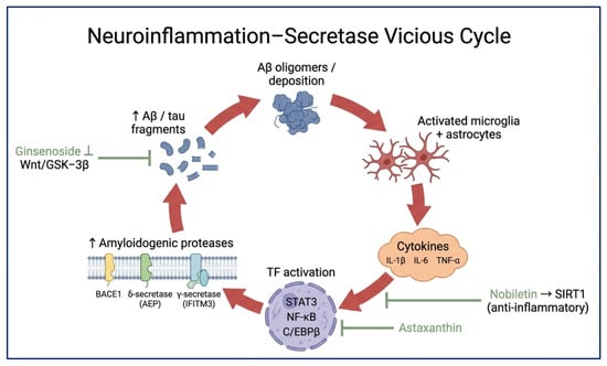
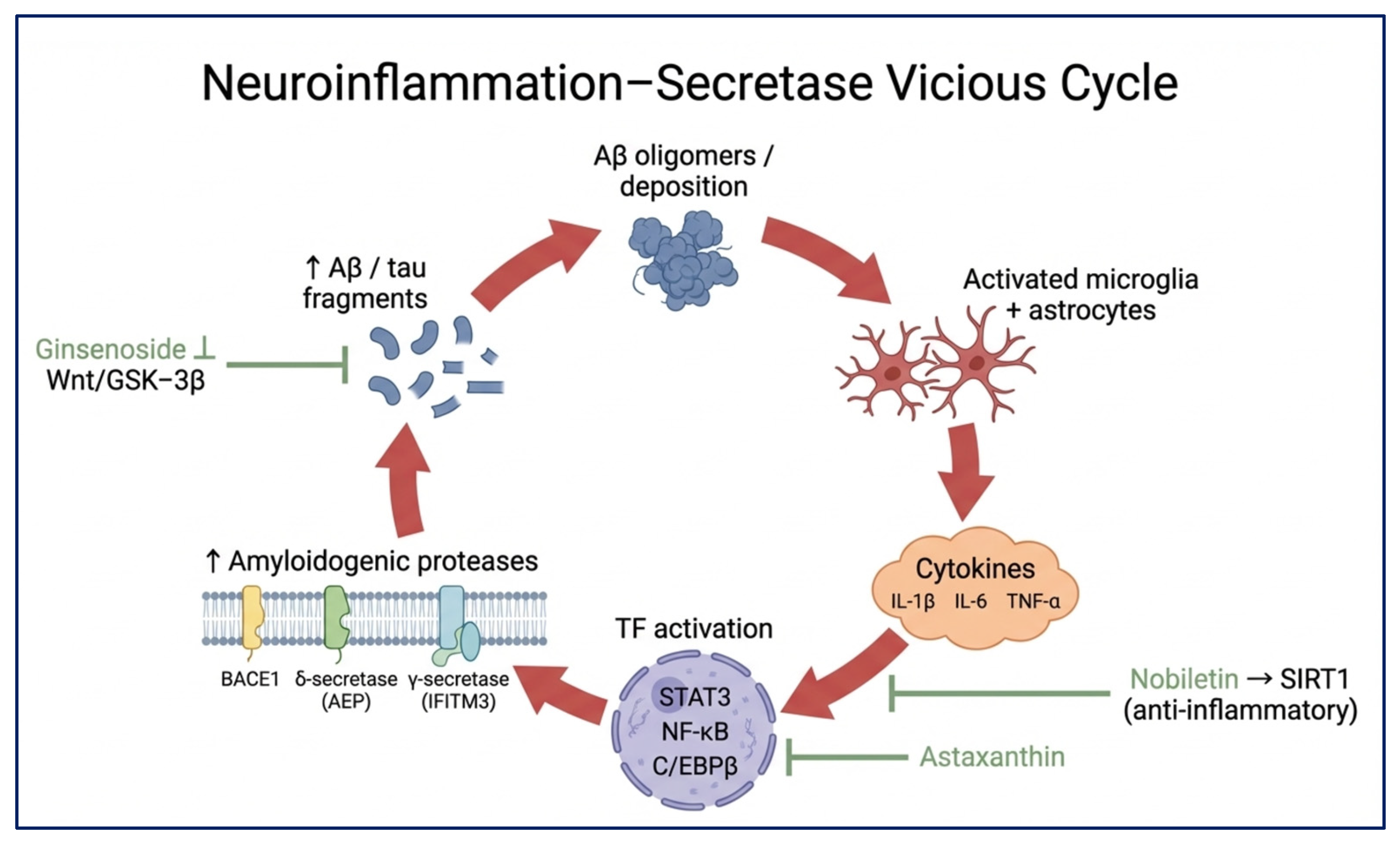
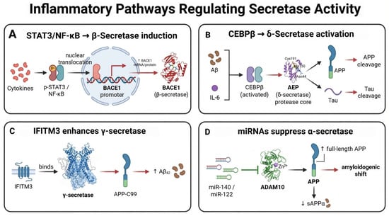
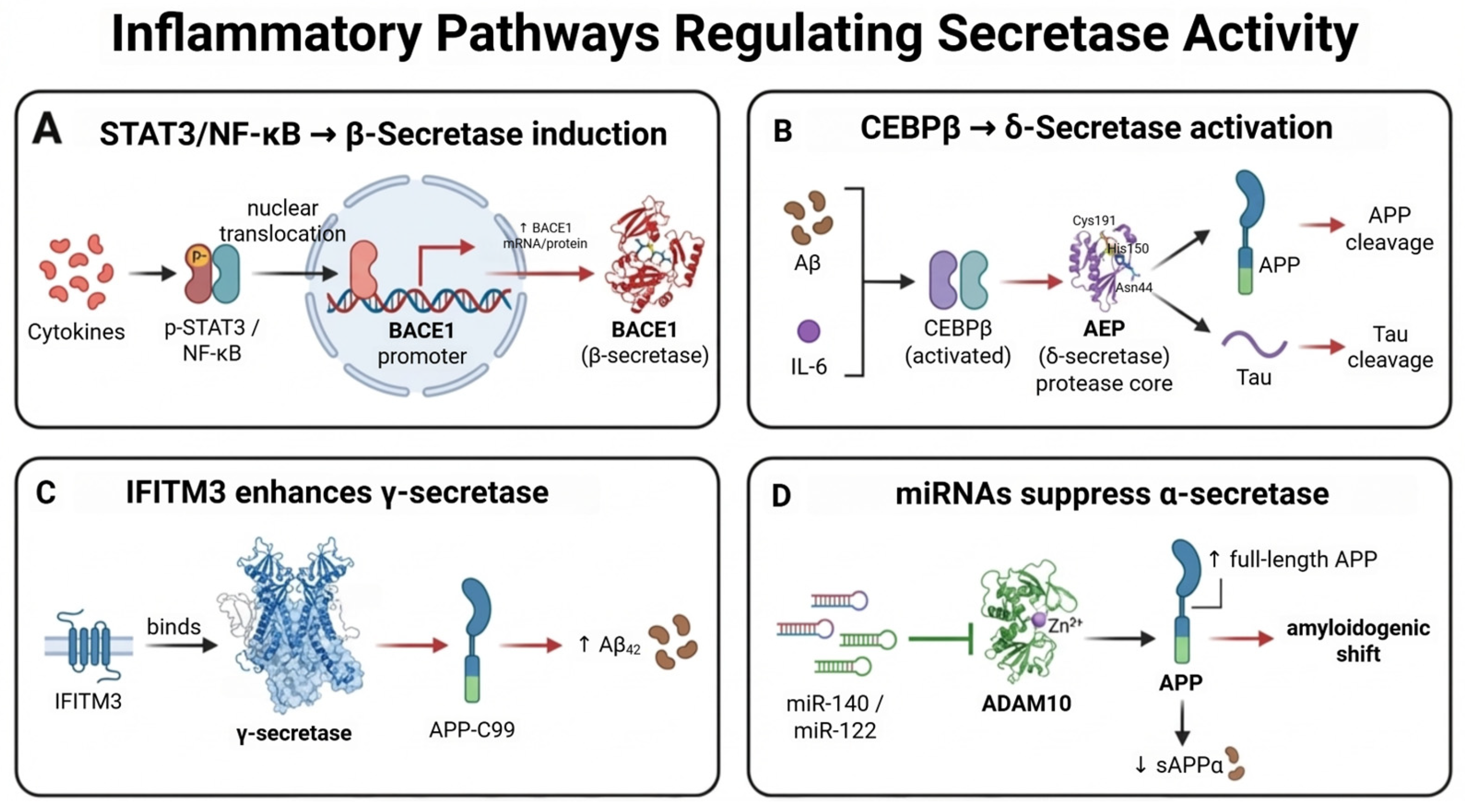
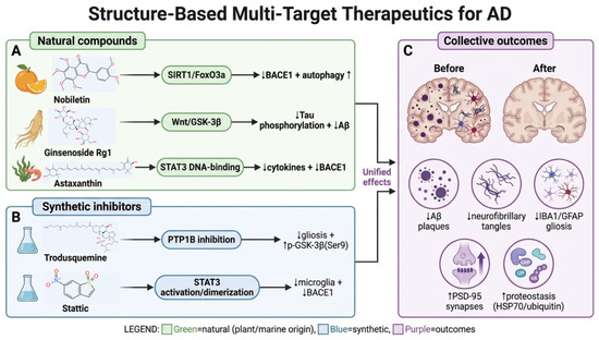
Disclaimer/Publisher’s Note: The statements, opinions and data contained in all publications are solely those of the individual author(s) and contributor(s) and not of MDPI and/or the editor(s). MDPI and/or the editor(s) disclaim responsibility for any injury to people or property resulting from any ideas, methods, instructions or products referred to in the content. |
© 2026 by the authors. Licensee MDPI, Basel, Switzerland. This article is an open access article distributed under the terms and conditions of the Creative Commons Attribution (CC BY) license.