Article

Biomineral Complex with Probiotic and Detoxifying Properties for Recovery After Radiotherapy

1
Department of Microbiology, Kazan (Volga-Region) Federal University, Kremlevskaya St., 18, Kazan 420008, Russia
2
Federal Center for Toxicological, Radiation and Biological Safety, Science City-2, Kazan 420075, Russia
3
Kazan Institute of Biochemistry and Biophysics of the Kazan Scientific Center of the Russian Academy of Sciences, Lobachevsky St., 2, Kazan 420111, Russia
*
Author to whom correspondence should be addressed.
Int. J. Mol. Sci. 2026, 27(11), 4794; https://doi.org/10.3390/ijms27114794
Submission received: 28 April 2026 / Revised: 18 May 2026 / Accepted: 25 May 2026 / Published: 26 May 2026
(This article belongs to the Special Issue Advanced Biomaterials for Tissue Regeneration)

Abstract

Radiotherapy is a highly effective, safe cancer treatment, and about half of all cancer treatments involve lifesaving radiotherapy. Despite huge advances in technology that have made it safer and more effective, it is still not without side effects. They differ from patient to patient and can include fatigue, nausea, skin reactions, and hair loss, but dysbiosis is the most common complication associated with radiotherapy. Probiotics aimed at restoring the microbiome have found widespread use, but the problem of their rapid inactivation in the gastrointestinal tract has not yet been solved. Our study aims to confirm the effectiveness of a novel biomineral complex, based on a powdered clinoptilolite containing a rock loaded with lactobacilli for restoring the intestinal microbiome of mice exposed to radiation. Based on the 16S rRNA gene analysis, alpha-diversity and dynamics of changes in the fecal metagenome, as well as the functional potential of mice exposed to radiation, were studied, and the prospects of administering the biomineral complex to achieve positive effects were assessed. NMR analysis of the mineral carrier was carried out, and its safety was confirmed. Moreover, per os administration of the complex following irradiation led to a reduction in the level of chromosomal aberrations induced by irradiation. Thus, the biomineral complex has a microbiome-restoring effect and reduces radiation-induced clastogenesis.

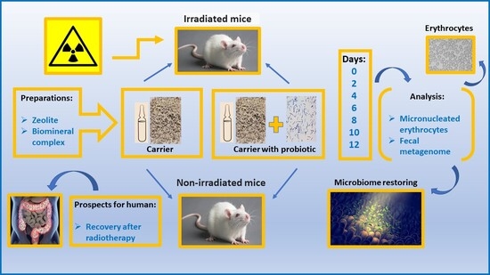
Graphical Abstract

1. Introduction

Radiotherapy (RT) is one of the primary methods in cancer management. About 30 to 50% of all cancer patients receive RT either alone or in combination with chemotherapy and surgery [1,2]. RT can cause side effects such as fatigue, depression, diarrhea, dysbiosis, sleep disorders, and so forth, impacting health-related quality of life. These side effects can be short- or long-term and vary among patients depending on the type of RT used. The composition and functional activity of the human microbiota play a crucial role in cancer pathogenesis, and the microbiota becomes dysregulated during tumorigenesis, as well as in response to RT. Advanced RT approaches such as the use of stereotactic body radiotherapy combined with microbiota modulation represent a promising strategy for sparing healthy tissue while maximizing immune-mediated antitumor effects [3].
Recent studies have revealed the gut microbiota’s active and crucial role not only in gastrointestinal cancers [4] but also in lung cancer patients with brain metastases. In these patients, key microorganisms associated with RT response were identified, and their potential as biomarkers was evaluated. The genera Flavonifractor and Negativibacillus, together with C-reactive protein and the systemic inflammation response index, were identified as independent predictors of radiotherapy response [5]. The identification of certain microorganisms in the microbiota can serve as a diagnostic marker for response to therapy. Moreover, not only for the intestinal microbiota but also for the respiratory microbiota of patients with advanced non-small cell lung cancer receiving PD-1/programmed death-ligand 1 blockade monotherapy, the objective response rate was higher in the high-diversity group compared with that of the low-diversity group [6]. Many modern studies confirm dysbiotic changes in the composition of the microbiome in cancer patients. Oral microbiome dysbiosis contributes to the development of oral cavity cancer [7]. Human colitis-associated colorectal carcinoma progression is accompanied by dysbiosis with enriched pathobionts. Microbiome alpha diversity in the nonneoplastic mucosa is positively correlated with colorectal cancer stage, and a high Shannon index predicts significantly shorter recurrence-free survival [4]. Colorectal laterally spreading tumors, early-stage lesions of colorectal cancer, are associated with gut microbiota dysbiosis [8]. Depletion of the bacterial microbiome induced by antibiotics leads to reduced tumor cell death following RT [9]. Thus, dysbiotic changes in the microbiome are characteristic of cancer; they persist throughout the course of cancer treatment and after its completion.
Probiotics, beneficial bacteria that can improve the balance of the gut microbiota, have shown promising potential in preventing cancer progression [10]. Several probiotic bacteria are currently used to restore the gut microbiota [11]. The lyophilized forms of the well-known probiotic bacteria Lactobacillus and Bifidobacterium are included in capsules that partially prevent their inactivation in the gastrointestinal tract (GIT) [12]. However, the release of lyophilized forms from capsules occurs almost immediately after entering the stomach [13]. During the 4–6 h spent in the stomach and small intestine, lyophilized bacteria do not have enough time to transform into vegetative forms [14]. Even some enteric capsules, which remain whole in the stomach and dissolve in the intestine, result in a massive emergence of vegetative forms from the lyophilizate only in the large intestine, which is densely populated by anaerobes, thereby making colonization of epithelia with probiotics difficult [15]. Therefore, the use of probiotic carriers that allow a gradual, prolonged release of bacteria is an important strategy for restoring the intestinal microbiota damaged during RT.
Nanosynergistic therapy is a novel therapeutic strategy that combines nanoparticles and probiotics or other agents to modulate the gut microbiota, increase drug bioavailability and stability, and reduce side effects. Dextran nanoparticles loaded with irinotecan, xylan-stearic acid conjugates loaded with capecitabine, and silver and gold nanoparticles have been shown to target specific gut pathogens to inhibit harmful microorganisms while promoting the growth of beneficial probiotics [4]. Zeolitic imidazolate frameworks (ZIFs), a subclass of metal-organic frameworks in which tetrahedrally coordinated transition-metal ions (e.g., Fe, Co, Zn) are connected by imidazolate linkers, are a platform for drug delivery and controlled drug release [16]. ZIF-8, which encapsulates mitoxantrone—an immunogenic cell death-inducing chemotherapeutic agent (MIT@ZIF-8)—was applied as a dual-functional nanomaterial for chemo-immunotherapy: a carrier to enhance tumor uptake of mitoxantrone for improved chemotherapy efficacy, and a pyroptosis inducer to amplify mitoxantrone-induced immunogenic cell death for augmented antitumor immune responses [17]. Different modifications of ZIFs are used to combat implant-associated infections [18] and serve as antibacterial platforms for the regeneration of infected skin wounds. [19]. ZIFs are topologically isomorphic to zeolites. The porous network of ZIFs consists of a central cavity (size ∼1.16 nm) connected to narrow apertures (size ∼0.34 nm) [20]. However, this pore size does not allow bacteria to be adsorbed. Therefore, natural minerals, in particular zeolites, are receiving increased attention for the delivery of probiotic bacteria into the GIT.
Clinoptilolite is a naturally occurring zeolite composed of microporous arrangements of silica and alumina tetrahedra, linked through shared oxygen atoms. It possesses a negatively charged open-framework porous structure, where the negative charge neutralization is achieved through cations (Na+, K+, Mg2+, Ca2+) capable of ion exchange. Compared to fibrous and lamellar zeolites, spherical clinoptilolite is less toxic [21,22,23] and is therefore used as a sorbent in agriculture, veterinary, and human medicine for contaminant removal and detoxification [24,25]. Clinoptilolite is not absorbed and is excreted with feces. There is no evidence that clinoptilolite is degraded during its passage through the gastrointestinal tract of animals [26].
Here, we investigated the effectiveness of a biomineral complex based on an active probiotic lactobacilli strain adsorbed on a mineral carrier—clinoptilolite-containing rock—in correcting the intestinal microbiota of experimental animals subjected to radiotherapy. The design of the preparation, the structure, porosity, and cytotoxicity of the carrier to eukaryotic cells, as well as important characteristics of the immobilized bacteria, such as survival, production of organic acids during long-term preservation, and resistance to gastrointestinal tract fluids, were described earlier [27]. In this study, the following objectives were addressed: (i) to investigate the gut microbiota of laboratory mice using 16S rRNA gene sequencing; (ii) to determine the alpha and beta diversity of the microbiota of non-treated and irradiated mice, and mice that received restorative therapy with the biomineral complex after irradiation; (iii) to identify the main groups of bacteria that changed quantitatively after irradiation and restorative therapy; and (iv) to analyze the frequency of chromosomal aberrations in the peripheral blood of mice before irradiation, after irraditation, and after restorative therapy. Finally, the main aim was to confirm the novel biomaterial’s potential for improving health status after RT.

2. Results

2.1. Frequency of Chromosomal Aberrations After Irradiation and Biomineral Complex Administration

The lifespan of red blood cells in mice is significantly shorter than that in humans, and averages 20–40 days. To capture chromosomal aberrations induced by radiation, we chose a time point of 12 days, approximately half the time required for red blood cell turnover. As a positive control, we analyzed the blood of mice given colchicine, a known inducer of chromosomal aberrations [28]. It was found that after 24 h of colchicine administration, the number of micronucleated erythrocytes increased by 2.6-fold compared to control mice (Table 1), confirming the validity of the test. Administration of the mineral carrier or the biomineral complex did not significantly alter the level of micronucleated erythrocytes. However, a single irradiation session, even after 12 days, increased the number of micronucleated erythrocytes by 1.6-fold. The administration of the mineral carrier or the biomineral complex following irradiation led to a significant reduction in the level of chromosomal aberrations in comparison to mice receiving only irradiation. The biomineral complex, lactobacilli immobilized on a carrier, was especially effective (Table 1).

2.2. Alpha Diversity of Mice Fecal Metagenome

Analysis of alpha diversity in the microbial communities of mice fecal samples revealed noticeable variations in the number of taxa (taxonomic richness index), the Shannon–Wiener, Simpson diversity, and evenness indices (Table 2). The highest number of OTUs was observed in the irradiated group with no follow-up treatment. The number of OTUs in this group on day 6 was 3486 and was 4434 on day 12 values that were 1.5- and 1.9-fold higher, respectively, than the mean across all other groups (2288 ± 420 OTUs). The same group showed the highest overall diversity coupled with the highest evenness (Shannon–Wiener index = 5.584 and evenness = 0.06005 on day 12).

2.3. Beta-Diversity of Mice Fecal Metagenomic Samples

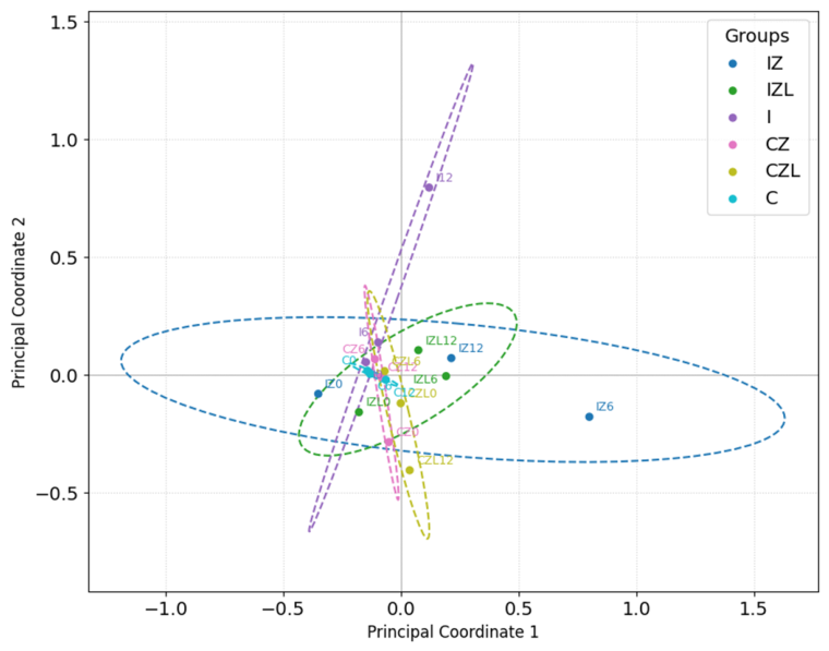
Between-group divergence relative to the control group, intragroup stability, and temporal dynamics were visualized using the principal coordinate analysis (PCoA) based on Bray–Curtis index values (Figure 1). Analysis of the distances between group centroids revealed different treatment effects. The greatest similarity to the control group was observed in the CZ group, where d = 0.0817. On the other hand, the IZ and I groups showed the greatest divergence from the control group, where d = 0.3395 and d = 0.3363, respectively. These values indicate a major change in the bacterial communities. Intermediate divergence values were observed in the IZL group, where d = 0.1431, and the CZL group, where d = 0.1981.
Examining the homogeneity of intra-group variability revealed a unique time dynamic. Group C demonstrated very high intra-group temporal stability (distance range: 0.0188–0.0530), providing a baseline for comparison with all other groups. Converging trajectories (CZ and IZL) showed a trend toward intra-group stability. For example, the CZ distance from the centroid decreased from 0.2125 on day 0 to 0.0679 on day 12, implying a shift toward homogeneity. Diverging trajectories (CZL and I) demonstrated a trend toward intra-group instability. The CZL group showed a progressive trend: from 0.0504 on day 0 to 0.2401 on day 12. The IZ group showed high-intensity dynamics and the greatest initial instability, with the largest intra-group distances from the centroid on day 0 (0.5727) and on day 6 (0.5910). However, the distance to the centroid decreased sharply on day 12, probably indicating recovery.

2.4. Changes in the Structure of the Mice Fecal Metagenome After Irradiation and Preparation Administration

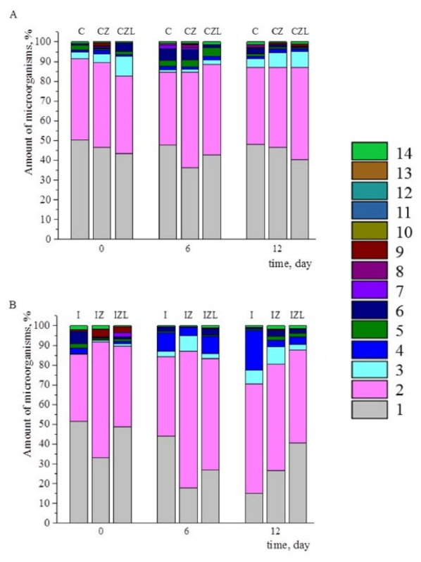
The metagenome of non-irradiated mice showed little change over the course of the experiment. A slight increase in the Actinomycetota phylum was noted toward the end of the experiment in mice receiving the mineral carrier or the biomineral complex (Figure 2A). However, the effect of radiation increased over time, manifesting as an increase in the relative abundance of Bacillota and Pseudomonadota and a decrease in the relative abundance of Bacteroidota (Figure 2B). In mice receiving the mineral carrier after irradiation, the destabilizing effect of radiation remained unchanged. Administration of the biomineral complex brought the microbiome composition closer to that of the control group by the end of the experiment (Figure 2).
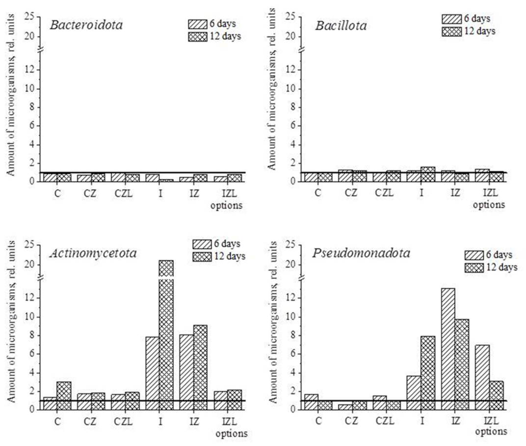
The standard representation of phyla in the mouse fecal metagenome underemphasizes phyla with low relative abundance (Figure 2). However, changes in the microbiome are particularly revealing when relative abundances on day 0 are taken as a reference, and changes caused by irradiation and treatment are calculated relative to the initial phyla abundance values. Figure 3 shows that irradiation caused significant changes in the abundance of Actinomycetota and Pseudomonadota, while the administration of the biomineral complex brought these values closer to those of the control group. The administration of the carrier showed a less pronounced restorative effect (Figure 3).
At the family level, irradiation had a strong impact on the relative abundance of different families. Among the representatives of the phylum Bacteroidota, the families Muribaculaceae, Prevotellaceae, and Bacteroidaceae predominated in all mice fecal samples. These families are constant components of the mammalian microbiota. On day 12 post-irradiation, the relative abundances of Muribaculaceae and Prevotellaceae were reduced in mouse fecal samples by 22.5-fold and 53.4-fold, respectively. The administration of the biomineral complex restored the abundance of these families (Figure S1).
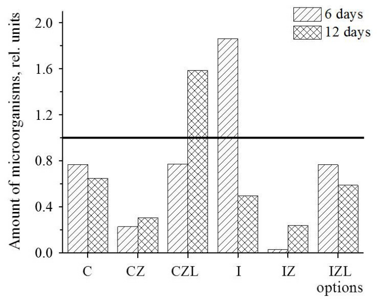
Among the representatives of the phylum Bacillota, two families predominated in all mice fecal samples, Lactobacillaceae and Lachnospiraceae (Figure S2). We paid special attention to the abundance of lactobacilli in the samples. Irradiation resulted in a 1.9-fold increase in Lactobacillaceae in mouse fecal samples on day 6 of the experiment, followed by a decrease by day 12 (Figure 4). Compared to the irradiated-only group (I), the biomineral complex group (IZL) showed a 37.3-fold and 4.2-fold lower fecal abundance of lactobacilli on days 6 and 12, respectively. This reduced fecal shedding—combined with the IZL group’s convergence toward control-level relative abundance in Figure 4—supports the hypothesis that the complex enhances mucosal adhesion of administered lactobacilli.
Among the representatives of the phylum Pseudomonadota, two families predominated in all mice fecal samples, Sutterellaceae and Enterobacteriaceae (Figure S3). Irradiation of mice resulted in a 2.1-fold decrease in the abundance of the Sutterellaceae family on day 12 of the experiment, while the administration of either the carrier or the preparation of both drugs increased their abundance by an average of 1.8 ± 0.3-fold. A similar pattern was observed for members of the Enterobacteriaceae family: a 5.6-fold decrease after irradiation and 3.2- and 1.6-fold increases after the administration of the carrier or the preparation, respectively. Based on the data obtained, it is possible to identify a positive trend in the influence of the biomineral complex on the representation of beneficial bacteria in the intestines of mice exposed to irradiation.

2.5. Functional Analysis of Mouse Fecal Metagenome

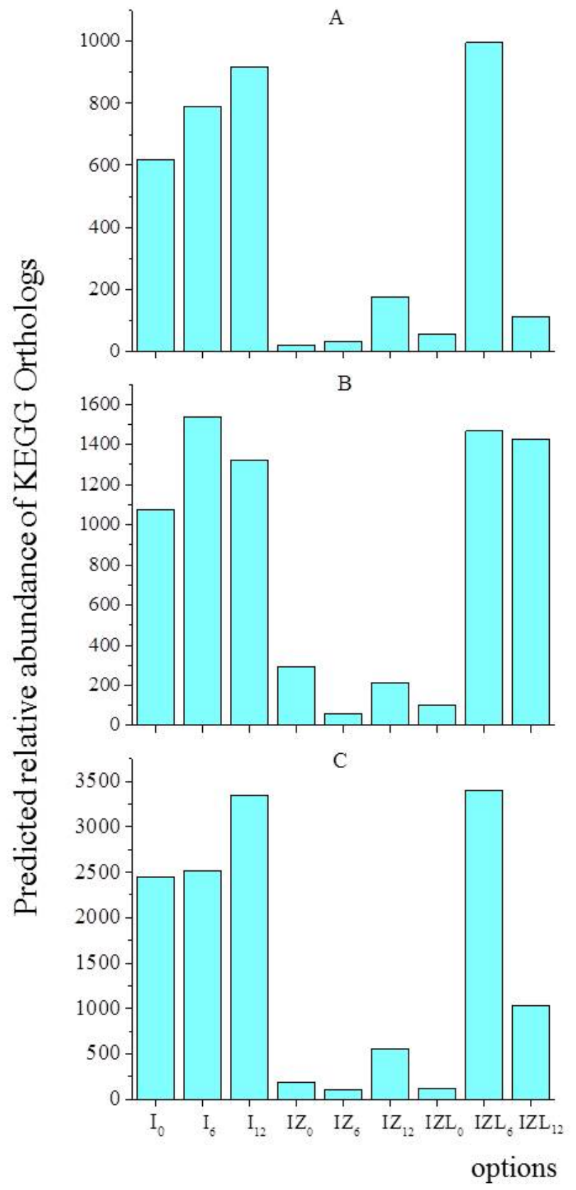
We performed a functional bioinformatics analysis of predicted bacterial metabolic pathways (Supplementary Table S1), identifying pathways counteracting radiation damage compared with one of the main metabolic pathways–lipopolysaccharide biosynthesis (Figure 5A). Patterns of genes responsible for nucleotide excision repair (Figure 5B) and glutathione metabolism (Figure 5C) are similar to those for lipopolysaccharide biosynthesis.
Compared to non-irradiated controls (Table S1), irradiated mice showed a time-dependent increase in fecal abundance of bacteria harboring genes involved in nucleotide excision repair, glutathione metabolism, and lipopolysaccharide biosynthesis (Figure 5), consistent with radiation-induced epithelial disruption and enhanced bacterial shedding.
Mineral carrier administration after irradiation reduced the abundance of the studied genes in mouse fecal samples; biomineral complex administration increased bacterial elimination from the intestines on day 6 and decreased it again on day 12. Presumably, the introduction of lactobacilli on day 6 hindered gut bacterial adsorption, and by day 12, some of them remained in the intestines, which indirectly indicates the restored epithelial adsorption of bacteria. This pattern can be traced in both the main phyla (Figure 3) and the family Lactobacillaceae (Figure 4). Dynamic changes in gene counts are similar for all metabolic pathways (Supplementary Table S1, Figure 5). These data show that there is a direct correlation between the number of OTUs and the number of functional genes.

2.6. NMR Spectrum of the Aqueous Extract of the Mineral Carrier

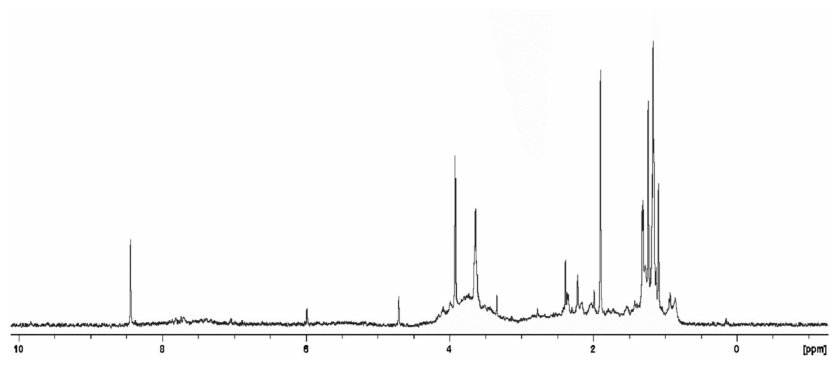
Figure 6 shows the nuclear magnetic resonance (NMR) spectrum of the aqueous extract of the carrier used for probiotic bacteria immobilization. The proton signals in the spectrum are low-intensity, indicating a low concentration of dissolved molecules in the solution. Relatively intense narrow signals in the 1–1.5 ppm region are related to the CH3 groups of low-molecular-weight compounds. The presence of low-intensity signals in the 2–4 ppm and 7–8 ppm regions may indicate the presence of a small amount of organic macromolecules in the sample, such as proteins and nucleic acids. In general, it is possible to conclude that the carrier sample contains only trace quantities of water-soluble substances. These data once again confirm the chemical inertness of the carrier and the absence of significant soluble organic impurities. The biological safety and non-toxicity of the carrier were previously established through in vitro cytotoxicity and in vivo evaluations [27].

3. Discussion

Gastrointestinal toxicity, including radiation enteritis and mental fatigue, is a common side effect of radiotherapy that substantially reduces patients’ quality of life. Among the various organs affected, the gut is particularly sensitive to radiation-induced damage [29]. Currently, there are no effective drugs available for the prevention or treatment of radiation-induced enteropathy or radiation-induced brain injury, both of which impair cognitive function, leading to severe complications or even death. Numerous current studies have confirmed that intestinal microbiota dysfunction is an important factor in the formation of these diseases, which are major adverse events following radiotherapy of malignant tumors. Irradiation causes changes in the composition of the flora and a decrease in its diversity, which is mainly manifested by a decline in beneficial bacterial species such as Lactobacilli and Bifidobacteria [30,31,32,33].
It is now established that probiotic interventions can protect the rectal mucosa by reducing inflammation and modulating the mucosa-associated microbiota. Preventive effects of combined live Bifidobacterium, Lactobacillus, Enterococcus, and Bacillus cereus tablets were shown in patients with radiation pneumonitis [34]. Probiotics, including Bacillus licheniformis, have been shown to mitigate intestinal inflammation and mucositis by modulating gut microbiota and immune responses. Bacillus licheniformis supplementation effectively alleviates craniospinal irradiation-induced gastrointestinal dysfunction and inflammation in pediatric patients with medulloblastoma, but does not significantly improve their survival rates [35]. Oral administration of Lactobacillus reuteri, which releases interleukin-22 at 24 h after total-body irradiation, mitigates damage to the intestine. Furthermore, this probiotic facilitated whole-abdomen irradiation when added to paclitaxel and carboplatin chemotherapy, thereby further increasing survival [36]. Probiotics effectively reduced the incidence of CyberKnife-associated radiation pneumonitis among patients with pulmonary malignancies, delayed radiation pneumonitis onset, and improved their quality of life [34]. The effect of probiotics extends not only to improving the gut microbiome but also to maintaining normal brain function. Patients taking Lactobacillus probiotics had better role functioning, emotional balance, and cognitive clarity after six weeks; probiotics helped patients adapt better to treatment stress and protect the gut–brain axis during radiotherapy [37]. Bacterial metabolites also have positive effects on human health damaged after irradiation. Indole-3-carboxaldehyde, derived from the intestinal microbiota, enhanced the abundance of probiotics, activated the AhR/IL-10/Wnt signaling pathway to promote intestinal epithelial proliferation, and demonstrated potential clinical application value for the treatment of radiation-induced damage [38].
Radiation-induced damage to the villi height and mucosal thickness was significantly mitigated by probiotic treatment (p < 0.01). Probiotics reduced radiation-induced numbers of pyknotic cells and neuronal inflammation in the cortex (p < 0.01). Altogether, probiotic treatment helped mitigate radiation-induced intestinal and neuronal damage [39]. Meta-analyses of 16 randomized controlled trials including 2097 patients have established that compared with the placebo groups, oral probiotics significantly reduced the side effects caused by radiotherapy and chemotherapy in various types of cancer, such as head and neck cancer, pelvic and abdominal cancer, breast cancer, lung cancer, etc. [40].
Thus, sufficient evidence has been obtained on the benefits of probiotics for the recovery of patients after radiotherapy. A modern understanding of the functional role of the gut microbiota in the body’s immunological, metabolic, and neurological status has prompted the development of new probiotic formulations incorporating a sorbent carrier or a polymer capsule to protect bacteria from the harsh gastric environment. Encapsulation technologies, particularly those using natural biopolymers such as alginate, chitosan, pectin, carrageenan, and gelatin, have significantly improved probiotic viability, shelf stability, and targeted release. Ensuring prolonged probiotic release in the gastrointestinal tract remains a serious and pressing challenge that cannot be addressed without accumulating fundamental knowledge about the carrier structure and its dynamic interactions with the target agent, as well as demonstrable data on the stability, efficacy, and safety of the formulation. Various technologies are used to ensure the stability of probiotics in gastric juice. Bacillus cereus, possessing antioxidant ability and covered with a polydopamine/chitosan layer-by-layer assembly, exhibited tolerance to ionizing radiation, freeze drying, long-term preservation at room temperature, and gastric acid [41]. We have developed a new Lactobacillus preparation immobilized on a mineral carrier. The clinoptilolite-containing carrier has been shown to be non-toxic [27] and contains only trace quantities of water-soluble substances (Figure 6), rendering it essentially inert. When loaded with lactobacilli, it provided prolonged stepwise release of bacteria over 12 h [27]. In this study, we demonstrated that this biomineral complex significantly reduced the frequency of chromosomal aberrations caused by radiation, restoring values to control levels (Table 1). Clinoptilolite was found to be the most effective adsorbent of cesium in vitro. When used in vivo, it significantly reduced the absorption of cesium by sheep fed contaminated herbage [42]. In veterinary medicine, zeolite improves the fitness of pets and removes radioactive elements, aflatoxins, and poisons. Zeolite also exhibits antioxidant, hemostatic, and antidiarrheal properties, projected for human care [43]. Moreover, in the Ames test, we have established that cell suspension and supernatant of Lactobacillus cultures possessed antimutagenic activity against sodium azide and 2-nitrofluorene [44], thereby further enhancing the detoxifying effect of the biomineral complex. The detoxifying properties are associated not only with clinoptilolite, but also with lactobacilli through direct binding of toxic compounds, influencing their biotransformation, and with the activity of secreted metabolites.
Alpha-diversity of mice fecal samples showed that the control group had very high intra-group temporal stability. The highest number of OTUs was observed in the irradiated group (Table 2), which demonstrated high-intensity dynamics and the greatest initial instability (Figure 1). Established irradiation-induced damage to the villi height and mucosal thickness [34] probably leads to disruption of intestinal microbiota adhesion and the release of many bacterial taxa into feces (Table 2). Beta-diversity assessment revealed a trend toward intra-group stability in the group of mice given the biomineral complex after irradiation (Figure 1). These data allow us to consider the effect of the complex to be positive. The influence of the complex on the abundance of Actinomycetota and Pseudomonadota can also be considered positive because significant changes in this parameter induced by irradiation shifted closer to the control group (Figure 2). It should be noted that on day 12 after irradiation, the ratio of Bacteroidota/Bacillota in the mouse fecal metagenome significantly decreased (Figure 2B), and administration of the complex restored it to the control level (Figure 2A). Overall, we found that families Muribaculaceae, Prevotellaceae, and Bacteroidaceae, which are constant components of the mammalian microbiota, predominated in all mouse fecal samples and significantly decreased on day 12 after irradiation (Figure 3). The administration of the biomineral complex restored the abundance of these families (Figure S1).
In similar experiments, Zhao et al. [45] found that during the recovery stage of acute radiation-induced intestinal injury, after the seventh day of radiation, the diversity of the mouse gut microbiota decreased overall, with the relative abundance of the phyla Proteobacteria and Bacteroides increasing. In our study, bacteria belonging to the phyla Pseudomonadota (previous name Proteobacteria) and Bacteroidota (previous name Bacteroides) also increased (Figure 3). Analysis of lactobacilli abundance in mouse feces revealed that on day 6 after irradiation, abundance increased, but on day 12, it decreased, which indicates the restoration of the normal intestinal metagenome (Figure 4). The biomineral complex reduced the abundance of lactobacilli by ~37-fold on day 6 and by ~4-fold on day 12, which indicates the adhesion of lactobacilli from the preparation to the mice’ intestinal epithelium. We adhere to the concept that the release of bacteria in feces indicates their elimination from the intestinal epithelium, which definitely indicates a disruption of the intestinal microbiome caused by irradiation. Thus, the restoration of the gut microbiome is associated with a reduction in the eliminated taxa in the feces of mice, which was shown in our experiments.
While pooled-sample metagenomics enabled robust detection of group-level microbiome trends, it precludes statistical evaluation of within-group variability. This design prioritizes detection of treatment-driven community shifts over assessment of inter-individual variability, consistent with exploratory microbiome study frameworks. Future studies with individual-level sequencing will be valuable to confirm these observations and assess inter-animal heterogeneity.
We focused on the functional bioinformatics analysis of metabolic pathways counteracting radiation damage, namely, glutathione metabolism, nucleotide excision repair, and biosynthesis of lipopolysaccharides as one of the main metabolic pathways (Figure 5). The patterns of dynamic changes in gene counts were similar for all metabolic pathways (Supplementary Table S1, Figure 5) and can be traced both in the main phyla (Figure 3) and in the Lactobacillaceae family (Figure 4), reflecting the direct relationship between the number of OTUs and the number of functional genes. The number of predicted gene copies in the feces of mice that did not receive the preparations increased after irradiation, while preparation administration reduced their number, indirectly indicating restored epithelial adsorption of gut bacteria and introduced lactobacilli.
Experimental data indicate that radiation exposure disrupts both the gut microbiota and metabolome during the acute injury phase, reducing beneficial bacteria such as Ruminococcaceae and Bifidobacterium [46]. Our results also indicated a high level of radiation-induced elimination of beneficial Lactobacillaceae from the intestines (Figure 4). However, we did not observe a significant difference between the yield of beneficial versus harmful bacteria. Radiation has been shown to damage not only the microbiome but also the intestinal epithelium. Intestinal organoids derived from the irradiated mice showed defects in budding and mucin expression, suggesting a detrimental effect of irradiation on the intestinal stemness and differentiation, whereas the indigenous gut bacterium L. acidophilus enhanced intestinal epithelial function [47]. Our data also confirm that the use of lactobacilli in a biomineral complex is justified for restoring the microbiome after radiotherapy. It is important to note that gut microbiota cannot be used as a sensitive biomarker at the prodromal stage in acute radiation-induced intestinal injury, but it is a potential biomarker at the critical stage, and interventions are needed to restore radiation-induced intestinal injury [45]. Thus, the administration of probiotics is an effective strategy to protect against irradiation and subsequent dysbiosis.

4. Materials and Methods

4.1. Animals

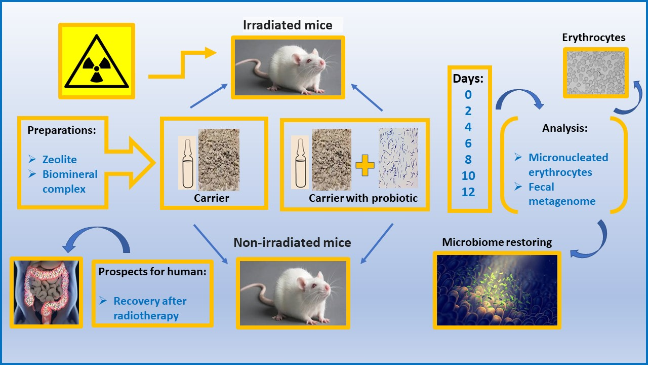
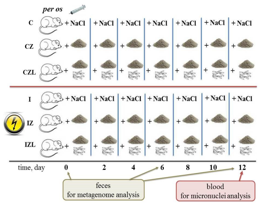
The experiments were performed on male white laboratory mice in accordance with the Directive of the Council of the European Communities (24.11. 86/609/EEC) and were approved by the Local Ethics Committee of Kazan Federal University (protocol No. 8 of 5 May 2015, protocol No. 33 of 25 November 2021). To simulate acute radiation sickness, animals were exposed to a single relatively uniform gamma irradiation in the biological gamma installation GUB-20, which contains sealed radionuclide sources of the IGI-Ts-9-1 type based on the radionuclide 137Cs, GUB-20 “Puma” (Russia, Saint Petersburg), a radiation source of 137Cs with an exposure dose rate of 1.99 × 10⁻⁵ A/kg (4.633 R/min) and with a dose field unevenness of 13.3%. The irradiation was delivered as a single dose of 4 Gy, sufficient to cause acute radiation sickness [45,48]. The animals were kept in standard vivarium conditions (air temperature 18–24 °C, relative air humidity 40–80%). Animals’ access to food and water was not limited (feeding regimen: ad libitum) according to the veterinary certificate of the Main Veterinary Directorate of the Republic of Tatarstan-216 No. 018687 dated 30 November 2021. Before the experiment, the animals were quarantined for 14 days, after which the mice were divided into 6 groups, with 5 individuals in each group: control mice without treatment (C); control mice receiving the mineral carrier (CZ); control mice receiving the biomineral complex (CZL); irradiated mice (I); irradiated mice receiving the mineral carrier (IZ) and irradiated mice receiving the biomineral complex (IZL). The mineral carrier (100 mg/mL of saline solution) and the biomineral complex (one ampule containing a 100 mg sample diluted in 1 mL of saline solution) were administered perorally at a dose of 0.2 mL per animal every two days for 12 days. The control group was administered a saline solution. The experimental scheme is shown in Figure 7.

4.2. Biomineral Complex

The clinoptilolite-containing rock from the Tatarsko-Shatrashanskoe zeolite deposit, Russia, was ground to microgranules smaller than 30 μm using an electric mill (Homaider, Guangzhou, China). High-temperature treatment of the samples was carried out for 30 min at a temperature of 500 °C (EKPS50 muffle furnace, model 5007, Smolensk, Russia). Thermal activation led to an increase in the porosity of the zeolite due to dehydration and combustion of residual organic matter, and is a process widely used in industry and agriculture [49]. Additionally, the zeolite samples were washed twice with 96% ethanol and dried at room temperature.
Lactobacillus plantarum B-11007 (new taxonomic name Lactiplantibacillus plantarum) registered in the VKPM collection, Russia, was isolated from commercial probiotic Lactobacterin dry (Biomed, Moscow, Russia) and cultivated in MRS medium (g/L: yeast extract—4.0, meat extract—10.0, casein hydrolyzate—10.0, glucose—20.0, ammonium citrate—2.0, sodium acetate—5.0, KH2PO4—2.0, MgSO4 × 7H2O—0.2, MnSO4 × 4H2O—0.05; pH 6.2–6.5) under microaerophilic conditions.
The Djuki’c–Vukovi’c technique, with some modifications, was used for immobilization [27,50]. A 16 h culture in a volume of 200 mL was subjected to centrifugation at 10,000 rpm for 5 min, and the sediment was washed twice with sterile 0.9% NaCl and resuspended in 200 mL of fresh MRS medium with the addition of clinoptilolite (5% of the total volume). The number of lactobacilli in the suspension averaged 5.8–6.0 × 10⁸ colony-forming units, CFU/mL. The suspension was incubated at 37 °C on a shaker (180 rpm, 20 h) and then centrifuged (1000 rpm, 5 min). The supernatant containing non-immobilized cells was removed; the sediment was washed twice with phosphate-buffered saline (0.01 M, pH 7.4) and used for lyophilization. The immobilization coefficient, reflecting the sorption capacity of the carrier, was calculated as the ratio between the number of viable cells (CFU/mL) in the suspension before immobilization and in the supernatant after immobilization, centrifugation, and washing twice with phosphate-buffered saline. The immobilization coefficient was expressed as a percentage, taking the initial number of lactobacilli in the suspension as 100%, and reached 96–98%. The bacterial pellet containing 100 mg of the clinoptilolite was suspended in 0.5 mL of lyoprotective sucrose–gelatin–milk medium (g/L: sucrose—100.0, gelatin—15.0, and skimmed milk powder—60.0) under aseptic conditions and packaged in glass ampoules using a polypropylene catheter. The necks of the ampoules were closed with sterile aluminum caps and frozen at −70 °C, after which they were subjected to dehydration for 5 h using a FreeZone Dryer (Labconco Corporation, Kansas City, MO, USA) at a vacuum depth of 0.035 torr.

4.3. Micronucleus Assay

Detection of micronuclei in normally anucleated erythrocytes is considered to be a consequence of the clastogenic and aneugenic effects of genotoxicants in vivo [51,52]. The count of micronuclei in mouse peripheral blood erythrocytes was performed 12 days after irradiation. As a negative control, animals were administered saline, and as a positive control, 0.125 mg/kg colchicine (Serva, Heidelberg, Germany). Colchicine is an aneugen whose main genotoxic effect is the induction of chromosomal segregation errors and aneuploidy by disrupting microtubule assembly. It induces micronuclei in the peripheral blood at 0.125–2 mg/kg [53]; therefore, the dose of 0.125 mg/kg was chosen for our experiments. Blood smears from the tail vein of mice were fixed with methanol, stained with Giemsa dye (Koch-Light Laboratories, Berkshire, England), and the number of micronuclei per 1000–2000 erythrocytes was counted in each animal group.

4.4. Metagenome Analysis

Molecular genetic analysis of the microbial community was performed according to the following protocol: mixed mouse fecal samples from each group were homogenized, and ~1 g of each sample was sterilely aspirated for DNA extraction using a Miniprep Kit (Axygen, Union City, CA, USA) according to the manufacturer’s protocol. Thus, metagenomic data represent composite group profiles (n = 1 per group/timepoint). This design prioritizes detection of treatment-driven community shifts over assessment of inter-individual variability, consistent with exploratory microbiome study frameworks. DNA concentration was determined and normalized to ≤10 ng/µL. The quality of DNA extraction was assessed by confirming amplification of the 16S rRNA gene targeting the V3–V4 region of the bacterial 16S rRNA gene using PCR and gel electrophoresis. Library preparation and sequencing were performed using the Illumina MiSeq. Preprocessing and subsequent sequence analysis were performed using the Mothur software package v.1.48.0 [54] on the Galaxy platform [55]. Data analysis was performed according to the Galaxy Training manual [56]. Operational taxonomic units (OTUs) were selected at a 97% identity threshold and classified using the SILVA 138.2 reference dataset [57].

4.5. Biodiversity and Functional Analysis

Alpha diversity indices, namely observed richness, Shannon, and Simpson, and beta-diversity Bray–Curtis and Dice–Sorensen similarities matrices were calculated using the PAST v4.02 software package. Principal coordinate analysis (PCoA) was used to illustrate the beta diversity and spatial distribution of all six groups in the experiment: C, CZ, CZL, I, IZ, and IZL. To assess group stability and treatment effects, 95% confidence ellipses were calculated, and Euclidean distances were determined to measure intra-group variance and between-group variance. PCoA ordination based on the Bray–Curtis dissimilarity matrix was performed and visualized in RStudio version 4.4.3 using the vegan and ggplot2 packages.
Functional prediction of bacterial communities’ pathways was carried out using PICRUSt2 [58] via the Galaxy platform (usegalaxy.org). The ASV table and representative sequences were processed with the default PICRUSt2 workflow to predict KEGG Ortholog abundances based on phylogenetic placement.

4.6. 1H NMR Spectroscopy

The mineral carrier (0.5 g) was suspended in 1.5 mL of deuterated water for 24 h. The supernatant was taken to fill the NMR ampule for further spectral measurements. NMR spectra were obtained on the Bruker Avance III 600 MHz spectrometer (Billerica, MA, USA) equipped with a triple-resonance TBI probe, z-gradients, and the BCU05 temperature control unit. The spectra were recorded at a temperature of 30 °C. One-dimensional proton spectra of the water system were recorded using a standard water-suppressed pulse sequence (zgesgp, rg = 2050, ns = 1024). The Topspin software 5.0 release was used for data processing.

4.7. Statistics

Data processing and plotting of taxa distribution among samples were performed using the standard Excel 7.0 software. To assess differences between mice blood samples when analyzing the results of the micronucleus test, the nonparametric Mann–Whitney criterion (U-test) was used, according to which U < 1 is the significance zone, U = 1–4 is the uncertainty zone, and U ≥ 4 is the zone of insignificance of differences.

5. Conclusions

Because of probiotics’ rapid inactivation in the gastrointestinal tract, the problem of restoring the intestinal microbiome after radiotherapy with beneficial bacteria remains relevant. Our results confirmed that the developed biomineral complex containing immobilized Lactobacillaceae on a mineral carrier, clinoptilolite-containing rock, restored irradiation-induced dysbiosis in mice thanks to its detoxifying properties and the gradual release of bacteria from the carrier, which gives them an enhanced likelihood of maintaining viability and adhering to intestinal epithelial cells. Irradiation caused significant changes in the abundance of Actinomycetota and Pseudomonadota, while the administration of the biomineral complex brought these values closer to those of the control group. Not only the restoration of the microbiome but also the elimination of clastogenic/aneugenic effects of radiation allow us to consider the biomineral complex as a promising drug for reducing side effects of radiotherapy.

Supplementary Materials

The following supporting information can be downloaded at: https://www.mdpi.com/article/10.3390/ijms27114794/s1.

Author Contributions

Conceptualization, O.I. and G.Y.; methodology, G.Y., N.K. and Y.Z.; software, P.Z.; validation, W.K. and A.K.; investigation, G.Y., W.K. and K.V.; data curation, O.I.; writing—original draft preparation, O.I.; writing—review and editing, W.K.; funding acquisition, O.I. All authors have read and agreed to the published version of the manuscript.

Funding

This research was funded by the Russian Science Foundation, grant number 24-14-00059. The APC was paid by the authors.

Institutional Review Board Statement

The animal study protocol was approved in accordance with the Directive of the Council of the European Communities (24.11. 86/609/EEC) and approved by the Local Ethics Committee of Kazan Federal University (protocol No. 8 of 5 May 2015, protocol No. 33 of 25 November 2021).

Informed Consent Statement

Not applicable.

Data Availability Statement

The raw data supporting the conclusions of this article will be made available by the authors on request.

Acknowledgments

During the preparation of this study, the authors used the biological gamma installation GUB-20 “Puma”, Russia, for the purposes of mice irradiation, and express their gratitude to the management of the Center for Toxicological, Radiation and Biological Safety for this possibility. The authors have reviewed and edited the output and take full responsibility for the content of this publication.

Conflicts of Interest

The authors declare no conflicts of interest. The funders had no role in the design of the study; in the collection, analyses, or interpretation of data; in the writing of the manuscript; or in the decision to publish the results.

Abbreviations

The following abbreviations are used in this manuscript:
RTRadiotherapy
OTUOperational taxonomic unit
ZIFZeolitic imidazolate framework
GITGastrointestinal tract
NMRNuclear magnetic resonance
CControl mice receiving saline solution
CPControl mice receiving colchicine, one day after (positive control)
CZControl mice receiving clinoptilolite
CZLControl mice receiving biomineral complex
IIrradiated mice (4 Gy)
IZIrradiated mice receiving clinoptilolite
IZLIrradiated mice receiving biomineral complex

References

  1. Majeed, H.; Gupta, V. Adverse effects of radiation therapy. In StatPearls [Internet]; StatPearls Publishing: Treasure Island, FL, USA, 2025. Available online: https://www.ncbi.nlm.nih.gov/books/NBK563259/ (accessed on 13 February 2026). [PubMed]
  2. Barton, M.B.; Jacob, S.; Shafiq, J.; Wong, K.; Thompson, S.R.; Hanna, T.P.; Delaney, G.P. Estimating the demand for radiotherapy from the evidence: A review of changes from 2003 to 2012. Radiother. Oncol. 2014, 112, 140–144. [Google Scholar] [CrossRef]
  3. Vučinić, D.; Redžović, A.; Hauser, G.; Mikolašević, I. Microbiota and radiotherapy: Unlocking the potential for improved gastrointestinal cancer treatment. Biomedicines 2025, 13, 526. [Google Scholar] [CrossRef]
  4. Han, Q.; Li, J.; Li, Z.; Aishajiang, R.; Yu, D. Novel therapeutic strategies and recent advances in gut microbiota synergy with nanotechnology for colorectal cancer treatment. Mater. Today Bio 2025, 31, 101601. [Google Scholar] [CrossRef]
  5. Liang, F.; Sun, Y.; Yang, J.; Shen, Z.; Wang, G.; Zhu, J.; Zhou, C.; Xia, Y. Gut microbiome is associated with radiotherapy response in lung cancer patients with brain metastases. Front. Cell. Infect. Microbiol. 2025, 15, 1562831. [Google Scholar] [CrossRef] [PubMed]
  6. Zhang, L.; Li, M.J.; Li, X.P.; Yang, B.; Xiao, T.; Wang, P.; Zhang, W.D. Respiratory microbiota diversity as a predictive biomarker for the efficacy of PD-1 blockades in patients with advanced non-small cell lung cancer: A retrospective exploratory study. Oncol. Lett. 2025, 29, 251. [Google Scholar] [CrossRef] [PubMed]
  7. Lian, Q.; Dong, L.; Zhou, Q.; Yuan, Q. Effect of the oral microbiota, blood metabolome, and inflammatory proteins on oral cavity cancer: A bidirectional two-sample Mendelian randomization study and mediation analysis. J. Stomatol. Oral Maxillofac. Surg. 2025, 126, 102372. [Google Scholar] [CrossRef] [PubMed]
  8. Masaadeh, A.H.; Eletrebi, M.; Parajuli, B.; De Jager, N.; Bosch, D.E. Human colitis-associated colorectal carcinoma progression is accompanied by dysbiosis with enriched pathobionts. Gut Microbes 2025, 17, 2479774. [Google Scholar] [CrossRef] [PubMed]
  9. Shiao, S.L.; Kershaw, K.M.; Limon, J.J.; You, S.; Yoon, J.; Ko, E.Y.; Guarnerio, J.; Potdar, A.A.; McGovern, D.P.B.; Bose, S.; et al. Commensal bacteria and fungi differentially regulate tumor responses to radiation therapy. Cancer Cell 2021, 39, 1202–1213.e6. [Google Scholar] [CrossRef]
  10. Min, S.; Zhang, Y.; Zhang, H.; Liu, Q. Gut microbiota drives cancer evolution and therapy resistance. Mol. Cancer 2026, 25, 39. [Google Scholar] [CrossRef]
  11. Hill, C.; Guarner, F.; Reid, G.; Gibson, G.R.; Merenstein, D.J.; Pot, B.; Morelli, L.; Canani, R.B.; Flint, H.J.; Salminen, S.; et al. The International Scientific Association for Probiotics and Prebiotics consensus statement on the scope and appropriate use of the term probiotic. Nat. Rev. Gastroenterol. Hepatol. 2014, 11, 506–514. [Google Scholar] [CrossRef] [PubMed]
  12. Gbassi, G.K.; Vandamme, T. Probiotic encapsulation technology: From microencapsulation to release into the gut. Pharmaceutics 2012, 4, 149–163. [Google Scholar] [CrossRef]
  13. Ding, W.K.; Shah, N.P. Effect of various encapsulating materials on the stability of probiotic bacteria. J. Food Sci. 2009, 74, M100–M107. [Google Scholar] [CrossRef] [PubMed]
  14. Corcoran, B.M.; Ross, R.P.; Fitzgerald, G.F.; Stanton, C. Comparative survival of probiotic lactobacilli spray-dried in the presence of prebiotic substances. J. Appl. Microbiol. 2004, 96, 1024–1039. [Google Scholar] [CrossRef] [PubMed]
  15. Zmora, N.; Zilberman-Schapira, G.; Suez, J.; Mor, U.; Dori-Bachash, M.; Bashiardes, S.; Kotler, E.; Zur, M.; Regev-Lehavi, D.; Brik, R.B.; et al. Personalized gut mucosal colonization resistance to empiric probiotics is associated with unique host and microbiome features. Cell 2018, 174, 1388–1405.e21. [Google Scholar] [CrossRef]
  16. Adibifar, A.; Salimi, M.; Rostamkhani, N.; Karami, Z.; Agh-Atabay, A.H.; Rostamizadeh, K. Folic acid-conjugated bovine serum albumin-coated selenium-ZIF-8 core/shell nanoparticles for dual target-specific drug delivery in breast cancer. Drug Deliv. Transl. Res. 2025, 15, 1786–1799. [Google Scholar] [CrossRef]
  17. Li, J.; Lv, W.; Han, Z.; Li, Y.; Deng, J.; Huang, Y.; Wan, S.; Sun, J.; Dai, B. Mitoxantrone-encapsulated ZIF-8 Enhances chemo-immunotherapy via amplified immunogenic cell death. Adv. Sci. 2025, 12, e2501542. [Google Scholar] [CrossRef]
  18. Guan, J.; Wang, X.; Tian, Z.; Jia, F.; Wang, J.; Xie, L.; Lan, J.; Han, P.; Lin, H.; Huang, X.; et al. Controlled-release of cinnamaldehyde from MXene/ZIF8/gelatin composite coatings: An integrated strategy to combat implant-associated infection. Colloids Surf. B Biointerfaces 2025, 251, 114615. [Google Scholar] [CrossRef]
  19. Zhang, J.; Xue, Y.; Zhang, L.; Chen, J.; Ma, D.; Zhang, Y.; Han, Y. A Targeted Core-Shell ZIF-8/Au@Fe3O4 Platform with multiple antibacterial pathways for infected skin wound regeneration. ACS Appl. Mater. Interfaces 2025, 17, 20901–20918. [Google Scholar] [CrossRef]
  20. Sharma, S.K.; Utpalla, P.; Bahadur, J.; Goutam, U.K.; Pujari, P.K. Micrometer scale pore-interconnectivity in nanoporous ZIF-8 films with Zn enriched surface terminations. Microporous Mesoporous Mater. 2020, 307, 110519. [Google Scholar] [CrossRef]
  21. Ivanova, I.I.; Knyazeva, E.E. Micro-mesoporous material obtained by zeolite recrystallization: Synthesis, characterization and catalytical applications. Chem. Soc. Rev. 2013, 42, 3671–3688. [Google Scholar] [CrossRef] [PubMed]
  22. Attanoos, R.L.; Churg, A.; Galateau-Salle, F.; Gibbs, A.R.; Roggli, V.L. Malignant mesothelioma and its non-asbestos cause. Arch. Pathol. Lab. Med. 2018, 142, 753–760. [Google Scholar] [CrossRef] [PubMed]
  23. Khojaewa, V.; Lopatin, O.; Zelenikhin, P.; Ilinskaya, O. Zeolites as carriers of antitumor ribonuclease binase. Front. Pharmacol. 2019, 10, 442. [Google Scholar] [CrossRef]
  24. Mastinu, A.; Kumar, A.; Maccarinelli, G.; Bonini, S.A.; Premoli, M.; Aria, F.; Gianoncelli, A.; Memo, M. Zeolite clinoptilolite: Therapeutic virtues of an ancient mineral. Molecules 2019, 24, 1517. [Google Scholar] [CrossRef]
  25. Kraljević, P.S.; Simović, M.J.; Gumbarević, D.; Filošević, A.; Pržulj, N.; Pavelić, K. Critical review on zeolite clinoptilolite safety and medical applications in vivo. Front. Pharmacol. 2018, 9, 1350. [Google Scholar] [CrossRef]
  26. EFSA Panel on Additives and Products or Substances used in Animal Feed (FEEDAP). Scientific Opinion on the safety and efficacy of clinoptilolite of sedimentary origin for all animal species. EFSA J. 2013, 11, 3039. [CrossRef]
  27. Ilinskaya, O.; Galeeva, A.; Glukhov, M.; Kurdy, W.; Zelinikhin, P.; Kolpakov, A.; Yakovleva, G.; Lopatin, O. New Design and Characteristics of Probiotics Immobilized on a Clinoptilolite-Containing Tuff. Sci. Pharm. 2024, 92, 46. [Google Scholar] [CrossRef]
  28. Singer, A.; Grinshpun-Cohen, J.; Sagi-Dain, L. Colchicine treatment increases the risk for fetal chromosomal aberrations-an observational study and systematic literature review. Rheumatology 2021, 60, 2342–2347. [Google Scholar] [CrossRef]
  29. Jian, Y.; Zhang, D.; Liu, M.; Wang, Y.; Xu, Z.X. The impact of gut microbiota on radiation-induced enteritis. Front. Cell. Infect. Microbiol. 2021, 11, 586392. [Google Scholar] [CrossRef]
  30. Yang, Q.; Qin, B.; Hou, W.; Qin, H.; Yin, F. Pathogenesis and therapy of radiation enteritis with gut microbiota. Front. Pharmacol. 2023, 14, 1116558. [Google Scholar] [CrossRef] [PubMed]
  31. Gerassy-Vainberg, S.; Blatt, A.; Danin-Poleg, Y.; Gershovich, K.; Sabo, E.; Nevelsky, A.; Daniel, S.; Dahan, A.; Ziv, O.; Dheer, R.; et al. Radiation induces proinflammatory dysbiosis: Transmission of inflammatory susceptibility by host cytokine induction. Gut 2018, 67, 97–107. [Google Scholar] [CrossRef] [PubMed]
  32. Cui, M.; Xiao, H.; Li, Y.; Zhou, L.; Zhao, S.; Luo, D.; Zheng, Q.; Dong, J.; Zhao, Y.; Zhang, X.; et al. Faecal microbiota transplantation protects against radiation-induced toxicity. EMBO Mol. Med. 2017, 9, 448–461. [Google Scholar] [CrossRef]
  33. Zhu, H.; Yan, X.; Shi, H.; Chen, Y.; Huang, C.; Zhou, Y.; Yan, S.; Zhang, N.; Wang, J.; Zhang, J.; et al. The role of gut microbiota and its metabolites in mitigating radiation damage. Microorganisms 2025, 13, 2151. [Google Scholar] [CrossRef] [PubMed]
  34. Wang, W.; Zhao, Y.; Guo, Y.; Wang, Q.; Hong, L. Preventive effects of combined live Bifidobacterium, Lactobacillus, Enterococcus, and Bacillus cereus tablets on radiation pneumonitis-associated with CyberKnife treatment for pulmonary malignancies. Ann. Clin. Microbiol. Antimicrob. 2025, 24, 66. [Google Scholar] [CrossRef] [PubMed]
  35. Wan, X.; Huang, J.; Zeng, Z.; Xu, Z.; Tu, Q.; Chen, H.; Tong, M.; Xu, H. Protective roles of Bacillus licheniformis preparation against gastrointestinal dysfunction and inflammation after radiotherapy in children with medulloblastoma. Br. J. Hosp. Med. 2025, 86, 1–11. [Google Scholar] [CrossRef]
  36. Hamade, D.F.; Espinal, A.; Yu, J.; Leibowitz, B.J.; Fisher, R.; Hou, W.; Shields, D.; van Pijkeren, J.P.; Mukherjee, A.; Epperly, M.W.; et al. Lactobacillus reuteri releasing IL-22 (LR-IL-22) facilitates intestinal radioprotection for whole-abdomen irradiation (WAI) of ovarian cancer. Radiat. Res. 2022, 198, 89–105. [Google Scholar] [CrossRef] [PubMed]
  37. Jheetay, H.M.; Menia, N.; Thakur, K.C.; Gupta, S. Probiotics and quality of life improvement in cancer therapy. Bioinformation 2025, 21, 3110–3113. [Google Scholar] [CrossRef]
  38. Xie, L.W.; Cai, S.; Lu, H.Y.; Tang, F.L.; Zhu, R.Q.; Tian, Y.; Li, M. Microbiota-derived I3A protects the intestine against radiation injury by activating AhR/IL-10/Wnt signaling and enhancing the abundance of probiotics. Gut Microbes 2024, 16, 2347722. [Google Scholar] [CrossRef]
  39. Venkidesh, B.S.; Shankar, S.R.; Narasimhamurthy, R.K.; Rao, S.B.S.; Mumbrekar, K.D. Radioprotective potential of probiotics against gastrointestinal and neuronal toxicity: A preclinical study. Clin. Transl. Oncol. 2023, 25, 3165–3173. [Google Scholar] [CrossRef] [PubMed]
  40. Lu, Y.; Luo, X.; Yang, D.; Li, Y.; Gong, T.; Li, B.; Cheng, J.; Chen, R.; Guo, X.; Yuan, W. Effects of probiotic supplementation on related side effects after chemoradiotherapy in cancer patients. Front. Oncol. 2022, 12, 1032145. [Google Scholar] [CrossRef]
  41. Zhang, F.; Zeng, L.; Zuo, J.; Tan, Y.; Wang, K.; Li, Z.; Su, Y.; Hu, Y.; Chen, Z.; Hong, J.; et al. Polydopamine/chitosan layer-by-layer coating of Bacillus cereus enhances the robustness of live anti-oxidation materials for the treatment of radiation enteritis. Mater. Today Bio 2025, 35, 102545. [Google Scholar] [CrossRef] [PubMed]
  42. Phillippo, M.; Gvozdanovic, S.; Gvozdanovic, D.; Chesters, J.K.; Paterson, E.; Mills, C.F. Reduction of radiocaesium absorption by sheep consuming feed contaminated with fallout from Chernobyl. Vet. Rec. 1988, 122, 560–563. [Google Scholar] [CrossRef] [PubMed]
  43. Laurino, C.; Palmieri, B. Zeolite: “The magic stone”; main nutritional, environmental, experimental and clinical fields of application. Nutr. Hosp. 2015, 32, 573–581. [Google Scholar] [CrossRef]
  44. Karamova, N.S.; Ilinskaya, O.N. Antimutagenic potential of four strains of bacteria of the genus Lactobacillus. Ecol. Genet. 2025, 23, 155–161. (In Russian) [Google Scholar] [CrossRef]
  45. Zhao, T.S.; Xie, L.W.; Cai, S.; Xu, J.Y.; Zhou, H.; Tang, L.F.; Yang, C.; Fang, S.; Li, M.; Tian, Y. Dysbiosis of gut microbiota is associated with the progression of radiation-induced intestinal injury and is alleviated by oral compound probiotics in mouse model. Front. Cell. Infect. Microbiol. 2021, 11, 717636. [Google Scholar] [CrossRef]
  46. Lu, L.; Huang, Q.; Sun, C.; Kang, S.; Jin, P.; Wang, X.; Luo, X.; Li, J. Effects of total abdominal irradiation on gut microbiota and metabolome during acute tissue injury. Front. Microbiol. 2025, 16, 1702694. [Google Scholar] [CrossRef]
  47. Sittipo, P.; Pham, H.Q.; Park, C.E.; Kang, G.U.; Zhi, Y.; Ji, H.J.; Jang, A.; Seo, H.S.; Shin, J.H.; Lee, Y.K. Irradiation-induced intestinal damage is recovered by the indigenous gut bacteria Lactobacillus acidophilus. Front. Cell. Infect. Microbiol. 2020, 10, 415. [Google Scholar] [CrossRef]
  48. Shukla, P.K.; Gangwar, R.; Manda, B.; Meena, A.S.; Yadav, N.; Szabo, E.; Balogh, A.; Lee, S.C.; Tigyi, G.; Rao, R. Rapid disruption of intestinal epithelial tight junction and barrier dysfunction by ionizing radiation in mouse colon in vivo: Protection by N-acetyl-l-cysteine. Am. J. Physiol.—Gastrointest. Liver Physiol. 2016, 310, G705–G715. [Google Scholar] [CrossRef]
  49. Seitzhanova, M.A.; Doszhanov, E.O.; Kuldeev, E.I.; Mansurov, Z.A.; Taju, K.; Tanirbergenova, S.K.; Kanzharkan, E.; Tazhkenova, G.K. The influence of heat treatment on the sorption characteristics of zeolite used in the water purification process. Combust. Plasma Chem. 2023, 21, 173–179. (In Russian) [Google Scholar] [CrossRef]
  50. Djukić-Vuković, A.P.; Mojović, L.V.; Jokić, B.M.; Nikolić, S.B.; Pejin, J.D. Lactic acid production on liquid distillery stillage by Lactobacillus rhamnosus immobilized onto zeolite. Bioresour. Technol. 2013, 135, 454–458. [Google Scholar] [CrossRef]
  51. Hayashi, M.; Tice, R.R.; MacGregor, J.T.; Anderson, D.; Blakey, D.H.; Kirsh-Volders, M.; Oleson, F.B., Jr.; Pacchierotti, F.; Romagna, F.; Shimada, H.; et al. In vivo rodent erythrocyte micronucleus assay. Mutat. Res. 1994, 312, 293–304. [Google Scholar] [CrossRef] [PubMed]
  52. Sommer, S.; Buraczewska, I.; Kruszewski, M. Micronucleus assay: The state of art, and future directions. Int. J. Mol. Sci. 2020, 21, 1534. [Google Scholar] [CrossRef]
  53. Zeng, Z.; Huo, J.; Zhu, X.; Liu, Y.; Li, R.; Chen, Y.; Zhang, L.; Chen, J. Characterization of benzo[a]pyrene and colchicine based on an in vivo repeat-dosing multi-endpoint genotoxicity quantitative assessment platform. Mutagenesis 2022, 37, 213–225. [Google Scholar] [CrossRef] [PubMed]
  54. Schloss, P.D.; Westcott, S.L.; Ryabin, T.; Hall, J.R.; Hartmann, M.; Hollister, E.B.; Lesniewski, R.A.; Oakley, B.B.; Parks, D.H.; Robinson, C.J.; et al. Introducing Mothur: Open-source, platform-independent, community-supported software for describing and comparing microbial communities. Appl. Environ. Microbiol. 2009, 75, 7537–7541. [Google Scholar] [CrossRef]
  55. Afgan, E.; Baker, D.; Batut, B.; van den Beek, M.; Bouvier, D.; Cech, M.; Chilton, J.; Clements, D.; Coraor, N.; Grüning, B.A.; et al. The Galaxy platform for accessible, reproducible and collaborative biomedical analyses: 2018 update. Nucleic Acids Res. 2018, 46, W537–W544. [Google Scholar] [CrossRef]
  56. Hiltemann, S.; Batut, B.; Clements, D. 16S Microbial Analysis with Mothur (Extended) (Galaxy Training Materials). Available online: https://training.galaxyproject.org/training-material/topics/metagenomics/tutorials/mothur-miseq-sop/tutorial (accessed on 15 December 2025).
  57. Chuvochina, M.; Gerken, J.; Frentrup, M.; Sandikci, Y.; Goldmann, R.; Freese, H.M.; Göker, M.; Sikorski, J.; Yarza, P.; Quast, C.; et al. SILVA in 2026: A global core biodata resource for rRNA within the DSMZ digital diversity. Nucleic Acids Res. 2026, 54, D334–D341. [Google Scholar] [CrossRef] [PubMed]
  58. Douglas, G.M.; Maffei, V.J.; Zaneveld, J.R.; Yurgel, S.N.; Brown, J.R.; Taylor, C.M.; Huttenhower, C.; Langille, M.G.I. PICRUSt2 for prediction of metagenome functions. Nat. Biotechnol. 2020, 38, 685–688. [Google Scholar] [CrossRef] [PubMed]
Figure 1. Beta-diversity and spatial distribution of six groups in the experiment.
Ijms 27 04794 g001
Figure 2. Metagenomic analysis of mouse feces at the phylum level: (A)—non-irradiated mice group; (B)—irradiated mice (dose 4 Gy); 1—Bacteroidota; 2—Bacillota; 3—Actinomycetota; 4—Pseudomonadota; 5—Thermodesulfobacteriota; 6—Campylobacterota; 7—Cyanobacteriota; 8—Deferribacterota; 9—Verrucomicrobiota; 10—Chloroflexota; 11—Dependentiae; 12—Rhodothermota; 13—Spirochaetota; 14—Other.
Ijms 27 04794 g002
Figure 3. Changes in the relative abundances of major phyla in mouse feces after irradiation. One relative unit is the abundance on day 0 for each group.
Ijms 27 04794 g003
Figure 4. Changes in the relative abundance of Lactobacillaceae family in mouse fecal samples exposed to irradiation. One relative unit is the abundance on day 0 for each group.
Ijms 27 04794 g004
Figure 5. The number of genes in mouse fecal samples after irradiation. (A)—lipopolysaccharide biosynthesis, (B)—nucleotide excision repair, (C)—glutathione metabolism. X-axis labels: the numbers represent sampling days, I—irradiation without preparation intake, IZ—mineral carrier intake, IZL—biomineral complex intake.
Ijms 27 04794 g005
Figure 6. 1H NMR spectrum of a zeolite sample in D2O.
Ijms 27 04794 g006
Figure 7. Scheme of experiments.
Ijms 27 04794 g007
Table 1. Clastogenic/aneugenic effect of irradiation and treatment with the mineral carrier and the biomineral complex in an in vivo micronucleus assay using mouse peripheral blood erythrocytes *.
Treatment Groups **AbbreviationNumber of Micronuclei per 1000 ErythrocytesMann–Whitney Criterion, U
Control mice receiving saline solution C6.8 ± 1.29
Control mice receiving colchicine, one day after (positive control) CP17.6 ± 1.26U = 0, compared to C, p-value is 0.00604, significant
Control mice receiving clinoptiloliteCZ6.4 ± 1.82U = 10, compared to C, p-value is 0.33724, insignificant
Control mice receiving biomineral complexCZL5.0 ± 1.82U = 3, compared to C, p-value is 0.03005, uncertain
Irradiated mice (4 Gy)I10.8 ± 0.50U = 0, compared to C, p-value is 0.00604, significant
Irradiated mice receiving clinoptilolite IZ8.0 ± 0.58U = 0, compared to I, p-value is 0.00604, significant U = 5.5, compared to C, p-value is 0.08692, insignificant
Irradiated mice receiving biomineral complex IZL6.6 ± 0.82U = 0, compared to I, p-value is 0.00604, significantU = 10.5, compared to C, p-value is 0.37828, insignificant
* Significance level was taken as 0.01. ** Blood samples for all groups except CP were collected on the final day of the experiment.
Table 2. Alpha-diversity of mouse fecal samples.
Variant_Days *OTUSimpsonShannon–WienerEvenness
C_029810.9414.050.01926
CZ_018150.93563.8920.02701
CZL_019780.92943.9780.02699
C_618790.9584.3050.03943
CZ_628260.96584.5640.03395
CZL_623060.95414.2350.02995
C_1224930.93874.1720.026
CZ_1224000.9124.0220.02326
CZL_1224790.93913.8720.01938
I_018510.92633.9220.02728
IZ_016230.94213.8620.02932
IZL_016570.92583.8420.02814
I_634860.95374.5120.02615
IZ_622240.92583.8280.02067
IZL_627260.95374.4340.0309
I_1244340.9895.5840.06005
IZ_1223800.97514.7410.04811
IZL_1225550.93534.240.02715
* Days since the beginning of the experiment.
Disclaimer/Publisher’s Note: The statements, opinions and data contained in all publications are solely those of the individual author(s) and contributor(s) and not of MDPI and/or the editor(s). MDPI and/or the editor(s) disclaim responsibility for any injury to people or property resulting from any ideas, methods, instructions or products referred to in the content.