The Gut–Pancreas Axis in Type 1 Diabetes: Emerging Insights into Microbiota and Immune Interactions


Abstract
1. Introduction
2. Conceptual Positioning of the Gut–Pancreas Axis
3. Interpretation of Causality and Strength of Evidence
4. Data from Longitudinal Cohorts
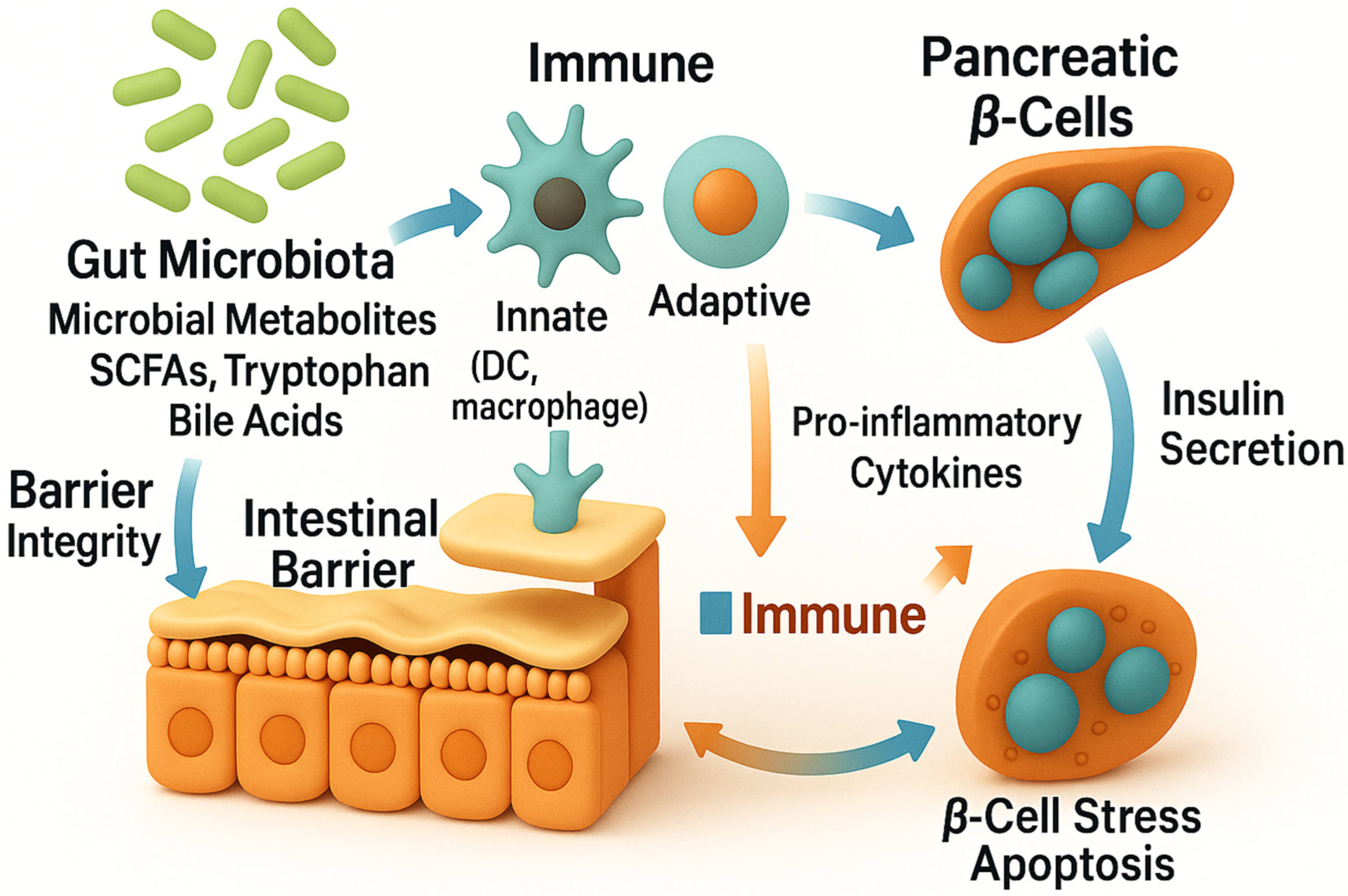
5. Mechanistic Links Along the Gut–Pancreas Axis (GPA)
5.1. Barrier Dysfunction and Microbial Dysbiosis
5.2. Microbial Translocation and Innate Immune Activation
5.3. Molecular Mimicry and Adaptive Immune Responses
5.4. Immunomodulatory Effects of SCFAs
5.5. Tryptophan Metabolism and AHR Signaling Pathway
5.6. Bile Acid Metabolism and Immune Modulation
5.7. Pattern Recognition Receptors and Immune Activation
5.8. Early-Life Microbiota and Immune Imprinting
6. Regulatory T Cells in the Gut–Pancreas Axis
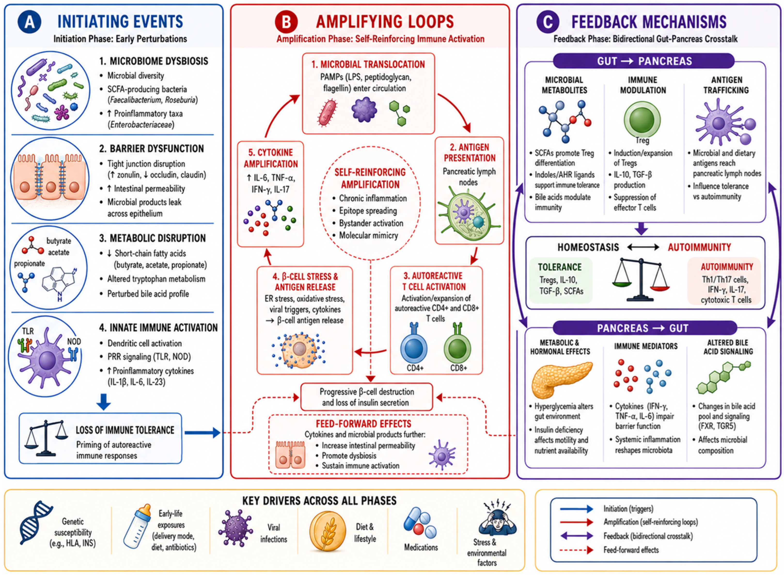
7. A Feedback Model of Microbiota–Immune Interactions
8. Therapeutic and Preventive Implications
9. Concluding Remarks and Future Directions
Author Contributions
Funding
Data Availability Statement
Acknowledgments
Conflicts of Interest
Abbreviations
| AHR | aryl hydrocarbon receptor |
| AMP | antimicrobial peptide |
| CRAMP | cathelicidin-related antimicrobial peptide |
| CVB | coxsackievirus group B |
| FMT | fecal microbiota transplantation |
| FXR | farnesoid X receptor |
| GAD65 | glutamic acid decarboxylase 65 |
| GPA | gut–pancreas axis |
| GPR | G-protein-coupled receptor |
| HDAC | histone deacetylase |
| HLA | human leukocyte antigen |
| IA-2 | islet antigen-2 |
| IL | interleukin |
| LPS | lipopolysaccharide |
| MAMP | microbe-associated molecular pattern |
| MASLD | metabolic dysfunction-associated steatotic liver disease |
| MUC | mucin |
| NOD mice | non-obese diabetic mice |
| NOD2 | nucleotide-binding oligomerization domain-containing protein 2 |
| pTreg | peripherally induced regulatory T cell |
| SCFA | short-chain fatty acid |
| T1D | type 1 diabetes |
| TGR5 | Takeda G-protein-coupled receptor 5 |
| TLR | Toll-like receptor |
| Treg | regulatory T cell (used as the standard collective abbreviation throughout) |
| tTreg | thymically derived regulatory T cell |
| ZO-1 | zonula occludens-1 |
References
- Jacobsen, L.M.; Schatz, D.A. Type 1 Diabetes: A Review. JAMA 2026, 335, 1070–1083. [Google Scholar] [CrossRef]
- Michels, A.W.; Redondo, M.J.; Atkinson, M.A. The pathogenesis, natural history, and treatment of type 1 diabetes: Time (thankfully) does not stand still. Lancet Diabetes Endocrinol. 2022, 10, 90–92. [Google Scholar] [CrossRef]
- Atkinson, M.A.; Mirmira, R.G. The pathogenic “symphony” in type 1 diabetes: A disorder of the immune system, β cells, and exocrine pancreas. Cell Metab. 2023, 35, 1500–1518. [Google Scholar] [CrossRef]
- Ogle, G.D.; Wang, F.; Haynes, A.; Gregory, G.A.; King, T.W.; Deng, K.; Dabelea, D.; James, S.; Jenkins, A.J.; Li, X.; et al. Global type 1 diabetes prevalence, incidence, and mortality estimates 2025: Results from the International diabetes Federation Atlas, 11th Edition, and the T1D Index Version 3.0. Diabetes Res. Clin. Pract. 2025, 225, 112277. [Google Scholar] [CrossRef]
- Evans-Molina, C.; Dor, Y.; Lernmark, Å.; Mathieu, C.; Millman, J.R.; Mirmira, R.G.; Pociot, F.; Redondo, M.J.; Rich, S.S.; Richardson, S.J.; et al. The heterogeneity of type 1 diabetes: Implications for pathogenesis, prevention, and treatment-2024 Diabetes, Diabetes Care, and Diabetologia Expert Forum. Diabetologia 2025, 68, 1859–1878. [Google Scholar] [CrossRef]
- Alkanani, A.K.; Hara, N.; Gottlieb, P.A.; Ir, D.; Robertson, C.E.; Wagner, B.D.; Frank, D.N.; Zipris, D. Alterations in Intestinal Microbiota Correlate with Susceptibility to Type 1 Diabetes. Diabetes 2015, 64, 3510–3520. [Google Scholar] [CrossRef] [PubMed]
- Yuan, X.; Wang, R.; Han, B.; Sun, C.; Chen, R.; Wei, H.; Chen, L.; Du, H.; Li, G.; Yang, Y.; et al. Functional and metabolic alterations of gut microbiota in children with new-onset type 1 diabetes. Nat. Commun. 2022, 13, 6356. [Google Scholar] [CrossRef] [PubMed]
- Bélteky, M.; Milletich, P.L.; Ahrens, A.P.; Triplett, E.W.; Ludvigsson, J. Infant gut microbiome composition correlated with type 1 diabetes acquisition in the general population: The ABIS study. Diabetologia 2023, 66, 1116–1128. [Google Scholar] [CrossRef] [PubMed]
- Stewart, C.J.; Ajami, N.J.; O’Brien, J.L.; Hutchinson, D.S.; Smith, D.P.; Wong, M.C.; Ross, M.C.; Lloyd, R.E.; Doddapaneni, H.; Metcalf, G.A.; et al. Temporal development of the gut microbiome in early childhood from the TEDDY study. Nature 2018, 562, 583–588. [Google Scholar] [CrossRef]
- Blok, L.; Hanssen, N.; Nieuwdorp, M.; Rampanelli, E. From Microbes to Metabolites: Advances in Gut Microbiome Research in Type 1 Diabetes. Metabolites 2025, 15, 138. [Google Scholar] [CrossRef]
- Mokhtari, P.; Metos, J.; Anandh Babu, P.V. Impact of type 1 diabetes on the composition and functional potential of gut microbiome in children and adolescents: Possible mechanisms, current knowledge, and challenges. Gut Microbes 2021, 13, 1926841. [Google Scholar] [CrossRef]
- Tillett, B.J.; Dwiyanto, J.; Secombe, K.R.; George, T.; Zhang, V.; Anderson, D.; Duggan, E.; Giri, R.; Loo, D.; Stoll, T.; et al. SCFA biotherapy delays diabetes in humanized gnotobiotic mice by remodeling mucosal homeostasis and metabolome. Nat. Commun. 2025, 16, 2893. [Google Scholar] [CrossRef]
- Ismail, H.M.; Perera, D.; Mandal, R.; DiMeglio, L.A.; Evans-Molina, C.; Hannon, T.; Petrosino, J.; Javornik Cregeen, S.; Schmidt, N.W. Gut Microbial Changes Associated with Obesity in Youth with Type 1 Diabetes. J. Clin. Endocrinol. Metab. 2025, 110, 364–373. [Google Scholar] [CrossRef] [PubMed]
- Wu, L.; Zhang, Q.; Tang, Z.; Li, Y.; Wu, T.; Chen, L.; Tan, C.; Zhang, L.; Ji, X.; Zhang, S.; et al. Gut microbiota-derived tryptophan indole metabolites ameliorate collagen-induced arthritis in mice via aryl hydrocarbon receptor activation in T cells and intestinal epithelial cells. Arthritis Rheumatol. 2026, 78, 613–629. [Google Scholar] [CrossRef]
- Mariño, E.; Richards, J.L.; McLeod, K.H.; Stanley, D.; Yap, Y.A.; Knight, J.; McKenzie, C.; Kranich, J.; Oliveira, A.C.; Rossello, F.J.; et al. Gut microbial metabolites limit the frequency of autoimmune T cells and protect against type 1 diabetes. Nat. Immunol. 2017, 18, 552–562, Erratum in Nat. Immunol. 2017, 18, 951. https://doi.org/10.1038/ni0817-951c. Erratum in Nat. Immunol. 2017, 18, 1271. https://doi.org/10.1038/ni1117-1271c. [Google Scholar] [CrossRef] [PubMed]
- Tisza, M.J.; Lloyd, R.E.; Hoffman, K.; Smith, D.P.; Rewers, M.; Javornik Cregeen, S.J.; Petrosino, J.F. Longitudinal phage-bacteria dynamics in the early life gut microbiome. Nat. Microbiol. 2025, 10, 420–430. [Google Scholar] [CrossRef]
- Zimmerman, S.; Tierney, B.T.; Nguyen, V.K.; Kostic, A.D.; Patel, C.J. Specification curve analysis of the TEDDY study reveals large variation in microbiome-based T1D predictive performance. Nat. Commun. 2025, 16, 9526. [Google Scholar] [CrossRef] [PubMed]
- Graef, F.A.; Berger, B.; Bahr, L.S.; Stange, R.; Michalsen, A.; Paul, F.; Vallance, B.A.; Jacobson, K. Fasting elicits gut microbiome signature changes that extend to type 1 diabetes patients. Front. Endocrinol. 2025, 16, 1623800. [Google Scholar] [CrossRef] [PubMed]
- Wen, L.; Ley, R.E.; Volchkov, P.Y.; Stranges, P.B.; Avanesyan, L.; Stonebraker, A.C.; Hu, C.; Wong, F.S.; Szot, G.L.; Bluestone, J.A.; et al. Innate immunity and intestinal microbiota in the development of Type 1 diabetes. Nature 2008, 455, 1109–1113. [Google Scholar] [CrossRef]
- Miani, M.; Le Naour, J.; Waeckel-Enée, E.; Verma, S.C.; Straube, M.; Emond, P.; Ryffel, B.; van Endert, P.; Sokol, H.; Diana, J. Gut Microbiota-Stimulated Innate Lymphoid Cells Support β-Defensin 14 Expression in Pancreatic Endocrine Cells, Preventing Autoimmune Diabetes. Cell Metab. 2018, 28, 557–572.e556. [Google Scholar] [CrossRef]
- Brugman, S.; Klatter, F.A.; Visser, J.T.; Wildeboer-Veloo, A.C.; Harmsen, H.J.; Rozing, J.; Bos, N.A. Antibiotic treatment partially protects against type 1 diabetes in the Bio-Breeding diabetes-prone rat. Is the gut flora involved in the development of type 1 diabetes? Diabetologia 2006, 49, 2105–2108. [Google Scholar] [CrossRef]
- Brown, K.; Godovannyi, A.; Ma, C.; Zhang, Y.; Ahmadi-Vand, Z.; Dai, C.; Gorzelak, M.A.; Chan, Y.; Chan, J.M.; Lochner, A.; et al. Prolonged antibiotic treatment induces a diabetogenic intestinal microbiome that accelerates diabetes in NOD mice. ISME J. 2016, 10, 321–332. [Google Scholar] [CrossRef]
- Wang, Z.; Gong, M.; Fang, Y.; Yuan, H.; Zhang, C. Reconstruction characteristics of gut microbiota from patients with type 1 diabetes affect the phenotypic reproducibility of glucose metabolism in mice. Sci. China Life Sci. 2025, 68, 176–188. [Google Scholar] [CrossRef]
- Rodrigues, V.F.; Elias-Oliveira, J.; Pereira, Í.S.; Pereira, J.A.; Barbosa, S.C.; Machado, M.S.G.; Guimarães, J.B.; Pacheco, T.C.F.; Bortolucci, J.; Zaramela, L.S.; et al. Akkermansia muciniphila restrains type 1 diabetes onset by eliciting cDC2 and Treg cell differentiation in NOD and STZ-induced experimental models. Life Sci. 2025, 372, 123624. [Google Scholar] [CrossRef]
- Gudi, R.R.; Taylor, H.; Johnson, B.M.; Maurya, R.; Mulligan, M.E.; Carter, L.; Westwater, C.; Vasu, C. Human Gut Commensal Bacteroides fragilis Suppresses Mucin Production and Alters Microbiota Composition Resulting in Accelerated Type 1 Diabetes in Mice. Immunology 2026, 177, 119–136. [Google Scholar] [CrossRef] [PubMed]
- Neuman, V.; Cinek, O.; Funda, D.P.; Hudcovic, T.; Golias, J.; Kramna, L.; Petruzelkova, L.; Pruhova, S.; Sumnik, Z. Human gut microbiota transferred to germ-free NOD mice modulate the progression towards type 1 diabetes regardless of the pace of beta cell function loss in the donor. Diabetologia 2019, 62, 1291–1296. [Google Scholar] [CrossRef] [PubMed]
- Costa, F.R.; Françozo, M.C.; de Oliveira, G.G.; Ignacio, A.; Castoldi, A.; Zamboni, D.S.; Ramos, S.G.; Câmara, N.O.; de Zoete, M.R.; Palm, N.W.; et al. Gut microbiota translocation to the pancreatic lymph nodes triggers NOD2 activation and contributes to T1D onset. J. Exp. Med. 2016, 213, 1223–1239. [Google Scholar] [CrossRef]
- Tai, N.; Peng, J.; Liu, F.; Gulden, E.; Hu, Y.; Zhang, X.; Chen, L.; Wong, F.S.; Wen, L. Microbial antigen mimics activate diabetogenic CD8 T cells in NOD mice. J. Exp. Med. 2016, 213, 2129–2146. [Google Scholar] [CrossRef] [PubMed]
- Vatanen, T.; Franzosa, E.A.; Schwager, R.; Tripathi, S.; Arthur, T.D.; Vehik, K.; Lernmark, Å.; Hagopian, W.A.; Rewers, M.J.; She, J.X.; et al. The human gut microbiome in early-onset type 1 diabetes from the TEDDY study. Nature 2018, 562, 589–594. [Google Scholar] [CrossRef]
- Zhang, L.; Jonscher, K.R.; Zhang, Z.; Xiong, Y.; Mueller, R.S.; Friedman, J.E.; Pan, C. Islet autoantibody seroconversion in type-1 diabetes is associated with metagenome-assembled genomes in infant gut microbiomes. Nat. Commun. 2022, 13, 3551. [Google Scholar] [CrossRef]
- Lowe, C.E.; Cooper, J.D.; Brusko, T.; Walker, N.M.; Smyth, D.J.; Bailey, R.; Bourget, K.; Plagnol, V.; Field, S.; Atkinson, M.; et al. Large-scale genetic fine mapping and genotype-phenotype associations implicate polymorphism in the IL2RA region in type 1 diabetes. Nat. Genet. 2007, 39, 1074–1082. [Google Scholar] [CrossRef]
- Zhang, C.; Li, G.; Lu, T.; Liu, L.; Sui, Y.; Bai, R.; Li, L.; Sun, B. The Interaction of Microbiome and Pancreas in Acute Pancreatitis. Biomolecules 2023, 14, 59. [Google Scholar] [CrossRef] [PubMed]
- Yu, X.F.; Gurung, C.; Yu, Z.; Xiao, W.; Kerem, G.; Teng, B.; Zhang, J.V.; Su, Z.; Zhou, Y.; Ren, P.G. Intestinal bacteria translocation promotes β-cell dysfunction in DIO mice. Sci. Rep. 2025, 15, 31034. [Google Scholar] [CrossRef] [PubMed]
- Nomura, K.; Yamasaki, Y.; Takeji, K.; Deha, S.; Yamashita, K.; Izumi-Mishima, Y.; Yasui-Yamada, S.; Kuroda, M.; Harada, N.; Kitamura, T.; et al. Gut-pancreas axis in the control of insulin secretion in streptozotocin-resistant rats. Biochem. Biophys. Res. Commun. 2025, 752, 151487. [Google Scholar] [CrossRef] [PubMed]
- Liang, J.; Chu, Y.; Chen, X.; Leng, Y. The gut-adipose/pancreas axis: A novel perspective on glycolipid metabolism dysregulation in MAFLD and T2DM pathogenesis. Front. Endocrinol. 2025, 16, 1664233, Erratum in Front. Endocrinol. 2025, 16, 1743993. https://doi.org/10.3389/fendo.2025.1743993. [Google Scholar] [CrossRef] [PubMed] [PubMed Central]
- Arikath, K.; Batra, S.K.; Ponnusamy, M.P. Unveiling the gut-pancreas axis: Microbial influence on stemness and tumor microenvironment of PDAC. Stem Cells 2026, 44, sxaf064. [Google Scholar] [CrossRef]
- Mauney, E.E.; Wibowo, M.C.; Tseng, Y.H.; Kostic, A.D. Adipose tissue-gut microbiome crosstalk in inflammation and thermogenesis. Trends Endocrinol. Metab. 2025, 36, 721–732. [Google Scholar] [CrossRef]
- Abildinova, G.Z.; Benberin, V.V.; Vochshenkova, T.A.; Afshar, A.; Mussin, N.M.; Kaliyev, A.A.; Zhussupova, Z.; Tamadon, A. The gut-brain-metabolic axis: Exploring the role of microbiota in insulin resistance and cognitive function. Front. Microbiol. 2024, 15, 1463958. [Google Scholar] [CrossRef]
- Zhang, Z.; Tanaka, I.; Pan, Z.; Ernst, P.B.; Kiyono, H.; Kurashima, Y. Intestinal homeostasis and inflammation: Gut microbiota at the crossroads of pancreas-intestinal barrier axis. Eur. J. Immunol. 2022, 52, 1035–1046. [Google Scholar] [CrossRef]
- Dubal, A.S.; Sarteau, A.C.; Azcarate-Peril, M.A.; Djukic, Z.; Ercolino, G.; Fruik, A.; Ho, J.; Lee, Z.; Roach, J.; Valancius, M.; et al. Exploring the Effects of Dietary Intake on Gut Microbiome Diversity and Composition of Older Adults with Type 1 Diabetes: A Pilot Study. Curr. Dev. Nutr. 2025, 9, 107550. [Google Scholar] [CrossRef]
- Tilg, H.; Adolph, T.E.; Trauner, M. Gut-liver axis: Pathophysiological concepts and clinical implications. Cell Metab. 2022, 34, 1700–1718. [Google Scholar] [CrossRef]
- Pabst, O.; Hornef, M.W.; Schaap, F.G.; Cerovic, V.; Clavel, T.; Bruns, T. Gut-liver axis: Barriers and functional circuits. Nat. Rev. Gastroenterol. Hepatol. 2023, 20, 447–461. [Google Scholar] [CrossRef]
- Milosevic, I.; Vujovic, A.; Barac, A.; Djelic, M.; Korac, M.; Radovanovic Spurnic, A.; Gmizic, I.; Stevanovic, O.; Djordjevic, V.; Lekic, N.; et al. Gut-Liver Axis, Gut Microbiota, and Its Modulation in the Management of Liver Diseases: A Review of the Literature. Int. J. Mol. Sci. 2019, 20, 395. [Google Scholar] [CrossRef] [PubMed]
- Marroncini, G.; Naldi, L.; Martinelli, S.; Amedei, A. Gut-Liver-Pancreas Axis Crosstalk in Health and Disease: From the Role of Microbial Metabolites to Innovative Microbiota Manipulating Strategies. Biomedicines 2024, 12, 1398. [Google Scholar] [CrossRef] [PubMed]
- Ma, L.; Wang, H.; Jin, Q.; Sun, Z.; Yu, S.; Zhang, Y. The Gut-Liver Axis: Molecular Mechanisms and Therapeutic Targeting in Liver Disease. Int. J. Gen. Med. 2025, 18, 7531–7546. [Google Scholar] [CrossRef] [PubMed]
- Zheng, Z.; Wang, B. The Gut-Liver Axis in Health and Disease: The Role of Gut Microbiota-Derived Signals in Liver Injury and Regeneration. Front. Immunol. 2021, 12, 775526. [Google Scholar] [CrossRef]
- Kwan, S.Y.; Jiao, J.; Qi, J.; Wang, Y.; Wei, P.; McCormick, J.B.; Fisher-Hoch, S.P.; Beretta, L. Bile Acid Changes Associated with Liver Fibrosis and Steatosis in the Mexican-American Population of South Texas. Hepatol. Commun. 2020, 4, 555–568. [Google Scholar] [CrossRef]
- Xiao, L.; Hu, L.; Chu, H.; Chen, L.; Yan, J.; Wang, W.; Yang, X.; Zhu, Q.; Du, F.; Song, Y.; et al. Retrorsine Cooperates with Gut Microbiota to Promote Hepatic Sinusoidal Obstruction Syndrome by Disrupting the Gut Barrier. J. Clin. Transl. Hepatol. 2022, 10, 1086–1098. [Google Scholar] [CrossRef]
- Kaur, I.; Juneja, P.; Tiwari, R.; Vasudevan, A.; Mourya, A.K.; Trauner, M.; Sarin, S.K.; Tripathi, D.M.; Kaur, S. Secondary bile acids in portal blood contribute to liver regeneration in a rat model of partial hepatectomy. Am. J. Physiol. Gastrointest. Liver Physiol. 2024, 327, G586–G597. [Google Scholar] [CrossRef]
- Bajaj, J.S.; Shamsaddini, A.; Fagan, A.; Sterling, R.K.; Gavis, E.; Khoruts, A.; Fuchs, M.; Lee, H.; Sikaroodi, M.; Gillevet, P.M. Fecal Microbiota Transplant in Cirrhosis Reduces Gut Microbial Antibiotic Resistance Genes: Analysis of Two Trials. Hepatol. Commun. 2021, 5, 258–271. [Google Scholar] [CrossRef]
- Sharma, A.; Roy, A.; Premkumar, M.; Verma, N.; Duseja, A.; Taneja, S.; Grover, S.; Chopra, M.; Dhiman, R.K. Fecal microbiota transplantation in alcohol-associated acute-on-chronic liver failure: An open-label clinical trial. Hepatol. Int. 2022, 16, 433–446. [Google Scholar] [CrossRef]
- Zheng, C.; Wang, L.; Zou, T.; Lian, S.; Luo, J.; Lu, Y.; Hao, H.; Xu, Y.; Xiang, Y.; Zhang, X.; et al. Ileitis promotes MASLD progression via bile acid modulation and enhanced TGR5 signaling in ileal CD8(+) T cells. J. Hepatol. 2024, 80, 764–777. [Google Scholar] [CrossRef]
- Kim, T.K.; Lee, J.C.; Im, S.H.; Lee, M.S. Amelioration of Autoimmune Diabetes of NOD Mice by Immunomodulating Probiotics. Front. Immunol. 2020, 11, 1832. [Google Scholar] [CrossRef]
- Okada, M.; Zhang, V.; Loaiza Naranjo, J.D.; Tillett, B.J.; Wong, F.S.; Steptoe, R.J.; Bergot, A.S.; Hamilton-Williams, E.E. Islet-specific CD8(+) T cells gain effector function in the gut lymphoid tissues via bystander activation not molecular mimicry. Immunol. Cell Biol. 2023, 101, 36–48. [Google Scholar] [CrossRef]
- Kartal, E.; Schmidt, T.S.B.; Molina-Montes, E.; Rodríguez-Perales, S.; Wirbel, J.; Maistrenko, O.M.; Akanni, W.A.; Alashkar Alhamwe, B.; Alves, R.J.; Carrato, A.; et al. A faecal microbiota signature with high specificity for pancreatic cancer. Gut 2022, 71, 1359–1372. [Google Scholar] [CrossRef]
- Brown, H.; Komnick, M.R.; Brigleb, P.H.; Dermody, T.S.; Esterházy, D. Lymph node sharing between pancreas, gut, and liver leads to immune crosstalk and regulation of pancreatic autoimmunity. Immunity 2023, 56, 2070–2085.e2011. [Google Scholar] [CrossRef]
- Jacob, N.; Jaiswal, S.; Maheshwari, D.; Nallabelli, N.; Khatri, N.; Bhatia, A.; Bal, A.; Malik, V.; Verma, S.; Kumar, R.; et al. Butyrate induced Tregs are capable of migration from the GALT to the pancreas to restore immunological tolerance during type-1 diabetes. Sci. Rep. 2020, 10, 19120. [Google Scholar] [CrossRef]
- Murri, M.; Leiva, I.; Gomez-Zumaquero, J.M.; Tinahones, F.J.; Cardona, F.; Soriguer, F.; Queipo-Ortuño, M.I. Gut microbiota in children with type 1 diabetes differs from that in healthy children: A case-control study. BMC Med. 2013, 11, 46. [Google Scholar] [CrossRef]
- Asante Baadu, F.; Ahsan, M.; Hussain, B.; Hussain, S.; Malik, H.; Naqvi, S.S.; Mustafa, Z.; Zahid, M.; Ahmad, R.T. Microbiome Imbalance and Pediatric Type 1 Diabetes Mellitus: An Updated Systematic Review of Gut Dysbiosis Evidence. Cureus 2025, 17, e89279. [Google Scholar] [CrossRef]
- Hummel, S.; Pflüger, M.; Hummel, M.; Bonifacio, E.; Ziegler, A.G. Primary dietary intervention study to reduce the risk of islet autoimmunity in children at increased risk for type 1 diabetes: The BABYDIET study. Diabetes Care 2011, 34, 1301–1305. [Google Scholar] [CrossRef]
- Beyerlein, A.; Chmiel, R.; Hummel, S.; Winkler, C.; Bonifacio, E.; Ziegler, A.G. Timing of gluten introduction and islet autoimmunity in young children: Updated results from the BABYDIET study. Diabetes Care 2014, 37, e194–e195. [Google Scholar] [CrossRef]
- Hakola, L.; Lund-Blix, N.A.; Takkinen, H.M.; Tapanainen, H.; Niinistö, S.; Korhonen, T.E.; Stene, L.C.; Hyöty, H.; Toppari, J.; Ilonen, J.; et al. Maternal gluten, cereal, and dietary fiber intake during pregnancy and lactation and the risk of islet autoimmunity and type 1 diabetes in the child. Clin. Nutr. ESPEN 2024, 62, 22–27. [Google Scholar] [CrossRef]
- Tapia, G.; Størdal, K.; Mårild, K.; Kahrs, C.R.; Skrivarhaug, T.; Njølstad, P.R.; Joner, G.; Stene, L.C. Antibiotics, acetaminophen and infections during prenatal and early life in relation to type 1 diabetes. Int. J. Epidemiol. 2018, 47, 1538–1548. [Google Scholar] [CrossRef] [PubMed]
- Cardwell, C.R.; Stene, L.C.; Joner, G.; Cinek, O.; Svensson, J.; Goldacre, M.J.; Parslow, R.C.; Pozzilli, P.; Brigis, G.; Stoyanov, D.; et al. Caesarean section is associated with an increased risk of childhood-onset type 1 diabetes mellitus: A meta-analysis of observational studies. Diabetologia 2008, 51, 726–735. [Google Scholar] [CrossRef]
- Donald, K.; Serapio-Palacios, A.; Bozorgmehr, T.; Ma, M.; Garcia, M.A.I.; Petersen, C.; Mandhane, P.; Subbarao, P.; Moraes, T.J.; Simons, E.; et al. Human milk IgA promotes normal immune development by limiting Th17-inducing Erysipelatoclostridium ramosum in the infant gut. Proc. Natl. Acad. Sci. USA 2025, 122, e2501030122. [Google Scholar] [CrossRef] [PubMed]
- Vatanen, T.; Kostic, A.D.; d’Hennezel, E.; Siljander, H.; Franzosa, E.A.; Yassour, M.; Kolde, R.; Vlamakis, H.; Arthur, T.D.; Hämäläinen, A.M.; et al. Variation in Microbiome LPS Immunogenicity Contributes to Autoimmunity in Humans. Cell 2016, 165, 842–853, Erratum in Cell 2016, 165, 1551. https://doi.org/10.1016/j.cell.2016.05.056. [Google Scholar] [CrossRef] [PubMed] [PubMed Central]
- Kondrashova, A.; Seiskari, T.; Ilonen, J.; Knip, M.; Hyöty, H. The ‘Hygiene hypothesis’ and the sharp gradient in the incidence of autoimmune and allergic diseases between Russian Karelia and Finland. Apmis 2013, 121, 478–493. [Google Scholar] [CrossRef]
- Aguayo-Patrón, S.V.; Trujillo-Rivera, O.A.; Cornejo-Granados, F.; Ochoa-Leyva, A.; Calderón de la Barca, A.M. HLA-Haplotypes Influence Microbiota Structure in Northwestern Mexican Schoolchildren Predisposed for Celiac Disease or Type 1 Diabetes. Microorganisms 2023, 11, 1412. [Google Scholar] [CrossRef] [PubMed]
- Yue, T.; Tan, H.; Wang, C.; Liu, Z.; Yang, D.; Ding, Y.; Xu, W.; Yan, J.; Zheng, X.; Weng, J.; et al. High-risk genotypes for type 1 diabetes are associated with the imbalance of gut microbiome and serum metabolites. Front. Immunol. 2022, 13, 1033393. [Google Scholar] [CrossRef] [PubMed]
- Russell, J.T.; Roesch, L.F.W.; Ördberg, M.; Ilonen, J.; Atkinson, M.A.; Schatz, D.A.; Triplett, E.W.; Ludvigsson, J. Genetic risk for autoimmunity is associated with distinct changes in the human gut microbiome. Nat. Commun. 2019, 10, 3621. [Google Scholar] [CrossRef]
- Parviainen, A.; Härkönen, T.; Ilonen, J.; But, A.; Knip, M. Heterogeneity of Type 1 Diabetes at Diagnosis Supports Existence of Age-Related Endotypes. Diabetes Care 2022, 45, 871–879. [Google Scholar] [CrossRef]
- Weston, C.S.; Boehm, B.O.; Pozzilli, P. Type 1 diabetes: A new vision of the disease based on endotypes. Diabetes Metab. Res. Rev. 2024, 40, e3770. [Google Scholar] [CrossRef]
- Endesfelder, D.; zu Castell, W.; Ardissone, A.; Davis-Richardson, A.G.; Achenbach, P.; Hagen, M.; Pflueger, M.; Gano, K.A.; Fagen, J.R.; Drew, J.C.; et al. Compromised gut microbiota networks in children with anti-islet cell autoimmunity. Diabetes 2014, 63, 2006–2014. [Google Scholar] [CrossRef]
- Kostic, A.D.; Gevers, D.; Siljander, H.; Vatanen, T.; Hyötyläinen, T.; Hämäläinen, A.M.; Peet, A.; Tillmann, V.; Pöhö, P.; Mattila, I.; et al. The dynamics of the human infant gut microbiome in development and in progression toward type 1 diabetes. Cell Host Microbe 2015, 17, 260–273, Erratum in Cell Host Microbe 2016, 20, 682–689. https://doi.org/10.1016/j.chom.2016.10.005. [Google Scholar] [CrossRef]
- Lamichhane, S.; Sen, P.; Dickens, A.M.; Alves, M.A.; Härkönen, T.; Honkanen, J.; Vatanen, T.; Xavier, R.J.; Hyötyläinen, T.; Knip, M.; et al. Dysregulation of secondary bile acid metabolism precedes islet autoimmunity and type 1 diabetes. Cell Rep. Med. 2022, 3, 100762. [Google Scholar] [CrossRef] [PubMed]
- Krischer, J.P.; Liu, X.; Lernmark, Å.; Hagopian, W.A.; Rewers, M.J.; She, J.X.; Toppari, J.; Ziegler, A.G.; Akolkar, B. Predictors of the Initiation of Islet Autoimmunity and Progression to Multiple Autoantibodies and Clinical Diabetes: The TEDDY Study. Diabetes Care 2022, 45, 2271–2281. [Google Scholar] [CrossRef] [PubMed]
- Kuusela, S.; Koskenniemi, J.J.; Valtanen, T.; Pokka, T.; Härkönen, T.; Ilonen, J.; Lempainen, J.; Kyrönniemi, A.; Toppari, J.; Knip, M.; et al. Islet autoimmunity and progression to type 1 diabetes in the Finnish DIPP study: Comparison between genetically susceptible children with and without an affected first-degree relative. Diabetologia 2026, 69, 93–102. [Google Scholar] [CrossRef] [PubMed]
- Salo, T.E.; Hakola, L.; Niinistö, S.; Takkinen, H.M.; Ahonen, S.; Puustinen, L.; Ilonen, J.; Toppari, J.; Veijola, R.; Hyöty, H.; et al. Gut Inflammation Markers, Diet, and Risk of Islet Autoimmunity in Finnish Children—A Nested Case-Control Study. J. Nutr. 2024, 154, 2244–2254. [Google Scholar] [CrossRef]
- Scharf, M.W.; Forsgård, R.A.; Prado, S.B.R.; Ganda Mall, J.P.; Repsilber, D.; Brummer, R.J.; Marques, T.M.; Wall, R. Acute effects of butyrate on intestinal permeability in patients with irritable bowel syndrome assessed by a novel colonoscopy research model. Gut Microbes 2025, 17, 2545414. [Google Scholar] [CrossRef]
- Chakraborty, P.; Laird, A.S. Understanding activity of butyrate at a cellular level. Neural Regen. Res. 2025, 20, 2323–2324. [Google Scholar] [CrossRef]
- Zhang, L.; Liu, C.; Jiang, Q.; Yin, Y. Butyrate in Energy Metabolism: There is Still More to Learn. Trends Endocrinol. Metab. 2021, 32, 159–169. [Google Scholar] [CrossRef]
- Gaskarth, D.A.; Fan, S.; Highton, A.J.; Kemp, R.A. The microbial metabolite butyrate enhances the effector and memory functions of murine CD8+ T cells and improves anti-tumor activity. Front. Med. 2025, 12, 1577906. [Google Scholar] [CrossRef]
- Lamichhane, S.; Sen, P.; Dickens, A.M.; Kråkström, M.; Ilonen, J.; Lempainen, J.; Hyöty, H.; Lahesmaa, R.; Veijola, R.; Toppari, J.; et al. Circulating metabolic signatures of rapid and slow progression to type 1 diabetes in islet autoantibody-positive children. Front. Endocrinol. 2023, 14, 1211015. [Google Scholar] [CrossRef]
- Webb-Robertson, B.M.; Nakayasu, E.S.; Frohnert, B.I.; Bramer, L.M.; Akers, S.M.; Norris, J.M.; Vehik, K.; Ziegler, A.G.; Metz, T.O.; Rich, S.S.; et al. Integration of Infant Metabolite, Genetic, and Islet Autoimmunity Signatures to Predict Type 1 Diabetes by Age 6 Years. J. Clin. Endocrinol. Metab. 2022, 107, 2329–2338. [Google Scholar] [CrossRef] [PubMed]
- Oresic, M. Metabolomics in the studies of islet autoimmunity and type 1 diabetes. Rev. Diabet. Stud. 2012, 9, 236–247. [Google Scholar] [CrossRef]
- Flammer, E.R.; Christopher, M.W.; Powers, E.R.; Broncucia, H.; Steck, A.K.; Gitelman, S.E.; Garrett, T.J.; Ismail, H.M. Serum Metabolomics Reveals Potential Differences in Gut Microbiota-Associated Metabolites in Twins Discordant for Type 1 Diabetes. Diabetes 2025, 74, 1957–1963. [Google Scholar] [CrossRef]
- Ma, L.; Liu, J.; Deng, M.; Zhou, L.; Zhang, Q.; Xiao, X. Metabolomics analysis of serum and urine in type 1 diabetes patients with different time in range derived from continuous glucose monitoring. Diabetol. Metab. Syndr. 2024, 16, 21. [Google Scholar] [CrossRef]
- Jing, W.; Dong, S.; Xu, Y.; Liu, J.; Ren, J.; Liu, X.; Zhu, M.; Zhang, M.; Shi, H.; Li, N.; et al. Gut microbiota-derived tryptophan metabolites regulated by Wuji Wan to attenuate colitis through AhR signaling activation. Acta Pharm. Sin. B 2025, 15, 205–223. [Google Scholar] [CrossRef] [PubMed]
- Marafini, I.; Monteleone, I.; Laudisi, F.; Monteleone, G. Aryl Hydrocarbon Receptor Signalling in the Control of Gut Inflammation. Int. J. Mol. Sci. 2024, 25, 4527. [Google Scholar] [CrossRef]
- Peesh, P.; Blasco-Conesa, M.P.; El Hamamy, A.; Khan, R.; Guzman, G.U.; Honarpisheh, P.; Mohan, E.C.; Goodman, G.W.; Nguyen, J.N.; Banerjee, A.; et al. Benefits of equilibrium between microbiota- and host-derived ligands of the aryl hydrocarbon receptor after stroke in aged male mice. Nat. Commun. 2025, 16, 1767. [Google Scholar] [CrossRef] [PubMed]
- Jia, Y.; He, M.; Wang, F.; Zhan, Y.; Deng, Q.; Shen, J.; Wang, X.; Ran, Q.; Huang, W.; Ling, Y.; et al. Indole-3-lactic acid protects the gut vascular barrier following intestinal ischemia injury through AhR/Nrf2/STAT3 mediated claudin 2 downregulation. Cell Commun. Signal 2025, 23, 447. [Google Scholar] [CrossRef]
- Kim, J.Y.; Cartwright, I.M.; Colgan, S.P. Indole dysbiosis and mucosal inflammation. Microbiota Host 2025, 3, e250004. [Google Scholar] [CrossRef]
- Hill, J.H.; Bell, R.; Barrios, L.; Baird, H.; Ost, K.; Greenewood, M.; Monts, J.K.; Tracy, E.; Meili, C.H.; Chiaro, T.R.; et al. Neonatal fungi promote lifelong metabolic health through macrophage-dependent β cell development. Science 2025, 387, eadn0953. [Google Scholar] [CrossRef]
- Huang, J.; Pearson, J.A.; Peng, J.; Hu, Y.; Sha, S.; Xing, Y.; Huang, G.; Li, X.; Hu, F.; Xie, Z.; et al. Gut microbial metabolites alter IgA immunity in type 1 diabetes. JCI Insight 2020, 5, e135718. [Google Scholar] [CrossRef]
- Park, S.H.; Cheon, D.H.; Kim, Y.M.; Choi, Y.; Cho, Y.J.; Hong, B.K.; Cho, S.H.; Kweon, M.N.; Kwon, H.M.; Chang, E.B.; et al. NFAT5 dictates crosstalk between intestinal epithelial regenerative capacity and microbiota in murine colitis models. J. Clin. Investig. 2025, 135, e183093. [Google Scholar] [CrossRef] [PubMed]
- Tepekule, B.; Lim, A.I.; Metcalf, C.J.E. A model of early-life interactions between the gut microbiome and adaptive immunity provides insights into the ontogeny of immune tolerance. PLoS Biol. 2025, 23, e3003263. [Google Scholar] [CrossRef]
- Girdhar, K.; Huang, Q.; Chow, I.T.; Vatanen, T.; Brady, C.; Raisingani, A.; Autissier, P.; Atkinson, M.A.; Kwok, W.W.; Kahn, C.R.; et al. A gut microbial peptide and molecular mimicry in the pathogenesis of type 1 diabetes. Proc. Natl. Acad. Sci. USA 2022, 119, e2120028119, Erratum in Proc. Natl. Acad. Sci. USA 2023, 120, e2309963120. https://doi.org/10.1073/pnas.2309963120. [Google Scholar] [CrossRef] [PubMed] [PubMed Central]
- Aalipanah, E.; Askarpour, M.; Eskandari, M.H.; Zare, M.; Famouri, M.; Bedeltavana, A.; Mohsenpour, M.A.; Sohrabi, Z. Comparing the effects of yogurt containing Akkermansia muciniphilia postbiotic with yogurt containing Lactobacillus rhamnosus postbiotic on body composition, biochemical indices, appetite, and depression scores in overweight or obese adults: A randomized, double-blind, controlled clinical trial. Clin. Nutr. ESPEN 2025, 68, 438–446. [Google Scholar] [CrossRef] [PubMed]
- Sun, J.; Xu, M.; Ortsäter, H.; Lundeberg, E.; Juntti-Berggren, L.; Chen, Y.Q.; Haeggström, J.Z.; Gudmundsson, G.H.; Diana, J.; Agerberth, B. Cathelicidins positively regulate pancreatic β-cell functions. FASEB J. 2016, 30, 884–894. [Google Scholar] [CrossRef]
- Lo Conte, M.; Cosorich, I.; Ferrarese, R.; Antonini Cencicchio, M.; Nobili, A.; Palmieri, V.; Massimino, L.; Lamparelli, L.A.; Liang, W.; Riba, M.; et al. Alterations of the intestinal mucus layer correlate with dysbiosis and immune dysregulation in human Type 1 Diabetes. eBioMedicine 2023, 91, 104567. [Google Scholar] [CrossRef]
- Robles-Vera, I.; Jarit-Cabanillas, A.; Brandi, P.; Martínez-López, M.; Martínez-Cano, S.; Rodrigo-Tapias, M.; Femenía-Muiña, M.; Redondo-Urzainqui, A.; Nuñez, V.; González-Correa, C.; et al. Microbiota translocation following intestinal barrier disruption promotes Mincle-mediated training of myeloid progenitors in the bone marrow. Immunity 2025, 58, 381–396.e389. [Google Scholar] [CrossRef]
- da Cruz Carvalho, M.C.; Mota, A.; do Nascimento, D.S.M.; de Sousa, I.N.F.; Bona, M.D.; da Silva Ribeiro, K.D.; Lima AÂ, M.; Maciel, B.L.L. Intestinal permeability and its relation to anthropometric and biochemical variables associated with cardiovascular risk in an elderly population. Sci. Rep. 2025, 15, 21919. [Google Scholar] [CrossRef]
- Ciurea, N.A.; Pantea, C.M.; Grama, P.; Kosovski, I.B.; Bataga, S. Fecal Zonulin as a Non-Invasive Marker of Intestinal Permeability: Findings from a Prospective Cohort Study. Medicina 2025, 61, 1527. [Google Scholar] [CrossRef]
- Turley, S.J.; Lee, J.W.; Dutton-Swain, N.; Mathis, D.; Benoist, C. Endocrine self and gut non-self intersect in the pancreatic lymph nodes. Proc. Natl. Acad. Sci. USA 2005, 102, 17729–17733. [Google Scholar] [CrossRef]
- Sorini, C.; Cosorich, I.; Lo Conte, M.; De Giorgi, L.; Facciotti, F.; Lucianò, R.; Rocchi, M.; Ferrarese, R.; Sanvito, F.; Canducci, F.; et al. Loss of gut barrier integrity triggers activation of islet-reactive T cells and autoimmune diabetes. Proc. Natl. Acad. Sci. USA 2019, 116, 15140–15149. [Google Scholar] [CrossRef]
- Bartosiak, J.T.; Hansen, P.A.; Schumacher, E.A.; Harty, K.R.; Stancill, J.S.; Corbett, J.A. Lipopolysaccharide-induced cytokine signaling activates a temporal innate defense program and represses pancreatic β-cell identity. J. Biol. Chem. 2025, 301, 110811. [Google Scholar] [CrossRef] [PubMed]
- Pipella, J.; Motlagh, R.A.; Rampazzo Morelli, N.; Thompson, P.J. Autoreactive T Cells and Cytokine Stress Drive β-Cell Senescence Entry and Accumulation in Type 1 Diabetes. Diabetes 2025, 74, 1562–1576. [Google Scholar] [CrossRef] [PubMed]
- Nanaware, P.P.; Calvo-Calle, J.M.; Redick, S.D.; Tarpley, M.W.; Cruz, J.; Clement, C.C.; Manganaro, A.; Velarde de la Cruz, E.E.; Muneeruddin, K.; Faulkner, M.; et al. The antigen presentation landscape of cytokine-stressed human pancreatic islets. Cell Rep. 2025, 44, 115927. [Google Scholar] [CrossRef]
- Chen, S.; Luo, Y.; Wei, G.; Liu, S. Molecular Mimicry at the Gut-Immune Interface: A Mechanistic Link to Type 1 Diabetes. Immunology 2026, 177, 701–712. [Google Scholar] [CrossRef]
- Girdhar, K.; Randall, A.; Mine, K.; Howard, C.; Pezzella, A.; Dogru, D.; Rhodes, L.; James, B.; Gautam, U.K.; Šrůtková, D.; et al. Impact of Parabacteroides distasonis colonization on host microbiome, metabolome, immunity, and diabetes onset. J. Mol. Endocrinol. 2025, 75, e250025. [Google Scholar] [CrossRef] [PubMed]
- Mittal, R.; Camick, N.; Lemos, J.R.N.; Hirani, K. Gene-environment interaction in the pathophysiology of type 1 diabetes. Front. Endocrinol. 2024, 15, 1335435. [Google Scholar] [CrossRef]
- Kotsiri, I.; Xanthi, M.; Domazinaki, C.M.; Magiorkinis, E. The Role of Viral Infections in the Immunopathogenesis of Type 1 Diabetes Mellitus: A Narrative Review. Biology 2025, 14, 981. [Google Scholar] [CrossRef] [PubMed]
- Jiang, Z.; He, L.; Li, D.; Zhuo, L.; Chen, L.; Shi, R.Q.; Luo, J.; Feng, Y.; Liang, Y.; Li, D.; et al. Human gut microbial aromatic amino acid and related metabolites prevent obesity through intestinal immune control. Nat. Metab. 2025, 7, 808–822. [Google Scholar] [CrossRef] [PubMed]
- Li, Q.; de Oliveira Formiga, R.; Puchois, V.; Creusot, L.; Ahmad, A.H.; Amouyal, S.; Campos-Ribeiro, M.A.; Zhao, Y.; Harris, D.M.M.; Lasserre, F.; et al. Microbial metabolite indole-3-propionic acid drives mitochondrial respiration in CD4(+) T cells to confer protection against intestinal inflammation. Nat. Metab. 2025, 7, 2510–2530. [Google Scholar] [CrossRef]
- Li, Q.; Zhang, J.; Fan, M.; Wu, N.; Li, T.; Wang, M.; Zhang, L. Gut microbiota-derived 4-hydroxyphenylacetic acid (4-HPAA) inhibits weight gain and is negatively associated with childhood obesity. Transl. Pediatr. 2025, 14, 1156–1167. [Google Scholar] [CrossRef]
- Zhang, Y.; Wang, T.; Han, J.; Song, J.; Yang, C.; Liang, L.; Li, H.; Wang, H. Targeting the gut microbiota and lipid metabolism: Potential mechanisms of natural products for the treatment of non-alcoholic fatty liver disease. Front. Pharmacol. 2025, 16, 1610498. [Google Scholar] [CrossRef]
- Saadh, M.J.; Allela, O.Q.B.; Ballal, S.; Mahdi, M.S.; Chahar, M.; Verma, R.; Al-Hussein, R.K.A.; Adil, M.; Jawad, M.J.; Al-Nuaimi, A.M.A. The effects of microbiota-derived short-chain fatty acids on T lymphocytes: From autoimmune diseases to cancer. Semin. Oncol. 2025, 52, 152398. [Google Scholar] [CrossRef]
- Mei, C.; Li, W.; Zhao, B.; He, Y.; Li, Q.; Zhang, T.; Li, X.; Zhang, K.; Zhang, Y.; Zhong, Z. Short-chain fatty acids mediate gut microbiota-brain communication and protect the blood-brain barrier integrity. Ann. N. Y. Acad. Sci. 2025, 1545, 116–131. [Google Scholar] [CrossRef]
- Shin, A.S.; Xing, Y.; Waseem, M.R.; Siwiec, R.; James-Stevenson, T.; Rogers, N.; Bohm, M.; Wo, J.; Lockett, C.; Gupta, A.; et al. Microbiota and short chain fatty acid relationships underlie clinical heterogeneity and identify key microbial targets in irritable bowel syndrome (IBS). Sci. Rep. 2025, 15, 35375. [Google Scholar] [CrossRef]
- O’Hara, J.R.; Ho, W.; Linden, D.R.; Mawe, G.M.; Sharkey, K.A. Enteroendocrine cells and 5-HT availability are altered in mucosa of guinea pigs with TNBS ileitis. Am. J. Physiol. Gastrointest. Liver Physiol. 2004, 287, G998–G1007. [Google Scholar] [CrossRef]
- Tang, Y.; Fu, A.; Wang, L.; Ge, Q. Microbiota-dependent metabolites—New engine for T cell warriors. Gut Microbes 2025, 17, 2523815. [Google Scholar] [CrossRef]
- Bui, T.N.Y.; Paul, A.; Guleria, S.; O’Sullivan, J.M.; Toldi, G. Short-chain fatty acids-a key link between the gut microbiome and T-lymphocytes in neonates? Pediatr. Res. 2025. [Google Scholar] [CrossRef]
- Korsten, S.; Vromans, H.; Garssen, J.; Willemsen, L.E.M. Butyrate Protects Barrier Integrity and Suppresses Immune Activation in a Caco-2/PBMC Co-Culture Model While HDAC Inhibition Mimics Butyrate in Restoring Cytokine-Induced Barrier Disruption. Nutrients 2023, 15, 2760. [Google Scholar] [CrossRef]
- Andrusaite, A.; Lewis, J.; Frede, A.; Farthing, A.; Kästele, V.; Montgomery, J.; Mowat, A.; Mann, E.; Milling, S. Microbiota-derived butyrate inhibits cDC development via HDAC inhibition, diminishing their ability to prime T cells. Mucosal Immunol. 2024, 17, 1199–1211. [Google Scholar] [CrossRef]
- Nshanian, M.; Gruber, J.J.; Geller, B.S.; Chleilat, F.; Lancaster, S.M.; White, S.M.; Alexandrova, L.; Camarillo, J.M.; Kelleher, N.L.; Zhao, Y.; et al. Short-chain fatty acid metabolites propionate and butyrate are unique epigenetic regulatory elements linking diet, metabolism and gene expression. Nat. Metab. 2025, 7, 196–211. [Google Scholar] [CrossRef]
- Kim, M.H.; Kang, S.G.; Park, J.H.; Yanagisawa, M.; Kim, C.H. Short-chain fatty acids activate GPR41 and GPR43 on intestinal epithelial cells to promote inflammatory responses in mice. Gastroenterology 2013, 145, 396–406.e10. [Google Scholar] [CrossRef]
- Chang, P.V.; Hao, L.; Offermanns, S.; Medzhitov, R. The microbial metabolite butyrate regulates intestinal macrophage function via histone deacetylase inhibition. Proc. Natl. Acad. Sci. USA 2014, 111, 2247–2252. [Google Scholar] [CrossRef]
- Lavoie, S.; Chun, E.; Bae, S.; Brennan, C.A.; Gallini Comeau, C.A.; Lang, J.K.; Michaud, M.; Hoveyda, H.R.; Fraser, G.L.; Fuller, M.H.; et al. Expression of Free Fatty Acid Receptor 2 by Dendritic Cells Prevents Their Expression of Interleukin 27 and is Required for Maintenance of Mucosal Barrier and Immune Response Against Colorectal Tumors in Mice. Gastroenterology 2020, 158, 1359–1372.e1359. [Google Scholar] [CrossRef]
- Xiu, W.; Chen, Q.; Wang, Z.; Wang, J.; Zhou, Z. Microbiota-derived short chain fatty acid promotion of Amphiregulin expression by dendritic cells is regulated by GPR43 and Blimp-1. Biochem. Biophys. Res. Commun. 2020, 533, 282–288. [Google Scholar] [CrossRef]
- Furusawa, Y.; Obata, Y.; Fukuda, S.; Endo, T.A.; Nakato, G.; Takahashi, D.; Nakanishi, Y.; Uetake, C.; Kato, K.; Kato, T.; et al. Commensal microbe-derived butyrate induces the differentiation of colonic regulatory T cells. Nature 2013, 504, 446–450, Erratum in Nature 2014, 506, 254. [Google Scholar] [CrossRef] [PubMed]
- Levé, M.; Manghi, P.; Bredon, M.; Lefevre, A.; Manara, S.; Armanini, F.; Emond, P.; Planchais, J.; Rolhion, N.; Segata, N.; et al. Metabolomics and metagenomics in mice reveal the role of the gut microbiota in tryptophan metabolism. iScience 2025, 28, 113751. [Google Scholar] [CrossRef]
- Liu, S.; Jiang, Q.; Li, W.; Shi, J.; Wu, B.; Xiong, M.; Huang, L. Identification and Validation of Tryptophan Metabolism-Related Genes in Diabetic Kidney Disease and Construction of a Clinical Prediction Model. J. Diabetes Res. 2025, 2025, 2736801. [Google Scholar] [CrossRef]
- Li, B.; Liang, X.; Li, Y.; Wang, R.; Wei, Y.; Liu, Q.; Zhang, J.; Wang, Q.; Miao, Q.; Xiao, X.; et al. Tryptophan catabolites from microbiota ameliorate immune-mediated hepatitis through activating aryl hydrocarbon receptor of T cells. Gut Microbes 2025, 17, 2557979. [Google Scholar] [CrossRef]
- Diaz, O.E.; Zhou, L.; Barrington, C.; Lindqvist, D.; Graelmann, F.; Wincent, E.; Stockinger, B. Beneficial and detrimental consequences of AHR activation in intestinal infection. Life Sci. Alliance 2025, 8, e202503414. [Google Scholar] [CrossRef]
- Kerkvliet, N.I.; Steppan, L.B.; Vorachek, W.; Oda, S.; Farrer, D.; Wong, C.P.; Pham, D.; Mourich, D.V. Activation of aryl hydrocarbon receptor by TCDD prevents diabetes in NOD mice and increases Foxp3+ T cells in pancreatic lymph nodes. Immunotherapy 2009, 1, 539–547. [Google Scholar] [CrossRef]
- Kang, H.; Chen, Z.; Wang, B.; Chen, Z. The AhR/IL-22 axis in chronic gut inflammation: Unraveling mechanisms and therapeutic prospects. Front. Immunol. 2025, 16, 1668173. [Google Scholar] [CrossRef]
- Chi, F.; Zhang, Q.; Shay, J.E.S.; Han, S.; Ten Hoeve, J.; Yuan, Y.; Yang, Z.; Shin, H.; Block, S.; Solanki, S.; et al. Dietary cysteine enhances intestinal stemness via CD8(+) T cell-derived IL-22. Nature 2025, 647, 706–715. [Google Scholar] [CrossRef]
- Støy, S.; D’Alessio, S.; Sandahl, T.D.; Dige, A.; Kjølbye, A.L.; Jorgensen, R.; Danese, S.; van de Bunt, M. Lipidated IL-22 Alone or Combined with Immunomodulatory Agents Improves Disease Endpoints and Promotes Mucosal Healing in a Mouse Model of Chronic Dextran Sodium Sulfate-Induced Colitis. Dig. Dis. Sci. 2025, 70, 2021–2031. [Google Scholar] [CrossRef]
- Xia, M.; Lu, J.; Lan, J.; Teng, T.; Shiao, R.; Sun, H.; Jin, Z.; Liu, X.; Wang, J.; Wu, H.; et al. Elevated IL-22 as a result of stress-induced gut leakage suppresses septal neuron activation to ameliorate anxiety-like behavior. Immunity 2025, 58, 218–231.e212. [Google Scholar] [CrossRef]
- Liu, Q.; Hua, Y.; He, R.; Xiang, L.; Li, S.; Zhang, Y.; Chen, R.; Qian, L.; Jiang, X.; Wang, C.; et al. Restoration of intestinal secondary bile acid synthesis: A potential approach to improve pancreatic β cell function in type 1 diabetes. Cell Rep. Med. 2025, 6, 102130. [Google Scholar] [CrossRef]
- Bunka, L.; Rozenberga, M.; Silamiķelis, I.; Saksis, R.; Birzniece, L.; Megnis, K.; Fedulovs, A.; Pahirko, L.; Zaļizko, P.; Krustiņš, E.; et al. Dual-approach analysis of gut microbiome in patients with type 1 diabetes and diabetic kidney disease. Ann. Med. 2025, 57, 2531254. [Google Scholar] [CrossRef]
- Tong, Y.; Lou, X. Interplay between bile acids, gut microbiota, and the tumor immune microenvironment: Mechanistic insights and therapeutic strategies. Front. Immunol. 2025, 16, 1638352. [Google Scholar] [CrossRef]
- Tyagi, A.; Kumar, V. The gut microbiota-bile acid axis: A crucial regulator of immune function and metabolic health. World J. Microbiol. Biotechnol. 2025, 41, 215. [Google Scholar] [CrossRef]
- Urbani, G.; Rondini, E.; Distrutti, E.; Marchianò, S.; Biagioli, M.; Fiorucci, S. Phenotyping the Chemical Communications of the Intestinal Microbiota and the Host: Secondary Bile Acids as Postbiotics. Cells 2025, 14, 595. [Google Scholar] [CrossRef]
- Li, C.; Adamson, C.; Ng, A.W.R.; Liang, Y.; Hong, Z.; Loh, J.T.; Ng, S.K.; Kwan, J.M.C.; Feng, S.; Ng, E.W.L.; et al. Gut microbiota-derived GlcNAc-MurNAc is a TLR4 agonist that protects the host gut. Nat. Commun. 2025, 16, 5577. [Google Scholar] [CrossRef]
- Arhire, A.I.; Papuc, T.; Ioacără, S.; Gradisteanu Pircalabioru, G.; Barbu, C.G. Unveiling the gut connection: Exploring the link between microbiota and type 1 diabetes onset in pediatric patients. Biomed. Rep. 2026, 24, 1. [Google Scholar] [CrossRef]
- Morsy, Y.; Shafie, N.S.; Amer, M. Integrative analysis of gut microbiota and metabolic pathways reveals key microbial and metabolomic alterations in diabetes. Sci. Rep. 2025, 15, 30686. [Google Scholar] [CrossRef]
- Lisicka, W.; Earley, Z.M.; Sifakis, J.J.; Erickson, S.A.; Mattingly, J.R.; Wu-Woods, N.J.; Krishnamurthy, S.R.; Belkaid, Y.; Ismagilov, R.F.; Cyster, J.G.; et al. Immunoglobulin A controls intestinal virus colonization to preserve immune homeostasis. Cell Host Microbe 2025, 33, 498–511.e410. [Google Scholar] [CrossRef]
- Adly, A.A.M.; Ismail, E.A.R.; Abd-Elgawad, M.M.; Salah, N.Y. Probiotic Supplementation Improves Glucose Homeostasis and Modulates Interleukin (IL)-21 and IL-22 Levels in Pediatric Patients with Type 1 Diabetes: A Randomized Placebo-Controlled Trial. Metabolites 2025, 15, 288. [Google Scholar] [CrossRef]
- Liang, W.; Enée, E.; Andre-Vallee, C.; Falcone, M.; Sun, J.; Diana, J. Intestinal Cathelicidin Antimicrobial Peptide Shapes a Protective Neonatal Gut Microbiota Against Pancreatic Autoimmunity. Gastroenterology 2022, 162, 1288–1302.e1216. [Google Scholar] [CrossRef]
- Tsai, Y.W.; Liu, Y.W.; Hsu, C.Y.; Fu, S.H.; Chien, M.W.; Dong, J.L.; Sun, C.C.; Chen, C.T.; Sytwu, H.K. Blimp-1 benefits gut-homing regulatory T cells by maintaining migration/suppressive function in autoimmune diabetes-prone mice. EBioMedicine 2025, 121, 106002. [Google Scholar] [CrossRef]
- de Groot, P.; Nikolic, T.; Pellegrini, S.; Sordi, V.; Imangaliyev, S.; Rampanelli, E.; Hanssen, N.; Attaye, I.; Bakker, G.; Duinkerken, G.; et al. Faecal microbiota transplantation halts progression of human new-onset type 1 diabetes in a randomised controlled trial. Gut 2021, 70, 92–105. [Google Scholar] [CrossRef]
- Koshida, K.; Ito, M.; Yakabe, K.; Takahashi, Y.; Tai, Y.; Akasako, R.; Kimizuka, T.; Takano, S.; Sakamoto, N.; Haniuda, K.; et al. Dysfunction of Foxp3(+) Regulatory T Cells Induces Dysbiosis of Gut Microbiota via Aberrant Binding of Immunoglobulins to Microbes in the Intestinal Lumen. Int. J. Mol. Sci. 2023, 24, 8549. [Google Scholar] [CrossRef]
- Ulbar, F.; Villanova, I.; Giancola, R.; Baldoni, S.; Guardalupi, F.; Fabi, B.; Olioso, P.; Capone, A.; Sola, R.; Ciardelli, S.; et al. Clinical-Grade Expanded Regulatory T Cells are Enriched with Highly Suppressive Cells Producing IL-10, Granzyme B, and IL-35. Biol. Blood Marrow Transplant. 2020, 26, 2204–2210. [Google Scholar] [CrossRef]
- Tekguc, M.; Wing, J.B.; Osaki, M.; Long, J.; Sakaguchi, S. Treg-expressed CTLA-4 depletes CD80/CD86 by trogocytosis, releasing free PD-L1 on antigen-presenting cells. Proc. Natl. Acad. Sci. USA 2021, 118, e2023739118. [Google Scholar] [CrossRef]
- Lin, C.H.; Wu, C.J.; Cho, S.; Patkar, R.; Huth, W.J.; Lin, L.L.; Chen, M.C.; Israelsson, E.; Betts, J.; Niedzielska, M.; et al. Selective IL-27 production by intestinal regulatory T cells permits gut-specific regulation of T(H)17 cell immunity. Nat. Immunol. 2023, 24, 2108–2120. [Google Scholar] [CrossRef] [PubMed]
- Griffith, J.W.; Faustino, L.D.; Cottrell, V.I.; Nepal, K.; Hariri, L.P.; Chiu, R.S.; Jones, M.C.; Julé, A.; Gabay, C.; Luster, A.D. Regulatory T cell-derived IL-1Ra suppresses the innate response to respiratory viral infection. Nat. Immunol. 2023, 24, 2091–2107. [Google Scholar] [CrossRef] [PubMed]
- Elsaghir, A.; El-Sabaa, E.M.W.; Zahran, A.M.; Mandour, S.A.; Salama, E.H.; Aboulfotuh, S.; El-Morshedy, R.M.; Tocci, S.; Mandour, A.M.; Ali, W.E.; et al. Elevated CD39+T-Regulatory Cells and Reduced Levels of Adenosine Indicate a Role for Tolerogenic Signals in the Progression from Moderate to Severe COVID-19. Int. J. Mol. Sci. 2023, 24, 17614. [Google Scholar] [CrossRef] [PubMed]
- Hu, W.; Wang, Z.M.; Feng, Y.; Schizas, M.; Hoyos, B.E.; van der Veeken, J.; Verter, J.G.; Bou-Puerto, R.; Rudensky, A.Y. Regulatory T cells function in established systemic inflammation and reverse fatal autoimmunity. Nat. Immunol. 2021, 22, 1163–1174. [Google Scholar] [CrossRef]
- Du, Y.; Zhao, M.; Zeng, X.; Wang, S.; Wang, Q.; Chen, L.; Yang, X.; Feng, X.; Lu, M.; Dittmer, U.; et al. Regulatory T cells suppress TLR9-induced formation of intrahepatic myeloid-cell aggregates for T cell population expansion in liver. Med. Microbiol. Immunol. 2025, 214, 24. [Google Scholar] [CrossRef]
- Ohkura, N.; Yasumizu, Y.; Kitagawa, Y.; Tanaka, A.; Nakamura, Y.; Motooka, D.; Nakamura, S.; Okada, Y.; Sakaguchi, S. Regulatory T Cell-Specific Epigenomic Region Variants Are a Key Determinant of Susceptibility to Common Autoimmune Diseases. Immunity 2020, 52, 1119–1132.e1114. [Google Scholar] [CrossRef]
- Lim, S.A.; Wei, J.; Nguyen, T.M.; Shi, H.; Su, W.; Palacios, G.; Dhungana, Y.; Chapman, N.M.; Long, L.; Saravia, J.; et al. Lipid signalling enforces functional specialization of T(reg) cells in tumours. Nature 2021, 591, 306–311. [Google Scholar] [CrossRef]
- Freuchet, A.; Roy, P.; Armstrong, S.S.; Oliaeimotlagh, M.; Kumar, S.; Orecchioni, M.; Ali, A.J.; Khan, A.; Makings, J.; Lyu, Q.; et al. Identification of human exT(reg) cells as CD16(+)CD56(+) cytotoxic CD4(+) T cells. Nat. Immunol. 2023, 24, 1748–1761. [Google Scholar] [CrossRef]
- Iamsawat, S.; Yu, R.; Kim, S.; Dvorina, N.; Qiu, K.; Choi, J.; Baldwin, W.M., 3rd; Min, B. Single-Cell Analysis Uncovers Striking Cellular Heterogeneity of Lung-Infiltrating Regulatory T Cells during Eosinophilic versus Neutrophilic Allergic Airway Inflammation. J. Immunol. 2024, 212, 1867–1876. [Google Scholar] [CrossRef] [PubMed]
- Santosh Nirmala, S.; Hu, Y.; Floegel, F.D.; Cruz, H.; Morgenstern, J.; Platz, A.; Vollroth, M.; Fuchs, A. Delineating phenotypic heterogeneity in human regulatory T cells across developmental stages and therapeutic sources. Front. Immunol. 2026, 17, 1697723. [Google Scholar] [CrossRef] [PubMed]
- Savage, P.A.; Klawon, D.E.J.; Miller, C.H. Regulatory T Cell Development. Annu. Rev. Immunol. 2020, 38, 421–453. [Google Scholar] [CrossRef] [PubMed]
- Morimoto, J.; Matsumoto, M.; Miyazawa, R.; Yoshida, H.; Tsuneyama, K.; Matsumoto, M. Aire suppresses CTLA-4 expression from the thymic stroma to control autoimmunity. Cell Rep. 2022, 38, 110384. [Google Scholar] [CrossRef]
- Heimli, M.; Tennebø Flåm, S.; Sagsveen Hjorthaug, H.; Bjørnstad, P.M.; Chernigovskaya, M.; Le, Q.K.; Tekpli, X.; Greiff, V.; Lie, B.A. Human thymic putative CD8αα precursors exhibit a biased TCR repertoire in single cell AIRR-seq. Sci. Rep. 2023, 13, 17714. [Google Scholar] [CrossRef] [PubMed]
- May, J.F.; Kelly, R.G.; Suen, A.Y.W.; Kim, J.; Kim, J.; Anderson, C.C.; Rayat, G.R.; Baldwin, T.A. Establishment of CD8+ T Cell Thymic Central Tolerance to Tissue-Restricted Antigen Requires PD-1. J. Immunol. 2024, 212, 271–283. [Google Scholar] [CrossRef]
- Beller, A.; Kruglov, A.; Durek, P.; von Goetze, V.; Werner, K.; Heinz, G.A.; Ninnemann, J.; Lehmann, K.; Maier, R.; Hoffmann, U.; et al. Specific microbiota enhances intestinal IgA levels by inducing TGF-β in T follicular helper cells of Peyer’s patches in mice. Eur. J. Immunol. 2020, 50, 783–794. [Google Scholar] [CrossRef]
- Chen, Z.; Zhang, T.; Kam, H.T.; Qiao, D.; Jin, W.; Zhong, Y.; Zhou, M.; Zhou, H.; Chong, W.P.; Chen, W.; et al. Induction of antigen-specific Treg cells in treating autoimmune uveitis via bystander suppressive pathways without compromising anti-tumor immunity. eBioMedicine 2021, 70, 103496. [Google Scholar] [CrossRef]
- Shaikh, N.A.; Zhang, X.B.; Abdalla, M.I.; Baylink, D.J.; Tang, X. Enhancing Human Treg Cell Induction through Engineered Dendritic Cells and Zinc Supplementation. Crit. Rev. Immunol. 2024, 44, 37–52. [Google Scholar] [CrossRef]
- Campos Canesso, M.C.; de Castro, T.B.R.; Nakandakari-Higa, S.; Lockhart, A.; Luehr, J.; Bortolatto, J.; Parsa, R.; Esterházy, D.; Lyu, M.; Liu, T.T.; et al. Identification of antigen-presenting cell-T cell interactions driving immune responses to food. Science 2025, 387, eado5088. [Google Scholar] [CrossRef]
- Guimarães, J.B.; Rodrigues, V.F.; Pereira, Í.S.; Manso, G.; Elias-Oliveira, J.; Leite, J.A.; Waldetario, M.; de Oliveira, S.; Gomes, A.; Faria, A.M.C.; et al. Inulin prebiotic ameliorates type 1 diabetes dictating regulatory T cell homing via CCR4 to pancreatic islets and butyrogenic gut microbiota in murine model. J. Leukoc. Biol. 2024, 115, 483–496. [Google Scholar] [CrossRef]
- Stojanović, I.; Saksida, T.; Miljković, Đ.; Pejnović, N. Modulation of Intestinal ILC3 for the Treatment of Type 1 Diabetes. Front. Immunol. 2021, 12, 653560. [Google Scholar] [CrossRef]
- Meynier, M.; Baudu, E.; Rolhion, N.; Defaye, M.; Straube, M.; Daugey, V.; Modoux, M.; Wawrzyniak, I.; Delbac, F.; Villéger, R.; et al. AhR/IL-22 pathway as new target for the treatment of post-infectious irritable bowel syndrome symptoms. Gut Microbes 2022, 14, 2022997. [Google Scholar] [CrossRef]
- Li, W.; Hang, S.; Fang, Y.; Bae, S.; Zhang, Y.; Zhang, M.; Wang, G.; McCurry, M.D.; Bae, M.; Paik, D.; et al. A bacterial bile acid metabolite modulates T(reg) activity through the nuclear hormone receptor NR4A1. Cell Host Microbe 2021, 29, 1366–1377.e1369. [Google Scholar] [CrossRef] [PubMed]
- Hang, S.; Paik, D.; Yao, L.; Kim, E.; Trinath, J.; Lu, J.; Ha, S.; Nelson, B.N.; Kelly, S.P.; Wu, L.; et al. Bile acid metabolites control T(H)17 and T(reg) cell differentiation. Nature 2019, 576, 143–148, Erratum in Nature 2020, 579, E7. https://doi.org/10.1038/s41586-020-2030-5. [Google Scholar] [CrossRef] [PubMed] [PubMed Central]
- Lamubol, J.; Ohto, N.; Kuwahara, H.; Mizuno, M. Lactiplantibacillus plantarum 22A-3-induced TGF-β1 secretion from intestinal epithelial cells stimulated CD103(+) DC and Foxp3(+) Treg differentiation and amelioration of colitis in mice. Food Funct. 2021, 12, 8044–8055. [Google Scholar] [CrossRef]
- Burton, O.T.; Bricard, O.; Tareen, S.; Gergelits, V.; Andrews, S.; Biggins, L.; Roca, C.P.; Whyte, C.; Junius, S.; Brajic, A.; et al. The tissue-resident regulatory T cell pool is shaped by transient multi-tissue migration and a conserved residency program. Immunity 2024, 57, 1586–1602.e1510. [Google Scholar] [CrossRef] [PubMed]
- Delacher, M.; Imbusch, C.D.; Hotz-Wagenblatt, A.; Mallm, J.P.; Bauer, K.; Simon, M.; Riegel, D.; Rendeiro, A.F.; Bittner, S.; Sanderink, L.; et al. Precursors for Nonlymphoid-Tissue Treg Cells Reside in Secondary Lymphoid Organs and are Programmed by the Transcription Factor BATF. Immunity 2020, 52, 295–312.e211. [Google Scholar] [CrossRef]
- Luo, Y.; Xu, C.; Wang, B.; Niu, Q.; Su, X.; Bai, Y.; Zhu, S.; Zhao, C.; Sun, Y.; Wang, J.; et al. Single-cell transcriptomic analysis reveals disparate effector differentiation pathways in human T(reg) compartment. Nat. Commun. 2021, 12, 3913. [Google Scholar] [CrossRef]
- Chauhan, S.K.; Bartolomé Casado, R.; Landsverk, O.J.B.; Johannessen, H.; Phung, D.; Nilsen, H.R.; Sætre, F.; Jahnsen, J.; Horneland, R.; Yaqub, S.; et al. Human small intestine contains 2 functionally distinct regulatory T-cell subsets. J. Allergy Clin. Immunol. 2023, 152, 278–289.e276. [Google Scholar] [CrossRef] [PubMed]
- Golden, G.J.; Wu, V.H.; Hamilton, J.T.; Amses, K.R.; Shapiro, M.R.; Sada Japp, A.; Liu, C.; Pampena, M.B.; Kuri-Cervantes, L.; Knox, J.J.; et al. Immune perturbations in human pancreas lymphatic tissues prior to and after type 1 diabetes onset. Nat. Commun. 2025, 16, 4621. [Google Scholar] [CrossRef]
- Vecchione, A.; Jofra, T.; Gerosa, J.; Shankwitz, K.; Di Fonte, R.; Galvani, G.; Ippolito, E.; Cicalese, M.P.; Schultz, A.R.; Seay, H.R.; et al. Reduced Follicular Regulatory T Cells in Spleen and Pancreatic Lymph Nodes of Patients with Type 1 Diabetes. Diabetes 2021, 70, 2892–2902. [Google Scholar] [CrossRef]
- Sun, H.; Kuk, W.; Rivera-Nieves, J.; Lopez-Ramirez, M.A.; Eckmann, L.; Ginsberg, M.H. β7 Integrin Inhibition Can Increase Intestinal Inflammation by Impairing Homing of CD25(hi)FoxP3(+) Regulatory T Cells. Cell Mol. Gastroenterol. Hepatol. 2020, 9, 369–385. [Google Scholar] [CrossRef]
- Su, Z.; Bian, L.; Zhao, H.; Cai, Y.; Yang, T.; Li, S.; Xu, X. Gut-tropic α4β7(+)CD8(+) T cells contribute to pancreatic β cell destruction in type 1 diabetes. Front. Immunol. 2025, 16, 1623428. [Google Scholar] [CrossRef]
- Campbell, C.; McKenney, P.T.; Konstantinovsky, D.; Isaeva, O.I.; Schizas, M.; Verter, J.; Mai, C.; Jin, W.B.; Guo, C.J.; Violante, S.; et al. Bacterial metabolism of bile acids promotes generation of peripheral regulatory T cells. Nature 2020, 581, 475–479. [Google Scholar] [CrossRef]
- Wen, X.; Yang, J.; James, E.; Chow, I.T.; Reijonen, H.; Kwok, W.W. Increased islet antigen-specific regulatory and effector CD4(+) T cells in healthy individuals with the type 1 diabetes-protective haplotype. Sci. Immunol. 2020, 5, eaax8767. [Google Scholar] [CrossRef]
- Wendering, D.J.; Amini, L.; Schlickeiser, S.; Farrera-Sal, M.; Schulenberg, S.; Peter, L.; Mai, M.; Vollmer, T.; Du, W.; Stein, M.; et al. Effector memory-type regulatory T cells display phenotypic and functional instability. Sci. Adv. 2024, 10, eadn3470. [Google Scholar] [CrossRef]
- Boldison, J.; Long, A.E.; Aitken, R.J.; Wilson, I.V.; Megson, C.; Hanna, S.J.; Wong, F.S.; Gillespie, K.M. Activated but functionally impaired memory Tregs are expanded in slow progressors to type 1 diabetes. Diabetologia 2022, 65, 343–355. [Google Scholar] [CrossRef]
- Dong, S.; Hiam-Galvez, K.J.; Mowery, C.T.; Herold, K.C.; Gitelman, S.E.; Esensten, J.H.; Liu, W.; Lares, A.P.; Leinbach, A.S.; Lee, M.; et al. The effect of low-dose IL-2 and Treg adoptive cell therapy in patients with type 1 diabetes. JCI Insight 2021, 6, e147474. [Google Scholar] [CrossRef]
- Bettini, M.; Scavuzzo, M.A.; Liu, B.; Kolawole, E.; Guo, L.; Evavold, B.D.; Borowiak, M.; Bettini, M.L. A Critical Insulin TCR Contact Residue Selects High-Affinity and Pathogenic Insulin-Specific T Cells. Diabetes 2020, 69, 392–400. [Google Scholar] [CrossRef]
- Höllbacher, B.; Duhen, T.; Motley, S.; Klicznik, M.M.; Gratz, I.K.; Campbell, D.J. Transcriptomic Profiling of Human Effector and Regulatory T Cell Subsets Identifies Predictive Population Signatures. Immunohorizons 2020, 4, 585–596. [Google Scholar] [CrossRef]
- Qin, Y.; Li, Y.; Wang, Y.; Wei, Q.; Dai, L.; Huang, M.; Chen, Y.; Gu, Y.; Yang, T.; Zhang, M. Plasticity deficits of Tregs remodeling toward Th1-like and Th17-like Tregs in individuals with type 1 diabetes. J. Endocrinol. Investig. 2025, 48, 1495–1509, Erratum in J. Endocrinol. Investig. 2025, 48, 1923–1924. https://doi.org/10.1007/s40618-025-02584-7. [Google Scholar] [CrossRef] [PubMed]
- Delacher, M.; Simon, M.; Sanderink, L.; Hotz-Wagenblatt, A.; Wuttke, M.; Schambeck, K.; Schmidleithner, L.; Bittner, S.; Pant, A.; Ritter, U.; et al. Single-cell chromatin accessibility landscape identifies tissue repair program in human regulatory T cells. Immunity 2021, 54, 702–720.e717. [Google Scholar] [CrossRef]
- Pouw, J.N.; Olde Nordkamp, M.A.M.; van Kempen, T.; Concepcion, A.N.; van Laar, J.M.; van Wijk, F.; Spierings, J.; Leijten, E.F.A.; Boes, M. Regulatory T cells in psoriatic arthritis: An IL-17A-producing, Foxp3(int)CD161 + RORγt + ICOS + phenotype, that associates with the presence of ADAMTSL5 autoantibodies. Sci. Rep. 2022, 12, 20675, Erratum in Sci. Rep. 2023, 13, 1348. https://doi.org/10.1038/s41598-023-28623-y. [Google Scholar] [CrossRef] [PubMed] [PubMed Central]
- Vatanen, T.; de Beaufort, C.; Marcovecchio, M.L.; Overbergh, L.; Brunak, S.; Peakman, M.; Mathieu, C.; Knip, M. Gut microbiome shifts in people with type 1 diabetes are associated with glycaemic control: An INNODIA study. Diabetologia 2024, 67, 1930–1942. [Google Scholar] [CrossRef] [PubMed]
- Shilo, S.; Godneva, A.; Rachmiel, M.; Korem, T.; Bussi, Y.; Kolobkov, D.; Karady, T.; Bar, N.; Wolf, B.C.; Glantz-Gashai, Y.; et al. The Gut Microbiome of Adults with Type 1 Diabetes and Its Association with the Host Glycemic Control. Diabetes Care 2022, 45, 555–563. [Google Scholar] [CrossRef] [PubMed]
- Roth-Schulze, A.J.; Penno, M.A.S.; Ngui, K.M.; Oakey, H.; Bandala-Sanchez, E.; Smith, A.D.; Allnutt, T.R.; Thomson, R.L.; Vuillermin, P.J.; Craig, M.E.; et al. Type 1 diabetes in pregnancy is associated with distinct changes in the composition and function of the gut microbiome. Microbiome 2021, 9, 167. [Google Scholar] [CrossRef]
- Bell, K.J.; Saad, S.; Tillett, B.J.; McGuire, H.M.; Bordbar, S.; Yap, Y.A.; Nguyen, L.T.; Wilkins, M.R.; Corley, S.; Brodie, S.; et al. Metabolite-based dietary supplementation in human type 1 diabetes is associated with microbiota and immune modulation. Microbiome 2022, 10, 9. [Google Scholar] [CrossRef]
- Song, X.; Sun, X.; Oh, S.F.; Wu, M.; Zhang, Y.; Zheng, W.; Geva-Zatorsky, N.; Jupp, R.; Mathis, D.; Benoist, C.; et al. Microbial bile acid metabolites modulate gut RORγ(+) regulatory T cell homeostasis. Nature 2020, 577, 410–415. [Google Scholar] [CrossRef]
- Kedmi, R.; Najar, T.A.; Mesa, K.R.; Grayson, A.; Kroehling, L.; Hao, Y.; Hao, S.; Pokrovskii, M.; Xu, M.; Talbot, J.; et al. A RORγt(+) cell instructs gut microbiota-specific T(reg) cell differentiation. Nature 2022, 610, 737–743, Erratum in Nature 2022 , 610, E7. https://doi.org/10.1038/s41586-022-05358-w. [Google Scholar] [CrossRef] [PubMed] [PubMed Central]
- Rankin, L.C.; Kaiser, K.A.; de Los Santos-Alexis, K.; Park, H.; Uhlemann, A.C.; Gray, D.H.D.; Arpaia, N. Dietary tryptophan deficiency promotes gut RORγt(+) Treg cells at the expense of Gata3(+) Treg cells and alters commensal microbiota metabolism. Cell Rep. 2023, 42, 112135. [Google Scholar] [CrossRef]
- Lo Conte, M.; Antonini Cencicchio, M.; Ulaszewska, M.; Nobili, A.; Cosorich, I.; Ferrarese, R.; Massimino, L.; Andolfo, A.; Ungaro, F.; Mancini, N.; et al. A diet enriched in omega-3 PUFA and inulin prevents type 1 diabetes by restoring gut barrier integrity and immune homeostasis in NOD mice. Front. Immunol. 2022, 13, 1089987. [Google Scholar] [CrossRef]
- Fernandez Trigo, N.; Kalbermatter, C.; Yilmaz, B.; Ganal-Vonarburg, S.C. The protective effect of the intestinal microbiota in type-1 diabetes in NOD mice is limited to a time window in early life. Front. Endocrinol. 2024, 15, 1425235. [Google Scholar] [CrossRef]
- Liu, Y.; Yang, M.; Tang, L.; Wang, F.; Huang, S.; Liu, S.; Lei, Y.; Wang, S.; Xie, Z.; Wang, W.; et al. TLR4 regulates RORγt(+) regulatory T-cell responses and susceptibility to colon inflammation through interaction with Akkermansia muciniphila. Microbiome 2022, 10, 98. [Google Scholar] [CrossRef]
- Chen, W.; Yu, Y.; Liu, Y.; Song, C.; Chen, H.; Tang, C.; Song, Y.; Zhang, X. Ursolic acid regulates gut microbiota and corrects the imbalance of Th17/Treg cells in T1DM rats. PLoS ONE 2022, 17, e0277061. [Google Scholar] [CrossRef]
- Yan, K.; Sun, X.; Wang, X.; Zheng, J.; Yu, H. Gut Microbiota and Metabolites: Biomarkers and Therapeutic Targets for Diabetes Mellitus and Its Complications. Nutrients 2025, 17, 2603. [Google Scholar] [CrossRef]
- Wu, M.H.; Chen, L.W.; Chen, J.H.; Lai, C.W. Goblet Cell-Mediated Pathway: A Major Contributor to Increased Intestinal Permeability in Streptozotocin-Induced Type 1 Diabetic Mice. Int. J. Mol. Sci. 2025, 26, 8890. [Google Scholar] [CrossRef]
- Buschard, K.; Krogvold, L.; Pociot, F.; Gerling, I.; Thea, R.; Dahl-Jørgensen, K.; Hansen, C.H.F. TLR5 influences the development of type 1 diabetes. BMJ Open Diabetes Res. Care 2025, 13, e005111. [Google Scholar] [CrossRef]
- Wirth, R.; Bódi, N.; Maróti, G.; Bagyánszki, M.; Talapka, P.; Fekete, É.; Bagi, Z.; Kovács, K.L. Regionally distinct alterations in the composition of the gut microbiota in rats with streptozotocin-induced diabetes. PLoS ONE 2014, 9, e110440. [Google Scholar] [CrossRef]
- Padakandla, S.R.; Das, T.; Sai Prashanthi, G.; Angadi, K.K.; Reddy, S.S.; Reddy, G.B.; Shivaji, S. Dysbiosis in the Gut Microbiome in Streptozotocin-Induced Diabetes Rats and Follow-Up During Retinal Changes. Investig. Ophthalmol. Vis. Sci. 2021, 62, 31. [Google Scholar] [CrossRef]
- de Groen, P.; Fuhri Snethlage, C.M.; Wortelboer, K.; Tokgöz, S.; Davids, M.; Verdoes, X.; Westerbeke, F.H.M.; Meijer, R.I.; Gotthardt, M.; de Vos, W.M.; et al. Autologous fecal microbiota capsules are safe and potentially preserve beta-cell function in individuals with type 1 diabetes. Gut Microbes 2025, 17, 2563155. [Google Scholar] [CrossRef]
- Li, J.; Chen, X.; Min, H.; Du, Z.; Zhang, L.; Hua, W.; Tian, L. Alternations of Gut Microbiome and Serum Metabolome with Prolongation of the Course of Type 1 Diabetes Mellitus. Diabetes Metab. Res. Rev. 2025, 41, e70076. [Google Scholar] [CrossRef]
- Gao, L.; Zhang, Y.; Hu, Z.; Chen, S.; Wang, Q.; Zeng, Y.; Yin, H.; Zhao, J.; Zhan, Y.; Gao, C.; et al. Microbiota-Derived Inosine Suppresses Systemic Autoimmunity via Restriction of B Cell Differentiation and Migration. Adv. Sci. 2025, 12, e2409837. [Google Scholar] [CrossRef]
- Girdhar, K.; Soto, M.; Huang, Q.; Orliaguet, L.; Cederquist, C.; Sundaresh, B.; Hu, J.; Figura, M.; Raisingani, A.; Canfora, E.E.; et al. Gut Microbiota Regulate Pancreatic Growth, Exocrine Function, and Gut Hormones. Diabetes 2022, 71, 945–960. [Google Scholar] [CrossRef]
- Antonio, J.M.; Liu, Y.; Suntornsaratoon, P.; Jones, A.; Ambat, J.; Bala, A.; Kanattu, J.J.; Flores, J.; Bandyopadhyay, S.; Upadhyay, R.; et al. Lacticaseibacillus rhamnosus GG-driven remodeling of arginine metabolism mitigates gut barrier dysfunction. Am. J. Physiol. Gastrointest. Liver Physiol. 2025, 329, G162–G185. [Google Scholar] [CrossRef]
- Allam-Ndoul, B.; Pulido-Mateos, E.C.; Bégin, F.; St-Arnaud, G.; Tinoco Mar, B.A.; Mayer, T.; Dumais, E.; Flamand, N.; Raymond, F.; Roy, D.; et al. Lactiplantibacillus plantarum strengthens the intestinal barrier: Involvement of the endocannabinoidome. Am. J. Physiol. Gastrointest. Liver Physiol. 2025, 329, G245–G260. [Google Scholar] [CrossRef]
- Kim, M.J.; Lee, Y.J.; Hussain, Z.; Park, H. Effect of Probiotics on Improving Intestinal Mucosal Permeability and Inflammation after Surgery. Gut Liver 2025, 19, 207–218. [Google Scholar] [CrossRef]
- Napier, B.A.; Allegretti, J.R.; Feuerstadt, P.; Kelly, C.R.; Van Hise, N.W.; Jäger, R.; Kassam, Z.; Reid, G. Multi-Species Synbiotic Supplementation Enhances Gut Microbial Diversity, Increases Urolithin A and Butyrate Production, and Reduces Inflammation in Healthy Adults: A Randomized, Placebo-Controlled Trial. Nutrients 2025, 17, 2734. [Google Scholar] [CrossRef]
- Jia, L.; Shan, K.; Pan, L.L.; Feng, N.; Lv, Z.; Sun, Y.; Li, J.; Wu, C.; Zhang, H.; Chen, W.; et al. Clostridium butyricum CGMCC0313.1 Protects against Autoimmune Diabetes by Modulating Intestinal Immune Homeostasis and Inducing Pancreatic Regulatory T Cells. Front. Immunol. 2017, 8, 1345. [Google Scholar] [CrossRef]
- Costabile, G.; Vitale, M.; Testa, R.; Rivieccio, A.; Palmnäs, M.; Lopez-Sanchez, P.; Landberg, R.; Riccardi, G.; Giacco, R. Daily profiles of plasma short-chain fatty acids after the intake of three different cereal fibers: A randomized controlled study. Eur. J. Nutr. 2025, 64, 217. [Google Scholar] [CrossRef]
- Jayasinghe, T.; Jenkins, J.; Medara, N.; Choowong, P.; Dharmarathne, G.; Kong, F.; Cho, H.; Kim, S.H.; Zhang, Y.; Franco-Duarte, R.; et al. Dietary Fibre Modulates Body Composition, Blood Glucose, Inflammation, Microbiome, and Metabolome in a Murine Model of Periodontitis. Nutrients 2025, 17, 1146. [Google Scholar] [CrossRef]
- Wegner, V.D.; Feile, A.; Alb, M.; Hudecek, M.; Hewitt, P.; Mosig, A.S. Short-Chain Fatty Acids Modulate Anti-ROR1 CAR T-Cell Function and Exhaustion in an Intestinal Adenocarcinoma-on-Chip Model. Adv. Healthc. Mater. 2025, 14, e2405003. [Google Scholar] [CrossRef]
- Nikam, B.; Jha, S.N.; Thakare, Y.; Coshic, P.; Gupta, N. Short-Chain Fatty Acid Sodium Butyrate Suppresses Protective Humoral Immunity by Inhibiting Follicular T Helper Cell Differentiation. Eur. J. Immunol. 2025, 55, e70076. [Google Scholar] [CrossRef]
- Florez, C.M.; Zaragoza, J.; Prather, J.; Parra, M.; Davis, J.; Vargas, A.; Ross, A.; Jäger, R.; Purpura, M.; Guglielmetti, S.; et al. Postbiotic Supplementation Increases Amino Acid Absorption from Plant-Based Meal: A Placebo-Controlled, Randomized, Double-Blind, Crossover Study. Probiotics Antimicrob. Proteins 2025, 17, 3641–3655. [Google Scholar] [CrossRef]
- Yavorov-Dayliev, D.; Pérez-Soto, M.I.; Gosálbez, L.; Iturria, I.; Ayo, J. Preliminary effect of the postbiotic pA1c®HI in glucose metabolism of obese individuals: A pilot, randomized, double-blind, controlled nutritional intervention. Nutrition 2026, 141, 112948. [Google Scholar] [CrossRef]
- Hansen, J.K.; Israelsen, M.; Nishijima, S.; Stinson, S.E.; Andersen, P.; Johansen, S.; Hansen, C.D.; Brol, M.J.; Klein, S.; Schierwagen, R.; et al. The postbiotic ReFerm® versus standard nutritional support in advanced alcohol-related liver disease (GALA-POSTBIO): A randomized controlled phase 2 trial. Nat. Commun. 2025, 16, 5969. [Google Scholar] [CrossRef]
- Ojetti, V.; Saviano, A.; Rizzi, A.; Tartaglione, L.; Di Leo, M.; Quaranta, G.; Masucci, L.; Pitocco, D. Efficacy of inulin supplementation in metabolic control and Akkermansia muciniphila levels in subjects with type 1 diabetes: A pilot study. Eur. Rev. Med. Pharmacol. Sci. 2026, 30, 15–26. [Google Scholar] [CrossRef]
- Huang, H.; Ma, D.; Zhou, Y.; Wu, L. Effect of probiotics and related supplements on glycemic control in pediatric patients with type 1 diabetes mellitus: A systematic review and meta-analysis of clinical trials. Front. Pediatr. 2025, 13, 1633694. [Google Scholar] [CrossRef] [PubMed]
- Kumar, S.; Kumar, R.; Rohilla, L.; Jacob, N.; Yadav, J.; Sachdeva, N. A high potency multi-strain probiotic improves glycemic control in children with new-onset type 1 diabetes mellitus: A randomized, double-blind, and placebo-controlled pilot study. Pediatr. Diabetes 2021, 22, 1014–1022. [Google Scholar] [CrossRef] [PubMed]
- Ho, J.; Reimer, R.A.; Doulla, M.; Huang, C. Effect of prebiotic intake on gut microbiota, intestinal permeability and glycemic control in children with type 1 diabetes: Study protocol for a randomized controlled trial. Trials 2016, 17, 347. [Google Scholar] [CrossRef]
- Moravejolahkami, A.R.; Shakibaei, M.; Fairley, A.M.; Sharma, M. Probiotics, prebiotics, and synbiotics in type 1 diabetes mellitus: A systematic review and meta-analysis of clinical trials. Diabetes Metab. Res. Rev. 2024, 40, e3655. [Google Scholar] [CrossRef]
- Ho, J.; Nicolucci, A.C.; Virtanen, H.; Schick, A.; Meddings, J.; Reimer, R.A.; Huang, C. Effect of Prebiotic on Microbiota, Intestinal Permeability, and Glycemic Control in Children with Type 1 Diabetes. J. Clin. Endocrinol. Metab. 2019, 104, 4427–4440. [Google Scholar] [CrossRef]
- Zare Javid, A.; Aminzadeh, M.; Haghighi-Zadeh, M.H.; Jamalvandi, M. The Effects of Synbiotic Supplementation on Glycemic Status, Lipid Profile, and Biomarkers of Oxidative Stress in Type 1 Diabetic Patients. A Placebo-Controlled, Double-Blind, Randomized Clinical Trial. Diabetes Metab. Syndr. Obes. 2020, 13, 607–617. [Google Scholar] [CrossRef]
- Zhang, X.; Zhang, Y.; Luo, L.; Le, Y.; Li, Y.; Yuan, F.; Wu, Y.; Xu, P. The Beneficial Effects of a Multispecies Probiotic Supplement on Glycaemic Control and Metabolic Profile in Adults with Type 1 Diabetes: A Randomised, Double-Blinded, Placebo-Controlled Pilot-Study. Diabetes Metab. Syndr. Obes. 2023, 16, 829–840. [Google Scholar] [CrossRef]
- Mittal, R.; Weiss, M.B.; Rendon, A.; Shafazand, S.; Lemos, J.R.N.; Hirani, K. Harnessing Machine Learning, a Subset of Artificial Intelligence, for Early Detection and Diagnosis of Type 1 Diabetes: A Systematic Review. Int. J. Mol. Sci. 2025, 26, 3935. [Google Scholar] [CrossRef]
- Yarmohammadi, H.; Soltanipur, M.; Rezaei, M.; Ejtahed, H.S.; Raei, M.; Razavi, A.; Mirhosseini, S.M.; Zangeneh, M.; Doroud, D.; Fateh, A.; et al. The Comparison of the Gut Microbiome Composition, Serum Inflammatory Markers and Faecal Short-Chain Fatty Acids Among Individuals with Type 1 and 2 Diabetes Mellitus with Healthy Controls: A Case-Control Study. Endocrinol. Diabetes Metab. 2025, 8, e70071. [Google Scholar] [CrossRef]
- Mittal, R.; Ho, A.; Adivikolanu, H.; Sawhney, M.; Lemos, J.R.N.; Mittal, M.; Hirani, K. Exploring the potential of computer simulation models in drug testing and biomedical research: A systematic review. Front. Pharmacol. 2025, 16, 1644907. [Google Scholar] [CrossRef] [PubMed]
- Aminian-Dehkordi, J.; Dickson, A.; Valiei, A.; Mofrad, M.R.K. MetaBiome: A multiscale model integrating agent-based and metabolic networks to reveal spatial regulation in gut mucosal microbial communities. mSystems 2025, 10, e0165224. [Google Scholar] [CrossRef]
- Molina Ortiz, J.P.; McClure, D.D.; Holmes, A.; Rice, S.A.; Read, M.N.; Shanahan, E.R. Genome-scale metabolic modelling of human gut microbes to inform rational community design. Gut Microbes 2025, 17, 2534673. [Google Scholar] [CrossRef]
- Xu, Y.; Gkoutos, G.V. A computational framework for inferring species dynamics and interactions with applications in microbiota ecology. npj Syst. Biol. Appl. 2025, 11, 87. [Google Scholar] [CrossRef]
- Hensen, T.; Thiele, I. Metabolic modeling links gut microbiota to metabolic markers of Parkinson’s disease. Gut Microbes 2025, 17, 2554195. [Google Scholar] [CrossRef]
- Taubenheim, J.; Kadibalban, A.S.; Zimmermann, J.; Taubenheim, C.; Tran, F.; Schreiber, S.; Rosenstiel, P.; Aden, K.; Kaleta, C. Metabolic modeling reveals a multi-level deregulation of host-microbiome metabolic networks in IBD. Nat. Commun. 2025, 16, 5120, Erratum in Nat. Commun. 2025, 16, 8978. https://doi.org/10.1038/s41467-025-64877-y. [Google Scholar] [CrossRef] [PubMed] [PubMed Central]



| Potential Mechanistic Pathway | Key Findings | Evidence Source | Level of Support | References |
|---|---|---|---|---|
| SCFA-mediated immune regulation |
| Human + Animal | Human: Associative; Animal: Experimental/Interventional | [15,29,74] |
| Intestinal barrier dysfunction |
| Human + Animal | Human: Associative; Animal: Experimental | [29,74] |
| Pattern recognition receptor activation |
| Animal + Limited Human | Experimental | [19,29] |
| Molecular mimicry |
| Animal | Experimental | [19,97] |
| Tryptophan metabolism and AHR signaling |
| Human + Animal | Human: Associative; Animal: Experimental | [74,98] |
| Bile acid metabolism |
| Human + Animal | Human: Associative; Animal: Experimental | [74] |
| Antimicrobial peptide regulation |
| Animal | Experimental | [19,99] |
| Microbial translocation and antigen presentation |
| Animal > Human | Experimental (strong in animal models; limited human data) | [19] |
| Intervention | Proposed Mechanism | Evidence Type | Stage of Development | Key Limitations/Considerations |
|---|---|---|---|---|
| Probiotics |
| Animal + Early Human | Preclinical → Early Clinical |
|
| Prebiotics and dietary fiber |
| Human + Animal | Early Clinical |
|
| Postbiotics |
| Preclinical | Preclinical |
|
| Fecal microbiota transplantation |
| Human + Animal | Early Clinical |
|
| Synbiotics |
| Limited Human + Animal | Early Clinical |
|
| Microbiome-based precision approaches |
| Conceptual + Early Research | Preclinical |
|
Disclaimer/Publisher’s Note: The statements, opinions and data contained in all publications are solely those of the individual author(s) and contributor(s) and not of MDPI and/or the editor(s). MDPI and/or the editor(s) disclaim responsibility for any injury to people or property resulting from any ideas, methods, instructions or products referred to in the content. |
© 2026 by the authors. Licensee MDPI, Basel, Switzerland. This article is an open access article distributed under the terms and conditions of the Creative Commons Attribution (CC BY) license.
Share and Cite
Mittal, R.; Sinha, P.; Doshi, J.; Goldmann, R.; Mittal, M.; Chaudhary, N.; Ravindra, V.; Hirani, K. The Gut–Pancreas Axis in Type 1 Diabetes: Emerging Insights into Microbiota and Immune Interactions. Int. J. Mol. Sci. 2026, 27, 4789. https://doi.org/10.3390/ijms27114789
Mittal R, Sinha P, Doshi J, Goldmann R, Mittal M, Chaudhary N, Ravindra V, Hirani K. The Gut–Pancreas Axis in Type 1 Diabetes: Emerging Insights into Microbiota and Immune Interactions. International Journal of Molecular Sciences. 2026; 27(11):4789. https://doi.org/10.3390/ijms27114789
Chicago/Turabian StyleMittal, Rahul, Priyanka Sinha, Jhanvi Doshi, Rebecca Goldmann, Mannat Mittal, Naisha Chaudhary, Vibha Ravindra, and Khemraj Hirani. 2026. "The Gut–Pancreas Axis in Type 1 Diabetes: Emerging Insights into Microbiota and Immune Interactions" International Journal of Molecular Sciences 27, no. 11: 4789. https://doi.org/10.3390/ijms27114789
APA StyleMittal, R., Sinha, P., Doshi, J., Goldmann, R., Mittal, M., Chaudhary, N., Ravindra, V., & Hirani, K. (2026). The Gut–Pancreas Axis in Type 1 Diabetes: Emerging Insights into Microbiota and Immune Interactions. International Journal of Molecular Sciences, 27(11), 4789. https://doi.org/10.3390/ijms27114789