Clinical Applications of Blood-Derived Extracellular Vesicle Biomarkers in Breast Cancer: A Scoping Review

 , and
, and
Abstract
1. Introduction
2. Methods
2.1. Research Questions
2.2. Eligibility Criteria
2.2.1. Population
2.2.2. Concept
2.2.3. Context
2.3. Search Strategy
2.4. Study Selection and Data Charting
2.5. Collating, Summarizing, and Reporting the Results
3. Results
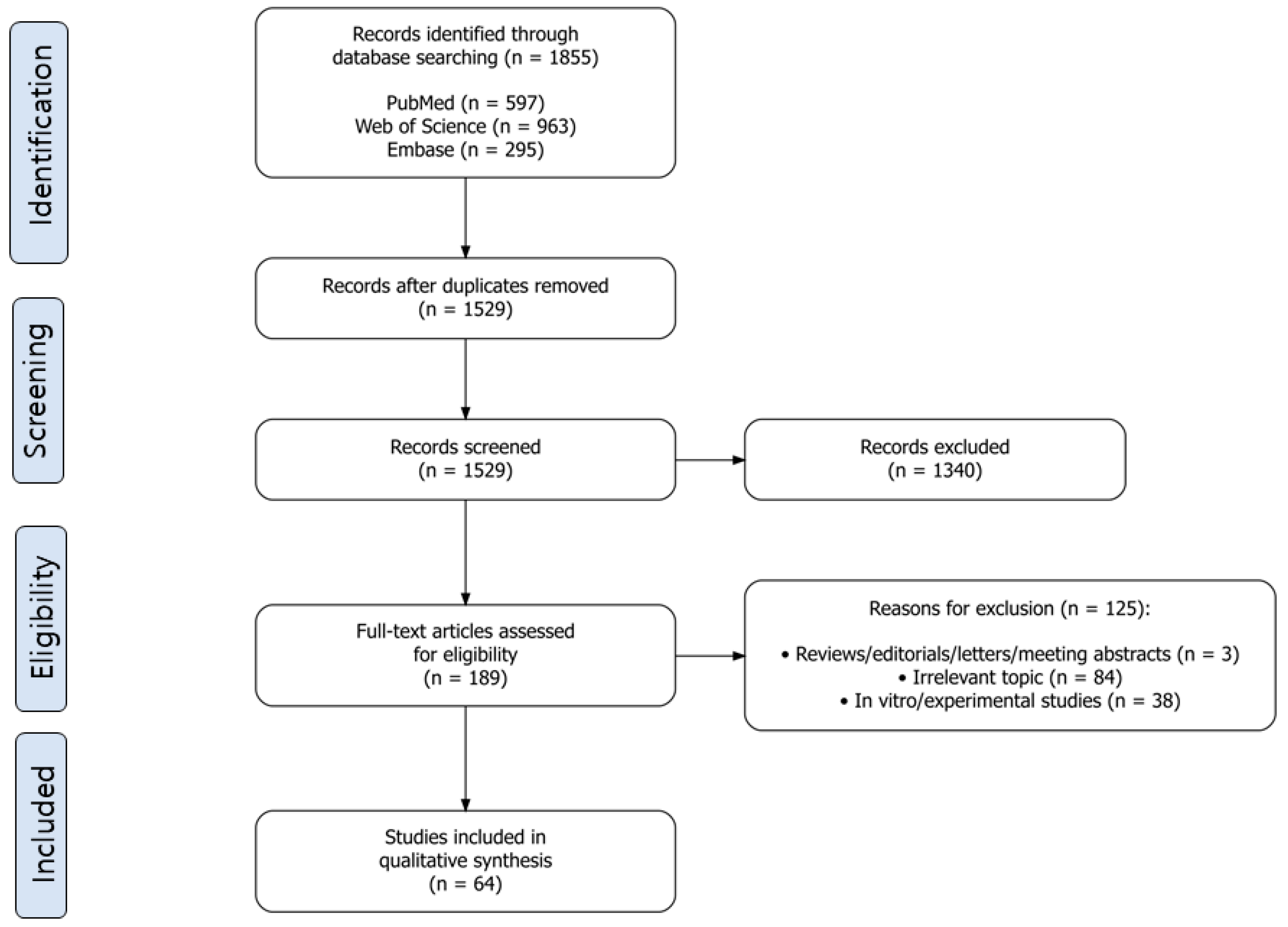
3.1. Selection of Evidence
3.2. Characteristics of Included Studies
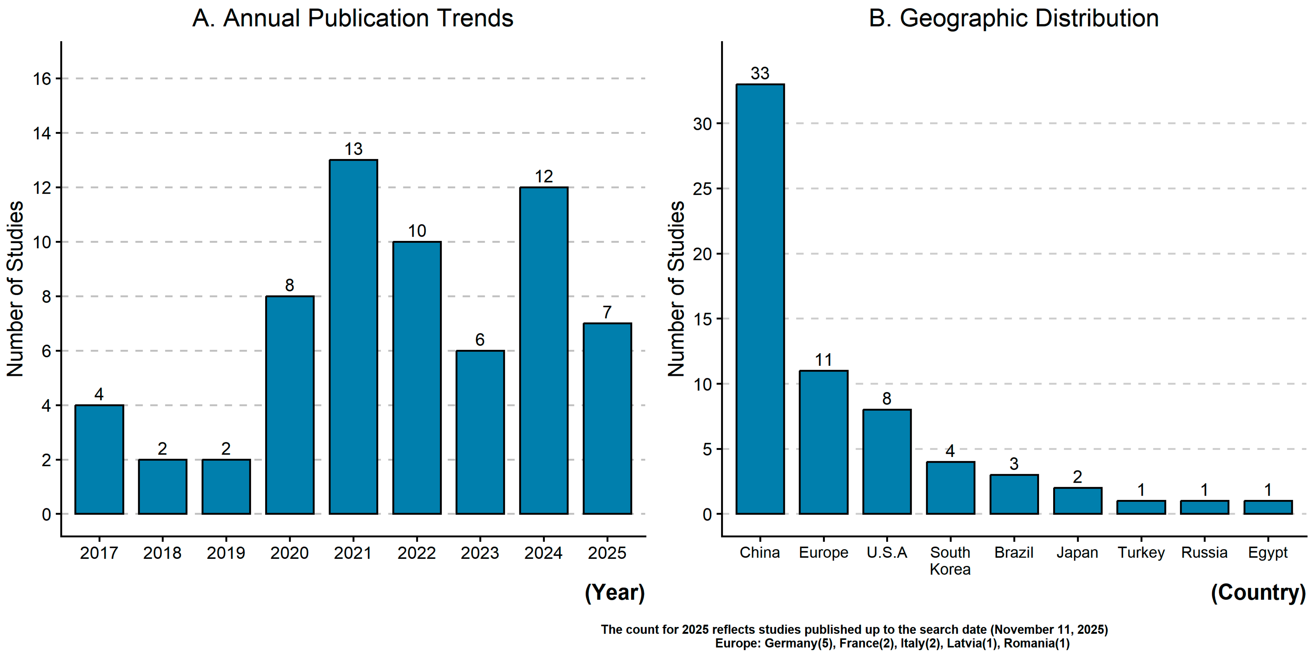
3.2.1. Publication Year and Geographic Distribution

| No. | Author (Year) | Biomarker Type | Clinical Endpoint | Molecular Subtype/Target Population | Key Biomarker(s) | Main Clinical Association | Sample (Method) | N |
|---|---|---|---|---|---|---|---|---|
| 1 | Curtaz (2022) [23] | miRNA | Prognosis | All subtypes | hsa-miR-576-3p, miR-130a-3p | Brain metastasis, grading | Serum (Kit) | 65 |
| 2 | Liu (2020) [43] | mRNA | Both | TNBC-focused | FBXO39 mRNA | Monitoring, OS, lymph node metastasis | Serum (UC + Kit) | 100 |
| 3 | Alvarez (2022) [16] | Protein | Response | All subtypes | 5-protein panel (GPIBA, etc.) | pCR | Plasma (SEC/PPLC) | 17 |
| 4 | Yuan (2021) [69] | miRNA | Prognosis | HER2+ only | hsa-miR-21, PDCD4 | Bone metastasis | Serum (Kit) | 51 |
| 5 | Li (2024) [38] | miRNA | Both | TNBC only | cirmiR-20a-5p, NPAT | Anti-PD-1 sensitivity, survival | Plasma (UC) | 50 |
| 6 | Shen (2021) [48] | miRNA | Prognosis | All subtypes | miR-7641 | OS, DFS | Plasma (UC) | 28 |
| 7 | Fan (2025) [25] | Protein | Prognosis | All subtypes | ITGB2 | OS, DFS | Serum (Kit) | 212 |
| 8 | Todorova (2022) [57] | miRNA | Response | All subtypes | miR-30b, miR-328, miR-423, miR-127 | pCR, RFS | Plasma (Kit) | 20 |
| 9 | Wang (2021) [61] | miRNA | Prognosis | HR+/HER2− disease | miR-363-5p, PDGFB | PFS | Plasma (UC) | 10 |
| 10 | Tkach (2022) [56] | Protein | Response | HR+ only | CD326, CD146, CD105 | Clinical response | Plasma (SEC) | 27 |
| 11 | Baldasici (2022) [17] | lncRNA | Response | All subtypes | HOTAIR, MALAT1 | pCR | Plasma (Kit) | 72 |
| 12 | Desai (2022) [24] | Protein | Response | TNBC only | Annexin A2 (AnxA2) | Treatment response | Serum (Kit) | 17 |
| 13 | Sadovska (2022) [47] | miRNA | Prognosis | TNBC-focused | miR-155, miR-181a, miR-181b | RFS, OS | Plasma (SEC) | 32 |
| 14 | Li (2021) [36] | miRNA | Response | All subtypes | miR-3662, miR-146a, miR-1290 | Treatment response | Serum (Kit) | 60 |
| 15 | Fontana (2025) [26] | miRNA | Prognosis | All subtypes | miR-3916, miR-3162-3p | OS, RFS | Plasma (Kit) | 296 |
| 16 | Cui (2020) [22] | mRNA | Prognosis | All subtypes | LDHC mRNA | OS, recurrence | Serum (Kit) | 75 |
| 17 | Ni (2018) [76] | miRNA | Prognosis | All subtypes | miR-16, miR-93, miR-494 | OS, recurrence | Plasma (Kit) | 153 |
| 18 | Sueta (2017) [73] | miRNA | Prognosis | All subtypes | miR-340, miR-17, miR-130a | OS, DFS | Serum (Kit) | 32 |
| 19 | Wu (2020) [63] | miRNA | Prognosis | All subtypes | miR-150-5p, miR-576-3p | OS, recurrence | Plasma (Kit) | 27 |
| 20 | Tamarindo (2025) [54] | Protein | Prognosis | TNBC only | HISTH2A, CSTA, HISTH2B | OS | Plasma (SEC) | 29 |
| 21 | Kim (2024) [32] | miRNA, Protein | Both | All subtypes | MDR1, miR-21, miR-221 | pCR, PFS | Plasma (UC) | 36 |
| 22 | Jung (2021) [31] | Protein | Response | All subtypes | NGF, IP-10, MMP-1 | Clinical response | Serum (UC) | 129 |
| 23 | König (2017) [72] | DNA | Both | All subtypes | EV-associated cfDNA | Clinical response, OS | Plasma (Kit) | 105 |
| 24 | Causin (2024) [20] | miRNA | Prognosis | All subtypes | miR-19a-3p, miR-130b-3p | OS | Plasma (UC) | 24 |
| 25 | Li (2021) [37] | Protein | Both | TNBC only | Annexin A6 | Clinical response | Serum (Kit) | 21 |
| 26 | Zhuang (2024) [71] | circRNA | Prognosis | All subtypes | circ-0100519 | OS, DFS | Serum (UC) | 20 |
| 27 | Liu (2023) [41] | lncRNA | Both | HER2+ only | Linc00969 | Response monitoring | Serum/Plasma (UC) | 108 |
| 28 | Wu (2021) [64] | miRNA, Protein | Prognosis | HR+/TNBC | miR-19a, IBSP | OS, bone metastasis | Serum (UC) | 87 |
| 29 | Li (2024) [39] | miRNA | Prognosis | All subtypes | miR-361-3p | OS, metastasis | Plasma (Kit) | 37 |
| 30 | Zhang (2020) [70] | miRNA | Both | HER2+ only | miR-1246, miR-155 | Response, survival | Plasma (Kit) | 183 |
| 31 | Sueta (2021) [51] | miRNA | Both | TNBC only | miR-4448, miR-2392 | pCR, OS | Serum (Kit) | 24 |
| 32 | Sun (2023) [52] | tRF | Both | HR+ only | tRF-16-K8J7K1B | Response, DFS | Serum (UC) | 56 |
| 33 | Kim (2024) [33] | miRNA | Both | All subtypes | 5-miRNA signature | Response, OS, DFS | Plasma (Immunoaffinity) | 35 |
| 34 | Bao (2021) [18] | miRNA | Prognosis | All subtypes | miGISig (3-miRNA panel) | OS, DRFS | Serum/Plasma (UC) | >1000 |
| 35 | Shi (2022) [49] | lncRNA | Prognosis | All subtypes | lncRNA DANCR | OS | Serum (Kit) | 120 |
| 36 | Li (2020) [35] | miRNA | Prognosis | N/A | miR-148a | OS | Serum (Kit) | 125 |
| 37 | Wang (2017) [74] | Protein | Both | All subtypes | TRPC5 | Response | Plasma (Kit) | 131 |
| 38 | Niu (2025) [45] | lncRNA, miRNA | Both | TNBC/HER2+/Luminal | LINC00899, miR-425 | Response | Plasma (Kit) | 119 |
| 39 | Carvalho (2022) [19] | miRNA | Prognosis | TNBC-focused | 4-miRNA panel (miR-142, etc.) | OS | Serum (Kit) | 150 |
| 40 | Richard (2024) [46] | Lipid | Both | HR+ only | 16 EV-sphingo scores | Response | Plasma (SEC) | 44 |
| 41 | Jiang (2024) [29] | Protein | Both | HER2+/TNBC | HER2-enriched EVs | Response | Plasma (SEC) | 11 |
| 42 | Tang (2019) [79] | lncRNA | Both | All subtypes | lncRNA HOTAIR | Response | Serum (Kit) | 65 |
| 43 | Del Re (2019) [78] | mRNA | Both | HR+ only | TK1, CDK9 mRNA | Response | Plasma (Kit) | 40 |
| 44 | Yang (2024) [67] | miRNA | Prognosis | All subtypes | miR-203a-3p | OS | Plasma (Kit) | 45 |
| 45 | Su (2021) [50] | mRNA, miRNA | Both | All subtypes | 11-exLR signature | Response | Plasma (Kit) | 112 |
| 46 | Yang (2025) [68] | Protein | Both | HER2+ focus | Exosomal HER2 | Response | Plasma (Kit) | 51 |
| 47 | Yang (2017) [75] | Protein | Both | All subtypes | GSTP1 | Response | Serum (UC) | 30 |
| 48 | Vikramdeo (2023) [59] | DNA | Prognosis | TNBC only | EV-mtDNA mutations | OS, DFS | Plasma (Kit) | 32 |
| 49 | Eskiler (2023) [27] | mRNA | Both | All subtypes | FGFR2, FGFR3 mRNA | Response | Serum (Kit) | 25 |
| 50 | Hoffmann (2023) [28] | Protein | Both | TNBC only | PD-L2 EVs | Response | Plasma (UC) | 54 |
| 51 | Tian (2021) [55] | Protein | Both | All subtypes | 8-EV protein signature | Response | Plasma (UC) | 85 |
| 52 | Vinik (2020) [60] | Protein | Prognosis | All subtypes | FAK, Fibronectin | OS, DFS | Plasma (SEC) | 46 |
| 53 | Tutanov (2020) [58] | Protein | Prognosis | All subtypes | SOCS3, IGF2R, FAK | OS | Plasma (UC) | 23 |
| 54 | Xu (2024) [66] | Protein | Prognosis | All subtypes | TALDO1 | OS | Serum (UC) | 126 |
| 55 | Jung (2023) [30] | Protein | Both | TNBC only | APRIL, CXCL13, VEGF | pCR, DFS | Serum (Kit) | 190 |
| 56 | Talat (2025) [53] | Protein | Prognosis | All subtypes | SDC2, Fibronectin | OS | Plasma (UC) | 169 |
| 57 | Lan (2021) [34] | lncRNA | Prognosis | TNBC only | lncRNA XIST | OS | Serum (Kit) | 91 |
| 58 | Chaudhary (2020) [21] | Protein | Prognosis | All subtypes | Annexin A2 | OS, DFS | Serum (Kit) | 169 |
| 59 | Stevic (2018) [77] | miRNA | Both | TNBC/HER2+ | miR-27a, miR-30e, miR-155 | pCR | Plasma (Kit) | 435 |
| 60 | Wang (2025) [80] | Protein | Both | TNBC only | RTN4 | Response | Plasma (UC) | 104 |
| 61 | Li (2024) [40] | Glycan | Both | TNBC-focused | EV glycan signature | Response | Plasma (SEC) | 72 |
| 62 | Xu (2024) [65] | Protein | Prognosis | All subtypes | TEVs (CD63+/EpCAM+) | OS | Serum (Capture) | 512 |
| 63 | Na-er (2021) [44] | lncRNA | Prognosis | TNBC-focused | SUMO1P3 | OS | Serum (Kit) | 190 |
| 64 | Liu (2022) [42] | circRNA | Prognosis | All subtypes | hsa_circ_0058514 | OS | Plasma (Kit) | 135 |
| Category | Subcategory | No. of Studies (%) | References |
|---|---|---|---|
| EV isolation method | Kit-based/precipitation | 36 (56.2%) | [17,19,21,22,23,24,25,26,27,28,30,34,35,36,37,39,42,44,45,49,50,51,57,59,63,67,68,69,70,72,73,74,76,77,78,79] |
| Ultracentrifugation (UC) | 18 (28.1%) | [18,20,31,32,38,41,43,48,52,53,55,58,61,64,66,71,75,80] | |
| Size-exclusion chromatography (SEC) | 8 (12.5%) | [16,29,40,46,47,54,56,60] | |
| Other specialized methods | 2 (3.1%) | [33,65] | |
| Biological sample source * | Plasma | 38 (59.4%) | [16,17,18,20,26,28,29,32,33,38,39,40,41,42,45,46,47,48,50,53,54,55,56,57,58,59,60,61,63,67,68,70,72,74,76,77,78,80] |
| Serum | 28 (43.8%) | [18,19,21,22,23,24,25,27,30,31,34,35,36,37,41,43,44,49,51,52,64,65,66,69,71,73,75,79] | |
| EV characterization methods | Western blotting (WB) | 48 (75.0%) | Supplementary Table S3 |
| Nanoparticle tracking analysis (NTA) | 41 (64.1%) | Supplementary Table S3 | |
| Transmission electron microscopy (TEM) | 36 (56.2%) | Supplementary Table S3 | |
| EV-associated markers | CD63 | 41 (64.1%) | Supplementary Table S3 |
| CD9 | 27 (42.2%) | Supplementary Table S3 | |
| TSG101 | 25 (39.1%) | Supplementary Table S3 | |
| CD81 | 17 (26.6%) | Supplementary Table S3 | |
| Molecular subtype focus * | TNBC | 10 (15.6%) | [24,28,30,34,37,38,51,54,59,80] |
| HR+/luminal | 5 (7.8%) | [46,52,56,61,78] | |
| HER2 + | 2 (3.1%) | [41,70] | |
| Disease stage * | Early-stage/ locally advanced | 31 (48.4%) | [16,20,22,25,27,28,29,30,31,32,34,36,42,44,45,47,51,52,53,54,57,58,60,61,67,69,72,73,76,77,79] |
| Stage IV | 29 (45.3%) | [17,18,19,23,24,26,33,35,37,39,40,41,43,46,48,49,50,55,56,59,64,65,66,68,70,71,74,75,78] | |
| Mixed non-metastatic and metastatic | 9 (14.1%) | [22,38,43,46,51,55,57,69,70] | |
| Biomarker category | RNA-based biomarkers | 39 (60.9%) | [17,18,19,20,22,23,26,27,32,33,34,35,36,38,39,41,42,43,44,45,47,48,49,50,51,52,57,61,63,64,67,69,70,71,73,76,77,78,79] |
| miRNA | 25 (39.1%) | [18,19,20,23,26,32,33,35,36,38,39,45,47,48,51,57,61,63,64,67,69,70,73,76,77] | |
| lncRNA | 7 (10.9%) | [17,34,41,44,45,49,79] | |
| mRNA | 4 (6.3%) | [22,27,43,78] | |
| circRNA | 2 (3.1%) | [42,71] | |
| tRF | 1 (1.6%) | [52] | |
| Protein-based biomarkers | 21 (32.8%) | [16,21,24,25,28,29,30,31,37,53,54,55,56,58,60,65,66,68,74,75,80] | |
| DNA-based biomarkers | 2 (3.1%) | [59,72] | |
| Lipid-based biomarkers | 1 (1.6%) | [46] | |
| Glycan-based biomarkers | 1 (1.6%) | [40] | |
| Clinical endpoints * | Treatment response | 33 (51.6%) | [16,17,24,27,28,29,30,31,32,33,36,37,38,40,41,43,45,46,50,52,55,56,57,68,70,72,74,75,77,78,79,80] |
| Prognostic outcomes | 57 (89.1%) | [18,19,20,21,22,23,25,26,27,28,29,30,32,33,34,35,37,38,39,40,41,42,43,44,45,46,47,48,49,50,51,52,53,54,55,58,59,60,61,63,64,65,66,67,68,69,70,71,72,73,74,75,76,77,78,79,80] | |
| Both response and prognosis | 26 (40.6%) | [27,28,29,30,32,33,37,38,40,41,45,46,50,51,52,55,68,70,72,74,75,77,78,79,80] | |
| Treatment response | pCR assessment | 14 (21.9%) | [16,17,27,30,32,33,40,45,47,50,51,57,68,77] |
| Longitudinal monitoring | 6 (9.4%) | [22,43,46,51,55,57] |

3.2.2. EV Isolation and Characterization Methods
3.2.3. Breast Cancer Subtype and Disease Stage
3.3. Biomarker Composition
3.3.1. Distribution of EV Biomarkers
3.3.2. miRNA and Other RNA-Based Biomarkers
3.3.3. Protein-Based and DNA-Based Biomarkers
3.4. Clinical Outcomes
3.4.1. Distribution of Clinical Endpoints
3.4.2. Treatment Response and Monitoring
3.4.3. Prognostic Indicators and Survival Outcomes
4. Discussion
4.1. Clinical Rationale for EV-Based Liquid Biopsy in Breast Cancer
4.2. Bridging the Technical Gap: Purity Versus Throughput
4.3. Standardization Challenges and MISEV Compliance
4.4. Geographic and Biological Concentration: Asia and TNBC
4.5. Multi-Omics EV Biomarkers
4.6. Artificial Intelligence-Enabled Analytical Technologies for EV Biomarker Applications
4.7. Limitations and Future Directions
5. Conclusions
Supplementary Materials
Author Contributions
Funding
Institutional Review Board Statement
Informed Consent Statement
Data Availability Statement
Conflicts of Interest
Abbreviations
| CA15-3 | Cancer antigen 15-3 |
| CEA | Carcinoembryonic antigen |
| cfDNA | Cell-free DNA |
| circRNA | Circular RNA |
| CTCs | Circulating tumor cells |
| ctDNA | Circulating tumor DNA |
| DFS | Disease-free survival |
| DNA | Deoxyribonucleic acid |
| EV | Extracellular vesicle |
| HER2+ | Human epidermal growth factor receptor 2-positive |
| HR+ | Hormone receptor-positive |
| JBI | Joanna Briggs Institute |
| lncRNA | Long non-coding RNA |
| mRNA | Messenger RNA |
| miRNA | MicroRNA |
| MISEV | Minimal Information for Studies of Extracellular Vesicles |
| NTA | Nanoparticle tracking analysis |
| OS | Overall survival |
| PCC | Population–Concept–Context |
| PFS | Progression-free survival |
| pCR | Pathological complete response |
| PRISMA-ScR | Preferred Reporting Items for Systematic Reviews and Meta-Analyses extension for Scoping Reviews |
| RFS | Relapse-free survival |
| RNA | Ribonucleic acid |
| SEC | Size-exclusion chromatography |
| TEM | Transmission electron microscopy |
| TNBC | Triple-negative breast cancer |
| tRF | Transfer RNA-derived fragments |
| UC | Ultracentrifugation |
| WB | Western blotting |
References
- Bray, F.; Laversanne, M.; Sung, H.; Ferlay, J.; Siegel, R.L.; Soerjomataram, I.; Jemal, A. Global cancer statistics 2022: GLOBOCAN estimates of incidence and mortality worldwide for 36 cancers in 185 countries. CA Cancer J. Clin. 2024, 74, 229–263. [Google Scholar] [CrossRef]
- Kim, J.; Harper, A.; McCormack, V.; Sung, H.; Houssami, N.; Morgan, E.; Mutebi, M.; Garvey, G.; Soerjomataram, I.; Fidler-Benaoudia, M.M. Global patterns and trends in breast cancer incidence and mortality across 185 countries. Nat. Med. 2025, 31, 1154–1162. [Google Scholar] [CrossRef]
- Bianchini, G.; De Angelis, C.; Licata, L.; Gianni, L. Treatment landscape of triple-negative breast cancer—Expanded options, evolving needs. Nat. Rev. Clin. Oncol. 2022, 19, 91–113. [Google Scholar] [CrossRef]
- Duffy, M.J. Serum tumor markers in breast cancer: Are they of clinical value? Clin. Chem. 2006, 52, 345–351. [Google Scholar] [CrossRef]
- Duffy, M.J.; Harbeck, N.; Nap, M.; Molina, R.; Nicolini, A.; Senkus, E.; Cardoso, F. Clinical use of biomarkers in breast cancer: Updated guidelines from the European Group on Tumor Markers (EGTM). Eur. J. Cancer 2017, 75, 284–298. [Google Scholar] [CrossRef] [PubMed]
- Wang, H.; Zhang, Y.; Zhang, H.; Cao, H.; Mao, J.; Chen, X.; Wang, L.; Zhang, N.; Luo, P.; Xue, J.; et al. Liquid biopsy for human cancer: Cancer screening, monitoring, and treatment. MedComm 2024, 5, e564. [Google Scholar] [CrossRef]
- Banys-Paluchowski, M.; Fehm, T.N.; Grimm-Glang, D.; Rody, A.; Krawczyk, N. Liquid Biopsy in Metastatic Breast Cancer: Current Role of Circulating Tumor Cells and Circulating Tumor DNA. Oncol. Res. Treat. 2022, 45, 4–11. [Google Scholar] [CrossRef]
- Wan, J.C.M.; Massie, C.; Garcia-Corbacho, J.; Mouliere, F.; Brenton, J.D.; Caldas, C.; Pacey, S.; Baird, R.; Rosenfeld, N. Liquid biopsies come of age: Towards implementation of circulating tumour DNA. Nat. Rev. Cancer 2017, 17, 223–238. [Google Scholar] [CrossRef] [PubMed]
- Zhang, X.; Wang, C.; Yu, J.; Bu, J.; Ai, F.; Wang, Y.; Lin, J.; Zhu, X. Extracellular vesicles in the treatment and diagnosis of breast cancer: A status update. Front. Endocrinol. 2023, 14, 1202493. [Google Scholar] [CrossRef] [PubMed]
- Lee, Y.; Ni, J.; Beretov, J.; Wasinger, V.C.; Graham, P.; Li, Y. Recent advances of small extracellular vesicle biomarkers in breast cancer diagnosis and prognosis. Mol. Cancer 2023, 22, 33. [Google Scholar] [CrossRef]
- Loric, S.; Denis, J.A.; Desbene, C.; Sabbah, M.; Conti, M. Extracellular Vesicles in Breast Cancer: From Biology and Function to Clinical Diagnosis and Therapeutic Management. Int. J. Mol. Sci. 2023, 24, 7208. [Google Scholar] [CrossRef] [PubMed]
- Doyle, L.M.; Wang, M.Z. Overview of Extracellular Vesicles, Their Origin, Composition, Purpose, and Methods for Exosome Isolation and Analysis. Cells 2019, 8, 727. [Google Scholar] [CrossRef] [PubMed]
- Arksey, H.; O’malley, L. Scoping studies: Towards a methodological framework. Int. J. Soc. Res. Methodol. 2005, 8, 19–32. [Google Scholar] [CrossRef]
- Peters, M.D.J.; Marnie, C.; Tricco, A.C.; Pollock, D.; Munn, Z.; Alexander, L.; McInerney, P.; Godfrey, C.M.; Khalil, H. Updated methodological guidance for the conduct of scoping reviews. JBI Evid. Synth. 2020, 18, 2119–2126. [Google Scholar] [CrossRef]
- Tricco, A.C.; Lillie, E.; Zarin, W.; O’Brien, K.K.; Colquhoun, H.; Levac, D.; Moher, D.; Peters, M.D.J.; Horsley, T.; Weeks, L.; et al. PRISMA Extension for Scoping Reviews (PRISMA-ScR): Checklist and Explanation. Ann. Intern. Med. 2018, 169, 467–473. [Google Scholar] [CrossRef]
- Alvarez, F.A.; Kaddour, H.; Lyu, Y.; Preece, C.; Cohen, J.; Baer, L.; Stopeck, A.T.; Thompson, P.; Okeoma, C.M. Blood plasma derived extracellular vesicles (BEVs): Particle purification liquid chromatography (PPLC) and proteomic analysis reveals BEVs as a potential minimally invasive tool for predicting response to breast cancer treatment. Breast Cancer Res. Treat. 2022, 196, 423–437. [Google Scholar] [CrossRef]
- Baldasici, O.; Balacescu, L.; Cruceriu, D.; Roman, A.; Lisencu, C.; Fetica, B.; Visan, S.; Cismaru, A.; Jurj, A.; Barbu-Tudoran, L.; et al. Circulating Small EVs miRNAs as Predictors of Pathological Response to Neo-Adjuvant Therapy in Breast Cancer Patients. Int. J. Mol. Sci. 2022, 23, 12625. [Google Scholar] [CrossRef]
- Bao, S.; Hu, T.; Liu, J.; Su, J.; Sun, J.; Ming, Y.; Li, J.; Wu, N.; Chen, H.; Zhou, M. Genomic instability-derived plasma extracellular vesicle-microRNA signature as a minimally invasive predictor of risk and unfavorable prognosis in breast cancer. J. Nanobiotechnology 2021, 19, 22. [Google Scholar] [CrossRef]
- Carvalho, T.M.; Brasil, G.O.; Jucoski, T.S.; Adamoski, D.; de Lima, R.S.; Spautz, C.C.; Anselmi, K.F.; Ozawa, P.M.M.; Cavalli, I.J.; Carvalho de Oliveira, J.; et al. MicroRNAs miR-142-5p, miR-150-5p, miR-320a-3p, and miR-4433b-5p in Serum and Tissue: Potential Biomarkers in Sporadic Breast Cancer. Front. Genet. 2022, 13, 865472. [Google Scholar] [CrossRef] [PubMed]
- Causin, R.L.; Polezi, M.R.; Freitas, A.J.A.; Calfa, S.; Altei, W.F.; Dias, J.O.; Laus, A.C.; Pessôa-Pereira, D.; Komoto, T.T.; Evangelista, A.F.; et al. EV-miRNAs from breast cancer patients of plasma as potential prognostic biomarkers of disease recurrence. Heliyon 2024, 10, e33933. [Google Scholar] [CrossRef]
- Chaudhary, P.; Gibbs, L.D.; Maji, S.; Lewis, C.M.; Suzuki, S.; Vishwanatha, J.K. Serum exosomal-annexin A2 is associated with African-American triple-negative breast cancer and promotes angiogenesis. Breast Cancer Res. 2020, 22, 11, Correction in Breast Cancer Res. 2020, 22, 31. [Google Scholar] [CrossRef]
- Cui, Z.; Chen, Y.; Hu, M.; Lin, Y.; Zhang, S.; Kong, L.; Chen, Y. Diagnostic and prognostic value of the cancer-testis antigen lactate dehydrogenase C4 in breast cancer. Clin. Chim. Acta 2020, 503, 203–209. [Google Scholar] [CrossRef]
- Curtaz, C.J.; Reifschläger, L.; Strähle, L.; Feldheim, J.; Feldheim, J.J.; Schmitt, C.; Kiesel, M.; Herbert, S.L.; Wöckel, A.; Meybohm, P.; et al. Analysis of microRNAs in Exosomes of Breast Cancer Patients in Search of Molecular Prognostic Factors in Brain Metastases. Int. J. Mol. Sci. 2022, 23, 3683. [Google Scholar] [CrossRef]
- Desai, P.P.; Narra, K.; James, J.D.; Jones, H.P.; Tripathi, A.K.; Vishwanatha, J.K. Combination of Small Extracellular Vesicle-Derived Annexin A2 Protein and mRNA as a Potential Predictive Biomarker for Chemotherapy Responsiveness in Aggressive Triple-Negative Breast Cancer. Cancers 2022, 15, 212. [Google Scholar] [CrossRef] [PubMed]
- Fan, J.; Sha, T.; Ma, B. Cancer-Derived Extracellular Vesicle ITGB2 Promotes the Progression of Triple-Negative Breast Cancer via the Activation of Cancer-Associated Fibroblasts. Glob. Chall. 2025, 9, 2400235. [Google Scholar] [CrossRef]
- Fontana, A.; Barbano, R.; Pasculli, B.; Mazza, T.; Palumbo, O.; Binda, E.; Trivieri, N.; Mencarelli, G.; Laurenzana, I.; Lamorte, D.; et al. Development of a microRNA-based prognostic model for accurate prediction of distant metastasis in breast cancer patients. Breast Cancer Res. 2025, 27, 170. [Google Scholar] [CrossRef]
- Guney Eskiler, G.; Kazan, N.; Haciefendi, A.; Deveci Ozkan, A.; Ozdemir, K.; Ozen, M.; Kocer, H.B.; Yilmaz, F.; Kaleli, S.; Sahin, E.; et al. The prognostic and predictive values of differential expression of exosomal receptor tyrosine kinases and associated with the PI3K/AKT/mTOR signaling in breast cancer patients undergoing neoadjuvant chemotherapy. Clin. Transl. Oncol. 2023, 25, 460–472. [Google Scholar] [CrossRef] [PubMed]
- Hoffmann, O.; Wormland, S.; Bittner, A.K.; Collenburg, M.; Horn, P.A.; Kimmig, R.; Kasimir-Bauer, S.; Rebmann, V. Programmed death receptor ligand-2 (PD-L2) bearing extracellular vesicles as a new biomarker to identify early triple-negative breast cancer patients at high risk for relapse. J. Cancer Res. Clin. Oncol. 2023, 149, 1159–1174. [Google Scholar] [CrossRef] [PubMed]
- Jiang, N.; Saftics, A.; Romano, E.; Ghaeli, I.; Resto, C.; Robles, V.; Das, S.; Van Keuren-Jensen, K.; Seewaldt, V.L.; Jovanovic-Talisman, T. Multiparametric profiling of HER2-enriched extracellular vesicles in breast cancer using Single Extracellular VEsicle Nanoscopy. J. Nanobiotechnology 2024, 22, 589. [Google Scholar] [CrossRef]
- Jung, H.H.; Kim, J.Y.; Cho, E.Y.; Lee, J.E.; Kim, S.W.; Nam, S.J.; Park, Y.H.; Ahn, J.S.; Im, Y.H. A Retrospective Exploratory Analysis for Serum Extracellular Vesicles Reveals APRIL (TNFSF13), CXCL13, and VEGF-A as Prognostic Biomarkers for Neoadjuvant Chemotherapy in Triple-Negative Breast Cancer. Int. J. Mol. Sci. 2023, 24, 15576. [Google Scholar] [CrossRef]
- Jung, H.H.; Kim, J.Y.; Cho, E.Y.; Oh, J.M.; Lee, J.E.; Kim, S.W.; Nam, S.J.; Park, Y.H.; Ahn, J.S.; Im, Y.H. Elevated Level of Nerve Growth Factor (NGF) in Serum-Derived Exosomes Predicts Poor Survival in Patients with Breast Cancer Undergoing Neoadjuvant Chemotherapy. Cancers 2021, 13, 5260. [Google Scholar] [CrossRef] [PubMed]
- Kim, M.W.; Lee, H.; Lee, S.; Moon, S.; Kim, Y.; Kim, J.Y.; Kim, S.I.; Kim, J.Y. Drug-resistant profiles of extracellular vesicles predict therapeutic response in TNBC patients receiving neoadjuvant chemotherapy. BMC Cancer 2024, 24, 185. [Google Scholar] [CrossRef] [PubMed]
- Kim, M.W.; Moon, S.; Lee, S.; Lee, H.; Kim, Y.; Kim, J.Y.; Kim, J.Y.; Kim, S.I. Exploring miRNA-target gene profiles associated with drug resistance in patients with breast cancer receiving neoadjuvant chemotherapy. Oncol. Lett. 2024, 27, 158. [Google Scholar] [CrossRef]
- Lan, F.; Zhang, X.; Li, H.; Yue, X.; Sun, Q. Serum exosomal lncRNA XIST is a potential non-invasive biomarker to diagnose recurrence of triple-negative breast cancer. J. Cell Mol. Med. 2021, 25, 7602–7607. [Google Scholar] [CrossRef] [PubMed]
- Li, D.; Wang, J.; Ma, L.J.; Yang, H.B.; Jing, J.F.; Jia, M.M.; Zhang, X.J.; Guo, F.; Gao, J.N. Identification of serum exosomal miR-148a as a novel prognostic biomarker for breast cancer. Eur. Rev. Med. Pharmacol. Sci. 2020, 24, 7303–7309. [Google Scholar]
- Li, S.; Zhang, M.; Xu, F.; Wang, Y.; Leng, D. Detection significance of miR-3662, miR-146a, and miR-1290 in serum exosomes of breast cancer patients. J. Cancer Res. Ther. 2021, 17, 749–755. [Google Scholar] [CrossRef]
- Li, T.; Tao, Z.; Zhu, Y.; Liu, X.; Wang, L.; Du, Y.; Cao, J.; Wang, B.; Zhang, J.; Hu, X. Exosomal annexin A6 induces gemcitabine resistance by inhibiting ubiquitination and degradation of EGFR in triple-negative breast cancer. Cell Death Dis. 2021, 12, 684. [Google Scholar] [CrossRef]
- Li, W.; Han, G.; Li, F.; Bu, P.; Hao, Y.; Huang, L.; Bai, X. Cancer cell-derived exosomal miR-20a-5p inhibits CD8(+) T-cell function and confers anti-programmed cell death 1 therapy resistance in triple-negative breast cancer. Cancer Sci. 2024, 115, 347–356. [Google Scholar] [CrossRef]
- Li, Y.; Fan, L.; Yan, A.; Ren, X.; Zhao, Y.; Hua, B. Exosomal miR-361-3p promotes the viability of breast cancer cells by targeting ETV7 and BATF2 to upregulate the PAI-1/ERK pathway. J. Transl. Med. 2024, 22, 112. [Google Scholar] [CrossRef]
- Li, Y.; Zhang, S.; Liu, C.; Deng, J.; Tian, F.; Feng, Q.; Qin, L.; Bai, L.; Fu, T.; Zhang, L.; et al. Thermophoretic glycan profiling of extracellular vesicles for triple-negative breast cancer management. Nat. Commun. 2024, 15, 2292. [Google Scholar] [CrossRef]
- Liu, C.; Lu, C.; Yixi, L.; Hong, J.; Dong, F.; Ruan, S.; Hu, T.; Zhao, X. Exosomal Linc00969 induces trastuzumab resistance in breast cancer by increasing HER-2 protein expression and mRNA stability by binding to HUR. Breast Cancer Res. 2023, 25, 124. [Google Scholar] [CrossRef]
- Liu, J.; Peng, X.; Yang, Y.; Zhang, Y.; Han, M.; Shi, X.; Zheng, J.; Li, T.; Chen, J.; Lv, W.; et al. The value of hsa_circ_0058514 in plasma extracellular vesicles for breast cancer. Front. Oncol. 2022, 12, 995196. [Google Scholar] [CrossRef]
- Liu, Y.C.; Yan, S.; Liu, D.M.; Pei, D.X.; Li, Y.W. Aberrant Expression of Cancer-Testis Antigen FBXO39 in Breast Cancer and its Clinical Significance. Clin. Lab. 2020, 66, 1877. [Google Scholar] [CrossRef]
- Na-Er, A.; Xu, Y.Y.; Liu, Y.H.; Gan, Y.J. Upregulation of serum exosomal SUMO1P3 predicts unfavorable prognosis in triple negative breast cancer. Eur. Rev. Med. Pharmacol. Sci. 2021, 25, 154–160. [Google Scholar] [PubMed]
- Niu, L.; Bai, Y.; Yu, M.; Sun, X. LINC00899 suppresses the progression of triple-negative breast cancer via the miRNA-425/PTEN axis and is a biomarker for neoadjuvant chemotherapy efficacy. J. Cancer 2025, 16, 1647–1655. [Google Scholar] [CrossRef]
- Richard, M.; Moreau, R.; Croyal, M.; Mathiot, L.; Frénel, J.S.; Campone, M.; Dupont, A.; Gavard, J.; André-Grégoire, G.; Guével, L. Monitoring concentration and lipid signature of plasma extracellular vesicles from HR(+) metastatic breast cancer patients under CDK4/6 inhibitors treatment. J. Extracell. Biol. 2024, 3, e70013, Correction in J. Extracell. Biol. 2025, 4, e70070. [Google Scholar] [CrossRef]
- Sadovska, L.; Zayakin, P.; Eglītis, K.; Endzeliņš, E.; Radoviča-Spalviņa, I.; Avotiņa, E.; Auders, J.; Keiša, L.; Liepniece-Karele, I.; Leja, M.; et al. Comprehensive characterization of RNA cargo of extracellular vesicles in breast cancer patients undergoing neoadjuvant chemotherapy. Front. Oncol. 2022, 12, 1005812. [Google Scholar] [CrossRef]
- Shen, S.; Song, Y.; Zhao, B.; Xu, Y.; Ren, X.; Zhou, Y.; Sun, Q. Cancer-derived exosomal miR-7641 promotes breast cancer progression and metastasis. Cell Commun. Signal 2021, 19, 20. [Google Scholar] [CrossRef]
- Shi, W.; Jin, X.; Wang, Y.; Zhang, Q.; Yang, L. High serum exosomal long non-coding RNA DANCR expression confers poor prognosis in patients with breast cancer. J. Clin. Lab. Anal. 2022, 36, e24186. [Google Scholar] [CrossRef] [PubMed]
- Su, Y.; Li, Y.; Guo, R.; Zhao, J.; Chi, W.; Lai, H.; Wang, J.; Wang, Z.; Li, L.; Sang, Y.; et al. Plasma extracellular vesicle long RNA profiles in the diagnosis and prediction of treatment response for breast cancer. npj Breast Cancer 2021, 7, 154, Correction in npj Breast Cancer 2022, 8, 34. [Google Scholar] [CrossRef] [PubMed]
- Sueta, A.; Fujiki, Y.; Goto-Yamaguchi, L.; Tomiguchi, M.; Yamamoto-Ibusuki, M.; Iwase, H.; Yamamoto, Y. Exosomal miRNA profiles of triple-negative breast cancer in neoadjuvant treatment. Oncol. Lett. 2021, 22, 819. [Google Scholar] [CrossRef]
- Sun, C.; Huang, X.; Li, J.; Fu, Z.; Hua, Y.; Zeng, T.; He, Y.; Duan, N.; Yang, F.; Liang, Y.; et al. Exosome-Transmitted tRF-16-K8J7K1B Promotes Tamoxifen Resistance by Reducing Drug-Induced Cell Apoptosis in Breast Cancer. Cancers 2023, 15, 899. [Google Scholar] [CrossRef]
- Talat, L.Y.; Mohamed, G.; Ibraheem, M.H.; WalyEldeen, A.A.; Hassan, H.; Ibrahim, S.A. SDC2 and FN as cargo proteins in circulating extracellular vesicles in obese breast cancer patients with lymph node metastasis. Sci. Rep. 2025, 15, 32498. [Google Scholar] [CrossRef]
- Tamarindo, G.H.; Novais, A.A.; Frigieri, B.M.; Alves, D.L.; de Souza, C.A.; Amadeu, A.; da Silveira, J.C.; Souza, F.F.; Bordin, N.A., Jr.; Chuffa, L.G.A.; et al. Distinct proteomic profiles of plasma-derived extracellular vesicles in healthy, benign, and triple-negative breast cancer: Candidate biomarkers for liquid biopsy. Sci. Rep. 2025, 15, 12122. [Google Scholar] [CrossRef]
- Tian, F.; Zhang, S.; Liu, C.; Han, Z.; Liu, Y.; Deng, J.; Li, Y.; Wu, X.; Cai, L.; Qin, L.; et al. Protein analysis of extracellular vesicles to monitor and predict therapeutic response in metastatic breast cancer. Nat. Commun. 2021, 12, 2536. [Google Scholar] [CrossRef] [PubMed]
- Tkach, M.; Hego, C.; Michel, M.; Darrigues, L.; Pierga, J.Y.; Bidard, F.C.; Théry, C.; Proudhon, C. Circulating extracellular vesicles provide valuable protein, but not DNA, biomarkers in metastatic breast cancer. J. Extracell. Biol. 2022, 1, e51, Correction in J. Extracell. Biol. 2025, 4, e70070. [Google Scholar] [CrossRef]
- Todorova, V.K.; Byrum, S.D.; Gies, A.J.; Haynie, C.; Smith, H.; Reyna, N.S.; Makhoul, I. Circulating Exosomal microRNAs as Predictive Biomarkers of Neoadjuvant Chemotherapy Response in Breast Cancer. Curr. Oncol. 2022, 29, 613–630. [Google Scholar] [CrossRef]
- Tutanov, O.; Proskura, K.; Kamyshinsky, R.; Shtam, T.; Tsentalovich, Y.; Tamkovich, S. Proteomic Profiling of Plasma and Total Blood Exosomes in Breast Cancer: A Potential Role in Tumor Progression, Diagnosis, and Prognosis. Front. Oncol. 2020, 10, 580891. [Google Scholar] [CrossRef]
- Vikramdeo, K.S.; Anand, S.; Sudan, S.K.; Pramanik, P.; Singh, S.; Godwin, A.K.; Singh, A.P.; Dasgupta, S. Profiling mitochondrial DNA mutations in tumors and circulating extracellular vesicles of triple-negative breast cancer patients for potential biomarker development. FASEB Bioadv. 2023, 5, 412–426. [Google Scholar] [CrossRef]
- Vinik, Y.; Ortega, F.G.; Mills, G.B.; Lu, Y.; Jurkowicz, M.; Halperin, S.; Aharoni, M.; Gutman, M.; Lev, S. Proteomic analysis of circulating extracellular vesicles identifies potential markers of breast cancer progression, recurrence, and response. Sci. Adv. 2020, 6, eaba5714. [Google Scholar] [CrossRef] [PubMed]
- Wang, X.; Qian, T.; Bao, S.; Zhao, H.; Chen, H.; Xing, Z.; Li, Y.; Zhang, M.; Meng, X.; Wang, C.; et al. Circulating exosomal miR-363-5p inhibits lymph node metastasis by downregulating PDGFB and serves as a potential noninvasive biomarker for breast cancer. Mol. Oncol. 2021, 15, 2466–2479. [Google Scholar] [CrossRef]
- Wang, X.; Wang, L.; Lin, H.; Zhu, Y.; Huang, D.; Lai, M.; Xi, X.; Huang, J.; Zhang, W.; Zhong, T. Research progress of CTC, ctDNA, and EVs in cancer liquid biopsy. Front. Oncol. 2024, 14, 1303335. [Google Scholar] [CrossRef]
- Wu, H.; Wang, Q.; Zhong, H.; Li, L.; Zhang, Q.; Huang, Q.; Yu, Z. Differentially expressed microRNAs in exosomes of patients with breast cancer revealed by next-generation sequencing. Oncol. Rep. 2020, 43, 240–250. [Google Scholar] [CrossRef]
- Wu, K.; Feng, J.; Lyu, F.; Xing, F.; Sharma, S.; Liu, Y.; Wu, S.Y.; Zhao, D.; Tyagi, A.; Deshpande, R.P.; et al. Exosomal miR-19a and IBSP cooperate to induce osteolytic bone metastasis of estrogen receptor-positive breast cancer. Nat. Commun. 2021, 12, 5196. [Google Scholar] [CrossRef] [PubMed]
- Xu, F.; Wang, K.; Zhu, C.; Fan, L.; Zhu, Y.; Wang, J.F.; Li, X.; Liu, Y.; Zhao, Y.; Zhu, C.; et al. Tumor-derived extracellular vesicles as a biomarker for breast cancer diagnosis and metastasis monitoring. iScience 2024, 27, 109506. [Google Scholar] [CrossRef] [PubMed]
- Xu, G.; Huang, R.; Wumaier, R.; Lyu, J.; Huang, M.; Zhang, Y.; Chen, Q.; Liu, W.; Tao, M.; Li, J.; et al. Proteomic Profiling of Serum Extracellular Vesicles Identifies Diagnostic Signatures and Therapeutic Targets in Breast Cancer. Cancer Res. 2024, 84, 3267–3285. [Google Scholar] [CrossRef]
- Yang, X.; Fan, L.; Huang, J.; Li, Y. Plasma Exosome miR-203a-3p is a Potential Liquid Biopsy Marker for Assessing Tumor Progression in Breast Cancer Patients. Breast Cancer Targets Ther. 2024, 16, 631–643. [Google Scholar] [CrossRef]
- Yang, X.; Xu, M.; Xia, Y.; Ba, Z.; Han, C.; Wang, Y.; Qu, J.; Wang, Y.; Zhou, Y.; Wang, R.; et al. Plasma-derived exosomal human epidermal growth factor receptor 2 (HER2) protein for distinguishing breast cancer from benign breast disease and assessing the efficacy of neoadjuvant therapy. Transl. Cancer Res. 2025, 14, 3186–3200. [Google Scholar] [CrossRef]
- Yuan, X.; Qian, N.; Ling, S.; Li, Y.; Sun, W.; Li, J.; Du, R.; Zhong, G.; Liu, C.; Yu, G.; et al. Breast cancer exosomes contribute to pre-metastatic niche formation and promote bone metastasis of tumor cells. Theranostics 2021, 11, 1429–1445. [Google Scholar] [CrossRef] [PubMed]
- Zhang, Z.; Zhang, L.; Yu, G.; Sun, Z.; Wang, T.; Tian, X.; Duan, X.; Zhang, C. Exosomal miR-1246 and miR-155 as predictive and prognostic biomarkers for trastuzumab-based therapy resistance in HER2-positive breast cancer. Cancer Chemother. Pharmacol. 2020, 86, 761–772. [Google Scholar] [CrossRef]
- Zhuang, M.; Zhang, X.; Ji, J.; Zhang, H.; Shen, L.; Zhu, Y.; Liu, X. Exosomal circ-0100519 promotes breast cancer progression via inducing M2 macrophage polarisation by USP7/NRF2 axis. Clin. Transl. Med. 2024, 14, e1763. [Google Scholar] [CrossRef] [PubMed]
- König, L.; Kasimir-Bauer, S.; Bittner, A.K.; Hoffmann, O.; Wagner, B.; Santos Manvailer, L.F.; Kimmig, R.; Horn, P.A.; Rebmann, V. Elevated levels of extracellular vesicles are associated with therapy failure and disease progression in breast cancer patients undergoing neoadjuvant chemotherapy. Oncoimmunology 2017, 7, e1376153. [Google Scholar] [CrossRef]
- Sueta, A.; Yamamoto, Y.; Tomiguchi, M.; Takeshita, T.; Yamamoto-Ibusuki, M.; Iwase, H. Differential expression of exosomal miRNAs between breast cancer patients with and without recurrence. Oncotarget 2017, 8, 69934–69944. [Google Scholar] [CrossRef]
- Wang, T.; Ning, K.; Lu, T.X.; Sun, X.; Jin, L.; Qi, X.; Jin, J.; Hua, D. Increasing circulating exosomes-carrying TRPC5 predicts chemoresistance in metastatic breast cancer patients. Cancer Sci. 2017, 108, 448–454. [Google Scholar] [CrossRef]
- Yang, S.J.; Wang, D.D.; Li, J.; Xu, H.Z.; Shen, H.Y.; Chen, X.; Zhou, S.Y.; Zhong, S.L.; Zhao, J.H.; Tang, J.H. Predictive role of GSTP1-containing exosomes in chemotherapy-resistant breast cancer. Gene 2017, 623, 5–14. [Google Scholar] [CrossRef] [PubMed]
- Ni, Q.; Stevic, I.; Pan, C.; Müller, V.; Oliveira-Ferrer, L.; Pantel, K.; Schwarzenbach, H. Different signatures of miR-16, miR-30b and miR-93 in exosomes from breast cancer and DCIS patients. Sci. Rep. 2018, 8, 12974, Correction in Sci. Rep. 2019, 9, 18700. [Google Scholar] [CrossRef]
- Stevic, I.; Müller, V.; Weber, K.; Fasching, P.A.; Karn, T.; Marmé, F.; Schem, C.; Stickeler, E.; Denkert, C.; van Mackelenbergh, M.; et al. Specific microRNA signatures in exosomes of triple-negative and HER2-positive breast cancer patients undergoing neoadjuvant therapy within the GeparSixto trial. BMC Med. 2018, 16, 179. [Google Scholar] [CrossRef]
- Del Re, M.; Bertolini, I.; Crucitta, S.; Fontanelli, L.; Rofi, E.; De Angelis, C.; Diodati, L.; Cavallero, D.; Gianfilippo, G.; Salvadori, B.; et al. Overexpression of TK1 and CDK9 in plasma-derived exosomes is associated with clinical resistance to CDK4/6 inhibitors in metastatic breast cancer patients. Breast Cancer Res. Treat. 2019, 178, 57–62. [Google Scholar] [CrossRef]
- Tang, S.; Zheng, K.; Tang, Y.; Li, Z.; Zou, T.; Liu, D. Overexpression of serum exosomal HOTAIR is correlated with poor survival and poor response to chemotherapy in breast cancer patients. J. Biosci. 2019, 44, 37. [Google Scholar] [CrossRef] [PubMed]
- Wang, H.; Huang, R.; Luo, L.; Wang, R.; Zhou, Z.; Hong, J.; Wu, J.; Huang, O.; He, J.; Chen, W.; et al. Targeting Reticulin 4 (RTN4) Within Small Extracellular Vesicles Combats Metastasis and Reinforces Immunotherapy in Triple-Negative Breast Cancer. J. Extracell. Vesicles 2025, 14, e70154. [Google Scholar] [CrossRef]
- Zhang, Y.; Lan, M.; Chen, Y. Minimal Information for Studies of Extracellular Vesicles (MISEV): Ten-Year Evolution (2014–2023). Pharmaceutics 2024, 16, 1394. [Google Scholar] [CrossRef]
- Shao, Y.; Sun, X.; He, Y.; Liu, C.; Liu, H. Elevated Levels of Serum Tumor Markers CEA and CA15-3 Are Prognostic Parameters for Different Molecular Subtypes of Breast Cancer. PLoS ONE 2015, 10, e0133830. [Google Scholar] [CrossRef] [PubMed]
- Geng, B.; Liang, M.M.; Ye, X.B.; Zhao, W.Y. Association of CA 15-3 and CEA with clinicopathological parameters in patients with metastatic breast cancer. Mol. Clin. Oncol. 2015, 3, 232–236. [Google Scholar] [CrossRef]
- Nicolò, E.; Gianni, C.; Pontolillo, L.; Serafini, M.S.; Munoz-Arcos, L.S.; Andreopoulou, E.; Curigliano, G.; Reduzzi, C.; Cristofanilli, M. Circulating tumor cells et al.: Towards a comprehensive liquid biopsy approach in breast cancer. Transl. Breast Cancer Res. 2024, 5, 10. [Google Scholar] [CrossRef]
- Guo, S.; Huang, J.; Li, G.; Chen, W.; Li, Z.; Lei, J. The role of extracellular vesicles in circulating tumor cell-mediated distant metastasis. Mol. Cancer 2023, 22, 193. [Google Scholar] [CrossRef]
- Maacha, S.; Bhat, A.A.; Jimenez, L.; Raza, A.; Haris, M.; Uddin, S.; Grivel, J.C. Extracellular vesicles-mediated intercellular communication: Roles in the tumor microenvironment and anti-cancer drug resistance. Mol. Cancer 2019, 18, 55. [Google Scholar] [CrossRef]
- Dong, Q.; Liu, X.; Cheng, K.; Sheng, J.; Kong, J.; Liu, T. Pre-metastatic Niche Formation in Different Organs Induced by Tumor Extracellular Vesicles. Front. Cell Dev. Biol. 2021, 9, 733627. [Google Scholar] [CrossRef]
- Jia, Y.; Yu, L.; Ma, T.; Xu, W.; Qian, H.; Sun, Y.; Shi, H. Small extracellular vesicles isolation and separation: Current techniques, pending questions and clinical applications. Theranostics 2022, 12, 6548–6575. [Google Scholar] [CrossRef]
- Suresh, P.S.; Zhang, Q. Comprehensive Comparison of Methods for Isolation of Extracellular Vesicles from Human Plasma. J. Proteome Res. 2025, 24, 2956–2967. [Google Scholar] [CrossRef] [PubMed]
- Zhang, P.; Wu, X.; Gardashova, G.; Yang, Y.; Zhang, Y.; Xu, L.; Zeng, Y. Molecular and functional extracellular vesicle analysis using nanopatterned microchips monitors tumor progression and metastasis. Sci. Transl. Med. 2020, 12, eaaz2878. [Google Scholar] [CrossRef] [PubMed]
- Welsh, J.A.; Goberdhan, D.C.I.; O’Driscoll, L.; Buzas, E.I.; Blenkiron, C.; Bussolati, B.; Cai, H.; Di Vizio, D.; Driedonks, T.A.P.; Erdbrügger, U.; et al. Minimal information for studies of extracellular vesicles (MISEV2023): From basic to advanced approaches. J. Extracell. Vesicles 2024, 13, e12404. [Google Scholar] [CrossRef] [PubMed]
- Jie, H.; Ma, W.; Huang, C. Diagnosis, Prognosis, and Treatment of Triple-Negative Breast Cancer: A Review. Breast Cancer Targets Ther. 2025, 17, 265–274. [Google Scholar] [CrossRef]
- Rayamajhi, S.; Sipes, J.; Tetlow, A.L.; Saha, S.; Bansal, A.; Godwin, A.K. Extracellular Vesicles as Liquid Biopsy Biomarkers across the Cancer Journey: From Early Detection to Recurrence. Clin. Chem. 2024, 70, 206–219. [Google Scholar] [CrossRef] [PubMed]
- Kwon, Y.; Kim, H.; Kwon, H.; Park, J. Multimodal Single Extracellular Vesicle Profiling Technologies to Unveil Clinical Heterogeneity. ACS Appl. Bio Mater. 2026, 9, 647–664. [Google Scholar] [CrossRef] [PubMed]
- Du, J.; Cao, C.; Xue, Z.; Wang, W.; Lu, X.; Wei, Y.; Huang, J.; Zhao, L.; Wang, L.; Xu, F. AI-Enhanced Lateral Flow Assay Enables 3-Minute Quantitative Detection with Laboratory-Grade Accuracy. Anal. Chem. 2025, 97, 24196–24208. [Google Scholar] [CrossRef]
- Han, G.-R.; Goncharov, A.; Eryilmaz, M.; Joung, H.-A.; Ghosh, R.; Yim, G.; Chang, N.; Kim, M.; Ngo, K.; Veszpremi, M. Deep learning-enhanced paper-based vertical flow assay for high-sensitivity. ACS Nano 2024, 18, 27933–27948. [Google Scholar] [CrossRef]

| Inclusion Criteria | Exclusion Criteria | |
|---|---|---|
| Population | Patients diagnosed with breast cancer Any molecular subtype (HR+, HER2+, TNBC) Any disease stage (Early, Locally Advanced, Metastatic) | Non-breast cancer populations Studies with no human clinical subjects |
| Outcome | Association between EV cargo and clinical outcomes Endpoints: Treatment response (pCR, RR) and/or Prognosis (OS, DFS, PFS, RFS) | Focused solely on diagnostic or early detection performance No report on therapeutic or prognostic outcomes |
| Publication type | Original peer-reviewed articles | Non-original studies (e.g., reviews, comments, editorials, notes, case reports, conference abstracts, etc.) Pre-clinical studies (In vitro/In vivo only) |
| Language | English | All other languages |
| Molecular Subtype | Author (Year) | Key Biomarker (Cargo) | Clinical Significance |
|---|---|---|---|
| TNBC | Todorova (2022) [57] | miR-30b, 141, 34a | Early prediction of pCR using dynamic miRNA changes during NAC. |
| Wang (2025) [80] | RTN4 (Protein) | Key driver for metastasis and immune evasion in TNBC. | |
| Vikramdeo (2023) [59] | EV-mtDNA mutations | Reflects tumor-specific mitochondrial genetic alterations in blood. | |
| Sueta (2021) [51] | miR-4448, 2392 | Prediction of pCR and future recurrence risk in TNBC patients. | |
| HER2+ | Liu (2020) [43] | FBXO39 mRNA | High correlation with HER2 expression and Ki-67 index; predicts OS. |
| Liu (2023) [41] | lncRNA Linc00969 | Transmission of Trastuzumab resistance via exosomal cargos. | |
| Zhang (2020) [70] | miR-1246, miR-155 | High expression in Trastuzumab-resistant cohorts; predictive of efficacy. | |
| Yang (2025) [68] | Exosomal HER2 protein | Diagnostic value (AUC > 0.85) for HER2+ breast cancer detection. | |
| HR+ | Richard (2024) [46] | EV-sphingo scores | Prediction of early resistance to CDK4/6 inhibitors (Palbociclib). |
| Del Re (2019) [78] | TK1 & CDK9 mRNA | Monitoring therapeutic response to CDK4/6 inhibitors in metastatic BC. | |
| Sun (2023) [52] | tRF-16-K8J7K1B | Exosomal transfer of Tamoxifen resistance in HR+ breast cancer. | |
| Wang (2021) [61] | miR-363-5p | Tumor suppressor role; inhibition of lymph node metastasis. |
| Author (Year) | Biomarker(s) | Sampling Timepoints | Key Longitudinal Finding & Clinical Link |
|---|---|---|---|
| Liu (2020) [43] | FBXO39 mRNA | Baseline vs. Post-surgery | Treatment response is reflected by a significant decrease in mRNA levels; baseline high expression predicts poor prognosis. |
| Todorova (2022) [57] | miR-141, 34a, 182, 183 | Baseline vs. After 1st Cycle | Dynamic changes in miRNA profiles after the first cycle of NAC can predict pCR achievement early. |
| Cui (2020) [22] | LDHC mRNA | Pre-op, Post-op, Recurrence | Marker levels decrease significantly after surgery and surge again upon recurrence, useful for monitoring relapse. |
| Sueta (2021) [51] | miR-4448, miR-2392, etc. | Baseline vs. Post-NAC | Pre-treatment profiles predict pCR, while post-treatment changes assess the risk of future recurrence. |
| Richard (2024) [46] | 16 EV-sphingo scores | Baseline vs. 2 months post-Tx | Sphingolipid signatures (Ceramide/SM) within EVs after 2 months of CDK4/6 inhibitors predict early drug resistance. |
| Tian (2021) [55] | 8-EV protein signature | Repeated cycles (Dynamic) | Serial profiling of surface proteins accurately reflects real-time therapeutic response in metastatic patients. 8-EV protein signature marker (EV CA 15-3, CA 125, CEA, HER2, EGFR, PSMA, EpCAM, and VEGF) |
Disclaimer/Publisher’s Note: The statements, opinions and data contained in all publications are solely those of the individual author(s) and contributor(s) and not of MDPI and/or the editor(s). MDPI and/or the editor(s) disclaim responsibility for any injury to people or property resulting from any ideas, methods, instructions or products referred to in the content. |
© 2026 by the authors. Licensee MDPI, Basel, Switzerland. This article is an open access article distributed under the terms and conditions of the Creative Commons Attribution (CC BY) license.
Share and Cite
Lee, E.-G.; Kim, K.-H.; Kim, S.B.; Chae, Y.C.; Kang, M.-C.; Kong, S.-Y. Clinical Applications of Blood-Derived Extracellular Vesicle Biomarkers in Breast Cancer: A Scoping Review. Int. J. Mol. Sci. 2026, 27, 4649. https://doi.org/10.3390/ijms27104649
Lee E-G, Kim K-H, Kim SB, Chae YC, Kang M-C, Kong S-Y. Clinical Applications of Blood-Derived Extracellular Vesicle Biomarkers in Breast Cancer: A Scoping Review. International Journal of Molecular Sciences. 2026; 27(10):4649. https://doi.org/10.3390/ijms27104649
Chicago/Turabian StyleLee, Eun-Gyeong, Kyung-Hee Kim, Se Bin Kim, Young Chan Chae, Min-Chae Kang, and Sun-Young Kong. 2026. "Clinical Applications of Blood-Derived Extracellular Vesicle Biomarkers in Breast Cancer: A Scoping Review" International Journal of Molecular Sciences 27, no. 10: 4649. https://doi.org/10.3390/ijms27104649
APA StyleLee, E.-G., Kim, K.-H., Kim, S. B., Chae, Y. C., Kang, M.-C., & Kong, S.-Y. (2026). Clinical Applications of Blood-Derived Extracellular Vesicle Biomarkers in Breast Cancer: A Scoping Review. International Journal of Molecular Sciences, 27(10), 4649. https://doi.org/10.3390/ijms27104649