Article

Chitooligosaccharide Seed Priming Enhances Photosynthetic Efficiency in Pea (Pisum sativum) Under Salinity


1
Institute of Biophysics and Biomedical Engineering, Bulgarian Academy of Sciences, 1113 Sofia, Bulgaria
2
Institute of Plant Physiology and Genetics, Bulgarian Academy of Sciences, 1113 Sofia, Bulgaria
3
Institute of Optical Materials and Technologies “Acad. Jordan Malinowski”, Bulgarian Academy of Sciences, 1113 Sofia, Bulgaria
*
Author to whom correspondence should be addressed.
These authors contributed equally to this work.
Int. J. Mol. Sci. 2026, 27(10), 4498; https://doi.org/10.3390/ijms27104498
Submission received: 17 April 2026 / Revised: 14 May 2026 / Accepted: 16 May 2026 / Published: 18 May 2026
(This article belongs to the Special Issue Plant Responses to Abiotic and Biotic Stresses: 2nd Editon)

Abstract

Low-molecular-weight chitosan derivatives emerged as promising plant growth biostimulants due to their favorable properties, such as biocompatibility, antibacterial and antifungal activity, enhancement of stress resistance, and yield improvement. In the present study, we evaluated the effect of pea seed priming with two types of chitooligosaccarides (aminochitooligosaccaride and chitooligosaccaride hydrochloride) applied at concentrations of 100 and 500 mg/L under non-stress conditions and 50 mM chronic NaCl stress. We characterized the seed surface topology by atomic force microscopy, the germination process by evaluation of seed germinability and synchrony, root emergence, seed imbibition capacity and ion leakage. Early plant growth and physiological performance were further evaluated in 14-day-old seedlings by measuring leaf water potential, Na+ accumulation in roots and leaves, photosystem II activity, leaf pigment content, and membrane stability. The results revealed changes in seed coat topology, i.e., higher surface roughness in 100 and 500 mg/L chitooligosaccaride hydrochloride and 500 mg/L aminochitooligosaccaride primed variants. Concentration-dependent effects of the two chitooligosaccarides under both non-stress and salt stress conditions were evident in 14-day-old seedlings. Under chronic salt stress, seed priming with 100 mg/L chitooligosaccharide hydrochloride and 500 mg/L aminochitooligosaccharide produced the most pronounced improvements in the primary photochemical reactions of photosynthesis, particularly the performance index on an absorption basis and the total performance index. Moreover, the investigated chitooligosaccharide, particularly chitooligosaccaride hydrochloride, preserved membrane integrity and maintained flavonol and anthocyanin levels, indicating a strong protective effect against salt stress. Overall, the data indicate beneficial effects on pea physiological status following seed priming with chitooligosaccarides under chronic salt stress conditions. This highlights the approach as a promising strategy for enhancing plant resilience in challenging environments, and it is worth further investigation and verification at the whole-plant level.

1. Introduction

Salt stress is among the most common stresses that plants experience, both in coastal regions and continental areas with prolonged drought periods. According to the Food and Agriculture Organization of the United Nations, up to 1.5 million hectares of arable land are lost annually due to increasing soil salinity [1]. The detrimental effects of salinity on plant growth arise from a combination of osmotic stress and ionic toxicity due to Na+ and Cl accumulation [2]. These factors lead to ion imbalance, membrane damage, reactive oxygen species (ROS) accumulation, and metabolic and photosynthetic abnormalities, which consequently impair plant growth [3]. The photosynthetic apparatus is among the main targets of salt stress in plants. Salination affects the number and ultrastructure of chloroplasts with respect to plastoglobules and grana formation, pigment composition and structural organization of photosynthetic pigment-protein complexes, as well as lipid metabolism and peroxidation [4,5,6,7,8,9]. These structural changes result in functional impairment of photosystem II (PSII) and the associated oxygen-evolving complex (OEC), and consequently the whole photosynthetic electron-transport chain [10,11,12,13,14]. Plants protect photosystems via non-photochemical quenching, cyclic electron flow, and activation of antioxidant defenses, preserving redox balance and photosynthetic performance [15,16]. Changes in chlorophyll content further modulate light-harvesting capacity, while anthocyanins and flavonoids act as non-enzymatic antioxidants, scavenging ROS, stabilizing membranes, and filtering excess light [17,18,19]. Their accumulation under salt stress is often species- and context-specific, yet essential for protecting electron transport and maintaining PSII functionality.
The large economic impact of salt stress worldwide triggers the development of diverse agronomic strategies to combat acute and persistent salinity. Different approaches and strategies are being developed to optimize photosynthesis, mainly relying on genetic modifications [20] whose main targets are the photophysical, photochemical or biochemical reactions of photosynthesis [21]. Photophysical reactions include the phase of light capture (with the help of specific pigments, mainly chlorophylls and carotenoids) and the transfer of excitation energy to various scattering paths, including the reaction centers (RC) of photosystems. The photochemical reactions follow the photophysical ones and include the splitting water (photolysis), electron transfer, proton transfer, and the formation of ATP and NADPH, which are then used in subsequent biochemical reactions in the Calvin−Benson cycle. Due to the complexity of photosynthetic reactions, often the modification of just one component of the photosynthetic apparatus can cause a “cascade” of changes with unknown consequences. Moreover, this often requires multiple gene transformation, which may not be feasible, at least in short times. Over the past two decades, interest in organically grown crops as well as the application of naturally driven plant biostimulants has been on the rise worldwide. Several studies have documented the benefits of plant biostimulant application on growth, crop productivity and quality, as well as on stress tolerance [22,23]. Among them, supplementation with chitosan and its derivatives stems as a promising novel ecological approach [24]. However, detailed information about the potential benefits from applications of plant biostimulants on the structure and functionality of the photosynthetic apparatus is very limited.
Chitooligosaccharides (COS) are considered a more beneficial alternative to chitosan. Several reports have demonstrated that these low-molecular-weight derivatives are more soluble than chitosan while preserving or even enhancing its beneficial properties, including biocompatibility, antibacterial and antifungal activity, resistance to abiotic and biotic stresses, and yield enhancement [25,26]. Furthermore, they positively affect photosynthesis, including under conditions of seed exposure to salt stress. An et al. [27] showed that when applied via soil supplementation under conditions of salt stress, COS induce a reduction in Na+ content, but an increase in leaf Ca2+ level, fresh biomass, transpiration rate, chlorophyll content, stomatal conductance and photosynthetic activity of cotton seedlings. Wheat seed soaking with COS exerts a protective effect against NaCl-induced stress at the stage of fully expanded seedlings, in terms of shoot and root length, chlorophyll content, photosynthetic rate, stomatal conductance, and antioxidant defense [28].
For decades, seed priming was utilized to enhance seed resistance and growth [29,30]. It relies on pre-soaking in water/solution for a certain period (typically reaching a lag phase in seed weigh increase) followed by a natural drying step to the initial seed weight and consequent rehydration before sowing [31]. During this process, the seeds are kept in a pre-germination state, allowing for the rapid resumption of metabolic activity. Several compounds, including inorganic salts, β-aminobutyric acid and plant hormones, have been identified as effective priming agents under salt stress conditions [32]. Chitosan seed priming was also reported to help combat salt stress [33], whereas the use of low-molecular-weight COS for this purpose remains unexplored. Moreover, to the best of our knowledge, no studies have yet addressed the effects of COS on seed coat structure or provided a detailed characterization of the photosynthetic electron transport chain, both of which may influence seed and photosynthetic performance under physiological and stress conditions. Therefore, the present study aimed to evaluate the effects of COS seed priming on plant physiological traits in pea plants under non-stress and salt stress conditions. To address this, two derivatives of COS (aminooligochitosan, NH2-COS and chitosan hydrochloride, HCl-COS) were applied in concentrations of 100 and 500 mg/L, and their effects on seed germination and seedling fitness were assessed under 50 mM NaCl stress in comparison with non-stress conditions.
By combining physiological measurements, chlorophyll a fluorescence induction kinetics OJIP curves analysis, and structural characterization of seed surface by atomic force microscopy (AFM), the present study provides mechanistic insights into COS-mediated priming effects, highlighting their potential to enhance plant resilience under saline conditions.

2. Results

Two types of COS, aminooligochitosan (denoted hereafter as NH2-COS100/500 for concentrations 100 and 500 mg/L, respectively) and chitosan hydrochloride (denoted hereafter as HCl-COS100/500 for concentrations 100 and 500 mg/L, respectively), were evaluated for their priming effects on a panel of growth and photosynthetic parameters. Water-soaked seeds served as the H2O-primed control.

2.1. Effect of COS-Priming on Seed Integrity and Surface Topology

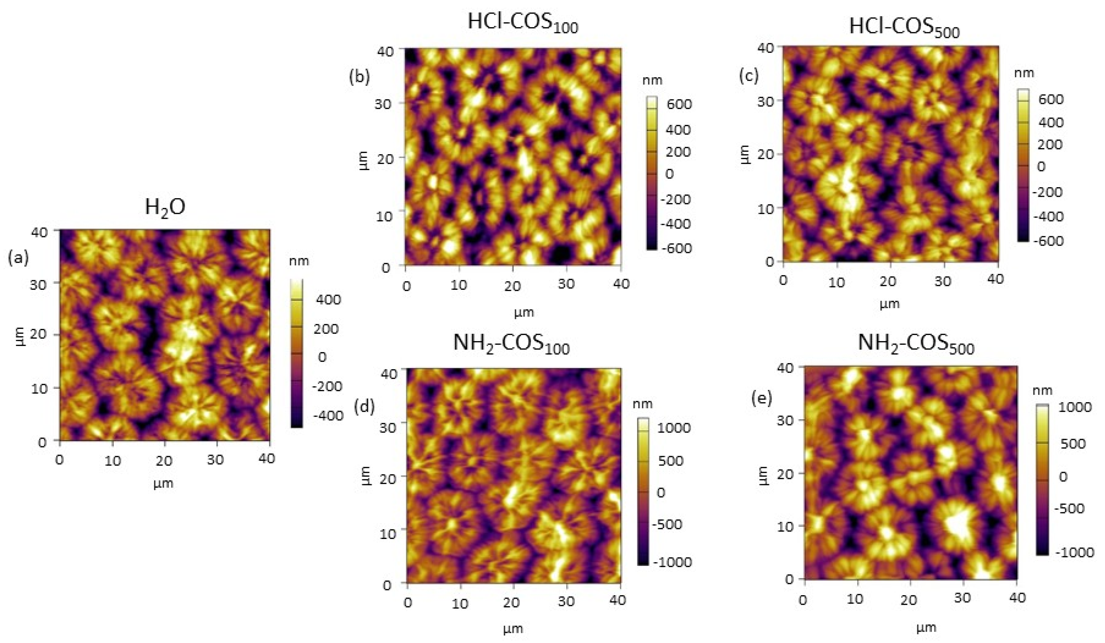
AFM was first utilized to probe the effect of the selected COS on the surface structure of pea seeds. As can be seen from Figure 1, all variants (H2O- and COS-primed) exhibited the typical surface topology for pea seeds, defined by the arrangement of macrosclereids into circular formations. The height of the formations differed among the various priming treatments (600–800 nm for H2O-priming, 800–1500 nm for HCl-COS-priming and 800–2000 nm for NH2-COS-priming), which was reflected in the estimated roughness (Rrms) values. The data revealed that the roughness of the HCl-COS100 (404 ± 22), HCl-COS500 (339 ± 30), and NH2-COS500 (421 ± 18) variants significantly exceeded that of the H2O-primed (266 ± 31) seeds, while NH2-COS100 (264 ± 30) retained values that did not differ significantly from the control (Table 1).
Seed imbibition capacity and ion leakage from the seed coat were also assessed to determine the effects of treatments on water uptake and seed coat integrity, respectively. Imbibition percentage varied in the range of 227–239% (i.e., about 5% variation). The conductivity values remained in the range of 828–978 µS/cm2 (i.e., about 18% variation) for all treatments (Figure S1). Thus, no significant differences were observed among the different priming treatments, indicating that COS application does not exert any negative impact on seed quality, i.e., it does not impair the uptake of water-soluble substances from the environment, and it does not compromise the seed coat integrity (Figure S1).

2.2. Effect of COS-Priming on Seed Germination Uunder Control and Salt Stress Conditions

The germination process and the consequent plant growth of the different seed treatments were compared under control and salt stress conditions. To mimic chronic salt stress in the environment, seeds and plants were grown in salt-supplemented medium from the onset of germination to the end of the experiment (i.e., 14-day-old seedlings).
As shown in Figure S2, the germination rates of seeds were high under control (non-stress) conditions (87–95%). Upon salting, the germinability of the H2O-primed variant dropped insignificantly (by ≈6%), and no statistically significant differences were detected among the different COS-primed variants. Salt stress exposure did not influence seed germination synchrony in any of the investigated treatments (Figure S2).
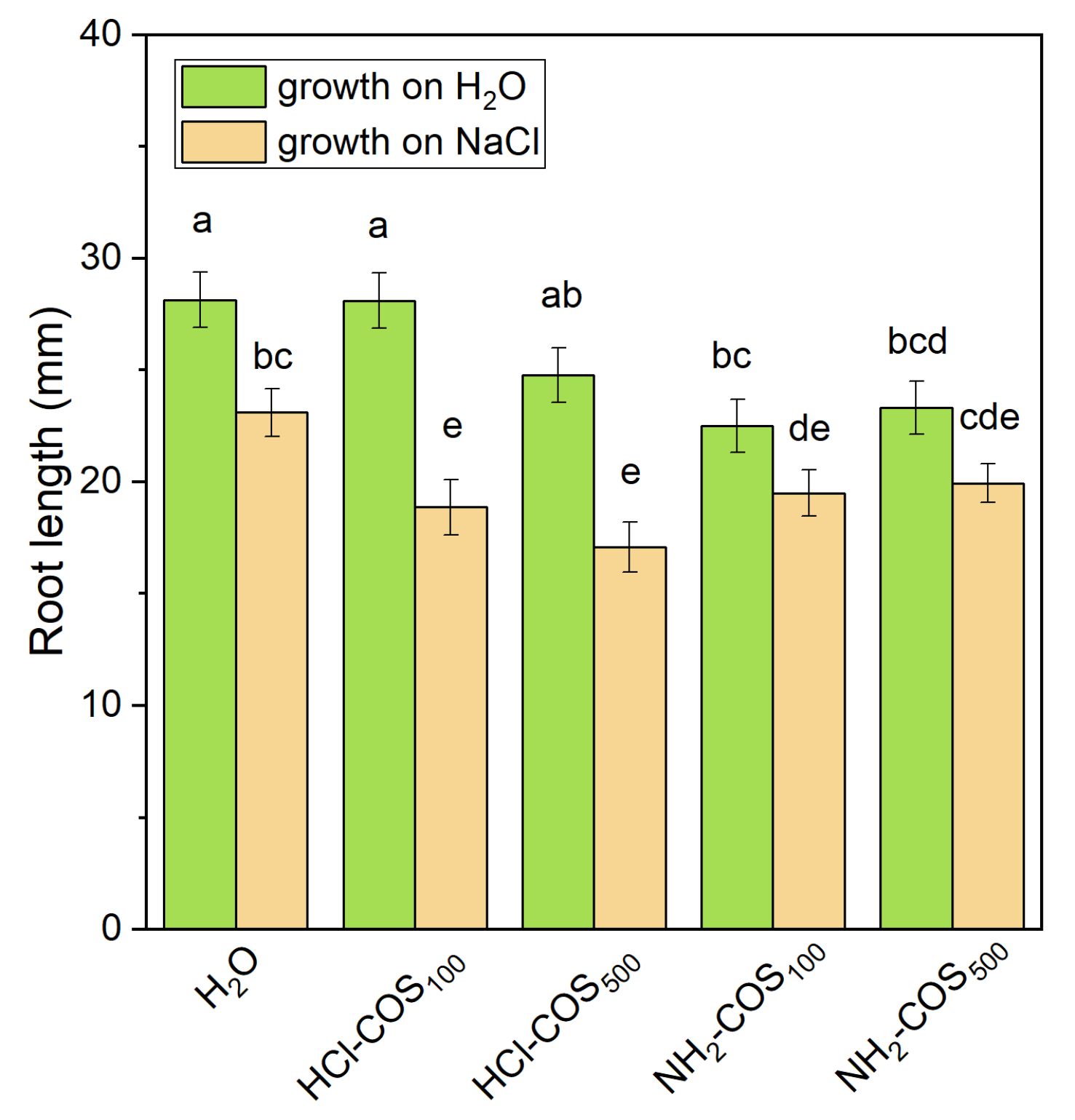
The root length measured on the fourth day of germination (see Figure S3) was approximately 28 mm in the H2O-primed variant under non-stress conditions. It did not change significantly in the HCl-COS-treated variants but was reduced by about 21% in both NH2-COS variants. Under salt stress, the root length in H2O-primed seeds decreased by approximately 18% compared with the non-stress control. In contrast, all COS-treated variants exhibited a more pronounced reduction in root length (14–26%) relative to the salt-stressed control (Figure 2).

2.3. Effect of COS-Priming on the Water Potential and Na+ Content in Pea Seedlings Under Control and Salt Stress Conditions

To assess the effects of COS pea seed priming on plant salt stress tolerance, we measured leaf water potential and Na+ accumulation in leaves and roots in non-stressed and salt-stressed seedlings.
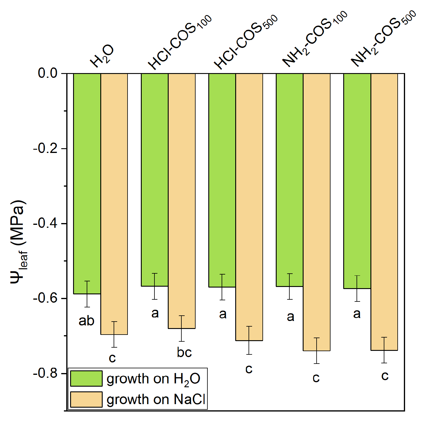
Under control conditions, leaf water potential (Ψleaf) in COS-primed variants was comparable to that of the H2O-primed control. Under salt stress, Ψleaf decreased in all treatments, i.e., by 17% in the H2O-primed variant and by 29% in COS-primed plants, with insignificant variation among the COS-primed variants (Figure 3).
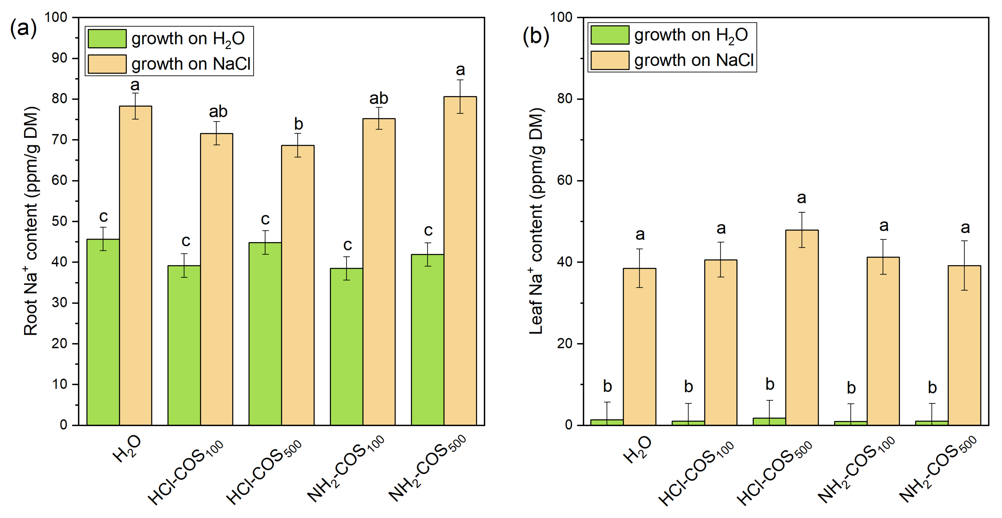
Under non-stress conditions, Na+ accumulation in H2O- and COS-primed variants ranged from 38.5 to 45.7 ppm/g dry mass (DM) in roots and from 1.05 to 1.83 ppm/g DM in leaves (Figure 4), with no statistically significant differences among treatments. Under salt stress, Na+ levels in the H2O-primed variant increased by ≈1.7-fold in roots and ≈28-fold in leaves, compared to the non-stressed control. No statistically significant differences in Na+ accumulation in roots and leaves were observed between COS-treated and H2O-primed variants under salt stress, except for the HCl-COS500 treatment, which showed a 12% lower Na+ content in roots relative to the H2O-primed variant (Figure 4).

2.4. Effect of COS-Priming on the Photosynthetic Activity of Pea Seedlings Under Control and Salt Stress Conditions

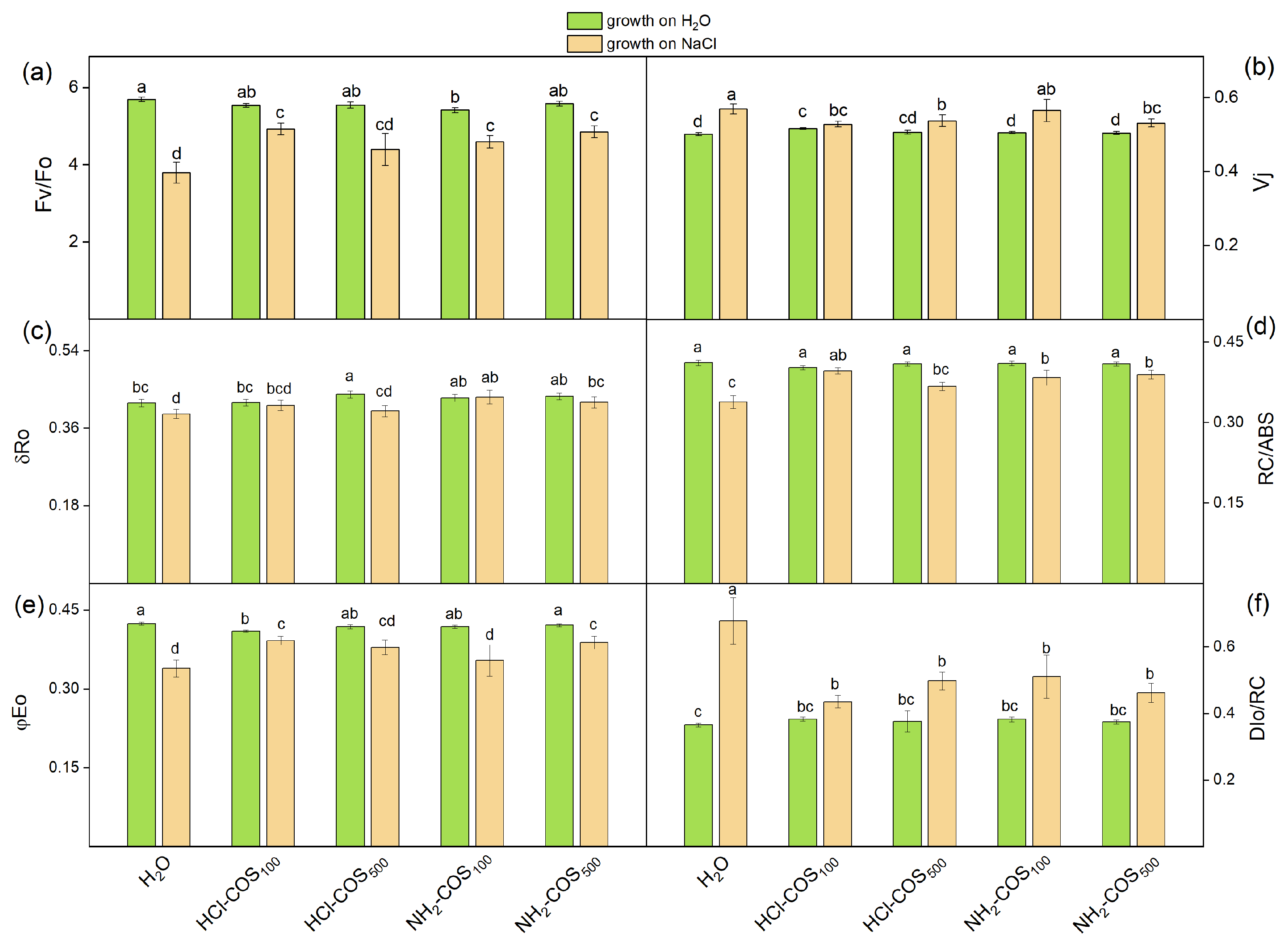
To evaluate the functional state of the photosynthetic apparatus, we analyzed chlorophyll a fluorescence induction (OJIP transitions). From the recorded OJIP curves (Figure S4), we calculated the following parameters [34,35]: the ratio of quantum yields of photochemical and concurrent non-photochemical processes (Fv/Fo); the relative variable fluorescence at the J step (Vj), indicating the proportion of closed PSII reaction centers; the efficiency of reduction in the end electron acceptors at the photosystem I (PSI) acceptor side (δRo); the density of the active reaction centers (RC/ABS); the quantum yield of electron transport beyond QA (φEo) and quantum yield of energy dissipation in the form of heat and fluorescence at the reaction center level (DIo/RC); the performance index on an absorption basis (PIABS) and the total performance index (PItotal). The PIABS index includes three parameters: the number of active PSII RC per antenna chlorophyll [γRC2/(1 − γRC2) = RC/ABS], the partial performance of primary photochemistry [φPo/(1 − φPo)], and the performance of thermal reactions of the intersystem electron carriers [ψ(Eo)/(1 − (ψEo))]. The PItotal index has an additional component [δ(Ro)/(1 − (δRo))], which characterizes the probability with which an electron is transferred to end PSI electron acceptors.
The values of the calculated JIP parameters obtained from leaves of non-treated and salt-treated plants developed from COS-primed seeds are shown in Figure 5 and Figure 6. The components defining PIABS and PItotal were also determined (Table 2).
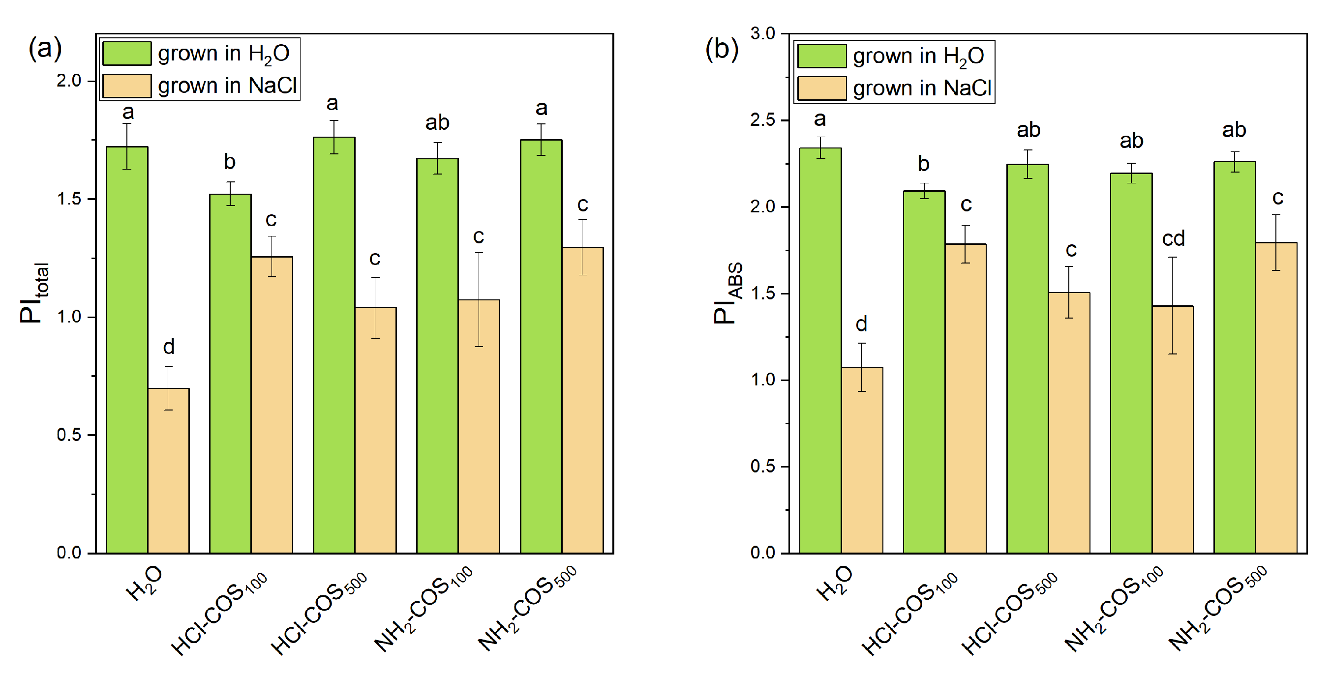
Under non-stress conditions, COS-priming induced only minor changes in the photosynthetic parameters reported in Figure 5, with values varying within 5% of those recorded for the H2O-primed control. The evaluation of PIABS and PItotal indices revealed a reduction by 10% and 12%, respectively, only in HCl-COS100 treatment compared to the H2O-primed variant, while the other variants remained close to the control values (Figure 6). As shown in Table 2, this slight inhibitory effect of HCl-COS100 under non-stress conditions is associated with a small decrease in the efficiency of the primary photochemical reactions [φ(Po)/(1 − φ (Po)] by ≈3% and in the efficiency of the dark reactions of the intersystem electron carriers in photosynthesis [ψ(Eo)/(1 − ψ(Eo)] by ≈6%.
Under NaCl stress, all evaluated photosynthetic parameters in the H2O-primed variant deviated markedly from those observed under non-stress conditions. The most pronounced effects included a decrease in Fv/Fo (by ≈33%), φEo (by 20%) and RC/ABS (by 19%), and an increase in DIo/RC by 86% (Figure 5). Consequently, the performance indices were strongly affected, with PIABS decreasing by more than 50% and PItotal by nearly 60% compared with the non-stressed H2O-primed variant (Figure 6).
Under salt stress, COS-primed variants exhibited distinct response patterns for the different photosynthetic parameters. The Vj parameter showed only minor changes (up to 8% decrease), while δRo increased by up to 11% relative to the H2O-primed variant. In contrast, several photosynthetic parameters were significantly affected by COS-priming. Specifically, Fv/Fo increased by 19–29% in HCl-COS100, NH2-COS100, and NH2-COS500 treatments. The quantum yield of electron transport beyond QA (φEo) increased by ≈16% in HCl-COS100 and NH2-COS500 variants. The density of the active reaction centers (RC/ABS) increased by 16–20% in HCl-COS100, NH2-COS100, and NH2-COS500, while the energy dissipation parameter (DIo/RC) strongly declined by 25–27% for all COS variants (Figure 5). Consistent with these changes, COS-priming significantly improved the PIABS and PItotal indices under salt stress by 42–65% and 49–80%, respectively, compared to the H2O-primed variant, except for PIABS in NH2-COS100, where the effect was not statistically significant (Figure 6). All COS-primed variants exhibited 12–30% higher values of the φ(Po)/((1 − φ(Po)) component compared with the H2O-primed stressed plants. For the remaining components of PIABS and PItotal, the effects varied in significance. Nevertheless, in HCl-COS100 and NH2-COS500 variants, all components PIABS and PItotal were significantly higher than in the H2O-primed variant (Table 2).

2.5. Effect of COS-Priming on the Leaf Pigment Content Under Control and Salt Stress Conditions

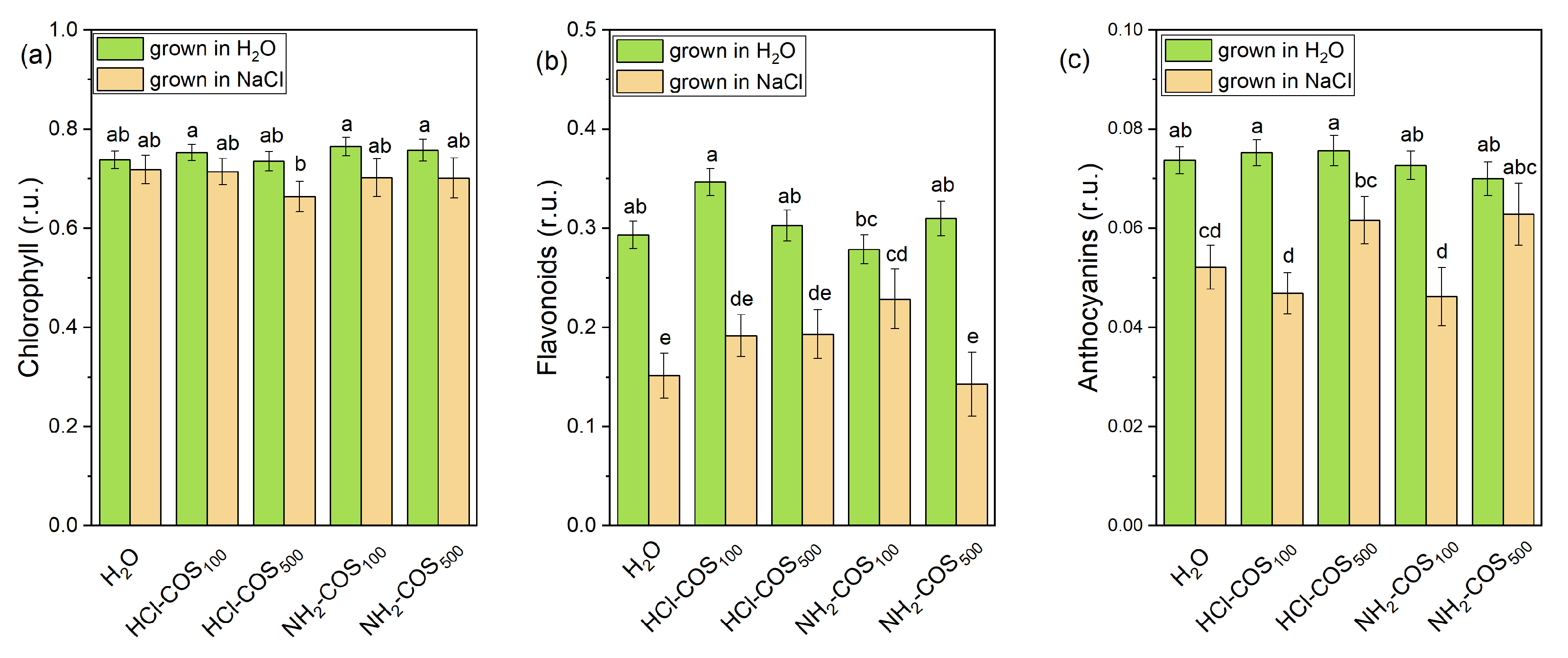
Under non-stress conditions, the total chlorophyll, flavonoid and anthocyanin contents in plants developed from COS-primed pea seeds varied in a narrow range (within 4%, 19%, and 5% from the H2O-primed variant, respectively) and remained close to the control values (Figure 7).
Salt stress reduced the mean chlorophyll content of the H2O-treated variant by only 2%, but markedly decreased the flavonoid and anthocyanin levels by 48% and 30%, respectively (Figure 7).
Among the COS-primed variants subjected to salt stress, only a slight (7%) and not statistically significant decrease in chlorophyll content was observed in the HCl-COS500 treatment. Notably, only the NH2-COS100 variant exhibited significantly higher flavonoid levels (by ≈54%) compared with the H2O-primed stressed plants, reaching values comparable to those under non-stress conditions. Anthocyanin content was higher in plants primed with 500 mg/L compared with 100 mg/L for both HCl-COS and NH2-COS treatments under salt stress. In particular, the NH2-COS500 variant maintained anthocyanin levels (0.063 ± 0.006 r.u.) close to those observed under non-stress conditions (0.070 ± 0.003 r.u.), indicating a strong protective effect of this treatment (Figure 7).

2.6. Effect of COS-Priming on Plants Membrane Integrity Under Control and Salt Stress Conditions

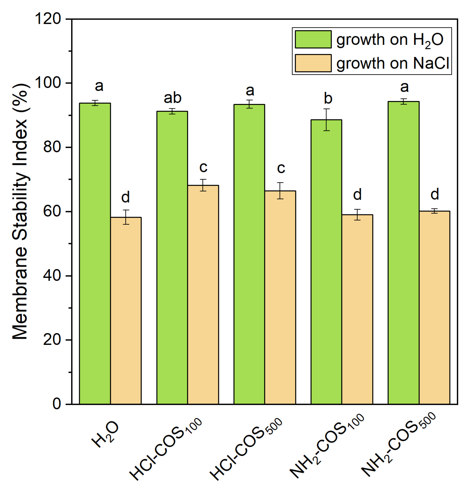
To assess the effect of COS-priming on the leaf membrane integrity, the Membrane Stability Index (MSI) was determined. Under non-stress conditions, only the NH2-COS100 variant showed a slight decrease (≈6%) compared with the H2O-primed control (Figure 8). Under salt stress, the MSI in both the H2O- and NH2-COS-primed plants decreased by approximately 38% relative to the non-stressed H2O-primed sample. In contrast, the decline in MSI was less pronounced in the HCl-COS variants, which maintained 14–17% higher values than the H2O-primed stressed plants.

2.7. Statistical Evaluation of the Individual and Combined COS-Priming and Salt Effects

ANOVA, followed by the Duncan’s multiple range test, was used to evaluate the individual and combined effects of COS-priming (both HCl-COS and NH2-COS) and salt stress on the physiological parameters determined in this study. The analysis revealed that continuous exposure to 50 mM NaCl stress affected almost all parameters assessed in 4- and 14-day-old seedlings, including root length, Na+ accumulation in roots and leaves, leaf water potential, photosynthetic activity, content of leaf pigments and MSI (Table 3). COS-priming alone significantly affected seed surface roughness, root length and flavonoid content, the non-photochemical dissipation of excitation energy, flavonoid level, and MSI. The evaluation of the F-values obtained for the combination of salt-stress and COS-priming (Fsalt×COS) revealed that many of the parameters remained unaffected, demonstrating the generally protective effect of the utilized COS under salt stress. The photosynthetic parameters Fv/Fo, Vj, DIo/RC, φ(Eo), PItotal related to the operation of PSII, the linear electron-transport, and the non-photochemical dissipation of the excess light energy, remained affected in the COS-primed variants under salt stress, although with relatively low Fsalt×COS values (ranging from 2.70 to 8.05). A notable high Fsalt×COS value for the MSI (144.11) was revealed, confirming the positive effect of COS-priming on the plant‘s cellular integrity.

3. Discussion

In this work, we investigated the effect of seed priming with aminooligochitosan (NH2-COS) and oligochitosan hydrochloride (HCl-COS) on pea seed germination, subsequent seedling growth, and photochemistry of photosynthesis under both physiological and salt stress conditions. The rationale for this work stems from the fact that, although the effects of various chitosan and chitosan derivatives have been extensively studied following foliar application, only a limited number of reports have examined seed treatment and priming protocols. Furthermore, to the best of our knowledge, there are no studies integrating detailed characterization of seed surface topology and PSII functionality in the context of COS seed priming. This gap is noteworthy, as seed priming is expected to provide long-term seed/plant protection and thereby preserve overall plant health. Our results demonstrate that HCl-COS and NH2-COS, applied as priming agents at concentrations in the range of 100–500 mg/L, had only minor effects on pea plant growth under non-stress conditions. However, under continuous salt stress (50 mM NaCl), both compounds exhibited dose- and type-dependent effects on pea seeds and seedlings. Furthermore, our findings have revealed a generally protective effect of both chitosan derivatives on plants fitness under saline conditions, as evidenced by positive changes in several physiological parameters associated with photosynthetic activity of photosystem II and cellular membrane integrity. In particular, the most pronounced beneficial effects on the primary photosynthetic steps were observed following treatments with HCl-COS100 and NH2-COS500.

3.1. Effect of COS on Seed and Plant Fitness in Non-Stress Conditions

The HCl-COS- and NH2-COS-priming treatments used in this study induced structural modifications on the pea seed surface, as revealed by AFM imaging (Figure 1). The seed roughness analysis showed that the observed circular structures in HCl-COS100, HCl-COS500 and NH2-COS500-primed variants were higher than those in the control H2O-primed variant (Table 1). This most likely indicates that at least a fraction of the COS molecules becomes adsorbed onto the seed surface, potentially forming a homogeneous film that does not disturb the overall surface structure, seed integrity, or germination properties (Figure 1, Figures S1 and S2). Furthermore, these observations demonstrate that the two different COS treatments exert distinct, type- and dose-dependent effects on the seat coat, which might result in different physiological consequences.
The negative effect of COS-priming on root length may be interpreted in the light of the findings reported by [36], who discuss the contrasting (positive and negative) effects of different chitosans on root and shoot growth. Specifically, in a number of examples root length was inhibited following chitosan application in hydroponic or nutrient-based systems, with the outcome depending on chitosan type and concentration, plant species, and overall experimental conditions. The suppression of genes related to root elongation, along with the stimulation of genes involved in the auxin biosynthesis pathway, resulting in auxin accumulation in the root meristem and subsequent root elongation [37,38], has been identified as a plausible mechanism underlying the observed root length inhibition. Nevertheless, to the best of our knowledge, the data on root growth following pea seed priming presented in the current work are reported for the first time. In our study, we did not perform genetic analysis but instead explored the potential effect of COS-induced structural changes on the seed coat that might affect the seed germination process. Our data confirm that the negative effect on root length is not caused by physical disruption of the seed, as evidenced by the preserved seed coat integrity. This finding justifies further research on the COS-induced hormonal and metabolic changes. Finally, the stability of total chlorophyll content in plants developed from COS-primed seeds under control conditions indicates that COS treatment does not interfere with basal photosynthetic capacity.
Furthermore, chitosan is well-known to boost the roots defense systems via a “growth-defense trade-off” mechanism that ensures higher resistance later in development [36]. The stimulation of flavonoid biosynthesis by HCl-COS100 under non-stress conditions (Figure 7) suggests that at this dose HCl-COS can activate the phenylpropanoid pathway, potentially enhancing the plant’s basal antioxidant capacity [24,39]. This observation, however, requires further experimental verification. The absence of similar effects in other treatments highlights the importance of dose- and compound-specific responses.

3.2. Effect of COS on Seed and Plant Fitness in Growth Under 50 mM NaCl Salt Stress

The applied salt stress resulted in root diminution for all COS treatments, but also in improved PSII capacity as compared to the H2O-primed control. This strongly suggests that both NH2-COS and HCl-COS applied at concentrations of 100 or 500 mg/L trigger the above-mentioned “growth-defense tradeoff” mechanism of pea seeds. This was evident from the chlorophyll fluorescence data presented in Figure 5 and Figure 6, which thus deserves a detailed discussion.
The data in the present study revealed a strong influence of salt stress on the examined JIP parameters in H2O-primed control seeds (Figure 5 and Figure 6, and Table 2), indicating impairment of the early steps of the photosynthetic process. The increase in the parameter Vj and the decrease in the Fv/Fo ratio in the H2O-variant (Figure 5) suggest changes in the acceptor and donor sides of PSII, respectively [40,41]. It is to be noted that the decrease in the parameter Fv/Fo is a result of an increase in Fo, which indicates the disconnection of the light-harvesting complex from the PSII reaction center, which strongly suggests structural reorganization of the photosynthetic complexes within the membrane. Similar salt-induced changes in the PSII complex have also been reported in previous studies [12,42,43]. Modifications of the PSII acceptor side influence QA reoxidation and its interaction with plastoquinone [12], leading to limitations in electron transport beyond QA (φEo). In addition, the decrease in the RC/ABS ratio reveals significant inactivation of the active PSII RC under the applied NaCl stress, as observed previously [44,45]. Prior studies with sorghum also showed that when seedlings were treated with 50 mM NaCl, the chlorophyll content did not change, but changes were observed in the PSII complex. Specifically, changes were found in absorption flux per reaction center and electron transport flux per reaction center [46]. Our data also revealed that salt stress decreased the probability of reduction in the end electron acceptors at the PSI acceptor side (δRo) (Figure 5), which suggests diminished reduction from NADP+ to NADPH via ferredoxin- NADP+ reductase and decreased efficiency of the Calvin–Benson cycle in line with [47]. It has also recently been shown that NaCl inhibits the activity of the Calvin−Benson cycle at least in two different sites [48].
All these salt-induced alterations in PSII resulted in a reduction in the performance indices (PIABS and PItotal), as these changes were associated with a stronger impact on the performance of thermal reactions between the intersystem electron carriers [ψ(Eo)/(1 − ψ(Eo))] and on the performance of the primary photochemistry [φ(Po)/(1 − φ(Po))]. The inhibition of the functions of the electron-transport chain under applied salt stress was accompanied by an increase in energy dissipation (DIo/RC), which is a protective mechanism preventing further damage to the photosynthetic apparatus under abiotic stress [49].
Previous works have also revealed that salt stress mainly destroys the OEC, inactivates PSII reaction centers, and blocks the electron flow from QA to QB in the photosynthetic electron chain [44,50]. The inhibition of PSII donor side activity is possibly related to the destabilization of the catalytic Mn complex due to salt-induced release of the Mn-stabilizing PsbO protein [51].
A study by [52] demonstrated that salt stress inhibits the activity of the PSII RC in plant leaves by reducing the activity of the OEC at the donor side of PSII and degrading D1 protein on the acceptor side of the PSII. A decrease in the electron transfer rate results in the accumulation of excess electrons from the electron transfer chain and leads to electron leakage, which could increase ROS and exacerbate damage to the PSII RC. Ultimately, it may result in peroxidation or dissociation of thylakoid membranes, i.e., alteration of the thylakoid ultrastructure [53]. A recent study on tomato varieties, characterized by different salt tolerance, demonstrated that salt stress inhibited the activity of both PSII and PSI by suppressing electron transfer efficiency, causing multiple damaged sites on the donor and acceptor sides of PSII, and disturbed energy flow distribution [54]. It is concluded that the salt tolerance mechanism in tomato is attributed to higher carbon assimilation efficiency and sugar accumulation, as well as the ability to protect PSII structures and maintain better PSI electron transfer.
Under salt stress conditions, all COS-primed variants exhibited values of the parameters Fv/Fo, Vj and RC/ABS similar to those of non-stressed H2O-primed plants, with a more pronounced effect in the HCl-COS100 and NH2-COS500 variants, suggesting enhanced protection of the PSII complex in these COS-primed plants (Figure 5). Moreover, COS-priming prevents the salt stress-induced decrease in δRo parameter (Figure 5), which suggests better reduction from NADP+ to NADPH and improved efficiency of the Calvin–Benson cycle. In addition, the performance indices (PIABS and PItotal) for COS-primed variants were also higher than those for stressed H2O-primed plants (Figure 6), thus revealing a preservation of the primary photochemical reactions. This effect may result from the activation of enzymes involved in the light-dependent photosynthetic reactions, which enhances antioxidant protection and helps preserve relative membrane integrity under salt stress [24,55,56]. However, we did not find significant differences in the chlorophyll content under non-stress and salt stress conditions, which shows that the protective effect of COS-priming on photosynthetic parameters is more likely due to structural rearrangements of the photosynthetic thylakoid membranes, which optimize the photosynthetic process—an issue that will be further explored by us in future.
Interestingly, the positive effects observed in PIABS and PItotal parameters were the highest (on average) for the two variants that exhibited the highest Rrms values, i.e., HCl-COS100 and NH2-COS500. This might be due to differences in the internalization and metabolism of the two COS due to their different chemical structures, which requires further detailed studies.
To quantify how well cell membranes of pea leaves maintain their integrity under conditions of COS-priming and salt stress, and their combined effect, here we evaluated the MSI, which reflects the degree of injury of leaf cellular membranes—one of the earliest and most sensitive response to salt stress [11,57,58]. Our data revealed lower stress-induced injury under salinization for both HCl-COS variants, meaning that only HCl-COS-priming exhibited a pronounced protective effect regarding the leaf membranes’ integrity under salt stress.
Under salt stress, the marked decrease in flavonoid content in H2O-primed and NH2-COS500 plants suggests a disruption of secondary metabolism or increased utilization of flavonoids as antioxidants in response to elevated ROS levels [17,19]. The relatively stable flavonoid levels in NH2-COS100 plants under salt stress (i.e., similar to those recorded under non-stress conditions) point to a partial protective effect at the 100 mg/L concentration, possibly through improved redox homeostasis. In HCl-COS-treated plants, the reduction in flavonoids under salinity, irrespective of concentration, may reflect a metabolic shift toward primary stress responses or an enhanced consumption of these compounds for ROS detoxification [59].
Anthocyanin responses further support the role of COS in modulating stress-related secondary metabolism [60]. While anthocyanin levels remained unchanged under control conditions, their significant reduction under salt stress (especially in H2O-primed, HCl-COS100, and NH2-COS100 plants) suggests that salinity suppresses their biosynthesis or accelerates their degradation. Notably, the maintenance of high anthocyanin levels in HCl-COS500 and NH2-COS500 plants under salt-stress (with respect to these treatments under non-stress conditions) may indicate a concentration-dependent protective effect, potentially linked to enhanced stress signaling or stabilization of pigment metabolism at higher COS doses. Given the role of anthocyanins as light filters and antioxidants, their relative preservation in these treatments may contribute to improved photoprotection under saline conditions [18].
In summary, this study provides the first experimental evidences of strong positive effects of seed priming with HCl-COS100 and NH2-COS500 on pea primary photosynthetic reactions under salt stress conditions. Our results strongly suggest that commercially available NH2-COS and HCl-COS are effective seed priming agents. These compounds preserve PSII operation and membrane cell integrity, and stimulate the biosynthesis of compounds with protective function under salt stress conditions, thus providing an affordable alternative for seed treatment with beneficial aspects for plant health, although further investigation and verification at the whole-plant level are necessary before any conclusions on whole-plant resilience can be drawn. To understand the exact mechanism(s) of action of these two oligochitosans, evaluation of the genetic, hormonal, metabolic, and antioxidant status of the plants is needed, as well as studies on shoot growth and biomass accumulation to confirm whole-plant tolerance.

4. Materials and Methods

4.1. Seed Treatment and Germination

For this study, we utilized two low-molecular-weight COS: chitosan hydrochloride (ChiBio, Qingdao, China) with deacetylation degree ≥ 98% and viscosity (1% in water) 20 cps, and aminooligochitosan (Realfine Chemical, Wuxi, China) with deacetylation degree ≥ 80% and viscosity (5% in water) < 15 cps. Both COS were dissolved in distilled water at a concentration of 100 mg/L and 500 mg/L and used fresh solutions for seed priming.
Sterilization of pea seeds with 0.1% KMnO4 for 1 min, followed by thorough washing with distilled water, was performed prior to the seed priming treatment. For each priming experiment, batches of 50 pea seeds (Pisum savitum L, cv. RAN-1) were soaked in 50 mL of either distilled water (H2O-priming) or the two aqueous solutions of chitosan oligosaccharides (COS-priming, denoted as NH2-COS100, NH2-COS500, HCl-COS100 and HCl-COS500 for concentration 100 and 500 mg/L, respectively). The priming procedure was conducted for 8 h under continuous and gentle rotation at 2 rpm and room temperature. Seed imbibition percentage was determined by weighing the dry mass of the seeds before the priming and immediately after 8 h of the incubation period. At that point, the seed coat integrity was also evaluated through the degree of electrolyte leakage from the seeds, estimated by measuring the electrical conductivity of all the priming solutions before and after the treatment (HI-5321 research grade EC/TDS meter, Hanna Instruments, Smithfield, RI, USA). The treated seeds were dried for 14–20 days to their initial weight in a dark and dry place. The priming procedure and all subsequent biological experiments were performed in triplicate.

4.2. Seed Surface Properties

Surface investigation of both H2O- and COS-primed pea seeds was carried out using atomic force microscopy (MFP-3D, Asylum Research, Oxford Instruments, Santa Barbara, CA, USA). Silicon AFM probes (AC160TS), with a resonance frequency of 300 kHz and a nominal spring constant of 20 N/m, were employed. All measurements were performed in air at room temperature in contact mode on primed seeds that were rehydrated for 3 h in distilled water and consequently dried out. Morphometric characterization, including surface roughness (Rrms) analysis, was conducted using IgorPro 6.37 software. Rrms was calculated according to the following formula:
R r m s = 1 N i = 1 N Z i 2
where Zi is the height at a given pixel i and N is the total number of pixels in the image.

4.3. Growth Conditions for Control and Stressed Seedlings

Before the start of germination, 25 seeds from each batch of 50 primed seeds were soaked in 50 mM NaCl for a 3-h rehydration period in order to induce salt stress at the earliest stage of plant development and to mimic natural conditions where seeds are exposed to salt stress already at their germination period. The other 25 seeds served as controls and were rehydrated and consequently grown in distilled water. All rehydrated seeds were placed in Petri dishes to germinate on filter paper saturated with tap water (pH = 7, conductivity 80 µS/cm2) for control seeds or a 50 mM NaCl solution for stressed seeds and were incubated in darkness. Germination and root length were recorded at 24-h intervals for 4 days. The germinability and synchrony of germination were calculated according to [61].
Young 4-day-old seedlings were transferred for hydroponic growth in containers filled with tap water (for control seedlings) or aqueous solution of 50 mM NaCl (for stressed seedlings), in controlled laboratory conditions, as previously described by us [62]. The plants were grown for a period of 14 days, which was sufficient for proper anatomical development of the third pair of true leaves. The second and third pairs of leaves were used for subsequent analyses.

4.4. Leaf Water Potential Measurements

Leaf water potential (ψleaf) was measured in situ using a PSY1 leaf psychrometer (ICT International, Armidale, NSW, Australia). For this purpose, the leaf chamber of PSY1 sensor was tightly attached onto the axial surface of the third leaf of intact control and salinity-stressed leaves, after gentle scratching and removal of the cuticular layer to ensure proper vapor equilibration and measurement accuracy. For each individual leaf, the measurement duration was set to 5 min.

4.5. Sodium Ion Content in Leaves and Roots

The Na+ content was determined by using a portable ion-selective meter L-AQUAtwin (Horiba, Kyoto, Japan). Measurements were performed on supernatant derived from centrifugation of homogenized roots and leaves (second and third pair) of individual plants. Prior to each measurement series, the instrument was calibrated using a two-point calibration procedure with standard solutions of 500 and 2000 ppm Na+. Sodium content was expressed as the [Na]+/g DM of the homogenized plant material ratio.

4.6. Fluorescence Induction Kinetics

Chlorophyll a fluorescence induction kinetics was recorded as OJIP transients using a Handy PEA fluorimeter (Hansatech Instruments Ltd., Narborough Road, Pentney, UK). Data acquisition and processing were performed with PEA Plus software (version 1.13). Prior to measurement, leaf samples were dark-adapted for 15 minutes using standard leaf clips to ensure full reopening of PSII RC. Fluorescence induction was initiated with a saturating red actinic pulse of 3200 µmol m−2 s−1, and the full OJIP transient was recorded. The primary fluorescence values obtained were Fo (minimal fluorescence), Fj (fluorescence at the J-step, ~2 ms), Fi (fluorescence at the I-step, ~20 ms), and Fm (maximal fluorescence). These raw values were used to calculate the updated JIP-test parameters included in this study. The following parameters were calculated according to [34]: FV/Fo—indicator of PSII photochemical efficiency and RC openness; Mo is defined as the initial slope of relative variable fluorescence; Mo = 4(F300μs − Fo)/(Fm − Fo); Vj = (Fj − Fo)/(Fm − Fo)—relative variable fluorescence at the J-step indicating the accumulation of QA; φEo = Fv/Fm × ψEo—quantum yield of electron transport beyond QA; ψEo = (1 − Vj)—probability that a trapped exciton moves an electron into the electron transport chain beyond QA; δRo = (1 − Vi)/(1 − Vj)—probability that an electron is transferred from the intersystem chain to PSI end acceptors; φPo = Fv/Fm—maximum quantum yield for primary photochemistry; ABS/RC = Mo × (1/Vj) × (1/φPo); RC/ABS or (1/ABS/RC)—density of active PSII RC per absorbed energy; DIo/RC = ABS/RC(1 − φPo)—energy dissipation per reaction center; PIABS—performance index on an absorption basis and PSII efficiency; PItotal—total performance index including PSI contribution [63]. The two performance indices were calculated according to the following equations [64]:
PIABS = [γRC2/(1 − γRC2) = RC/ABS] × [φPo/(1 − φPo)] × [ψEo/(1 − ψEo)]
and
PItotal = PIABS × [(δRo/(1 − δRo)]

4.7. Measurements of Leaf Pigments

Total chlorophyll, flavonols and anthocyanin contents were non-destructively determined using a multi-pigment meter (MPM-100, Opti-Sciences Ins., Hudson, NH, USA).

4.8. Determination of Membrane Stability Index

Membrane integrity of pea leaves was assessed by calculating the membrane stability index (MSI) using the following equation: MSI (%) = [1 − (EC1/EC2)] × 100, where EC1 and EC2 are the measured conductivities of leaf material submerged in distilled water after incubation in a water bath at 40 °C for 30 min and after heating at 100 °C, respectively, for 15 min as in [57,58].

4.9. Graphical Representation and Statistical Evaluation

Data were graphically presented using Origin 2018 (Origin Lab., Northampton, MA, USA). The comparisons between the responses to salt and chitosan treatments were made using a two-way ANOVA, followed by the Duncan’s multiple range test (p < 0.05). R-4.5.2 (R-project, https://cran.r-project.org/, R Foundation, Vienna, Austria) software with agricolae package (https://cran.r-project.org/package=agricolae (accessed on 1 April 2026)) was used for the calculations.

Supplementary Materials

The following supporting information can be downloaded at: https://www.mdpi.com/article/10.3390/ijms27104498/s1.

Author Contributions

Conceptualization V.V. and S.K.; methodology, S.S., D.I., G.R., E.A., A.D., V.S. and V.K.; software, V.S., V.K. and T.T.; formal analysis, T.T., G.R., V.V. and S.K.; investigation, S.S., D.I., G.R., A.D., V.S. and V.K.; resources, V.V. and S.K.; data curation, V.V. and S.K.; writing—original draft preparation, V.V. and S.K.; writing—review and editing, V.V., S.K., E.A., A.D., T.T., S.S., V.S., V.K., D.I. and G.R.; visualization, S.K., G.R. and V.S.; supervision, V.V. and S.K.; project administration, S.K. and V.V.; funding acquisition, S.K. and V.V. All authors have read and agreed to the published version of the manuscript.

Funding

This research was funded by the Bulgarian National Science Fund, project number KΠ-06-H86/2.

Institutional Review Board Statement

Not applicable.

Informed Consent Statement

Not applicable.

Data Availability Statement

All data are contained within the manuscript.

Acknowledgments

The authors are grateful for the free oligochitosan samples kindly provided by Realfine Chemical (China) and ChiBio (China). Research equipment of Distributed Research Infrastructure INFRAMAT, part of the Bulgarian National Roadmap for Research Infrastructures, supported by the Bulgarian Ministry of Education and Science was used in this investigation.

Conflicts of Interest

The donors of free samples of chitooligosaccharides had no role in the design of the study; in the collection, analyses, or interpretation of data; in the writing of the manuscript; or in the decision to publish the results.

Abbreviations

The following abbreviations are used in this manuscript:
AFMatomic force microscopy
COSchitooligosaccarides
DMdry mass
MSIMembrane Stability Index
NH2-COS100aminooligochitosan in concentration 100 mg/L
NH2-COS500aminooligochitosan in concentration 500 mg/L
HCl-COS100oligochitosan hydrochloride in concentration 100 mg/L
HCl-COS500oligochitosan hydrochloride in concentration 500 mg/L
OECoxygen-evolving complex
PSIphotosystem I
PSIIphotosystem II
RCreaction center
ROSreactive oxygen species
Fv/Foratio of quantum yields of photochemical and concurrent non-photochemical processes
Vjrelative variable fluorescence at the J step, indicating the proportion of closed photosystem II reaction centers
δRoefficiency/probability of reduction in the end electron acceptors at the photosystem I acceptor side
RC/ABSdensity of the active reaction centers
φEoquantum yield of electron transport beyond QA
DIo/RCquantum yield of energy dissipation in the form of heat and fluorescence at the reaction center level
Rrmsseed surface roughness
PIABSperformance index on absorption basis
PItotaltotal performance index
Ψleafleaf water potential

References

  1. FAO. The State of the World’s Land and Water Resources for Food and Agriculture—Systems at Breaking Point (SOLAW 2021); FAO: Rome, Italy, 2021. [Google Scholar]
  2. Munns, R.; Tester, M. Mechanisms of salinity tolerance. Annu. Rev. Plant Biol. 2008, 59, 651–681. [Google Scholar] [CrossRef]
  3. Parida, A.K.; Das, A.B. Salt tolerance and salinity effects on plants: A review. Ecotoxicol. Environ. Saf. 2005, 60, 324–349. [Google Scholar] [CrossRef] [PubMed]
  4. Barhoumi, Z.; Djebali, W.; Chaïbi, W.; Abdelly, C.; Smaoui, A. Salt impact on photosynthesis and leaf ultrastructure of Aeluropus littoralis. J. Plant Res. 2007, 120, 529–537. [Google Scholar] [CrossRef] [PubMed]
  5. Hameed, A.; Ahmed, M.Z.; Hussain, T.; Aziz, I.; Ahmad, N.; Gul, B.; Nielsen, B.L. Effects of salinity stress on chloroplast structure and function. Cells 2021, 10, 2023. [Google Scholar] [CrossRef]
  6. Mehraban, A.; Kadali, F.; Miri, M. Influence of salt stress on lipids metabolism, photorespiration, photosynthesis and chlorophyll fluorescence in crop plants. Chem. Res. J. 2017, 2, 127–132. [Google Scholar]
  7. Ashraf, M.; Harris, P.J.C. Photosynthesis under stressful environments: An overview. Photosynthetica 2013, 51, 163–190. [Google Scholar] [CrossRef]
  8. Huang, L.; Li, Z.; Liu, Q.; Pu, G.; Zhang, Y.; Li, J. Research on the adaptive mechanism of photosynthetic apparatus under salt stress: New directions to increase crop yield in saline soils. Ann. Appl. Biol. 2019, 175, 1–17. [Google Scholar] [CrossRef]
  9. Liu, Z.; Zou, L.; Chen, C.; Zhao, H.; Yan, Y.; Wang, C.; Liu, X. ITRAQ-based quantitative proteomic analysis of salt stress in Spica Prunellae. Sci. Rep. 2019, 9, 9590. [Google Scholar] [CrossRef]
  10. Hao, S.; Wang, Y.; Yan, Y.; Liu, Y.; Wang, J.; Chen, S. A review on plant responses to salt stress and their mechanisms of salt resistance. Horticulturae 2021, 7, 132. [Google Scholar] [CrossRef]
  11. Rashkov, G.D.; Stefanov, M.A.; Yotsova, E.K.; Borisova, P.B.; Dobrikova, A.G.; Apostolova, E.L. Exploring nitric oxide as a regulator in salt tolerance: Insights into photosynthetic efficiency in maize. Plants 2024, 13, 1312. [Google Scholar] [CrossRef]
  12. Stefanov, M.A.; Rashkov, G.D.; Apostolova, E.L. Assessment of the photosynthetic apparatus functions by chlorophyll fluorescence and P700 absorbance in C3 and C4 plants under physiological conditions and under salt stress. Int. J. Mol. Sci. 2022, 23, 3768. [Google Scholar] [CrossRef]
  13. Stefanov, M.A.; Rashkov, G.D.; Yotsova, E.K.; Dobrikova, A.G.; Apostolova, E.L. Impact of salinity on the energy transfer between pigment–protein complexes in photosynthetic apparatus, functions of the oxygen-evolving complex and photochemical activities of photosystem II and photosystem I in two Paulownia lines. Int. J. Mol. Sci. 2023, 24, 3108. [Google Scholar] [CrossRef]
  14. Stefanov, M.A.; Rashkov, G.D.; Borisova, P.B.; Apostolova, E.L. Changes in photosystem II complex and physiological activities in pea and maize plants in response to salt stress. Plants 2024, 13, 1025. [Google Scholar] [CrossRef]
  15. Demmig-Adams, B.; Adams, W.W., III. Photoprotection and Other Responses of Plants to High Light Stress. Annu. Rev. Plant Physiol. Plant Mol. Biol. 1992, 43, 599–626. [Google Scholar] [CrossRef]
  16. Kramer, D.M.; Johnson, G.; Kiirats, O.; Edwards, G.E. New fluorescence parameters for the determination of QA redox state and excitation energy fluxes. Photosynth. Res. 2004, 79, 209–218. [Google Scholar] [CrossRef] [PubMed]
  17. Agati, G.; Azzarello, E.; Pollastri, S.; Tattini, M. Flavonoids as antioxidants in plants: Location and functional significance. Plant Sci. 2012, 196, 67–76. [Google Scholar] [CrossRef]
  18. Li, Z.; Ahammed, G.J. Plant stress response and adaptation via anthocyanins: A review. Plant Stress 2023, 10, 100230. [Google Scholar] [CrossRef]
  19. Fini, A.; Brunetti, C.; Di Ferdinando, M.; Ferrini, F.; Tattini, M. Stress-induced flavonoid biosynthesis and the antioxidant machinery of plants. Plant Signal. Behav. 2011, 6, 709–711. [Google Scholar] [CrossRef]
  20. Chen, Z.; Debernardi, J.M.; Dubcovsky, J.; Gallavotti, A. Recent advances in crop transformation technologies. Nat. Plants 2022, 8, 1343–1351. [Google Scholar] [CrossRef]
  21. da Fonseca-Pereira, P.; Siqueira, J.A.; Monteiro-Batista, R.d.C.; Vaz, M.G.M.V.; Nunes-Nesi, A.; Araújo, W.L. Using synthetic biology to improve photosynthesis for sustainable food production. J. Biotechnol. 2022, 359, 1–14. [Google Scholar] [CrossRef]
  22. De Pascale, S.; Rouphael, Y.; Colla, G. Plant biostimulants: Innovative tool for enhancing plant nutrition in organic farming. Eur. J. Hortic. Sci. 2018, 82, 277–285. [Google Scholar] [CrossRef]
  23. Shukla, P.S.; Mantin, E.G.; Adil, M.; Bajpai, S.; Critchley, A.T.; Prithiviraj, B. Ascophyllum nodosum-Based Biostimulants: Sustainable Applications in Agriculture for the Stimulation of Plant Growth, Stress Tolerance, and Disease Management. Front. Plant Sci. 2019, 10, 655. [Google Scholar] [CrossRef]
  24. Mukhtar Ahmed, K.B.; Khan, M.M.A.; Siddiqui, H.; Jahan, A. Chitosan and its oligosaccharides, a promising option for sustainable crop production—A review. Carbohydr. Polym. 2020, 227, 115331. [Google Scholar] [CrossRef]
  25. Yuan, X.; Zheng, J.; Jiao, S.; Cheng, G.; Feng, C.; Du, Y.; Liu, H. A review on the preparation of chitosan oligosaccharides and application to human health, animal husbandry and agricultural production. Carbohydr. Polym. 2019, 220, 60–70. [Google Scholar] [CrossRef]
  26. Liu, Y.; Yang, H.; Wen, F.; Bao, L.; Zhao, Z.; Zhong, Z. Chitooligosaccharide-induced plant stress resistance. Carbohydr. Polym. 2023, 302, 120344. [Google Scholar] [CrossRef]
  27. An, M.; Zhang, L.; Wang, Q.; Ren, K.; Wang, Q.; Lin, D.; Zhu, Y.; Fan, Y. Chito-oligosaccharide composites enhanced the adaptability of cotton seedlings to salinized soil by modulating photosynthetic efficiency and metabolite. Front. Plant Sci. 2025, 16, 1615321, Correction in Front. Plant Sci. 2025, 16, 1668787. https://doi.org/10.3389/fpls.2025.1615321. [Google Scholar] [PubMed]
  28. Ma, L.; Li, Y.; Yu, C.; Wang, Y.; Li, X.; Li, N.; Chen, Q.; Bu, N. Alleviation of exogenous oligochitosan on wheat seedlings growth under salt stress. Protoplasma 2012, 249, 393–399. [Google Scholar] [CrossRef]
  29. Heydecker, W.; Higgins, J.; Gulliver, R.L. Accelerated germination by osmotic seed treatment. Nature 1973, 246, 42–44. [Google Scholar] [CrossRef]
  30. Fu, Y.; Ma, L.; Li, J.; Hou, D.; Zeng, B.; Zhang, L.; Liu, C.; Bi, Q.; Tan, J.; Yu, X.; et al. Factors influencing seed dormancy and germination and advances in seed priming technology. Plants 2024, 13, 1319. [Google Scholar] [CrossRef]
  31. Jisha, K.C.; Vijayakumari, K.; Puthur, J.T. Seed priming for abiotic stress tolerance: An overview. Acta Physiol. Plant. 2013, 35, 1381–1396. [Google Scholar] [CrossRef]
  32. Ibrahim, E.A. Seed priming to alleviate salinity stress in germinating seeds. J. Plant Physiol. 2016, 192, 38–46. [Google Scholar] [CrossRef]
  33. Mahdavi, B.; Rahimi, A. Seed priming with chitosan improves the germination and growth performance of ajowan. EurAsian J. Biosci. 2013, 7, 69–76. [Google Scholar] [CrossRef]
  34. Strasser, R.J.; Tsimilli-Michael, M.; Srivastava, A. Analysis of the Chlorophyll a Fluorescence Transient. In Chlorophyll a Fluorescence a Signature of Photosynthesis; Papageorgiou, G.C., Govindjee, G., Eds.; Springer: Dordrecht, The Netherlands, 2004; pp. 321–362. [Google Scholar]
  35. Tsimilli-Michael, M.; Strasser, R.J. The energy flux theory 35 years later: Formulations and applications. Photosynth. Res. 2013, 117, 289–320. [Google Scholar] [CrossRef] [PubMed]
  36. Suwanchaikasem, P.; Idnurm, A.; Selby-Pham, J.; Walker, R.; Boughton, B.A. The impacts of chitosan on plant root systems and Its potential to be Used for controlling fungal diseases in agriculture. J. Plant Growth Regul. 2024, 43, 3424–3445. [Google Scholar] [CrossRef]
  37. Lopez-Moya, F.; Suarez-Fernandez, M.; Lopez-Llorca, L.V. Molecular mechanisms of chitosan interactions with fungi and plants. Int. J. Mol. Sci. 2019, 20, 332. [Google Scholar] [CrossRef]
  38. Lopez-Moya, F.; Escudero, N.; Zavala-Gonzalez, E.A.; Esteve-Bruna, D.; Blázquez, M.A.; Alabadí, D.; Lopez-Llorca, L.V. Induction of auxin biosynthesis and WOX5 repression mediate changes in root development in Arabidopsis exposed to chitosan. Sci. Rep. 2017, 7, 16813. [Google Scholar] [CrossRef]
  39. Hadwiger, L.A. Multiple effects of chitosan on plant systems: Solid science or hype. Plant Sci. 2013, 208, 42–49. [Google Scholar] [CrossRef]
  40. Moustakas, M.; Bayçu, G.; Sperdouli, I.; Eroğlu, H.; Eleftheriou, E.P. Arbuscular mycorrhizal symbiosis enhances photosynthesis in the medicinal herb Salvia fruticosa by improving photosystem II photochemistry. Plants 2020, 9, 962. [Google Scholar] [CrossRef]
  41. Kalaji, H.M.; Schansker, G.; Brestic, M.; Bussotti, F.; Calatayud, A.; Ferroni, L.; Goltsev, V.; Guidi, L.; Jajoo, A.; Li, P.; et al. Frequently asked questions about chlorophyll fluorescence, the sequel. Photosynth. Res. 2017, 132, 13–66, Erratum in Photosynth. Res. 2017, 132, 67–68. https://doi.org/10.1007/s11120-016-0318-y. [Google Scholar] [CrossRef]
  42. Athar, H.U.R.; Zafar, Z.U.; Ashraf, M. Glycinebetaine improved photosynthesis in canola under salt stress: Evaluation of chlorophyll fluorescence parameters as potential indicators. J. Agron. Crop Sci. 2015, 201, 428–442. [Google Scholar] [CrossRef]
  43. Stefanov, M.; Yotsova, E.; Rashkov, G.D.; Ivanova, K.; Markovska, Y.; Apostolova, E.L. Effects of salinity on the photosynthetic apparatus of two Paulownia lines. Plant Physiol. Biochem. 2016, 101, 54–59. [Google Scholar] [CrossRef]
  44. Kan, X.; Ren, J.; Chen, T.; Cui, M.; Li, C.; Zhou, R.; Zhang, Y.; Liu, H.; Deng, D.; Yin, Z. Effects of salinity on photosynthesis in maize probed by prompt fluorescence, delayed fluorescence and P700 signals. Environ. Exp. Bot. 2017, 140, 56–64. [Google Scholar] [CrossRef]
  45. Stefanov, M.A.; Rashkov, G.D.; Yotsova, E.K.; Borisova, P.B.; Dobrikova, A.G.; Apostolova, E.L. Protective effects of sodium nitroprusside on photosynthetic performance of Sorghum bicolor L. under salt stress. Plants 2023, 12, 832. [Google Scholar] [CrossRef]
  46. Stefanov, M.A.; Rashkov, G.D.; Yotsova, E.K.; Borisova, P.B.; Dobrikova, A.G.; Apostolova, E.L. Different sensitivity levels of the photosynthetic apparatus in Zea mays L. and Sorghum bicolor L. under salt stress. Plants 2021, 10, 1469. [Google Scholar] [CrossRef] [PubMed]
  47. Michelet, L.; Zaffagnini, M.; Morisse, S.; Sparla, F.; Pérez-Pérez, M.E.; Francia, F.; Danon, A.; Marchand, C.H.; Fermani, S.; Trost, P.; et al. Redox regulation of the Calvin–Benson cycle: Something old, something new. Front. Plant Sci. 2013, 4, 470. [Google Scholar] [CrossRef] [PubMed]
  48. Patil, P.P.; Kodru, S.; Szabó, M.; Vass, I. Investigation of the effect of salt stress on photosynthetic electron transport pathways in the Synechocystis PCC 6803 cyanobacterium. Physiol. Plant. 2025, 177, e70066. [Google Scholar] [CrossRef]
  49. Derks, A.; Schaven, K.; Bruce, D. Diverse mechanisms for photoprotection in photosynthesis. Dynamic regulation of photosystem II excitation in response to rapid environmental change. Biochim. Biophys. Acta Bioenerg. 2015, 1847, 468–485. [Google Scholar] [CrossRef]
  50. Lu, C.; Vonshak, A. Effects of salinity stress on photosystem II function in cyanobacterial Spirulina platensis cells. Physiol. Plant. 2002, 114, 405–413. [Google Scholar] [CrossRef]
  51. Gong, H.; Tang, Y.; Wang, J.; Wen, X.; Zhang, L.; Lu, C. Characterization of photosystem II in salt-stressed cyanobacterial Spirulina platensis cells. Biochim. Biophys. Acta Bioenerg. 2008, 1777, 488–495. [Google Scholar] [CrossRef] [PubMed]
  52. Zhang, H.; Xu, N.; Wu, X.; Wang, J.; Ma, S.; Li, X.; Sun, G. Effects of four types of sodium salt stress on plant growth and photosynthetic apparatus in sorghum leaves. J. Plant Interact. 2018, 13, 506–513. [Google Scholar] [CrossRef]
  53. Mitsuya, S.; Takeoka, Y.; Miyake, H. Effects of sodium chloride on foliar ultrastructure of sweet potato (Ipomoea batatas Lam.) plantlets grown under light and dark conditions in vitro. J. Plant Physiol. 2000, 157, 661–667. [Google Scholar] [CrossRef]
  54. Li, X.; Han, Y.; Cong, Y.; Wang, L.; Shi, Y.; Liu, H.; Liu, H. Mechanisms of salt tolerance: Insights into photosystem function and carbon metabolism in tomato seedlings. Plant Soil 2025, 516, 1489–1513. [Google Scholar] [CrossRef]
  55. Tabassum, M.; Noreen, Z.; Aslam, M.; Shah, A.N.; Usman, S.; Waqas, A.; Alsherif, E.A.; Korany, S.M.; Nazim, M. Chitosan modulated antioxidant activity, inorganic ions homeostasis and endogenous melatonin to improve yield of Pisum sativum L. accessions under salt stress. Sci. Hortic. 2024, 323, 112509. [Google Scholar] [CrossRef]
  56. Amooaghaie, R.; Rajaie, N. Exploring effect of chitosan on antioxidant system and hypericin content in Hypericum perforatum L. under various irrigation regimes. BMC Plant Biol. 2025, 25, 799. [Google Scholar] [CrossRef] [PubMed]
  57. Rady, M.M. Effect of 24-epibrassinolide on growth, yield, antioxidant system and cadmium content of bean (Phaseolus vulgaris L.) plants under salinity and cadmium stress. Sci. Hortic. 2011, 129, 232–237. [Google Scholar] [CrossRef]
  58. Ru, C.; Liu, Y.; Yu, X.; Xie, C.; Hu, X. Melatonin enhances tomato salt tolerance by improving water use efficiency, photosynthesis, and redox homeostasis. Agronomy 2025, 15, 1746. [Google Scholar] [CrossRef]
  59. Brunetti, C.; Di Ferdinando, M.; Fini, A.; Pollastri, S.; Tattini, M. Flavonoids as Antioxidants and Developmental Regulators: Relative Significance in Plants and Humans. Int. J. Mol. Sci. 2013, 14, 3540–3555. [Google Scholar] [CrossRef]
  60. Li, J.; Han, A.; Zhang, L.; Meng, Y.; Xu, L.; Ma, F.; Liu, R. Chitosan oligosaccharide alleviates the growth inhibition caused by physcion and synergistically enhances resilience in maize seedlings. Sci. Rep. 2022, 12, 162. [Google Scholar] [CrossRef]
  61. Ranal, M.A.; Santana, D.G.d.; Ferreira, W.R.; Mendes-Rodrigues, C. Calculating germination measurements and organizing spreadsheets. Rev. Bras. Botânica 2009, 32, 849–855. [Google Scholar] [CrossRef]
  62. Krumova, S.; Stoichev, S.; Ilkov, D.; Strijkova, V.; Katrova, V.; Crespo, A.; Álvarez, J.; Martínez, E.; Martínez-Ramírez, S.; Tsonev, T.; et al. Pea seed priming with pluronic P85-grafted single-walled carbon nanotubes affects photosynthetic gas exchange but not photosynthetic light reactions. Int. J. Mol. Sci. 2024, 25, 7901. [Google Scholar] [CrossRef]
  63. Banks, J.M. Continuous excitation chlorophyll fluorescence parameters: A review for practitioners. Tree Physiol. 2017, 37, 1128–1136. [Google Scholar] [CrossRef]
  64. Maliba, B.G.; Inbaraj, P.M.; Berner, J.M. The use of OJIP fluorescence transients to monitor the effect of elevated ozone on biomass of canola plants. Water Air Soil Pollut. 2019, 230, 75, Correction in Water Air Soil Pollut. 2019, 230, 99. https://doi.org/10.1007/s11270-019-4124-y. [Google Scholar] [CrossRef]
Figure 1. Representative AFM images of the seed topology of H2O- (a) and COS-primed (be) variants.
Ijms 27 04498 g001
Figure 2. Root length of 4-day-old seedlings derived from H2O- and COS-primed seeds, grown under control conditions or under 50 mM NaCl stress. Values are expressed as means ± SE (n = 53−63). Means with no letters in common differ significantly according to the Duncan’s multiple range test (p < 0.05).
Ijms 27 04498 g002
Figure 3. Leaf water potential (Ψleaf) of 14-day-old seedlings derived from H2O- and COS-primed seeds, grown under non-stress conditions or under 50 mM NaCl stress. Values are expressed as means ± SE (n = 6). Means with no letters in common differ significantly according to the Duncan’s multiple range test (p < 0.05).
Ijms 27 04498 g003
Figure 4. Na+ content in roots (a) and leaves (b) of 14-day-old seedlings derived from H2O- and COS-primed seeds, grown under control conditions or under 50 mM NaCl stress. Values are expressed as means ± SE (n = 6). Means with no letters in common differ significantly according to the Duncan’s multiple range test (p < 0.05).
Ijms 27 04498 g004
Figure 5. Selected JIP parameters determined on leaves of 14-day-old seedlings derived from H2O- and COS-primed variants grown in H2O or 50 mM NaCl: (a) ratio of quantum yields of photochemical and concurrent non-photochemical processes (Fv/Fo); (b) relative variable fluorescence at the J step (Vj); (c) efficiency with which an electron from the intersystem electron carriers is transferred to reduce end electron acceptors at the PSI acceptor side (δRo); (d) density of the active reaction centers (RC/ABS); (e) quantum yield of electron transport beyond QA (φEo); (f) quantum yield of energy dissipation in the form of heat and fluorescence at the reaction center level (DIo/RC). Values are expressed as means ± SE (n = 20). Means with no letters in common differ significantly according to the Duncan’s multiple range test (p < 0.05).
Ijms 27 04498 g005
Figure 6. Performance indices PItotal (a) and PIABS (b), calculated from OJIP curves recorded for leaves of 14-day-old seedlings derived from H2O- and COS-primed variants grown in H2O or 50 mM NaCl. Values are expressed as means ± SE (n = 20). Means with no letters in common differ significantly according to the Duncan’s multiple range test (p < 0.05).
Ijms 27 04498 g006
Figure 7. Leaf chlorophyll (a), flavonoids (b) and anthocyanin (c) content of 14-day-old seedlings derived from H2O- and COS-primed variants grown in H2O or 50 mM NaCl. Values are expressed as means ± SE (n = 23–29 for non-stress conditions and n = 7–15 for salt stress). Means with no letters in common differ significantly according to the Duncan’s multiple range test (p < 0.05).
Ijms 27 04498 g007
Figure 8. Membrane Stability Index determined for 14-day-old seedlings derived from H2O- and COS-primed variants grown in H2O or 50 mM NaCl. Values are expressed as means ± SE (n = 6). Means with no letters in common differ significantly according to the Duncan’s multiple range test (p < 0.05).
Ijms 27 04498 g008
Table 1. Seed surface roughness (Rrms) values determined for H2O- and COS-primed variants.
TreatmentsRrms (nm)
H2O266 ± 31 c
HCl-COS100404 ± 22 ab
HCl-COS500339 ± 30 b
NH2-COS100264 ± 30 c
NH2-COS500421 ± 18 a
Values are expressed as means ± SE (n = 10–16 images for each variant). Means with no letters in common differ significantly according to the Duncan’s multiple range test (p < 0.05).
Table 2. Components of the performance indices PIABS and PItotal estimated for 14-day-old seedlings derived from H2O- and COS-primed seeds grown in H2O or 50 mM NaCl.
Treatmentsγ(RC)/((1 − γ(RC))φ(Po)/((1 − φ(Po))ψ(Eo)/(1 − ψ(Eo))δ(Ro)/(1 − δ(Ro))
H2O0.411 ± 0.005 a5.698 ± 0.061 a0.999 ± 0.018 a0.729 ± 0.028 ab
HCl-COS1000.402 ± 0.004 ab5.538 ± 0.056 b0.938 ± 0.009 b0.731 ± 0.022 ab
HCl-COS5000.411 ± 0.005 a5.540 ± 0.087 ab0.979 ± 0.018 a0.789 ± 0.026 a
NH2-COS1000.410 ± 0.004 a5.419 ± 0.069 ab0.983 ± 0.011 a0.765 ± 0.027 ab
NH2-COS5000.409 ± 0.004 a5.585 ± 0.062 ab0.987 ± 0.014 a0.775 ± 0.023 ab
NaCl0.338 ± 0.012 d3.797 ± 0.273 d0.769 ± 0.041 d0.655 ± 0.029 c
HCl-COS100 + NaCl0.396 ± 0.006 bc4.928 ± 0.148 bc0.898 ± 0.026 bc0.718 ± 0.041 b
HCl-COS500 + NaCl0.367 ± 0.008 cd4.596 ± 0.209 c0.877 ± 0.058 c0.674 ± 0.034 bc
NH2-COS100 + NaCl0.383 ± 0.014 bc4.399 ± 0.418 c0.792 ± 0.085 d0.769 ± 0.054 ab
NH2-COS500 + NaCl0.389 ± 0.008 bc4.858 ± 0.211 bc0.901 ± 0.038 bc0.743 ± 0.039 ab
Values are expressed as means ± SE (n = 20). Means with no letters in common differ significantly according to the Duncan’s multiple range test (p < 0.05).
Table 3. Summary of one-way and two-way ANOVA for the effects of salt (Fsalt), COS-priming of seeds (FCOS), and their interaction (Fsalt×COS) on seed structural and plant physiological traits in Pisum sativum.
TraitFsaltFCOSFsalt×COS
Rrms 11.92 ***
Imbibition 2.48
Conductivity 2.47
Germination0.480.760.41
Synchrony0.120.330.23
Root length62.33 ***6.33 ***2.81 *
Leaf water potential40.58 ***0.270.36
Root Na+ content290.15 ***1.621.78
Leaves Na+ content206.59 ***0.390.31
Fv/Fo92.87 ***2.63 *6.05 ***
Vj17.60 ***1.362.70 *
DIo/RC55.80 ***3.62 **8.05 ***
φ(Eo)32.60 ***1.573.74 **
RC/ABS27.44 ***1.200.93
δ(Ro)33.54 ***1.400.21
PIABS34.13 ***2.372.05
PItotal71.02 ***1.833.81 **
Chlorophyll8.58 **0.630.34
Flavonoids93.29 ***2.80 *2.10
Anthocyanins65.16 ***0.782.07
Membrane Stability Index430.64 ***4.91 *144.11 ***
*** indicates 99.9% significance, ** indicates 99% significance, * indicates 95% significance.
Disclaimer/Publisher’s Note: The statements, opinions and data contained in all publications are solely those of the individual author(s) and contributor(s) and not of MDPI and/or the editor(s). MDPI and/or the editor(s) disclaim responsibility for any injury to people or property resulting from any ideas, methods, instructions or products referred to in the content.