Amino Acid-Derived Metabolic Signature Across Stages of Systolic Dysfunction: Derivation and Internal Evaluation of the HASI (Heart Failure Amino Acid-Derived Systolic Index)—40 Index
 , , , ,
  , , and
Abstract
1. Introduction
2. Results and Discussion
2.1. Groups Characterization
2.2. Laboratory Results
2.3. Metabolomic Profiling
2.4. HASI-40 Index
2.5. Correlations Between Circulating Amino Acids and Left Ventricular Ejection Fraction
2.6. Uni- and Multivariable Models
2.7. Receiver Operator Curve Analysis (ROC) for Combined Models
2.8. Discussion
2.9. Limitations
3. Materials and Methods
3.1. Study Population
3.2. Clinical Evaluation
3.3. Amino Acids/Metabolomic Profiling
- Proteinogenic Amino Acids—directly incorporated into proteins; reflect nutritional status, protein turnover, and anabolic balance. In HFrEF, they reflect global protein turnover, anabolic resistance, and substrate availability. The proteinogenic amino acids include essential (histidine, isoleucine, leucine, lysine, methionine, phenylalanine, threonine, tryptophan, and valine) and non-essential/conditionally essential (alanine, arginine, asparagine, aspartate, cysteine, glutamate, glutamine, glycine, proline, serine, and tyrosine).
- Methylation Pathway Intermediates, which are linked to one-carbon metabolism, epigenetics, mitochondrial function, and redox balance. Their role in HFrEF is linked with impaired methylation flux, disrupted one-carbon metabolism, mitochondrial dysfunction, and oxidative stress. This group includes the following amino acids: sarcosine, glycine, serine, methionine.
- Muscle Catabolism Markers that reflect proteolysis, cachexia, and systemic catabolic activation, including core markers such as 3-Methylhistidine (3-MH), followed by alanine and glutamine.
3.4. Biomarker Derivation
3.5. Statistical Analysis
4. Conclusions
Author Contributions
Funding
Institutional Review Board Statement
Informed Consent Statement
Data Availability Statement
Acknowledgments
Conflicts of Interest
References
- Paraskevaidis, I.; Tsougos, E.; Kourek, C. One Syndrome, Many Faces: A Unified Perspective on Heart Failure Phenotypes. Int. J. Mol. Sci. 2025, 26, 8960. [Google Scholar] [CrossRef] [Scilit]
- Gatto, C.; Rusciano, M.R.; Visco, V.; Vecchione, C.; Ciccarelli, M. Pathophysiology of HFpEF: Insights from a Metabolic-Mitochondrial Perspective. Int. J. Mol. Sci. 2025, 27, 284. [Google Scholar] [CrossRef] [Scilit]
- Adamu, U.G.; Badianyama, M.; Mayisela, M.; Amoni, J.; Tsabedze, D.; Maseko, M.; Tsabedze, N. The Use of Metabolomes in Risk Stratification of Patients with Heart Failure: A Scoping Review. Life 2026, 16, 514. [Google Scholar] [CrossRef] [Scilit]
- Hunter, W.G.; Kelly, J.P.; McGarrah, R.W., 3rd; Kraus, W.E.; Shah, S.H. Metabolic Dysfunction in Heart Failure: Diagnostic, Prognostic, and Pathophysiologic Insights From Metabolomic Profiling. Curr. Heart Fail. Rep. 2016, 13, 119–131. [Google Scholar] [CrossRef] [Scilit] [PubMed]
- Jaltotage, B.; Dwivedi, G.; Ooi, D.E.L.; Mahadavan, G. The Utility of Circulating and Imaging Biomarkers Alone and in Combination in Heart Failure. Curr. Cardiol. Rev. 2021, 17, e160721193557. [Google Scholar] [CrossRef] [Scilit]
- Mavroudis, A.; Fragoulis, C.; Mavromoustakou, K.; Iliakis, P.; Tsioufis, K.; Chrysohoou, C. Mechanisms of Cardiac Inflammation in Heart Failure: Role of Dietary Patterns, Nutrients, and Therapeutic Strategies. Nutrients 2026, 18, 1005. [Google Scholar] [CrossRef] [Scilit] [PubMed]
- Yun, H.R.; Singh, M.K.; Han, S.; Ranbhise, J.S.; Ha, J.; Kim, S.S.; Kang, I. Biomarkers of Cardiac Metabolic Flexibility in Health, HFrEF and HFpEF. Int. J. Mol. Sci. 2026, 27, 879. [Google Scholar] [CrossRef] [Scilit] [PubMed]
- Evans, P.C.; Vilahur, G.; Kleinbongard, P.; Osto, E.; Remme, C.A.; Madonna, R.; Aksentijevic, D.; Sourij, H.; Davidson, S.M.; Santovito, D.; et al. Novel cardiovascular metabolic risk factor mechanisms and therapeutic opportunities. Eur. Heart J. 2026. epub ahead of print. [Google Scholar] [CrossRef] [Scilit]
- Li, Q.; Hoppe, T. Role of amino acid metabolism in mitochondrial homeostasis. Front. Cell Dev. Biol. 2023, 11, 1127618. [Google Scholar] [CrossRef] [Scilit]
- Urbanowicz, T.; Pietkiewicz, D.; Plewa, S.; Krasińska, B.; Spasenenko, I.; Gabriel, K.; Jezierska, K.; Krasiński, Z.; Kowalewski, M.; Matysiak, J.; et al. Metabolic Predictors of CAD: Focus on Cystine, Methionine, Proline, and Threonine Circulating Levels-Exploratory Pilot Study. J. Clin. Med. 2025, 14, 8356. [Google Scholar] [CrossRef] [Scilit]
- Li, X.; Zhang, H.S. Amino acid metabolism, redox balance and epigenetic regulation in cancer. FEBS J. 2024, 291, 412–429. [Google Scholar] [CrossRef] [Scilit]
- Castiglione, V.; Aimo, A.; Vergaro, G.; Saccaro, L.; Passino, C.; Emdin, M. Biomarkers for the diagnosis and management of heart failure. Heart Fail. Rev. 2022, 27, 625–643. [Google Scholar] [CrossRef] [Scilit] [PubMed]
- Spyropoulos, F.; Sorrentino, A.; van der Reest, J.; Yang, P.; Waldeck-Weiermair, M.; Steinhorn, B.; Eroglu, E.; Saeedi Saravi, S.S.; Yu, P.; Haigis, M.; et al. Metabolomic and transcriptomic signatures of chemogenetic heart failure. Am. J. Physiol. Heart Circ. Physiol. 2022, 322, H451–H465. [Google Scholar] [CrossRef] [Scilit]
- Palazzuoli, A.; Tramonte, F.; Beltrami, M. Laboratory and Metabolomic Fingerprint in Heart Failure with Preserved Ejection Fraction: From Clinical Classification to Biomarker Signature. Biomolecules 2023, 13, 173. [Google Scholar] [CrossRef] [Scilit]
- Yang, C.; Shi, Z.; Bao, L.; Xv, X.; Jiang, D.; You, L. Targeted metabolomic analysis of serum amino acids in heart failure patients. Amino Acids 2024, 56, 22–32. [Google Scholar] [CrossRef] [Scilit]
- Schaffer, S.W.; Ito, T.; Azuma, J.; Jong, C.J.; Kramer, J.H. Mechanisms Underlying Development of Taurine-Deficient Cardiomyopathy. Hearts 2020, 1, 86–98. [Google Scholar] [CrossRef] [Scilit]
- Ur Rehman, M.; Saeed, H.; Babar, Z.U.D.; Omer, O.; Tashfeen, S. Metabolomic Biomarkers in Heart Failure: A Systematic Review of Diagnostic and Prognostic Significance. Cureus 2025, 17, e97636. [Google Scholar] [CrossRef] [Scilit] [PubMed]
- Savarese, G.; Stolfo, D.; Sinagra, G.; Lund, L.H. Heart failure with mid-range or mildly reduced ejection fraction. Nat. Rev. Cardiol. 2022, 19, 100–116. [Google Scholar] [CrossRef] [Scilit]
- Wu, L.; Rizwan, A.; Rodriguez, M.; El Hachem, K.; Hassan Virk, H.U.; Khawaja, M.; Strauss, M.; Krittanawong, C. Heart Failure With Mildly Reduced Ejection Fraction: A Call for a Precision Medicine Approach. JACC Adv. 2026, 5, 102476–102493. [Google Scholar] [CrossRef] [Scilit] [PubMed]
- Fonseka, O.; Gare, S.R.; Chen, X.; Zhang, J.; Alatawi, N.H.; Ross, C.; Liu, W. Molecular Mechanisms Underlying Heart Failure and Their Therapeutic Potential. Cells 2025, 14, 324. [Google Scholar] [CrossRef] [Scilit]
- Zhou, Q.; Li, P.; Zhao, H.; Xu, X.; Li, S.; Zhao, J.; Xu, D.; Zeng, Q. Heart Failure With Mid-range Ejection Fraction: A Distinctive Subtype or a Transitional Stage? Front. Cardiovasc. Med. 2021, 8, 678121. [Google Scholar] [CrossRef] [Scilit] [PubMed]
- Schmitt, A.; Behnes, M.; Weidner, K.; Abumayyaleh, M.; Reinhardt, M.; Abel, N.; Lau, F.; Forner, J.; Ayoub, M.; Mashayekhi, K.; et al. Prognostic impact of prior LVEF in patients with heart failure with mildly reduced ejection fraction. Clin. Res. Cardiol. 2025, 114, 570–588. [Google Scholar] [CrossRef] [Scilit] [PubMed]
- Ragni, M.; Greco, C.M.; Felicetta, A.; Ren, S.V.; Kunderfranco, P.; Ruocco, C.; Carullo, P.; Larcher, V.; Tedesco, L.; Severi, I.; et al. Dietary essential amino acids for the treatment of heart failure with reduced ejection fraction. Cardiovasc. Res. 2023, 119, 982–997. [Google Scholar] [CrossRef] [Scilit] [PubMed]
- Xin, Y.; Zhang, Y.; Yuan, Z.; Li, S. Methionine is an essential amino acid in doxorubicin-induced cardiotoxicity through modulating mitophagy. Free Radic. Biol. Med. 2025, 232, 28–39. [Google Scholar] [CrossRef] [Scilit]
- Simmonds, S.J.; Cuijpers, I.; Heymans, S.; Jones, E.A.V. Cellular and Molecular Differences between HFpEF and HFrEF: A Step Ahead in an Improved Pathological Understanding. Cells 2020, 9, 242. [Google Scholar] [CrossRef] [Scilit]
- Manolis, A.A.; Manolis, T.A.; Manolis, A.S. Neurohumoral Activation in Heart Failure. Int. J. Mol. Sci. 2023, 24, 15472. [Google Scholar] [CrossRef] [Scilit]
- Callahan, D.M.; Toth, M.J. Skeletal muscle protein metabolism in human heart failure. Curr. Opin. Clin. Nutr. Metab. Care 2013, 16, 66–71. [Google Scholar] [CrossRef] [Scilit]
- Reina-Couto, M.; Pereira-Terra, P.; Quelhas-Santos, J.; Silva-Pereira, C.; Albino-Teixeira, A.; Sousa, T. Inflammation in Human Heart Failure: Major Mediators and Therapeutic Targets. Front. Physiol. 2021, 12, 746494–746519. [Google Scholar] [CrossRef] [Scilit]
- Rasooly, D.; Giambartolomei, C.; Peloso, G.M.; Dashti, H.; Ferolito, B.R.; Golden, D.; Horimoto, A.R.V.R.; Pietzner, M.; Farber-Eger, E.H.; Wells, Q.S.; et al. Large-scale multi-omics identifies drug targets for heart failure with reduced and preserved ejection fraction. Nat. Cardiovasc. Res. 2025, 4, 293–311. [Google Scholar] [CrossRef] [Scilit]
- Liu, H.; Wang, S.; Wang, J.; Guo, X.; Song, Y.; Fu, K.; Gao, Z.; Liu, D.; He, W.; Yang, L.L. Energy metabolism in health and diseases. Signal Transduct. Target. Ther. 2025, 10, 69–140. [Google Scholar] [CrossRef] [Scilit]
- Kozhevnikova, M.V.; Belenkov, Y.N.; Shestakova, K.M.; Ageev, A.A.; Markin, P.A.; Kakotkina, A.V.; Korobkova, E.O.; Moskaleva, N.E.; Kuznetsov, I.V.; Khabarova, N.V.; et al. Metabolomic profiling in heart failure as a new tool for diagnosis and phenotyping. Sci. Rep. 2025, 15, 11849. [Google Scholar] [CrossRef] [Scilit] [PubMed]
- Kozhevnikova, M.V.; Kakotkina, A.V.; Korobkova, E.O.; Kuznetsov, I.V.; Shestakova, K.M.; Moskaleva, N.E.; Appolonova, S.A.; Belenkov, Y.N. Metabolomic Panel for the Diagnosis of Heart Failure with Preserved Ejection Fraction. Int. J. Mol. Sci. 2025, 26, 2102. [Google Scholar] [CrossRef] [Scilit] [PubMed]
- Urbanowicz, T.; Plewa, S.; Pietkiewicz, D.; Krasińska, B.; Matysiak, J.; Tykarski, A. Association between serum amino acid profiles and clinical severity in heart failure with reduced ejection fraction: An exploratory study. Pol. Arch. Intern. Med. 2026, 136, 17267–17271. [Google Scholar] [CrossRef] [Scilit]
- Józefczuk, E.; Guzik, T.J.; Siedlinski, M. Novel biomarkers and emerging tools to identify causal molecular pathways in hypertension and associated cardiovascular diseases. Kardiol. Pol. 2023, 81, 221–231. [Google Scholar] [CrossRef] [Scilit] [PubMed]
- Grigore, I.; Ciobotaru, O.R.; Hînganu, D.; Gurau, G.; Stamate, E.; Tutunaru, D.; Gavril, R.S.; Ciobotaru, O.C.; Hînganu, M.V. Lipid, Metabolomic and Gut Microbiome Profiles in Long-Term-Hospitalized Cardiac Patients-An Observational and Retrospective Study. Diagnostics 2025, 15, 2874. [Google Scholar] [CrossRef] [Scilit]
- Liu, G.; Nguyen, N.Q.H.; Wong, K.E.; Agarwal, S.K.; Boerwinkle, E.; Chang, P.P.; Claggett, B.L.; Loehr, L.R.; Ma, J.; Matsushita, K.; et al. Metabolomic Association and Risk Prediction With Heart Failure in Older Adults. Circ. Heart Fail. 2024, 17, e010896. [Google Scholar] [CrossRef] [Scilit]
- Cheng, M.L.; Wang, C.H.; Shiao, M.S.; Liu, M.H.; Huang, Y.Y.; Huang, C.Y.; Mao, C.T.; Lin, J.; Ho, H.Y.; Yang, N.I. Metabolic disturbances identified in plasma are associated with outcomes in patients with heart failure: Diagnostic and prognostic value of metabolomics. J. Am. Coll. Cardiol. 2015, 65, 1509–1520. [Google Scholar] [CrossRef] [Scilit]
- Shah, S.H.; Kraus, W.E.; Newgard, C.B. Metabolomic profiling for the identification of novel biomarkers and mechanisms related to common cardiovascular diseases: Form and function. Circulation 2012, 126, 1110–1120. [Google Scholar] [CrossRef] [Scilit]
- Wang, X.; Liu, C.; Chen, Y.; Wang, M.; Zhao, K.; Jiang, W. Metabolomic Signatures of Recovery: A Secondary Analysis of Public Longitudinal LC-MS Datasets Shows Polyphenol-Rich Interventions Attenuate Purine Degradation and Oxidative Stress Following Exhaustive Exercise. Metabolites 2026, 16, 79. [Google Scholar] [CrossRef] [Scilit]
- Fogo, G.M.; Raghunayakula, S.; Emaus, K.J.; Torres Torres, F.J.; Shangguan, G.; Wider, J.M.; Hüttemann, M.; Sanderson, T.H. Mitochondrial dynamics and quality control regulate proteostasis in neuronal ischemia-reperfusion. Autophagy 2025, 21, 1492–1506. [Google Scholar] [CrossRef] [Scilit]
- Jagaraj, C.J.; Parakh, S.; Atkin, J.D. Emerging Evidence Highlighting the Importance of Redox Dysregulation in the Pathogenesis of Amyotrophic Lateral Sclerosis (ALS). Front. Cell Neurosci. 2021, 14, 581950–581979. [Google Scholar] [CrossRef] [Scilit]
- Day, S.M. The ubiquitin proteasome system in human cardiomyopathies and heart failure. Am. J. Physiol. Heart Circ. Physiol. 2013, 304, H1283–H1293. [Google Scholar] [CrossRef] [Scilit]
- Foster, M.W.; Riley, J.M.; Kaki, P.C.; Al Soueidy, A.; Aligholiazadeh, E.; Rame, J.E. Metabolic Adaptation in Heart Failure and the Role of Ketone Bodies as Biomarkers. Curr. Heart Fail. Rep. 2024, 21, 498–503. [Google Scholar] [CrossRef] [Scilit] [PubMed]
- Doenst, T.; Nguyen, T.D.; Abel, E.D. Cardiac metabolism in heart failure: Implications beyond ATP production. Circ. Res. 2013, 113, 709–724. [Google Scholar] [CrossRef] [Scilit] [PubMed]
- Lygate, C.A. Maintaining energy provision in the heart: The creatine kinase system in ischaemia-reperfusion injury and chronic heart failure. Clin. Sci. 2024, 138, 491–514. [Google Scholar] [CrossRef] [Scilit]
- Adaniya, S.M.; O-Uchi, J.; Cypress, M.W.; Kusakari, Y.; Jhun, B.S. Posttranslational modifications of mitochondrial fission and fusion proteins in cardiac physiology and pathophysiology. Am. J. Physiol. Cell Physiol. 2019, 316, C583–C604. [Google Scholar] [CrossRef] [Scilit] [PubMed]
- Hartupee, J.; Mann, D.L. Neurohormonal activation in heart failure with reduced ejection fraction. Nat. Rev. Cardiol. 2017, 14, 30–38. [Google Scholar] [CrossRef] [Scilit]
- Masarone, D.; Caiazza, M.; Amodio, F.; Melillo, E.; Pacileo, R.; Limongelli, G.; Pacileo, G. Genome Editing and Heart Failure. Adv. Exp. Med. Biol. 2023, 1396, 75–85. [Google Scholar]


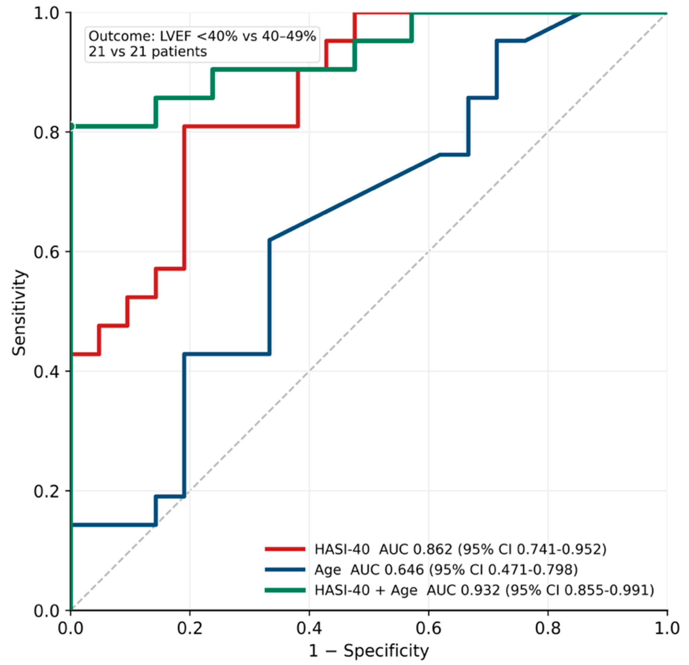
| Parameters | LVEF 40–49% Group n = 21 | LVEF < 40 Group n = 21 | p |
|---|---|---|---|
| Demographical: | |||
| Age (years) (median (Q1–Q3)) | 66 (50–69) | 68 (63–74) | 0.107 |
| Sex (male (%)) | 12 (57) | 14 (67) | 0.540 |
| BMI (kg/m2) (median (Q1–Q3)) | 28.3 (27.2–31.1) | 26.9 (25.0–30.9) | 0.191 |
| Obesity (BMI > 30 kg/ms) (n (%)) | 8 (38) | 6 (29) | 0.528 |
| Clinical status (NYHA classification): | |||
| I/II (n (%)) | 12 (57) | 3 (14) | 0.009 |
| II (n (%)) | 9 (43) | 10 (48) | 1.000 |
| II/III (n (%)) | 0 (0) | 1 (5) | 1.000 |
| III (n (%)) | 0 (0) | 7 (33) | 0.008 |
| Co-morbidities: | |||
| Arterial hypertension (n (%)) | 15 (71) | 13 (62) | 0.506 |
| Diabetes mellitus type 2 (n (%)) | 4 (19) | 7 (33) | 0.302 |
| Dyslipidemia (n (%)) | 20 (95) | 17 (81) | 0.343 |
| COPD (n (%)) | 2 (10) | 1 (5) | 0.511 |
| Kidney dysfunction * (n (%)) | 6 (29) | 5 (24) | 0.740 |
| Family CVD history (n (%)) | 3 (13) | 6 (29) | 0.137 |
| Therapy: | |||
| B-blockers (n (%)) | 13 (62) | 8 (38) | 0.131 |
| ACE-I (n (%)) | 9 (43) | 6 (29) | 0.348 |
| ARB (n (%)) | 4 (19) | 2 (10) | 0.395 |
| Statins (n (%)) | 15 (71) | 13 (62) | 0.528 |
| Metformin (n (%)) | 4 (19) | 7 (33) | 0.302 |
| SGLT2-i (n (%)) | 7 (33) | 7 (33) | 1.000 |
| Loop diuretics (n (%)) | 12 (57) | 14 (67) | 0.760 |
| MRA (n (%)) | 12 (57) | 14 (67) | 0.760 |
| Parameters | LVEF 40–49% Group n = 21 | LVEF < 40% Group n = 21 | p |
|---|---|---|---|
| Laboratory results | |||
| Hematocrit (median (Q1–Q3)) | 43 (40–45) | 44 (41–45) | 0.293 |
| Glomerular filtration rate (GFR) (mL/kg/min) (median (Q1–Q3)) | 73 (64–81) | 70 (63–74) | 0.531 |
| ALT (IU/L) (median (Q1–Q3)) | 40 (24–55) | 26 (21–38) | 0.106 |
| BNP (pg/mL) (median (Q1–Q3)) | 402 (304–470) | 787 (337–978) | 0.222 |
| Uric acid (μmol/L)(median (Q1–Q3)) | 329 (294–379) | 338 (217–363) | 0.661 |
| Lipoprotein (a) (mg/dL) (median (Q1–Q3)) | 2.4 (2.1–2.9) | 2.6 (1.4–4.8) | 0.915 |
| Total cholesterol (mmol/L) (median (Q1–Q3)) | 4.8 (3.6–5.7) | 4.3 (3.3–5.6) | 0.871 |
| Echocardiography | |||
| Transthoracic echocardiography results: | |||
| LVEDD (mm) (median (Q1–Q3)) | 50 (45–52) | 54 (50–57) | 0.053 |
| IVS (mm) (median (Q1–Q3)) | 11 (10–12) | 10 (9–12) | 0.447 |
| LA (mm) (median (Q1–Q3)) | 37 (35–41) | 42 (39–46) | 0.065 |
| MItral insufficiency moderate (n (%)) | 2 (10) | 4 (19) | 0.395 |
| LVEF (mm) (median (Q1–Q3)) | 45 (42–46) | 35 (30–38) | <0.001 |
| Coronary angiography results | |||
| Coronary angiography proven significant disease: | |||
| 1—vessel disease (n (%)) | 4 (19) | 4 (19) | 1.000 |
| 2—vessel disease (n (%)) | 1 (5) | 3 (14) | 0.311 |
| 3—vessel disease (n (%)) | 5 (24) | 1 (5) | 0.085 |
| Amino Acids (μmol/L) (Median (Q1–Q3)) | LVEF 40–49% Group n = 21 | LVEF < 40% Group n = 21 | p | FDR q |
|---|---|---|---|---|
| Proteinogenic Amino Acids: | ||||
| Essential: | ||||
| Histidine | 95.6 (87.9–102.6) | 90.7 (81.5–109.8) | 0.435 | 0.639 |
| Isoleucine | 61.6 (57.4–68.8) | 58.3 (52.8–62.4) | 0.724 | 0.839 |
| Leucine | 145 (136–161) | 141 (119–150) | 0.132 | 0.412 |
| Lysine | 202 (161–217) | 199 (172–228) | 0.715 | 0.839 |
| Phenylalanine | 74.8 (68.9–87.3) | 73.2 (61.6–88.0) | 0.132 | 0.412 |
| Methionine | 26.8 (23.6–28.2) | 25.3 (19.7–27.1) | 0.035 * | 0.268 |
| Threonine | 117 (99–140) | 107 (95–123) | 0.187 | 0.422 |
| Tryptophan | 64.3 (59.0–68.3) | 53.0 (45.8–61.3) | 0.061 | 0.268 |
| Valine | 253 (239–297) | 246 (230–264) | 0.220 | 0.422 |
| Non-essential/conditionally essential: | ||||
| Alanine | 417 (376–443) | 412 (352–465) | 0.687 | 0.839 |
| Arginine | 83.7 (76.5–97.1) | 83.2 (74.3–97.5) | 0.980 | 0.980 |
| Asparagine | 47.8 (45.0–52.2) | 46.1 (42.1–50.4) | 0.320 | 0.513 |
| Aspartic acid | 25.6 (20.6–35.3) | 20.5 (17.1–29.0) | 0.187 | 0.422 |
| Cystine | 56.4 (49.1–65.7) | 56.2 (50.9–66.8) | 0.980 | 0.980 |
| Glutamine | 541 (503–595) | 612 (572–661) | 0.131 | 0.412 |
| Glycine | 250 (209–275) | 230 (207–281) | 0.881 | 0.968 |
| Proline | 192 (171–221) | 179 (168–196) | 0.222 | 0.422 |
| Tyrosine | 70.7 (65.5–84.1) | 62.3 (53.9–75.9) | 0.032 * | 0.238 |
| One-carbon metabolism: | ||||
| Sarcosine | 1.7 (1.4–2.1) | 1.2 (1.0–1.5) | 0.004 * | 0.080 |
| Glycine | 250 (209–275) | 230 (207–281) | 0.881 | 0.968 |
| Serine | 147 (130–161) | 123 (108–146) | 0.012 * | 0.135 |
| Methionine | 26.8 (23.6–28.2) | 25.3 (19.7–27.1) | 0.053 | 0.268 |
| Taurine | 161 (143–176) | 116 (99–152) | 0.018 * | 0.120 |
| Muscle catabolism: | ||||
| 3-Methylhistidine (3-MH) | 12.2 (7.0–22.2) | 14.1 (7.9–29.1) | 0.725 | 0.839 |
| Beta-Alanine | 3.0 (2.5–3.9) | 2.9 (2.4–3.5) | 0.614 | 0.839 |
| Glutamine | 541 (503–595) | 612 (572–661) | 0.131 | 0.412 |
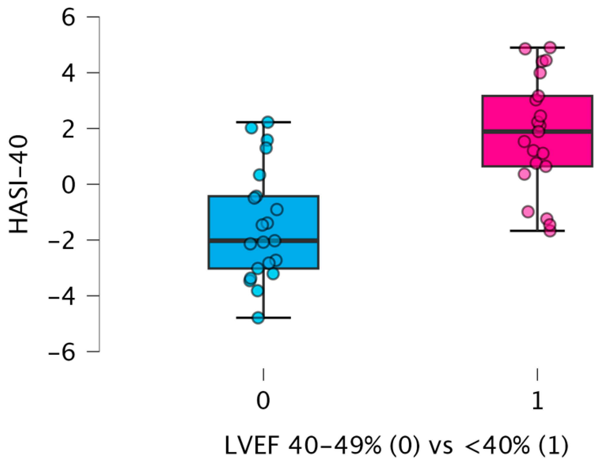
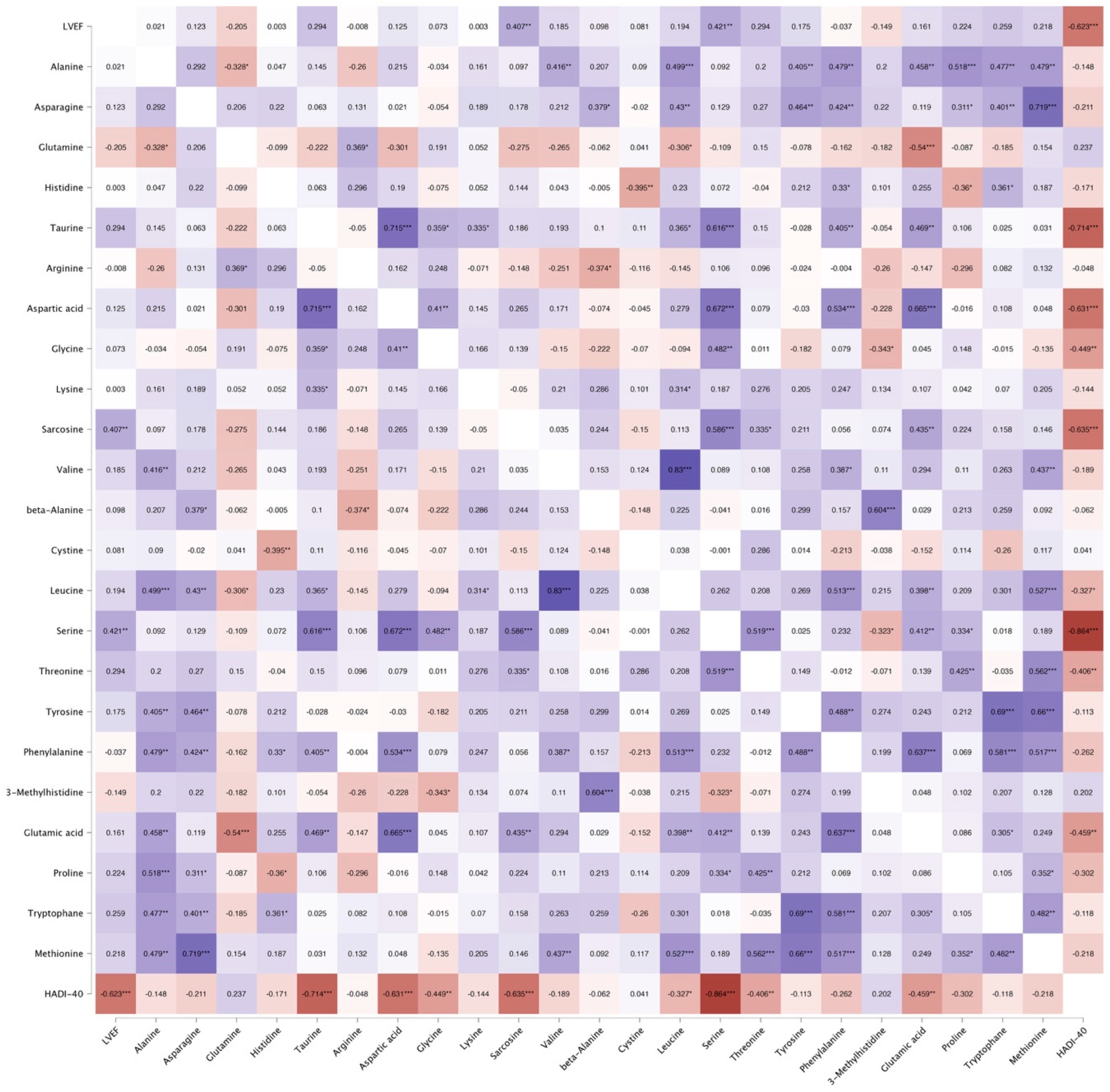
| HASI-40 index (median (Q1–Q3)) | −2.025 (−3.021–−0.429) | 1.890 (0.642–3.166) | <0.001 | 0.011 |
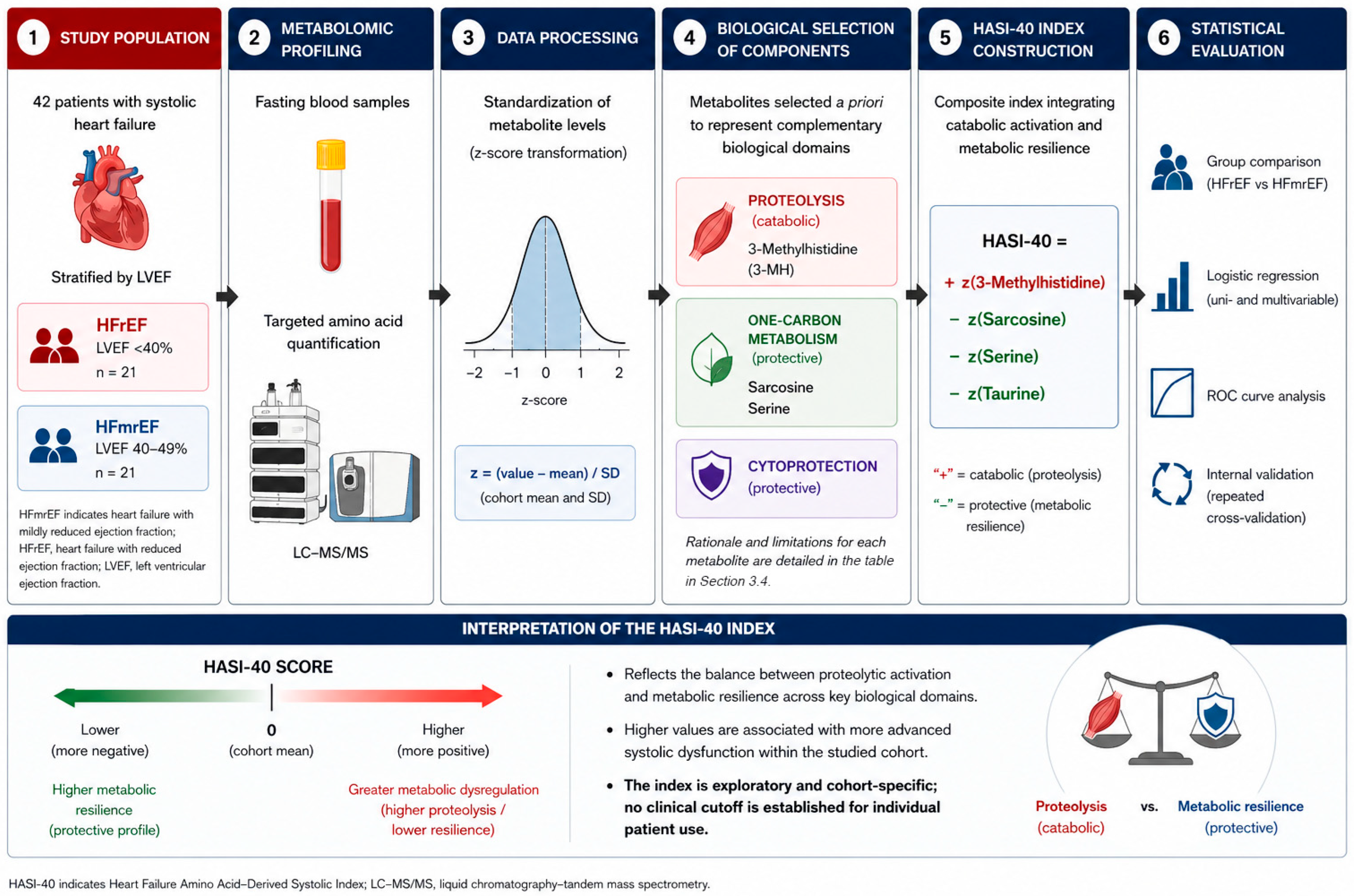
| Metabolite | Biological Domain | Direction in Index | Reason for Inclusion | Limitation |
|---|---|---|---|---|
| 3-Methylhistidine (3-MH) | Muscle proteolysis/catabolic activity | Positive (+) | Established marker of myofibrillar protein breakdown; reflects systemic catabolic activation observed in advanced heart failure | Not specific to heart failure; influenced by muscle mass, diet (meat intake), and protein-energy malnutrition |
| Sarcosine | One-carbon metabolism/methylation flux | Negative (−) | Intermediate of glycine–serine metabolism; reflects methylation capacity and mitochondrial function | Levels may be influenced by renal function and folate metabolism; not specific to cardiac pathology |
| Serine | One-carbon metabolism/cytoprotective metabolism | Negative (−) | Central substrate for nucleotide synthesis, redox balance, and mitochondrial pathways; reduced levels suggest impaired metabolic resilience | Broad metabolic role reduces specificity; influenced by nutritional status and systemic illness |
| Taurine | Cytoprotective buffering/mitochondrial stability | Negative (−) | Involved in calcium handling, antioxidant defense, and membrane stabilization; depletion linked to impaired cellular protection | Affected by diet, renal handling, and comorbidities; not unique to heart failure |
Disclaimer/Publisher’s Note: The statements, opinions and data contained in all publications are solely those of the individual author(s) and contributor(s) and not of MDPI and/or the editor(s). MDPI and/or the editor(s) disclaim responsibility for any injury to people or property resulting from any ideas, methods, instructions or products referred to in the content. |
© 2026 by the authors. Licensee MDPI, Basel, Switzerland. This article is an open access article distributed under the terms and conditions of the Creative Commons Attribution (CC BY) license.
Share and Cite
Krasińska, B.; Spasenenko, I.; Pietkiewicz, D.; Plewa, S.; Filipiak, K.J.; Pawlaczyk-Gabriel, K.; Bartkowski, J.; Tykarski, A.; Krasiński, Z.; Matysiak, J.; et al. Amino Acid-Derived Metabolic Signature Across Stages of Systolic Dysfunction: Derivation and Internal Evaluation of the HASI (Heart Failure Amino Acid-Derived Systolic Index)—40 Index. Int. J. Mol. Sci. 2026, 27, 4459. https://doi.org/10.3390/ijms27104459
Krasińska B, Spasenenko I, Pietkiewicz D, Plewa S, Filipiak KJ, Pawlaczyk-Gabriel K, Bartkowski J, Tykarski A, Krasiński Z, Matysiak J, et al. Amino Acid-Derived Metabolic Signature Across Stages of Systolic Dysfunction: Derivation and Internal Evaluation of the HASI (Heart Failure Amino Acid-Derived Systolic Index)—40 Index. International Journal of Molecular Sciences. 2026; 27(10):4459. https://doi.org/10.3390/ijms27104459
Chicago/Turabian StyleKrasińska, Beata, Ievgen Spasenenko, Dagmara Pietkiewicz, Szymon Plewa, Krzysztof J. Filipiak, Katarzyna Pawlaczyk-Gabriel, Jarosław Bartkowski, Andrzej Tykarski, Zbigniew Krasiński, Jan Matysiak, and et al. 2026. "Amino Acid-Derived Metabolic Signature Across Stages of Systolic Dysfunction: Derivation and Internal Evaluation of the HASI (Heart Failure Amino Acid-Derived Systolic Index)—40 Index" International Journal of Molecular Sciences 27, no. 10: 4459. https://doi.org/10.3390/ijms27104459
APA StyleKrasińska, B., Spasenenko, I., Pietkiewicz, D., Plewa, S., Filipiak, K. J., Pawlaczyk-Gabriel, K., Bartkowski, J., Tykarski, A., Krasiński, Z., Matysiak, J., & Urbanowicz, T. (2026). Amino Acid-Derived Metabolic Signature Across Stages of Systolic Dysfunction: Derivation and Internal Evaluation of the HASI (Heart Failure Amino Acid-Derived Systolic Index)—40 Index. International Journal of Molecular Sciences, 27(10), 4459. https://doi.org/10.3390/ijms27104459