Review

The Protective and Regenerative Potential of Lactoferrin in Hair and Skin Health

by
Nicole Kaplan
1,* and
Giorgio Dell’Acqua
2
1
Helaina Inc., New York, NY 10010, USA
2
Dellacqua Consulting, Jersey City, NJ 07302, USA
*
Author to whom correspondence should be addressed.
Int. J. Mol. Sci. 2026, 27(10), 4451; https://doi.org/10.3390/ijms27104451
Submission received: 24 March 2026 / Revised: 11 May 2026 / Accepted: 12 May 2026 / Published: 15 May 2026
(This article belongs to the Section Biochemistry)

Abstract

Lactoferrin is a naturally occurring bioactive glycoprotein that is part of the body’s innate immune system and has essential roles in iron metabolism, microbial defense, inflammation regulation, and tissue repair. It supports the natural regulation of iron bioavailability in skin and hair follicles, helping to reduce excess free-iron-driven oxidative stress while preserving levels of necessary iron for cellular functions. Lactoferrin promotes cell regeneration by increasing proliferation across in vitro systems, stimulating wound healing in scratch assays, and boosting matrix production in fibroblast models. Lactoferrin can also modulate inflammatory signaling involved in skin and hair physiology by providing balanced cytokine suppression, suggesting potential value in cosmetic or dermatological applications. Here, we present the first focused summary of lactoferrin’s role specifically in skin and hair biology, distinguished from prior reviews in systemic or multi-system broad health contexts. We link mechanistic insights with clinical and preclinical evidence and uniquely map molecular functions to dermatologic and trichologic outcomes. We also provide an overview of clinical skin studies that have explored lactoferrin as a supportive agent in conditions such as acne, and highlight that, despite mechanistic plausibility, there are no existing available reports of well-controlled human clinical trials leveraging lactoferrin for hair-focused outcomes. In summary, we propose lactoferrin as not just an anti-inflammatory molecule, but also as a microenvironment stabilizer, and particularly relevant for hair and skin support as an alternative to pharmacological interventions. By addressing both established and underexplored applications, this review provides a translational framework for clinical development and provides a comprehensive rationale behind leveraging lactoferrin for hair and skin epithelial health.

1. Introduction

Lactoferrin is a multifunctional, iron-binding glycoprotein that has protective and tissue healing capacities throughout the life of an organism [1]. It is present in exocrine secretions—including milk, saliva, and tears—and is released during inflammation by neutrophils and synthesized by epithelial cells. Structurally, lactoferrin is an ~80 kDa protein belonging to the transferrin family, composed of N- and C-terminal lobes, and connected by a flexible linker. Lactoferrin’s capacity to fight oxidative stress, reduce inflammation, and promote immune balance has been reported in many studies [1,2,3]. The structural heterogeneity of the protein arises in part from its ability to be differentially modified by phosphorylation, acetylation, glycosylation, and other post-translational modifications [4]. This post-translational diversity enables lactoferrin to have different binding interactions, biological activities, and downstream effects. Thus, lactoferrin’s structural heterogeneity is instrumental in its diverse bioactivities and functional capabilities [1,4].
Lactoferrin’s structural and functional flexibility is further highlighted when lactoferrin is processed at sites of inflammation (specifically by neutrophil proteases) [5] or during digestion (by gastric enzymes and partially in the duodenum) [6], delivering functional peptides with different active domains. These peptides have been associated with immune regulation, anti-inflammatory, and antimicrobial properties [7,8], possibly modulating the gut microbiome [9] and exhibiting systemic activity to mitigate microbial infections [10].
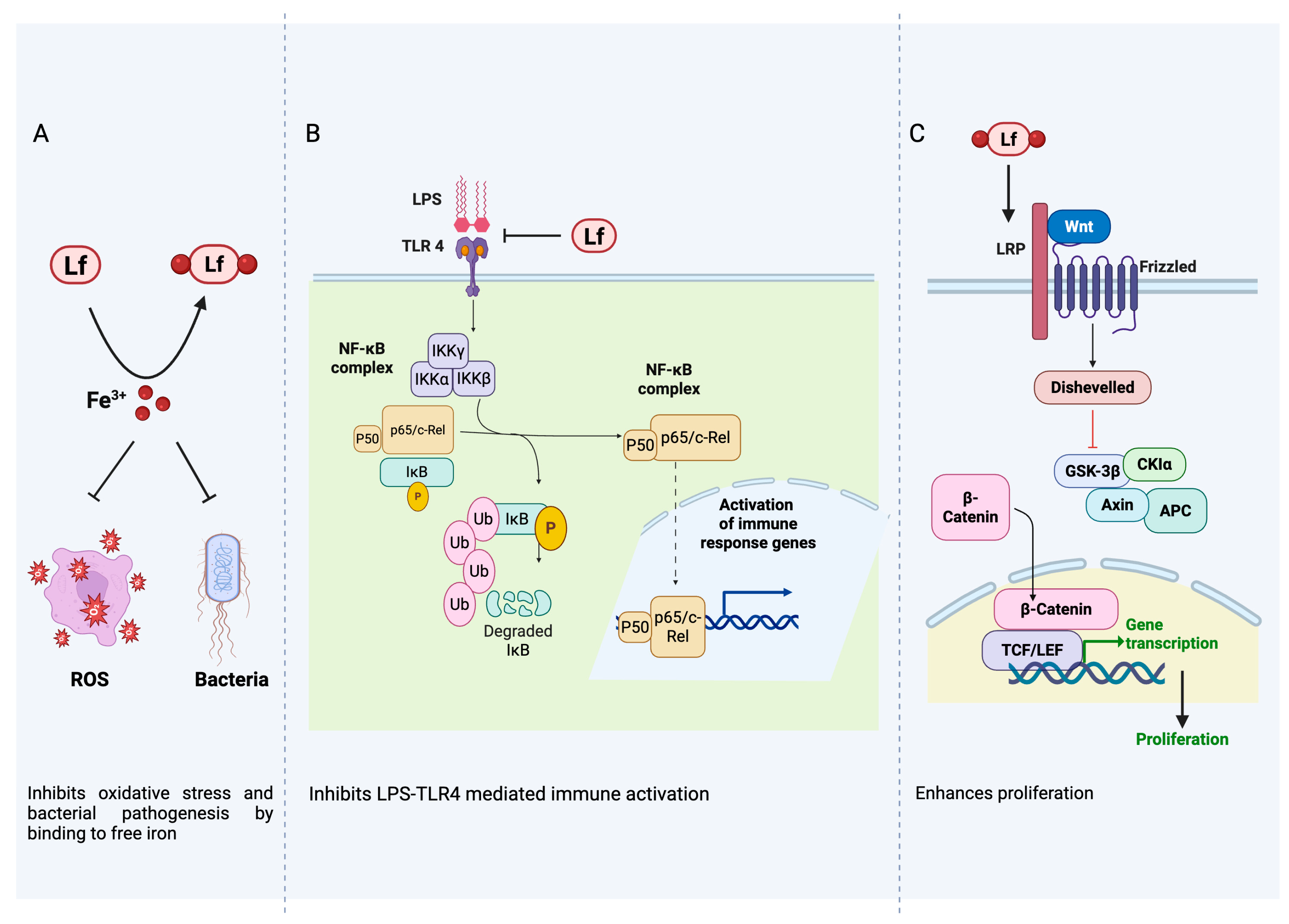
The chemistry of lactoferrin is complex, allowing lactoferrin to act multifunctionally, where different forms and structures of the protein may be more effective. Furthermore, the structural flexibility of lactoferrin enables multiple mechanisms of action (MoA) and engagement of various signaling pathways (Figure 1). Lactoferrin is well recognized for its strong binding capacity to free iron and its ability to reduce oxidative stress [1]. This property not only aids in limiting pathogen activity [11], but also plays an important role in regulating the activity of certain iron-dependent enzymes and proteins involved in protective and regenerative processes [12]. Lactoferrin’s regulation of iron availability also links host defense to oxidative stress control [13]. Free iron catalyzes the formation of reactive oxygen species (ROS) through Fenton chemistry, contributing to oxidative damage during infection and inflammation. By tightly binding free iron, lactoferrin reduces ROS generation and thereby protects cellular components from oxidative damage [14,15,16].
Lactoferrin exhibits context-dependent immunomodulatory activity, characterized by the attenuation of excessive pro-inflammatory signaling and the promotion of immune homeostasis. Lactoferrin and its peptides have been shown to reduce levels of pro-inflammatory cytokines such as TNF-α and IL-6 by suppressing the NF–κB signaling pathway, a central driver of pro-inflammatory responses [7,17]. In a clinical study, lactoferrin supplementation has been linked to a pronounced increase in circulating neutrophil precursors, and an associated reduction in TNF-α and IL-6 production [18]. Although the mechanisms described here are associated with direct immune system regulation, lactoferrin also works through biochemical and cellular mechanisms that make it relevant for protection against environmental stressors such as pathogens and UV radiation [19,20], making it particularly relevant for hair and skin applications. Thus, lactoferrin is not strictly an immune switch that controls activation or suppression per se, but rather is an immune and oxidative balancing factor with potential use in the cosmetics industry.
Previous reviews have discussed the general nutritional, antioxidant, and regulatory properties of lactoferrin across multiple systems and applications [21,22,23]. The present review specifically focuses on hair and skin as the primary biological targets. Its novelty lies in integrating both preclinical and clinical evidence related to dermatological and follicular outcomes, while evaluating lactoferrin within the context of skin and hair health applications.

2. The Role of Lactoferrin in Host Defense Against Infection

Lactoferrin’s role in host defense against infection represents its most fundamental biological function and provides a framework for its diverse activities. Its wide distribution in mucosal secretions, as well as its release at sites of infection and inflammation, enables it to contribute to first-line defense against microbial challenge [24,25]. Its antimicrobial activity is mediated through both iron-dependent and iron-independent mechanisms.
The primary antimicrobial function of lactoferrin is its high-affinity sequestration of ferric iron (Fe3+), an innate immune process described as nutritional immunity. Iron is a critical requirement for the growth of many microorganisms. By limiting iron availability, lactoferrin restricts microbial growth [24]. In addition to this mechanism, lactoferrin exerts antimicrobial effects through direct interactions with bacterial, viral, and fungal surfaces. It can bind to pathogen-associated molecular patterns such as lipopolysaccharide (LPS) in Gram-negative bacteria, destabilize microbial membranes, and interfere with pathogen adhesion and entry into host cells [24,26]. These activities extend across a broad spectrum of pathogens, including enteric bacteria, respiratory viruses, and opportunistic fungi, highlighting lactoferrin’s role as a non-specific but effective antimicrobial agent.
Beyond direct pathogen control, lactoferrin plays a critical role in shaping host immune responses by simultaneously controlling microbial burden and preventing excessive inflammation, which is essential for maintaining tissue integrity at sites of host–pathogen interaction [3,15]. Lactoferrin’s antioxidant activity complements its antimicrobial and immunomodulatory functions, reinforcing its role in maintaining tissue homeostasis [14].
At epithelial interfaces where microbial exposure is continuous, these functions are highly integrated. Lactoferrin contributes to barrier protection by modulating microbial communities, regulating immune responses, and supporting epithelial integrity [13,26,27]. This integrated role in host defense provides a mechanistic foundation for its broader applications in epithelial systems, including skin and hair.

3. Lactoferrin-Mediated Iron Regulation in Skin and Hair Biology

3.1. Iron Trafficking in the Skin and Hair Follicle Microenvironment

Immune cells, such as macrophages and activated Natural Killer (NK) cells, are recruited to injured, inflamed, or proliferating skin and hair follicles, where they regulate iron balance and metabolism [28,29,30]. Tissue macrophages regulate iron by recycling it, storing it in ferritin, and releasing it via the iron exporter ferroportin, depending on metabolic need [28,29,30]. Macrophage ferroportin-mediated iron export and release is essential for the proliferation of skin and follicle epithelial cells. A study in mice demonstrated that when ferroportin is knocked out specifically in macrophages, the animals failed to develop hair, highlighting the importance of localized iron trafficking in the hair follicle and the need for balanced iron control. Wound healing following skin damage was also reduced in these mice, suggesting that cellular repair and growth are both very iron-dependent, as healing was delayed when macrophages could not deliver iron [29].
An in vivo study in mice with transferrin deficiency demonstrated that iron is essential for skin development. The study reported that invariant Natural Killer T (iNKT) cells reside in the skin during early life and serve as a source of transferrin by upregulating genes encoding transferrin and transferrin receptor [30]. Upregulation of transferrin, an iron-regulating protein, supported hair growth by promoting the proliferation of hair follicle progenitor cells in these mice. These iNKT cells remain present in later life to support follicle homeostasis, highlighting the important role of iron regulation throughout the full life cycle of the hair follicle to support healthy hair growth [30].
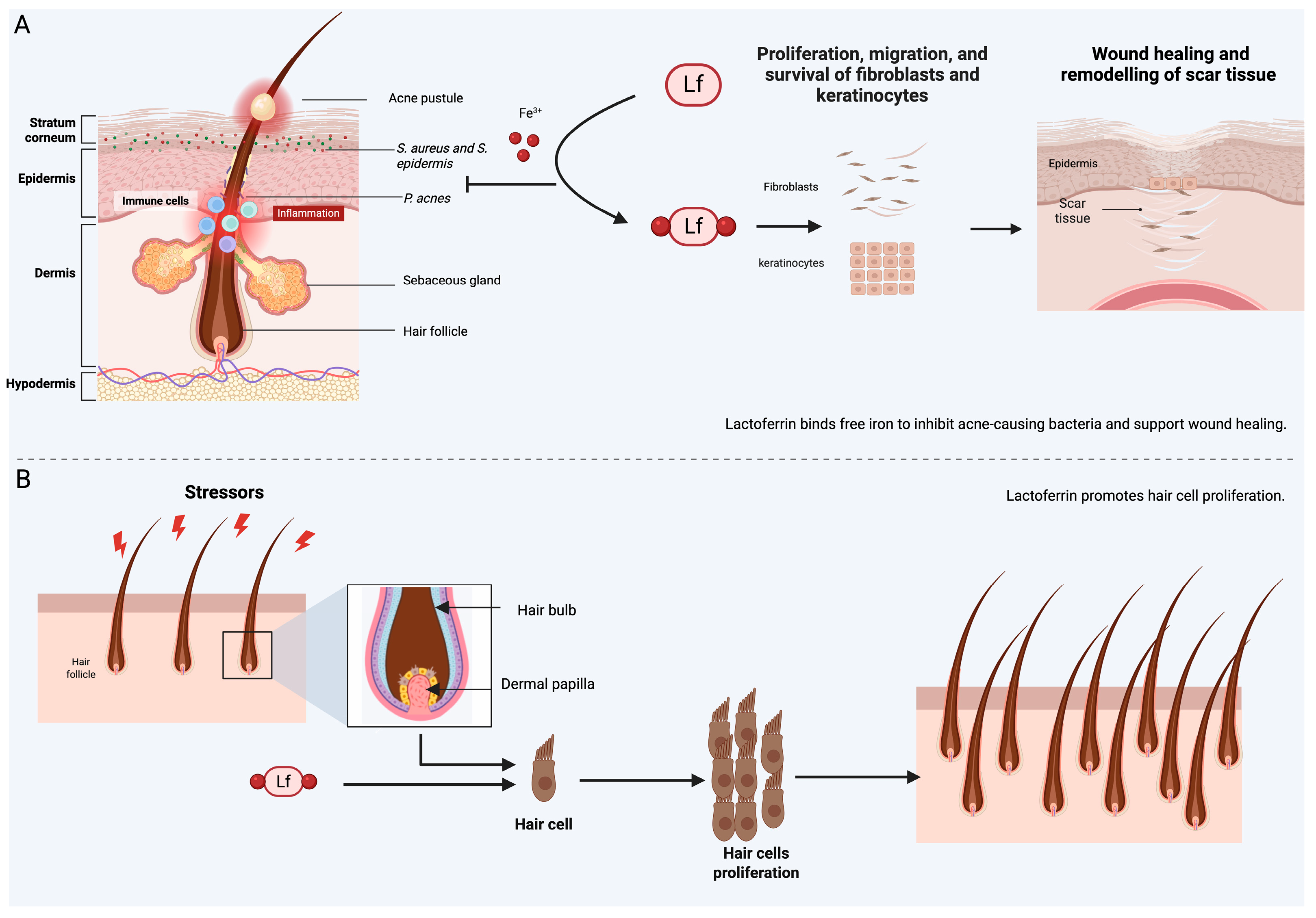
Iron is an essential co-factor for enzymes involved in DNA replication, DNA repair, [31] and collagen biosynthesis [32]. Iron metabolism in the cell is also essential for oxygenation and energy production; however, iron levels need to be controlled. When in excess, free iron can accelerate oxidative damage in the skin and contribute to skin aging, especially when combined with UVA exposure [33]. Reducing local iron availability in the hair follicular niche can reduce oxidative stress by shifting macrophages to a healing state and suppressing pro-inflammatory signaling [34]. Thus, it is not simply binding versus delivering iron that is biologically relevant but rather the regulated balance of iron (Figure 2). Here, we propose lactoferrin as a highly sophisticated molecule capable of regulating that balance.

3.2. Systemic Iron and Hair Loss

As discussed previously, iron serves as an essential cofactor for several enzymes involved in cell replication [35]. Ribonucleotide reductase, a rate-limiting enzyme in DNA synthesis, is one such enzyme that is highly dependent on iron for its activity [36]. In iron-deficient conditions, there is a reduction in the ribonucleotide reductase activity decreasing the DNA synthesis [37].
Ferritin is an iron storage protein, and serum ferritin reflects the iron levels in the body. Thus, serum ferritin is a standard indicator of iron status in hair loss studies. Hair loss is often observed in women with iron deficiency [38], and there also seems to be a link between low serum ferritin level and Telogen Effluvium (TE), a hair loss condition associated with stress [39,40]. However, the association between hair loss and low serum ferritin levels has been debated for many years, and there is no clear consensus on optimal ferritin levels to reduce hair loss, although a range of 40 to 70 ng/dL has been suggested [40].
Figure 2. Lactoferrin’s potential in hair and skin applications: (A) Lactoferrin (Lf), by binding and sequestering the free iron (Fe3+), limits iron availability for acne causing bacteria, also facilitates iron regulation to support the wound healing process by promoting fibroblast and keratinocyte proliferation; (B) Lactoferrin promotes the proliferation of hair cells by activating signaling pathways within dermal papilla cells within the hair bulb, enhancing cellular regeneration. Created with BioRender.com.
Ijms 27 04451 g002
While the data linking ferritin levels and hair loss are not entirely consistent [39], a recent study on individuals with TE found a significant correlation between TE and low iron levels [41]. This study showed that individuals taking lactoferrin experienced less TE and less hair loss, suggesting a potential link between systemic lactoferrin intake and protection against stress-induced hair loss [41].
While age-related hair loss is chronic and progressive, stress-related disruptions in hair cycling are typically acute and potentially reversible. Aging-associated hair loss is driven in part by inflammaging, cumulative oxidative stress, and impaired iron homeostasis [42,43], whereas stress-related shedding is characterized by transient cytokine surges and oxidative imbalance that may resolve if the follicular environment recovers [44]. Although the downstream pathways underlying these conditions differ, both share upstream contributors, including oxidative stress and inflammation [42,44] or other citations…?). Lactoferrin acts at these shared nodes through modulation of iron availability, redox balance, inflammatory signaling, and cell survival [3,16,25], suggesting a role in regulating the follicular microenvironment across distinct hair loss phenotypes. By targeting these upstream processes, lactoferrin may address both chronic (age-related) and acute (stress-induced) forms of hair loss through systems-level normalization rather than single-pathway activation, warranting further investigation in hair-focused clinical trials [15].

3.3. Lactoferrin as an Iron-Binding Protein to Target Iron-Dependent Conditions in Hair and Skin

Lactoferrin, as the name suggests, is an iron-binding protein with a hundred times more affinity to iron than transferrin [45]. Lactoferrin has two lobes, and each lobe of the protein can bind to an iron ion [46]. The capacity of lactoferrin to reversibly bind iron makes this protein key for regulating iron homeostasis in the body, but it also helps prevent iron-dependent pathogens from proliferating [11].
In vitro studies suggest that iron chelators show potential as an intervention strategy in hypoxic and inflammatory microenvironments in the human hair follicle and skin. By reducing excess free iron—an element implicated in hair loss—iron chelators may stimulate hair growth and rejuvenation [47]. In low oxygen conditions, Hypoxia-Inducible Factor-1 (HIF-1) is activated, promoting tissue formation, cell proliferation, and migration. During wound healing, HIF-1 activates processes such as neovascularization, collagen, and elastin production [47,48].
Hair conditions such as Androgenetic Alopecia (AGA) have been linked to reduced oxygenation and vascularization due to a lack of nutrients reaching the hair bulb, where HIF-1 expression was strongly reduced in AGA patients [49]. Studies conducted in hair cells reported that subsequent activation of HIF-1 can stimulate gene activation responsible for cellular growth [49,50]. Indeed, the HIF-1 signaling pathway has been recently identified as one of the main growth-stimulating pathways in the human follicle, together with other signaling pathways [50].
Minoxidil, commonly used to stimulate hair growth, has been shown to inhibit prolyl-hydroxylase-2 (PHD-2), an enzyme responsible for the degradation of HIF-1 through hydroxylation [51]. Inhibition of PHD-2 can therefore activate HIF-1-dependent transcription of genes such as Vascular Endothelial Growth Factor-1 (VEGF-1), crucial for neovascularization and growth during the anagen phase of the hair follicle [52].
Since PHD-2 is highly dependent on iron as a cofactor, iron chelating agents can effectively inhibit PHD-2-dependent degradation of HIF-1 [34]. Thor D et al. [53] tested the above hypothesis in a clinical study using a topically administered iron chelator. After nine months, hair thickness, density and shine improved significantly compared to baseline. Hair shedding was reduced by 66.8% as early as 6 months [53]. The authors followed up with an additional review suggesting that iron chelators have a potential benefit in autoimmune hair conditions such as alopecia areata (AA) when used as an adjuvant [34]. In fact, iron chelators, by creating a mimetic hypoxic environment, seem to stabilize HIF-1 and at the same time stimulate macrophage polarization and cytokine expression. Reduction in intracellular iron can therefore shift macrophages toward an anti-inflammatory M2 phenotype and suppress NF–κB signaling, with consequent reduction in TNF-α and IL-15, key cytokines involved in autoimmune follicular degeneration [34].
Unlike iron chelators such as Deferoxamine (DFO), which primarily functions through direct depletion of bioavailable ferric iron [54], lactoferrin more adaptively regulates iron availability within inflammatory and epithelial environments. Through its reversible iron-binding capacity, lactoferrin could be leveraged as a naturally occurring iron-regulatory protein to support hair loss [34,53] and skin health [47,48].

4. Lactoferrin as a Protective and Regenerative Factor in Hair and Skin: Evidence from Cellular and Animal Models

Beyond its iron-dependent mechanisms, lactoferrin activates distinct signaling pathways that drive protective and regenerative responses. In vitro studies demonstrate that lactoferrin, in its various forms, can interact with tissue-specific receptors to induce different physiological responses, including stress protection, wound healing, cellular migration, and regeneration [55,56,57,58].
Stem cell populations within the skin are central to tissue maintenance, regeneration, and the cell cycle. The hair follicle contains multiple stem cell niches, including epithelial, melanocyte, and mesenchymal compartments, that regulate hair growth, pigmentation, and follicular remodeling [59]. Among these, mesenchymal stem cells (MSCs) contribute to follicular inductivity and pigmentation by supporting epithelial–mesenchymal signaling networks essential for hair cycling [60,61]. In parallel, dermal MSCs are involved in cutaneous repair and promote wound healing, extracellular matrix remodeling, and attenuation of inflammation in skin lesions [62,63].
A key feature of these stem cell populations is their sensitivity to redox balance. Hair-follicle-derived MSCs exhibit pronounced susceptibility to oxidative injury, with ROS accumulation driving apoptosis and functional decline [64]. This vulnerability becomes especially relevant in translational contexts, where ex vivo expansion and transplantation expose MSCs to oxidative environments that can significantly diminish their functional utility in clinical applications. Together, these observations highlight redox regulation as a key determinant of stem cell function in skin and hair biology and show that antioxidant systems are critical modulators of regenerative outcomes. Given lactoferrin’s antioxidant and regenerative potential, here we review specific in vitro studies that are important for predicting its potential clinical benefits.
An in vitro study has shown that lactoferrin is able to protect MSCs from oxidative-stress-induced cellular senescence (aging) and programmed cell death (apoptosis) [56]. Lactoferrin was able to significantly reduce the production of intracellular ROS in hMSCs induced by hydrogen peroxide exposure, restore growth, and promote self-renewal. Mechanistically, this was linked to inhibition of caspase-3 activation, a key enzyme driving apoptosis, and modulation of Akt signaling, involved in cell survival pathways [56]. These data highlight the capacity of lactoferrin as an antioxidant in mesenchymal cells, helping to limit ROS-induced damage at the cellular level. This positions lactoferrin as a protective and anti-senescence agent in contexts where oxidative stress limits regenerative capacity, such as in hair and skin development and repair.
Essential to our understanding of hair growth and repair are studies in dermal papilla (DP) cells, specialized mesenchymal cells that sit at the control center of the hair follicle. DP cells have regenerative properties, are associated with stem cells, and are critical for hair development and hair cycling [65]. They are often used in experimental settings to study the molecular basis of hair growth interventions [66]. One such study focused on colostrum-derived exosomes containing lactoferrin and proposed them as a cell-free regenerative factor that can increase DP cell proliferation and stimulate cell cycle progression [67]. A key biological switch controlling hair regeneration is the transition of the hair follicle from the telogen (resting) phase into the anagen (growth) phase. In mice, these exosomes were able to accelerate the transition from the telogen to the anagen phase via strong activation of the Wnt/β-catenin signaling pathway [67]. Thus, this cell-free system triggered a key regenerative hair switch in vivo, acting through the Wnt pathway to regulate regeneration.
Another study showed that lactoferrin can significantly increase the proliferation of cultured DP cells via modulation of growth-factor signaling [57]. Treatment with bovine lactoferrin (bLF) enhanced the phosphorylation of Erk1/2 and Akt in DP cells, important factors for survival and proliferation. Lactoferrin also induced the expression of several Wnt signaling-related components, including Wnt3a, Wnt7a, Lymphoid Enhancer-Binding Factor 1 (Lef1), and β-catenin [57]. This pathway plays a central role in hair follicle development, induction and cycling. Furthermore, topical application of lactoferrin in shaved mice significantly increased hair growth compared to controls, suggesting that bLF’s proliferative effects on DP cells in vitro can translate into enhanced hair growth potential in vivo [57].
Lactoferrin’s potential in cosmetic and skin care applications has been explored in skin keratinocytes and reconstructed epidermal models. Using a recombinant human lactoferrin from transgenic rice, research has shown that lactoferrin can activate growth and survival pathways in keratinocytes through the ERK/MAPK signaling pathway, mediated by the Low-Density Lipoprotein Receptor-Related Protein 1 (LRP-1) receptor [58]. Lactoferrin increased keratinocyte proliferation and motility, enhanced migration-associated cytoskeletal reorganization, and inhibited cell apoptosis. In a wound healing in vivo swine model, the protein promoted wound re-epithelialization by accelerating epidermal closure and overall wound healing rate following second-degree burn when applied topically [58]. These studies suggest that lactoferrin possesses healing and regeneration capabilities that can be leveraged in skin aging.
Recent work by Xie and colleagues using a reconstructed human epidermis (RHE) model provides compelling evidence for lactoferrin’s anti-inflammatory and barrier-supportive activity in epidermal tissue. In this three-dimensional keratinocyte-based system, lactoferrin reduced secretion of the pro-inflammatory markers IL-1α and Thymic Stromal Lymphopoietin (TSLP), while simultaneously improving the skin barrier as measured by increasing transepithelial electric resistance (TEER). Lactoferrin also upregulated skin barrier- and hydration-associated proteins, including involucrin, filaggrin, claudin-1, aquaporin-3, and hyaluronan synthase-1 [55]. These findings suggest that lactoferrin not only suppresses inflammatory signaling but also promotes barrier maturation within a physiologically relevant 3D skin model.
Studies across multiple epithelial cell types have consistently demonstrated lactoferrin’s ability to suppress inflammatory signaling and cytokine production. In intestinal epithelial models, lactoferrin treatment significantly reduced pro-inflammatory cytokine expression, including IL-1β, IL-6, TNF-α, and IL-8, while simultaneously downregulating NK-kB phosphorylation associated with MAPK signaling pathways [68,69,70]. A study in primary intestinal epithelial cells further revealed that lactoferrin’s anti-inflammatory effects involve modulation of the ELAVL1/PI3K/NF–κB pathway, with iron-free lactoferrin showing potent activity in reducing LPS-induced inflammatory responses [71]. Similar suppressive effects have been observed in bovine mammary epithelial cells, where lactoferrin pretreatment inhibited TLR4-mediated NF–κB activation and reduced expression of key inflammatory mediators [72]. Collectively, these studies establish lactoferrin as a key regulator of epithelial immune responses.
Unlike corticosteroids, which broadly suppress inflammatory gene transcription through glucocorticoid receptor signaling [73], lactoferrin is a systems-level microenvironment regulator rather than a generalized immunosuppressive agent. A comprehensive meta-analysis of lactoferrin studies demonstrated significant inhibitory effects on both upstream inflammatory cytokines and downstream NF–κB signaling across multiple epithelial models [7]. Contrary to general anti-inflammatory agents, lactoferrin exerts more moderate context-dependent immunomodulatory effects while maintaining tissue homeostasis at host-environment interfaces. Overall, lactoferrin functions as a key regulator of the epithelial microenvironment, coordinating antimicrobial defense, inflammatory balance, and oxidative stress control.

5. Lactoferrin Clinical Applications in Dermatology

There are limited studies currently exploring lactoferrin as an oral or topical formulation. No registered clinical trials have evaluated lactoferrin for hair-related conditions. However, evidence from in vitro and in vivo models suggests that lactoferrin may have the potential to mitigate hair loss or graying [57,74,75]. By contrast, clinical evidence exists exploring oral or topical lactoferrin in the context of acne and skin wound healing, with emerging data on broader inflammatory skin responses [76]. Studies conducted between 2010 and 2022 have reported that lactoferrin, when administered orally, can significantly reduce acne severity (Table 1) [77,78,79,80]. Results are indicated in the table below.
Collectively, these clinical studies suggest that lactoferrin can reach the skin target, modulate inflammation, and exert clinically measurable effects within epithelial tissues following oral administration. This supports lactoferrin’s potential as a supportive agent in the field of dermatology.

6. Future Directions

Current and past research in dermatology and cosmetics using lactoferrin has primarily focused on cell lines, animal models, and, in one case, reconstructed skin [55]. Limited clinical studies exist (on skin applications only), and ex vivo studies using explanted hair follicles or human skin are currently lacking.
Ex vivo explants represent the closest model to clinical testing and provide invaluable insights into optimal safe dosage, ensuring no toxicity, and accurate MoA. These data are empirical prior to initiating clinical trials, where MoA and dosage determine inclusion criteria, treatment regimens, and relevant endpoints. In addition, explants are particularly valuable in cosmetic and dermatology research, where increasing regulatory limitations on preclinical animal testing make alternative methods essential.
Ex vivo models have been previously established and validated in the literature for both hair follicles [81] and skin explants [82,83], providing reliable platforms for studying follicular and dermatological processes. Building on this established foundation, we propose to leverage these well-characterized systems to evaluate lactoferrin’s potential in hair and skin health. Studies would be conducted in specialized laboratories with access to clinical samples and established expertise in hair follicular and skin biology. The experimental approach would compare lactoferrin, with or without its peptide derivatives, using hypoxic versus normoxic skin and hair follicle explants. This design would help to identify safe and optimal dosing parameters ahead of clinical trials, while better defining lactoferrin’s MoA.
Following ex vivo validation, subsequent clinical studies could be conducted in a defined target population over a predetermined time frame, with endpoints tailored to the specific research objectives. The formulation vehicle for lactoferrin, whether topical or ingestible, would be selected and optimized for each treatment approach. A double-blind, controlled trial design would be preferred, particularly for studies involving oral lactoferrin administration.
One current limitation is the relatively small number of well-controlled clinical studies aimed at defining optimal effective dosing strategies for lactoferrin in distinct applications, such as skin health or hair support. Future studies focusing on evaluating comprehensive dose–response characterization in humans, along with standardized dosing regimens and long-term outcomes, will be important to better understand lactoferrin as a dietary supplement for hair and skin applications.

7. Conclusions on Lactoferrin in Hair and Skin Regeneration

This review provides an in-depth discussion about the role of lactoferrin and similar molecules in skin and hair applications and explores their potential use in nutraceutical formulations, with a special focus on hair growth and skin regeneration. Advances in recombinant protein production, including precision fermentation-derived human lactoferrin, further expand its potential as a scalable and biologically relevant active ingredient. Distinct from drug-like agents, lactoferrin is an endogenously produced balanced regulator of iron homeostasis, inflammation, and epithelial regeneration. Lactoferrin is not the most potent agent in any single category, but based on its mechanism of action, it can deliver coordinated multi-pathway improvements with potential to collectively outperform single-mechanism agents in complex epithelial environments.
Due to its high iron-binding capacity, lactoferrin and its derivatives can modulate iron levels in the extracellular environment. It can limit pathogen growth and protect from oxidative stress by sequestering excess free iron, and depending on the local microenvironment, it can facilitate the controlled delivery of iron to the cells required for metabolism and proliferation. This dual capacity of lactoferrin makes it an ideal candidate for skin and hair applications.
In several experimental models, lactoferrin and its derivatives have demonstrated the ability to protect against oxidative stress through both direct (ROS scavenging and chelation) and indirect (receptor binding) MoA (Figure 1). They also have the capacity to reduce pro-inflammatory molecules involved in chronic low-grade inflammation (“inflammaging”) and auto-immune responses. Additionally, lactoferrin exhibits regenerative properties, either by directly binding to the LRP1 receptor or indirectly through HIF-1 stabilization.
Ex vivo studies combined with targeted clinical trials can establish lactoferrin as a safe and effective nutritional supplement for skin and hair applications. Available research demonstrates encouraging consistency across studies evaluating inflammatory skin conditions, particularly acne [77,78,79,80]. Across acne-focused clinical studies conducted between 2010 and 2022, oral lactoferrin supplementation was associated with improvements in inflammatory lesion counts, acne severity, sebum production, and overall skin appearance, with reporter reductions in inflammatory lesions ranging from approximately 20–40% in placebo-controlled settings. Although formulations, dosing strategies, and study designs varied, findings were directly consistent and collectively suggest that orally administered lactoferrin can exert biologically relevant effects in the skin.
In contrast, no registered clinical trials have specifically evaluated lactoferrin for hair-related conditions, despite emerging mechanistic and preclinical evidence supporting potential relevance in follicular biology, oxidative stress regulation, and inflammatory modulation. While direct clinical evidence in AGA and AA is currently lacking, the high vascularization and immunological activity of the hair follicle support its potential as a target for circulating lactoferrin, warranting further investigation.
Importantly, the observed improvements in acne outcomes support broader mechanistic concepts relevant to skin and hair biology, including modulation of inflammatory signaling, oxidative stress, microbial interactions, and epithelial homeostasis. Based on the current available preclinical and clinical literature, a daily dose of 100–200 mg lactoferrin is a reasonable recommended supplementation range supporting intended biological applications. However, as mentioned above, further dose–response and long-term clinical studies are needed to optimize dosing strategies according to specific health outcomes. In conclusion, lactoferrin represents a promising dietary supplement for hair and skin conditions, and future studies will provide a critical foundation for lactoferrin’s rational use in the fields of dermatology and cosmetics.

Author Contributions

Writing—original draft submission, G.D. and N.K.; writing—review and editing, N.K.; visualization—N.K.; revisions—N.K. All authors have read and agreed to the published version of the manuscript.

Funding

This research received no external funding.

Institutional Review Board Statement

Not applicable.

Informed Consent Statement

Not applicable.

Data Availability Statement

No new data were created or analyzed in this study. Data sharing is not applicable to this article.

Acknowledgments

The authors would like to thank Rajitha Gadde, Helaina Inc., New York, USA, for her assistance with figure editing and preparation, and for insightful discussions during the writing of this review. The authors also thank Ross Peterson, Helaina Inc., Colorado, USA, and Anthony Clark, Helaina Inc., New York, USA. The PubMed database was systematically searched in January 2026 for research studies on pre-clinical models, review articles, meta-analysis studies, clinical trials, and randomized controlled studies between 1975 and 2026. In PubMed, the MeSH terms used were “lactoferrin, hair loss, alopecia, acne vulgaris, in vitro, in vivo, pre-clinical models, and clinical trials”. Two independent investigators performed the search. The review is narrative in nature and not intended to be a meta-analysis or systematic review.

Conflicts of Interest

Nicole Kaplan is an employee of Helaina Inc.; New York, NY, USA. Giorgio Dell’Acqua is a consultant of Helaina Inc., New York, NY, USA.

Abbreviations

The following abbreviations are used in this manuscript:
AAAlopecia Areata
AGAAndrogenic alopecia
AKTv-akt murine thymoma viral oncogene homolog
DPDermal papilla
ERKExtracellular signal-regulated kinase
HIF-1Hypoxia- inducible factor-1
IL-6Interleukin 6
LRP1Low-Density Lipoprotein Receptor-Related Protein 1
MAPKMitogen-activated protein kinase
MoAMechanism of action
MSCMesenchymal stem cells
NFkBNuclear factor kappa light chain enhancer of activated B cells
PHD-2Prolyl-hydroxylase-2
ROSReactive oxygen species
TETelogen effluvium
TNFαTumor necrosis factor alpha
UVUltraviolet

References

  1. Kowalczyk, P.; Kaczyńska, K.; Kleczkowska, P.; Bukowska-Ośko, I.; Kramkowski, K.; Sulejczak, D. The Lactoferrin Phenomenon—A Miracle Molecule. Molecules 2022, 27, 2941. [Google Scholar] [CrossRef]
  2. Hao, L.; Shan, Q.; Wei, J.; Ma, F.; Sun, P. Lactoferrin: Major Physiological Functions and Applications. Curr. Protein Pept. Sci. 2019, 20, 139–144. [Google Scholar] [CrossRef]
  3. Actor, J.K.; Hwang, S.-A.; Kruzel, M.L. Lactoferrin as a Natural Immune Modulator. Curr. Pharm. Des. 2009, 15, 1956–1973. [Google Scholar] [CrossRef]
  4. Albar, A.; Almehdar, H.; Uversky, V.; Redwan, E. Structural Heterogeneity and Multifunctionality of Lactoferrin. Curr. Protein Pept. Sci. 2014, 15, 778–797. [Google Scholar] [CrossRef]
  5. Drago-Serrano, M.E.; Campos-Rodriguez, R.; Carrero, J.C.; De La Garza, M. Lactoferrin and Peptide-Derivatives: Antimicrobial Agents with Potential Use in Nonspecific Immunity Modulation. Curr. Pharm. Des. 2018, 24, 1067–1078. [Google Scholar] [CrossRef] [PubMed]
  6. Gifford, J.L.; Hunter, H.N.; Vogel, H.J. Lactoferricin: A Lactoferrin-Derived Peptide with Antimicrobial, Antiviral, Antitumor and Immunological Properties. Cell. Mol. Life Sci. 2005, 62, 2588–2598. [Google Scholar] [CrossRef] [PubMed]
  7. Yami, H.A.; Tahmoorespur, M.; Javadmanesh, A.; Tazarghi, A.; Sekhavati, M.H. The Immunomodulatory Effects of Lactoferrin and Its Derived Peptides on NF-κB Signaling Pathway: A Systematic Review and Meta-analysis. Immun. Inflamm. Dis. 2023, 11, e972. [Google Scholar] [CrossRef]
  8. Gruden, Š.; Poklar Ulrih, N. Diverse Mechanisms of Antimicrobial Activities of Lactoferrins, Lactoferricins, and Other Lactoferrin-Derived Peptides. Int. J. Mol. Sci. 2021, 22, 11264. [Google Scholar] [CrossRef]
  9. Baldi, A.; Ioannis, P.; Chiara, P.; Eleonora, F.; Roubini, C.; Vittorio, D. Biological Effects of Milk Proteins and Their Peptides with Emphasis on Those Related to the Gastrointestinal Ecosystem. J. Dairy Res. 2005, 72, 66–72. [Google Scholar] [CrossRef]
  10. Martin, L.; Van Meegern, A.; Doemming, S.; Schuerholz, T. Antimicrobial Peptides in Human Sepsis. Front. Immunol. 2015, 6, 404. [Google Scholar] [CrossRef] [PubMed]
  11. Arnold, R.R.; Brewer, M.; Gauthier, J.J. Bactericidal Activity of Human Lactoferrin: Sensitivity of a Variety of Microorganisms. Infect. Immun. 1980, 28, 893–898. [Google Scholar] [CrossRef]
  12. Zakharova, E.T.; Kostevich, V.A.; Sokolov, A.V.; Vasilyev, V.B. Human Apo-Lactoferrin as a Physiological Mimetic of Hypoxia Stabilizes Hypoxia-Inducible Factor-1 Alpha. BioMetals 2012, 25, 1247–1259. [Google Scholar] [CrossRef]
  13. Drago-Serrano, M.E.; Campos-Rodríguez, R.; Carrero, J.C.; De la Garza, M. Lactoferrin: Balancing Ups and Downs of Inflammation Due to Microbial Infections. Int. J. Mol. Sci. 2017, 18, 501. [Google Scholar] [CrossRef]
  14. Ward, P.P.; Conneely, O.M. Lactoferrin: Role in Iron Homeostasis and Host Defense against Microbial Infection. Biometals 2004, 17, 203–208. [Google Scholar] [CrossRef]
  15. Legrand, D. Overview of Lactoferrin as a Natural Immune Modulator. J. Pediatr. 2016, 173, S10–S15. [Google Scholar] [CrossRef] [PubMed]
  16. Kell, D.B.; Heyden, E.L.; Pretorius, E. The Biology of Lactoferrin, an Iron-Binding Protein That Can Help Defend against Viruses and Bacteria. Front. Immunol. 2020, 11, 550441. [Google Scholar] [CrossRef] [PubMed]
  17. Machnicki, M.; Zimecki, M.; Zagulskit, T. Lactoferrin Regulates the Release of Tumour Necrosis Factor Alpha and Interleukin 6 In Vivo. Int. J. Exp. Pathol. 1993, 74, 433. [Google Scholar]
  18. Zimecki, M.; Spiegel, K.; Właszczyk, A.; Kübler, A.; Kruzel, M.L. Lactoferrin Increases the Output of Neutrophil Precursors and Attenuates the Spontaneous Production of TNF-Alpha and IL-6 by Peripheral Blood Cells. Arch. Immunol. Ther. Exp. 1999, 47, 113–118. [Google Scholar]
  19. Yang, S.; Fang, F.; Yu, X.; Yang, C.; Zhang, X.; Wang, L.; Zhu, L.; Shao, K.; Zhu, T. Knockdown of H19 Inhibits the Pathogenesis of Acne Vulgaris by Targeting the miR-196a/TLR2/NF-κB Axis. Inflammation 2020, 43, 1936–1947. [Google Scholar] [CrossRef]
  20. Wang, Y.; Wang, L.; Wen, X.; Hao, D.; Zhang, N.; He, G.; Jiang, X. NF-κB Signaling in Skin Aging. Mech. Ageing Dev. 2019, 184, 111160. [Google Scholar] [CrossRef]
  21. Rascón-Cruz, Q.; Siqueiros-Cendón, T.S.; Siañez-Estrada, L.I.; Villaseñor-Rivera, C.M.; Ángel-Lerma, L.E.; Olivas-Espino, J.A.; León-Flores, D.B.; Espinoza-Sánchez, E.A.; Arévalo-Gallegos, S.; Iglesias-Figueroa, B.F. Antioxidant Potential of Lactoferrin and Its Protective Effect on Health: An Overview. Int. J. Mol. Sci. 2024, 26, 125. [Google Scholar] [CrossRef]
  22. Jańczuk, A.; Brodziak, A.; Czernecki, T.; Król, J. Lactoferrin—The Health-Promoting Properties and Contemporary Application with Genetic Aspects. Foods 2022, 12, 70. [Google Scholar] [CrossRef] [PubMed]
  23. Brouwer, C.; Welling, M.M.; Alwasel, S.; Boekhout, T. Potential Health Benefits of Lactoferrin and Derived Peptides—How to Qualify as a Medical Device? Crit. Rev. Microbiol. 2025, 51, 1041–1065. [Google Scholar] [CrossRef] [PubMed]
  24. Valenti, P.; Antonini, G. Lactoferrin: Lactoferrin: An Important Host Defence against Microbial and Viral Attack. Cell. Mol. Life Sci. 2005, 62, 2576–2587. [Google Scholar] [CrossRef]
  25. Legrand, D.; Elass, E.; Carpentier, M.; Mazurier, J. Lactoferrin: Lactoferrin: A Modulator of Immune and Inflammatory Responses. Cell. Mol. Life Sci. 2005, 62, 2549. [Google Scholar] [CrossRef]
  26. Ochoa, T.; Cleary, T. Effect of Lactoferrin on Enteric Pathogens. Biochimie 2009, 91, 30–34. [Google Scholar] [CrossRef] [PubMed]
  27. Lönnerdal, B. Nutritional Roles of Lactoferrin. Curr. Opin. Clin. Nutr. Metab. Care 2009, 12, 293–297. [Google Scholar] [CrossRef]
  28. Sukhbaatar, N.; Weichhart, T. Iron Regulation: Macrophages in Control. Pharmaceuticals 2018, 11, 137. [Google Scholar] [CrossRef]
  29. Recalcati, S.; Gammella, E.; Buratti, P.; Doni, A.; Anselmo, A.; Locati, M.; Cairo, G. Macrophage Ferroportin Is Essential for Stromal Cell Proliferation in Wound Healing. Haematologica 2019, 104, 47–58. [Google Scholar] [CrossRef]
  30. Wang, W.-B.; Lin, Y.-D.; Zhao, L.; Liao, C.; Zhang, Y.; Davila, M.; Sun, J.; Chen, Y.; Xiong, N. Developmentally Programmed Early-Age Skin Localization of iNKT Cells Supports Local Tissue Development and Homeostasis. Nat. Immunol. 2023, 24, 225–238. [Google Scholar] [CrossRef]
  31. Zhang, C. Essential Functions of Iron-Requiring Proteins in DNA Replication, Repair and Cell Cycle Control. Protein Cell 2014, 5, 750–760. [Google Scholar] [CrossRef]
  32. Hutton, J.J.; Tappel, A.L.; Udenfriend, S. Cofactor and Substrate Requirements of Collagen Proline Hydroxylase. Arch. Biochem. Biophys. 1967, 118, 231–240. [Google Scholar] [CrossRef]
  33. Pouillot, A.; Polla, A.; Polla, B.S. Iron and Iron Chelators: A Review on Potential Effects on Skin Aging. Curr. Aging Sci. 2013, 6, 225–231. [Google Scholar] [CrossRef] [PubMed]
  34. Pagani, A.; Thor, D.; Panayi, A.C.; Klein, S.M.; Geis, S.; Knoedler, L.; Knoedler, S.; Perozzo, F.A.G.; Sofo, G.; Loucas, R.; et al. Iron Chelators in the Management of Autoimmune-Induced Alopecia: A Focus on Hypoxia-Inducible Factor 1 Modulation and Hair Restoration. J. Clin. Med. 2025, 14, 2133. [Google Scholar] [CrossRef]
  35. Cairo, G.; Bernuzzi, F.; Recalcati, S. A Precious Metal: Iron, an Essential Nutrient for All Cells. Genes Nutr. 2006, 1, 25–39. [Google Scholar] [CrossRef]
  36. Puig, S.; Ramos-Alonso, L.; Romero, A.M.; Martínez-Pastor, M.T. The Elemental Role of Iron in DNA Synthesis and Repair. Met. Integr. Biometal Sci. 2017, 9, 1483–1500. [Google Scholar] [CrossRef]
  37. Furukawa, T.; Naitoh, Y.; Kohno, H.; Tokunaga, R.; Taketani, S. Iron Deprivation Decreases Ribonucleotide Reductase Activity and DNA Synthesis. Life Sci. 1992, 50, 2059–2065. [Google Scholar] [CrossRef]
  38. Trost, L.B.; Bergfeld, W.F.; Calogeras, E. The Diagnosis and Treatment of Iron Deficiency and Its Potential Relationship to Hair Loss. J. Am. Acad. Dermatol. 2006, 54, 824–844. [Google Scholar] [CrossRef]
  39. Thompson, J.M.; Mirza, M.A.; Park, M.K.; Qureshi, A.A.; Cho, E. The Role of Micronutrients in Alopecia Areata: A Review. Am. J. Clin. Dermatol. 2017, 18, 663–679. [Google Scholar] [CrossRef]
  40. Almohanna, H.M.; Ahmed, A.A.; Tsatalis, J.P.; Tosti, A. The Role of Vitamins and Minerals in Hair Loss: A Review. Dermatol. Ther. 2019, 9, 51–70. [Google Scholar] [CrossRef] [PubMed]
  41. Bedair, N.I.; Abdelaziz, A.S.; Abdelrazik, F.S.; El-kassas, M.; AbouHadeed, M.H. Post Covid Telogen Effluvium: The Diagnostic Value of Serum Ferritin Biomarker and the Preventive Value of Dietary Supplements. a Case Control Study. Arch. Dermatol. Res. 2024, 316, 336. [Google Scholar] [CrossRef]
  42. Trueb, R. Oxidative Stress in Ageing of Hair. Int. J. Trichology 2009, 1, 6. [Google Scholar] [CrossRef]
  43. Rinnerthaler, M.; Bischof, J.; Streubel, M.; Trost, A.; Richter, K. Oxidative Stress in Aging Human Skin. Biomolecules 2015, 5, 545–589. [Google Scholar] [CrossRef]
  44. Botchkarev, V.A. Stress and the Hair Follicle: Exploring the Connections. Am. J. Pathol. 2003, 162, 709–712. [Google Scholar] [CrossRef]
  45. Mazurier, J.; Spik, G. Comparative Study of the Iron-Binding Properties of Human transferrins: I. Complete and Sequential Iron Saturation and Desaturation of the Lactotransferrin. Biochim. Biophys. Acta BBA—Gen. Subj. 1980, 629, 399–408. [Google Scholar] [CrossRef] [PubMed]
  46. Rascón-Cruz, Q.; Espinoza-Sánchez, E.A.; Siqueiros-Cendón, T.S.; Nakamura-Bencomo, S.I.; Arévalo-Gallegos, S.; Iglesias-Figueroa, B.F. Lactoferrin: A Glycoprotein Involved in Immunomodulation, Anticancer, and Antimicrobial Processes. Molecules 2021, 26, 205. [Google Scholar] [CrossRef]
  47. Pagani, A.; Aitzetmüller, M.M.; Duscher, D. Iron Chelators & HIF-1α: A New Frontier for Skin Rejuvenation. In Regenerative Medicine and Plastic Surgery; Duscher, D., Shiffman, M.A., Eds.; Springer International Publishing: Cham, Switzerland, 2019; pp. 201–209. ISBN 978-3-030-19957-9. [Google Scholar]
  48. Pagani, A.; Aitzetmüller, M.M.; Brett, E.A.; König, V.; Wenny, R.; Thor, D.; Radtke, C.; Huemer, G.M.; Machens, H.-G.; Duscher, D. Skin Rejuvenation through HIF-1α Modulation. Plast. Reconstr. Surg. 2018, 141, 600e–607e. [Google Scholar] [CrossRef] [PubMed]
  49. Seo, J.; Yan, L.; Kageyama, T.; Nanmo, A.; Chun, Y.-S.; Fukuda, J. Hypoxia Inducible Factor-1α Promotes Trichogenic Gene Expression in Human Dermal Papilla Cells. Sci. Rep. 2023, 13, 1478. [Google Scholar] [CrossRef]
  50. Wang, Q.; Zhou, M.; Zhang, H.; Hou, Z.; Liu, D. Hypoxia Treatment of Adipose Mesenchymal Stem Cells Promotes the Growth of Dermal Papilla Cells via HIF-1α and ERK1/2 Signaling Pathways. Int. J. Mol. Sci. 2023, 24, 11198. [Google Scholar] [CrossRef]
  51. Yum, S.; Jeong, S.; Kim, D.; Lee, S.; Kim, W.; Yoo, J.-W.; Kim, J.-A.; Kwon, O.; Kim, D.-D.; Min, D.; et al. Minoxidil Induction of VEGF Is Mediated by Inhibition of HIF-Prolyl Hydroxylase. Int. J. Mol. Sci. 2017, 19, 53. [Google Scholar] [CrossRef] [PubMed]
  52. Yano, K.; Brown, L.F.; Detmar, M. Control of Hair Growth and Follicle Size by VEGF-Mediated Angiogenesis. J. Clin. Investig. 2001, 107, 409–417. [Google Scholar] [CrossRef]
  53. Thor, D.; Pagani, A.; Bukowiecki, J.; Houschyar, K.S.; Kølle, S.-F.T.; Wyles, S.P.; Duscher, D. A Novel Hair Restoration Technology Counteracts Androgenic Hair Loss and Promotes Hair Growth in A Blinded Clinical Trial. J. Clin. Med. 2023, 12, 470. [Google Scholar] [CrossRef]
  54. Porter, J.B. Practical Management of Iron Overload. Br. J. Haematol. 2001, 115, 239–252. [Google Scholar] [CrossRef] [PubMed]
  55. Xie, T.; Qiao, W.; Jia, T.; Kaku, K. Skin Care Function of Lactoferrin Was Characterized Using Recombinant Human Epidermal Model. Cosmetics 2024, 11, 98. [Google Scholar] [CrossRef]
  56. Park, S.Y.; Jeong, A.-J.; Kim, G.-Y.; Jo, A.; Lee, J.E.; Leem, S.-H.; Yoon, J.-H.; Ye, S.K.; Chung, J.W. Lactoferrin Protects Human Mesenchymal Stem Cells from Oxidative Stress-Induced Senescence and Apoptosis. J. Microbiol. Biotechnol. 2017, 27, 1877–1884. [Google Scholar] [CrossRef]
  57. Huang, H.-C.; Lin, H.; Huang, M.-C. Lactoferrin Promotes Hair Growth in Mice and Increases Dermal Papilla Cell Proliferation through Erk/Akt and Wnt Signaling Pathways. Arch. Dermatol. Res. 2019, 311, 411–420. [Google Scholar] [CrossRef] [PubMed]
  58. Tang, L.; Wu, J.J.; Ma, Q.; Cui, T.; Andreopoulos, F.M.; Gil, J.; Valdes, J.; Davis, S.C.; Li, J. Human Lactoferrin Stimulates Skin Keratinocyte Function and Wound Re-Epithelialization. Br. J. Dermatol. 2010, 163, 38–47. [Google Scholar] [CrossRef]
  59. Wang, B.; Liu, X.-M.; Liu, Z.-N.; Wang, Y.; Han, X.; Lian, A.-B.; Mu, Y.; Jin, M.-H.; Liu, J.-Y. Human Hair Follicle-Derived Mesenchymal Stem Cells: Isolation, Expansion, and Differentiation. World J. Stem Cells 2020, 12, 462–470. [Google Scholar] [CrossRef]
  60. Ma, C.; Cheng, M.; Wu, Y.; Xu, X. The Role of Mesenchymal Stem Cells in Hair Regeneration and Hair Cycle. Stem Cells Dev. 2024, 33, 1–10. [Google Scholar] [CrossRef]
  61. Liang, A.; Fang, Y.; Ye, L.; Meng, J.; Wang, X.; Chen, J.; Xu, X. Signaling Pathways in Hair Aging. Front. Cell Dev. Biol. 2023, 11, 1278278. [Google Scholar] [CrossRef] [PubMed]
  62. Jeon, Y.K.; Jang, Y.H.; Yoo, D.R.; Kim, S.N.; Lee, S.K.; Nam, M.J. Mesenchymal Stem Cells’ Interaction with Skin: Wound-Healing Effect on Fibroblast Cells and Skin Tissue: Mesenchymal Stem Cells and Wound Healing. Wound Repair Regen. 2010, 18, 655–661. [Google Scholar] [CrossRef]
  63. Sellheyer, K.; Krahl, D. Skin Mesenchymal Stem Cells: Prospects for Clinical Dermatology. J. Am. Acad. Dermatol. 2010, 63, 859–865. [Google Scholar] [CrossRef]
  64. Wang, Y.; Sui, Y.; Lian, A.; Han, X.; Liu, F.; Zuo, K.; Liu, M.; Sun, W.; Wang, Z.; Liu, Z.; et al. PBX1 Attenuates Hair Follicle-Derived Mesenchymal Stem Cell Senescence and Apoptosis by Alleviating Reactive Oxygen Species-Mediated DNA Damage Instead of Enhancing DNA Damage Repair. Front. Cell Dev. Biol. 2021, 9, 739868. [Google Scholar] [CrossRef]
  65. Yang, C.-C.; Cotsarelis, G. Review of Hair Follicle Dermal Cells. J. Dermatol. Sci. 2010, 57, 2–11. [Google Scholar] [CrossRef]
  66. Madaan, A.; Verma, R.; Singh, A.T.; Jaggi, M. Review of Hair Follicle Dermal Papilla Cells as in Vitro Screening Model for Hair Growth. Int. J. Cosmet. Sci. 2018, 40, 429–450. [Google Scholar] [CrossRef]
  67. Kim, H.; Jang, Y.; Kim, E.H.; Jang, H.; Cho, H.; Han, G.; Song, H.K.; Kim, S.H.; Yang, Y. Potential of Colostrum-Derived Exosomes for Promoting Hair Regeneration Through the Transition from Telogen to Anagen Phase. Front. Cell Dev. Biol. 2022, 10, 815205. [Google Scholar] [CrossRef]
  68. Berlutti, F.; Schippa, S.; Morea, C.; Sarli, S.; Perfetto, B.; Donnarumma, G.; Valenti, P. Lactoferrin Downregulates Pro-Inflammatory Cytokines Upexpressed in Intestinal Epithelial Cells Infected with Invasive or Noninvasive Escherichia coli strains. Biochem. Cell Biol. 2006, 84, 351–357. [Google Scholar] [CrossRef]
  69. Hu, P.; Zhao, F.; Wang, J.; Zhu, W. Lactoferrin Attenuates Lipopolysaccharide-Stimulated Inflammatory Responses and Barrier Impairment through the Modulation of NF-κB/MAPK/Nrf2 Pathways in IPEC-J2 Cells. Food Funct. 2020, 11, 8516–8526. [Google Scholar] [CrossRef]
  70. Wu, H.; Fan, L.; Gao, Y.; Wang, J.; Zheng, N. The Protective Effects of Iron Free Lactoferrin on Lipopolysaccharide-Induced Intestinal Inflammatory Injury via Modulating the NF-κB/PPAR Signaling Pathway. Foods 2022, 11, 3378. [Google Scholar] [CrossRef]
  71. Li, C.; Liu, X.; Huang, Z.; Zhai, Y.; Li, H.; Wu, J. Lactoferrin Alleviates Lipopolysaccharide-Induced Infantile Intestinal Immune Barrier Damage by Regulating an ELAVL1-Related Signaling Pathway. Int. J. Mol. Sci. 2022, 23, 13719. [Google Scholar] [CrossRef]
  72. Zhang, K.; Zhang, R.; Zhang, Y.; Zhang, M.; Su, H.; Zhao, F.; Wang, D.; Cao, G.; Zhang, Y. Regulation of LPS-Induced Inflammatory Responses in Bovine Mammary Epithelial Cells via TLR4-Mediated NF-κB and MAPK Signaling Pathways by Lactoferrin. Life 2025, 15, 69. [Google Scholar] [CrossRef]
  73. Barnes, P.J. Anti-Inflammatory Actions of Glucocorticoids: Molecular Mechanisms. Clin. Sci. 1998, 94, 557–572. [Google Scholar] [CrossRef]
  74. Sikkink, S.K.; Mine, S.; Freis, O.; Danoux, L.; Tobin, D.J. Stress-Sensing in the Human Greying Hair Follicle: Ataxia Telangiectasia Mutated (ATM) Depletion in Hair Bulb Melanocytes in Canities-Prone Scalp. Sci. Rep. 2020, 10, 18711. [Google Scholar] [CrossRef]
  75. Du, F.; Li, J.; Zhang, S.; Zeng, X.; Nie, J.; Li, Z. Oxidative Stress in Hair Follicle Development and Hair Growth: Signalling Pathways, Intervening Mechanisms and Potential of Natural Antioxidants. J. Cell. Mol. Med. 2024, 28, e18486. [Google Scholar] [CrossRef]
  76. Hassoun, L.A.; Sivamani, R.K. A Systematic Review of Lactoferrin Use in Dermatology. Crit. Rev. Food Sci. Nutr. 2017, 57, 3632–3639. [Google Scholar] [CrossRef]
  77. Kazandjieva, J.; Dimitrova, J.; Sankeva, M.; Yankov, D.; Bocheva, V.; Kircheva, K.; Gincheva, V.; Gospodinova, K.; Andasorova, R.; Milanova, M.; et al. Efficacy of a Retinoid Complex plus Anti-inflammatory Component Cream Alone or in Combination with Prebiotic Food Supplement in Adult Acne: A Randomized, Assessor-blinded, Parallel-group, Multicenter Trial on 184 Women. J. Cosmet. Dermatol. 2022, 21, 5716–5722. [Google Scholar] [CrossRef]
  78. Kim, J.; Ko, Y.; Park, Y.K.; Kim, N.I.; Ha, W.K.; Cho, Y. Dietary Effect of Lactoferrin-Enriched Fermented Milk on Skin Surface Lipid and Clinical Improvement of Acne Vulgaris. Nutrition 2010, 26, 902–909. [Google Scholar] [CrossRef]
  79. Mueller, E.A.; Trapp, S.; Frentzel, A.; Kirch, W.; Brantl, V. Efficacy and Tolerability of Oral Lactoferrin Supplementation in Mild to Moderate Acne Vulgaris: An Exploratory Study. Curr. Med. Res. Opin. 2011, 27, 793–797. [Google Scholar] [CrossRef]
  80. Chan, H.; Chan, G.; Santos, J.; Dee, K.; Co, J.K. A Randomized, Double-Blind, Placebo-Controlled Trial to Determine the Efficacy and Safety of Lactoferrin with Vitamin E and Zinc as an Oral Therapy for Mild to Moderate Acne Vulgaris. Int. J. Dermatol. 2017, 56, 686–690. [Google Scholar] [CrossRef]
  81. Langan, E.A.; Philpott, M.P.; Kloepper, J.E.; Paus, R. Human Hair Follicle Organ Culture: Theory, Application and Perspectives. Exp. Dermatol. 2015, 24, 903–911. [Google Scholar] [CrossRef]
  82. Pruniéras, M.; Delescluse, C.; Regnier, M. The Culture of Skin a Review of Theories and Experimental Methods. J. Investig. Dermatol. 1976, 67, 58–65. [Google Scholar] [CrossRef][Green Version]
  83. Eberlin, S.; Silva, M.S.D.; Facchini, G.; Silva, G.H.D.; Pinheiro, A.L.T.A.; Eberlin, S.; Pinheiro, A.D.S. The Ex Vivo Skin Model as an Alternative Tool for the Efficacy and Safety Evaluation of Topical Products. Altern. Lab. Anim. 2020, 48, 10–22. [Google Scholar] [CrossRef] [PubMed]
Figure 1. Mechanisms of action of lactoferrin: (A) Lactoferrin (Lf) by binding to free iron (Fe3+), limits the availability of iron to pathogens and mitigates oxidative stress, contributing to antimicrobial and antioxidant activity; (B) By binding to TLR4 ligands/agonists such as LPS, lactoferrin inhibits NF-kB downstream signaling and inhibits the production of pro-inflammatory cytokines; (C) By upregulating β-catenin and activating the Wnt signaling pathway, lactoferrin enhances cell proliferation and promotes tissue regeneration during wound healing. Created with BioRender.com.
Ijms 27 04451 g001
Table 1. Summary of human clinical trials (2010–2022) exploring lactoferrin supplementation for acne severity reduction.
StudynAge/
Gender
DesignInterventionDurationClinical Outcome
Kim et al., 2010 [78]36
(18 per group)
18–30 years; mixed genderRandomized placebo-controlled study200 mg of lactoferrin daily in fermented milk 12 weeksSignificant multi-parameter improvements
Inflammatory lesion count (38.6% lower), total lesion count (23.1% lower), acne grade (20.3% lower, and sebum content (31.1% lower)
Mueller et al., 2011 [79]43
(39 completed)
Adolescents and young adults; mixed genderOpen-label, single-arm study100 mg of lactoferrin administered twice daily in a chewable form8 weeks76.9% of subjects showed improvement
Reductions in total acne lesions and inflammatory activity
Chan et al., 2017 [80]168
(164 completed)
13–40 years; mixed genderRandomized, double-blinded, placebo-controlled study100 mg lactoferrin (2× daily)
Capsule form with 11 IU vitamin D and 5 mg zinc
12 weeks32.5% reduction in acne lesions
Maximal effects at week 10. Significant reduction in comedones and inflammatory lesions (44%) plus sebum secretion (6.2%) after 12 weeks compared to the placebo group
Kazandjieva et al., 2022 [77]18425–40 years; female onlyRandomized, assessor-blinded, parallel group, multicenter trialTopical cream + oral supplement
Sebogard Elle cream (2× daily) plus daily oral prebiotic with fructose/galactose oligosaccharides, zinc, lactoferrin, and niacinamide, vs. cream alone
12 weeksSuperior combination therapy results
Combination group: 73% reduction in inflammatory acne lesions vs. topical only group: 63% reduction
Table 1 summarizes human clinical trials between 2010 and 2022 exploring lactoferrin as a supplement to reduce the severity of acne.
Disclaimer/Publisher’s Note: The statements, opinions and data contained in all publications are solely those of the individual author(s) and contributor(s) and not of MDPI and/or the editor(s). MDPI and/or the editor(s) disclaim responsibility for any injury to people or property resulting from any ideas, methods, instructions or products referred to in the content.