Correction: Ribeiro et al. Liposomal Formulations of a New Zinc(II) Complex Exhibiting High Therapeutic Potential in a Murine Colon Cancer Model. Int. J. Mol. Sci. 2022, 23, 6728

 ,
,  , ,
, ,  , ,
, ,  ,
,  and
and 
Excerpt
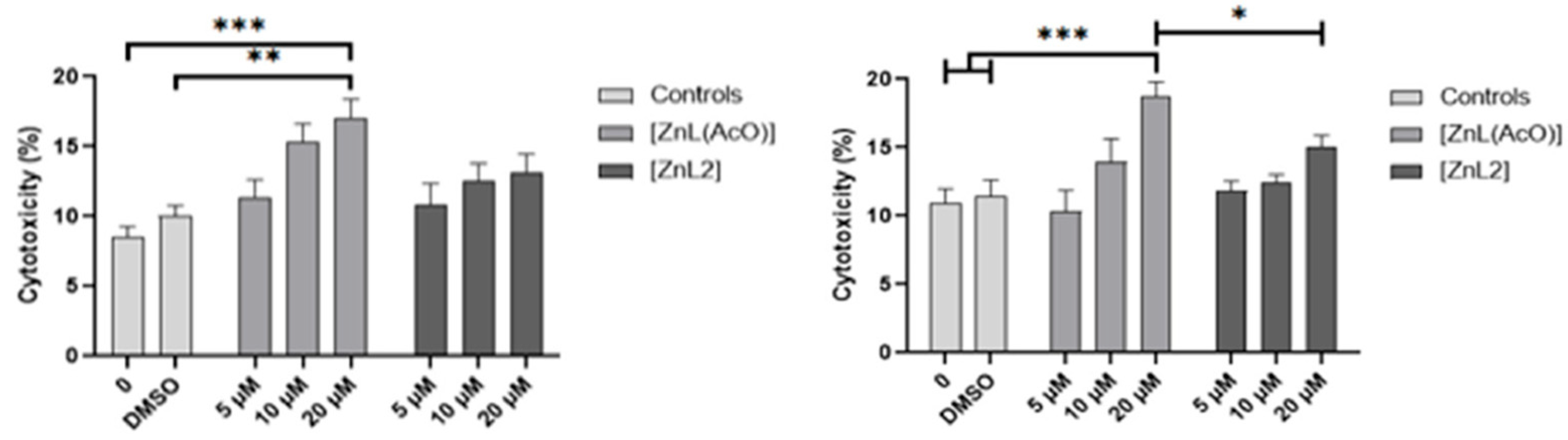
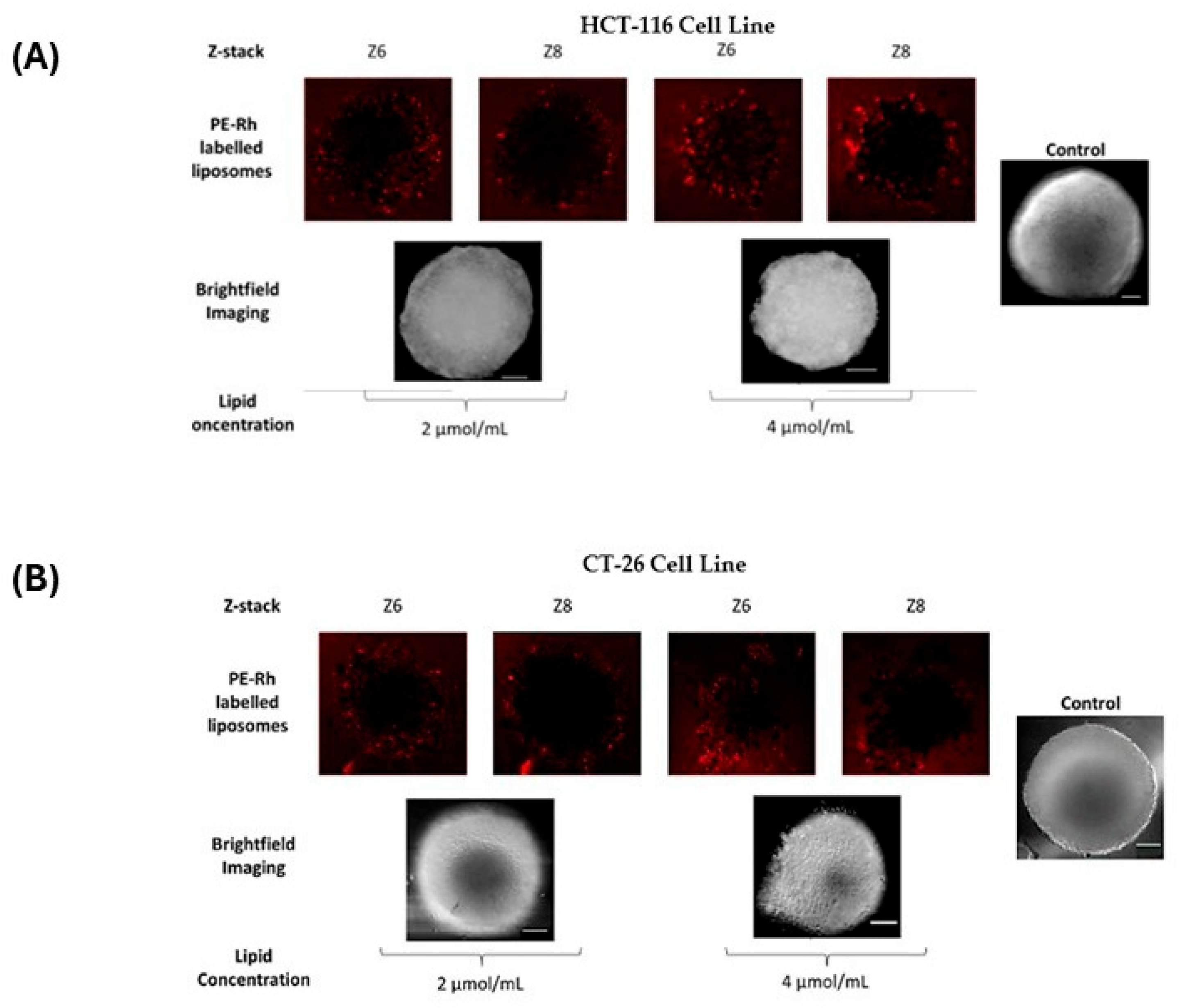
In the original publication [...]
Share and Cite
Ribeiro, N.; Albino, M.; Ferreira, A.; Escrevente, C.; Barral, D.C.; Pessoa, J.C.; Reis, C.P.; Gaspar, M.M.; Correia, I. Correction: Ribeiro et al. Liposomal Formulations of a New Zinc(II) Complex Exhibiting High Therapeutic Potential in a Murine Colon Cancer Model. Int. J. Mol. Sci. 2022, 23, 6728. Int. J. Mol. Sci. 2026, 27, 4339. https://doi.org/10.3390/ijms27104339
Ribeiro N, Albino M, Ferreira A, Escrevente C, Barral DC, Pessoa JC, Reis CP, Gaspar MM, Correia I. Correction: Ribeiro et al. Liposomal Formulations of a New Zinc(II) Complex Exhibiting High Therapeutic Potential in a Murine Colon Cancer Model. Int. J. Mol. Sci. 2022, 23, 6728. International Journal of Molecular Sciences. 2026; 27(10):4339. https://doi.org/10.3390/ijms27104339
Chicago/Turabian StyleRibeiro, Nádia, Melissa Albino, Andreia Ferreira, Cristina Escrevente, Duarte C. Barral, João Costa Pessoa, Catarina Pinto Reis, Maria Manuela Gaspar, and Isabel Correia. 2026. "Correction: Ribeiro et al. Liposomal Formulations of a New Zinc(II) Complex Exhibiting High Therapeutic Potential in a Murine Colon Cancer Model. Int. J. Mol. Sci. 2022, 23, 6728" International Journal of Molecular Sciences 27, no. 10: 4339. https://doi.org/10.3390/ijms27104339
APA StyleRibeiro, N., Albino, M., Ferreira, A., Escrevente, C., Barral, D. C., Pessoa, J. C., Reis, C. P., Gaspar, M. M., & Correia, I. (2026). Correction: Ribeiro et al. Liposomal Formulations of a New Zinc(II) Complex Exhibiting High Therapeutic Potential in a Murine Colon Cancer Model. Int. J. Mol. Sci. 2022, 23, 6728. International Journal of Molecular Sciences, 27(10), 4339. https://doi.org/10.3390/ijms27104339