Article

m6A Methylation Mediated Autophagy and Nucleotide-Binding Oligomerization Domain-like Receptors Signaling Pathway Provides New Insight into the Mitigation of Oxidative Damage by Mulberry Leaf Polysaccharides

by
Wenqiang Jiang
1,2,
Yan Lin
1,
Linjie Qian
2,
Siyue Lu
1,
Zhengyan Gu
1,
Xianping Ge
1,2 and
Linghong Miao
1,2,*
1
Key Laboratory of Freshwater Fisheries and Germplasm Resources Utilization, Ministry of Agriculture and Rural Affairs, Freshwater Fisheries Research Center, Chinese Academy of Fishery Sciences, Wuxi 214081, China
2
Wuxi Fisheries College, Nanjing Agricultural University, Wuxi 214081, China
*
Author to whom correspondence should be addressed.
Int. J. Mol. Sci. 2025, 26(9), 4345; https://doi.org/10.3390/ijms26094345
Submission received: 30 March 2025 / Revised: 27 April 2025 / Accepted: 29 April 2025 / Published: 2 May 2025
(This article belongs to the Special Issue Fish Nutrition Program and Epigenetic Regulation)

Abstract

m6A methylation modification is an important genetic modification involved in biological processes such as sexual maturation, antibacterial, and antiviral in aquatic animals. However, few studies have been conducted in aquatic animals on the relationship between m6A methylation modification and autophagy-inflammation induced by lipid metabolism disorders. In the present study, a high-fat (HF) group and HF-MLP group (1 g mulberry leaf polysaccharides (MLPs)/1 kg HF diet) were set up. The mid-hind intestines of Megalobrama amblycephala juveniles from the two groups were collected for MeRIP-seq and RNA-seq after an 8-week feeding trial. The m6A peaks in the HF and HF-MLP groups were mainly enriched in the 3′ Untranslated Region (3′UTR), Stop codon, and coding sequence (CDS) region. Compared with the HF group, the m6A peaks in the HF-MLP group were shifted toward the 5′UTR region. ‘RRACH’ was the common m6A methylation motif in the HF and HF-MLP groups. Methyltransferase mettl14 and wtap expression in the intestines of the HF-MLP group were significantly higher compared with the HF group (p < 0.05). A total of 21 differentially expressed genes(DEGs) with different peaks were screened by the combined MeRIP-seq and RNA-seq analysis. Kyoto encyclopedia of genes and genomes (KEGG) enrichment analysis enriched BCL2 interacting protein 3 (bnip3) to autophagy–animal and mitophagy–animal signaling pathways, etc., and nucleotide-binding domain leucine-rich repeat protein 1 (nlrp1) was enriched to the Nucleotide-binding oligomerization domain (NOD)-like receptor signaling pathway. Combined MeRIP-seq and RNA-seq analysis indicated that the expression pattern of bnip3 was hyper-up and that of nlrp1 was hyper-down. Gene Set Enrichment Analysis (GSEA) analysis confirmed that the intestinal genes of HF-MLP group positively regulate lysosomal and autophagy–animal signaling pathways. In the present study, we demonstrated that m6A methylation modification plays a role in regulating autophagy-inflammatory responses induced by HF diets by MLPs, and further explored the molecular mechanisms by which MLPs work from the epigenetic perspective.

1. Introduction

Megalobrama amblycephala belongs to Cypriniformes, Cyprinidae, and Megalobrama (Dybowsky, 1792). The total farmed production of bream in China was 767,000 tons in 2023 (more than 90% of which was accounted for by M. amblycephala), which ranked eighth in China’s freshwater farmed fish production [1]. With the rapid development of the intensive aquaculture model in recent years, the farming production of M. amblycephala has been increasing, while its food safety problems have also become increasingly prominent [2]. High-fat diets have been used in intensive aquaculture models for their protein-sparing and growth-promoting properties to decrease production costs and enhance aquaculture production [3,4]. However, M. amblycephala chronically feeding on high-fat diets induces excessive lipid deposition, raises lipid peroxidation levels, inhibits autophagy, and induces oxidative stress and inflammation [5,6,7]. It has been found that the active substances extracted from plant resources with green, efficient, and non-polluting characteristics can improve the growth performance and fat utilization of fish and eliminate the adverse effects of a high−fat diet on fish [8,9].
Plant polysaccharides are important natural macromolecules made from glucose, fructose, galactose, arabinose, and other monosaccharides linked by α− or β-glycosidic bonds in plant cells [10]. The supplementation of plant polysaccharides in high-fat diets for aquatic animals promotes lipolysis and alleviates inflammation and oxidative stress caused by lipid metabolism disorders; for example, 2–4 g/kg sea buckthorn polysaccharide in the Danio rerio diet [11], 10 g/kg Lycium barbarum polysaccharide in the hybrid grouper (Epinephelus fuscoguttatus♀ × Epinephelus lanceolatus♂) diet [12], and 0.8 g/kg Citrus maxima polysaccharide in the hybrid grouper (E. fuscoguttatus♀ × E. lanceolatus♂) diet [13]. The area of mulberry plantations in China was 796,700 hm2 in 2021 [14], and the annual production of mulberry leaves exceeds 15 million tons [15]. Mulberry leaf polysaccharides (MLPs) are important active substances found in mulberry leaf, exhibiting various biological activities such as hypoglycemic, hypolipidemic, immune-boosting, anti-bacterial, anti-oxidant, and anti-aging effects, etc. [2,16,17,18,19]. MLPs also have favorable effects on improving the growth and development of the animal, immune function, and the quality of animal products in 14-day-old chickens and immunosuppressed mice in research [18,20]. MLPs supplementation improves metabolic disorders, repairs histopathological damage, and regulates intestinal flora in the high-fat diet-induced obese mouse model [21,22,23], yet little research has been conducted in aquaculture.
Autophagy is a highly conserved intracellular degradation pathway in eukaryotes which participates in biological processes related to cellular quality control, metabolism, and innate/acquired immunity [24]. Autophagy flux is affected by chronic high-fat diets or other causes of overnutrition, potentially inhibiting autophagy [25]. Infections, autoimmune disorders, and metabolic disorders may result when autophagy is dysfunctional [26]. Dietary high-fat diets in yellow catfish (Pelteobagrus fulvidraco) inhibited the formation of hepatic autolysosome. In vitro studies have shown that the formation of autolysosome in P. fulvidraco hepatocytes was significantly inhibited by incubation with palmitic acid [27]. Ultrastructural observations show that liver mitochondrial biogenesis and mitophagy were inhibited when spotted seabass (Lateolabrax maculatus) fed a high-fat diet [28]. Inflammasomes are multiprotein complexes that are activated and assembled in response to cellular stress or infection [29]. Research on the nucleotide-binding oligomerization domain-like receptors (NLR) family, an intracellular pattern recognition receptor with similar function and structure, such as NLRP1 and NLRP3, has centered on the relationship between autophagy and NLRP1/NLRP3 to explore the interactions between autophagy and inflammasomes [30,31]. NLRP3 inflammasome was activated by the inhibition of podocyte autophagy in high-fat diet-induced diabetic nephropathy mice, and silencing of NLRP3 effectively restored autophagy in podocytes, suggesting that NLRP3 is a negative regulator of autophagy [32]. Caspase-1 activation induced by NLRP1 inflammatory vesicle assembly leads to the release of pro-inflammatory cytokines interleukin 1β and interleukin 18 [33]. NLRP1 and NLRP3 mediate lipopolysaccharide-induced apoptosis in fibroblast-like synoviocytes, and inhibition of nlrp1 and nlrp3 markedly suppressed the expression of apoptosis-related cytokines [34].
Previous studies revealed that epigenetic mechanisms regulate the expression of autophagy genes, and that epigenetic mechanisms also affect autophagy by influencing the expression of genes upstream and downstream of autophagy [35]. Meanwhile, the nlrp1 expression is also mediated by m6A methylation modification [36]. Dietary levels of carbohydrates, vitamins, and other substances have been found to be strongly associated with epigenetic modifications in aquatic animals [37,38,39]. Briefly, nutritional factors affect gene expression and their regulation of biological processes through epigenetic modifications at the molecular level [38]. High-fat diets lead to extensive gene promoter methylation alterations in humans and mice, affecting organ development and function [40,41]. In this study, MLPs were supplemented to high-fat diets for M. amblycephala, aiming to establish an epigenetic regulatory network of the MLPs-regulated autophagy and NOD-like receptor signaling pathway in mitigating high-fat diet-induced intestinal damage by integrating the intestinal transcriptome and m6A methylation sequencing.

2. Results

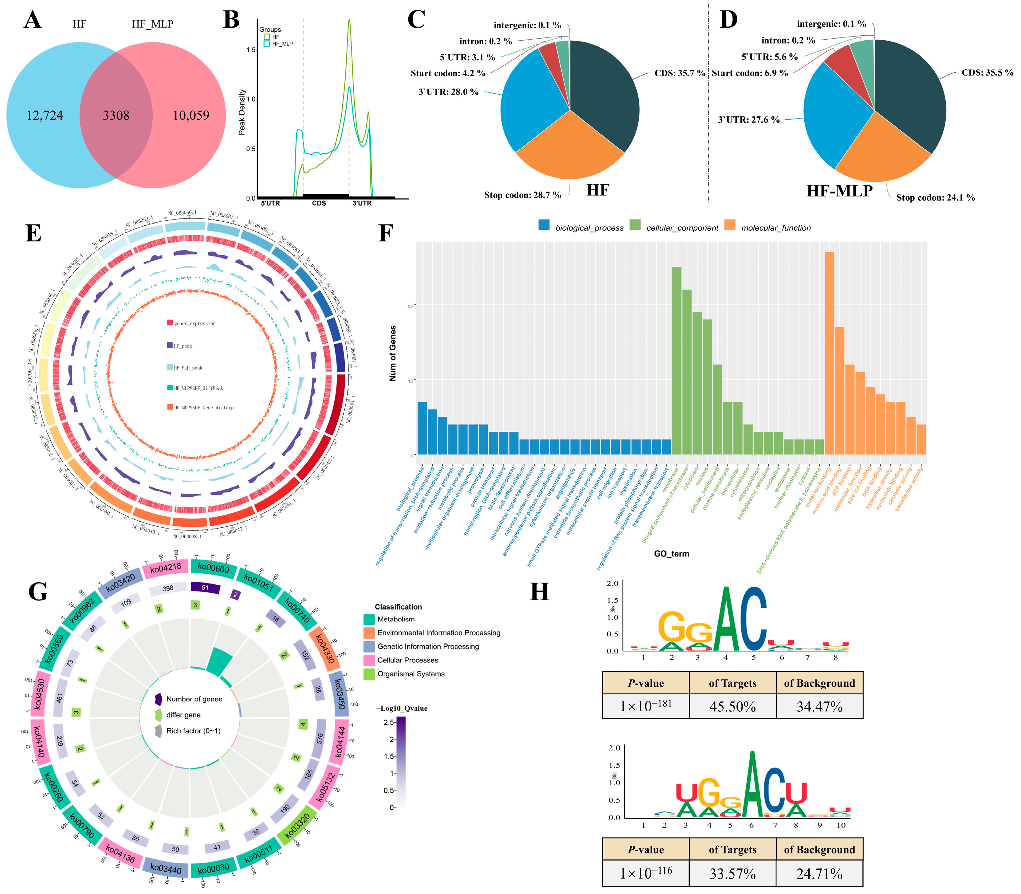
2.1. m6A Methylation Modification in Intestines of HF and HF-MLP Groups

High-throughput sequencing results of the intestinal IP (MeRIP-seq) and input (RNA-seq) libraries showed that more than 82.0% of the clean data mapped the reference genome of M. amblycephala, with excellent concordance between different replicates, Q30 > 94.4% and Q20 > 98.0% (Tables S1 and S2). Venn diagram analysis of the HF and HF-MLP groups revealed 3308 common peaks, and 12,724 unique peaks to the HF group as well as 10,059 unique peaks to the HF-MLP group (Figure 1A). In terms of the m6A peak distribution, the m6A peaks were shifted with MLPs supplementation, and the m6A peak density in the 5′UTR region of the HF-MLP group increased by 2.5% compared with that of the HF group (Figure 1B–D). Briefly, 35.7%, 28.0%, and 3.1% of the m6A peaks were located in the CDS, 3′UTR, and 5′UTR regions in the HF group, while in the HF-MLP group, these percentages were 35.5%, 27.6%, and 5.6%, respectively. Over 87.9% m6A peaks in HF group and 96.2% m6A peaks in HF-MLP group were mapped to the CDS region of the reference genome (Table S3, Figure S1). Gene expression and genomic distribution of m6A peaks were almost identical on the 24 major chromosomes of the reference genome of M. amblycephala (Figure 1E).
In Gene Ontology (GO) categorization analysis, the category with the greatest number of differential m6A peaks was highly enriched in the biological process (biological process ontology), membrane (cellular component ontology), and metal ion binding (molecular function ontology) (Figure 1F, Table S4). Kyoto Encyclopedia of Genes and Genomes (KEGG) enrichment analysis significantly enriched 35 pathways (Figure 1G, Table S5). The top 10 KEGG pathways (based on p-value) that differential m6A peaks most significantly enriched were sphingolipid metabolism (ko00600), biosynthesis of ansamycins (ko01051), riboflavin metabolism (ko00740), notch signaling pathway (ko04330), non-homologous end-joining (ko03450), endocytosis (ko04144), salmonella infection (ko05132), PPAR signaling pathway (ko03320), other glycan degradation (ko00511), and the pentose phosphate pathway (ko00030).
Furthermore, we found that the RDACW motif was identified as highly enriched within the m6A site in the HF and HF-MLP groups by motif analysis (Figure 1H). According to the abbreviated base symbols correspondence table, “A/G” is denoted by “R”, “U/A/G” is denoted by “D”, “A/U” is denoted by “W”.

2.2. Identification of DEGs in Intestinal of HF and HF-MLP Groups

Transcriptome sequencing of input libraries constructed in the HF and HF-MLP groups showed that the number of DEGs in the HF-MLP group vs. the HF group was 1829, of which 1079 DEGs were up-regulated and 750 DEGs were down-regulated (Figure 2A). The top 100 DEGs with significant differences were subjected to expression pattern clustering analysis (Table S6). Figure 2B clearly presents that the DEGs within the same group have similar expression patterns and expression levels, while the differential genes between different groups show significant separation effects.
GO enrichment analysis significantly enriched 2695 functions (Figure 2C, Table S7). The top 10 GO functions (based on p-value) that DEGs most significantly enriched were cholesterol biosynthetic process (GO:0006695), sterol biosynthetic process (GO:0016126), triglyceride catabolic process (GO:0019433), positive regulation of triglyceride catabolic process (GO:0010898), cholesterol homeostasis (GO:0042632), neutral amino acid transmembrane transporter activity (GO:0015175), negative regulation of mitotic cell cycle (GO:0045930), phosphatidylcholine-sterol O-acyltransferase activator activity (GO:0060228), phosphatidylcholine metabolic process (GO:0046470), and positive regulation of cholesterol esterification (GO:0010873).
KEGG enrichment analysis significantly enriched 197 pathways (Figure 2D, Table S8). The top 10 KEGG pathways (based on p-value) that DEGs most significantly enriched were lysosome (ko04142), steroid biosynthesis (ko00100), FoxO signaling pathway (ko04068), autophagy–animal (ko04140), sphingolipid metabolism (ko00600), PPAR signaling pathway (ko03320), glycine, serine and threonine metabolism (ko00260), primary bile acid biosynthesis (ko00120), other glycan degradation (ko00511), and NOD-like receptor signaling pathway (ko04621).The statistical information on the autophagy–animal and NOD-like receptor signaling pathway-related DEGs in the intestines of the HF-MLP group compared to the HF group was shown in Table 1.

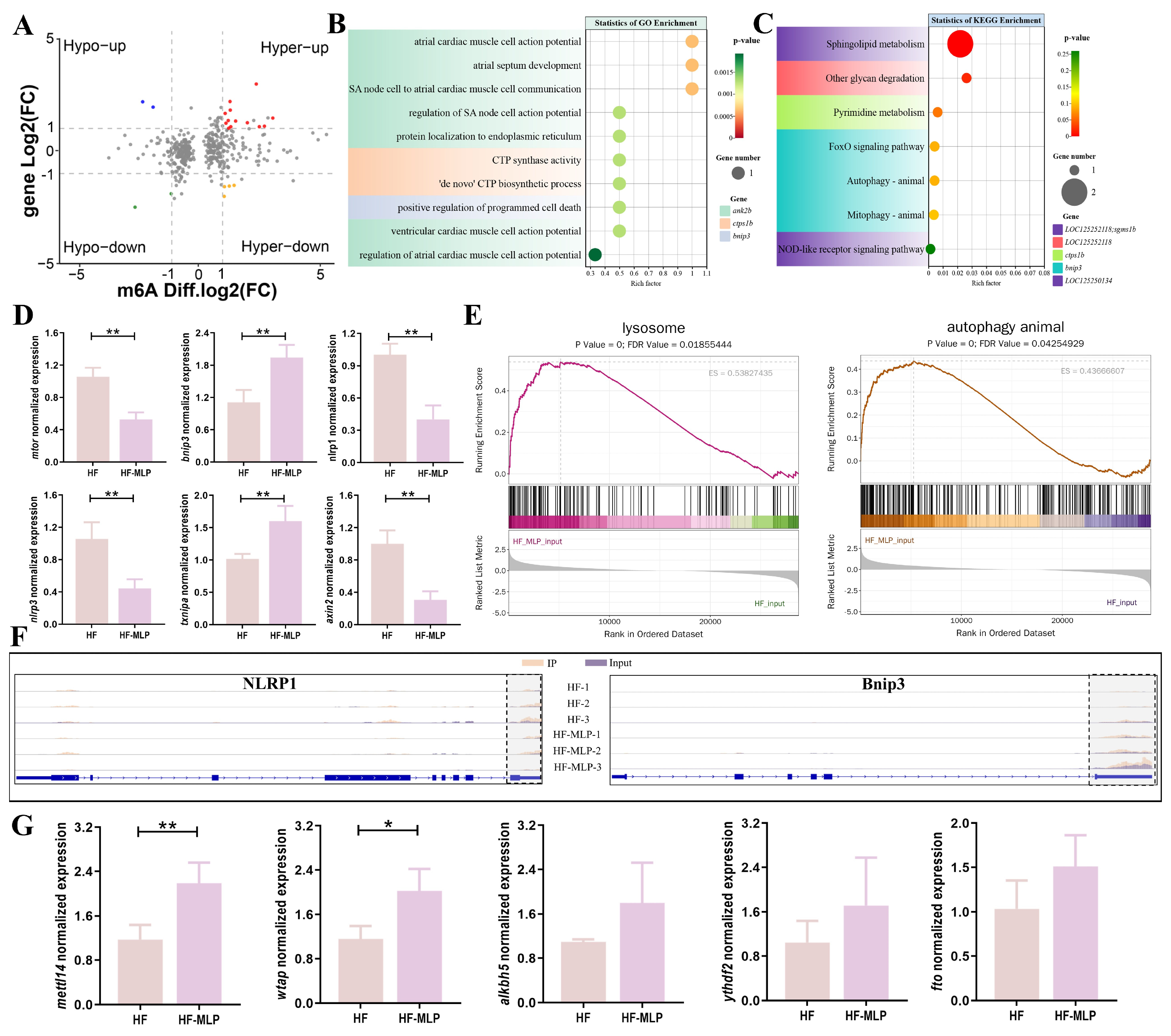
2.3. Assessment of the m6A-Modified Gene Transcription in Intestinal of HF and HF-MLP Groups

Figure 3A shows a four-quadrant graph of genes with significant differences in both m6A methylation and genes expression (Threshold: |Log2(Fc)| > 1). The expression pattern of 13 genes was m6A hypermethylation and gene expression was up-regulated (Hyper-up). The expression pattern of four genes was m6A hypermethylation and gene expression was down-regulated (Hyper-down). The expression pattern of two genes was m6A hypomethylation and gene expression was up-regulated (Hypo-up). The expression pattern of two genes was m6A hypomethylation and gene expression was down-regulated (Hypo-down) (Table 2).
GO enrichment analysis significantly enriched 107 functions (Figure 3B, Table S9). The top 10 GO functions (based on p-value) that m6A-modified DEGs most significantly enriched were atrial cardiac muscle cell action potential (GO:0086014), atrial septum development (GO:0003283), SA node cell to atrial cardiac muscle cell communication (GO:0086070), regulation of SA node cell action potential (GO:0098907), protein localization to the endoplasmic reticulum (GO:0070972), CTP synthase activity (GO:0003883), ‘de novo’ CTP biosynthetic process (GO:0044210), positive regulation of programmed cell death (GO:0043068), ventricular cardiac muscle cell action potential (GO:0086005), and regulation of atrial cardiac muscle cell action potential (GO:0098910). KEGG enrichment analysis significantly enriched seven pathways (Figure 3C, Table S10), sphingolipid metabolism (ko00600), other glycan degradation (ko00511), pyrimidine metabolism (ko00240), FoxO signaling pathway (ko04068), autophagy–animal (ko04140), mitophagy–animal (ko04137), and NOD-like receptor signaling pathway (ko04621).
Genes related to the autophagy–animal (mtor, bnip3), NOD-like receptor signaling pathway (txnipa, nlrp1, nlrp3), and Wnt signaling pathway (axin2) were randomly selected for verification (Figure 3D). All the candidate genes verified by qRT-PCR were identical to the results of transcriptome sequencing, indicating that the sequencing results were credible (Figure 3D, Table S11). GSEA confirmed that the intestinal genes of the HF-MLP group positively regulate lysosomal and autophagy–animal signaling pathways (Figure 3E).
Figure 3F exhibits the m6A peak expression patterns of two representative genes in the autophagy–animal signaling pathway (bnip3) and NOD-like receptor signaling pathway (nlrp1). Bnip3 and nlrp1 undergo hypermethylation in the 3′UTR region and cause changes in gene expression. In details, the expression of nlrp1 was significantly down-regulated in the HF-MLP group versus the HF group, and the bnip3 expression was significantly up-regulated in the HF-MLP group than in the HF group (|Diffgene.Log2(Fc)| > 1).
Expression of m6A methylation modification-related enzymes (mettl14, wtap, ythdf2, fto, and alkbh5) was detected by qRT-PCR. Notably, the changes in wtap and mettl14 were more significant compared to several other key genes (Figure 3G). Noticeable increases in mettl14 and wtap expression were exhibited in the HF-MLP group compared to the HF group (p < 0.05). The expression of ythdf2, fto, and alkbh5 were not affected by dietary MLPs supplementation (p > 0.05).

3. Discussion

m6A methylation modification, the most abundantly expressed form of epigenetic regulation, is a dynamically reversible process [42]. m6A methylation genomics could be co-analyzed with metabolomics for a more comprehensive understanding of gene function. Epigenetic changes attributed to diet-induced m6A methylation modification were found to mediate phenotypic plasticity in humans and mammals [43,44,45]. However, researchers in the fisheries field have primarily focused on the dynamic m6A methylation modification characteristics of aquatic animals in different physiological states [46,47].
In the present study, the m6A modification profile of the M. amblycephala intestines was first characterized, and the m6A methylation modification sites of intestinal mRNAs were enriched near the 3′UTR, stop codon, and CDS region. Interestingly, m6A methyla-tion modifications of mRNAs were found to show differences in spatial distribution de-pending on the species. The m6A methylation modifications were mainly enriched in the exon region, stop codon region, and 3′UTR region in humans [43], rats [44], and finishing pigs [45] suggesting that the overall distributions of m6A methylation modification sites in the human, mammal, and M. amblycephala are similar [48]. By contrast, studies in plants have revealed that m6A methylation modification sites are enriched not only in the stop codon region and the 3′UTR region, but also around the start codon and the 5′UTR region [49,50]. The proportion of m6A peaks in the 5′UTR region and stop codon region in the HF-MLP group was higher than the proportion of m6A peaks in the HF group, suggesting that the distribution of m6A peaks may be affected by MLPs supplementation. Furthermore, the localization of m6A peaks shifted to the 5′UTR and CDS regions after MLP supplementation in the present study. The dynamic distribution of m6A methylation modifications is highly responsive to regulating animals’ physiological conditions to accommodate various changes in the external environment [51]. Consistent with previous studies, the m6A peak of the mRNA was also found to move toward the 5′UTR region following changes in the external environment in humans, large yellow croaker (Larimichthys crocea), and Arabidopsis [47,51,52]. Annotation analysis showed that the common base sequence of the different methylation modification sites was RDACW in the intestines of the HF and HF-MLP groups. The distribution characteristics of RDACW are consistent with the common m6A motif pattern RRACH, indicating that a highly conserved type of m6A methylation modification sites, which further confirms that the function of m6A methylation modification is indeed present in M. amblycephala intestine [53,54].
m6A methylation modification plays an essential regulatory role in animal reproduction, growth development, and immunization. m6A methylation modification is undertaken by writers (mettl3, mettl14, wtap, etc.), erasers (fto and alkbh5), and readers (ythdc1, ythdf2, ythdf3, etc.) for the addition, deletion, or recognition of m6A methylation modifications. In the present study, intestinal mettl14 and wtap (writers) expression was significantly higher in the HF-MLP group compared with the HF group. Conserved heterodimers formed by methyltransferase 13 (METTL3) and METTL14 are recruited by wtap to form a complex that mediates the m6A methylation modification process [55]. Overexpression/knockdown of METTL14 mediates osteogenic differentiation capacity of bone marrow mesenchymal stem cells by activating/suppressing autophagy through m6A methylation modification of beclin-1 expression [56]. METTL14 overexpression activates the autophagy pathway in the mitochondria of I/R cardiomyocytes to ameliorate cardiomyocytes injury [57]. Hypotheses based on classical physiology and biochemistry suggest that epigenetic regulation is a potential factor in dietary modification of animal phenotype. Betaine potentiates lipolysis in finishing pigs feeding on low-energy diets by up-regulating the expression of writer genes/proteins (mettl3, mettl14, wtap) [45]. Notoginsenoside R1 repairs DNA damage in skin under UV irradiation by up-regulating wtap expression in skin keratinocytes [58].
m6A methylation modification as a pivotal mediator in response to dietary and external environmental changes has received mounting awareness [43,45,50]. In the present study, we analyzed and identified 21 DEGs (|DiffExp.gene.Log2(Fc)| > 1) with differential m6A Peak (|DiffPeak.Log2(Fc)| > 1). KEGG signaling pathway enrichment analysis showed that these DEGs were enriched in the autophagy–animal signaling pathway, and NOD-like receptor signaling pathway. In the autophagy–animal signaling pathway, bnip3 was enriched, with an expression pattern of up-regulation of methylation levels and up-regulation of gene expression. A positive correlation between gene expression and methylation levels has been reported in most transcripts of Arabidopsis and human studies as compared with the expression pattern in which methylation levels are negatively correlated with gene expression [48,59]. Consistent with our research, up-regulation expression of bnip3 in breast tumors via methylation in the 3′UTR plays a proapoptotic function [60]. Astragalus polysaccharide promotes cellular autophagy and ameliorates femoral head necrosis by elevating bnip3 expression [61]. In the NOD-like receptor signaling pathway, nlrp1 was enriched, with a classical expression pattern of up-regulation of methylation levels and down-regulation of gene expression. NLRs are pattern recognition receptors in the cytoplasm, and the activation of partial members (NLRP1, NLRP3, and NLRC4, etc.) leads to the assembly of inflammasome, which subsequently triggers the activation of inflammatory caspases [62]. The pro-inflammatory downstream effectors IL-1β and IL-18 were cleaved by enzymatically active Caspase-1 into their biologically active forms to induce inflammation [63]. Antrodia camphorate polysaccharides reduced reactive oxygen species (ROS) content to inhibit reduced nicotinamide adenine dinucleotide phosphate oxidase 2 (NOX2)-NLRP1 activation, thereby reducing inflammatory damage of cortical neurons [64]. The activation of NLRP1 inflammasome mediating inflammatory responses in aquatic animals has been reported in both zebrafish [63] and common carp (Cyprinus carpio) [65]. NLRP1 was found to play an essential role in the immune organs of bony fish, and its expression was up-regulated in common carp (Cyprinus carpio) treated with spring viremia of carp, Edwardsiella tarda, and Aeromonas hydrophila [65].
In the present study, KEGG analysis of DEGs in the transcriptome and DEGs with differential peaking following coanalysis revealed that the differential genes were enriched in the autophagy–animal and NOD-like receptor signaling pathways. Furthermore, GSEA analysis showed that the expression of major genes of autophagy–animal and lysosomal signaling pathways were up-regulated in the HF-MLP group. Autophagy, an important cellular mechanism in eukaryotic cells, degrades damaged organelles and macromolecules via lysosomes [24]. In aquatic animals such as M. amblycephala [6,66], zebrafish [67], and spotted seabass (Lateolabrax maculatus) [28], etc., it was found that high-fat diets down-regulated the expression of autophagy-related genes and inhibited autophagic flux. MLPs in this present study were heteropolysaccharides (201.4 KDa) mainly consisting of rhamnose, arabinose, and galactose, a structure with an extreme immune and antioxidant activity [2]. Plant polysaccharides were attenuated by high-fat diet-induced lipid accumulation, oxidative stress, and inflammation by activating autophagy [68,69,70]. It has been found that polysaccharides might play an immunoregulatory and inflammatory function by modifying the levels of non-coding RNA [71], histone modifications [72], etc. Tremella fuciformis polysaccharides attenuate lipopolysaccharide-induced inflammatory responses by down-regulating miR-155 expression in RAW264.7 cells that suppress the expression of protein kinase B (AKT), p38MAPK, and nuclear factor kappa-B (NF-κB) [73]. Stimulus factors such as pathogen/injury initiate the assembly of NLRP1/NLRP3 inflammasome to activate inflammasome [29]. Autophagy inhibits inflammatory responses by phagocytosis and degradation of inflammasome and downstream active components through endocytosis and phagocytosis [74]. Micheliolide mediates autophagy to degrade some components of the NLRP3 inflammasome, blocking inflammasome activation and attenuating pro-inflammatory cytokine release in radiation-damaged intestinal tissues [75]. Autophagy dysfunction results in blocked clearance of activated NLRP3 inflammasome to induce maturation and release of pro-inflammatory cytokines [29,32]. Moreover, activation of inflammasome-mediated IL-1β secretion aggravates intestinal ischemia-reperfusion injury by inhibiting autophagy in mice, whereas knockdown of NLRP3 reverses these effects [76].

4. Materials and Methods

4.1. MLPs

MLPs was obtained from Xi’an Ciyuan Biotech Co., Ltd., Xi’an, China. MLPs were heteropolysaccharides (201.4 KDa) mainly consisting of rhamnose, glucose, arabinose, and galactose. UV-Vis spectroscopy (Thermo Fisher, Cleveland, OH, USA) showed that the MLPs were extremely pure, with minimal protein and nucleic acid content. MLPs are pyranose rings polysaccharides linked by β-glycosidic bonds as revealed by FT-IR spectra (Thermo Fisher, Cleveland, OH, USA) [3].

4.2. Diets

Crushed ingredients (sieved through a 60-mesh) such as soybean meal, rapeseed meal, and cottonseed meal were mixed well according to the feed formula, with soybean oil and water added, and 2.0 mm sink pellets were made with an F-26 (II) granulator (South China University of Technology, Guangzhou, China). Two types of diets were made in our experiment: an HF diet (crude protein 31.7%, crude fat 14.8%) and an HF-MLP diet (1 g/kg of MLPs was added to the formula of HF diet to make HF-MLP diet). Diets dried in a cool dry place were packaged and stored at -20 °C until the feeding trial.

4.3. Fish and Feeding Trial

Experimental fish species was M. amblycephala ‘Huahai No.1’, which was supplied by the National M. amblycephala stock (Ezhou, China). Juveniles of homogeneous size (9.67 ± 0.23 g) were selected for the feeding trial, with 15 juveniles per aquarium in six aquariums (0.32 m3). The HF and HF-MLP groups were set up in the feeding trial, with three replicates in each group. M. amblycephala juveniles were fed with commercial feed (Tongwei Co., Ltd., Wuxi, China) during the domestication period. Fish were fed experimental diets three times a day (7:00, 12:00, and 17:00) since the completion of domestication. During the 8-week feeding trial, the conditions of the aquaculture water were kept constant with dissolved oxygen ≥ 6 mg/L, water temperature 26 °C~28 °C, ammonia nitrogen ≤ 0.05 mg/L, and pH 7.0 ± 0.1.

4.4. Sample Collection

Four fish were randomly selected from each aquarium while the feeding trial was ended, anesthetized with 100 mg/L tricaine methanesulfonate (MS-222) (Sigma, Saint Louis, MO, USA), and the emptied hind intestine was sampled on ice. Collected samples were snap-frozen in liquid nitrogen and stored at −80 ℃, awaiting MeRIP-seq, RNA-seq, real-time PCR analysis.

4.5. RNA Isolation, Library Construction, and Sequencing

RNA was extracted from the mid-hind intestine of the HF and HF-MLP groups using TRIzol reagent (Takara Co., Ltd., Dalian, China), with three replicates in each group, and downstream experiments were performed after quality control and integrity testing of the RNA. Poly (A) RNA specifically purified by Dynabeads Oligo (dT) (Thermo Fisher, Cleveland, OH, USA) was fragmented into small pieces at 86 °C for 7 min. The cleaved RNA fragments were incubated for 2 h at 4 °C with m6A-specific antibody (No. 202003, Synaptic Systems, Göttingen, Germany) in IP buffer. The IP RNA was reverse-transcribed to cDNA, which was next used to synthesize Ulabeled second-stranded DNAs. PCR was used to construct sequencing libraries following double-stranded digestion. At last, we performed paired-end sequencing (PE150) on an Illumina Novaseq™ 6000 platform (Biozeron Co., Ltd., Shanghai, China).

4.6. Bioinformatics Analysis

The clean data obtained after FastQC and RseQC from all IP samples and Input samples were analyzed. Reads were mapped to the reference genome of M. amblycephala using HISAT2 (http://daehwankimlab.github.io/hisat2). Peak calling and diff peak analysis were performed by R package exomePeak (https://www.bioconductor.org/packages/3.3/bioc/html/exomePeak.html). HOMER (http://homer.ucsd.edu/homer/motif) was used for motif analysis.
StringTie was used for quantified expression for all genes in the input libraries by calculating FPKM. The differentially expressed genes (DEGs) were selected with |Log2 (Fold change)| ≥ 1 and p value < 0.05 by R package edgeR.

4.7. Real-Time PCR Analysis (qRT-PCR)

Intestinal RNA (400 ng/μL, OD260/280 = 1.8–2.0) extracted using TRIzol reagent (Takara, Dalian, China) was synthesized to cDNA by PrimeScript RT reagent Kit (Takara, China). The qRT-PCR analysis was conducted on the CFX96 instrument (Bio-Rad, Hercules, CA, USA) with TB Green (Takara, Dalian, China) as the fluorescent dye. Primers for qRT-PCR are listed in Table 3. The relative expression of genes was determined by the 2−∆∆CT method with β−actin as the reference gene.

4.8. Statistical Analysis

An independent sample t-test was performed to identify differences between the HF and HF-MLP groups using SPSS Version 20.0 software (SPSS Inc., Chicago, IL, USA) when data conformed to normality (Shapiro–Wilk test) and homogeneity of variance (Levene’s test). Values were expressed as the mean ± standard error. A statistically significant difference was considered at the p < 0.05.

5. Conclusions

In conclusion, diets of HF and HF-MLP induced a dynamic change in intestinal m6A methylation modification of M. amblycephala juveniles (Figure 4). Differential methylation modification of intestinal DEGs, mainly in the 3′UTR region, modulates the physiological status of the intestine in the M. amblycephala juveniles following the supplementation of MLPs. Combined MeRIP-seq and RNA-seq analyses identified key DEGs and pathways, autophagy–animal and mitophagy–animal signaling pathways (bnip3, Hyper-up), and NOD-like receptor signaling pathway (nlrp1, Hyper-down). This study provides strong support for diet (MLPs)-mediated epigenetic regulation to alter aquatic animal phenotypes.

Supplementary Materials

The supporting information can be downloaded at: https://www.mdpi.com/article/10.3390/ijms26094345/s1.

Author Contributions

Conceptualization, Y.L. and L.M.; data curation, W.J. and Z.G.; investigation, W.J., L.Q. and S.L.; methodology, W.J., L.Q. and Y.L.; project administration, L.M. and X.G.; software, L.Q. and Z.G.; supervision, L.M. and X.G.; validation, S.L.; writing—original draft, W.J.; writing—review and editing, L.M. All authors have read and agreed to the published version of the manuscript.

Funding

This research was funded by the China Agriculture Research System (Grant number: CARS-45), Central Public-interest Scientific Institution Basal Research Fund, Freshwater Fisheries Research Center, CAFS (Grant number: 2025JBFR02), National Key Research and Development Plan Program of China (Grant number: 2022YFD2400600/2022YFD2400603), Science and Technology Innovation Team (Grant number: 2023TD63).

Institutional Review Board Statement

The operation of the feeding trial and the sampling process for the M. amblycephala juveniles was carried out in strict accordance with the instructions (LAECFFRC-2023-04-19) of the Freshwater Fishery Research Center Animal Care and Use Committee, Approval Date: 19 April 2023.

Informed Consent Statement

Not applicable.

Data Availability Statement

The authors confirm that the data supporting the findings of this study are available within the manuscript and tables.

Conflicts of Interest

The authors declare no conflict of interest.

References

  1. The Bureau of Fisheries and Fishery Administration tMoA, National Fisheries Technology Extension Center, Fisheries. CSo. 2023 China Fishery Statistical Yearbook; China Agriculture Press: Beijing, China, 2023. [Google Scholar]
  2. Jiang, W.; Lin, Y.; Qian, L.; Lu, S.; Shen, H.; Ge, X.; Miao, L. Mulberry Leaf Polysaccharides Attenuate Oxidative Stress Injury in Peripheral Blood Leukocytes by Regulating Endoplasmic Reticulum Stress. Antioxidants 2024, 13, 136. [Google Scholar] [CrossRef]
  3. Ma, Q.; Li, L.Y.; Le, J.Y.; Lu, D.L.; Qiao, F.; Zhang, M.L.; Du, Z.Y.; Li, D.L. Dietary microencapsulated oil improves immune function and intestinal health in Nile tilapia fed with high-fat diet. Aquaculture 2018, 496, 19–29. [Google Scholar] [CrossRef]
  4. Tang, T.; Hu, Y.; Peng, M.; Chu, W.; Hu, Y.; Zhong, L. Effects of high-fat diet on growth performance, lipid accumulation and lipid metabolism-related MicroRNA/gene expression in the liver of grass carp (Ctenopharyngodon idella). Comp. Biochem. Physiol. Part B Biochem. Mol. Biol. 2019, 234, 34–40. [Google Scholar] [CrossRef]
  5. Cao, X.F.; Dai, Y.J.; Liu, M.Y.; Yuan, X.Y.; Wang, C.C.; Huang, Y.Y.; Liu, W.B.; Jiang, G.Z. High-fat diet induces aberrant hepatic lipid secretion in blunt snout bream by activating endoplasmic reticulum stress-associated IRE1/XBP1 pathway. BBA-Mol. Cell Biol. Lipids 2019, 1864, 213–223. [Google Scholar] [CrossRef] [PubMed]
  6. Dong, Y.; Xia, T.; Yu, M.; Wang, L.; Song, K.; Zhang, C.; Lu, K. Hydroxytyrosol attenuates high-fat-diet-induced oxidative stress, apoptosis and inflammation of blunt snout bream (Megalobrama amblycephala) through its regulation of mitochondrial homeostasis. Fishes 2022, 7, 78. [Google Scholar] [CrossRef]
  7. Abasubong, K.P.; Jiang, G.Z.; Guo, H.X.; Wang, X.; Li, X.F.; Yan-Zou, D.; Liu, W.B.; Desouky, H.E. High-fat diet alters intestinal microbiota and induces endoplasmic reticulum stress via the activation of apoptosis and inflammation in blunt snout bream. Fish Physiol. Biochem. 2023, 49, 1079–1095. [Google Scholar] [CrossRef]
  8. Zhao, W.; Yao, R.; Wei, H.; Guo, Y.; Chen, A.; Chen, B. Astaxanthin, bile acid and chlorogenic acid attenuated the negative effects of high-fat diet on the growth, lipid deposition, and liver health of Oncorhynchus mykiss. Aquaculture 2023, 567, 739255. [Google Scholar] [CrossRef]
  9. Yin, P.; Xie, S.; Zhuang, Z.; He, X.; Tang, X.; Tian, L.; Liu, Y.; Niu, J. Dietary supplementation of bile acid attenuate adverse effects of high-fat diet on growth performance, antioxidant ability, lipid accumulation and intestinal health in juvenile largemouth bass (Micropterus salmoides). Aquaculture 2021, 531, 735864. [Google Scholar] [CrossRef]
  10. Cao, W.; Wu, J.; Zhao, X.; Li, Z.; Yu, J.; Shao, T.; Hou, X.; Zhou, L.; Wang, C.; Wang, G.; et al. Structural elucidation of an active polysaccharide from Radix Puerariae lobatae and its protection against acute alcoholic liver disease. Carbohydr. Polym. 2024, 325, 121565. [Google Scholar] [CrossRef]
  11. Lan, Y.; Wang, C.; Zhang, C.; Li, P.; Zhang, J.; Ji, H.; Yu, H. Dietary sea buckthorn polysaccharide reduced lipid accumulation, alleviated inflammation and oxidative stress, and normalized imbalance of intestinal microbiota that was induced by high-fat diet in zebrafish Danio rerio. Fish Physiol. Biochem. 2022, 48, 1717–1735. [Google Scholar] [CrossRef]
  12. Tan, X.; Sun, Z.; Ye, C. Dietary Lycium barbarum extract administration improved growth, meat quality and lipid metabolism in hybrid grouper (Epinephelus lanceolatus♂ × E. fuscoguttatus♀) fed high lipid diets. Aquaculture 2019, 504, 190–198. [Google Scholar] [CrossRef]
  13. Zou, C.; Fang, Y.; Lin, N.; Liu, H. Polysaccharide extract from pomelo fruitlet ameliorates diet-induced nonalcoholic fatty liver disease in hybrid grouper (Epinephelus lanceolatus♂ × Epinephelus fuscoguttatus♀). Fish Shellfish. Immunol. 2021, 119, 114–127. [Google Scholar] [CrossRef] [PubMed]
  14. Li, J.; Gu, W. Development Trends and Policy Recommendations for China’s Silkworm and Mulberry Industry in 2022. Chin. J. Anim. Sci. 2022, 58, 270–274. [Google Scholar]
  15. Li, L.; Zhang, S.; Xu, F.; Ma, H.; Zhuo, Y.; Qiao, Y. Research Progress of Mulberry Leaf Comprehensive Utilization. Sci. Technol. Food Ind. 2022, 43, 397–404. [Google Scholar]
  16. Yuan, Q.; Xie, Y.; Wang, W.; Yan, Y.; Ye, H.; Jabbar, S.; Zeng, X. Extraction optimization, characterization and antioxidant activity in vitro of polysaccharides from mulberry (Morus alba L.) leaves. Carbohydr. Polym. 2015, 128, 52–62. [Google Scholar] [CrossRef]
  17. Zhang, Y.; Ren, C.; Lu, G.; Cui, W.; Mu, Z.; Gao, H.; Wang, Y. Purification, characterization and anti-diabetic activity of a polysaccharide from mulberry leaf. Regul. Toxicol. Pharm. 2014, 70, 687–695. [Google Scholar] [CrossRef]
  18. Chen, X.; Sheng, Z.; Qiu, S.; Yang, H.; Jia, J.; Wang, J.; Jiang, C. Purification, characterization and in vitro and in vivo immune enhancement of polysaccharides from mulberry leaves. PLoS ONE 2019, 14, 0208611. [Google Scholar] [CrossRef]
  19. Hu, Y.; Zhang, Y.; Cui, X.; Wang, D.; Hu, Y.; Wang, C. Structure-function relationship and biological activity of polysaccharides from mulberry leaves: A review. Int. J. Biol. Macromol. 2024, 268, 131701. [Google Scholar] [CrossRef]
  20. Chen, X.; Cai, B.; Wang, J.; Sheng, Z.; Yang, H.; Wang, D.; Chen, J.; Ning, Q. Mulberry leaf-derived polysaccharide modulates the immune response and gut microbiota composition in immunosuppressed mice. J. Funct. foods 2021, 83, 104545. [Google Scholar] [CrossRef]
  21. Li, R.; Xue, Z.; Li, S.; Zhou, J.; Liu, J.; Zhang, M.; Panichayupakaranant, P.; Chen, H. Mulberry leaf polysaccharides ameliorate obesity through activation of brown adipose tissue and modulation of the gut microbiota in high-fat diet fed mice. Food Funct. 2022, 13, 561–573. [Google Scholar] [CrossRef]
  22. Li, R.; Xue, Z.; Jia, Y.; Wang, Y.; Li, S.; Zhou, J.; Liu, J.; Zhang, M.; He, C.; Chen, H. Polysaccharides from mulberry (Morus alba L.) leaf prevents obesity by inhibiting pancreatic lipase in high-fat diet induced mice. Int. J. Biol. Macromol. 2021, 192, 452–460. [Google Scholar] [CrossRef]
  23. Zhao, X.; Fu, Z.; Yao, M.; Cao, Y.; Zhu, T.; Mao, R.; Huang, M.; Pang, Y.; Meng, X.; Li, L.; et al. Mulberry (Morus alba L.) leaf polysaccharide ameliorates insulin resistance-and adipose deposition-associated gut microbiota and lipid metabolites in high-fat diet-induced obese mice. Food. Sci. Nutr. 2022, 10, 617–630. [Google Scholar] [CrossRef] [PubMed]
  24. Ichimiya, T.; Yamakawa, T.; Hirano, T.; Yokoyama, Y.; Hayashi, Y.; Hirayama, D.; Wagatsuma, K.; Itoi, T.; Nakase, H. Autophagy and autophagy-related diseases: A review. Int. J. Mol. Sci. 2020, 21, 8974. [Google Scholar] [CrossRef]
  25. Kim, K.H.; Lee, M.S. Autophagy as a crosstalk mediator of metabolic organs in regulation of energy metabolism. Rev. Endocr. Metab. Disord. 2014, 15, 11–20. [Google Scholar] [CrossRef]
  26. Painter, J.D.; Galle-Treger, L.; Akbari, O. Role of autophagy in lung inflammation. Front. Immunol. 2020, 11, 1337. [Google Scholar] [CrossRef] [PubMed]
  27. Wu, L.; Wei, C.; Yang, S.; Zhao, T.; Luo, Z. Effects of fat and fatty acids on the formation of autolysosomes in the livers from yellow catfish Pelteobagrus fulvidraco. Genes 2019, 10, 751. [Google Scholar] [CrossRef] [PubMed]
  28. Dong, Y.; Xia, T.; Lin, J.; Wang, L.; Song, K.; Zhang, C. Quercetin attenuates high-fat diet-induced excessive fat deposition of spotted seabass (Lateolabrax maculatus) through the regulatory for mitochondria and endoplasmic reticulum. Front. Mar. Sci. 2021, 8, 746811. [Google Scholar] [CrossRef]
  29. Liu, X.C.; Huang, M.; Huang, X.P.; Guan, J.F.; Li, X.F.; Xie, D.Z.; Xu, C. NLRP3 is a promising target for regulating high glucose-induced inflammatory response in Megalobrama amblycephala. Aquaculture 2022, 555, 738220. [Google Scholar] [CrossRef]
  30. Su, S.; Wu, Y.; Lin, Q.; Wang, D.; Hai, J. URB597 protects against NLRP3 inflammasome activation by inhibiting autophagy dysfunction in a rat model of chronic cerebral hypoperfusion. J. Neuroinflammation 2019, 16, 260. [Google Scholar] [CrossRef]
  31. Lin, Q.; Li, S.; Jiang, N.; Shao, X.; Zhang, M.; Xi, H.; Zhang, Z.; Shen, J.; Zhou, Y.; Zhou, W.; et al. PINK1-parkin pathway of mitophagy protects against contrast-induced acute kidney injury via decreasing mitochondrial ROS and NLRP3 inflammasome activation. Redox Biol. 2019, 26, 101254. [Google Scholar] [CrossRef]
  32. Hou, Y.; Lin, S.; Qiu, J.; Sun, W.; Dong, M.; Xiang, Y.; Wang, L.; Du, P. NLRP3 inflammasome negatively regulates podocyte autophagy in diabetic nephropathy. Biochem. Biophys. Res. Commun. 2020, 521, 791–798. [Google Scholar] [CrossRef]
  33. Vrentas, C.E.; Boggiatto, P.M.; Olsen, S.C.; Leppla, S.H.; Moayeri, M. Characterization of the NLRP1 inflammasome response in bovine species. Innate Immun. 2020, 26, 301–311. [Google Scholar] [CrossRef] [PubMed]
  34. Zhao, L.R.; Xing, R.L.; Wang, P.M.; Zhang, N.S.; Yin, S.J.; Li, X.C.; Zhang, L. NLRP1 and NLRP3 inflammasomes mediate LPS/ATP-induced pyroptosis in knee osteoarthritis. Mol. Med. Rep. 2018, 17, 5463–5469. [Google Scholar] [CrossRef] [PubMed]
  35. Liang, J.; Sun, J.; Zhang, W.; Wang, X.; Xu, Y.; Peng, Y.; Zhang, L.; Xiong, W.; Liu, Y.; Liu, H. Novel insights into the roles of N6-methyladenosine (m6A) modification and autophagy in human diseases. Int. J. Biol. Sci. 2023, 19, 705. [Google Scholar] [CrossRef] [PubMed]
  36. Zhang, G.; Li, X.; Huang, X. m6A-related bioinformatics analysis and functional characterization reveals that METTL3-mediated NPC1L1 mRNA hypermethylation facilitates progression of atherosclerosis via inactivation of the MAPK pathway. Inflamm. Res. 2023, 72, 429–442. [Google Scholar] [CrossRef]
  37. Panserat, S.; Marandel, L.; Geurden, I.; Veron, V.; Dias, K.; Plagnes-Juan, E.; Pegourié, G.; Arbenoits, E.; Santigosa, E.; Weber, G.; et al. Muscle catabolic capacities and global hepatic epigenome are modified in juvenile rainbow trout fed different vitamin levels at first feeding. Aquaculture 2017, 468, 515–523. [Google Scholar] [CrossRef]
  38. Xie, N.; Tian, J.; Meng, X.; Dong, L.; Jiang, M.; Wen, H.; Lu, X. DNA methylation profiling and transcriptome sequencing reveal the molecular mechanism of the high-carbohydrate diet on muscle growth of grass carp (Ctenopharyngodon idella). Aquac. Rep. 2023, 30, 101545. [Google Scholar] [CrossRef]
  39. Terova, G.; Díaz, N.; Rimoldi, S.; Ceccotti, C.; Gliozheni, E.; Piferrer, F. Effects of sodium butyrate treatment on histone modifications and the expression of genes related to epigenetic regulatory mechanisms and immune response in European sea bass (Dicentrarchus labrax) fed a plant-based diet. PLoS ONE 2016, 11, 0160332. [Google Scholar] [CrossRef]
  40. Jacobsen, S.C.; Brøns, C.; Bork-Jensen, J.; Ribel-Madsen, R.; Yang, B.; Lara, E.; Hall, E.; Calvanese, V.; Nilsson, E.; Jørgensen, S.W.; et al. Effects of short-term high-fat overfeeding on genome-wide DNA methylation in the skeletal muscle of healthy young men. Diabetologia 2012, 55, 3341–3349. [Google Scholar] [CrossRef]
  41. Zhang, Y.; Wang, H.; Zhou, D.; Moody, L.; Lezmi, S.; Chen, H.; Pan, Y.X. High-fat diet caused widespread epigenomic differences on hepatic methylome in rat. Physiol. Genom. 2015, 47, 514–523. [Google Scholar] [CrossRef]
  42. Knuckles, P.; Lence, T.; Haussmann, I.U.; Jacob, D.; Kreim, N.; Carl, S.H.; Masiello, I.; Hares, T.; Villaseñor, R.; Hess, D.; et al. Zc3h13/Flacc is required for adenosine methylation by bridging the mRNA-binding factor Rbm15/Spenito to the m6A machinery component Wtap/Fl(2)d. Genes Dev. 2018, 32, 415–429. [Google Scholar] [CrossRef]
  43. Sun, J.; Li, M.; Xiong, Y.; Zhai, L.; Zhao, J. Oxidative stress mediated by n6-methyladenosine methylation contributes to high-fat diet induced male reproductive dysfunction. Mol. Nutr. Food Res. 2023, 67, e2101052. [Google Scholar] [CrossRef] [PubMed]
  44. Li, N.; Zhang, D.; Cao, S.; Qiao, M.; Zhang, P.; Zhao, Q.; Shen, Y.; Huang, X.; Song, L. The effects of folic acid on RNA m6A methylation in hippocampus as well as learning and memory ability of rats with acute lead exposure. J. Funct. Foods 2021, 76, 104276. [Google Scholar] [CrossRef]
  45. Fu, R.; Chen, D.; Tian, G.; Zheng, P.; He, J.; Yu, J.; Mao, X.; Huang, Z.; Pu, J.; Yu, B. Betaine affects abdominal flare fat metabolism via regulating m6A RNA methylation in finishing pigs fed a low-energy diet. J. Funct. Foods 2023, 107, 105620. [Google Scholar] [CrossRef]
  46. Shao, Y.A.; Duan, X.M.; Zhao, X.L.; Lv, Z.M.; Li, C.H. Global N6-methyladenosine methylation analysis reveals the positive correlation between m6A modification and mRNA abundance during Apostichopus japonicus disease development. Dev. Comp. Immunol. 2022, 133, 104434. [Google Scholar] [CrossRef]
  47. Ji, Q.; Xie, Z.L.; Li, L.Z.; Han, X.L.; Song, W. A Characterization of the RNA modification response to starvation under low temperatures in large yellow croaker (Larimichthys crocea). Fishes 2024, 9, 41. [Google Scholar] [CrossRef]
  48. Dominissini, D.; Moshitch-Moshkovitz, S.; Schwartz, S.; Salmon-Divon, M.; Ungar, L.; Osenberg, S.; Cesarkas, K.; Jacob-Hirsch, J.; Amariglio, N.; Kupiec, M.; et al. Topology of the human and mouse m6A RNA methylomes revealed by m6A-seq. Nature 2012, 485, 201–206. [Google Scholar] [CrossRef]
  49. Su, T.; Chen, J.; Huo, X.; Kuang, L.; Yan, T.; Gao, F.; Wu, D. Transcriptome-wide m6A methylation and metabolomic analysis reveal regulatory networks in rice roots under manganese stress. Environ. Exp. Bot. 2024, 226, 105906. [Google Scholar] [CrossRef]
  50. Chen, J.; Cao, H.; Chen, D.; Kuang, L.; Wu, D. Transcriptome-wide analysis of m6A methylation reveals genetic responses to cadmium stress at germination stage in rice. Environ. Exp. Bot. 2023, 205, 105130. [Google Scholar] [CrossRef]
  51. Zhou, J.; Wan, J.; Gao, X.; Zhang, X.; Jaffrey, S.R.; Qian, S.B. Dynamic m6A mRNA methylation directs translational control of heat shock response. Nature 2015, 526, 591–594. [Google Scholar] [CrossRef]
  52. Kramer, M.C.; Janssen, K.A.; Palos, K.; Nelson, A.D.; Vandivier, L.E.; Garcia, B.A.; Lyons, E.; Beilstein, M.A.; Gregory, B.D. N6-methyladenosine and RNA secondary structure affect transcript stability and protein abundance during systemic salt stress in Arabidopsis. Plant Direct 2020, 4, e00239. [Google Scholar] [CrossRef] [PubMed]
  53. Clancy, M.J.; Shambaugh, M.E.; Timpte, C.S.; Bokar, J.A. Induction of sporulation in Saccharomyces cerevisiae leads to the formation of N6-methyladenosine in mRNA: A potential mechanism for the activity of the IME4 gene. Nucleic Acids Res. 2002, 30, 4509–4518. [Google Scholar] [CrossRef]
  54. Harper, J.E.; Miceli, S.M.; Roberts, R.J.; Manley, J.L. Sequence specificity of the human messenger-RNA n6-adenosine methylase invitro. Nucleic Acids Res. 1990, 18, 5735–5741. [Google Scholar] [CrossRef] [PubMed]
  55. Jiang, X.; Liu, B.; Nie, Z.; Duan, L.; Xiong, Q.; Jin, Z.; Ynag, C.; Chen, Y. The role of m6A modification in the biological functions and diseases. Signal Transduct. Target. Ther. 2021, 6, 74. [Google Scholar] [CrossRef]
  56. He, M.; Lei, H.; He, X.; Liu, Y.; Wang, A.; Ren, Z.; Liu, X.; Yan, G.; Wang, W.; Wang, Y.; et al. METTL14 regulates osteogenesis of bone marrow mesenchymal stem cells via inducing autophagy through m6A/IGF2BPs/Beclin-1 signal axis. Stem Cells Transl. Med. 2022, 11, 987–1001. [Google Scholar] [CrossRef]
  57. Huang, M.; He, J.; He, X. Mechanism of METTL14 affecting IR injury of cardiomyocytes by m6A methylation regulation sirt6-mediated mitochondrial autophagy. Lab. Med. Clin. 2024, 21, 3669–3674+3681. [Google Scholar]
  58. Liu, X.K. Mechanism Study on the Restorative Effect of Notoginsenoside R1 to RNA Methylation Mediated Skin Ultraviolet Damage. Ph.D. Thesis, Changchun University of Chinese Medicine, Changchun, China, June 2024. [Google Scholar]
  59. Luo, G.Z.; MacQueen, A.; Zheng, G.; Duan, H.; Dore, L.C.; Lu, Z.; Liu, J.; Chen, K.; Jia, G.; Bergelson, J.; et al. Unique features of the m6A methylome in Arabidopsis thaliana. Nat. Commun. 2014, 5, 5630. [Google Scholar] [CrossRef]
  60. Niu, Y.; Lin, Z.Y.; Wan, A.; Chen, H.L.; Liang, H.; Sun, L.; Wang, G. RNA N6-methyladenosine demethylase FTO promotes breast tumor progression updates through inhibiting BNIP3. Mol. Cancer 2019, 18, 46. [Google Scholar] [CrossRef]
  61. Zhang, S.Y.; Wang, F.; Zeng, X.J.; Huang, Z.; Dong, K.F. Astragalus polysaccharide ameliorates steroid-induced osteonecrosis of femoral head through miR-206/HIF-1α/BNIP3 axis. Kaohsiung J. Med. Sci. 2021, 37, 1089–1100. [Google Scholar] [CrossRef]
  62. Chen, G.; Shaw, M.H.; Kim, Y.G.; Nuñez, G. NOD-Like Receptors: Role in Innate Immunity and Inflammatory Disease. Annu. Rev. Pathol. Mech. Dis. 2009, 4, 365–398. [Google Scholar] [CrossRef]
  63. Li, J.Y.; Gao, K.; Shao, T.; Fan, D.D.; Hu, C.B.; Sun, C.C.; Dong, W.R.; Lin, A.F.; Xiang, L.X.; Shao, J.Z. Characterization of an NLRP1 Inflammasome from Zebrafish Reveals a Unique Sequential Activation Mechanism Underlying Inflammatory Caspases in Ancient Vertebrates. J. Immunol. 2018, 201, 1946–1966. [Google Scholar] [CrossRef] [PubMed]
  64. Shen, H.; Guang, Q.; Hao, Y.; Yu, X.; Wang, Y.; Zhai, L. Antrodia camphorata polysaccharide alleviates inflammatory damage of cortical neurons through ROS-NOX2-NLRP1 signal pathway. Zhejiang Med. J. 2021, 43, 244–249+346. [Google Scholar]
  65. Zhao, H.; Wang, H.; Liu, R.; Liang, Y.; Li, K.; Shan, S.; An, L.; Yang, G.; Li, H. Activation of the NLRP1 inflammasome and its ligand recognition in the antibacterial immune response of common carp (Cyprinus carpio). Fish Shellfish. Immunol. 2022, 125, 238–246. [Google Scholar] [CrossRef]
  66. Xu, W.N.; Chen, D.H.; Liu, W.B.; Xu, J.X.; Yang, S.S. Molecular characterization of microtubule-associated protein 1-light chain 3B in Megalobrama amblycephala fed with high fat/berberine diets. J. Appl. Genet. 2018, 59, 345–355. [Google Scholar] [CrossRef]
  67. Meng, X.H.; Chen, B.; Zhang, J.P. Intracellular insulin and impaired autophagy in a zebrafish model and a cell model of Type 2 diabetes. Int. J. Biol. Sci. 2017, 13, 985–995. [Google Scholar] [CrossRef]
  68. Liu, S.Y.; Chen, L.; Li, X.C.; Hu, Q.K.; He, L.J. Lycium barbarum polysaccharide protects diabetic peripheral neuropathy by enhancing autophagy via mTOR/p70S6K inhibition in Streptozotocin-induced diabetic rats. J. Chem. Neuroanat. 2018, 89, 37–42. [Google Scholar] [CrossRef]
  69. Chen, D.D.; Xu, R.; Zhou, J.Y.; Chen, J.Q.; Wang, L.; Liu, X.S.; Liang, C.L.; Liu, B.H.; Lu, R.R.; Wu, J.B.; et al. Cordyceps militaris polysaccharides exerted protective effects on diabetic nephropathy in mice via regulation of autophagy. Food Funct. 2019, 10, 5102–5114. [Google Scholar] [CrossRef] [PubMed]
  70. Li, X.; Gong, H.; Yang, S.; Yang, L.; Fan, Y.; Zhou, Y. Pectic bee pollen polysaccharide from Rosa rugosa alleviates diet-induced hepatic steatosis and insulin resistance via induction of AMPK/mTOR-mediated autophagy. Molecules 2017, 22, 699. [Google Scholar] [CrossRef]
  71. Li, A.; Shuai, X.; Jia, Z.; Li, H.; Liang, X.; Su, D.; Guo, W. Ganoderma lucidum polysaccharide extract inhibits hepatocellular carcinoma growth by downregulating regulatory T cells accumulation and function by inducing microRNA-125b. J. Transl. Med. 2015, 13, 100. [Google Scholar] [CrossRef]
  72. Chen, H.L.; Tan, H.L.; Yang, J.; Wei, Y.Y.; Hu, T.J. Sargassum polysaccharide inhibits inflammatory response in PCV2 infected-RAW264. 7 cells by regulating histone acetylation. Carbohydr. Polym. 2018, 200, 633–640. [Google Scholar] [CrossRef]
  73. Ruan, Y.; Li, H.; Pu, L.; Shen, T. Tremella fuciformis Polysaccharides Attenuate Oxidative Stress and Inflammation in Macrophages through miR-155. Anal. Cell. Pathol. 2018, 2018, 5762371. [Google Scholar] [CrossRef] [PubMed]
  74. Ko, J.H.; Yoon, S.O.; Lee, H.J.; Oh, J.Y. Rapamycin regulates macrophage activation by inhibiting NLRP3 inflammasome-p38 MAPK-NFκB pathways in autophagy- and p62-dependent manners. Oncotarget 2017, 8, 40817–40831. [Google Scholar] [CrossRef] [PubMed]
  75. Wu, D.M.; Li, J.; Shen, R.; Li, J.; Yu, Y.; Li, L.; Deng, S.H.; Liu, T.; Zhang, T.; Xu, Y.; et al. Autophagy induced by micheliolide alleviates acute irradiation-induced intestinal injury via inhibition of the NLRP3 inflammasome. Front. Pharmacol. 2022, 12, 773150. [Google Scholar] [CrossRef] [PubMed]
  76. Wang, Z.; Li, Z.; Feng, D.; Zu, G.; Li, Y.; Zhao, Y.; Wang, G.; Ning, S.; Zhu, J.; Zhang, F.; et al. Autophagy Induction Ameliorates Inflammatory Responses in Intestinal Ischemia—Reperfusion Through Inhibiting NLRP3 Inflammasome Activation. Shock 2019, 52, 387–395. [Google Scholar] [CrossRef]
Figure 1. MLPs altered m6A modification in the intestines of HF and HF-MLP groups. (A) Classification of methylated genes in the intestines of the HF and HF-MLP groups. (BD) Peak density analysis and classification of m6A methylation modification sites in intestinal mRNA of the HF and HF-MLP groups. (E) Circos plot of the levels of m6A peaks and expression abundance on 24 chromosomes in the HF and HF-MLP groups. (F) GO categorization analysis of genes with differentially methylated m6A peaks. (G) KEGG pathway analysis of genes with differential methylated m6A peaks. (H) The same motif which was identified in the intestines of the HF and HF-MLP groups.
Ijms 26 04345 g001
Figure 2. MLPs affected mRNA expression in the intestines of HF and HF-MLP groups. (A) Volcano plots were used to characterize significantly different genes based on Log2 (Fold change). (B) Heat map of transcriptome profile for HF and HF-MLP groups. Rows represent biological replicates and columns represent individual genes. Transcripts with higher expression levels in a sample are displayed in red and yellow, whereas those with lower expression levels are displayed in blue. (C) Enriched GO terms performed with Goatools package (https://github.com/tanghaibao/goatools) for DEGs between the HF and HF-MLP groups. (D) Enriched KEGG pathways performed with KOBAS 3.0 (http://bioinfo.org/kobas) for DEGs between HF and HF-MLP groups.
Ijms 26 04345 g002
Figure 3. Integrating analysis of differentially modified m6A methylation and expressed genes in the intestines of HF and HF-MLP groups. (A) Four-quadrant graph representing the relationship between m6A methylation and gene expression. (B) GO analysis of genes with differential methylated m6A peaks and differential expression. (C) KEGG analysis of genes with differential methylated m6A peaks and differential expression. (D) Relative transcript levels of key genes (qRT-PCR validation, n = 8). ** indicates an extremely significant difference (p < 0.01; Independent sample t-test). (E) GSEA analysis for the lysosome and autophagy–animal pathway. (F) Peak abundance of m6A (IP) and expression (input) in nlrp1 and bnip3 in intestines of HF and HF-MLP groups. The dashed box represents the 3′UTR region of the gene. (G) Relative gene expression levels of methylase-related genes (n = 8). * indicates a significant difference (p < 0.05; Independent sample t-test); ** indicates an extremely significant difference (p < 0.01; Independent sample t-test).
Ijms 26 04345 g003
Figure 4. The epigenetic regulatory mechanisms by which the inclusion of mulberry leaf polysaccharides (MLPs) in high-fat diet affect intestinal health in Megalobrama amblycephala. Upward arrows indicate significant up-regulation in mRNA level or m6A level, and downward arrows indicate significant down-regulation in mRNA level.
Ijms 26 04345 g004
Table 1. Information statistics of autophagy–animal and NOD-like receptor signaling pathway-related DEGs in the intestines of HF-MLP group compared with HF group.
Predict FunctionGene NameTranscript IDRegulationLog2FCp-Value
Autophagy–animalddit4XM_048171097.1Up4.330.00
hif1al2XM_048166513.1Up2.170.01
bnip3XM_048170148.1Up1.320.02
mtorXM_048210663.1Down−1.560.02
ulk2XM_048161082.1Up3.470.00
ulk1aXM_048209404.1Up1.230.00
irs2bXM_048193118.1Up4.790.00
irs2aXM_048197081.1Up3.620.00
zfyve1XM_048206589.1Up1.260.02
cflaraXM_048173454.1Down−1.130.01
ctsbbXM_048207873.1Up2.910.00
ctsl.1XM_048191905.1Up3.140.00
ctslaXM_048189060.1Up2.110.00
NOD-like receptor signaling pathwaymapk12bXM_048157097.1Up2.130.03
traf3XM_048191482.1Down−1.050.01
nlrc3XM_048175695.1Up1.940.01
nlrp3XM_048210961.1Down−3.500.00
nlrp12XM_048190599.1Down−3.120.00
nlrp1XM_048162530.1Down−1.590.00
Table 2. The expression pattern of m6A methylation-modified differentially expressed genes.
GeneNameMeRIP-SeqRNA−SeqExpression
Pattern
Peak
Annotation
Diffpeak.
Log2(Fc)
m6A
Regulation
Diffgene.
Log2(Fc)
Gene
Regulation
wnk1aUTR3−1.02down−1.90downHypo-down
loc125252147exonic−2.44down−2.50down
ank2bUTR3−2.14down2.18upHypo-up
znhit6UTR3−1.73down1.94up
znf420UTR31.26up−1.56downHyper-down
loc125275941UTR31.06up−2.02down
nlrp1UTR31.07up−1.59down
map4exonic1.45up−1.53down
ubald2UTR32.32up2.97upHyper-up
mmadhcaUTR31.29up2.19up
zgc:110699UTR31.32up1.07up
tmem119bUTR31.24up1.01up
zranb1bexonic2.44up1.07up
sh3d19exonic2.64up1.10up
ctps1bUTR31.10up1.66up
dock4bUTR31.30up1.81up
mntaUTR31.13up1.23up
sialidase-4UTR52.97up1.46up
mtss1laUTR31.22up1.35up
sgms1bUTR51.97up1.25up
bnip3UTR31.51up1.32up
Table 3. Primer sequences used for qRT-PCR.
Genes Primer Sequence (5′–3′)Product Length (bps)Accession No.
mettl14ForwardTCGGCCGACATGGTACAAAT120XM_048197582.1
ReverseTGGTCTTGCCAGGGTTGTTT
wtapForwardAGAGCTCAAGAGCAGCCAAG200XM_048206845.1
ReverseGTTCAGAGGCCGTTGAAGGA
alkbh5ForwardTGCACACAGGCCTCGTATTT131XM_048197746.1
ReverseAGCCCGGCTCTCTATCTTCA
ftoForwardACGGCACAGGAGAACAGAAG107XM_048184628.1
ReverseGCCTGAAGGATTGTCCTGCT
ythdf2ForwardCAAAGGGCCCCTCTATCTGC221XM_048191909.1
ReverseTGGTCACCGGCTTATTCTCG
txnipaForwardGAGAACACCTGCTCTCGCAT168XM_048201110.1
ReverseCACACGAATGCTCTTCCCCT
nlrp1ForwardACTCAGCAAAGCAGGAAAAGC161XM_048162530.1
ReverseAGGTCTCAACGAGGGAAATG
nlrp3ForwardTGGAGTTGTGTCTCTCCAACG163XM_048194926.1
ReverseCCTTCCGGACCAGTCCATTC
axin2ForwardGTCTGAAGCGGGAACAGGAA121XM_048190722.1
ReverseAAAGGCAGAGAGTGGGATGC
bnip3ForwardGAGGTGGCAGCAGTCCTAAA125XM_048170148.1
ReverseATCACATGGCAGGCTTCCTC
mtorForwardGCCTCAAGTTATGCCCACCT91XM_048210663.1
ReverseCACAACCATCCCCATCTGCT
β-actinForwardTCGTCCACCGCAAATGCTTCTA152XM_048192430.1
ReverseCCGTCACCTTCACCGTTCCAGT
Abbreviation: methyltransferase 14 (mettl14), WT1-associated protein (wtap), alkB homolog 5 (alkbh5), FTO alpha-ketoglutarate dependent dioxygenase (fto), YTH N6-methyladenosine RNA binding protein F2 (ythdf2), thioredoxin interacting protein a (txnipa), NLR family pyrin domain containing 1 (nlrp1), NLR family pyrin domain containing 3 (nlrp3), Axis inhibition protein 2 (axin 2), BCL2 interacting protein 3 (bnip3), mechanistic target of rapamycin kinase (mtor), beta-cytoskeletal actin (β-actin).
Disclaimer/Publisher’s Note: The statements, opinions and data contained in all publications are solely those of the individual author(s) and contributor(s) and not of MDPI and/or the editor(s). MDPI and/or the editor(s) disclaim responsibility for any injury to people or property resulting from any ideas, methods, instructions or products referred to in the content.

