
Post–Column Guanosine Addition as a Screening Tool in the Search for Effective G–Quadruplex Binders—A Case Study of Achyrocline satureioides Phenolic Compounds

 , , ,
, , , 
Abstract
1. Introduction
2. Results and Discussion
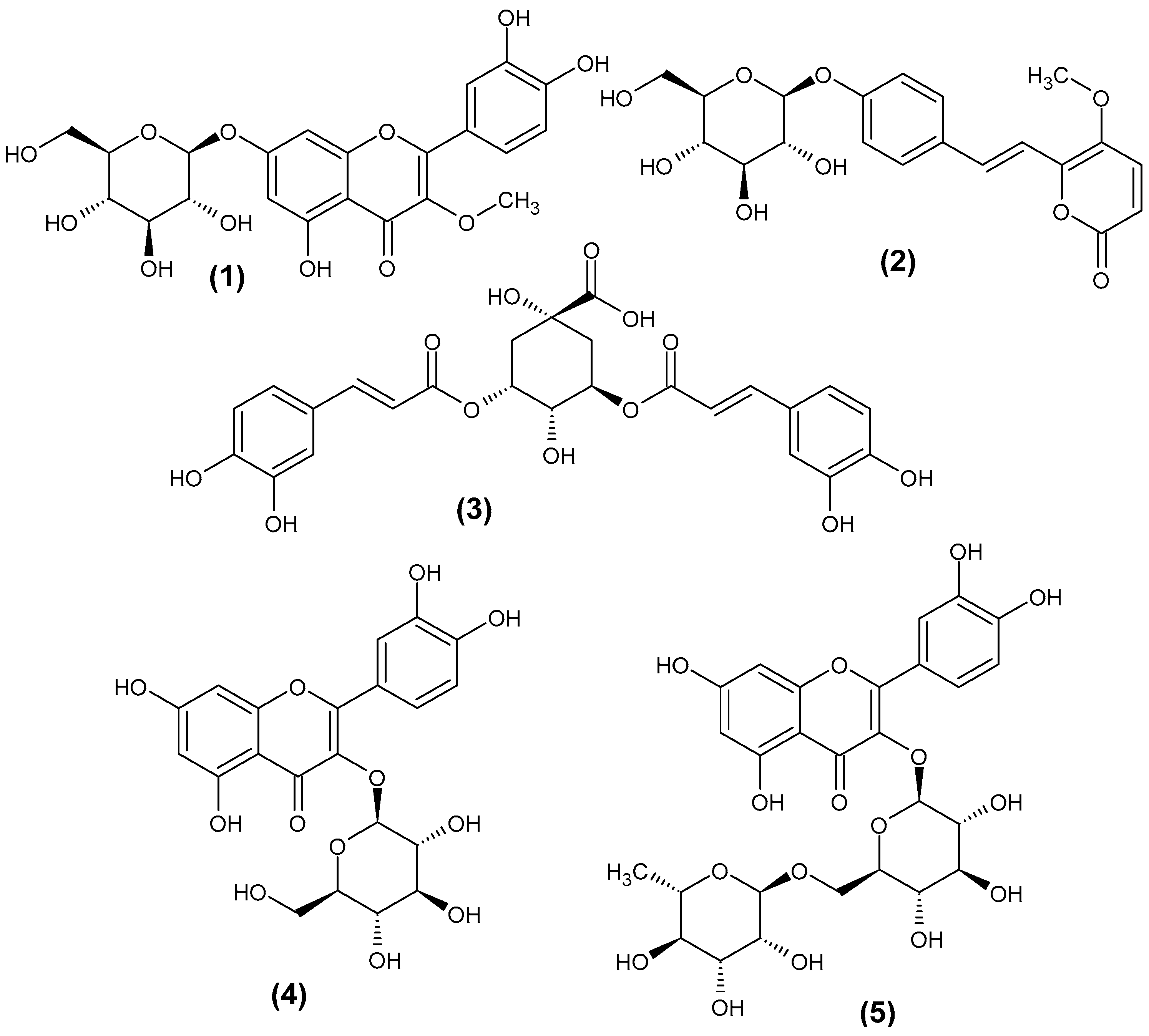
2.1. HPLC–MS Identification of Compounds 1–3
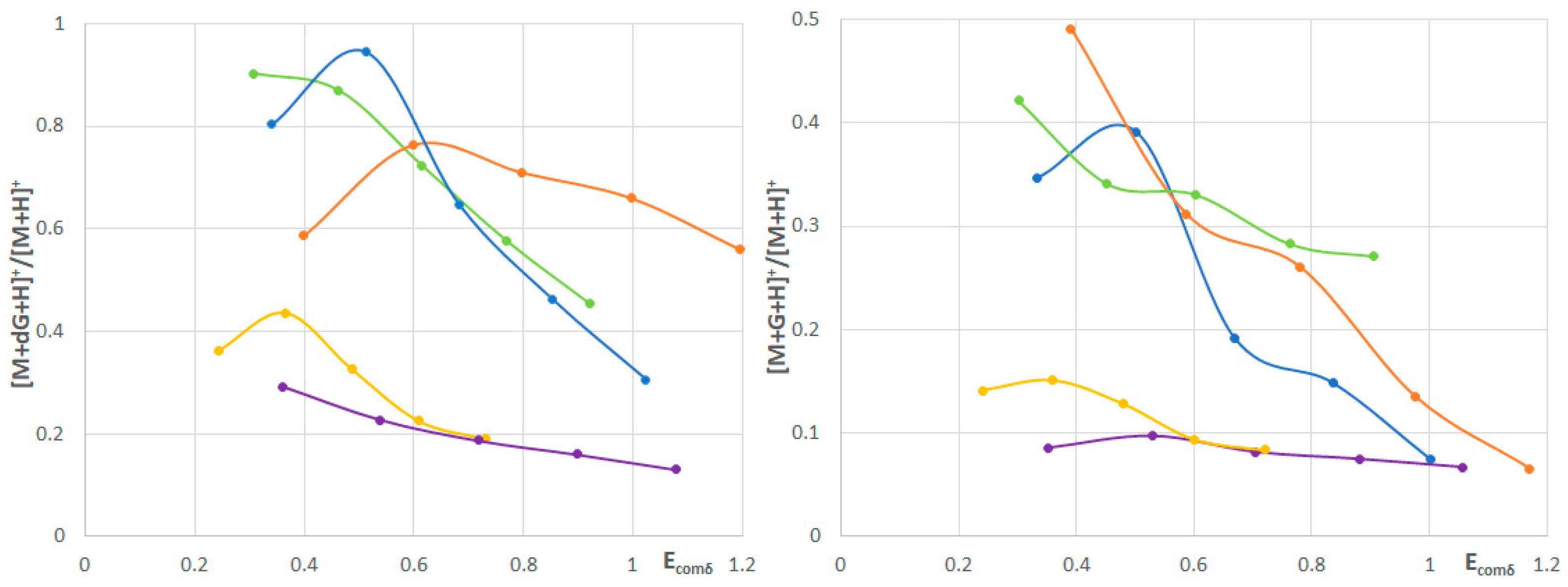
2.2. Stabilities of the Adducts of 1–5 with Deoxyguanosine and Guanosine
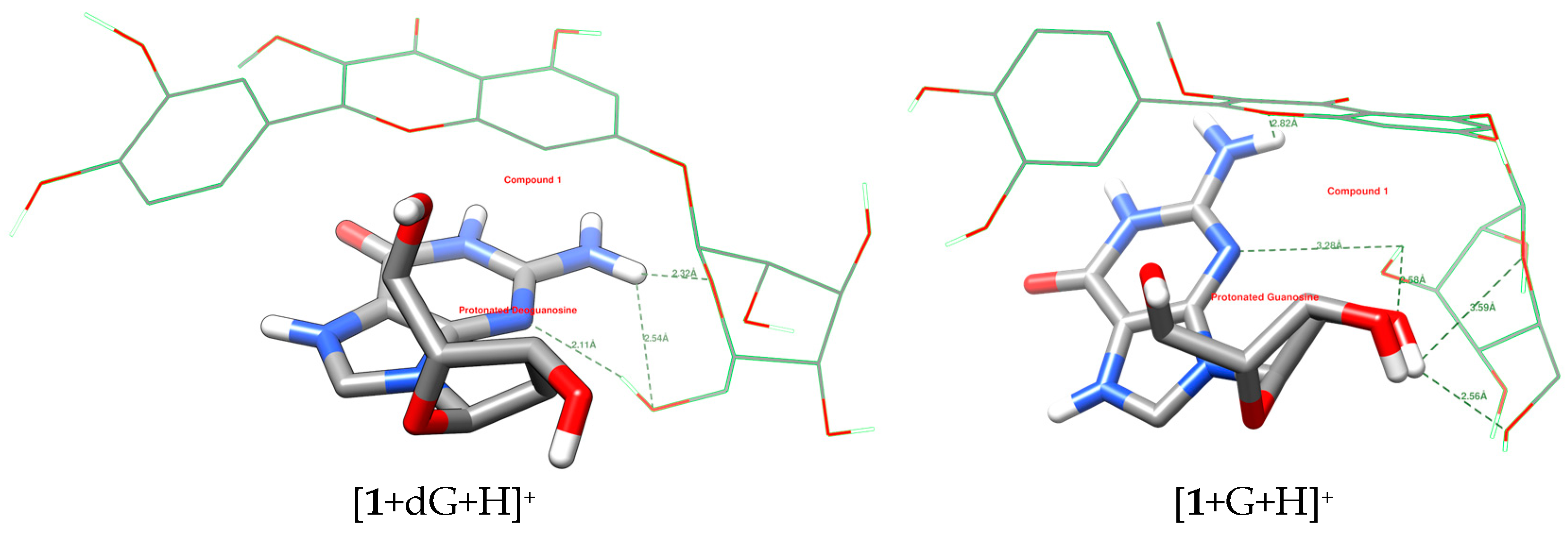
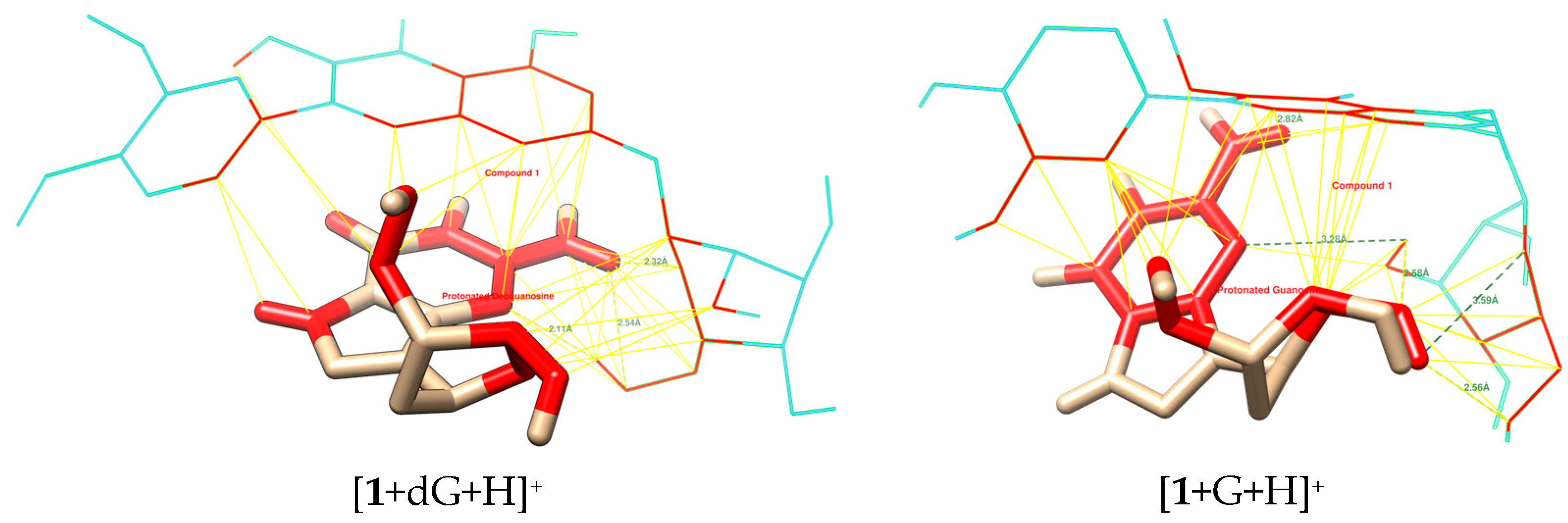
2.3. Molecular Docking
3. Materials and Methods
3.1. Sample Preparation
3.2. HPLC-MS Analysis and Post-Column Addition Experiments
3.3. Molecular Docking
4. Conclusions
Supplementary Materials
Author Contributions
Funding
Institutional Review Board Statement
Informed Consent Statement
Data Availability Statement
Conflicts of Interest
References
- Manousi, N.; Zacharis, C.K. Automated post-column sample manipulation prior to detection in liquid chromatography: A review of pharmaceutical and bioanalytical applications. Curr. Anal. Chem. 2019, 15, 759–775. [Google Scholar] [CrossRef]
- Shi, E.; Zuo, L.; Yao, H.; Sun, Z.; Chen, D. Recent advances in post-column derivatization: Enhancing sensitivity and selectivity in liquid chromatography analysis. TrAC Trends Anal. Chem. 2024, 178, 117799. [Google Scholar] [CrossRef]
- Shi, S.Y.; Zhang, Y.P.; Jiang, X.Y.; Chen, X.Q.; Huang, K.L.; Zhou, H.H. Coupling HPLC to on-line, post-column (bio) chemical assays for high-resolution screening of bioactive compounds from complex mixtures. TrAC Trends Anal. Chem. 2009, 28, 865–877. [Google Scholar] [CrossRef]
- Kool, J.; Giera, M.; Irth, H.; Niessen, W.M. Advances in mass spectrometry-based post-column bioaffinity profiling of mixtures. Anal. Bioanal. Chem. 2011, 399, 2655–2668. [Google Scholar] [CrossRef] [PubMed]
- Waridel, P.; Wolfender, J.L.; Lachavanne, J.B.; Hostettmann, K. Identification of the polar constituents of Potamogeton species by HPLC-UV with post-column derivatization, HPLC-MSn and HPLC-NMR, and isolation of a new ent-labdane diglycoside. Phytochemistry 2004, 65, 2401–2410. [Google Scholar] [CrossRef]
- Colombo, R.; Yariwake, J.H.; Queiroz, E.F.; Ndjoko, K.; Hostettmann, K. On-line identification of sugarcane (Saccharum officinarum L.) methoxyflavones by liquid chromatography-UV detection using post-column derivatization and liquid chromatography-mass spectrometry. J. Chromatogr. A 2005, 1082, 51–59. [Google Scholar] [CrossRef] [PubMed]
- Polasek, J.; Queiroz, E.F.; Hostettmann, K. On-line identification of phenolic compounds of Trifolium species using HPLC-UV-MS and post-column UV-derivatisation. Phytochem. Anal. 2007, 18, 13–23. [Google Scholar] [CrossRef] [PubMed]
- Colombo, R.; Yariwake, J.H.; Queiroz, E.F.; Ndjoko, K.; Hostettmann, K. On-line identification of minor flavones from sugarcane juice by LC/UV/MS and post-column derivatization. J. Braz. Chem. Soc. 2009, 20, 1574–1579. [Google Scholar] [CrossRef]
- Chen, H.J.; Inbaraj, B.S.; Chen, B.H. Determination of phenolic acids and flavonoids in Taraxacum formosanum Kitam by liquid chromatography-tandem mass spectrometry coupled with a post-column derivatization technique. Int. J. Mol. Sci. 2011, 13, 260–285. [Google Scholar] [CrossRef]
- Zhou, H.; Li, T.; Li, B.; Sun, S. Skin health properties of Paeonia lactiflora flower extracts and tyrosinase inhibitors and free radical scavengers identified by HPLC post-column bioactivity assays. Heliyon 2023, 9, e18569. [Google Scholar] [CrossRef]
- Niu, Y.; Yin, L.; Luo, S.; Dong, J.; Wang, H.; Hashi, Y.; Chen, S. Identification of the anti-oxidants in Flos chrysanthemi by HPLC-DAD-ESI/MSn and HPLC coupled with a post-column derivatisation system. Phytochem. Anal. 2013, 24, 59–68. [Google Scholar] [CrossRef] [PubMed]
- Raudonis, R.; Bumblauskiene, L.; Jakstas, V.; Pukalskas, A.; Janulis, V. Optimization and validation of post-column assay for screening of radical scavengers in herbal raw materials and herbal preparations. J. Chromatogr. A 2010, 1217, 7690–7698. [Google Scholar] [CrossRef] [PubMed]
- Fu, Q.; Zhang, C.; Lin, Z.; Sun, H.; Liang, Y.; Jiang, H.; Song, Z.; Wang, H.; Chen, S. Rapid screening and identification of compounds with DNA-binding activity from Folium Citri Reticulatae using on-line HPLC-DAD-MSn coupled with a post column fluorescence detection system. Food Chem. 2016, 192, 250–259. [Google Scholar] [CrossRef]
- Wang, K.-B.; Wang, Y.; Dickerhoff, J.; Yang, D. DNA G-Quadruplexes as Targets for Natural Product Drug Discovery. Engineering 2024, 38, 39–51. [Google Scholar] [CrossRef]
- Shan, C.; Tan, J.-H.; Ou, T.-M.; Huang, Z.-S. Natural products and their derivatives as G-quadruplex binding ligands. Sci. China Chem. 2013, 56, 1351–1363. [Google Scholar] [CrossRef]
- Che, T.; Wang, Y.-Q.; Huang, Z.-L.; Tan, J.-H.; Huang, Z.-S.; Chen, S.-B. Natural alkaloids and heterocycles as G-quadruplex ligands and potential anticancer agents. Molecules 2018, 23, 493. [Google Scholar] [CrossRef] [PubMed]
- Lu, Y.; Yu, S.; Lin, F.; Lin, F.; Zhao, X.; Wu, L.; Miao, Y.; Li, H.; Deng, Y.; Geng, L. Simultaneous label-free screening of G-quadruplex active ligands from natural medicine via a microfluidic chip electrophoresis-based energy transfer multi-biosensor strategy. Analyst 2017, 142, 4257–4264. [Google Scholar] [CrossRef]
- Artese, A.; Costa, G.; Ortuso, F.; Parrotta, L.; Alcaro, S. Identification of new natural DNA G-quadruplex binders selected by a structure-based virtual screening approach. Molecules 2013, 18, 12051–12070. [Google Scholar] [CrossRef]
- Monsen, R.C.; Trent, J.O. G-quadruplex virtual drug screening: A review. Biochimie 2018, 152, 134–148. [Google Scholar] [CrossRef]
- Rocca, R.; Moraca, F.; Costa, G.; Nadai, M.; Scalabrin, M.; Talarico, C.; Distinto, S.; Maccioni, E.; Ortuso, F.; Artese, A.; et al. Identification of G-quadruplex DNA/RNA binders: Structure-based virtual screening and biophysical characterization. Biochim. Et Biophys. Acta (BBA)-Gen. Subj. 2017, 1861, 1329–1340. [Google Scholar] [CrossRef]
- Ye, H.; Zhang, H.; Xiang, J.; Shen, G.; Yang, F.; Wang, F.; Wang, J.; Tang, Y. Advances and prospects of natural dietary polyphenols as G-quadruplex stabilizers in biomedical applications. Int. J. Biol. Macromol. 2024, 254, 127825. [Google Scholar] [CrossRef] [PubMed]
- Wang, H.; Chen, T.; Wu, S.; Chu, X.; Yu, R. A novel biosensing strategy for screening G-quadruplex ligands based on graphene oxide sheets. Biosens. Bioelectron. 2012, 34, 88–93. [Google Scholar] [CrossRef]
- Bai, G.; Cao, X.; Zhang, H.; Xiang, J.; Ren, H.; Tan, L.; Tang, Y. Direct screening of G-quadruplex ligands from Kalopanax septemlobus (Thunb.) Koidz extract by high performance liquid chromatography. J. Chromatogr. A 2011, 1218, 6433–6438. [Google Scholar] [CrossRef]
- Bianchi, S.E.; de Carvalho Meirelles, G.; Raabe, V.B.; de Souza, K.C.B.; Bassani, V.L. Achyrocline satureioides review: From the pharmacochemical diversity to the technological development of products. Fitoterapia 2023, 168, 105537. [Google Scholar] [CrossRef]
- Carini, J.P.; Klamt, F.; Bassani, V.L. Flavonoids from Achyrocline satureioides: Promising biomolecules for anticancer therapy. RSC Adv. 2014, 4, 3131–3144. [Google Scholar] [CrossRef]
- Doneda, E.; Bianchi, S.E.; Pittol, V.; Kreutz, T.; Scholl, J.N.; Ibañez, I.L.; Bracalente, C.; Durán, H.; Figueiró, F.; Klamt, F.; et al. 3-O-Methylquercetin from Achyrocline satureioides-Cytotoxic activity against A375-derived human melanoma cell lines and its incorporation into cyclodextrins-hydrogels for topical administration. Drug Deliv. Transl. Res. 2021, 11, 2151–2168. [Google Scholar] [CrossRef] [PubMed]
- De Souza, K.C.B.; Schapoval, E.E.S.; Bassani, V.L. LC determination of flavonoids: Separation of quercetin, luteolin and 3-O-methylquercetin in Achyrocline satureioides preparations. J. Pharm. Biomed. Anal. 2002, 28, 771–777. [Google Scholar] [CrossRef]
- Fernández-Fernández, A.M.; Dumay, E.; Lazennec, F.; Migues, I.; Heinzen, H.; Lema, P.; López-Pedemonte, T.; Medrano-Fernandez, A. Antioxidant, antidiabetic, and antiobesity properties, TC7-cell cytotoxicity and uptake of Achyrocline satureioides (Marcela) conventional and high pressure-assisted extracts. Foods 2021, 10, 893. [Google Scholar] [CrossRef]
- Grassi-Zampieron, R.; França, L.V.; Carollo, C.A.; Vieira, M.D.C.; Oliveros-Bastidas, A.; Siqueira, J.M.D. Comparative profiles of Achyrocline alata (Kunth) DC. and A. satureioides (Lam.) DC.; Asteraceae, applying HPLC-DAD-MS. Rev. Bras. Farmacogn. 2010, 20, 575–579. [Google Scholar] [CrossRef]
- Martínez-Busi, M.; Arredondo, F.; González, D.; Echeverry, C.; Vega-Teijido, M.A.; Carvalho, D.; Rodríguez-Haralambides, A.; Rivera, F.; Dajas, F.; Abin-Carriquiry, J.A. Purification, structural elucidation, antioxidant capacity and neuroprotective potential of the main polyphenolic compounds contained in Achyrocline satureioides (Lam) DC (Compositae). Bioorg. Med. Chem. 2019, 27, 2579–2591. [Google Scholar] [CrossRef]
- Holzschuh, M.H.; Gosmann, G.; Schneider, P.H.; Schapoval, E.E.S.; Bassani, V.L. Identification and stability of a new bichalcone in Achyrocline satureioides spray dried powder. Pharmazie 2010, 65, 650–656. [Google Scholar] [PubMed]
- Carini, J.P.; Kaiser, S.; Ortega, G.G.; Bassani, V.L. Development, optimisation and validation of a stability-indicating HPLC method of achyrobichalcone quantification using experimental designs. Phytochem. Anal. 2013, 24, 193–200. [Google Scholar] [CrossRef] [PubMed]
- Carini, J.P.; Leitao, G.G.; Schneider, P.H.; Santos, C.C.; Costa, F.N.; Holzschuh, M.H.; Klamt, F.; Bassani, V.L. Isolation of achyrobichalcone from Achyrocline satureioides by high-speed countercurrent chromatography. Curr. Pharm. Biotechn. 2015, 16, 66–71. [Google Scholar] [CrossRef] [PubMed]
- Kaloga, M.; Hänsel, R.; Cybulski, E.M. Isolierung eines kawapyrons aus Achyrocline satureioides. Planta Med. 1983, 48, 103–104. [Google Scholar] [CrossRef]
- March, R.E.; Lewars, E.G.; Stadey, C.J.; Miao, X.-S.; Zhao, X.; Metcalfe, C.D. A comparison of flavonoid glycosides by electrospray tandem mass spectrometry. Int. J. Mass Spectrom. 2006, 248, 61–85. [Google Scholar] [CrossRef]
- Yang, W.-Z.; Qiao, X.; Bo, T.; Wang, Q.; Guo, D.-A.; Ye, M. Low energy induced homolytic fragmentation of flavonol 3-O-glycosides by negative electrospray ionization tandem mass spectrometry. Rapid Commun. Mass Spectrom. 2014, 28, 385–395. [Google Scholar] [CrossRef]
- Feng, S.; Bagia, C.; Mpourmpakis, G. Determination of proton affinities and acidity constants of sugars. J. Phys. Chem. A 2013, 117, 5211–5219. [Google Scholar] [CrossRef]
- Szymborska, K.; Frański, R.; Beszterda-Buszczak, M. Extraction with acidified methanol-an easy and effective method of methyl chlorogenate formation, as studied by ESI-MS. Molecules 2022, 27, 7543. [Google Scholar] [CrossRef]
- Clifford, M.N.; Johnston, K.L.; Knight, S.; Kuhnert, N. Hierarchical scheme for LC-MSn identification of chlorogenic acids. J. Agric. Food Chem. 2003, 51, 2900–2911. [Google Scholar] [CrossRef]
- Jellen, E.E.; Chappell, A.M.; Ryzhov, V. Effects of size of noncovalent complexes on their stability during collision-induced dissociation. Rapid Commun. Mass Spectrom. 2002, 16, 1799–1804. [Google Scholar] [CrossRef]
- Forbes, M.W.; Volmer, D.A.; Francis, G.J.; Böhme, D.K. A comparison of data analysis methods for determining gas phase stabilities by cid: Alkali metal complexes of polyether ionophore antibiotics. J. Am. Soc. Mass Spectrom. 2005, 16, 779–791. [Google Scholar] [CrossRef]
- Stężycka, O.; Frańska, M.; Beszterda-Buszczak, M. Exploring glycosylated soy isoflavones affinities toward g-tetrads as studied by survival yield method. ChemPhysChem 2023, 24, e202300056. [Google Scholar] [CrossRef]
- Tomar, J.S. In-Silico Modeling studies of G-quadruplex with soy isoflavones having anticancerous activity. J. Mol. Model. 2015, 21, 193. [Google Scholar] [CrossRef] [PubMed]
- Li, W.; Zhang, M.; Zhang, J.L.; Li, H.Q.; Zhang, X.C.; Sun, Q.; Qiu, C.M. Interactions of daidzin with intramolecular G-quadruplex. FEBS Lett. 2006, 580, 4905–4910. [Google Scholar] [CrossRef]
- Stężycka, O.; Frańska, M. Binding of quercetin derivatives toward g-tetrads as studied by the survival yield method. ACS Omega 2023, 8, 39816–39821. [Google Scholar] [CrossRef] [PubMed]
- Ribaudo, G.; Oselladore, E.; Ongaro, A.; Zagotto, G.; Memo, M.; Gianoncelli, A. Enhanced G-quadruplex selectivity of flavonoid glycoside rutin over quercetin. Nat. Prod. Res. 2022, 36, 3469–3473. [Google Scholar] [CrossRef]
- Stężycka, O.; Kasperkowiak, M.; Frańska, M.; Nowak, D.; Hoffmann, M. Oxygen atom from carbonyl group as an important binding agent to the G-quadruplex-study case of flavonoids. ChemPlusChem 2024, 89, e202400186. [Google Scholar] [CrossRef] [PubMed]
- Chan, E.W.C.; Kezuka, M.; Chan, H.T.; Wong, S.K. Alpinia zerumbet: A review of the chemistry, quantity, and pharmacological properties of selected kavalactones. J. Nat. Remedies 2023, 23, 699–709. [Google Scholar] [CrossRef]
- Avogadro: An Open-Source Molecular Builder and Visualization Tool. Version 1.2.0. Available online: http://avogadro.cc/ (accessed on 24 February 2025).
- Hanwell, M.D.; Curtis, D.E.; Lonie, D.C.; Vandermeersch, T.; Zurek, E.; Hutchison, G.R. Avogadro: An advanced semantic chemical editor, visualization, and analysis platform. J. Cheminform. 2012, 4, 17. [Google Scholar] [CrossRef]
- Trott, O.; Olson, A.J. AutoDock Vina: Improving the speed and accuracy of docking with a new scoring function, efficient optimization, and multithreading. J. Comput. Chem. 2010, 31, 455–461. [Google Scholar] [CrossRef]
- O’Boyle, N.M.; Banck, M.; James, C.A.; Morley, C.; Vandermeersch, T.; Hutchison, G.R. Open Babel: An open chemical toolbox. J. Cheminform. 2011, 3, 33. [Google Scholar] [CrossRef] [PubMed]
- Open Babel Development Team. Open Babel. 2020. Version: 3.1.1. Available online: https://openbabel.org/index.html (accessed on 24 February 2025).
- Pettersen, E.F.; Goddard, T.D.; Huang, C.C.; Couch, G.S.; Greenblatt, D.M.; Meng, E.C.; Ferrin, T.E. UCSF Chimera-A visualization system for exploratory research and analysis. J. Comput. Chem. 2004, 25, 1605–1612. [Google Scholar] [CrossRef] [PubMed]


| Compound | Detected Ions m/z |
|---|---|
| 3-O-methylquercetin-7-O–glucoside (1) | 477 ([M–H]−), 462 ([M–H–CH3]−•), 315 ([aglycone–H]−), 300 ([aglycone–H–CH3]−•), 299 ([aglycone–H–CH4]−), 271 ([aglycone–H–CH4–CO]−); 479 ([M+H]+), 317 ([aglycone+H]+), 302 ([aglycone+H–CH3]+•) |
| 4′-hydroxydehydrokawain-4′-O-glucoside (2) | 451 ([M+HCOO]−), 441 ([M+Cl]−), 243 ([aglycone–H]−); 407 ([M+H]+), 245 ([aglycone+H]+) |
| 3,5-di-O-caffeoylquinic acid (3) | 515 ([M–H]−), 353 ([M–H–(OC–CH=CH–C6H4O2)]−), 191 ([M–H–(OC–CH=CH–C6H4O2)2]− or [quinic acid–H]−), 173 ([quinic acid–H–H2O]−), 179 ([caffeoyl acid–H]−), 135 ([CH=CH–C6H5O2]−) 517 ([M+H]+), 163 ([OC–CH=CH–C6H5O2]+) |
| Compound | Binding Energy to Protonated Deoxyguanosine [kcal/mol] | Binding Energy to Protonated Guanosine [kcal/mol] |
|---|---|---|
| 1 | −3.2 | −3.3 |
| 2 | −2.8 | −2.9 |
| 3 | −2.3 | −2.2 |
| 4 | −3.0 | −3.1 |
| 5 | −3.5 | −3.5 |
| Adduct | dG | G |
|---|---|---|
| [1+dG/G+H]+ | 2 | 3 |
| [2+dG/G+H]+ | 3 | 3 |
| [3+dG/G+H]+ | 3 | 5 |
| [4+dG/G+H]+ | 4 | 4 |
| [5+dG/G+H]+ | 6 | 6 |
Disclaimer/Publisher’s Note: The statements, opinions and data contained in all publications are solely those of the individual author(s) and contributor(s) and not of MDPI and/or the editor(s). MDPI and/or the editor(s) disclaim responsibility for any injury to people or property resulting from any ideas, methods, instructions or products referred to in the content. |
© 2025 by the authors. Licensee MDPI, Basel, Switzerland. This article is an open access article distributed under the terms and conditions of the Creative Commons Attribution (CC BY) license (https://creativecommons.org/licenses/by/4.0/).
Share and Cite
Stężycka, O.; Frańska, M.; Nowak, D.; Hoffmann, M.; Kasperkowiak, M.; Beszterda-Buszczak, M. Post–Column Guanosine Addition as a Screening Tool in the Search for Effective G–Quadruplex Binders—A Case Study of Achyrocline satureioides Phenolic Compounds. Int. J. Mol. Sci. 2025, 26, 4312. https://doi.org/10.3390/ijms26094312
Stężycka O, Frańska M, Nowak D, Hoffmann M, Kasperkowiak M, Beszterda-Buszczak M. Post–Column Guanosine Addition as a Screening Tool in the Search for Effective G–Quadruplex Binders—A Case Study of Achyrocline satureioides Phenolic Compounds. International Journal of Molecular Sciences. 2025; 26(9):4312. https://doi.org/10.3390/ijms26094312
Chicago/Turabian StyleStężycka, Olga, Magdalena Frańska, Damian Nowak, Marcin Hoffmann, Małgorzata Kasperkowiak, and Monika Beszterda-Buszczak. 2025. "Post–Column Guanosine Addition as a Screening Tool in the Search for Effective G–Quadruplex Binders—A Case Study of Achyrocline satureioides Phenolic Compounds" International Journal of Molecular Sciences 26, no. 9: 4312. https://doi.org/10.3390/ijms26094312
APA StyleStężycka, O., Frańska, M., Nowak, D., Hoffmann, M., Kasperkowiak, M., & Beszterda-Buszczak, M. (2025). Post–Column Guanosine Addition as a Screening Tool in the Search for Effective G–Quadruplex Binders—A Case Study of Achyrocline satureioides Phenolic Compounds. International Journal of Molecular Sciences, 26(9), 4312. https://doi.org/10.3390/ijms26094312