Review

Evaluation of Illumina and Oxford Nanopore Sequencing for the Study of DNA Methylation in Alzheimer’s Disease and Frontotemporal Dementia

1
Molecular Markers Laboratory, IRCCS Istituto Centro San Giovanni di Dio Fatebenefratelli, 25125 Brescia, Italy
2
Department of Biology, Ecology and Earth Sciences, University of Calabria, 87036 Rende, Italy
3
Department of Neuroscience, Psychology, Drug Research and Child Health, University of Florence, 50139 Florence, Italy
4
IRCCS Fondazione Don Carlo Gnocchi, 50143 Florence, Italy
5
Azienda Ospedaliero-Universitaria Careggi SOD Neurologia 1, 50100 Florence, Italy
6
Regional Neurogenetic Centre (CRN), Department of Primary Care, ASP Catanzaro, 88046 Lamezia Terme, Italy
7
Department of Clinical and Experimental Sciences, University of Brescia, 25123 Brescia, Italy
*
Author to whom correspondence should be addressed.
These authors contributed equally to this work.
Int. J. Mol. Sci. 2025, 26(9), 4198; https://doi.org/10.3390/ijms26094198
Submission received: 27 February 2025 / Revised: 24 April 2025 / Accepted: 25 April 2025 / Published: 28 April 2025
(This article belongs to the Special Issue New Advances in Epigenetics and Epigenomics)

Abstract

DNA methylation is a critical epigenetic mechanism involved in numerous physiological processes. Alterations in DNA methylation patterns are associated with various brain disorders, including dementias such as Alzheimer’s disease (AD) and frontotemporal dementia (FTD). Investigating these alterations is essential for understanding the pathogenesis and progression of these disorders. Among the various methods for detecting DNA methylation, DNA sequencing is one of the most widely employed. Specifically, two main sequencing approaches are commonly used for DNA methylation analysis: bisulfite sequencing and single-molecule long-read sequencing. In this review, we compared the performances of CpG methylation detection obtained using two popular sequencing platforms, Illumina for bisulfite sequencing and Oxford Nanopore (ON) for long-read sequencing. Our comparison considers several factors, including accuracy, efficiency, genomic regions, costs, wet-lab protocols, and bioinformatics pipelines. We provide insights into the strengths and limitations of both methods with a particular focus on their application in research on AD and FTD.

1. Introduction

The term epigenetics refers to the study of heritable changes in gene expression that do not involve alterations to the underlying DNA sequence. These modifications can influence how genes are turned on or off and are crucial for various biological processes, including development, differentiation, and diseases [1,2,3]. Epigenetic changes can be stable and heritable through mitosis/meiosis, without altering the DNA sequence itself [4]. Importantly, they can also be reversible, which has significant implications in treating diseases and developing novel therapeutic strategies [5]. Recent advances in epigenetics have highlighted its role in various diseases, including cancer, cardiovascular disorders, and neurodegenerative diseases [6,7,8,9,10]. Epigenetic mechanisms include DNA methylation, histone modification, and non-coding RNA interactions [6].
DNA methylation is a critical epigenetic process involving the addition of methyl groups to the C-5 position of the cytosine, forming 5-methylcytosine (5 mC). It has a role in many physiological processes such as the regulation of tissue-specific gene expression, genetic imprinting, X-inactivation, and transposons silencing to ensure genomic stability [11]. In mammals, DNA methylation mainly occurs in cytosines followed by a guanine nucleotide (CpG) and accounts for around 1% of the genome in healthy human tissues [4,12]. The majority of CpGs in the genome are hypermethylated while CpG-rich regions known as CpG islands (CGI), predominantly found at gene promoters, remain unmethylated [13]. CpG islands are short stretches of DNA where the frequency of CpG dinucleotides is significantly higher than in the rest of the genome. These regions are approximately 1 kb in length with a GC content greater than 50% and a ratio of CpG to GpC greater than 0.6 [14,15]. DNA methylation can modulate gene expression by inhibiting transcription when methyl groups are present in gene promoters. On the contrary, unmethylated CpG islands in promoters result in the transcription of a particular gene [16,17]. DNA methylation patterns are established and regulated by a family of enzymes called DNA methyltransferases (DNMTs). DNMT1 maintains the DNA methylation in somatic cells while DNMT3a and DNMT3b perform de novo methylation during development [18,19]. Active DNA demethylation is mediated by enzymes from the ten-eleven translocation (TET) family, which oxidize 5 mC to form 5-hydroxymethylcytosine (5 hmC). Further oxidation steps produce intermediate products, ultimately leading to the erasing of DNA methylation [20,21].
DNA methylation is highly tissue-specific and varies substantially between cell types. In the human brain, it is a key process for the proliferation and differentiation of neural stem cells, as well as in synaptic plasticity, learning, and memory. Alterations in DNA methylation patterns are associated with several brain disorders, including dementias such as Alzheimer’s disease (AD) and frontotemporal dementia (FTD) [22]. AD and FTD are two neurodegenerative dementias characterized by cognitive decline, leading to function disability [23]. They are considered multifactorial diseases, due to the complex interactions between genetic and environmental factors.
Genetic factors play an essential role in familial forms of both AD and FTD. In a very small proportion of cases, both diseases follow a Mendelian inheritance pattern with autosomal dominant transmission observed. High-penetrant mutations in three genes were identified to cause autosomal dominant AD (APP, PSEN1, and PSEN2), while causative, high-penetrant mutations in either one of the other three distinct genes (MAPT, GRN, and C9orf72) were identified as causative of monogenic FTD. Nevertheless, the majority of patients with AD and FTD appear to have a late-onset or sporadic form, with disease onset after the age of 65. In these cases, the interaction between genetic and environmental factors plays a crucial role in the multifactorial etiology of the disease [24,25,26]. Accordingly, neurotoxic agent exposures, alcohol consumption, smoking habits, or brain injuries can significantly raise the risk of AD and FTD by interfering with neurodegenerative genes through DNA methylation. Alterations in DNA methylation have been identified as key factors in the pathogenesis of these disorders [27,28,29,30,31,32,33,34,35,36].
Several epigenome-wide association studies (EWAS) conducted on AD and FTD patients revealed differentially methylated CpG sites (DMPs) and regions (DMRs), showing that specific DNA methylation alterations can influence the expression of genes involved in neurodegenerative processes [37,38,39,40]. DNA methylation changes have been observed in genes associated with the autosomal-dominant form of early-onset Alzheimer’s disease (EOAD), such as APP and PSEN1. Abnormal methylation of the APP gene can influence its expression and contribute to amyloid plaque formation, with increased methylation reducing gene expression [41,42]. In the case of the PSEN1 gene, low methylation is associated with higher gene expression and disease progression [43]. Conversely, no studies have yet been found linking PSEN2 methylation to AD. Altered methylation levels have also been found in genes associated with the autosomal dominant form of FTD, including MAPT, GRN, and C9orf72. Methylation of the MAPT gene is linked to an increased risk and severity of AD [44]; however, no significant differences in methylation levels have been found for FTD [39,45]. Specifically, in FTD, there is an increase in methylation of the GRN gene promoter, leading to a decrease in its expression [46]. Regarding the C9orf72 gene, carriers of the repeat expansion exhibit significantly higher levels of methylation in the CpG island compared to non-carriers, with a clear association between expansion size, age of onset, and methylation status in FTD patients [47,48]. Different studies have identified various hypermethylated and hypomethylated loci in other critical genes associated with AD and FTD. There are strong evidences that ankyrin 1 (ANK1) gene is hypermethylated in AD cortex and involved in the neuropathology of the disease [49,50]. Brain DNA methylation differences have been found in genes classically associated with AD such as SORL1, ABCA7, HLA-DRB5, SCL2A4, and BIN1 [51]. EWAS meta-analysis on FTD patients revealed differentially methylated loci in OTUD4 and in NFATC1. Of these loci, OTUD4 showed consistent upregulation of mRNA and protein expression in FTD [40].
Currently, various effective methods are employed for detecting DNA methylation, depending on the specific application, design of the study, or requirements. Array-based technologies represent a traditional approach that generates DNA methylation profiles for hundreds to thousands of samples at single-CpG resolution. Their main advantage is the economic efficiency, but this comes at the cost of a targeted design that interrogates only a small fraction of CpG sites across the genome.
For a more comprehensive understanding of epigenetic modifications that regulate gene expression and their impact on diseases, DNA sequencing-based approaches are essential [52]. When it comes to DNA methylation analysis, bisulfite sequencing technique is still considered the gold standard [53]. However, recent technological advancements have made the single molecule long-read sequencing technique an interesting alternative [54]. To date, a wide range of companies such as Illumina, Thermo Fisher Scientific, Roche, QIAGEN, PacBio, Oxford Nanopore, and BGI offer solutions for epigenetics research. Illumina confirms itself as the leader of the Next Generation Sequencing (NGS) market [55]. Considering the long-read sequencing (Third Generation Sequencing) segment, though, Oxford Nanopore accounts for the largest share [55].
No attempt has been made to compare these two technologies in the context of neurodegenerative dementias. This review aims to explore the advantages and limitations of Illumina and Oxford Nanopore sequencing techniques for DNA methylation analysis, and evaluate their application in research on neurodegenerative dementias such as AD and FTD.

2. Sequencing Techniques and Technologies

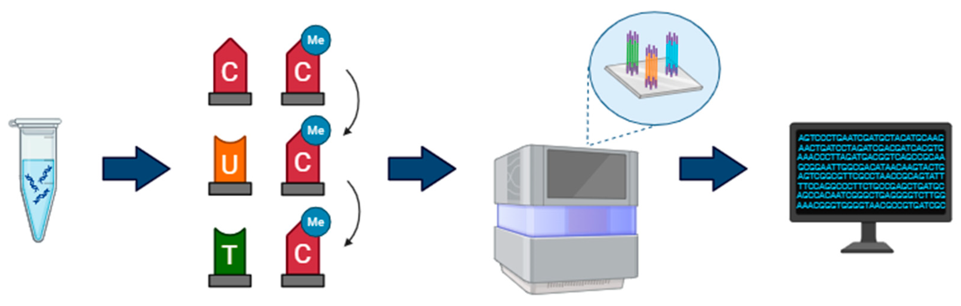
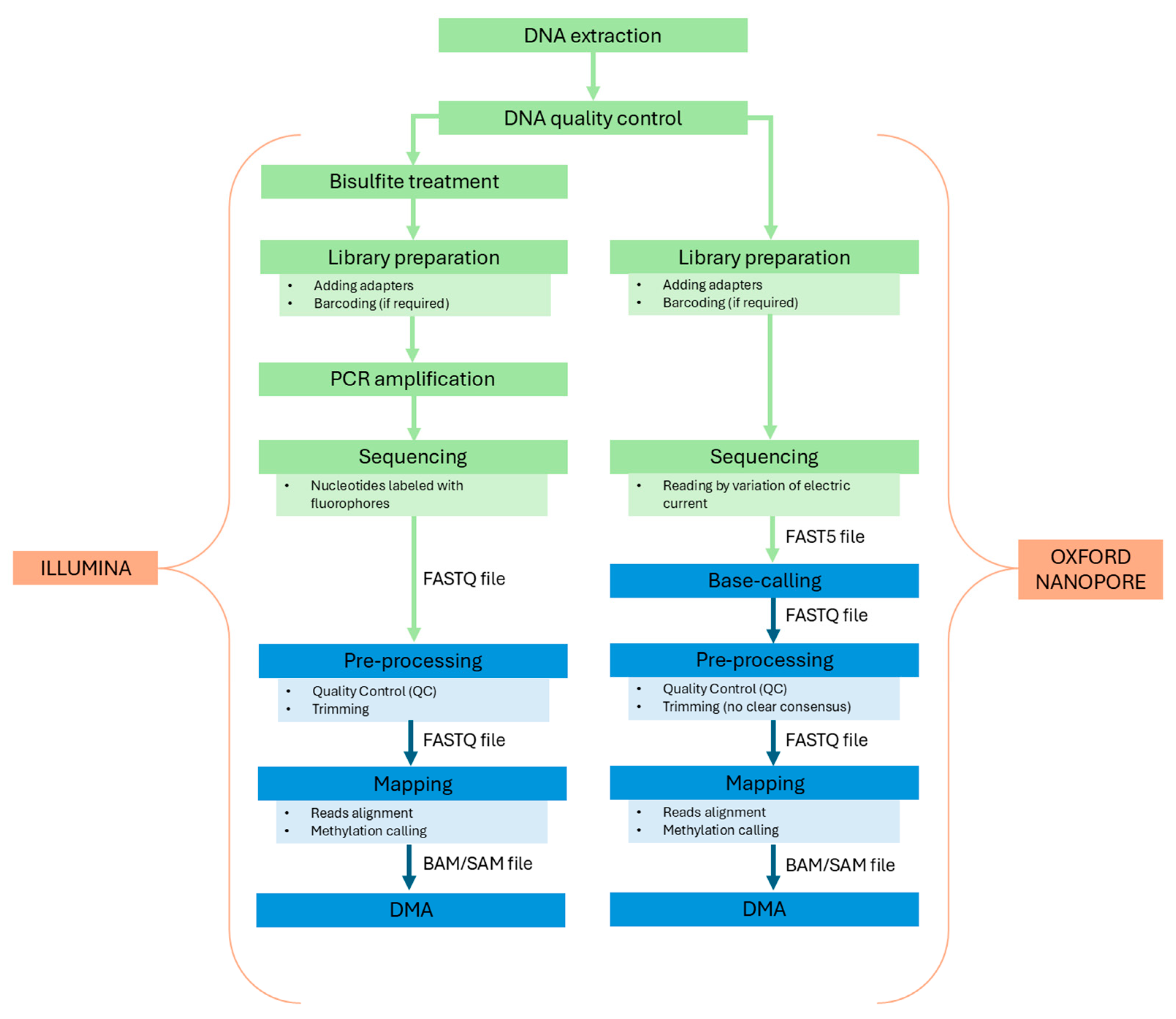
2.1. Illumina Bisulfite Sequencing

In bisulfite sequencing, the genomic DNA is treated with sodium bisulfite, resulting in a deamination of unmethylated cytosines to uracil and leaving methylated cytosines intact (Figure 1). The harsh treatment breaks the DNA into fragments; specific adapters are added to the ends of DNA fragments to assist in amplification and binding to the flow cell surface. PCR is then used to amplify the template, causing the conversion of the uracil to thymine. Subsequently, DNA is sequenced using short-read sequencing technologies like Illumina [56,57]. Illumina uses a sequencing-by-synthesis process, where DNA fragments bind to the flow cell and are locally amplified by PCR, creating clusters of identical molecules that enhance the reading signal. Reads are obtained by detecting fluorescent signals emitted by nucleotides added sequentially. Each sequencing cycle involves adding a nucleotide, detecting the fluorescent signal and removing the reversible terminator, allowing the next nucleotide to be incorporated [58]. Sequences are compared to a bisulfite-converted reference genome, and the percentage of methylation is estimated. As a result, 5 mC can be identified at single base-pair resolution using a qualitative, quantitative, and effective method [59,60]. Methylation studies using bisulfite sequencing can be employed through two methods: whole genome bisulfite sequencing (WGBS) and reduced representation bisulfite sequencing (RRBS). WGBS gives a full assessment of cytosine methylation covering most of the ~28 million CpGs in the human genome (≥2 CpG/100 bp); however, it remains the most expensive and resource-intensive technique [61,62,63]. As an alternative to WGBS, RRBS targets CpG-rich regions using a restriction enzyme that cuts at specific sites, allowing a reduction of costs and resources for the analysis. RRBS covers approximately 4 million human CpG sites [61,64,65].

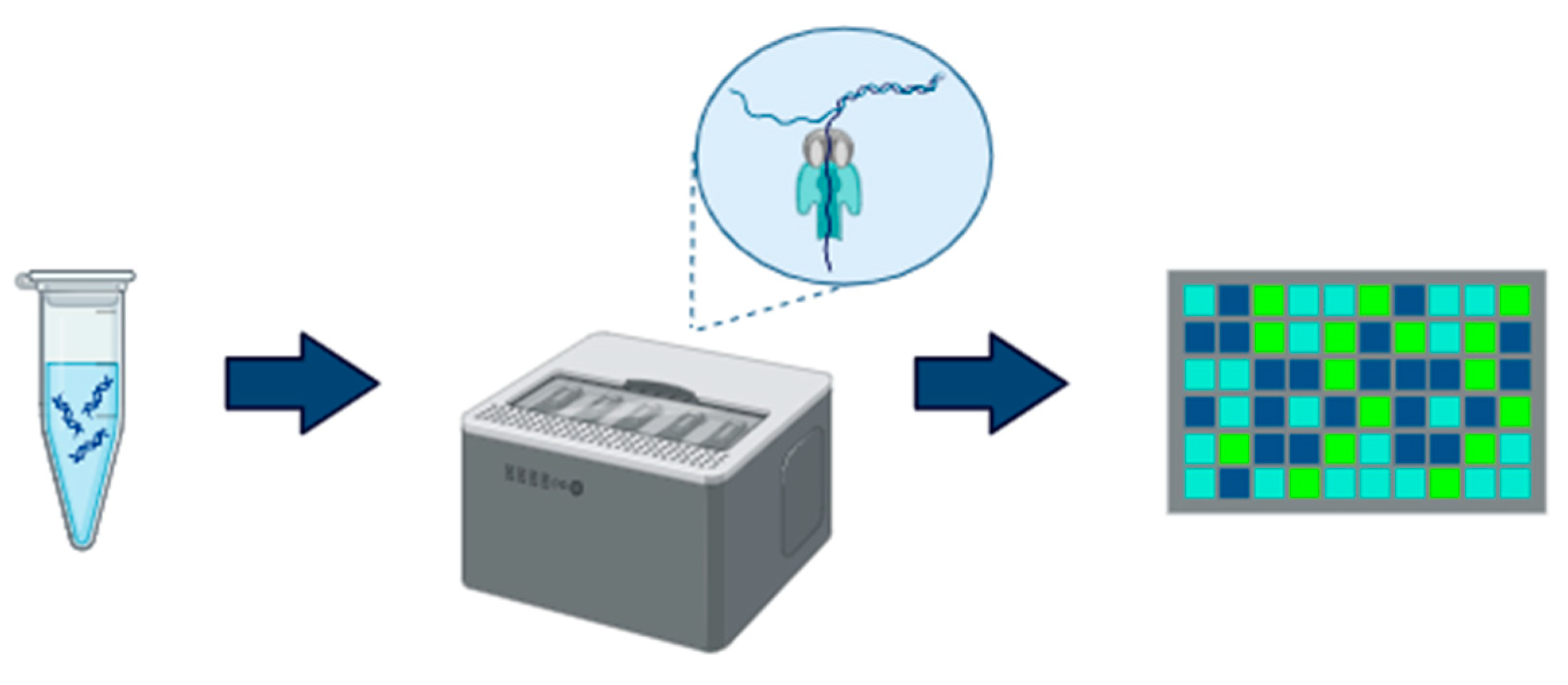
2.2. Oxford Nanopore

Thanks to the advancement of long-read sequencing, it is now possible to detect methylation without inducing a chemical treatment like bisulfite conversion to the DNA (Figure 2). Long-read sequencing technologies can generate longer sequences (ranging from 10 kb to >1 Mb in length) compared to short-read approaches (150–300 bp), and they have the capability to directly sequence native DNA, allowing the identification of methylation from raw sequence data [66,67]. Oxford Nanopore (ON) uses an innovative method involving the passage of DNA molecules through a nanopore fixed in a synthetic membrane. Unlike NGS sequencing methods, ON does not require PCR amplification or DNA fragmentation, thereby preserving the native structure of long DNA strands. Specific adapters are attached to the DNA to facilitate its passage through the nanopores. The membrane of each nanopore is exposed to an electrical current, and as the DNA moves through the pore, variations in the electrical signal are detected. These signal variations are translated into nucleotide sequences using advanced computational algorithms. Nanopores are sensitive enough to distinguish electric shifts between methylated and unmethylated cytosines, enabling detailed mapping of methylation profiles on a large scale [66,68]. The difference can be determined by comparing the electric current pattern to an in silico reference or an unmodified control, or through pre-trained models, such as neural networks [69], machine learning [70], or Hidden Markov Models (HMM) [71]. However, a key challenge arises in detecting methylation at closely CpG sites, as it is often assumed that all CpGs within a 10-bp region share the same methylation status. The nanopore sequencer can sequence the whole genome (WGM) or select molecules in real-time, determining whether to continue sequencing or discard them based on predefined regions of interest. Therefore, it is possible to design target genomic regions such as CpG islands or CpG rich promoters, essentially achieving reduced representation methylation sequencing (RRMS), which is conceptually similar to the commonly used RRBS [72].

3. Comparison

3.1. Accuracy and Efficiency

In this context, accuracy refers to how precisely the sequencing output reflects the fidelity of both nucleotide identification and methylation detection. Efficiency, on the other hand, encompasses factors such as the alignment success rate and the number of CpG sites detected. A comparative genome-wide DNA methylation study across sequencing technology showed that the quality scores above Q20 and Q30 on Illumina NovaSeq 6000 are 92% and 83% for RRBS, 95% and 90% for WGBS [73]. However, bisulfite treatment is a highly aggressive method. This chemical process can induce a significant degradation of DNA, increasing the probability of base-calling error and leading to lower alignment rates and data loss. This may result in variance in methylation levels due to technical biases [74,75]. Several tools have been developed to enhance efficiency in bisulfite sequencing data analysis. A comprehensive benchmarking study compared 14 alignment algorithms, concluding that BSMAP achieved the highest efficiency for detecting CpG sites and methylation levels, with an alignment rate exceeding 90% and approximately 26.5 million CpG sites detected [76]. On the other hand, ON sequencing using R9.4.1 pore version and the Guppy version 6.1.2 in super accuracy mode for base and methylation calls demonstrated a raw read accuracy of almost Q20 (98.81%) and a median read identity mapped to the reference genome of 96%. Methylation calls in ON covered 28.83 million CpG sites, representing 98.8% of the total CpG sites [77,78]. Interestingly, a recent study comparing ON and Illumina bisulfite sequencing found a high correlation (r = 0.967) between the methylation levels detected by the two platforms. This suggests that despite differences in sequencing chemistry, both platforms provide reliable and qualitative insights into methylation patterns [79]. Nonetheless, it has been observed that ON is able to reach a much higher confidence in CpGs detection at a much lower depth than bisulfite whole-genome sequencing [78]. Additionally, bisulfite sequencing has a known limitation in distinguishing between 5 mC and 5-hmC modifications, leading to an overestimation of 5 mC levels [80]. In contrast, ON allows for direct, real-time, and simultaneous sequencing and detection of both 5 mC and 5 hmC modifications [68].

3.2. Genome Regions

Illumina WGBS allows for comprehensive genome-wide DNA methylation analysis at single-nucleotide resolution. This approach covers key genomic regions, including promoters, introns, exons, as well as 5′ and 3′ untranslated regions (UTRs). Alternatively, Illumina RRBS can be employed to enrich for CpG-dense regions, particularly CpG islands found in gene promoters. However, highly repetitive regions of the genome remain difficult to sequence and characterize using short-read sequencing technologies like Illumina, due to limited sequencing depth and poor mapping accuracy [81,82]. In the human genome, two predominant types of repeat elements are tandem repeats (TRs), which range from a single base to megabases, and interspersed repeats, primarily consisting of transposable elements (TEs) [82]. The advent of ON has enabled the resolution of many of these previously inaccessible “dark loci”, thanks to the longer read lengths it produces. As a result, new methylation patterns within repetitive DNA, including those associated with genes linked to neurodegenerative dementias, can now be identified.

3.3. Costs

The cost per sample for WGBS is relatively high, about 300 $ per sample depending on the required coverage and the option of paired- or single-end sequencing. This makes the technology more suitable for large-scale studies or projects requiring a comprehensive view of genomic methylation. RRBS is a more economical technique, with costs ranging from $100 to $300 per sample, significantly reducing expenses for studies with a large number of samples or for analyses focused on specific genomic regions [64].
The cost per sample with ON is variable and higher compared to Illumina, ranging from $500 to $1500 per sample, depending on the required coverage and the desired read length. Although it may seem expensive initially, the ability to monitor sequencing progress in real-time can reduce overall costs if sufficient coverage is quickly achieved.

3.4. Wet-Lab Protocols

Regardless of the sequencing methods employed, all wet-lab workflows begin with DNA extraction from the biological samples. WGBS requires at least 100 ng of high-quality DNA. To ensure reliable results, the input DNA should be intact and minimally fragmented, as bisulfite treatment can cause additional fragmentation. Before sequencing, fragments should have a length of 200–400 base pairs [83]. RRBS requires less DNA than WGBS, with a minimum amount of 50 ng. Although it focuses only on specific portions of the genome (CpG-rich regions), high DNA quality is still necessary [64,84].
ON requires a larger amount of DNA compared to Illumina, with 1 µg needed to obtain high-quality reads. It is crucial that the DNA is highly intact and not fragmented to ensure long and accurate reads, with DNA fragments exceeding 30 kb. Samples must follow specific purity requirements; contaminants such as salts or proteins can compromise sequencing quality and can lead to a reduction in the measured A260/280 and A260/230 ratios (required 260/280 ratio of 1.8 and 260/230 ratio of 2.0–2.2) [85]. Regarding the library preparation, both Illumina and ON protocols include steps for adapter ligation and barcoding. However, as previously mentioned, Illumina bisulfite sequencing has the disadvantage of requiring two additional steps which can introduce several errors and biases: the sodium bisulfite tratment and PCR amplification. Furthermore, the preservation of bisulfite-treated samples is crucial for maintaining DNA integrity. It is recommended to store converted DNA at −80 °C to prevent degradation, as inadequate temperatures can cause DNA instability due to the conversion of cytosines to uracils. Although converted and purified DNA can be stored at −20 °C for at least 9 months without significant quality loss, for longer periods it is preferable to store it at −80 °C [86]. The two workflows are summarized in Figure 3.

3.5. Bioinformatics Pipelines

The bioinformatics pipeline for bisulfite sequencing is almost identical for both WGBS and RRBS experiments [87,88]. Shafi et al. [89] provided a detailed overview of this multi-step process, with a particular focus on the many tools that can be employed at each step. The workflow can be summarized in four main phases: data preprocessing, alignment, methylation calling, and differential methylation analysis (DMA). Data preprocessing begins with a quality check of the short reads (in FASTA/FASTQ format), usually performed with the FastQC (http://www.bioinformatics.babraham.ac.uk/projects/fastqc/ (accessed on 24 April 2025)) program. Low-quality bases and adapters are then trimmed to improve alignment efficiency. There are many trimming tools that can be employed for this task, such as Cutadapt [90], Trimmomatic [91], and Trim Galore! (https://github.com/FelixKrueger/TrimGalore (accessed on 24 April 2025)). After preprocessing, reads are ready for alignment to the reference genome and methylation calling, which estimates methylation levels. These two steps can be performed with specific bisulfate sequencing aligners. Three-letter aligners—such as Bismark [92], BS Seeker [93], Bwa-Meth (https://github.com/brentp/bwa-meth (accessed on 24 April 2025)), and BRAT [94]—convert all Cs into Ts in the forward strand and all Gs into As in the reverse strand of the reference genome. Wildcard aligners (such as BSMAP [95], RRBSMAP [96], GSNAP [97], and BRAT-BW [98]) replace all the Cs in the reference genome with a wildcard letter Y instead, allowing it to match both Cs and Ts in the bisulfite sequencing reads. The methylation state of each cytosine in the reads is determined afterward. As with standard aligners, the output is a file in the SAM/BAM format. An optional post-alignment phase of quality control, methylation statistics, and visualization of methylated sites should be added for a more comprehensive analysis. Software tools like BiQ Analyzer [99] or aligners like BRAT can accomplish this. Finally, methylation levels between CpG sites are compared through DMA. A wide variety of hypothesis testing methods have been developed for this task. The classification proposed by Shafi et al. helps in bringing order to this vast array of approaches. Some of the existing methods use count values (the number of CpG sites or read counts) to test whether the difference in methylation between samples is significant or due to chance. Other methods use ratio values (such as methylation percentage) instead. Count-based approaches include those based on logistic regression (methylKit [100]), beta-binomial distribution (methylSig [101]), or Hidden Markov Models (ComMet [102]). Ratio-based approaches include methods based on smoothing (BSmooth [60]), entropy (QDMR [103]), binary segmentation (metilene [104]), or again, Hidden Markov Models (HMM-DM [105]). Some approaches, like COHCAP [106], integrate classical statistical tests such as Fisher’s exact test, t-test, and ANOVA. An accurate evaluation of the advantages and disadvantages of each method is strongly recommended before making a choice. In particular, it is important to weigh crucial parameters such as spatial correlation between CpG sites, error control, and confounding effects, including read depth variance and biological covariation. For example, logistic regression-based approaches typically consider read depth but often ignore biological variation among replicates and spatial correlation. Moreover, some methods may be specific to a particular sequencing protocol, i.e., WGBS or RRBS. Regardless of the chosen method, the output of DMA will always yield a list of differentially methylated sites and/or regions (i.e., clusters of adjacent CpG sites).
The bioinformatics workflow for the detection of 5 mC on ON starts with the translation of raw electrical signal data into nucleotide sequences using base-caller softwares. The number of base-calling programs has grown and their efficiency has increased in the last years. Some of the most used base-calling programs developed by ON are Guppy (https://nanoporetech.com/document/Guppy-protocol (accessed on 24 April 2025)) and Flappie (https://github.com/nanoporetech/flappie (accessed on 24 April 2025)). Guppy is a general purpose base-caller available only to ON customers, while Dorado and Flappie are open-source software [107]. After a QC check, nucleotide sequences are aligned to the human reference genome. Minimap2 is a widely used aligner thanks to its capability to work with long genomic reads of hundreds of megabases in length [108]. The subsequent step is the methylation calling. Over the years, several different methylation-calling tools have been developed. Liu et al. assessed the performance of different methylation-calling tools, concluding that Guppy and Nanopolish (https://github.com/jts/nanopolish (accessed on 24 April 2025)) are the best options for users with limited computational resources, while Megaladon (https://github.com/nanoporetech/megalodon (accessed on 24 April 2025)) showed better performances at the cost of high-demanding resources [109]. Base-calling, alignment to a reference genome, and detection of modified bases can also be performed using Dorado (https://github.com/nanoporetech/dorado (accessed on 24 April 2025)) as a single tool.
Regarding DMA, very few softwares are specifically designed for ON, although existing methods designed for bisulfite sequencing can be used. Recently, it has been demonstrated that pycoMeth, a toolbox for differential methylation for long-read sequencing data, performs comparably to or better than previous tools for the segmentation and further identification of differential methylated regions (DMRs) [110].
The great majority of available software tools for bioinformatics data processing are specifically designed for bisulfite sequencing. Some of these tools can also be employed for ON sequencing, but results should be carefully evaluated. ON-specific tools, despite being few, are often a better option. Regardless of the tools used, BS and ON sequencing pipelines are quite similar. However, a few differences can be highlighted.
ON requires an additional base-calling step to obtain FASTQ files, whereas Illumina outputs them directly. The adapter/quality trimming step is a key step in the BS pipeline, where well-documented and benchmarked tools are usually employed. In contrast, for ON there is no clear consensus on how to address this task. The effectiveness of the trimming step in reducing errors for ON sequencing is also in question [111]. Among the few available ON-specific trimming tools are Porechop (https://github.com/rrwick/Porechop/ (accessed on 24 April 2025)), Porechop_ABI [112], ProwlerTrimmer [113], Chopper (https://github.com/wdecoster/chopper (accessed on 24 April 2025)), and fastplong (https://github.com/OpenGene/fastplong (accessed on 24 April 2025)), the new fastp-based tool optimized for long-read data. Some aligners, such as Guppy and Dorado, can also perform adapter trimming. The alignment step also differs between BS and ON sequencing. BS aligners are designed to deal with the bisulfite treatment of the DNA, which involves some kind of base conversion as described previously. Conversely, ON aligners do not modify reads or references but are optimized to deal with the higher error rates caused by ON technologies and by the inherent difficulties of long-read alignments. A key challenge in methylation calling using ON sequencing lies in accurately detecting methylation at closely spaced CpG sites, due to signal interference resulting from the overlapping influence of adjacent bases during sequencing [109], a difficulty not encountered in BS. Nanopolish explicitly assumes that all CpGs within a 10-bp window share the same methylation state, which can result in inaccurate predictions when this assumption is violated [114]. Although the extent to which Guppy and Megalodon address this specific issue remains unclear, Liu et al. demonstrated that their performance, like Nanopolish, declines in the presence of non-concordant methylation states at neighboring CpG sites. While Dorado’s performance in this context has not been comprehensively assessed, its reliance on training datasets with fixed ratios of methylated and unmethylated bases suggests a potential susceptibility to misclassification [115]. In contrast, the deep learning-based method DeepBAM has been shown to achieve improved model performance by incorporating contextual information from extended k-mer windows, enabling more reliable detection of complex, heterogeneous methylation patterns [116].

4. Discussion

DNA methylation is a fundamental epigenetic mechanism involved in various physiological processes. Alterations in this mechanism can potentially lead to severe dysfunctions and disorders. Several brain disorders, including dementias such as AD and FTD are associated with modified methylation patterns. DNA sequencing is one of the most commonly used methods for detecting DNA methylation. More specifically, two sequencing approaches are currently dominant for DNA methylation detection: sequencing of bisulfite-converted DNA and single-molecule long-read sequencing [117].
To determine the most suitable approach for AD and FTD methylation studies, we compared Illumina sequencing, which involves DNA bisulfite conversion, with ON sequencing, which employs single-molecule long-read technology. We evaluated these two platforms across several aspects, including accuracy, efficiency, genomic regions covered, costs, wet-lab protocols and bioinformatics pipelines (Table 1). Illumina-based bisulfite sequencing, particularly WGBS, shows high accuracy, typically producing a significant percentage of bases above Q20 and Q30 and with single-nucleotide resolution across the genome. On the other hand, the quality of ON long-read sequencing has made substantial improvements in accuracy over the past decade, now reaching the Q20 accuracy for the detection of 5 mC. Although the accuracy of Illumina bisulfite sequencing remains slightly superior, Oxford Nanopore has the advantage of directly detecting methylation without the need of bisulfite treatment, thereby preserving the DNA original structure and avoiding the degradation or amplification biases that may occur with bisulfite conversion and PCR amplification [118]. Both technologies demonstrate comparable efficiency in terms of alignment rate and number of detected CpG sites. Additionally, it has been shown a high concordance in methylation levels between ON and Illumina (r > 0.95). In terms of initial DNA quality and quantity, ON sequencing imposes more stringent requirements than Illumina. Cost considerations are also essential: ON is generally more expensive, making Illumina a more feasible option when resources are limited. Consequently, cost per sample should be a primary factor in study planning and experimental design. Another often-overlooked aspect when selecting the best suitable platform is the bioinformatics processing. Due to the recent emergence and growing adoption of ON technology, there are relatively few tools specifically developed for its data. Furthermore, these tools have not undergone the extensive testing and benchmarking that tools designed for Illumina platforms have. One major challenge in methylation calling using ON tools is the reliable identification of methylation at CpG sites that are located close together. While it has been suggested that closer neighboring CpG sites are more likely to share the same methylation status, with co-methylation decreasing as the distance between CpG sites increases [119], a more recent study has been shown that a substantial number of adjacent CpG loci in human genome display discordant and cell-specific methylation patterns, and they are involved in regulation of the activity of enhancers [120]. Disregulation of enhancer methylation in neurons has been associated with AD pathology and cognitive symptoms. A large cluster of significantly hypomethylated enhancers has been identified within the DSCAML1 gene that targets BACE1. Hypomethylation of these enhancers in AD is associated with an upregulation of BACE1 transcripts and an increase in amyloid plaques, neurofibrillary tangles, and cognitive decline [121]. Since DNA methylation landscapes vary significantly between brain regions and across different cell types [122,123,124], it is not well known whether assuming uniform methylation across adjacent CpG sites may obscure important regulatory regions and lead to oversimplified interpretations of epigenetic mechanisms underlying neurodegenerative disease processes. An experimental comparison of ON and Illumina in detecting DNA methylation at closely spaced CpG sites, particularly within enhancers, may highlight important differences and consequently guide the selection of the most appropriate technology for studies on neurodegenerative diseases focused on specific regulatory elements. Despite these limitations, nanopore sequencing offers a unique advantage by enabling the simultaneous detection of both 5 mC and 5 hmC modifications, a capability not achievable with bisulfite-based methods. This approach allows for more accurate real-time estimates of both modifications, eliminating the need for separate experiments that may alter or complicate downstream analyses. The 5 hmC has been observed to be enriched in the human brain, particularly in neurons, and have a role in neurological disorders [125,126,127,128].
During brain development and the early stages of adulthood, the 5 hmC accumulates, indicating a crucial role in the growth and function of neurons [129,130]. Different brain areas, including the entorhinal cortex, cerebellum, and neocortex, showed reduced 5 hmC levels in late-stage AD patients [131,132], while the temporal gyrus presented elevated 5 hmC levels [132,133]. However, a number of disagreements are still present, especially for temporal gyrus and hippocampus [134,135]. Smith et al. [136] demonstrated that hypohydroxymethylation had previously confounded estimates of DNA hypermethylation at the ANK1 gene in AD patients. As 5 hmC is a precursor of active DNA demethylation, its reduced levels may reflect a loss of this mechanism in AD. Global levels of 5 hmC have also been found to be altered in FTD patients, compared to controls [137], but no studies have expanded our understanding of which brain regions, cell types, or specific genes exhibit these differences. The lack of studies and the discrepancies indicate the need to amplify researches to pinpoint the precise function of 5 hmC in AD and FTD. To date, no studies have considered the possibility of studying 5 hmC in AD and FTD using nanopore sequencing. Most studies that have identified methylation differences in genes and regions related to AD and FTD have primarily used immunohistochemical techniques and microarray-based methods, which only interrogate a small fractions of CpG sites. There is still a significant need to elucidate DNA methylation patterns and provide conclusive results in the genomic regions implicated in these pathologies. WGBS or RRBS on Illumina platforms can provide a comprehensive methylation signature in relevant regions previously associated with AD and FTD, and potentially identify novel regions. Nevertheless, short-read sequencing platforms like Illumina face challenges in accurately sequencing highly repetitive regions of the genome, which can be overcome by ON sequencing. A direct long-read sequencing can effectively span large tandem repeats, providing insights into their length, nucleotide content, and associated base modifications [138]. In a recent study, Ramirez et al. [139] investigated the role and the impact of DNA methylation on repetitive regions in patients with AD, using ON sequencing. They identified differentially methylated promoters within repetitive/dark regions and alterations in bulk 5 mC modifications of alpha satellites at a chromosomal scale. These regions are not detectable with Illumina-based methods, but they could reveal processes such as inflammation and genomic instability in neurodegeneration [140,141]. The GGGGCC repeat (G4C2)n expansion in the C9orf72 gene is the most frequently observed genetic cause of FTD. It has been shown that C9orf72 gene expression is reduced in multiple brain regions of patients with the repeat expansion [142,143,144].
Although various studies have employed different methods to evaluate C9orf72 promoter methylation, few have explored the potential of using long-read sequencing to better characterize epigenetic modifications in this gene. To our knowledge, only one study—by Udine et al. [145]—investigated the methylation levels in C9orf72 using long-read sequencing, primarily in patients with amyotrophic lateral sclerosis (ALS). Their findings indicate that higher methylation levels are associated with longer repeat expansions, and that methylation levels correlate with age at disease onset.
To date, no studies have specifically explored the potential of using nanopore sequencing to investigate DNA methylation within C9orf72 or to identify novel methylation signatures in repetitive regions in FTD patients. In summary, our review indicates that both technologies exhibit comparable efficacy in detecting DNA methylation; however, their suitability depends on the specific objectives and constraints of each study. Thus, we recommend a careful evaluation of each technology’s respective advantages and limitations when selecting the most appropriate platform for a given study. ON offers distinct advantages in assessing methylation patterns in AD and FTD, as it can target additional genomic regions beyond those accessible through Illumina bisulfite sequencing, including repetitive sequences potentially implicated in these pathologies. Furthermore, ON allows for the differentiation of 5 hmC, offering additional insights into its role in neurodegenerative dementias. Conversely, Illumina bisulfite sequencing may be more suitable for studies where DNA quality or quantity is suboptimal or when financial constraints demand a cost-effective approach. Illumina may also be preferable when the regions of interest are already known to be effectively sequenced through bisulfite sequencing.

5. Conclusions

This review compares Illumina bisulfite sequencing and ON sequencing for DNA methylation profiling, emphasizing the strengths and limitations of each technology, particularly in the context of AD and FTD research. The choice between Illumina and ON should be carefully considered, accounting for study objectives, sample quality, bioinformatics analysis, and budgetary constraints, as platform selection can significantly impact research outcomes. To date, few studies have directly compared these two technologies in their application to DNA methylation analysis, and none have focused specifically on neurodegenerative diseases. This review aims to serve as a preliminary guide for researchers investigating DNA methylation in AD and FTD through sequencing. However, our comparison provides only a general overview on the topic, and more targeted benchmarking studies are needed. Future research will also be essential to validate and expand ON’s application for detecting 5 hmC and methylation in repetitive regions, potentially unlocking new insights into the methylation landscape of AD and FTD. Choosing the appropriate platform could be a game changer in the discovery of novel epigenetic mechanisms that modulate the pathogenesis and/or progression of these diseases. Aberrant DNA methylation patterns that precede overt neurodegeneration may facilitate the early diagnosis of AD and FTD, particularly in sporadic forms. As epigenetics serves as a bridge between genes and the environment, identifying such patterns could also help clarify how environmental factors contribute to neurodegeneration. Lastly, given that DNA methylation is a reversible epigenetic modification, the development of novel compounds capable of inducing site-specific methylation or demethylation could enable precise therapeutic modulation of gene expression, offering new opportunities for the treatment of neurodegenerative disorders.

Author Contributions

Conceptualization, B.N., A.M. (Alberto Montesanto) and R.G.; methodology, L.P., D.L. and A.F.; formal analysis, L.P., D.L. and A.F; investigation, L.P., D.L. and A.F.; resources, R.M., B.N., A.M. (Alberto Montesanto), and R.G.; writing—original draft preparation, L.P., D.L. and A.F.; writing—review and editing, C.S., A.L., S.B. (Sonia Bellini), A.I., S.B. (Silvia Bagnoli), T.D., A.M. (Alessandra Mingrino), V.L., E.P., B.B., R.M., B.N. and A.M. (Alberto Montesanto) and R.G.; visualization, L.P., D.L. and A.F.; supervision, B.B., R.M., B.N., A.M. (Alberto Montesanto), and R.G.; project administration, R.G.; funding acquisition, R.G., B.N., R.M. and A.M. (Alberto Montesanto). All authors have read and agreed to the published version of the manuscript.

Funding

This research was funded by the European Union—Next Generation EU—NRRP M6C2—Investment 2.1 Enhancement and strengthening of biomedical research in the NHS—PNRR-MR1-2022-12375654—Cup Code: C83C22001300001. Title: Genetic and epigenetic modulAtors in Rare neurodegenerative diseases with DEmentia: a National study on autosomal dominant Alzheimer disease and genetic frontotemporal degeneration with dementIA (GARDENIA).

Conflicts of Interest

The authors declare no conflicts of interest. The funders had no role in the design of the study; in the collection, analyses, or interpretation of data; in the writing of the manuscript; or in the decision to publish the results.

References

  1. Gibney, E.R.; Nolan, C.M. Epigenetics and Gene Expression. Heredity 2010, 105, 4–13. [Google Scholar] [CrossRef] [PubMed]
  2. Farsetti, A.; Illi, B.; Gaetano, C. How Epigenetics Impacts on Human Diseases. Eur. J. Intern. Med. 2023, 114, 15–22. [Google Scholar] [CrossRef] [PubMed]
  3. Kiefer, J.C. Epigenetics in Development. Dev. Dyn. 2007, 236, 1144–1156. [Google Scholar] [CrossRef] [PubMed]
  4. Bird, A. DNA Methylation Patterns and Epigenetic Memory. Genes Dev. 2002, 16, 6–21. [Google Scholar] [CrossRef]
  5. Rasmi, Y.; Shokati, A.; Hassan, A.; Aziz, S.G.; Bastani, S.; Jalali, L.; Moradi, F.; Alipour, S. The Role of DNA Methylation in Progression of Neurological Disorders and Neurodegenerative Diseases as Well as the Prospect of using DNA Methylation Inhibitors as Therapeutic Agents for such Disorders. IBRO Neurosci. Rep. 2022, 14, 28–37. [Google Scholar] [CrossRef]
  6. Bird, A. Perceptions of Epigenetics. Nature 2007, 447, 396–398. [Google Scholar] [CrossRef]
  7. Jones, P.A.; Baylin, S.B. The Epigenomics of Cancer. Cell 2007, 128, 683–692. [Google Scholar] [CrossRef]
  8. Krolevets, M.; Cate, V.T.; Prochaska, J.H.; Schulz, A.; Rapp, S.; Tenzer, S.; Andrade-Navarro, M.A.; Horvath, S.; Niehrs, C.; Wild, P.S. DNA Methylation and Cardiovascular Disease in Humans: A Systematic Review and Database of Known CpG Methylation Sites. Clin. Epigenet. 2023, 15, 56. [Google Scholar] [CrossRef]
  9. Moosavi, A.; Motevalizadeh Ardekani, A. Role of Epigenetics in Biology and Human Diseases. Iran. Biomed. J. 2016, 20, 246–258. [Google Scholar] [CrossRef]
  10. Vodovotz, Y.; Barnard, N.; Hu, F.B.; Jakicic, J.; Lianov, L.; Loveland, D.; Buysse, D.; Szigethy, E.; Finkel, T.; Sowa, G.; et al. Prioritized Research for the Prevention, Treatment, and Reversal of Chronic Disease: Recommendations from the Lifestyle Medicine Research Summit. Front. Med. 2020, 7, 585744. [Google Scholar] [CrossRef]
  11. Moore, L.D.; Le, T.; Fan, G. DNA Methylation and its Basic Function. Neuropsychopharmacology 2013, 38, 23–38. [Google Scholar] [CrossRef] [PubMed]
  12. Ehrlich, M.; Gama-Sosa, M.A.; Huang, L.H.; Midgett, R.M.; Kuo, K.C.; McCune, R.A.; Gehrke, C. Amount and Distribution of 5-Methylcytosine in Human DNA from Different Types of Tissues of Cells. Nucleic Acids Res. 1982, 10, 2709–2721. [Google Scholar] [CrossRef]
  13. Schübeler, D. Function and Information Content of DNA Methylation. Nature 2015, 517, 321–326. [Google Scholar] [CrossRef] [PubMed]
  14. Illingworth, R.S.; Bird, A.P. CpG Islands—‘A Rough Guide’. FEBS Lett. 2009, 583, 1713–1720. [Google Scholar] [CrossRef]
  15. Long, M.D.; Smiraglia, D.J.; Campbell, M.J. The Genomic Impact of DNA CpG Methylation on Gene Expression; Relationships in Prostate Cancer. Biomolecules 2017, 7, 15. [Google Scholar] [CrossRef] [PubMed]
  16. Boyes, J.; Bird, A. Repression of Genes by DNA Methylation Depends on CpG Density and Promoter Strength: Evidence for Involvement of a Methyl-CpG Binding Protein. EMBO J. 1992, 11, 327–333. [Google Scholar] [CrossRef]
  17. Weber, M.; Hellmann, I.; Stadler, M.B.; Ramos, L.; Pääbo, S.; Rebhan, M.; Schübeler, D. Distribution, Silencing Potential and Evolutionary Impact of Promoter DNA Methylation in the Human Genome. Nat. Genet. 2007, 39, 457–466. [Google Scholar] [CrossRef]
  18. Jeltsch, A. On the Enzymatic Properties of Dnmt1: Specificity, Processivity, Mechanism of Linear Diffusion and Allosteric Regulation of the Enzyme. Epigenetics 2006, 1, 63–66. [Google Scholar] [CrossRef]
  19. Ren, W.; Gao, L.; Song, J. Structural Basis of DNMT1 and DNMT3A-Mediated DNA Methylation. Genes 2018, 9, 620. [Google Scholar] [CrossRef]
  20. Ito, S.; Shen, L.; Dai, Q.; Wu, S.C.; Collins, L.B.; Swenberg, J.A.; He, C.; Zhang, Y. Tet Proteins can Convert 5-Methylcytosine to 5-Formylcytosine and 5-Carboxylcytosine. Science 2011, 333, 1300–1303. [Google Scholar] [CrossRef]
  21. Tahiliani, M.; Koh, K.P.; Shen, Y.; Pastor, W.A.; Bandukwala, H.; Brudno, Y.; Agarwal, S.; Iyer, L.M.; Liu, D.R.; Aravind, L.; et al. Conversion of 5-Methylcytosine to 5-Hydroxymethylcytosine in Mammalian DNA by MLL Partner TET1. Science 2009, 324, 930–935. [Google Scholar] [CrossRef] [PubMed]
  22. Younesian, S.; Yousefi, A.; Momeny, M.; Ghaffari, S.H.; Bashash, D. The DNA Methylation in Neurological Diseases. Cells 2022, 11, 3439. [Google Scholar] [CrossRef] [PubMed]
  23. Rascovsky, K.; Salmon, D.P.; Ho, G.J.; Galasko, D.; Peavy, G.M.; Hansen, L.A.; Thal, L.J. Cognitive Profiles Differ in Autopsy-Confirmed Frontotemporal Dementia and AD. Neurology 2002, 58, 1801–1808. [Google Scholar] [CrossRef]
  24. Stozická, Z.; Zilka, N.; Novák, M. Risk and Protective Factors for Sporadic Alzheimer’s Disease. Acta Virol. 2007, 51, 205–222. [Google Scholar] [PubMed]
  25. Rosso, S.M.; Landweer, E.; Houterman, M.; Donker Kaat, L.; van Duijn, C.M.; van Swieten, J.C. Medical and Environmental Risk Factors for Sporadic Frontotemporal Dementia: A Retrospective Case-Control Study. J. Neurol. Neurosurg. Psychiatry 2003, 74, 1574–1576. [Google Scholar] [CrossRef]
  26. Saraceno, C.; Pagano, L.; Laganà, V.; Geviti, A.; Bagnoli, S.; Ingannato, A.; Mazzeo, S.; Longobardi, A.; Fostinelli, S.; Bellini, S.; et al. Mutational Landscape of Alzheimer’s Disease and Frontotemporal Dementia: Regional Variances in Northern, Central, and Southern Italy. Int. J. Mol. Sci. 2024, 25, 7035. [Google Scholar] [CrossRef]
  27. Fenoglio, C.; Scarpini, E.; Serpente, M.; Galimberti, D. Role of Genetics and Epigenetics in the Pathogenesis of Alzheimer’s Disease and Frontotemporal Dementia. J. Alzheimers Dis. 2018, 62, 913–932. [Google Scholar] [CrossRef]
  28. Adani, G.; Filippini, T.; Garuti, C.; Malavolti, M.; Vinceti, G.; Zamboni, G.; Tondelli, M.; Galli, C.; Costa, M.; Vinceti, M.; et al. Environmental Risk Factors for Early-Onset Alzheimer’s Dementia and Frontotemporal Dementia: A Case-Control Study in Northern Italy. Int. J. Environ. Res. Public Health 2020, 17, 7941. [Google Scholar] [CrossRef]
  29. De Plano, L.M.; Saitta, A.; Oddo, S.; Caccamo, A. Epigenetic Changes in Alzheimer’s Disease: DNA Methylation and Histone Modification. Cells 2024, 13, 719. [Google Scholar] [CrossRef]
  30. Shireby, G.; Dempster, E.L.; Policicchio, S.; Smith, R.G.; Pishva, E.; Chioza, B.; Davies, J.P.; Burrage, J.; Lunnon, K.; Seiler Vellame, D.; et al. DNA Methylation Signatures of Alzheimer’s Disease Neuropathology in the Cortex are Primarily Driven by Variation in Non-Neuronal Cell-Types. Nat. Commun. 2022, 13, 5620–5627. [Google Scholar] [CrossRef]
  31. Giannini, L.A.A.; Boers, R.G.; van der Ende, E.L.; Poos, J.M.; Jiskoot, L.C.; Boers, J.B.; van IJcken, W.F.J.; Dopper, E.G.; Pijnenburg, Y.A.L.; Seelaar, H.; et al. Distinctive Cell-Free DNA Methylation Characterizes Presymptomatic Genetic Frontotemporal Dementia. Ann. Clin. Transl. Neurol. 2024, 11, 744–756. [Google Scholar] [CrossRef] [PubMed]
  32. Ramesh, N.; Evans, A.; Wojta, K.; Yang, Z.; Boks, M.M.; Kahn, R.S.; de Boer, S.C.M.; van der Lee, S.J.; Pijnenburg, Y.A.L.; Reus, L.M.; et al. Accurate DNA Methylation Predictor for C9orf72 Repeat Expansion Alleles in the Pathogenic Range. bioRxiv 2025, Preprint. [Google Scholar] [CrossRef]
  33. Macías, M.; Alba-Linares, J.J.; Acha, B.; Blanco-Luquin, I.; Fernández, A.F.; Álvarez-Jiménez, J.; Urdánoz-Casado, A.; Roldan, M.; Robles, M.; Cabezon-Arteta, E.; et al. Advancing Personalized Medicine in Alzheimer’s Disease: Liquid Biopsy Epigenomics Unveil APOE Ε4-Linked Methylation Signatures. Int. J. Mol. Sci. 2025, 26, 3419. [Google Scholar] [CrossRef]
  34. Harvey, J.; Imm, J.; Kouhsar, M.; Smith, A.R.; Creese, B.; Smith, R.G.; Wheildon, G.; Chouliaras, L.; Shireby, G.; Jaunmuktane, Z.; et al. Interrogating DNA Methylation Associated with Lewy Body Pathology in a Cross Brain-Region and Multi-Cohort Study. medRxiv 2025, Preprint. [Google Scholar] [CrossRef]
  35. Zhang, W.; Young, J.I.; Gomez, L.; Schmidt, M.A.; Lukacsovich, D.; Kunkle, B.W.; Chen, X.S.; Martin, E.R.; Wang, L. Blood DNA Methylation Signature for Incident Dementia: Evidence from Longitudinal Cohorts. Alzheimers Dement. 2025, 21, e14496. [Google Scholar] [CrossRef]
  36. Ruan, T.; Ling, Y.; Wu, C.; Niu, Y.; Liu, G.; Xu, C.; Lv, Z.; Yuan, Y.; Zhou, X.; Wang, Q.; et al. Abnormal Epigenetic Modification of Lysosome and Lipid Regulating Genes in Alzheimer’s Disease. J. Alzheimers Dis. 2025, 104, 1185–1200. [Google Scholar] [CrossRef]
  37. Smith, R.G.; Pishva, E.; Shireby, G.; Smith, A.R.; Roubroeks, J.A.Y.; Hannon, E.; Wheildon, G.; Mastroeni, D.; Gasparoni, G.; Riemenschneider, M.; et al. A Meta-Analysis of Epigenome-Wide Association Studies in Alzheimer’s Disease Highlights Novel Differentially Methylated Loci Across Cortex. Nat. Commun. 2021, 12, 3517. [Google Scholar] [CrossRef] [PubMed]
  38. Zhang, L.; Silva, T.C.; Young, J.I.; Gomez, L.; Schmidt, M.A.; Hamilton-Nelson, K.L.; Kunkle, B.W.; Chen, X.; Martin, E.R.; Wang, L. Epigenome-Wide Meta-Analysis of DNA Methylation Differences in Prefrontal Cortex Implicates the Immune Processes in Alzheimer’s Disease. Nat. Commun. 2020, 11, 6114. [Google Scholar] [CrossRef]
  39. Taskesen, E.; Mishra, A.; van der Sluis, S.; Ferrari, R.; International FTD-Genomics Consortium; Veldink, J.H.; van Es, M.A.; Smit, A.B.; Posthuma, D.; Pijnenburg, Y. Susceptible Genes and Disease Mechanisms Identified in Frontotemporal Dementia and Frontotemporal Dementia with Amyotrophic Lateral Sclerosis by DNA-Methylation and GWAS. Sci. Rep. 2017, 7, 8899. [Google Scholar] [CrossRef]
  40. Fodder, K.; Murthy, M.; Rizzu, P.; Toomey, C.E.; Hasan, R.; Humphrey, J.; Raj, T.; Lunnon, K.; Mill, J.; Heutink, P.; et al. Brain DNA Methylomic Analysis of Frontotemporal Lobar Degeneration Reveals OTUD4 in Shared Dysregulated Signatures Across Pathological Subtypes. Acta Neuropathol. 2023, 146, 77–95. [Google Scholar] [CrossRef]
  41. Park, H.; Shin, J.; Kim, Y.; Saito, T.; Saido, T.C.; Kim, J. CRISPR/dCas9-Dnmt3a-Mediated Targeted DNA Methylation of APP Rescues Brain Pathology in a Mouse Model of Alzheimer’s Disease. Transl. Neurodegener 2022, 11, 41. [Google Scholar] [CrossRef] [PubMed]
  42. Iwata, A.; Nagata, K.; Hatsuta, H.; Takuma, H.; Bundo, M.; Iwamoto, K.; Tamaoka, A.; Murayama, S.; Saido, T.; Tsuji, S. Altered CpG Methylation in Sporadic Alzheimer’s Disease is Associated with APP and MAPT Dysregulation. Hum. Mol. Genet. 2014, 23, 648–656. [Google Scholar] [CrossRef]
  43. Monti, N.; Cavallaro, R.A.; Stoccoro, A.; Nicolia, V.; Scarpa, S.; Kovacs, G.G.; Fiorenza, M.T.; Lucarelli, M.; Aronica, E.; Ferrer, I.; et al. CpG and Non-CpG Presenilin1 Methylation Pattern in Course of Neurodevelopment and Neurodegeneration is Associated with Gene Expression in Human and Murine Brain. Epigenetics 2020, 15, 781–799. [Google Scholar] [CrossRef] [PubMed]
  44. Mori, H.; Yoshino, Y.; Ueno, M.; Funahashi, Y.; Kumon, H.; Ozaki, Y.; Yamazaki, K.; Ochi, S.; Iga, J.; Ueno, S. Blood MAPT Expression and Methylation Status in Alzheimer’s Disease. PCN Rep. 2022, 1, e65. [Google Scholar] [CrossRef]
  45. Caillet-Boudin, M.; Buée, L.; Sergeant, N.; Lefebvre, B. Regulation of Human MAPT Gene Expression. Mol. Neurodegener. 2015, 10, 28. [Google Scholar] [CrossRef] [PubMed]
  46. Galimberti, D.; D’Addario, C.; Dell’osso, B.; Fenoglio, C.; Marcone, A.; Cerami, C.; Cappa, S.F.; Palazzo, M.C.; Arosio, B.; Mari, D.; et al. Progranulin Gene (GRN) Promoter Methylation is Increased in Patients with Sporadic Frontotemporal Lobar Degeneration. Neurol. Sci. 2013, 34, 899–903. [Google Scholar] [CrossRef]
  47. Xi, Z.; Rainero, I.; Rubino, E.; Pinessi, L.; Bruni, A.C.; Maletta, R.G.; Nacmias, B.; Sorbi, S.; Galimberti, D.; Surace, E.I.; et al. Hypermethylation of the CpG-Island Near the C9orf72 G₄C₂-Repeat Expansion in FTLD Patients. Hum. Mol. Genet. 2014, 23, 5630–5637. [Google Scholar] [CrossRef]
  48. Gijselinck, I.; Van Mossevelde, S.; van der Zee, J.; Sieben, A.; Engelborghs, S.; De Bleecker, J.; Ivanoiu, A.; Deryck, O.; Edbauer, D.; Zhang, M.; et al. The C9orf72 Repeat Size Correlates with Onset Age of Disease, DNA Methylation and Transcriptional Downregulation of the Promoter. Mol. Psychiatry 2016, 21, 1112–1124. [Google Scholar] [CrossRef]
  49. De Jager, P.L.; Srivastava, G.; Lunnon, K.; Burgess, J.; Schalkwyk, L.C.; Yu, L.; Eaton, M.L.; Keenan, B.T.; Ernst, J.; McCabe, C.; et al. Alzheimer’s Disease: Early Alterations in Brain DNA Methylation at ANK1, BIN1, RHBDF2 and Other Loci. Nat. Neurosci. 2014, 17, 1156–1163. [Google Scholar] [CrossRef]
  50. Lunnon, K.; Smith, R.; Hannon, E.; De Jager, P.L.; Srivastava, G.; Volta, M.; Troakes, C.; Al-Sarraj, S.; Burrage, J.; Macdonald, R.; et al. Methylomic Profiling Implicates Cortical Deregulation of ANK1 in Alzheimer’s Disease. Nat. Neurosci. 2014, 17, 1164–1170. [Google Scholar] [CrossRef]
  51. Yu, L.; Chibnik, L.B.; Srivastava, G.P.; Pochet, N.; Yang, J.; Xu, J.; Kozubek, J.; Obholzer, N.; Leurgans, S.E.; Schneider, J.A.; et al. Association of Brain DNA Methylation in SORL1, ABCA7, HLA-DRB5, SLC24A4, and BIN1 with Pathological Diagnosis of Alzheimer Disease. JAMA Neurol. 2015, 72, 15–24. [Google Scholar] [CrossRef] [PubMed]
  52. Li, S.; Tollefsbol, T.O. DNA Methylation Methods: Global DNA Methylation and Methylomic Analyses. Methods 2021, 187, 28–43. [Google Scholar] [CrossRef] [PubMed]
  53. Derbala, D.; Garnier, A.; Bonnet, E.; Deleuze, J.; Tost, J. Whole-Genome Bisulfite Sequencing Protocol for the Analysis of Genome-Wide DNA Methylation and Hydroxymethylation Patterns at Single-Nucleotide Resolution. Methods Mol. Biol. 2024, 2842, 353–382. [Google Scholar] [CrossRef]
  54. Hook, P.W.; Timp, W. Beyond Assembly: The Increasing Flexibility of Single-Molecule Sequencing Technology. Nat. Rev. Genet. 2023, 24, 627–641. [Google Scholar] [CrossRef]
  55. Markets and Markets. Available online: https://www.Marketsandmarkets.Com/ResearchInsight/Next-Generation-Sequencing-Ngs-Technologies-Market.Asp (accessed on 17 April 2025).
  56. Li, Y.; Tollefsbol, T.O. DNA Methylation Detection: Bisulfite Genomic Sequencing Analysis. Methods Mol. Biol. 2011, 791, 11–21. [Google Scholar] [CrossRef]
  57. Frommer, M.; McDonald, L.E.; Millar, D.S.; Collis, C.M.; Watt, F.; Grigg, G.W.; Molloy, P.L.; Paul, C.L. A Genomic Sequencing Protocol that Yields a Positive Display of 5-Methylcytosine Residues in Individual DNA Strands. Proc. Natl. Acad. Sci. USA 1992, 89, 1827–1831. [Google Scholar] [CrossRef]
  58. Slatko, B.E.; Gardner, A.F.; Ausubel, F.M. Overview of Next-Generation Sequencing Technologies. Curr. Protoc. Mol. Biol. 2018, 122, e59. [Google Scholar] [CrossRef]
  59. Lister, R.; Pelizzola, M.; Dowen, R.H.; Hawkins, R.D.; Hon, G.; Tonti-Filippini, J.; Nery, J.R.; Lee, L.; Ye, Z.; Ngo, Q.; et al. Human DNA Methylomes at Base Resolution show Widespread Epigenomic Differences. Nature 2009, 462, 315–322. [Google Scholar] [CrossRef] [PubMed]
  60. Hansen, K.D.; Langmead, B.; Irizarry, R.A. BSmooth: From Whole Genome Bisulfite Sequencing Reads to Differentially Methylated Regions. Genome Biol. 2012, 13, R83. [Google Scholar] [CrossRef]
  61. Beck, D.; Ben Maamar, M.; Skinner, M.K. Genome-Wide CpG Density and DNA Methylation Analysis Method (MeDIP, RRBS, and WGBS) Comparisons. Epigenetics 2022, 17, 518–530. [Google Scholar] [CrossRef]
  62. Adusumalli, S.; Mohd Omar, M.F.; Soong, R.; Benoukraf, T. Methodological Aspects of Whole-Genome Bisulfite Sequencing Analysis. Brief. Bioinform. 2015, 16, 369–379. [Google Scholar] [CrossRef] [PubMed]
  63. Yong, W.; Hsu, F.; Chen, P. Profiling Genome-Wide DNA Methylation. Epigenet. Chromatin 2016, 9, 26. [Google Scholar] [CrossRef] [PubMed]
  64. Meissner, A.; Gnirke, A.; Bell, G.W.; Ramsahoye, B.; Lander, E.S.; Jaenisch, R. Reduced Representation Bisulfite Sequencing for Comparative High-Resolution DNA Methylation Analysis. Nucleic Acids Res. 2005, 33, 5868–5877. [Google Scholar] [CrossRef] [PubMed]
  65. Chatterjee, A.; Stockwell, P.A.; Rodger, E.J.; Morison, I.M. Comparison of Alignment Software for Genome-Wide Bisulphite Sequence Data. Nucleic Acids Res. 2012, 40, e79. [Google Scholar] [CrossRef]
  66. Logsdon, G.A.; Vollger, M.R.; Eichler, E.E. Long-Read Human Genome Sequencing and its Applications. Nat. Rev. Genet. 2020, 21, 597–614. [Google Scholar] [CrossRef]
  67. Mantere, T.; Kersten, S.; Hoischen, A. Long-Read Sequencing Emerging in Medical Genetics. Front. Genet. 2019, 10, 426. [Google Scholar] [CrossRef]
  68. Wang, Y.; Zhao, Y.; Bollas, A.; Wang, Y.; Au, K.F. Nanopore Sequencing Technology, Bioinformatics and Applications. Nat. Biotechnol. 2021, 39, 1348–1365. [Google Scholar] [CrossRef]
  69. Zhang, Y.; Hatakeyama, S.; Yamaguchi, K.; Furukawa, Y.; Miyano, S.; Yamaguchi, R.; Imoto, S. On the Application of BERT Models for Nanopore Methylation Detection. In Proceedings of the 2021 IEEE International Conference on Bioinformatics and Biomedicine (BIBM), Houston, TX, USA, 9–12 December 2021. [Google Scholar]
  70. Yuen, Z.W.; Srivastava, A.; Daniel, R.; McNevin, D.; Jack, C.; Eyras, E. Systematic Benchmarking of Tools for CpG Methylation Detection from Nanopore Sequencing. Nat. Commun. 2021, 12, 3438. [Google Scholar] [CrossRef]
  71. Rand, A.C.; Jain, M.; Eizenga, J.M.; Musselman-Brown, A.; Olsen, H.E.; Akeson, M.; Paten, B. Mapping DNA Methylation with High-Throughput Nanopore Sequencing. Nat. Methods 2017, 14, 411–413. [Google Scholar] [CrossRef]
  72. Loose, M.; Malla, S.; Stout, M. Real-Time Selective Sequencing using Nanopore Technology. Nat. Methods 2016, 13, 751–754. [Google Scholar] [CrossRef]
  73. Liu, X.; Pang, Y.; Shan, J.; Wang, Y.; Zheng, Y.; Xue, Y.; Zhou, X.; Wang, W.; Sun, Y.; Yan, X.; et al. Beyond the Base Pairs: Comparative Genome-Wide DNA Methylation Profiling Across Sequencing Technologies. Brief. Bioinform. 2024, 25, bbae440. [Google Scholar] [CrossRef] [PubMed]
  74. Tanaka, K.; Okamoto, A. Degradation of DNA by Bisulfite Treatment. Bioorg. Med. Chem. Lett. 2007, 17, 1912–1915. [Google Scholar] [CrossRef] [PubMed]
  75. Raine, A.; Liljedahl, U.; Nordlund, J. Data Quality of Whole Genome Bisulfite Sequencing on Illumina Platforms. PLoS ONE 2018, 13, e0195972. [Google Scholar] [CrossRef]
  76. Gong, W.; Pan, X.; Xu, D.; Ji, G.; Wang, Y.; Tian, Y.; Cai, J.; Li, J.; Zhang, Z.; Yuan, X. Benchmarking DNA Methylation Analysis of 14 Alignment Algorithms for Whole Genome Bisulfite Sequencing in Mammals. Comput. Struct. Biotechnol. J. 2022, 20, 4704–4716. [Google Scholar] [CrossRef]
  77. Kolmogorov, M.; Billingsley, K.J.; Mastoras, M.; Meredith, M.; Monlong, J.; Lorig-Roach, R.; Asri, M.; Alvarez Jerez, P.; Malik, L.; Dewan, R.; et al. Scalable Nanopore Sequencing of Human Genomes Provides a Comprehensive View of Haplotype-Resolved Variation and Methylation. Nat. Methods 2023, 20, 1483–1492. [Google Scholar] [CrossRef] [PubMed]
  78. Nanoporetech. Available online: https://nanoporetech.com/platform/accuracy (accessed on 17 April 2025).
  79. Genner, R.; Akeson, S.; Meredith, M.; Jerez, P.A.; Malik, L.; Baker, B.; Miano-Burkhardt, A.; CARD-Long-Read Team; Paten, B.; Billingsley, K.J.; et al. Assessing Methylation Detection for Primary Human Tissue Using Nanopore Sequencing. bioRxiv, 2024; Preprint. [Google Scholar] [CrossRef]
  80. Huang, Y.; Pastor, W.A.; Shen, Y.; Tahiliani, M.; Liu, D.R.; Rao, A. The Behaviour of 5-Hydroxymethylcytosine in Bisulfite Sequencing. PLoS ONE 2010, 5, e8888. [Google Scholar] [CrossRef]
  81. Ebbert, M.T.W.; Jensen, T.D.; Jansen-West, K.; Sens, J.P.; Reddy, J.S.; Ridge, P.G.; Kauwe, J.S.K.; Belzil, V.; Pregent, L.; Carrasquillo, M.M.; et al. Systematic Analysis of Dark and Camouflaged Genes Reveals Disease-Relevant Genes Hiding in Plain Sight. Genome Biol. 2019, 20, 97. [Google Scholar] [CrossRef]
  82. Liao, X.; Zhu, W.; Zhou, J.; Li, H.; Xu, X.; Zhang, B.; Gao, X. Repetitive DNA Sequence Detection and its Role in the Human Genome. Commun. Biol. 2023, 6, 954. [Google Scholar] [CrossRef]
  83. Suzuki, M.; Liao, W.; Wos, F.; Johnston, A.D.; DeGrazia, J.; Ishii, J.; Bloom, T.; Zody, M.C.; Germer, S.; Greally, J.M. Whole-Genome Bisulfite Sequencing with Improved Accuracy and Cost. Genome Res. 2018, 28, 1364–1371. [Google Scholar] [CrossRef]
  84. Gu, H.; Smith, Z.D.; Bock, C.; Boyle, P.; Gnirke, A.; Meissner, A. Preparation of Reduced Representation Bisulfite Sequencing Libraries for Genome-Scale DNA Methylation Profiling. Nat. Protoc. 2011, 6, 468–481. [Google Scholar] [CrossRef]
  85. Nanoporetech. Available online: https://nanoporetech.com/document/input-dna-rna-qc (accessed on 17 April 2025).
  86. Zymoresearch. Available online: https://zymoresearch.eu/pages/bisulfite-beginner-guide?srsltid=AfmBOop4jvOAIPlX9lDmWCdHMu3q-7J9ZxxXIJf26R7IV81H3vZiwpfr (accessed on 17 April 2025).
  87. Gong, T.; Borgard, H.; Zhang, Z.; Chen, S.; Gao, Z.; Deng, Y. Analysis and Performance Assessment of the Whole Genome Bisulfite Sequencing Data Workflow: Currently Available Tools and a Practical Guide to Advance DNA Methylation Studies. Small Methods 2022, 6, e2101251. [Google Scholar] [CrossRef] [PubMed]
  88. Cd-Genomics. Available online: https://www.cd-genomics.com/overview-of-rrbs-data-analysis-pipeline-alignment-tools-databases-and-challenges.html (accessed on 17 April 2025).
  89. Shafi, A.; Mitrea, C.; Nguyen, T.; Draghici, S. A Survey of the Approaches for Identifying Differential Methylation using Bisulfite Sequencing Data. Brief. Bioinform. 2018, 19, 737–753. [Google Scholar] [CrossRef]
  90. Martin, M. CUTADAPT Removes Adapter Sequences from High-Throughput Sequencing Reads. EMBnet. J. 2011, 17, 10–12. [Google Scholar] [CrossRef]
  91. Bolger, A.M.; Lohse, M.; Usadel, B. Trimmomatic: A Flexible Trimmer for Illumina Sequence Data. Bioinformatics 2014, 30, 2114–2120. [Google Scholar] [CrossRef] [PubMed]
  92. Krueger, F.; Andrews, S.R. Bismark: A Flexible Aligner and Methylation Caller for Bisulfite-Seq Applications. Bioinformatics 2011, 27, 1571–1572. [Google Scholar] [CrossRef]
  93. Chen, P.; Cokus, S.J.; Pellegrini, M. BS Seeker: Precise Mapping for Bisulfite Sequencing. BMC Bioinform. 2010, 11, 203. [Google Scholar] [CrossRef] [PubMed]
  94. Harris, E.Y.; Ponts, N.; Levchuk, A.; Roch, K.L.; Lonardi, S. BRAT: Bisulfite-Treated Reads Analysis Tool. Bioinformatics 2010, 26, 572–573. [Google Scholar] [CrossRef]
  95. Xi, Y.; Li, W. BSMAP: Whole Genome Bisulfite Sequence MAPping Program. BMC Bioinform. 2009, 10, 232. [Google Scholar] [CrossRef]
  96. Xi, Y.; Bock, C.; Müller, F.; Sun, D.; Meissner, A.; Li, W. RRBSMAP: A Fast, Accurate and User-Friendly Alignment Tool for Reduced Representation Bisulfite Sequencing. Bioinformatics 2012, 28, 430–432. [Google Scholar] [CrossRef]
  97. Wu, T.D.; Nacu, S. Fast and SNP-Tolerant Detection of Complex Variants and Splicing in Short Reads. Bioinformatics 2010, 26, 873–881. [Google Scholar] [CrossRef]
  98. Harris, E.Y.; Ponts, N.; Le Roch, K.G.; Lonardi, S. BRAT-BW: Efficient and Accurate Mapping of Bisulfite-Treated Reads. Bioinformatics 2012, 28, 1795–1796. [Google Scholar] [CrossRef]
  99. Bock, C.; Reither, S.; Mikeska, T.; Paulsen, M.; Walter, J.; Lengauer, T. BiQ Analyzer: Visualization and Quality Control for DNA Methylation Data from Bisulfite Sequencing. Bioinformatics 2005, 21, 4067–4068. [Google Scholar] [CrossRef]
  100. Akalin, A.; Kormaksson, M.; Li, S.; Garrett-Bakelman, F.E.; Figueroa, M.E.; Melnick, A.; Mason, C.E. methylKit: A Comprehensive R Package for the Analysis of Genome-Wide DNA Methylation Profiles. Genome Biol. 2012, 13, R87. [Google Scholar] [CrossRef] [PubMed]
  101. Park, Y.; Figueroa, M.E.; Rozek, L.S.; Sartor, M.A. MethylSig: A Whole Genome DNA Methylation Analysis Pipeline. Bioinformatics 2014, 30, 2414–2422. [Google Scholar] [CrossRef]
  102. Saito, Y.; Mituyama, T. Detection of Differentially Methylated Regions from Bisulfite-Seq Data by Hidden Markov Models Incorporating Genome-Wide Methylation Level Distributions. BMC Genom. 2015, 16 (Suppl. S12), S3. [Google Scholar] [CrossRef] [PubMed]
  103. Zhang, Y.; Liu, H.; Lv, J.; Xiao, X.; Zhu, J.; Liu, X.; Su, J.; Li, X.; Wu, Q.; Wang, F.; et al. QDMR: A Quantitative Method for Identification of Differentially Methylated Regions by Entropy. Nucleic Acids Res. 2011, 39, e58. [Google Scholar] [CrossRef] [PubMed]
  104. Jühling, F.; Kretzmer, H.; Bernhart, S.H.; Otto, C.; Stadler, P.F.; Hoffmann, S. Metilene: Fast and Sensitive Calling of Differentially Methylated Regions from Bisulfite Sequencing Data. Genome Res. 2016, 26, 256–262. [Google Scholar] [CrossRef]
  105. Yu, X.; Sun, S. HMM-DM: Identifying Differentially Methylated Regions using a Hidden Markov Model. Stat. Appl. Genet. Mol. Biol. 2016, 15, 69–81. [Google Scholar] [CrossRef]
  106. Warden, C.D.; Lee, H.; Tompkins, J.D.; Li, X.; Wang, C.; Riggs, A.D.; Yu, H.; Jove, R.; Yuan, Y. COHCAP: An Integrative Genomic Pipeline for Single-Nucleotide Resolution DNA Methylation Analysis. Nucleic Acids Res. 2019, 47, 8335–8336. [Google Scholar] [CrossRef]
  107. Wick, R.R.; Judd, L.M.; Holt, K.E. Performance of Neural Network Basecalling Tools for Oxford Nanopore Sequencing. Genome Biol. 2019, 20, 129. [Google Scholar] [CrossRef]
  108. Li, H. Minimap2: Pairwise Alignment for Nucleotide Sequences. Bioinformatics 2018, 34, 3094–3100. [Google Scholar] [CrossRef]
  109. Liu, Y.; Rosikiewicz, W.; Pan, Z.; Jillette, N.; Wang, P.; Taghbalout, A.; Foox, J.; Mason, C.; Carroll, M.; Cheng, A.; et al. DNA Methylation-Calling Tools for Oxford Nanopore Sequencing: A Survey and Human Epigenome-Wide Evaluation. Genome Biol. 2021, 22, 295. [Google Scholar] [CrossRef]
  110. Snajder, R.; Leger, A.; Stegle, O.; Bonder, M.J. pycoMeth: A Toolbox for Differential Methylation Testing from Nanopore Methylation Calls. Genome Biol. 2023, 24, 83. [Google Scholar] [CrossRef] [PubMed]
  111. Bush, S.J. Read Trimming has Minimal Effect on Bacterial SNP-Calling Accuracy. Microb. Genom. 2020, 6, mgen000434. [Google Scholar] [CrossRef]
  112. Bonenfant, Q.; Noé, L.; Touzet, H. Porechop_ABI: Discovering Unknown Adapters in Oxford Nanopore Technology Sequencing Reads for Downstream Trimming. Bioinform. Adv. 2022, 3, vbac085. [Google Scholar] [CrossRef] [PubMed]
  113. Lee, S.; Nguyen, L.T.; Hayes, B.J.; Ross, E.M. Prowler: A Novel Trimming Algorithm for Oxford Nanopore Sequence Data. Bioinformatics 2021, 37, 3936–3937. [Google Scholar] [CrossRef]
  114. Simpson, J.T.; Workman, R.E.; Zuzarte, P.C.; David, M.; Dursi, L.J.; Timp, W. Detecting DNA Cytosine Methylation using Nanopore Sequencing. Nat. Methods 2017, 14, 407–410. [Google Scholar] [CrossRef] [PubMed]
  115. Kong, Y.; Zhang, Y.; Mead, E.A.; Chen, H.; Loo, C.E.; Fan, Y.; Ni, M.; Zhang, X.; Kohli, R.M.; Fang, G. Critical Assessment of Nanopore Sequencing for the Detection of Multiple Forms of DNA Modifications. bioRxiv, 2024; Preprint. [Google Scholar] [CrossRef]
  116. Bai, X.; Yao, H.; Wu, B.; Liu, L.; Ding, Y.; Xiao, C. DeepBAM: A High-Accuracy Single-Molecule CpG Methylation Detection Tool for Oxford Nanopore Sequencing. Brief. Bioinform. 2024, 25, bbae413. [Google Scholar] [CrossRef]
  117. Gouil, Q.; Keniry, A. Latest Techniques to Study DNA Methylation. Essays Biochem. 2019, 63, 639–648. [Google Scholar] [CrossRef]
  118. Xu, L.; Seki, M. Recent Advances in the Detection of Base Modifications using the Nanopore Sequencer. J. Hum. Genet. 2020, 65, 25–33. [Google Scholar] [CrossRef]
  119. Affinito, O.; Palumbo, D.; Fierro, A.; Cuomo, M.; De Riso, G.; Monticelli, A.; Miele, G.; Chiariotti, L.; Cocozza, S. Nucleotide Distance Influences Co-Methylation between Nearby CpG Sites. Genomics 2020, 112, 144–150. [Google Scholar] [CrossRef] [PubMed]
  120. Taryma-Leśniak, O.; Bińkowski, J.; Przybylowicz, P.K.; Sokolowska, K.E.; Borowski, K.; Wojdacz, T.K. Methylation Patterns at the Adjacent CpG Sites within Enhancers are a Part of Cell Identity. Epigenet. Chromatin 2024, 17, 30–35. [Google Scholar] [CrossRef] [PubMed]
  121. Li, P.; Marshall, L.; Oh, G.; Jakubowski, J.L.; Groot, D.; He, Y.; Wang, T.; Petronis, A.; Labrie, V. Epigenetic Dysregulation of Enhancers in Neurons is Associated with Alzheimer’s Disease Pathology and Cognitive Symptoms. Nat. Commun. 2019, 10, 2246–2247. [Google Scholar] [CrossRef] [PubMed]
  122. Kozlenkov, A.; Roussos, P.; Timashpolsky, A.; Barbu, M.; Rudchenko, S.; Bibikova, M.; Klotzle, B.; Byne, W.; Lyddon, R.; Di Narzo, A.F.; et al. Differences in DNA Methylation between Human Neuronal and Glial Cells are Concentrated in Enhancers and Non-CpG Sites. Nucleic Acids Res. 2014, 42, 109–127. [Google Scholar] [CrossRef]
  123. Rizzardi, L.F.; Hickey, P.F.; Rodriguez DiBlasi, V.; Tryggvadóttir, R.; Callahan, C.M.; Idrizi, A.; Hansen, K.D.; Feinberg, A.P. Neuronal Brain-Region-Specific DNA Methylation and Chromatin Accessibility are Associated with Neuropsychiatric Trait Heritability. Nat. Neurosci. 2019, 22, 307–316. [Google Scholar] [CrossRef]
  124. Rizzardi, L.F.; Hickey, P.F.; Idrizi, A.; Tryggvadóttir, R.; Callahan, C.M.; Stephens, K.E.; Taverna, S.D.; Zhang, H.; Ramazanoglu, S.; GTEx Consortium; et al. Human Brain Region-Specific Variably Methylated Regions are Enriched for Heritability of Distinct Neuropsychiatric Traits. Genome Biol. 2021, 22, 116. [Google Scholar] [CrossRef]
  125. Khoodoruth, M.A.S.; Chut-Kai Khoodoruth, W.N.; Al Alwani, R. Exploring the Epigenetic Landscape: The Role of 5-Hydroxymethylcytosine in Neurodevelopmental Disorders. Camb. Prism. Precis. Med. 2024, 2, e5. [Google Scholar] [CrossRef]
  126. Zhao, J.; Gu, T.; Gao, C.; Miao, G.; Palma-Gudiel, H.; Yu, L.; Yang, J.; Wang, Y.; Li, Y.; Lim, J.; et al. Brain 5-Hydroxymethylcytosine Alterations are Associated with Alzheimer’s Disease Neuropathology. Nat. Commun. 2025, 16, 2842. [Google Scholar] [CrossRef]
  127. Kuehner, J.N.; Chen, J.; Bruggeman, E.C.; Wang, F.; Li, Y.; Xu, C.; McEachin, Z.T.; Li, Z.; Chen, L.; Hales, C.M.; et al. 5-Hydroxymethylcytosine is Dynamically Regulated during Forebrain Organoid Development and Aberrantly Altered in Alzheimer’s Disease. Cell. Rep. 2021, 35, 109042. [Google Scholar] [CrossRef]
  128. Zhang, Y.; Zhang, Z.; Li, L.; Xu, K.; Ma, Z.; Chow, H.; Herrup, K.; Li, J. Selective Loss of 5hmC Promotes Neurodegeneration in the Mouse Model of Alzheimer’s Disease. FASEB J. 2020, 34, 16364–16382. [Google Scholar] [CrossRef]
  129. Song, C.; Szulwach, K.E.; Fu, Y.; Dai, Q.; Yi, C.; Li, X.; Li, Y.; Chen, C.; Zhang, W.; Jian, X.; et al. Selective Chemical Labeling Reveals the Genome-Wide Distribution of 5-Hydroxymethylcytosine. Nat. Biotechnol. 2011, 29, 68–72. [Google Scholar] [CrossRef]
  130. Lister, R.; Mukamel, E.A.; Nery, J.R.; Urich, M.; Puddifoot, C.A.; Johnson, N.D.; Lucero, J.; Huang, Y.; Dwork, A.J.; Schultz, M.D.; et al. Global Epigenomic Reconfiguration during Mammalian Brain Development. Science 2013, 341, 1237905. [Google Scholar] [CrossRef]
  131. Mastroeni, D.; McKee, A.; Grover, A.; Rogers, J.; Coleman, P.D. Epigenetic Differences in Cortical Neurons from a Pair of Monozygotic Twins Discordant for Alzheimer’s Disease. PLoS ONE 2009, 4, e6617. [Google Scholar] [CrossRef]
  132. Condliffe, D.; Wong, A.; Troakes, C.; Proitsi, P.; Patel, Y.; Chouliaras, L.; Fernandes, C.; Cooper, J.; Lovestone, S.; Schalkwyk, L.; et al. Cross-Region Reduction in 5-Hydroxymethylcytosine in Alzheimer’s Disease Brain. Neurobiol. Aging 2014, 35, 1850–1854. [Google Scholar] [CrossRef] [PubMed]
  133. Coppieters, N.; Dieriks, B.V.; Lill, C.; Faull, R.L.M.; Curtis, M.A.; Dragunow, M. Global Changes in DNA Methylation and Hydroxymethylation in Alzheimer’s Disease Human Brain. Neurobiol. Aging 2014, 35, 1334–1344. [Google Scholar] [CrossRef] [PubMed]
  134. Delgado-Morales, R.; Esteller, M. Opening Up the DNA Methylome of Dementia. Mol. Psychiatry 2017, 22, 485–496. [Google Scholar] [CrossRef] [PubMed]
  135. Yokoyama, A.S.; Rutledge, J.C.; Medici, V. DNA Methylation Alterations in Alzheimer’s Disease. Environ. Epigenet. 2017, 3, dvx008. [Google Scholar] [CrossRef]
  136. Smith, A.R.; Smith, R.G.; Pishva, E.; Hannon, E.; Roubroeks, J.A.Y.; Burrage, J.; Troakes, C.; Al-Sarraj, S.; Sloan, C.; Mill, J.; et al. Parallel Profiling of DNA Methylation and Hydroxymethylation Highlights Neuropathology-Associated Epigenetic Variation in Alzheimer’s Disease. Clin. Epigenet. 2019, 11, 52. [Google Scholar] [CrossRef]
  137. Ellison, E.M.; Abner, E.L.; Lovell, M.A. Multiregional Analysis of Global 5-Methylcytosine and 5-Hydroxymethylcytosine Throughout the Progression of Alzheimer’s Disease. J. Neurochem. 2017, 140, 383–394. [Google Scholar] [CrossRef]
  138. Giesselmann, P.; Brändl, B.; Raimondeau, E.; Bowen, R.; Rohrandt, C.; Tandon, R.; Kretzmer, H.; Assum, G.; Galonska, C.; Siebert, R.; et al. Analysis of Short Tandem Repeat Expansions and their Methylation State with Nanopore Sequencing. Nat. Biotechnol. 2019, 37, 1478–1481. [Google Scholar] [CrossRef]
  139. Ramirez, P.; Sun, W.; Kazempour Dehkordi, S.; Zare, H.; Fongang, B.; Bieniek, K.F.; Frost, B. Nanopore-Based DNA Long-Read Sequencing Analysis of the Aged Human Brain. bioRxiv 2024, Preprint. [Google Scholar] [CrossRef]
  140. Black, E.M.; Giunta, S. Repetitive Fragile Sites: Centromere Satellite DNA as a Source of Genome Instability in Human Diseases. Genes 2018, 9, 615. [Google Scholar] [CrossRef] [PubMed]
  141. Barra, V.; Fachinetti, D. The Dark Side of Centromeres: Types, Causes and Consequences of Structural Abnormalities Implicating Centromeric DNA. Nat. Commun. 2018, 9, 4340. [Google Scholar] [CrossRef] [PubMed]
  142. Udine, E.; DeJesus-Hernandez, M.; Tian, S.; das Neves, S.P.; Crook, R.; Finch, N.A.; Baker, M.C.; Pottier, C.; Graff-Radford, N.R.; Boeve, B.F.; et al. Abundant Transcriptomic Alterations in the Human Cerebellum of Patients with a C9orf72 Repeat Expansion. Acta Neuropathol. 2024, 147, 73. [Google Scholar] [CrossRef]
  143. Dickson, D.W.; Baker, M.C.; Jackson, J.L.; DeJesus-Hernandez, M.; Finch, N.A.; Tian, S.; Heckman, M.G.; Pottier, C.; Gendron, T.F.; Murray, M.E.; et al. Extensive Transcriptomic Study Emphasizes Importance of Vesicular Transport in C9orf72 Expansion Carriers. Acta Neuropathol. Commun. 2019, 7, 150. [Google Scholar] [CrossRef] [PubMed]
  144. Hasan, R.; Humphrey, J.; Bettencourt, C.; Newcombe, J.; NYGC ALS Consortium; Lashley, T.; Fratta, P.; Raj, T. Transcriptomic Analysis of Frontotemporal Lobar Degeneration with TDP-43 Pathology Reveals Cellular Alterations Across Multiple Brain Regions. Acta Neuropathol. 2022, 143, 383–401. [Google Scholar] [CrossRef]
  145. Udine, E.; Finch, N.A.; DeJesus-Hernandez, M.; Jackson, J.L.; Baker, M.C.; Saravanaperumal, S.A.; Wieben, E.; Ebbert, M.T.W.; Shah, J.; Petrucelli, L.; et al. Targeted Long-Read Sequencing to Quantify Methylation of the C9orf72 Repeat Expansion. Mol. Neurodegener. 2024, 19, 99. [Google Scholar] [CrossRef]
Figure 1. Illumina bisulfite sequencing (DNA; bisulfite treatment; sequencing; data analysis).
Ijms 26 04198 g001
Figure 2. Oxford Nanopore sequencing (DNA; sequencing; data analysis).
Ijms 26 04198 g002
Figure 3. Workflow of Illumina bisulfite sequencing and Oxford Nanopore sequencing; green = wet lab; blue = bioinformatics pipelines.
Ijms 26 04198 g003
Table 1. Differences between Illumina and Oxford Nanopore for accuracy, efficiency, genome regions, costs, wet-lab protocols, and bioinformatics pipelines.
AspectIlluminaOxford Nanopore
AccuracyReaching Q20 and Q30;
5 hmC not detected
Almost Q20;
real-time sequencing of 5 mC and 5 hmC
EfficiencyAlignment rate exceeding 90%;
26.5 million CpG sites
Alignment rate reaching 96%;
28.8 million CpG sites
Genome RegionsRepetitive regions are hard to sequenceEffective in resolving repetitive and dark genomic regions
CostsWGBS~300$ per sample
RRBS~200$ per sample
WGM ~ 1000$ per sample
Wet-lab protocolsneed of bisulfite treatment and PCR;
~100 ng input;
DNA fragments 200–400 bp
no need of bisulfite treatment and PCR;
~1 µg input;
DNA fragments > 30 kb;
260/280 ratio of 1.8 and 260/230 ratio of 2.0–2.2
Bioinformatics pipelinesSpecific tools with extensive testing and benchmarkingFew specific tools developed,
especially for DMA
Disclaimer/Publisher’s Note: The statements, opinions and data contained in all publications are solely those of the individual author(s) and contributor(s) and not of MDPI and/or the editor(s). MDPI and/or the editor(s) disclaim responsibility for any injury to people or property resulting from any ideas, methods, instructions or products referred to in the content.

