Article

Enhanced Disease Susceptibility1 Regulates Immune Response in Lotus japonicus

1
Key Laboratory of South China Agricultural Plant Molecular Analysis and Genetic Improvement, Guangzhou 510650, China
2
Guangdong Provincial Key Laboratory of Applied Botany, South China Botanical Garden, Chinese Academy of Sciences, Guangzhou 510650, China
3
University of Chinese Academy of Sciences, Beijing 100049, China
*
Author to whom correspondence should be addressed.
Int. J. Mol. Sci. 2025, 26(8), 3848; https://doi.org/10.3390/ijms26083848
Submission received: 26 February 2025 / Revised: 7 April 2025 / Accepted: 16 April 2025 / Published: 18 April 2025
(This article belongs to the Section Molecular Plant Sciences)

Abstract

Enhanced disease susceptibility1 (EDS1) is a key node in the plant immune signaling network, regulating salicylic acid (SA) levels and other immune responses in Arabidopsis thaliana. We previously reported that modulation of SA by AGD2-like defense response protein 1 (ALD1) has been shown to influence the immune response in Lotus japonicus, but the role of LjEDS1 in this species remains unclear. Here, we identified and characterized the LjEDS1 gene in L. japonicus. The LjEDS1 protein contains a lipase-like domain and an EP domain similar to the Arabidopsis EDS1 protein. Subcellular localization studies revealed that the LjEDS1 protein is distributed in both the cytoplasm and nucleus. Heterologous expression of LjEDS1 in the Arabidopsis ateds1 mutant increased resistance to Pseudomonas syringae pv. Tomato (Pst) strain DC3000. In L. japonicus, roots of the ljeds1 mutants exhibited heightened susceptibility to Ralstonia solanacearum, with increased lesion areas and bacterial titers. Conversely, the overexpression of LjEDS1 reduced the lesion areas and bacterial titers in roots infected with R. solanacearum compared to those in the wild-type. Gene expression analysis showed that LjEDS1 regulates defense-related, basal immunity, and oxidative stress response genes in L. japonicus roots. These findings establish LjEDS1 as an important regulator of disease resistance in L. japonicus.

1. Introduction

Plants are continuously exposed to a variety of pathogens that can restrict nutrients, disrupt physiological processes, and cause tissue damage through toxins, cell-wall-degrading enzymes, and virulence proteins. These disruptions result in symptoms such as wilting, decay, excessive growth, dysplasia, and even death [1]. To counter these threats, plants have developed innate immune responses relying on two primary recognition systems. The first detects pathogen-associated molecular patterns (PAMPs) via pattern recognition receptors (PRRs) on the plasma membrane, activating PAMP-triggered immunity (PTI) through mitogen-activated protein kinase (MAPK) cascades. If pathogens secrete effectors that inhibit PTI, plants activate effector-triggered immunity (ETI) through nucleotide-binding leucine-rich repeat (LRR) receptors (NLRs), triggering robust defense responses, including transcriptional reprogramming and developmental adjustments to thwart pathogen invasion [2,3,4].
Enhanced Disease Susceptibility 1 (EDS1) is central to plant immune response activation, coordinating multiple regulatory pathways to maintain immune system functionality when one pathway is disrupted by pathogen effectors. PTI triggered by leucine-rich repeat receptor proteins in Arabidopsis depends on the EDS1-PAD4-activated disease resistance1 (ADR1) complex [5]. Pathogens often target EDS1 by secreting effectors to suppress PTI. In response, plants utilize EDS1 as bait to sense pathogen effectors, activating ETI and protecting EDS1 via resistant protein (R protein), triggering programmed cell death and inducing the expression of resistance-related genes in the nucleus, ultimately inhibiting further infection with pathogens [6,7]. Additionally, systemic acquired resistance (SAR) requires salicylic acid (SA) [8], which is promoted by the EDS1/phytoalexin deficient 4 (PAD4) heterodimer through activation of genes such as Isochoristmate synthase1 (ICS1) and AGD2-like defense response protein 1 (ALD1), leading to SA accumulation [9,10].
Lotus japonicus is a model legume species whose roots interact with beneficial microorganisms, and many pathogenic microbes in the soil must be identified and resisted. This dual interaction necessitates precise and efficient defense mechanisms. Ralstonia solanacearum, a compatible pathogen of L. japonicus, invades root tissues, colonizes xylem vessels, and replicates within the host, ultimately leading to wilting and death [4,11].
The function of LjEDS1 in the immune response of L. japonicus has been poorly understood. Our previous studies found that LjALD1 regulates SA levels in L. japonicus, thereby positively affecting resistance to R. solanacearum [11,12]. In this study, we identified the LjEDS1 gene and analyzed its expression patterns in L. japonicus. We investigated its role in disease resistance using Arabidopsis ateds1 mutants and confirmed LjEDS1’s positive role in resistance to R. solanacearum by analyzing LjEDS1-overexpressing and ljeds1 mutant plants. Our findings elucidate the function of LjEDS1 in the immunity of L. japonicus.

2. Results

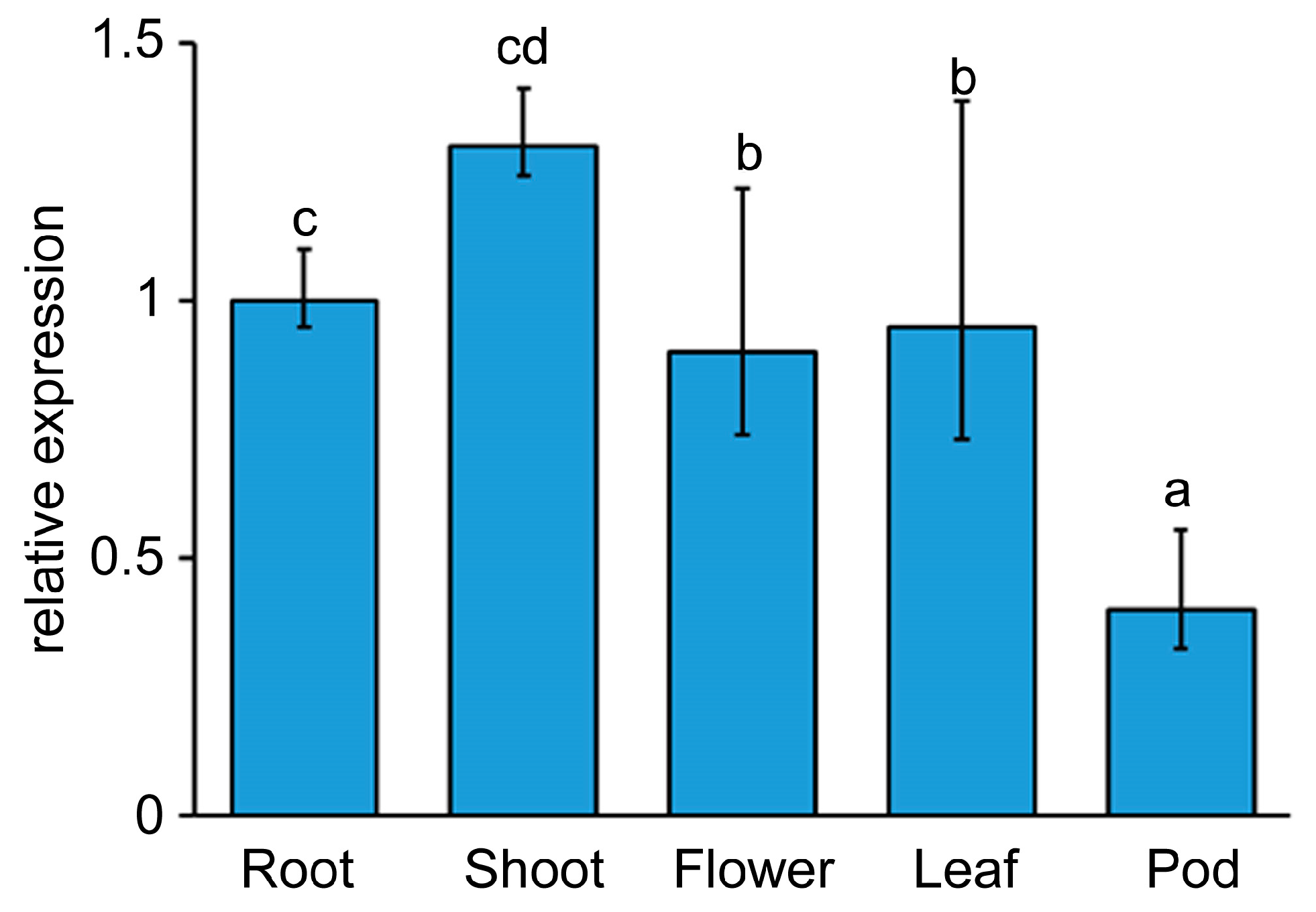
2.1. LjEDS1 Is an EDS1 Orthologs in L. japonicus

The coding sequence (CDS) of the EDS1 gene in L. japonicus is 1836 bp and is named LjEDS1 (Lj1g3v0416380). This sequence is divided into three exons within the genome sequence of 3260 bp and encodes a protein that contains both a lipase-like domain belonging to the Abhydrolase superfamily and the EP (EDS1-PAD4) domain of the EP superfamily. A homologous gene, LjEDS1-like (Lj1g3v0416340), designated LjEDS1L, was also identified (Supplementary Figure S1). Phylogenetic analysis of EDS1, PAD4, and senescence-associated gene101 (SAG101) in the EDS1 family showed that LjEDS1 clusters in the same evolutionary branch as EDS1 in Glycine max (soybean) and Arabidopsis (Supplementary Figure S2). Protein sequence alignments revealed that LjEDS1 and LjEDS1L possess characteristic EDS1 motifs, including SDH in the lipase-like domain and EPLDIA in the EP domain [13,14,15]. However, LjEDS1L lacks the KNEDT motif, located in the EP domain (Supplementary Figure S3). Consequently, this study focused on the role of LjEDS1 in disease resistance.
Tissue-specific expression analysis of LjEDS1 in L. japonicus showed the highest expression levels in shoots, followed by roots, flowers, and leaves, with the lowest expression observed in pods (Figure 1). The LjEDS1 protein was expressed in Arabidopsis mesophyll protoplasts and localized to both the cytoplasm and nucleus (Figure 2).

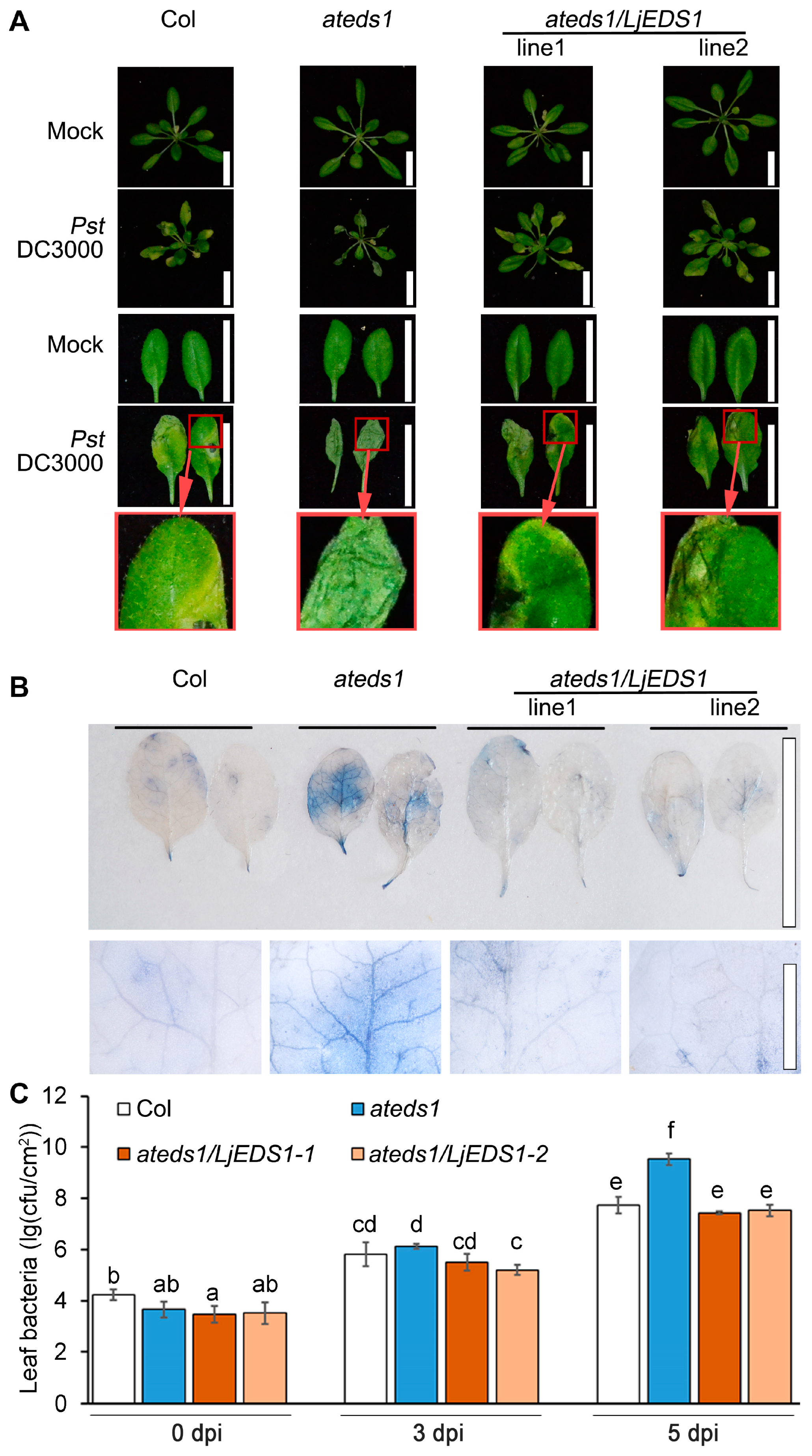
2.2. LjEDS1 Positively Regulates Disease Resistance to Pst DC3000 in Arabidopsis

To investigate the biological roles of the LjEDS1 gene in plant immunity, LjEDS1 was overexpressed in the eds1 null mutant line of eds1-2 (ateds1) in Arabidopsis [13]. Positive transgenic plants were identified using genomic sequence fusion of LjEDS1 with the β-glucuronidase (GUS) reporter gene to generate p35S::LjEDS1- GUS transgenic Arabidopsis lines (Supplementary Figure S4A). Two homozygous transgenic Arabidopsis eds1-2 lines expressing LjEDS1 were selected for subsequent studies. GUS signals confirmed their positive transgenic status (Supplementary Figure S4B). Semi-quantitative RT-PCR demonstrated increased expression levels of the LjEDS1 gene in transgenic lines (Supplementary Figure S4C).
Under normal growth conditions, no morphological differences were observed among Col, ateds1, and ateds1/LjEDS1 plants. Leaves from 4-week-old plants (positions 5–9) of wild-type, ateds1, and ateds1/LjEDS1 lines were inoculated with Pst DC3000. At 5 days post infection (dpi), ateds1 leaves exhibited almost complete necrosis and wilting, whereas Col and ateds1/LjEDS1 plants showed significantly reduced lesion areas (Figure 3A). Trypan blue staining confirmed extensive cell death in ateds1 leaves, which was mitigated in Col and ateds1/LjEDS1 lines (Figure 3B). Additionally, bacterial titers in ateds1/LjEDS1 leaves were 10–20% lower than those in ateds1 leaves at 5 dpi (Figure 3C). These results indicate that LjEDS1 can complement the eds1-2 defect in regards to pathogen resistance.

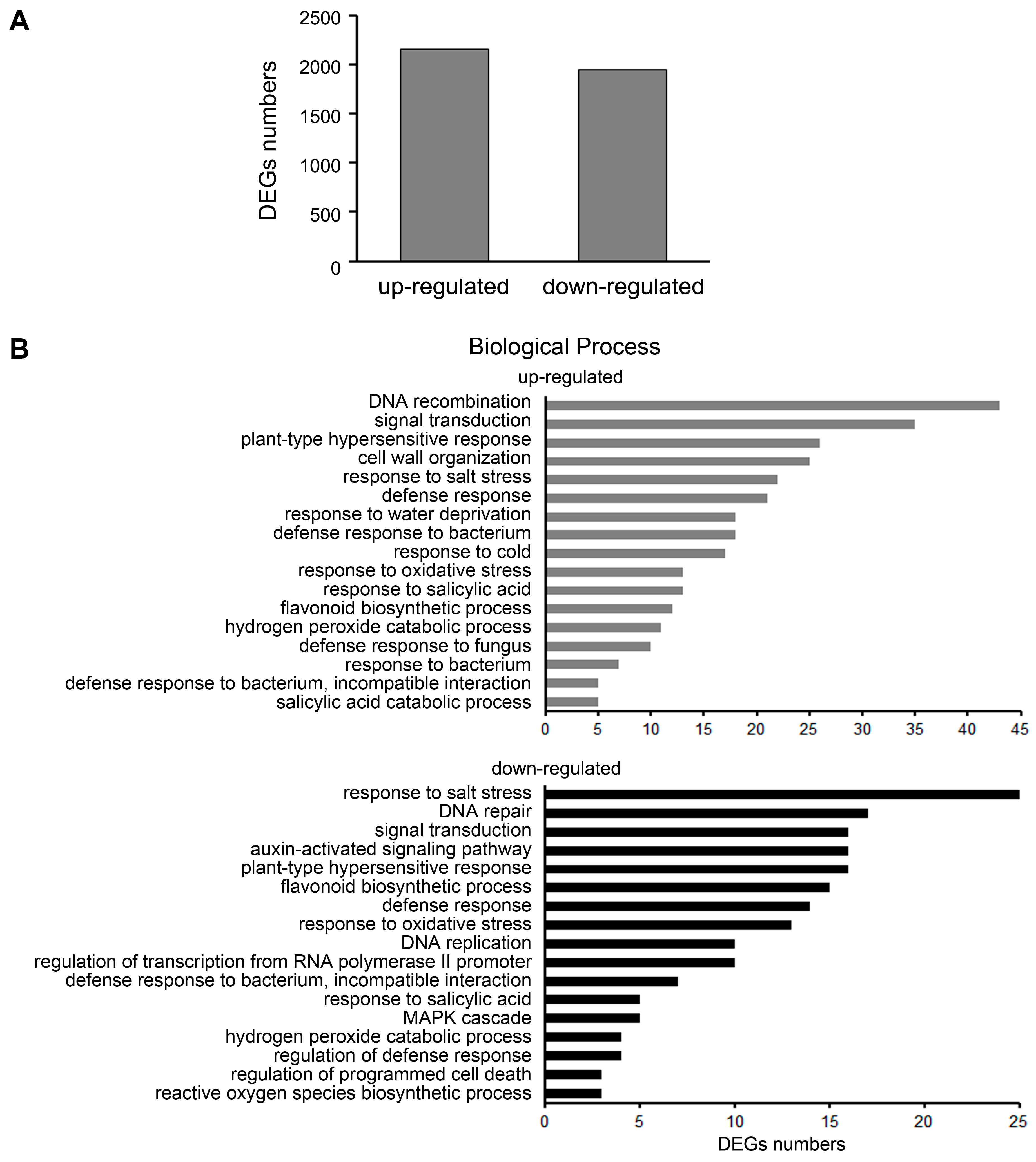
2.3. Differential Gene Expression Analysis in ljeds1 Roots

We obtained two LORE1 insertion mutant lines (plant IDs in Lotus Base: 30014411 and 30012794) with a L. japonicus Gifu B-129 background (https://lotus.au.dk/, accessed on 11 November 2014). Quantitative RT-PCR analysis showed that LjEDS1 was not expressed in these mutants; thus, these alleles were named ljeds1-1 and ljeds1-2 (Supplementary Figures S1 and S5). To investigate the molecular mechanism of LjEDS1’s role in immune regulation in L. japonicus, transcriptome analysis was performed on seedling roots of Gifu B-129 and its mutant ljeds1-1 (plant IDs in Lotus Base: 30014411). A total of 12,801 differentially expressed genes (DEGs) were identified between ljeds1-1 and Gifu B-129, of which 2160 were upregulated and 1949 were downregulated (Figure 4A). To validate the accuracy of the RNA-seq data, we tested the relative expression levels of seven representative genes involved in plant–microbe interaction by qRT-PCR and found that their expression trends were consistent with the transcriptome data (Supplementary Figure S6). Among the DEGs, the transcription levels of multiple defense response-related genes were significantly downregulated (Supplementary Table S2), including homologous genes predicted by Lotus Base for pathogenesis-related protein1 (Lj0g3v0215119.1), which decreased more than 100-fold. Reactive oxygen species (ROS)-related genes, such as homologous genes of peroxidase 3-like (Lj3g3v2890760.1 and Lj3g3v2890770.2), homologous genes of cationic peroxidase 1-like (Lj0g3v0012529.1), and homologous genes of peroxidase 10-like (Lj2g3v1836090.1), showed a 3–10-fold decrease. Transcription levels of homologous genes (Lj2g3v3246900.1 and Lj2g3v3315570.1) of red chlorophyll catabolite reductase, associated with programmed cell death, decreased by more than 10-fold [16].
Gene ontology (GO) enrichment analysis was conducted to determine the primary biological functions of DEGs in ljeds1. Upregulated categories included DNA recombination, signal transduction, plant-type hypersensitive response, responses to abiotic stress, defense responses to biotic stress, and responses to SA. Downregulated categories included responses to salt stress, defense responses to biotic stress, DNA repair, signal transduction, ROS synthesis and metabolism, SA synthesis, MAPK cascade, and regulation of programmed cell death (Figure 4B). These findings indicate that LjEDS1 plays a critical role in regulating the defense response in L. japonicus. Furthermore, a Kyoto Encyclopedia of Genes and Genomes (KEGG) enrichment analysis was performed to identify the associated signaling pathways. Significant enrichment was observed in pathways including flavonoid biosynthesis (homologous genes of chalcone synthase: Lj2g3v2125830.1, Lj4g3v2574990.1, and Lj2g3v2124310.1; homologous genes of chalcone isomerase: Lj5g3v2288880.1, etc.), MAPK signaling pathway (homologous genes of mitogen-activated protein kinase kinase kinase 1-like: Lj6g3v2256790.1 and Lj4g3v0335750.1, etc.), and phenylpropanoid biosynthesis (homologous genes of isoliquiritigenin 2′-O-methyltransferase-like: Lj0g3v0215539.1 and Lj0g3v0273599.1; homologous genes of phenylalanine ammonia-lyase: Lj3g3v0602630.2 and Lj3g3v0602620.1; homologous genes of peroxidase 3-like Lj3g3v2890760.1; homologous genes of cationic peroxidase 1-like: Lj0g3v0012529.1; homologous genes of peroxidase 10-like: Lj2g3v1836090.1, etc.). These results demonstrate the crucial role of LjEDS1 in signaling pathways such as MAPK and in plant hormones (Supplementary Table S3).

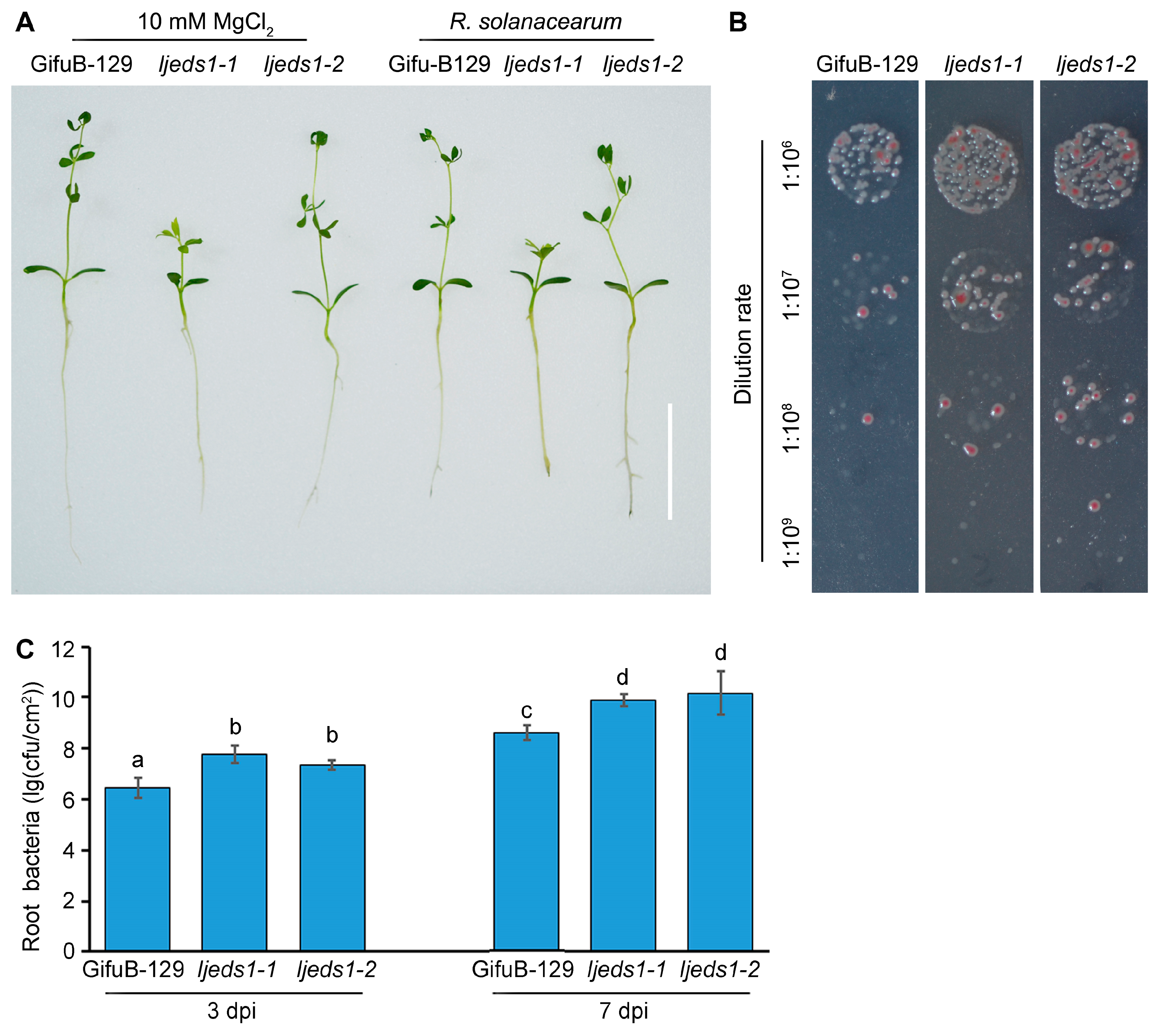
2.4. ljeds1 Mutants Are Susceptible to R. solanacearum

To evaluate the role of LjEDS1 in the immune response of L. japonicus, the disease resistance phenotype of Gifu B-129 and ljeds1 mutants (ljeds1-1 and ljeds1-2) was assessed. Roots of 3-day-old seedlings were inoculated with R. solanacearum, and observations were recorded at 3 dpi and 7 dpi. At 7 dpi, browning lesions were more pronounced in ljeds1-1 and ljeds1-2 compared to in Gifu B-129 (Figure 5A). Root bacterial titers were significantly higher in ljeds1 mutants, with titers approximately 10-fold higher at 3 dpi and 20-fold higher at 7 dpi compared to those for Gifu B-129 (Figure 5B,C). These results indicate that the ljeds1 mutation accelerates R. solanacearum proliferation in the Gifu B-129 ecotype.

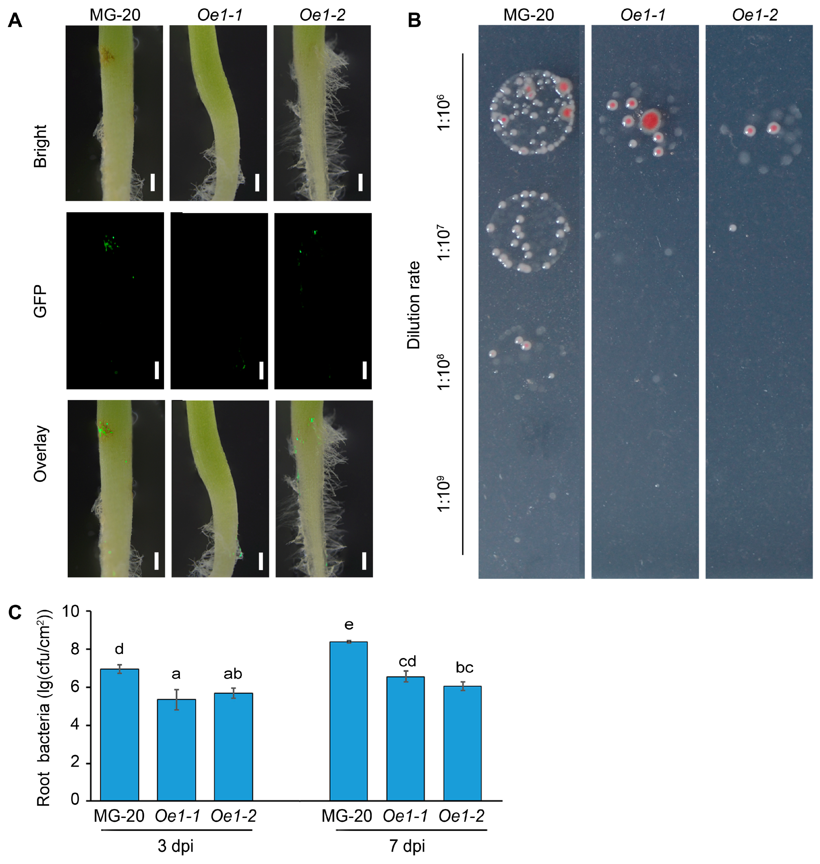
2.5. LjEDS1 Overexpression Enhances Resistance to R. solanacearum
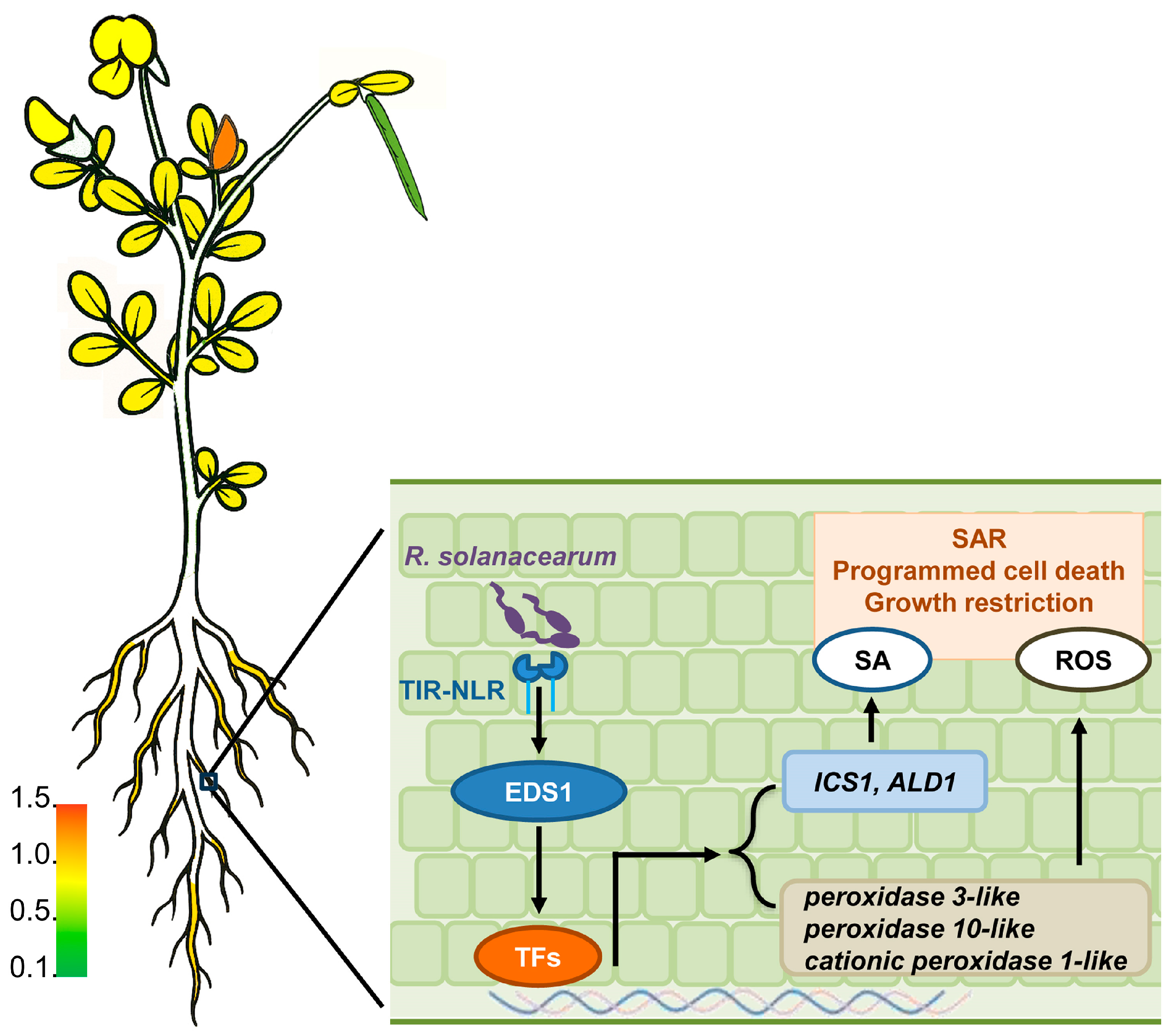
To further confirm the role of LjEDS1 in the immune response of L. japonicus, two LjEDS1 overexpression (Oe) lines (Oe1-1 and Oe1-2) were generated in the MG-20 ecotype. Quantitative RT-PCR analysis showed high LjEDS1 expression in Oe lines (Supplementary Figure S7C). Roots of 3-day-old seedlings were inoculated with R. solanacearum-GFP, and observations were made at 3 dpi and 7 dpi. At 7 dpi, brown lesions were observed on MG-20 roots but were absent in the Oe1-1 and Oe1-2 lines. Fluorescence microscopy revealed reduced R. solanacearum aggregation in the Oe lines compared to the results for MG-20 (Figure 6A). Statistical analysis showed that bacterial titers in the Oe lines were reduced by approximately 100-fold at both 3 dpi and 7 dpi compared to those for MG-20 (Figure 6B,C). These results indicate that the expression level of LjEDS1 positively correlates with the defense response of L. japonicus to R. solanacearum.

3. Discussion

Studies on Arabidopsis, soybean, and tobacco have shown that EDS1 contains two domains: the lipase-like domain of the abhydrolase superfamily and the EP domain of the EP superfamily. Among these, the EP domain is critical for the immune function of EDS1 protein family members [14,17,18,19,20]. Domain prediction analysis revealed that the LjEDS1 protein sequence contains both domains (Supplementary Figure S3). Additionally, it possesses the KNEDT motif, which is unique to EDS1 compared to the composition of other family members such as SAG101 and PAD4. This motif indicates that LjEDS1 is likely a homolog of AtEDS1 [13,14,15]. The conserved structure of EDS1 across evolution is further supported by the evolutionary relationship of EDS1 family members in L. japonicus, Arabidopsis, and soybean. The phylogenetic analysis shows that LjEDS1 clusters within the EDS1 clade, forming an independent branch distinct from SAG101 and PAD4 (Supplementary Figure S2).
Previous research on EDS1 has primarily focused on its function in the leaves. For instance, AtEDS1 collaborates with immune partners like PAD4 to regulate immune responses in Arabidopsis leaves via SA-dependent and SA-independent pathways [21]. OsEDS1 regulates pathogen susceptibility in rice leaves through the jasmonic acid signaling pathway [22], while GmEDS1 modulates SA accumulation, enhancing soybean leaf sensitivity to virulent pathogens [17]. In contrast, LjEDS1 exhibits a unique expression pattern, with higher expression levels in shoots and roots compared to those in flowers, leaves, and pods (Figure 1). Stem-cell-triggered immunity safeguards plant shoot apexes from pathogen infection [23], suggesting that this expression pattern may implicate LjEDS1 in a unique immune mechanism in shoot or root apexes. Meanwhile, its high expression in roots supports a flexible and efficient immune response when roots interact with the complex, microbe-rich soil environment. The subcellular localization of EDS1 between the nucleus and cytoplasm is essential for its function. The EDS1-SAG101 heterodimer predominantly localizes to the nucleus, while the EDS1-PAD4 heterodimer is found in both the cytoplasm and nucleus. The SAG101-EDS1-PAD4 ternary complex resides in the nucleus [24]. SAG101 promotes the nuclear entry of EDS1, while PAD4 maintains a cytoplasmic–nuclear balance [25,26]. LjEDS1 localization to both the cytoplasm and nucleus (Figure 2) suggests that its regulatory mechanism in L. japonicus may be similar to that of EDS1 in other plants, such as Arabidopsis, where cytoplasmic and nuclear shuttling enables immune regulation.
To preliminarily assess the immune function of LjEDS1, we conducted heterologous complementation experiments. Expression of LjEDS1 in the Arabidopsis ateds1 mutant restored pathogen resistance, reducing lesion formation and bacterial titers to wild-type levels (Figure 3). This finding is consistent with studies on GmEDS1, which partially compensates for pathogen resistance defects in Arabidopsis ateds1 mutants [17]. These results suggest that LjEDS1 shares structural and functional similarities with known EDS1 proteins and plays a significant role in the immune pathways of L. japonicus, particularly in the roots, which are critical for legumes.
Despite substantial studies on EDS1 in leaves, its role in roots remains underexplored. Specific expression analysis revealed that LjEDS1 exhibits relatively high expression in the roots of L. japonicus (Figure 1). Roots employ cooperative or defensive strategies when encountering foreign microbes. What role does LjEDS1 play in this defense process? Our study shows that although the targeted tissues differ, EDS1 performs similar functions within L. japonicus roots. Suppression of EDS1 expression during pathogen infection results in more pronounced necrotic lesions and increased bacterial titers in Arabidopsis, wilting and chlorosis in cotton seedlings, and heightened chlorosis and cell death in soybeans [17,27,28]. In L. japonicus, ljeds1 mutants exhibited accelerated pathogen proliferation and larger root lesions upon R. solanacearum infection compared to the results for wild-type plants (Figure 5). Conversely, overexpression of LjEDS1 reduced pathogen proliferation and alleviated disease symptoms (Figure 6). These results indicate that LjEDS1 positively regulates defense responses in L. japonicus roots.
After confirming the regulatory role of EDS1 in the immune function of L. japonicus, we investigated its underlying regulatory mechanisms. GO enrichment analysis indicated that the mutation of ljeds1 affected several mechanisms related to defense responses (Figure 4B), highlighting the transcriptional regulatory role of LjEDS1 in immune function. Previous studies on Arabidopsis have shown that excessive accumulation of EDS1 in the nucleus can trigger autoimmunity, leading to symptoms resembling pathogen infection, such as inhibited root growth, increased root hair formation, and programmed cell death [29,30,31]. Additionally, the EDS1-SAG101 complex interacts with NRG1 (N requirement gene 1) to induce host cell death. GO analysis revealed that the ljeds1 mutation affected DEGs related to programmed cell death and plant-type hypersensitive responses (Figure 4B). This suggests that the significantly shorter root length observed in LjEDS1-overexpressing lines might result from autoimmunity triggered by LjEDS1 (Supplementary Figure S5A). Collectively, these results demonstrate that LjEDS1 regulates immune responses in L. japonicus, including root growth inhibition and programmed cell death.
Plant physiological responses require various signaling molecules. One of the earliest markers of plant immune responses is the production of ROS in different subcellular compartments [32]. In this study, biosynthesis and metabolic processes related to ROS were altered in ljeds1 mutants (Figure 4B). Under conditions such as pathogen infection or cold stress, EDS1 regulates ROS production and scavenging through interactions with proteins like PAD4 and SAG101, thereby modulating plant resistance and immune responses [5,33]. SA maintains the ROS balance upstream [32]. Additionally, the SA-induced EDS1-NPR1 complex binds to promoters of defense genes, activating their roles in Arabidopsis immunity [34]. EDS1 enhances resistance to biotic stress by regulating SA accumulation [13]. In Chrysanthemum morifolium, transcriptional regulation mediated by CmEDS1 plays a vital role in early-stage infection before lesion formation, supporting SA accumulation for defense [35]. Similarly, in cotton, GbEDS1 enhances resistance by regulating SA and H2O2 production during disease development [27]. HIR3 in tobacco also promotes resistance through an EDS1- and SA-dependent pathway, causing cell death and SA accumulation [36]. Throughout the defense process, AtEDS1 forms a complex with DELLA protein, negatively regulated by gibberellin (GA), which interacts with SA to balance growth and defense [28,37]. In this study, GO enrichment analysis revealed that mechanisms related to the SA catabolic process and response to SA were regulated by LjEDS1 (Figure 4B). These results align with those from previous studies on EDS1, suggesting that LjEDS1 modulates plant defense by regulating SA and ROS accumulation.
Additionally, GO and KEGG enrichment analysis revealed that the MAPK cascade was downregulated in L. japonicus upon ljeds1 mutation (Figure 4B). MAPKs are crucial in plant immune responses [10,38]. MAPKs can bypass SA to induce SA-responsive genes and act upstream of ROS bursts [39,40]. In Arabidopsis, MAP kinase 4 (MPK4) regulates SA-dependent responses through EDS1 and PAD4. Our findings suggest that LjEDS1 may influence MAPK pathways in L. japonicus. While LjEDS1 modulates immune responses in L. japonicus roots through a complex regulatory network, further investigation is required to elucidate its specific regulatory mechanisms.
In conclusion, during R. solanacearum infection of L. japonicus roots, the plant’s NLRs detect pathogen effectors, thereby activating EDS1. The active EDS1 triggers downstream transcription factors (TFs), which subsequently induce the transcription of defense-related genes (Figure 7). These include the SA biosynthesis gene ICS1 and ALD1 oxidation-reduction reaction related genes such as peroxidase 3-like, which trigger pathogen resistance.

4. Materials and Methods

4.1. Growth Conditions and Treatments of L. japonicus

Two ecotypes of L. japonicus, Gifu B-129 and MG-20, were primarily used in this study. LORE1 insertion mutant lines of ljeds1-1 and ljeds1-2 (plant IDs in Lotus Base: 30014411 and 30012794) with a Gifu B-129 background were obtained from Lotus Base (https://lotus.au.dk/, accessed on 11 November 2014). Two LjEDS1 overexpression lines (Oe1-1 and Oe1-2) were generated by transforming MG-20 with Agrobacterium tumefaciens strain AGL1. The L. japonicus ecotypes Gifu B-129 and MG-20 were used as wild-type (WT) controls in this study. After surface disinfection, the seeds were placed in sterile dishes for 2 days and grown in a growth chamber under a long-day photoperiod (16 h light/8 h dark) at 22 ± 2 °C.
For bacterial growth assays, R. solanacearum was cultured in SMSA medium at 28 °C in the dark, and 2-day-old seedlings were infected by adding R. solanacearum (suspended in sterile 10 mM MgCl2 to OD600 = 0.01, equivalent to ~2 × 10⁶ cfu mL⁻1) or a mock suspension (10 mM MgCl2). The images were observed under a Leica M165 FC Fluorescent Stereo Microscope. The root tips (1 cm) were excised at specific points in time post-inoculation, ground, and cultured on SMSA solid medium for 2 days. Data were statistically analyzed [11]. The experiments were repeated three times, with three plants per sample.

4.2. Growth Conditions and Treatments of Arabidopsis

The Arabidopsis ecotype Columbia-0 (Col-0) was used as the wild-type (WT) in this study. After surface disinfection, the seeds were incubated for 2 days in the dark at 4 °C and then grown in a growth chamber under a short-day photoperiod (12 h light/12 h dark) at 22 ± 2 °C.
For bacterial growth assays, the Pseudomonas syringae pv. Tomato DC3000 was cultured in King’s B medium containing 25 mg mL⁻1 rifampicin at 28 °C. Leaves of 4-week-old plants were dip-inoculated with Pst DC3000 (suspended in sterile 10 mM MgCl2 to OD600 = 0.0002, equivalent to ~105 cfu mL⁻1) or a mock suspension (10 mM MgCl2). Samples were collected at 0, 3, and 5 days post infection (dpi). Each sample included three independent biological replicates, with three infected leaf discs per replicate. Leaf discs were placed in microcentrifuge tubes, ground with a pestle in 1 mL of 10 mM MgCl2, then diluted, cultured on King’s B solid medium, and statistically analyzed [41].

4.3. RNA-Seq and Transcriptome Data Analysis

Root samples from 10-day-old seedlings were collected and sent to Shanghai OE Biotech Co., Ltd. (Shanghai, China) for RNA sequencing. The expression abundance of each gene across the samples was determined through sequence similarity alignment against a database comprising known reference genome sequences and annotation files, using HTSeq count software (Version number: 0.6.0) to obtain the number of reads aligned to genes in each sample and Cufflinks software (Version number: 2.2.1) to calculate the FPKM (fragments per kilobase per million reads) value of gene expression. Sequencing data were mapped to the genome sequence using Hisat 2 software (Version number: 2.0.5). The calculation of gene expression levels was performed using the FPKM method [42]. According to DESeq software (Version number: 1.18.0), the negative binomial distribution test was used to identify differentially expressed genes (DEGs). Only those unigenes with a p-value ≤ 0.05 and a fold change ≥ 2 were designated as differentially expressed. The DEGs were subjected to GO enrichment and KEGG enrichment analyses.

4.4. RNA Isolation and Expression Analysis

For expression analysis, the tissues were collected and immediately placed in liquid nitrogen or stored at −80 °C. RNA was extracted using the Plant RNA Kit (Magen R4151-02, Guangdong, China). First-strand cDNAs were synthesized using the GoScript™ Reverse Transcription System (Promega, Madison, WI, USA), according to the manufacturer’s instructions. AtActin, LjActin, or LjATPase were used as internal controls for RT-PCR analysis. All qRT-PCR experiments were performed on a LightCycler® 480 Real-Time PCR System (Mannheim, Germany) under the following amplification conditions: 10 min at 95 °C, followed by 40 cycles of 95 °C for 5 s, 60 °C for 20 s, and 72 °C for 20 s. LjATPase was used as the internal control for normalization, and the 2−△△Ct method was applied for real-time RT-PCR analysis [11]. The primers used are listed in Supplementary Table S1.

4.5. Plasmid Construction and Plant Transformation

To generate LjEDS1 (Lj1g3v0416380) overexpression lines in L. japonicus and Arabidopsis, the LjEDS1 coding sequence (1836 bp) was amplified by PCR and used to construct pUBI::LjEDS1-3×FLAG and p35S::LjEDS1-GUS vectors. The primers used are listed in Supplementary Table S1. The expression vector pUBI::LjEDS1-3×FLAG was transformed into A. tumefaciens strain AGL1 and used to infect L. japonicus [43]. The p35S::LjEDS1-GUS vector was transformed into A. tumefaciens strain GV3101 and used to infect Arabidopsis [44]. Positive transformants were identified by GUS staining and RT-PCR.

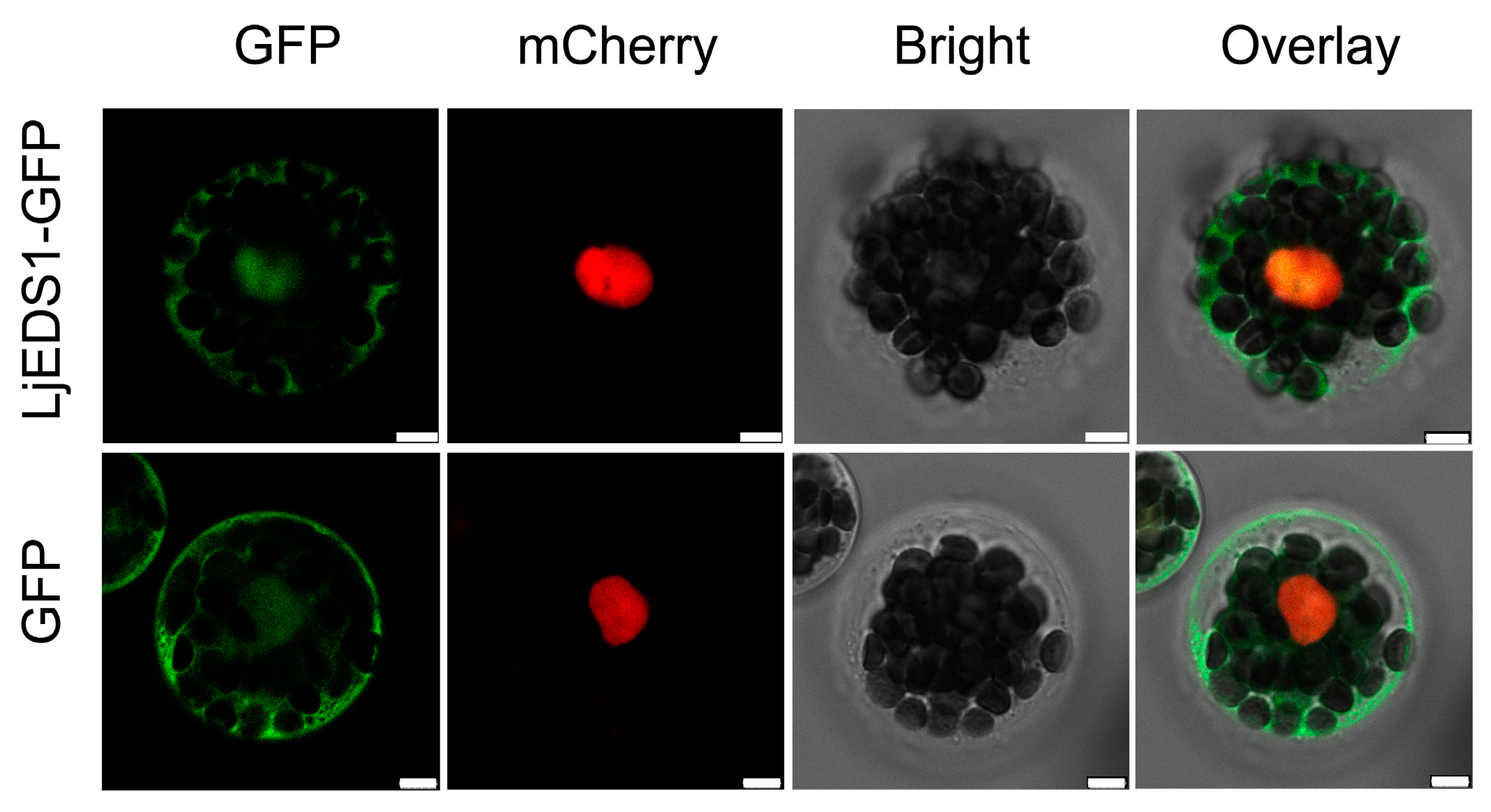
4.6. Subcellular Localization

To confirm the localization of the LjEDS1 protein, the LjEDS1 coding region was cloned into a vector containing an N-terminal EGFP fusion to generate 35S::LjEDS1-nEGFP. The primers used are listed in Supplementary Table S1. A nuclear marker was generated by fusing mCherry to a nuclear localization signal (NLS) peptide. These plasmids were co-transformed into Arabidopsis mesophyll protoplasts [45]. Fluorescence signals were observed using a Leica TCS SP8 confocal laser scanning microscope (Leica, Wetzlar, Germany).

4.7. Histochemistry Analysis

Leaves of p35S::LjEDS1-GUS Arabidopsis transgenic lines were stained with GUS staining buffer (1 mM X-Gluc, 0.5 mM K3[Fe(CN)6], 0.5 mM K4[Fe(CN)6], 1 mM EDTA, pH 8.0, and 0.1 mM PBS buffer, pH 7.0) at 37 °C for 2–4 h following vacuum infiltration for 15 min. Stained tissues were stored in a decolorizing solution (70% ethanol) for observation [11].
To observe dead cells in Arabidopsis leaves, inoculated leaves were immersed in trypan blue solution (30 mL lactic acid, 30 mL glycerol, 30 g phenol, and 900 mg trypan blue) and gently shaken for 40 min. Stained tissues were destained overnight in chloral hydrate solution [46]. Images were captured using a Leica M165 C Stereo Microscope (Leica, Wetzlar, Germany).

4.8. Sequence Analysis and Phylogenetic Tree Construction

Relevant nucleotide and protein sequences were obtained from Lotus Base (https://lotus.au.dk/, accessed on 1 September 2014), NCBI (https://www.ncbi.nlm.nih.gov/, accessed on 20 March 2023), and Phytozome (https://jgi.doe.gov/, accessed on 20 March 2023). Analysis of gene structure was conducted using GSDS 2.0 (http://gsds.gao-lab.org/, accessed on 12 March 2018). A phylogenetic tree was constructed using MEGA 6.0 with the neighbor-joining (NJ) method and 1000 bootstrap replicates. Multiple sequence alignments of protein sequences across different plant species were conducted using the T-Coffee Server (https://tcoffee.crg.eu/, accessed on 25 March 2023).

4.9. Statistical Analysis

All experiments included three biological replicates, and data are presented as means ± SDs (standard deviations). Statistical significance was assessed using SPSS software (version 21.0). One-way ANOVA, followed by Duncan’s post hoc test, were applied to identify significant differences, with p < 0.05 indicated by different letters above bars.

5. Conclusions

Our findings reveal that LjEDS1’s structure closely resembles that of EDS1 in Arabidopsis and other plants, with localization in both the nucleus and cytoplasm. Additionally, LjEDS1 enhances disease resistance in the ateds1 mutant of Arabidopsis. The mutation of LjEDS1 in L. japonicus exacerbates the symptoms of R. solanacearum infection, while overexpression mitigates these symptoms. LjEDS1 regulates the disease-resistance response in L. japonicus by modulating the transcription levels of genes associated with biotic (and abiotic) stress responses, ROS and SA synthesis, and the MAPK cascade.

Supplementary Materials

The following supporting information can be downloaded at: https://www.mdpi.com/article/10.3390/ijms26083848/s1.

Author Contributions

Conceptualization, H.J.; methodology, M.Y.; validation, Q.L. and H.H.; formal analysis, M.H.; investigation, H.J. and C.S.; resources, Y.C.; data curation, M.Y. and Q.L.; writing—original draft preparation, M.Y.; writing—review and editing, H.J.; supervision, H.J.; project administration, G.W. and Y.C.; funding acquisition, Y.C. All authors have read and agreed to the published version of the manuscript.

Funding

This research was funded by National Natural Science Foundation of China (grant number 31570242).

Institutional Review Board Statement

Not applicable.

Informed Consent Statement

Not applicable.

Data Availability Statement

Data are contained within the article and Supplementary Materials. The raw transcriptome sequencing data generated in this study are not publicly available, but are available from the corresponding author upon reasonable request.

Acknowledgments

We thank Xingliang Hou for providing the Arabidopsis eds1-2 mutant.

Conflicts of Interest

The authors declare no conflicts of interest.

References

  1. Dou, D.; Zhou, J.M. Phytopathogen effectors subverting host immunity: Different foes, similar battleground. Cell Host Microbe. 2012, 12, 484–495. [Google Scholar] [CrossRef] [PubMed]
  2. Dodds, P.N.; Rathjen, J.P. Plant immunity: Towards an integrated view of plant-pathogen interactions. Nat. Rev. Genet. 2010, 11, 539–548. [Google Scholar] [CrossRef]
  3. Chen, J.; Zhang, J.; Kong, M.; Freeman, A.; Chen, H.; Liu, F. More stories to tell: NONEXPRESSOR OF PATHOGENESIS-RELATED GENES1, a salicylic acid receptor. Plant Cell Environ. 2021, 44, 1716–1727. [Google Scholar] [CrossRef] [PubMed]
  4. Yu, G.; Zhang, L.; Xue, H.; Chen, Y.; Liu, X.; Del Pozo, J.C.; Zhao, C.; Lozano-Duran, R.; Macho, A.P. Cell wall-mediated root development is targeted by a soil-borne bacterial pathogen to promote infection. Cell Rep. 2024, 43, 114179. [Google Scholar]
  5. Pruitt, R.N.; Locci, F.; Wanke, F.; Zhang, L.; Saile, S.C.; Joe, A.; Karelina, D.; Hua, C.; Frohlich, K.; Wan, W.L.; et al. The EDS1-PAD4-ADR1 node mediates Arabidopsis pattern-triggered immunity. Nature 2021, 598, 495–499. [Google Scholar] [CrossRef]
  6. Heidrich, K.; Wirthmueller, L.; Tasset, C.; Pouzet, C.; Deslandes, L.; Parker, J.E. Arabidopsis EDS1 Connects Pathogen Effector Recognition to Cell Compartment-Specific Immune Responses. Science 2011, 334, 1401–1404. [Google Scholar] [CrossRef]
  7. Halane, M.K.; Kim, S.H.; Spears, B.J.; Garner, C.M.; Rogan, C.J.; Okafor, E.C.; Su, J.; Bhattacharjee, S.; Gassmann, W. The bacterial type III-secreted protein AvrRps4 is a bipartite effector. PLoS Pathog. 2018, 14, e1006984. [Google Scholar] [CrossRef] [PubMed]
  8. Shah, J.; Chaturvedi, R.; Chowdhury, Z.; Venables, B.; Petros, R.A. Signaling by small metabolites in systemic acquired resistance. Plant J. 2014, 79, 645–658. [Google Scholar] [CrossRef]
  9. Cecchini, N.M.; Jung, H.W.; Engle, N.L.; Tschaplinski, T.J.; Greenberg, J.T. ALD1 Regulates Basal Immune Components and Early Inducible Defense Responses in Arabidopsis. Mol. Plant Microbe Interact. 2015, 28, 455–466. [Google Scholar] [CrossRef]
  10. Cui, H.; Gobbato, E.; Kracher, B.; Qiu, J.; Bautor, J.; Parker, J.E. A core function of EDS1 with PAD4 is to protect the salicylic acid defense sector in Arabidopsis immunity. New Phytol. 2017, 213, 1802–1817. [Google Scholar] [CrossRef]
  11. Huang, M.; Yuan, M.; Sun, C.; Li, M.; Wu, P.; Jiang, H.; Wu, G.; Chen, Y. Roles of AGD2a in Plant Development and Microbial Interactions of Lotus japonicus. Int. J. Mol. Sci. 2022, 23, 6863. [Google Scholar] [CrossRef] [PubMed]
  12. Chen, W.; Li, X.; Tian, L.; Wu, P.; Li, M.; Jiang, H.; Chen, Y.; Wu, G. Knockdown of LjALD1, AGD2-like defense response protein 1, influences plant growth and nodulation in Lotus japonicus. J. Integr. Plant Biol. 2014, 56, 1034–1041. [Google Scholar] [CrossRef] [PubMed]
  13. Feys, B.J.; Moisan, L.J.; Newman, M.A.; Parker, J.E. Direct interaction between the Arabidopsis disease resistance signaling proteins, EDS1 and PAD4. EMBO J. 2001, 20, 5400–5411. [Google Scholar] [CrossRef]
  14. Wagner, S.; Stuttmann, J.; Rietz, S.; Guerois, R.; Brunstein, E.; Bautor, J.; Niefind, K.; Parker, J.E. Structural basis for signaling by exclusive EDS1 heteromeric complexes with SAG101 or PAD4 in plant innate immunity. Cell Host Microbe 2013, 14, 619–630. [Google Scholar] [CrossRef]
  15. Lapin, D.; Bhandari, D.D.; Parker, J.E. Origins and Immunity Networking Functions of EDS1 Family Proteins. Annu. Rev. Phytopathol. 2020, 58, 253–276. [Google Scholar] [CrossRef] [PubMed]
  16. Tang, Y.; Li, M.; Chen, Y.; Wu, P.; Wu, G.; Jiang, H. Knockdown of OsPAO and OsRCCR1 cause different plant death phenotypes in rice. J. Plant Physiol. 2011, 168, 1952–1959. [Google Scholar] [CrossRef]
  17. Wang, J.; Shine, M.B.; Gao, Q.M.; Navarre, D.; Jiang, W.; Liu, C.; Chen, Q.; Hu, G.; Kachroo, A. Enhanced Disease Susceptibility1 Mediates Pathogen Resistance and Virulence Function of a Bacterial Effector in Soybean. Plant Physiol. 2014, 165, 1269–1284. [Google Scholar] [CrossRef]
  18. Lapin, D.; Kovacova, V.; Sun, X.; Dongus, J.A.; Bhandari, D.; Von Born, P.; Bautor, J.; Guarneri, N.; Rzemieniewski, J.; Stuttmann, J.; et al. A Coevolved EDS1-SAG101-NRG1 Module Mediates Cell Death Signaling by TIR-Domain Immune Receptors. Plant Cell 2019, 31, 2430–2455. [Google Scholar] [CrossRef]
  19. Bhandari, D.D.; Lapin, D.; Kracher, B.; Von Born, P.; Bautor, J.; Niefind, K.; Parker, J.E. An EDS1 heterodimer signalling surface enforces timely reprogramming of immunity genes in Arabidopsis. Nat. Commun. 2019, 10, 772. [Google Scholar]
  20. Gantner, J.; Ordon, J.; Kretschmer, C.; Guerois, R.; Stuttmann, J. An EDS1-SAG101 Complex Is Essential for TNL-Mediated Immunity in Nicotiana benthamiana. Plant Cell 2019, 31, 2456–2474. [Google Scholar] [CrossRef]
  21. Zeng, H.Y.; Liu, Y.; Chen, D.K.; Bao, H.N.; Huang, L.Q.; Yin, J.; Chen, Y.L.; Xiao, S.; Yao, N. The immune components ENHANCED DISEASE SUSCEPTIBILITY 1 and PHYTOALEXIN DEFICIENT 4 are required for cell death caused by overaccumulation of ceramides in Arabidopsis. Plant J. 2021, 107, 1447–1465. [Google Scholar] [CrossRef] [PubMed]
  22. Ke, Y.; Kang, Y.; Wu, M.; Liu, H.; Hui, S.; Zhang, Q.; Li, X.; Xiao, J.; Wang, S. Jasmonic Acid-Involved OsEDS1 Signaling in Rice-Bacteria Interactions. Rice 2019, 12, 25. [Google Scholar] [CrossRef]
  23. Naseem, M.; Srivastava, M.; Dandekar, T. Stem-cell-triggered immunity safeguards cytokinin enriched plant shoot apexes from pathogen infection. Front. Plant Sci. 2014, 5, 588. [Google Scholar] [CrossRef] [PubMed]
  24. Zhu, S.; Jeong, R.D.; Venugopal, S.C.; Lapchyk, L.; Navarre, D.; Kachroo, A.; Kachroo, P. SAG101 forms a ternary complex with EDS1 and PAD4 and is required for resistance signaling against turnip crinkle virus. PLoS Pathog. 2011, 7, e1002318. [Google Scholar] [CrossRef] [PubMed]
  25. Garcia, A.V.; Blanvillain-Baufume, S.; Huibers, R.P.; Wiermer, M.; Li, G.; Gobbato, E.; Rietz, S.; Parker, J.E. Balanced nuclear and cytoplasmic activities of EDS1 are required for a complete plant innate immune response. PLoS Pathog. 2010, 6, e1000970. [Google Scholar] [CrossRef]
  26. Khan, M.S.S.; Islam, F.; Chen, H.; Chang, M.; Wang, D.; Liu, F.; Fu, Z.Q.; Chen, J. Transcriptional Coactivators: Driving Force of Plant Immunity. Front. Plant Sci. 2022, 13, 823937. [Google Scholar] [CrossRef]
  27. Yan, Z.; Xingfen, W.; Wei, R.; Jun, Y.; Zhiying, M. Island Cotton Enhanced Disease Susceptibility 1 Gene Encoding a Lipase-Like Protein Plays a Crucial Role in Response to Verticillium dahliae by Regulating the SA Level and H(2)O(2) Accumulation. Front. Plant Sci. 2016, 7, 1830. [Google Scholar] [CrossRef]
  28. Li, Y.; Yang, Y.; Hu, Y.; Liu, H.; He, M.; Yang, Z.; Kong, F.; Liu, X.; Hou, X. DELLA and EDS1 Form a Feedback Regulatory Module to Fine-Tune Plant Growth-Defense Tradeoff in Arabidopsis. Mol. Plant. 2019, 12, 1485–1498. [Google Scholar] [CrossRef]
  29. Stuttmann, J.; Peine, N.; Garcia, A.V.; Wagner, C.; Choudhury, S.R.; Wang, Y.; James, G.V.; Griebel, T.; Alcazar, R.; Tsuda, K.; et al. Arabidopsis thaliana DM2h (R8) within the Landsberg RPP1-like Resistance Locus Underlies Three Different Cases of EDS1-Conditioned Autoimmunity. PLoS Genet. 2016, 12, e1005990. [Google Scholar] [CrossRef]
  30. Poncini, L.; Wyrsch, I.; Denervaud Tendon, V.; Vorley, T.; Boller, T.; Geldner, N.; Metraux, J.P.; Lehmann, S. In roots of Arabidopsis thaliana, the damage-associated molecular pattern AtPep1 is a stronger elicitor of immune signalling than flg22 or the chitin heptamer. PLoS ONE 2017, 12, e0185808. [Google Scholar] [CrossRef]
  31. Jing, Y.P.; Zheng, X.J.; Zhang, D.L.; Shen, N.; Wang, Y.; Yang, L.; Fu, A.G.; Shi, J.S.; Zhao, F.G.; Lan, W.Z.; et al. Danger-Associated Peptides Interact with PIN-Dependent Local Auxin Distribution to Inhibit Root Growth in Arabidopsis. Plant Cell. 2019, 31, 1767–1787. [Google Scholar] [CrossRef]
  32. Lukan, T.; Coll, A. Intertwined Roles of Reactive Oxygen Species and Salicylic Acid Signaling Are Crucial for the Plant Response to Biotic Stress. Int. J. Mol. Sci. 2022, 23, 5568. [Google Scholar] [CrossRef] [PubMed]
  33. Chen, Q.F.; Xu, L.; Tan, W.J.; Chen, L.; Qi, H.; Xie, L.J.; Chen, M.X.; Liu, B.Y.; Yu, L.J.; Yao, N.; et al. Disruption of the Arabidopsis Defense Regulator Genes SAG101, EDS1, and PAD4 Confers Enhanced Freezing Tolerance. Mol. Plant 2015, 8, 1536–1549. [Google Scholar]
  34. Chen, H.; Li, M.; Qi, G.; Zhao, M.; Liu, L.; Zhang, J.; Chen, G.; Wang, D.; Liu, F.; Fu, Z.Q. Two interacting transcriptional coactivators cooperatively control plant immune responses. Sci. Adv. 2021, 7, eabl7173. [Google Scholar] [CrossRef] [PubMed]
  35. Liu, Y.; Xin, J.; Liu, L.; Song, A.; Guan, Z.; Fang, W.; Chen, F. A temporal gene expression map of Chrysanthemum leaves infected with Alternaria alternata reveals different stages of defense mechanisms. Hortic. Res. 2020, 7, 23. [Google Scholar] [CrossRef]
  36. Li, S.; Zhao, J.; Zhai, Y.; Yuan, Q.; Zhang, H.; Wu, X.; Lu, Y.; Peng, J.; Sun, Z.; Lin, L.; et al. The hypersensitive induced reaction 3 (HIR3) gene contributes to plant basal resistance via an EDS1 and salicylic acid-dependent pathway. Plant J. 2019, 98, 783–797. [Google Scholar] [CrossRef] [PubMed]
  37. Dong, S.; Tarkowska, D.; Sedaghatmehr, M.; Welsch, M.; Gupta, S.; Mueller-Roeber, B.; Balazadeh, S. The HB40-JUB1 transcriptional regulatory network controls gibberellin homeostasis in Arabidopsis. Mol. Plant 2022, 15, 322–339. [Google Scholar] [CrossRef]
  38. Bigeard, J.; Colcombet, J.; Hirt, H. Signaling mechanisms in pattern-triggered immunity (PTI). Mol. Plant 2015, 8, 521–539. [Google Scholar]
  39. Zhang, J.; Shao, F.; Li, Y.; Cui, H.; Chen, L.; Li, H.; Zou, Y.; Long, C.; Lan, L.; Chai, J.; et al. A Pseudomonas syringae effector inactivates MAPKs to suppress PAMP-induced immunity in plants. Cell Host Microbe. 2007, 1, 175–185. [Google Scholar] [CrossRef]
  40. Tsuda, K.; Mine, A.; Bethke, G.; Igarashi, D.; Botanga, C.J.; Tsuda, Y.; Glazebrook, J.; Sato, M.; Katagiri, F. Dual regulation of gene expression mediated by extended MAPK activation and salicylic acid contributes to robust innate immunity in Arabidopsis thaliana. PLoS Genet. 2013, 9, e1004015. [Google Scholar]
  41. Yang, L.; Li, B.; Zheng, X.-Y.; Li, J.; Yang, M.; Dong, X.; He, G.; An, C.; Deng, X.W. Salicylic acid biosynthesis is enhanced and contributes to increased biotrophic pathogen resistance in Arabidopsis hybrids. Nat. Commun. 2015, 6, 7309. [Google Scholar] [CrossRef] [PubMed]
  42. Trapnell, C.; William, B.A.; Pertea, G.; Mortazavi, A.; Kwan, G.; Baren, M.J.; Salzberg, L.S.; Wold, B.J.; Pachter, L. Transcript assembly and quantification by RNA-Seq reveals unannotated transcripts and isoform switching during cell differentiation. Nat. Biotechnol. 2010, 28, 511–515. [Google Scholar] [CrossRef] [PubMed]
  43. Stiller, J.; Martirani, L.; Tuppale, S.; Chian, R.J.; Chiurazzi, M.; Gresshoff, P.M. High frequency transformation and regeneration of transgenic plants in the model legume Lotus japonicus. J. Exp. Bot. 1997, 48, 1357–1365. [Google Scholar] [CrossRef]
  44. Clough, S.J.; Bent, A.F. Floral dip: A simplified method for Agrobacterium-mediated transformation of Arabidopsis thaliana. Plant J. 1998, 16, 735–743. [Google Scholar] [CrossRef] [PubMed]
  45. Yoo, S.D.; Cho, Y.H.; Sheen, J. Arabidopsis mesophyll protoplasts: A versatile cell system for transient gene expression analysis. Nat. Protoc. 2007, 2, 1565–1572. [Google Scholar]
  46. Son, G.H.; Moon, J.; Shelake, R.M.; Vuong, U.T.; Ingle, R.A.; Gassmann, W.; Kim, J.Y.; Kim, S.H. Conserved Opposite Functions in Plant Resistance to Biotrophic and Necrotrophic Pathogens of the Immune Regulator SRFR1. Int. J. Mol. Sci. 2021, 22, 6427. [Google Scholar] [CrossRef]
Figure 1. Specific expression pattern analysis of LjEDS1 in L. japonicus. RNA was extracted from 10-day-old roots, shoots, leaves, flowers, and pods of MG-20, and qRT-PCR analysis was performed. The expression level in the root was used as a reference. The data are the mean ± SD of three biological replicates. Lowercase letters above the bars indicate significant differences (one-way ANOVA, p < 0.05). Relative expression was normalized to that of the reference gene LjATPase (internal control).
Ijms 26 03848 g001
Figure 2. Subcellular localization of LjEDS1 protein. Confocal images of Arabidopsis protoplasts transiently expressing LjEDS1-GFP fusions. Green fluorescence of GFP and LjEDS1-GFP fusions; red fluorescence from mCherry fused with a nuclear localization signal (NLS) peptide; bar = 5 μm.
Ijms 26 03848 g002
Figure 3. LjEDS1 restores plant pathogen resistance of the eds1-2 mutated allele in Arabidopsis. (A) Disease resistance phenotypes of Col, ateds1, and two representative LjEDS1 complementation transgenic lines (ateds1/LjEDS1). Four-week-old plants were dip-inoculated with Pst DC3000 (OD600 = 0.0002). Leaves shown were photographed at 5 days post infection (dpi); bar = 2 cm. (B) Cell death analysis of Col, ateds1, and ateds1/LjEDS1 complementation transgenic plants. Stained leaves as described in (A) at 5 dpi with trypan blue. Representative leaves were from six plants per genotype. Experiments were independently repeated three times; bar = 1 cm. (C) Bacterial growth of Pst DC3000 on plants described in (A) at 0, 3, and 5 dpi. cfu/cm2, colony-forming units per cm2 of leaves. Data are the mean ± SD of three biological replicates. Lowercase letters indicate significant differences (one-way ANOVA, p < 0.05).
Ijms 26 03848 g003
Figure 4. Analysis of differentially expressed genes (DEGs) in Gifu B-129 ecotype and its ljeds1 mutant of L. japonicus (Gifu B-129 ecotype). (A) Histogram showing the number of upregulated and downregulated DEGs between ljeds1 mutant and WT plants. (B) GO enrichment analysis of DEGs in biological processes between ljeds1 mutant and WT plants.
Ijms 26 03848 g004
Figure 5. The disease resistance phenotype of LjEDS1-related lines under R. solanacearum infection in L. japonicus (Gifu B-129 ecotype). (A) Three-day-old plants were infected with R. solanacearum (OD600 = 0.01), and roots were photographed at 7 dpi; bar = 2 cm. (B) Colonization of R. solanacearum in roots at 7 dpi on SMSA medium. (C) Bacterial growth of R. solanacearum in LjEDS1-related lines at 3 dpi and 7 dpi. cfu/cm, colony forming units per cm of roots. Data are the mean ± SD of three biological replicates. Lowercase letters indicate significant differences (one-way ANOVA, p < 0.05).
Ijms 26 03848 g005
Figure 6. The disease resistance phenotype of LjEDS1 overexpression lines under R. solanacearum infection in L. japonicus (MG-20 ecotype). (A) Three-day-old plants were infected with R. solanacearum-GFP (OD600 = 0.01), and roots were photographed at 7 dpi; bar = 1 mm. (B) Colonization of R. solanacearum in roots at 7 dpi on SMSA medium. (C) Bacterial growth of R. solanacearum in LjEDS1-related lines at 3 dpi and 7 dpi. cfu/cm, colony-forming units per cm of roots. Data are the mean ± SD of three biological replicates. Lowercase letters indicate significant differences (one-way ANOVA, p < 0.05).
Ijms 26 03848 g006
Figure 7. Gene expression model of the response of L. japonicus to R. solanacearum infection. The left image shows the tissue-specific expression pattern of LjEDS1 genes. The right image shows the working model for LjEDS1 immune signaling in L. japonicus.
Ijms 26 03848 g007
Disclaimer/Publisher’s Note: The statements, opinions and data contained in all publications are solely those of the individual author(s) and contributor(s) and not of MDPI and/or the editor(s). MDPI and/or the editor(s) disclaim responsibility for any injury to people or property resulting from any ideas, methods, instructions or products referred to in the content.

