Review

Insights into Active Site Cysteine Residues in Mycobacterium tuberculosis Enzymes: Potential Targets for Anti-Tuberculosis Intervention

by
Abayomi S. Faponle
1,*,
James W. Gauld
2,3 and
Sam P. de Visser
4,*
1
Department of Biochemistry, Faculty of Basic Medical Sciences, Sagamu Campus, Olabisi Onabanjo University, Ago-Iwoye 120107, Nigeria
2
Department of Chemistry and Biochemistry, University of Windsor, Windsor, ON N9B 3P4, Canada
3
Department of Chemistry, Memorial University of Newfoundland, St. John’s, NL A1C 5S7, Canada
4
Department of Chemical Engineering, Manchester Institute of Biotechnology, The University of Manchester, 131 Princess Street, Manchester M1 7DN, UK
*
Authors to whom correspondence should be addressed.
Int. J. Mol. Sci. 2025, 26(8), 3845; https://doi.org/10.3390/ijms26083845
Submission received: 19 March 2025 / Revised: 14 April 2025 / Accepted: 15 April 2025 / Published: 18 April 2025

Abstract

Cysteine, a semi-essential amino acid, is found in the active site of a number of vital enzymes of the bacterium Mycobacterium tuberculosis (Mtb) and in particular those that relate to its survival, adaptability and pathogenicity. Mtb is the causative agent of tuberculosis, an infectious disease that affects millions of people globally. Common anti-tuberculosis targets are focused on immobilizing a vital cysteine amino acid residue in enzymes that plays critical roles in redox and non-redox catalysis, the modulation of the protein, enzyme activity, protein structure and folding, metal coordination, and posttranslational modifications of newly synthesized proteins. This review examines five Mtb enzymes that contain an active site cysteine residue and are considered as key targets for anti-tuberculosis drugs, namely alkyl hydroperoxide reductase (AhpC), dihydrolipoamide dehydrogenase (Lpd), aldehyde dehydrogenase (ALDH), methionine aminopeptidase (MetAP) and cytochromes P450. AhpC and Lpd protect Mtb against oxidative and nitrosative stress, whereas AhpC neutralizes peroxide/peroxynitrite substrates with two active site cysteine residues. Mtb ALDH detoxifies aldehydes, using a nucleophilic active site cysteine to form an oxyanion thiohemiacetal intermediate, whereas MtMetAP’s active site cysteine is essential for substrate recognition. The P450s metabolize various endogenous and exogenous compounds. Targeting these critical active site cysteine residues could disrupt enzyme functions, presenting a promising avenue for developing anti-mycobacterial agents.

1. Introduction

1.1. Mycobacterium tuberculosis and the Importance of Enzyme Function

Mycobacterium tuberculosis (Mtb, M. tuberculosis) is the causative microorganism of human tuberculosis (TB) diseases [1,2]. M. tuberculosis has and continues to infect millions of people worldwide and is found in virtually all countries [2]. In fact, according to the World Health Organization, TB is second only to COVID-19 as the top infectious global disease, with an estimated 10.6 million people with TB infections in 2022. This is due in part to the fact that M. tuberculosis is adaptive and can thrive in various anatomical sites of the host [3]. However, it mostly affects the lungs, but in serious cases, it affects other body parts as well, including the lymph nodes, the eyes, the bones and/or bone joints, and the gut [4]. People with “active TB” show symptoms such as coughing, sometimes accompanied by sputum or blood, chest pains, fever and/or weight loss. These symptoms, if not treated immediately, can be fatal. Notably, most infected people have “latent TB” and show no symptoms but risk the disease progressing to “active TB”. In addition to the challenges faced by infection latency in TB, it can cause complications in those with other diseases such as HIV/AIDS, due in part to drug-resistant Mtb strains. As a result, there is a clear and increased need to develop new and more effective anti-tuberculosis therapeutic drugs. To support this effort, scientific studies have focused on understanding essential enzymes of Mtb and particularly those Mtb enzymes that are used to drive its metabolic activities, its virulence, its defense mechanisms and its persistence. Many of these vital Mtb enzymes contain an active site cysteine residue, and pharmaceutical efforts have focused on inactivating these cysteine residues. Reviewing and providing insights into the active site cysteine in enzymes of the various metabolic processes in M. tuberculosis opens a new perspective of the biocatalysis and drug discovery/development community working in this area of human infectious disease.

1.2. Metalloenzymes as Potential Therapeutic Targets

Metalloenzymes have diverse and ubiquitous roles in pathogenic organisms and, therefore, play central roles in the propagation of many diseases, including tuberculosis. As a result, such metalloenzymes are often present as potential targets for therapeutic interventions [5]. Furthermore, the active sites of a number of these critical metalloenzymes possess one or more cysteine residues. Thus, a cysteinate residue in a protein often binds metal ions, and, for instance, in the cytochromes P450, it links the heme to the protein scaffold [6,7,8,9]. Moreover, in the cytochromes P450 the cysteinate axial ligand causes a push effect toward the heme and makes its active species highly active for oxygen atom transfer reactions such as substrate hydroxylation (aliphatic as well as aromatic), substrate epoxidation and desaturation [10,11,12,13,14], although recently even defluorination reactions have been reported [15]. In addition, cysteinate is part of the catalytic triad in cysteine proteases that are involved in the hydrolysis of peptides and the biodegradation of proteins [16,17,18]. Finally, cysteine residues in proteins can form disulfide linkages that stabilize the structure and conformation of the protein [19,20,21]. Clearly, cysteine residues play important roles in enzyme structure and function, and inactivating these residues may affect their function. As such, cysteine residues in enzymes have become potential targets for drug therapies against Mtb due to the fact that they are often crucial for catalytic activity of the metalloenzymes [22]. Moreover, these groups may be susceptible to modification or reaction, and, therefore, are ideal targets for developing novel anti-tubercular therapies.
Metabolic processes, such as the shikimate pathway, which produces the aromatic amino acids phenylalanine, tyrosine and tryptophan, or aromatic intermediates, such as chorismite or phenolic compounds, are required for the adaptability, viability and survival of plants and apicomplexan protozoans [23,24,25]. The enzyme of the first committed reaction of the shikimate pathway is the metalloenzyme 3-deoxy-D-arabino-heptulosonate 7-phosphate synthase (DAHPS), which catalyzes the conversion of phosphoenolpyruvate (PEP) and erythrose-4-phosphate (E4P) into inorganic phosphate and 3-deoxy-D-arabino-heptulosonate 7-phosphate [26]. Furthermore, it possesses two key active site cysteine residues. Due to the critical role of the enzyme and biosynthesis function, it represents a potential key target for inhibition or inactivation, with the focus being on disrupting the entire metabolic pathway of the organism. Fortunately, this pathway is not present in humans and, therefore, is an attractive target for anti-parasitic and anti-tubercular agents [27]. A recent finding, through computational studies, suggested that the two cysteine residues present in the active site of DHAPS are important for structural stabilization of the catalytic scaffold, as well as regulating its catalytic activity [28].
In this review, an overview of the roles and mechanisms of several key enzymes essential to the viability and pathology of M. tuberculosis are presented. In particular, the focus is placed on those enzymes with active site cysteine residues and their potential as targets for drugs and other therapeutic strategies against tuberculosis. More specifically, the roles, mechanisms and potential therapeutic strategies of alkyl hydroperoxide reductase (AhpC), dihydrolipoamide dehydrogenase (Lpd), aldehyde dehydrogenase (ALDH), the lyase methionine aminopeptidase (MetAP), and the cytochromes P450 are reviewed.

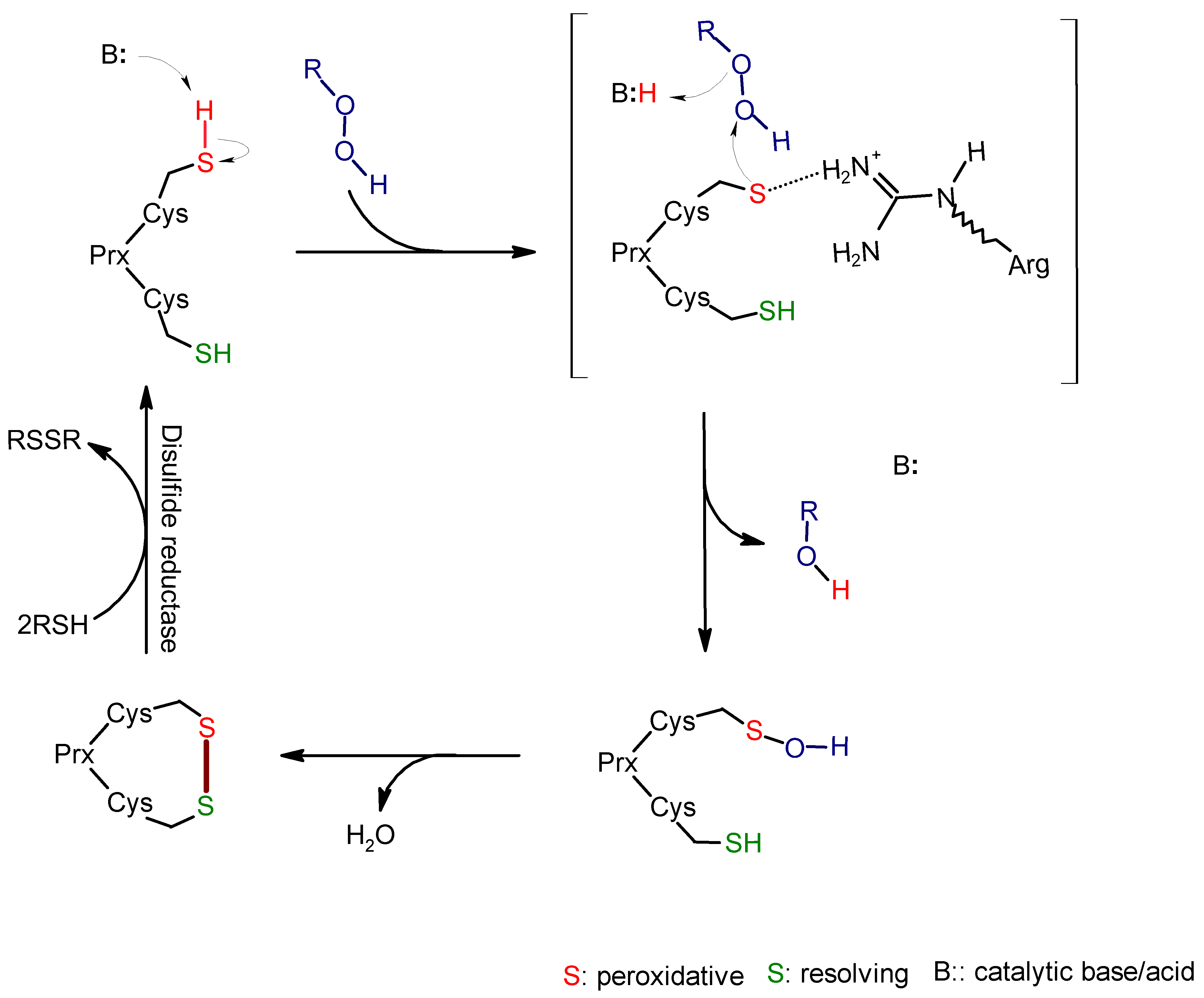
2. Alkyl Hydroperoxide Reductase C (AhpC)

2.1. Function and Significance of AhpC in M. tuberculosis

Alkyl hydroperoxide reductase C (AhpC) is involved in the oxidative stress defense of M. tuberculosis against reactive nitrogen species, peroxides and peroxynitrite, and contributes to the survival of the microorganism within macrophages [29]. It catalyzes the reduction of organic hydroperoxides, which are generated by the host cells upon infection, to the corresponding alcohols [30]. AhpC is dependent on the interaction between an adaptor protein AhpD, and (reduced) NADH for its full peroxidase activity against peroxides and peroxynitrites [31]. AhpC peroxidase activity has been found to aid the bacterial virulence and mechanism of resistance to anti-tubercular drug, isoniazid [32,33]. AhpC gene regulation is uniquely marked with periods of silencing and activation, which relates to Mtb’s ability for antioxidant defense, especially during dormancy or transmission to new hosts [34]. AhpC belongs to the peroxiredoxin family of enzymes with evolutionarily conserved cysteine within the catalytic site, where it forms sulfenic acid intermediate during the reduction process [35].

2.2. Role of Active Site Cysteine in AhpC Activity and Potential as a Drug Target

This family of enzymes comprises the typical 2-Cys peroxiredoxins that use two cysteine residues for the catalytic oxidoreductive reactions [35]. They usually exist as functional dimers, but they undergo structural rearrangement to the oligomeric form in their catalytic intermediate states. Unlike other Gram-negative bacteria peroxidoxins that have structural folds of a dimeric pentamer in the reduced state, the mycobacterial AhpC would rather assume a ring-shaped hexamer of dimers [36]. In general, these enzymes’ reaction involves a peroxidative cysteine to react with the peroxide/peroxynitrite substrates to form cysteine sulfenic acid, which is subsequently attacked by a second resolving cysteine to form a disulfide crosslink (Scheme 1). Each cysteine residue is located on a subunit of the dimer. The inter-subunit disulfide bond is reduced by associated proteins/oxidoreductases, again returning the peroxidative cysteine to the initial state.
In MtbAhpC, the reducing equivalents for reducing the disulfide crosslink are supplied by AhpD, which is a thioredoxin-like protein present in a small number of organisms [31]. A third cysteine, whose catalytic role has not yet been determined, has been implicated in the catalytic mechanism of Mtb AhpC [37,38]. In contrast to other peroxiredoxins that feature local unfolding of the active site that exposes the peroxidative cysteine to the solvent environment [35], the MtbAhpC has the capability of an entire conformational flexibility of the helix α1 of the loop-helix region that bears the peroxidative cysteine. This rigid-body movement of the entire helix brings the peroxidative cysteine in close proximity to resolving cysteine for disulfide crosslinking (Figure 1) [36]. The resolving cysteine provides an important function, which is to prevent further oxidation of the cysteine sulfenate by peroxide to form inactive sulfinic (-SO2H) or sulfonic acid (-SO3H). As such, a high concentration of resolving cysteine is maintained at the region complementary to the region bearing the peroxidative cysteine [35]. The chemical transformation is distinct from cysteine dioxygenase that uses dioxygen on an iron center to convert a free cysteine amino acid to cysteine sulfinic acid products [39,40,41,42,43].
Since the MtbAhpC-AhpD peroxidase system is vital in protecting M. tuberculosis against oxidative stress triggered by peroxides/peroxynitrites [31] and is not present in humans, it has potential for anti-tubercular drug interventions. Studies have shown that MtbAhpC gene is expressed in compensation for the lack of KatG, a gene that encodes the catalase-peroxidase system that detoxifies hydrogen peroxide, in isoniazid-resistant M. tuberculosis in which the KatG gene was deleted [37,44]. There is a tendency to alter the role of the reducing partner protein, the AhpD, but this may not completely destroy the peroxide defense. For instance, a competitive inhibitor which competes for the substrate binding site of AhpD protein was unable to completely suppress the in vitro activity of AhpC/AhpD, and there was still a small amount of AhpD when isoniazid-resistant M. tuberculosis was studied in infected mouse lungs [45]. Still, the most effective strategy is to target the MtbAhpC to cause total loss of the oxidative stress defense mechanism in the pathogenic organism. Guimarães et al. showed that there are implications for structure-based drug design that will mitigate or arrest the completion of the catalytic cycle of Mtb AhpC [36]. This was because of the unique manner in which the peroxidative cysteine moved close to the complementary second cysteine, which involved moving the entire helix α1, which generates a cavity that can potentially accommodate an inhibitor.

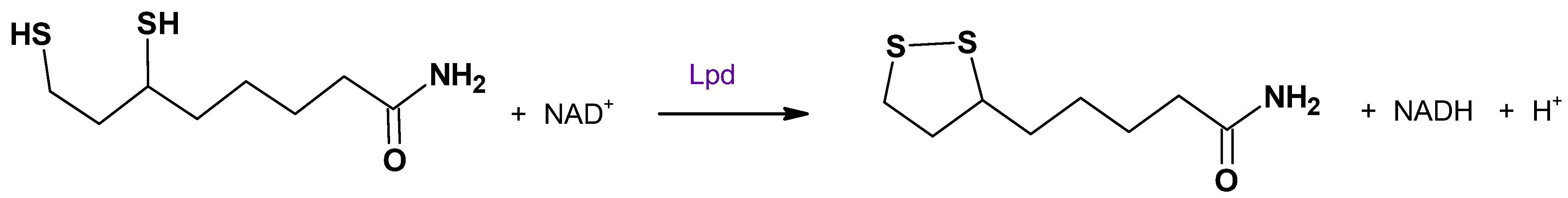
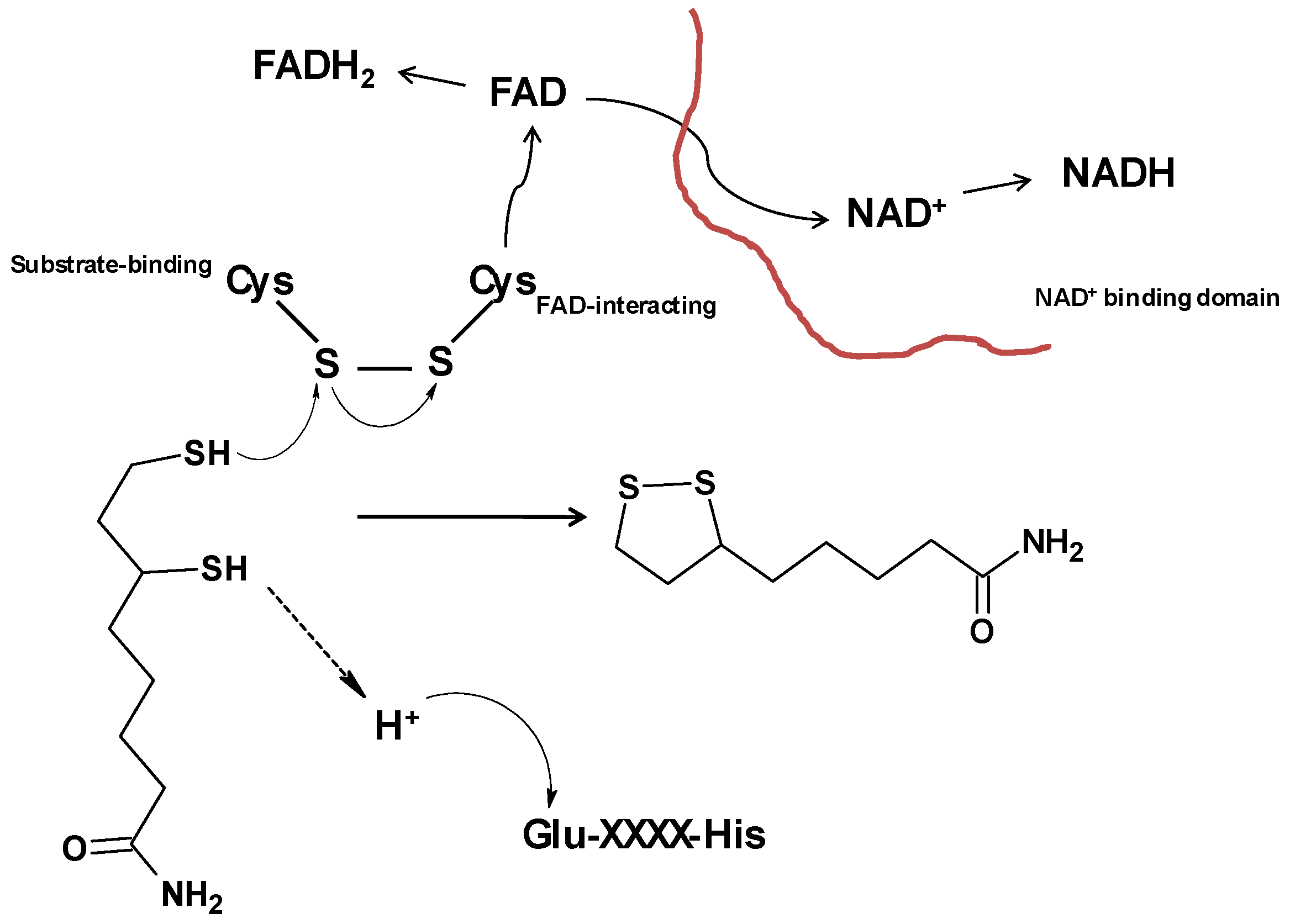
3. Dihydrolipoamide Dehydrogenase (Lpd)

3.1. Role of Lpd in M. tuberculosis Metabolism

Dihydrolipoamide dehydrogenase (Lpd) has its catalytic roles strategically placed for central energy metabolism and/or biosynthetic functions. It is one of the three components of a set of multienzymes that include the pyruvate dehydrogenase (PDH), the branched chain ketoacid dehydrogenase (BCKADH) and the α-ketoglutarate dehydrogenase (αKGD) complexes, which exist in both eukaryotic and prokaryotic organisms [46], although the αKGD enzyme is lacking in Mtb [47]. Lpd is the last enzyme (E3) of the PDH and BCKADH complexes and contains a flavoprotein disulfide reductase, which catalyzes the oxidation of dihydrolipoyl cofactor with the help of NAD+, as an acceptor of the reducing equivalent (Scheme 2) [48,49].
In Mtb, Lpd’s function is involved in three different enzyme complexes (PDH, BCKADH, PNR/P) in a well-coordinated manner: the Lpd, Dlat (dihydrolipoyl acyltransferase, E2) and AceE (E1) multi-components synthesize acetyl-coA in the PDH complex; the Lpd, Dlat and pdhABC complexes produce the branched-chain fatty acyl-coA in the BCKADH complex; and the Lpd, Dlat and AhpC/AhpD system detoxifies peroxides/peroxynitrites (PNR/P) [50].
The enzymatic products of these complexes are essential for the metabolic homeostasis, cellular survival and adaptability of Mtb in the macrophages, ensuring that the energy requirements of the organism are met continuously even when the host cells are in a nutritional deficient state. They also ensure that the organism continues to mount defense responses against host-induced oxidative and nitrosative stress, as well as the reduction in the over-accumulation of metabolites such as pyruvate, branched-chain amino acids and branched-chain keto acids, which can become potentially harmful to the organism itself.

3.2. Impact of Active Site Cysteine on Lpd Function and Drug Development

Dihydrolipoamide dehydrogenase (Lpd) operates a disulfide-based redox active center [51]. The disulfide crosslink is contributed by two active site cysteine residues that are evolutionarily conserved in many organisms [46,51,52,53]. The structure of Lpd’s active site is arranged in such a way that the electron flow from the dihydrolipoyl substrate to NAD+, via FAD (flavin adenine dinucleotide) is functionally related to the formation of NADH, which regenerates the disulfide crosslink to complete the catalytic cycle (Scheme 3).
Lpd is a homodimeric enzyme, but each monomer contains four distinct domains which are sites for FAD binding and NAD+ binding. In addition, the structure contains a central domain and an interface domain [54]. The two active site cysteine residues are located in the FAD binding domain, where they interact with the tightly but non-covalently bound FAD. The interface domain has a conserved His-Glu motif, which donates a proton (H+) for the catalysis [55]. The NAD+ molecules are located on their own separate domain, which indicates the requirement for the enzyme to devise an electron flow in this manner. Disruption of this electron flow will negatively affect enzyme functions and thus the upstream metabolic pathways, leading to energy starvation and the accumulation of toxic metabolic intermediates such as pyruvate or lactate [56].
One of the two active site cysteine residues is substrate-binding (proximal) and the other interacts with the FAD molecule. Interestingly, it has been shown that the substrate-binding cysteinyl residue is more reactive than the distal one, which indicates that their thiol groups are chemically inequivalent [57]. As such, the proximal cysteine is more amenable to further reactions such as nitrosylation and sulfenation [58]. These chemical modifications could cause protein misfolding [59] and, in the case of Lpd, might result in loss of its enzymatic activity [60].
In Mtb, the electron flow arrangement is such that the first enzyme component that accepts electrons from NADH is Lpd (direction of flow of electrons: NADH → Lpd → DlaT → AhpD → AhpC → ROOH), which has its redox center regenerated in the process. As such, Lpd’s function is critical in this arrangement. Electron flow would be disrupted when there is impairment of function or even absence of any of the components [31]. Any chemotherapy agent that can target any of these components can disrupt the electron flow. Therefore, such an agent will have potential to attenuate Mtb’s survival because the organism becomes susceptible to oxidative and nitrosative stress induced by the host immune response [31,50]. It has been shown that chemical inhibitors like triazaspirodimethoxybenzoyls have the potential to selectively inhibit MtbLpd, with >100-fold selectivity compared to human Lpd [61]. Although both share some evolutionary conservation, there are variations in the active sites of the MtbLpd and human Mtb [49]. The differences in the active sites could be explored to design and develop useful species-selective inhibitors of Lpd, particularly those targeting the active site cysteine residues, as potent anti-tubercular therapeutic agents.

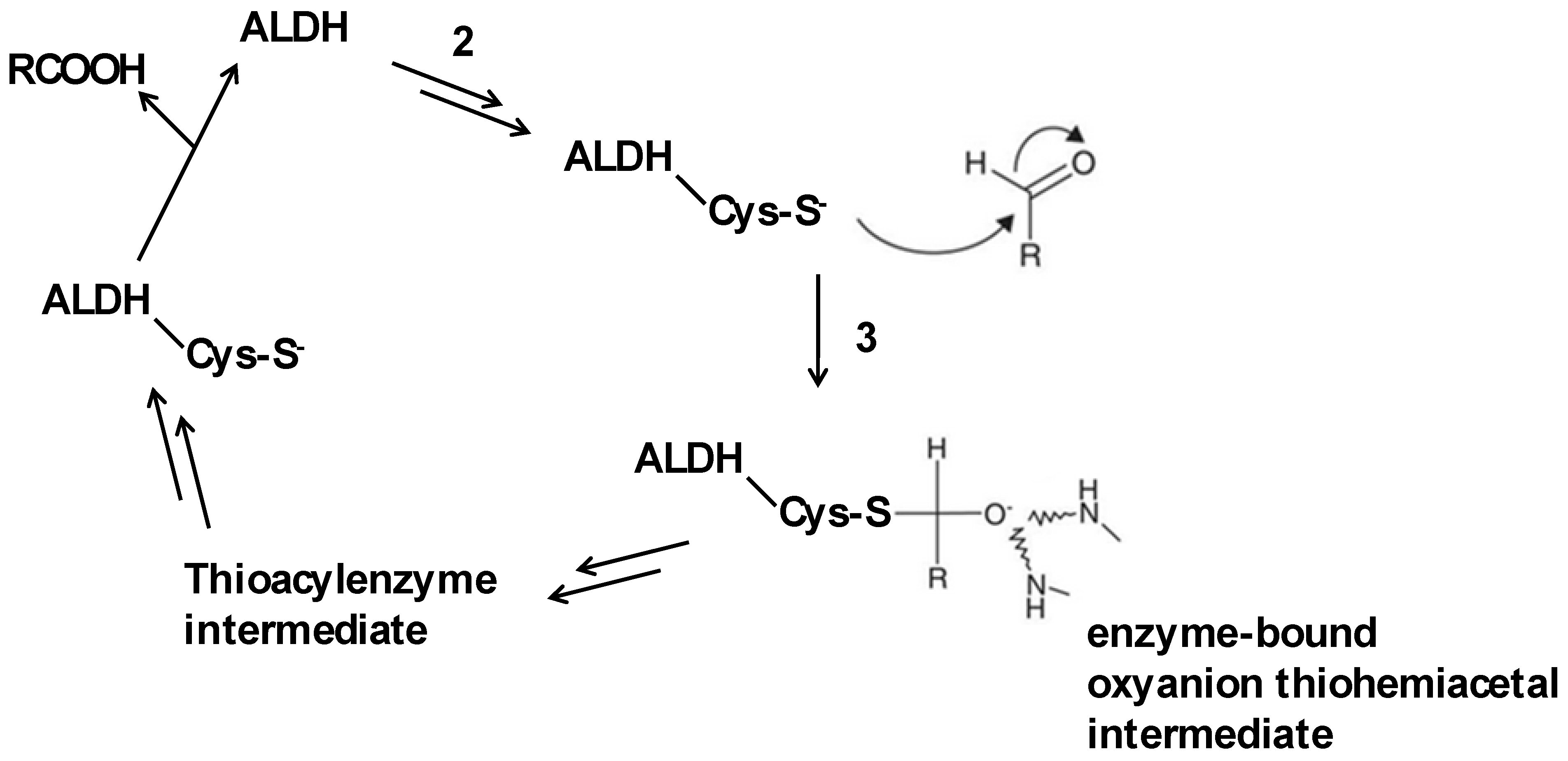
4. Aldehyde Dehydrogenase (ALDH)

4.1. Function of ALDH in Detoxification Pathways

Aldehyde dehydrogenases (ALDH) are a superfamily of NAD(P)+-dependent proteins that catalyze the oxidation of a wide range of aldehydes (aliphatic and aromatic, both endogenous and exogenous) to their corresponding more soluble carboxylic acids [62,63]. These enzymes are present in most organisms including prokaryotes, eukaryotes and Archaea. ALDH have various (non)catalytic functions that involve detoxification, biosynthesis, antioxidant functions, and structural and regulatory mechanisms, including roles in embryogenesis, development and neurotransmission, oxidative stress, and cancer [64,65]. There is more than one ALDH gene in humans [66,67]. Thus, the human ALDH-2 protein has many functions, including nitrate reduction [68,69] and the removal of toxic aldehydes. Accumulation of aldehydes can lead to protein/enzyme dysfunction, oxidative damage, the generation of reactive oxygen species, and lipid peroxidation, which causes the formation of more aldehydes [65,70]. Mtb is also known to have ten putative ALDH proteins. Bioinformatics analyses have associated them with seven different Mtb ALDH classes. Notably, only one of the Mtb ALDH proteins, Mtb Rv0223c, has been experimentally solved and was comparable to the human ALDH-2 because of their close sequence and structural similarity to each other [66].
In this case, they share three domains (catalytic domain, coenzyme domain and oligomerization domain) that align very highly when superimposed (Figure 2). However, Mtb Rv0223c is a monomer, in contrast to the octameric human ALDH-2 [71,72,73]. Despite this difference, both proteins retain the evolutionarily conserved cysteine and glutamic acid at identical positions within their active sites. The active site cysteine is directly involved in the detoxification mechanism of Mtb ALDH.

4.2. Involvement of Active Site Cysteine in ALDH Activity and Drug Targeting Strategies

Aldehydes make Mtb sensitive to nitric oxide (NO) and Cu, which are natural antimicrobial agents produced by the host immune cells [74,75]. Recently, evidence has been put together in a review that shows aldehydes of host origin have the ability to control intracellular growth of M. tuberculosis [76]. Limón et al. showed that the wild-type M. tuberculosis became susceptible to Cu toxicity when an exogenous aldehyde, para-hydroxybenzaldehyde (pHBA), was added to the mycobacterial strain cultures [77]. As such, the presence of some aldehydes may be important for controlling M. tuberculosis infections. Since the human and Mtb ALDH2 are highly homologous, it is presumed that both have similar catalysis. Importantly, the active site cysteine acts as a nucleophile that attacks the aldehyde functional group (the carbonyl carbon), resulting in the formation of an oxyanion thiohemiacetal intermediate, which is stabilized by amide/peptide nitrogen atoms of nearby amino acid residues [78,79]. This step is crucial in the aldehyde detoxification mechanism mediated by Mtb. Overall, the implications of a defective or dysfunctional Mtb ALDH will be apparent absence or perturbed catalysis leading to the buildup of aldehydes, which attenuate the growth of the bacteria within the macrophages.
There is potential to design and develop drugs that will interfere with this critical step in the catalysis of Mtb ALDH, particularly by preventing the active site cysteine from performing the nucleophilic attack (step 2 in Scheme 4) upon cofactor binding. For instance, it has been shown before that disulfiram, a sobriety drug used to treat alcoholism, which was repurposed against Mycobacterium tuberculosis showed significant anti-tubercular activities against both multidrug- and extensively drug-resistant strains of the microbe in mice [80]. Also, there was a synergistic action of Cu ions and disulfiram in killing Mtb, which suggests that the antibacterial effects of the drug are copper-dependent [81]. While the mechanism of the bactericidal activity of the drug was unclear, another study had revealed the possibility of the drug inducing interactions between two active site cysteine residues, thereby forming a stable intramolecular disulfide bond [82] that halts catalysis. This is very insightful and opens a broad window of opportunities to develop and screen a wide range of inhibitors that have promising potential to interfere with the active site cysteine in Mtb. It is useful to research species-specific inhibitors that target the pathogenic enzyme, as well as co-agents that induce the innate host immune defenses, resulting in the accumulation of reactive aldehydes and thus leaving the enzyme susceptible to their actions. A novel chemotherapy strategy will be to induce physiological events that increase the amount of metabolic aldehydes that kill the bacteria while sparing the human host cells. By these means, the chemotherapy agents can be developed to perturb the catalytic role of the active cysteine of M. tuberculosis, leaving the accumulated aldehydes cytotoxic only to the invading pathogen.

5. Methionine Aminopeptidase (MetAP)

5.1. Role of MetAP in Protein Processing

Methionine aminopeptidases (MetAP) are present in all living organisms, including bacteria and humans, where they are involved in the translational modification of proteins in the cells. They are dinuclear metalloenzymes that rely on first-row transition metals as cofactors for catalytic activity. Specifically, these metalloproteases cleave the N-terminal methionyl residue from all newly synthesized proteins (Scheme 5) [83,84].
This translational processing occurs in most nascent proteins and is an important process in endothelial cell growth and the differentiation for angiogenesis, which is the formation of new blood vessels, and cell cycle/proliferation. In humans, annihilation or suppression of this reaction would prevent the N-terminal residues from undergoing further modifications, leading to blockage of angiogenesis [5,85,86] and control of cell proliferation [87,88,89]. This signifies the importance of MetAP inhibitors as potential anticancer agents. Nonetheless, the focus here is to illuminate the structure and functions of mycobacterial MetAP in an effort to reveal the potential of developing species-specific inhibitors that target their active site cysteines. There are two classes of MetAP—namely type 1 (MetAP1) and type 2 (MetAP2)—that are present in all organisms. The difference between the two types is found in type 2, in which there is an approximately 60-residue insert within the α-helix fold of the catalytic domain [5,90,91]. While both types are present in eukaroytes and humans, the prokaryotes possess either one of the two types with several homologues [84]. There are subclasses, designated a, b, c and d, for type 1 MetAPs, with unique features in the N-terminal domains, such as the presence of a zinc finger domain in subclass b [92]. Two isoforms of the enzyme are found in Mtb, MtMetAP1a and MtMetAP1c; the latter possesses a highly conserved proline-rich N-terminal extension [85]. Both isoforms are enzymatically active and not functionally redundant [93]. There was mycobacterial growth when either one of them was over-expressed in the presence of inhibitors. However, knockdown of only MtMetAP1a resulted in reduced growth, which suggests its essentiality in the viability of Mtb [93]. Therefore, efforts to develop anti-mycobacterial agents could be directed at the mitigation of the enzymatic activity of MtMetAP1a.

5.2. Significance of Active Site Cysteine in MetAP Function and Drug Targeting Strategies
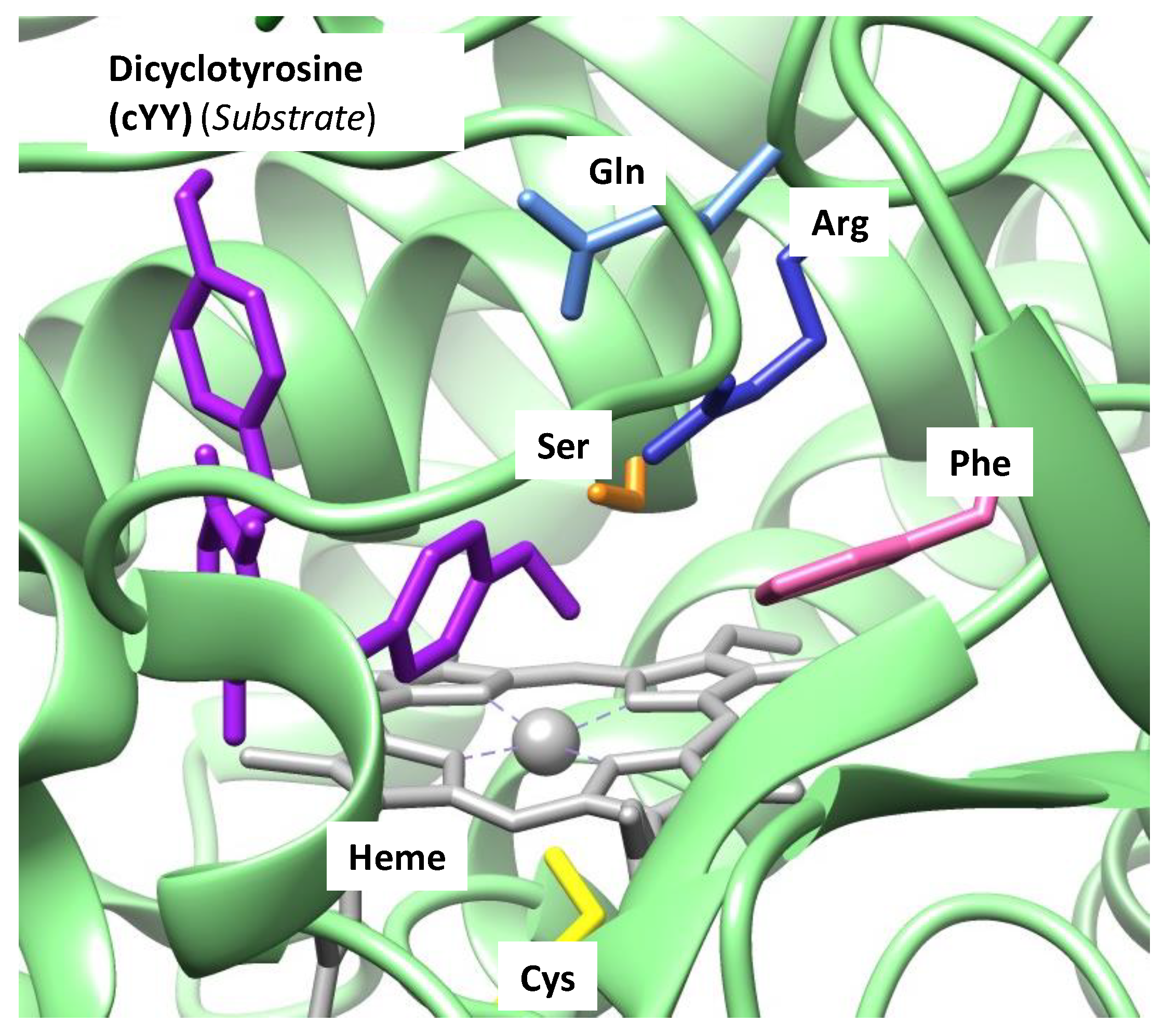
The active sites of MetAPs contain a dinuclear metal center, and the residues coordinating these metals are highly conserved in all organisms [5,90]. The substrate-binding pocket is surrounded by non-conserved but homologous amino acid residues. Figure 3A depicts the dinuclear metal coordination motif and the substrate-pocket residues of MtMetAP1c. Importantly, a cysteine is found to be conserved among these residues in all MetAP1, although another residue, glycine, is present in the homologous position in MetAP2 [94,95]. Reddi et al. performed a sequence alignment test of MtMetAP1c with other type 1 MetAPs from other species and identified the conserved Cys105 residue at the same location in all strains (Figure 3B) [96]. On the other hand, only the E. coli MetAP1 active site Cys70 has been revealed to participate directly in catalysis [97], while the Cys105 residue is required to play an essential role in substrate positioning. The active site Cys105 role may be harnessed in designing specific MetAP1 inhibitors. Reduced enzymatic activities of E. coli and human MetAP1s when the active site cysteine was mutated [94] may reinforce the plausibility of designing specific inhibitors for therapeutic interventions against pathogenic diseases.
The active site Cys105 can be covalently modified only by some specific inhibitors. The mechanism of selective modification is dependent on the stereochemistry of the cysteine and the presence of divalent metal ions [96]. Generally, MetAPs are capable of using any first-row transition metals for catalytic activity [98], although cobalt ions have been used frequently to reconstitute the enzyme in many studies [5]. Both the cysteine-specific inhibitory agent and cysteine must be in an orientation that favors a nucleophilic addition. The metal ions help to stabilize the complex through a bridging water molecule. Since MetAPs have the capacity to utilize a range of metal ions for catalysis, it may present some difficulties in designing potent inhibitors against them. However, a promising approach is developing species- and site-specific inhibitors that target the active site cysteine through covalent modification, disrupting its functions within the enzyme.

6. Cytochromes P450

The cytochrome P450s (P450s or CYPs) are enzymes present in most organisms, ranging from prokaryotes to eukaryotes, in which they mediate a wide range of metabolic reactions, including the biosynthesis and biodegradation of compounds in both endogenous and exogenous molecules [6,7,8,9,10,11,12,13,14,15,99,100,101,102,103,104]. The P450s are monooxygenases that insert a single oxygen atom derived from a dioxygen molecule into their substrates, thus changing the physicochemical properties of the substrates, as happens in the detoxification of xenobiotics, for instance. Generally, the P450 active site consists of a heme iron (porphyrin) cofactor, bound through a heme–thiolate interaction from an active site cysteinate residue, while the substrate binding pocket has important amino acid residues that relate to substrate positioning and proton and electron relay [105]. As an example, we show in Figure 4 the active site of a typical P450 enzyme, namely an extract from the protein databank file 6UPI [106,107] that represents the dicyclotyrosine-bound isozyme CYP121. The heme iron (grey) lies on the equatorial plane of the active site pocket and has a distal site, where the dioxygen and substrate bind, and a proximal site that links it to the axial Cys345 residue of the protein.
Usually, the P450 reaction cycle goes through a series of pre-catalytic events, which produce the active oxidant (known as Compound I, Cpd I) of the enzyme reaction, before returning to the enzyme resting state to complete the catalytic cycle [6,7,8,13,14,101,103,104,105,108]. Briefly, when the substrate binds into the P450 active site, a water molecule that occupies the distal site is displaced and leads to a change of spin state from low-spin to high-spin. Thereafter, the heme iron(III) is reduced to iron(II), where the electron is supplied by a reducing partner, e.g., NAD(P)H or P450 reductase. Next, the heme iron(II) readily binds dioxygen to form an adduct. A second electron is supplied by the redox partner that changes the iron-dioxygen into an iron-peroxo group. This heme peroxo intermediate is protonated to form the heme iron(III)-hydroperoxo complex and ultimately forms Cpd I through a subsequent protonation and heterolytic O–O bond cleavage that releases a water molecule. CpdI is a highly reactive intermediate with substrates and typically reacts through oxygen atom transfer. The Cpd I was an elusive species until Rittle and Green were able to characterize it spectroscopically in 2010 [109]. Several studies have shown the importance of the thiolate contribution to the reactive nature of Cpd I—the oxidizing equivalents are shared between the porphyrin architecture and the thiolate ligands [10,11,12,13,14,15,110,111,112,113]. The influence of the heme-thiolate is termed the “push effect” whereby the axial ligand thiolate promotes an increased electron density on the heme iron and the oxo atom, thereby enhancing the reactivity of Cpd I active oxidant of P450 enzyme. With enhanced reactivity occasioned by the push effect of the heme-thiolate, Compound I typically reacts with substrates through oxygen atom transfer reactions, including aliphatic and aromatic hydroxylation, sulfoxidation, epoxidation, dealkylation, decarboxylation and desaturation, among others [6,7,8,9,101,102,103,104,114,115,116,117].
Mycobacteria contain several CYP enzymes; Mycobacterium tuberculosis encodes 20 CYPs, which contribute to its survival and pathogenicity. For example, Mtb CYP121 is involved in the biosynthesis of mycocyclosin, an important natural compound in the bacterium that regulates cellular homeostasis and controls its survival and growth [118,119,120]. Additionally, the heme-thiolate has been shown to act as a redox switch that activates and deactivates P450 catalytic activity through the (reversible) formation of sulfenic acid in the presence of hydrogen peroxide and thus inhibits the enzyme function [10]. Also, the P450 2B4 isozyme nearly lost its monooxygenase activity when its active site cysteine was substituted with serine, emphasizing the vital role the heme-thiolate plays in the catalytic life of the P450s [111]. Again, the heme-thiolate plays a role in spin state and redox switching within the P450 catalytic cycle by ensuring low energy transition between the high- and low-spin states [113]. Largely, since the P450 active cysteine is sine qua non to P450 catalysis, it is an ideal target for enzyme function disruption, particularly in mycobacterial agents such as M. tuberculosis.

7. Conclusions

Cysteine, a semi-essential amino acid residue found in proteins, may be evolutionarily conserved in certain protein sequence regions due to needs such as the catalysis, regulation and modulation of protein and enzyme activity, protein structure and folding, its structural motif—particularly in cofactor metal coordination—and its role in posttranslational modifications of nascent proteins. The cysteinate group often participates in redox and non-redox enzymatic reactions and serves as a nucleophile. Cysteine is found in the active site of a wide range of enzymes, including those present in pathogenic organisms, in which they aid in propagating diseases such as tuberculosis, which has M. tuberculosis as the etiological agent. M. tuberculosis possesses enzymes that play roles in cellular defenses against oxidative and nitrosative stress caused by host-derived reactive oxygen/nitrogen species, e.g., peroxides and peroxynitrite, as well as removal of cytotoxic agents such as over-accumulated host cell aldehydes. These enzymes ensure Mtb’s survival, virulence and persistence in the host’s macrophages. The enzymes discussed in this review are involved in Mtb’s survival and adaptability. For example, alkyl hydroperoxide reductase (AhpC) and dihydrolipoamide dehydrogenase (Lpd) are parts of the enzymatic anti-oxidative arsenal that ensure Mtb’s viability; aldehyde dehydrogenase (ALDH) clears a wide range of aldehydes from both endogenous and exogenous sources which are toxic to its own cell, ensuring its survival. Methionine aminopeptidase (MetAP), a metalloenzyme that is active with many of the first-row transition metals, cleaves the N-terminal methionine during the translational modification that prepares the nascent protein for further protein modification, which is essential in the regulation of the cell cycle and cell proliferation, thus ensuring Mtb’s survival and virulence in the host cells. The cytochromes P450 mediate several important reactions involving both endogenous and exogenous compounds in the microorganism, ensuring its adaptability, survival and pathogenicity.

7.1. Implications for Drug Discovery

The active site cysteine of these Mtb enzymes is important for their enzymatic functions. The two active site cysteine residues in AhpC work in a coordinated manner to neutralize peroxide/peroxynitrite substrates. While the peroxidative cysteine forms cysteine sulfenic acid with the substrates, the resolving cysteine returns the former to the initial disulfide state. Interference or blocking of these reactions would halt the enzyme function. Any chemical agents that can target Mtb and alter this step in the catalysis are potential anti-tuberculosis interventions. Also, Lpd has two active site cysteine residues, which form a disulfide link and are involved in electron transport to NAD+ via FAD. Disruption of the electron flow would block the multienzyme complex function. This could be achieved by targeting the reactive substrate-binding cysteine of Lpd in the Mtb enzyme. Since this reaction is vital for the organism’s central intermediary metabolism, lack of it will lead to microbial death due to energy starvation and over-accumulation of toxic metabolic intermediates. Again, an inhibitor of this enzyme function is a promising anti-tubercular agent. ALDH detoxifies aldehydes with its active site cysteine. In ALDH catalysis, cysteine provides the thiol nucleophile that attacks the aldehyde carbonyl carbon to form an oxyanion thiohemiacetal intermediate in the oxidation reaction, leading to the formation of carboxylic acid. Interfering with the cysteine nucleophilic attack would block this crucial oxidative reaction. This is also a target for potential anti-tubercular agent. In MtMetAP, the active site cysteine plays an essential role in substrate recognition and positioning, rather than being involved in direct catalysis, as found in E.coliMetAP. Indeed, this reveals the possibility of designing a site-specific compound that can block the enzyme’s ability to recognize its native substrates. Lastly, by targeting or blocking the catalytic contribution of the heme-thiolate moiety of the P450 enzymes, the metabolic processes mediated by them can be disrupted, thus providing a lasting solution to the problem of Mtb infections.

7.2. Challenges and Future Research Directions

The Mtb enzymes discussed in this review are essential for Mtb’s survival and pathogenicity. However, some of them are present and/or have overlapping functions in human host cells. The challenges lie in the efforts of being able to target microbial enzymes while sparing the human enzyme functions. Some of the microbial enzymes have evolved somewhat dissimilar mechanisms, perhaps to evade host cells’ defenses. When such differences are recognized, they may open the possibility of developing specific inhibitory agents. For instance, although AhpC are found in M. tuberculosis and humans, the MtbAhpC-AhpD peroxidase system that is vital in protecting Mtb against oxidative stress is not present in humans, which makes it a potential specific target for drug discovery and development. Thus, it has become imperative to continually find not only target-specific inhibitors of enzyme functions but also those that are species-specific, leaving the human host enzymes intact.

Author Contributions

Conceptualization, A.S.F.; writing—original draft preparation, A.S.F.; writing—review and editing, J.W.G. and S.P.d.V. All authors have read and agreed to the published version of the manuscript.

Funding

This research was funded by the Natural Science and Engineering Research Council of Canada (NSERC) under grant code 04840-2018 to J.W.G.

Conflicts of Interest

The authors declare no conflicts of interest. The funders had no role in the design of the study, in the collection, analyses, or interpretation of data, in the writing of the manuscript, or in the decision to publish the results.

Abbreviations

The following abbreviations are used in this manuscript:
TBTuberculosis
MtbMycobacterium tuberculosis
AhpCAlkyl hydroperoxide reductase
LpdDihydrolipoamide dehydrogenase
ALDHAldehyde dehydrogenase
MetAPMethionine aminopeptidase

References

  1. Stokstad, E. Infectious disease: Drug-resistant TB on the rise. Science 2000, 287, 2391–2392. [Google Scholar] [CrossRef] [PubMed]
  2. World Health Organization (WHO). “Tuberculosis (TB)” 7 November 2023. Available online: https://www.who.int/news-room/fact-sheets/detail/tuberculosis (accessed on 24 October 2024).
  3. Chai, Q.; Zhang, Y.; Liu, C.H. Mycobacterium tuberculosis: An Adaptable Pathogen Associated with Multiple Human Diseases. Front. Microbiol. 2018, 8, 158. [Google Scholar] [CrossRef]
  4. Shah, M.; Chida, N. Extrapulmonary tuberculosis. In Handbook of Tuberculosis; Grosset, J.H., Chaisson, R.E., Eds.; Springer International Publishing: Cham, Switzerland, 2017; pp. 91–118. [Google Scholar]
  5. Chen, A.Y.; Adamek, R.N.; Dick, B.L.; Credille, C.V.; Morrison, C.N.; Cohen, S.M. Targeting Metalloenzymes for Therapeutic Intervention. Chem. Rev. 2019, 119, 1323–1455. [Google Scholar] [CrossRef]
  6. Sono, M.; Roach, M.P.; Coulter, E.D.; Dawson, J.H. Heme-Containing Oxygenases. Chem. Rev. 1996, 96, 2841–2888. [Google Scholar] [CrossRef]
  7. Meunier, B.; de Visser, S.P.; Shaik, S. Mechanism of Oxidation Reactions Catalyzed by Cytochrome P450 Enzymes. Chem. Rev. 2004, 104, 3947–3980. [Google Scholar] [CrossRef]
  8. Denisov, I.G.; Makris, T.M.; Sligar, S.G.; Schlichting, I. Structure and Chemistry of Cytochrome P450. Chem. Rev. 2005, 105, 2253–2277. [Google Scholar] [CrossRef]
  9. Munro, A.W.; Girvan, H.M.; McLean, K.J. Variations on a (T)Heme—Novel Mechanisms, Redox Partners and Catalytic Functions in the Cytochrome P450 Superfamily. Nat. Prod. Rep. 2007, 24, 585–609. [Google Scholar] [CrossRef]
  10. Green, M.T. C-H Bond Activation in Heme Proteins: The Role of Thiolate Ligation in Cytochrome P450. Curr. Opin. Chem. Biol. 2009, 13, 84–88. [Google Scholar] [CrossRef]
  11. Kadish, K.M.; Smith, K.M.; Guilard, R. (Eds.) Handbook of Porphyrin Science; World Scientific Publishing Co.: Hackensack, NJ, USA, 2010. [Google Scholar]
  12. Guengerich, F.P. Mechanisms of Cytochrome P450-Catalyzed Oxidations. ACS Catal. 2018, 8, 10964–10976. [Google Scholar] [CrossRef]
  13. Dunham, N.P.; Arnold, F.H. Nature’s Machinery, Repurposed: Expanding the Repertoire of Iron-Dependent Oxygenases. ACS Catal. 2020, 10, 12239–12255. [Google Scholar] [CrossRef]
  14. de Visser, S.P.; Lin, Y.-T.; Ali, H.S.; Bagha, U.K.; Mukherjee, G.; Sastri, C.V. Negative Catalysis or Non-Bell-Evans-Polanyi Reactivity by Metalloenzymes: Examples from Mononuclear Heme and Non-Heme Iron Oxygenases. Coord. Chem. Rev. 2021, 439, 213914. [Google Scholar] [CrossRef]
  15. Zhang, Y.; Mokkawes, T.; de Visser, S.P. Insights into Cytochrome P450 Enzyme Catalyzed Defluorination of Aromatic Fluorides. Angew. Chem. Int. Ed. 2023, 62, e202310785. [Google Scholar] [CrossRef] [PubMed]
  16. Chapman, H.A.; Riese, R.J.; Shi, G.-P. Emerging roles for cysteine proteases in human biology. Annu. Rev. Physiol. 1997, 59, 63–88. [Google Scholar] [CrossRef] [PubMed]
  17. Otto, H.-H.; Schirmeister, T. Cysteine proteases and their inhibitors. Chem. Rev. 1997, 97, 133–171. [Google Scholar] [CrossRef]
  18. Madala, P.K.; Tyndall, J.D.A.; Nall, T.; Fairlie, D.P. Update 1 of: Proteases universally recognize beta strands in their active sites. Chem. Rev. 2010, 110, PR1–PR31. [Google Scholar] [CrossRef]
  19. Patil, N.A.; Tailhades, J.; Hughes, R.A.; Separovic, F.; Wade, J.D.; Hossain, M.A. Cellular disulfide bond formation in bioactive peptides and proteins. Int. J. Mol. Sci. 2015, 16, 1791–1805. [Google Scholar] [CrossRef]
  20. Bošnjak, I.; Bojović, V.; Šegvić-Bubić, T.; Bielen, A. Occurrence of protein disulfide bonds in different domains of life: A comparison of proteins from the Protein Data Bank. Protein Eng. Des. Sel. 2014, 27, 65–72. [Google Scholar] [CrossRef]
  21. Feige, M.J.; Braakman, I.; Hendershot, L.M. Oxidative Folding of Proteins: Basic Principles, Cellular Regulation and Engineering; Feige, M.J., Ed.; The Royal Society of Chemistry: London, UK, 2018; pp. 1–33. [Google Scholar]
  22. Fomenko, D.E.; Marino, S.M.; Gladyshev, V.N. Functional diversity of cysteine residues in proteins and unique features of catalytic redox-active cysteines in thiol oxidoreductases. Mol. Cell 2008, 26, 228–235. [Google Scholar] [CrossRef]
  23. Parish, T.; Stoker, N.G. The common aromatic amino acid biosynthesis pathway is essential in Mycobacterium tuberculosis. Microbiology 2002, 148, 3069–3077. [Google Scholar] [CrossRef]
  24. Roberts, F.; Roberts, C.W.; Johnson, J.J.; Kyle, D.E.; Krell, T.; Coggins, J.R.; Coombs, G.H.; Milhous, W.K.; Tzipori, S.; Ferguson, D.J.P.; et al. Evidence for the shikimate pathway in apicomplexan parasites. Nature 1998, 393, 801–805. [Google Scholar] [CrossRef]
  25. Campbell, S.A.; Richards, T.A.; Mui, E.J.; Samuel, B.U.; Coggins, J.R.; McLeod, R.; Roberts, C.W. A complete shikimate pathway in Toxoplasma gondii: An ancient eukaryotic innovation. Int. J. Parasitol. 2004, 34, 5–13. [Google Scholar] [CrossRef] [PubMed]
  26. Herrmann, K.M.; Weaver, L.M. The Shikimate Pathway. Annu. Rev. Plant Physiol. Plant Mol. Biol. 1999, 50, 473–503. [Google Scholar] [CrossRef]
  27. Tzin, V.; Galili, G.; Aharoni, A. Shikimate Pathway and Aromatic Amino Acid Biosynthesis. eLS 2012, 1–10. [Google Scholar] [CrossRef]
  28. Faponle, A.S.; Fagbohunka, B.S.; Gauld, J.W. Influence of Cysteine440 on the Active Site Properties of 3-Deoxy-d-Arabino-Heptulosonate 7-Phosphate Synthase in Mycobacterium tuberculosis (MtDAHPS). ACS Omega 2023, 8, 14401–14409. [Google Scholar] [CrossRef] [PubMed]
  29. Wong, C.F.; Shin, J.; Subramanian Manimekalai, M.S.; Saw, W.G.; Yin, Z.; Bhushan, S.; Kumar, A.; Ragunathan, P.; Grüber, G. AhpC of the mycobacterial antioxidant defense system and its interaction with its reducing partner Thioredoxin-C. Sci. Rep. 2017, 7, 5159. [Google Scholar] [CrossRef] [PubMed]
  30. Chen, L.; Xie, Q.W.; Nathan, C. Alkyl hydroperoxide reductase subunit C (AhpC) protects bacterial and human cells against reactive nitrogen intermediates. Mol. Cell 1998, 1, 795–805. [Google Scholar] [CrossRef]
  31. Bryk, R.; Lima, C.D.; Erdjument-Bromage, H.; Tempst, P.; Nathan, C. Metabolic enzymes of mycobacteria linked to antioxidant defense by a thioredoxin-like protein. Science 2002, 295, 1073–1077. [Google Scholar] [CrossRef]
  32. Bhargavi, G.; Singh, A.K.; Deenadayalan, A.; Ponnuraja, C.; Patil, S.A.; Palaniyandi, K. Role of a Putative Alkylhydroperoxidase Rv2159c in the Oxidative Stress Response and Virulence of Mycobacterium tuberculosis. Pathogens 2022, 11, 684. [Google Scholar] [CrossRef]
  33. Wilson, T.; de Lisle, G.W.; Marcinkeviciene, J.A.; Blanchardand, J.S.; Collins, D.M. Antisense RNA to ahpC, an oxidative stress defence gene involved in isoniazid resistance, indicates that AhpC of Mycobacterium bovis has virulence properties. Microbiology 1998, 144, 2687–2695. [Google Scholar] [CrossRef]
  34. Master, S.S.; Springer, B.; Sander, P.; Boettger, E.C.; Deretic, V.; Timmins, G.S. Oxidative stress response genes in Mycobacterium tuberculosis: Role of ahpC in resistance to peroxynitrite and stage-specific survival in macrophages. Microbiology 2002, 148, 3139–3144. [Google Scholar] [CrossRef]
  35. Wood, Z.A.; Schröder, E.; Robin Harris, J.; Poole, L.B. Structure, mechanism and regulation of peroxiredoxins. Trends Biochem. Sci. 2003, 28, 32–40. [Google Scholar] [CrossRef]
  36. Guimarães, B.G.; Souchon, H.; Honoré, N.; Saint-Joanis, B.; Brosch, R.; Shepard, W.; Cole, S.T.; Alzari, P.M. Structure and mechanism of the alkyl hydroperoxidaseAhpC, a key element of the Mycobacterium tuberculosis defense system against oxidative stress. J. Biol. Chem. 2005, 280, 25735–25742. [Google Scholar] [CrossRef] [PubMed]
  37. Chauhan, R.; Mande, S.C. Site-directed mutagenesis reveals a novel catalytic mechanism of Mycobacterium tuberculosis alkylhydroperoxidase C. Biochem. J. 2002, 367, 255–261. [Google Scholar] [CrossRef] [PubMed]
  38. Koshkin, A.; Knudsen, G.M.; Ortiz De Montellano, P.R. Intermolecular interactions in the AhpC/AhpD antioxidant defense system of Mycobacterium tuberculosis. Arch. Biochem. Biophys. 2004, 427, 41–47. [Google Scholar] [CrossRef] [PubMed]
  39. Aluri, S.; de Visser, S.P. The mechanism of cysteine oxygenation by cysteine dioxygenase enzymes. J. Am. Chem. Soc. 2007, 129, 14846–14847. [Google Scholar] [CrossRef]
  40. Kumar, D.; Thiel, W.; de Visser, S.P. Theoretical study on the mechanism of the oxygen activation process in cysteine dioxygenase enzymes. J. Am. Chem. Soc. 2011, 133, 3869–3882. [Google Scholar] [CrossRef]
  41. Tchesnokov, E.P.; Faponle, A.S.; Davies, C.G.; Quesne, M.G.; Turner, R.; Fellner, M.; Souness, R.J.; Wilbanks, S.M.; de Visser, S.P.; Jameson, G.N.L. An iron-oxygen intermediate formed during the catalytic cycle of cysteine dioxygenase. Chem. Commun. 2016, 52, 8814–8817. [Google Scholar] [CrossRef]
  42. Faponle, A.S.; Seebeck, F.P.; de Visser, S.P. Sulfoxide synthase versus cysteine dioxygenase reactivity in a nonheme iron enzyme. J. Am. Chem. Soc. 2017, 139, 9259–9270. [Google Scholar] [CrossRef]
  43. Yeh, C.-C.G.; Pierides, C.; Jameson, G.N.L.; de Visser, S.P. Structure and functional differences of cysteine and 3-mercaptopropionate dioxygenases. A computational study. Chem. Eur. J. 2021, 27, 13793–13806. [Google Scholar] [CrossRef]
  44. Sherman, D.R.; Mdluli, K.; Hickey, M.J.; Arain, T.M.; Morris, S.L.; Barry, C.E.; Stover, C.K. Compensatory ahpC gene expression in isoniazid-resistant Mycobacterium tuberculosis. Science 1996, 272, 1641–1643. [Google Scholar] [CrossRef]
  45. Koshkin, A.; Zhou, X.T.; Kraus, C.N.; Brenner, J.M.; Bandyopadhyay, P.; Kuntz, I.D.; Barry, C.E.; Ortiz de Montellano, P.R. Inhibition of Mycobacterium tuberculosis AhpD, an element of the peroxiredoxin defense against oxidative stress. Antimicrob. Agents Chemother. 2004, 48, 2424–2430. [Google Scholar] [CrossRef] [PubMed]
  46. Carothers, D.J.; Pons, G.; Patel, M.S. Dihydrolipoamide dehydrogenase: Functional similarities and divergent evolution of the pyridine nucleotide-disulfide oxidoreductases. Arch. Biochem. Biophys. 1989, 268, 409–425. [Google Scholar] [CrossRef] [PubMed]
  47. Tian, J.; Bryk, R.; Itoh, M.; Suematsu, M.; Nathan, C. Variant tricarboxylic acid cycle in Mycobacterium tuberculosis: Identification of alpha-ketoglutarate decarboxylase. Proc. Natl. Acad. Sci. USA 2005, 102, 10670–10675. [Google Scholar] [CrossRef] [PubMed]
  48. Argyrou, A.; Blanchard, J.S.; Palfey, B.A. The lipoamide dehydrogenase from Mycobacterium tuberculosis permits the direct observation of flavin intermediates in catalysis. Biochemistry 2002, 41, 14580–14590. [Google Scholar] [CrossRef]
  49. Rajashankar, K.R.; Bryk, R.; Kniewel, R.; Buglino, J.A.; Nathan, C.F.; Lima, C.D. Crystal structure and functional analysis of lipoamide dehydrogenase from Mycobacterium tuberculosis. J. Biol. Chem. 2005, 280, 33977–33983. [Google Scholar] [CrossRef]
  50. Venugopal, A.; Bryk, R.; Shi, S.; Rhee, K.; Rath, P.; Schnappinger, D.; Ehrt, S.; Nathan, C. Virulence of Mycobacterium tuberculosis depends on lipoamide dehydrogenase, a member of three multienzyme complexes. Cell Host Microbe 2011, 9, 21–31. [Google Scholar] [CrossRef]
  51. Hopkins, N.; Williams, C.H. Characterization of lipoamide dehydrogenase from Escherichia coli lacking the redox active disulfide: C44S and C49S. Biochemistry 1995, 34, 11757–11765. [Google Scholar] [CrossRef]
  52. Williams, C.H.J. Chemistry and Biochemistry of Flavoenzymes; Muller, F., Ed.; CRC Press: Boca Raton, FL, USA, 1992; pp. 121–212. [Google Scholar]
  53. Brautigam, C.A.; Chuang, J.L.; Tomchick, D.R.; Machius, M.; Chuang, D.T. Crystal structure of human dihydrolipoamide dehydrogenase: NAD+/NADH binding and the structural basis of disease-causing mutations. J. Mol. Biol. 2005, 350, 543–552. [Google Scholar] [CrossRef]
  54. Yan, L.J.; Wang, Y. Roles of Dihydrolipoamide Dehydrogenase in Health and Disease. Antioxid. Redox Signal. 2023, 39, 794–806. [Google Scholar] [CrossRef]
  55. Benen, J.; van Berkel, W.; Dieteren, N.; Arscott, D.; Williams, C.; Veeger, C.; de Kok, A. Lipoamide dehydrogenase from Azotobacter vinelandii: Site-directed mutagenesis of the His450-Glu455 diad. Kinetics of wild-type and mutated enzymes. Eur. J. Biochem. 1992, 207, 487–497. [Google Scholar] [CrossRef]
  56. Broxton, C.N.; Kaur, P.; Lavorato, M.; Ganesh, S.; Xiao, R.; Mathew, N.D.; Nakamaru-Ogiso, E.; Anderson, V.E.; Falk, M.J. Dichloroacetate and thiamine improve survival and mitochondrial stress in a C. elegans model of dihydrolipoamide dehydrogenase deficiency. JCI Insight 2022, 7, e156222. [Google Scholar] [CrossRef] [PubMed]
  57. Thorpe, C.; Williams, C.H. Differential reactivity of the two active site cysteine residues generated on reduction of pig heart lipoamide dehydrogenase. J. Biol. Chem. 1976, 251, 3553–3557. [Google Scholar] [CrossRef]
  58. Becker, K.; Savvides, S.N.; Keese, M.; Schirmer, R.H.; Karplus, P.A. Enzyme inactivation through sulfhydryl oxidation by physiologic NO-carriers. Nat. Struct. Biol. 1998, 5, 267–271. [Google Scholar] [CrossRef] [PubMed]
  59. Nakamura, T.; Lipton, S.A. Redox modulation by S-nitrosylation contributes to protein misfolding, mitochondrial dynamics, and neuronal synaptic damage in neurodegenerative diseases. Cell Death Differ. 2011, 18, 1478–1486. [Google Scholar] [CrossRef]
  60. Yan, L.J.; Liu, L.; Forster, M.J. Reversible inactivation of dihydrolipoamide dehydrogenase by Angeli’s salt. Sheng Wu Wu Li Hsueh Bao 2012, 28, 341–350. [Google Scholar] [PubMed]
  61. Bryk, R.; Arango, N.; Venugopal, A.; Warren, J.D.; Park, Y.H.; Patel, M.S.; Lima, C.D.; Nathan, C. Triazaspirodimethoxybenzoyls as selective inhibitors of mycobacterial lipoamide dehydrogenase. Biochemistry 2010, 49, 1616–1627. [Google Scholar] [CrossRef]
  62. Kono, H.; Rusyn, I.; Yin, M.; Gäbele, E.; Yamashina, S.; Dikalova, A.; Kadiiska, M.B.; Connor, H.D.; Mason, R.P.; Segal, B.H.; et al. NADPH oxidase-derived free radicals are key oxidants in alcohol-induced liver disease. J. Clin. Investig. 2000, 106, 867–872. [Google Scholar] [CrossRef]
  63. Szöcs, K.; Lassègue, B.; Wenzel, P.; Wendt, M.; Daiber, A.; Oelze, M.; Meinertz, T.; Münzel, T.; Baldus, S. Increased superoxide production in nitrate tolerance is associated with NAD(P)H oxidase and aldehyde dehydrogenase 2 downregulation. J. Mol. Cell. Cardiol. 2007, 42, 1111–1118. [Google Scholar] [CrossRef]
  64. Vasiliou, V.; Nebert, D.W. Analysis and update of the human aldehyde dehydrogenase (ALDH) gene family. Human Genomics 2005, 2, 138–143. [Google Scholar] [CrossRef]
  65. Marchitti, S.A.; Brocker, C.; Stagos, D.; Vasiliou, V. Non-P450 aldehyde oxidizing enzymes: The aldehyde dehydrogenase superfamily. Expert Opin. Drug Metab. Toxicol. 2008, 4, 697–720. [Google Scholar] [CrossRef]
  66. Kim, C.Y.; Webster, C.; Roberts, J.K.; Moon, J.H.; Alipio Lyon, E.Z.; Kim, H.; Yu, M.; Hung, L.W.; Terwilliger, T.C. Analysis of nucleoside-binding proteins by ligand-specific elution from dye resin: Application to Mycobacterium tuberculosis aldehyde dehydrogenases. J. Struct. Funct. Genom. 2009, 10, 291–301. [Google Scholar] [CrossRef] [PubMed]
  67. Shortall, K.; Djeghader, A.; Magner, E.; Soulimane, T. Insights into Aldehyde Dehydrogenase Enzymes: A Structural Perspective. Front. Mol. Biosci. 2021, 8, 659550. [Google Scholar] [CrossRef] [PubMed]
  68. Chen, Z.; Zhang, J.; Stamler, J.S. Identification of the enzymatic mechanism of nitroglycerin bioactivation. Proc. Natl. Acad. Sci. USA 2002, 99, 8306–8311. [Google Scholar] [CrossRef]
  69. Sydow, K.; Daiber, A.; Oelze, M.; Chen, Z.; August, M.; Wendt, M.; Ullrich, V.; Mülsch, A.; Schulz, E.; Keaney, J.F.; et al. Central role of mitochondrial aldehyde dehydrogenase and reactive oxygen species in nitroglycerin tolerance and cross-tolerance. J. Clin. Investig. 2004, 113, 482–489. [Google Scholar] [CrossRef]
  70. Cederbaum, A.I. Role of lipid peroxidation and oxidative stress in alcohol toxicity. Free Radical Biol. Med. 1989, 7, 537–539. [Google Scholar] [CrossRef] [PubMed]
  71. Ni, L.; Zhou, J.; Hurley, T.D.; Weiner, H. Human liver mitochondrial aldehyde dehydrogenase: Three-dimensional structure and the restoration of solubility and activity of chimeric forms. Protein Sci. 1999, 8, 2784–2790. [Google Scholar] [CrossRef]
  72. Moon, J.H.; Lyon, A.E.; Yu, M.; Hung, L.-W.; Terwilliger, T.; Kim, C.-Y. X-ray Crystal Structure of Aldehyde Dehydrogenase from Mycobacterium Tuberculosis Complexed with NAD+ (PDB: 3B4W). Protein Data Bank. 2007. Available online: https://www.rcsb.org/structure/3B4W (accessed on 11 March 2025).
  73. Perez-Miller, S.J.; Hurley, T.D. Coenzyme isomerization is integral to catalysis in aldehyde dehydrogenase. Biochemistry 2003, 42, 7100–7109. [Google Scholar] [CrossRef]
  74. Nathan, C.; Shiloh, M.U. Reactive oxygen and nitrogen intermediates in the relationship between mammalian hosts and microbial pathogens. Proc. Natl. Acad. Sci. USA 2000, 97, 8841–8848. [Google Scholar] [CrossRef]
  75. Wolschendorf, F.; Ackart, D.; Shrestha, T.B.; Hascall-Dove, L.; Nolan, S.; Lamichhane, G.; Wang, Y.; Bossmann, S.H.; Basaraba, R.J.; Niederweis, M. Copper resistance is essential for virulence of Mycobacterium tuberculosis. Proc. Natl. Acad. Sci. USA 2011, 108, 1621–1626. [Google Scholar] [CrossRef]
  76. Darwin, K.H.; Stanley, S.A. The aldehyde hypothesis: Metabolic intermediates as antimicrobial effectors. Open Biol. 2022, 12, 220010. [Google Scholar] [CrossRef]
  77. Limón, G.; Samhadaneh, N.M.; Pironti, A.; Darwin, K.H. Aldehyde accumulation in Mycobacterium tuberculosis with defective proteasomal degradation results in copper sensitivity. mBio 2023, 14, e0036323. [Google Scholar] [CrossRef] [PubMed]
  78. Farrés, J.; Wang, T.T.; Cunningham, S.J.; Weiner, H. Investigation of the active site cysteine residue of rat liver mitochondrial aldehyde dehydrogenase by site-directed mutagenesis. Biochemistry 1995, 34, 2592–2598. [Google Scholar] [CrossRef]
  79. Steinmetz, C.G.; Xie, P.; Weiner, H.; Hurley, T.D. Structure of mitochondrial aldehyde dehydrogenase: The genetic component of ethanol aversion. Structure 1997, 5, 701–711. [Google Scholar] [CrossRef] [PubMed]
  80. Horita, Y.; Takii, T.; Yagi, T.; Ogawa, K.; Fujiwara, N.; Inagaki, E.; Kremer, L.; Sato, Y.; Kuroishi, R.; Lee, Y.; et al. Antitubercular activity of disulfiram, an antialcoholism drug, against multidrug- and extensively drug-resistant Mycobacterium tuberculosis isolates. Antimicrob. Agents Chemother. 2012, 56, 4140–4145. [Google Scholar] [CrossRef] [PubMed]
  81. Dalecki, A.G.; Haeili, M.; Shah, S.; Speer, A.; Niederweis, M.; Kutsch, O.; Wolschendorf, F. Disulfiram and Copper Ions Kill Mycobacterium tuberculosis in a Synergistic Manner. Antimicrob. Agents Chemother. 2015, 59, 4835–4844. [Google Scholar] [CrossRef]
  82. Shen, M.L.; Lipsky, J.J.; Naylor, S. Role of disulfiram in the in vitro inhibition of rat liver mitochondrial aldehyde dehydrogenase. Biochem. Pharmacol. 2000, 60, 947–953. [Google Scholar] [CrossRef]
  83. Giglione, C.; Vallon, O.; Meinnel, T. Control of protein life-span by N-terminal methionine excision. EMBO J. 2003, 22, 13–23. [Google Scholar] [CrossRef]
  84. Lowther, W.T.; Matthews, B.W. Structure and function of the methionine aminopeptidases. Biochim. Biophys. Acta 2000, 1477, 157–167. [Google Scholar] [CrossRef]
  85. Griffith, E.C.; Su, Z.; Turk, B.E.; Chen, S.; Chang, Y.H.; Wu, Z.; Biemann, K.; Liu, J.O. Methionine aminopeptidase (type 2) is the common target for angiogenesis inhibitors AGM-1470 and ovalicin. Chem. Biol. 1997, 4, 461–471. [Google Scholar] [CrossRef]
  86. Ehlers, T.; Furness, S.; Robinson, T.P.; Zhong, H.A.; Goldsmith, D.; Aribser, J.; Bowen, J.P. Methionine AminoPeptidase Type-2 Inhibitors Targeting Angiogenesis. Curr. Top. Med. Chem. 2016, 16, 1478–1488. [Google Scholar] [CrossRef]
  87. Bernier, S.G.; Taghizadeh, N.; Thompson, C.D.; Westlin, W.F.; Hannig, G. Methionine aminopeptidases type I and type II are essential to control cell proliferation. J. Cell. Biochem. 2005, 95, 1191–1203. [Google Scholar] [CrossRef] [PubMed]
  88. Hu, X.; Addlagatta, A.; Lu, J.; Matthews, B.W.; Liu, J.O. Elucidation of the function of type 1 human methionine aminopeptidase during cell cycle progression. Proc. Natl. Acad. Sci. USA 2006, 103, 18148–18153. [Google Scholar] [CrossRef] [PubMed]
  89. Yin, S.Q.; Wang, J.J.; Zhang, C.M.; Liu, Z.P. The development of MetAP-2 inhibitors in cancer treatment. Curr. Med. Chem. 2012, 19, 1021–1035. [Google Scholar] [CrossRef]
  90. Arfin, S.M.; Kendall, R.L.; Hall, L.; Weaver, L.H.; Stewart, A.E.; Matthews, B.W.; Bradshaw, R.A. Eukaryotic methionyl aminopeptidases: Two classes of cobalt-dependent enzymes. Proc. Natl. Acad. Sci. USA 1995, 92, 7714–7718. [Google Scholar] [CrossRef]
  91. Bradshaw, R.A.; Brickey, W.W.; Walker, K.W. N-terminal processing: The methionine aminopeptidase and N alpha-acetyl transferase families. Trends Biochem. Sci. 1998, 23, 263–267. [Google Scholar] [CrossRef] [PubMed]
  92. Addlagatta, A.; Quillin, M.L.; Omotoso, O.; Liu, J.O.; Matthews, B.W. Identification of an SH3-binding motif in a new class of methionine aminopeptidases from Mycobacterium tuberculosis suggests a mode of interaction with the ribosome. Biochemistry 2005, 44, 7166–7174. [Google Scholar] [CrossRef]
  93. Olaleye, O.; Raghunand, T.R.; Bhat, S.; He, J.; Tyagi, S.; Lamichhane, G.; Gu, P.; Zhou, J.; Zhang, Y.; Grosset, J.; et al. Methionine aminopeptidases from Mycobacterium tuberculosis as novel antimycobacterial targets. Chem. Biol. 2010, 17, 86–97. [Google Scholar] [CrossRef]
  94. Li, J.Y.; Cui, Y.M.; Chen, L.L.; Gu, M.; Li, J.; Nan, F.J.; Ye, Q.Z. Mutations at the S1 sites of methionine aminopeptidases from Escherichia coli and Homo sapiens reveal the residues critical for substrate specificity. J. Biol. Chem. 2004, 279, 21128–21134. [Google Scholar] [CrossRef]
  95. Swierczek, K.; Copik, A.J.; Swierczek, S.I.; Holz, R.C. Molecular discrimination of type-I over type-II methionyl aminopeptidases. Biochemistry 2005, 44, 12049–12056. [Google Scholar] [CrossRef]
  96. Reddi, R.; Arya, T.; Kishor, C.; Gumpena, R.; Ganji, R.J.; Bhukya, S.; Addlagatta, A. Selective targeting of the conserved active site cysteine of Mycobacterium tuberculosis methionine aminopeptidase with electrophilic reagents. FEBS J. 2014, 281, 4240–4248. [Google Scholar] [CrossRef]
  97. Chiu, C.H.; Lee, C.Z.; Lin, K.S.; Tam, M.F.; Lin, L.Y. Amino acid residues involved in the functional integrity of Escherichia coli methionine aminopeptidase. J. Bacteriol. 1999, 181, 4686–4689. [Google Scholar] [CrossRef] [PubMed]
  98. Lowther, W.T.; Matthews, B.W. Metalloaminopeptidases: Common functional themes in disparate structural surroundings. Chem. Rev. 2002, 102, 4581–4608. [Google Scholar] [CrossRef]
  99. Ortiz de Montellano, P.R. (Ed.) Cytochrome P450: Structure, Mechanism and Biochemistry, 3rd ed.; Kluwer Academic/Plenum Publishers: New York, NY, USA, 2005. [Google Scholar]
  100. Krauser, J.A.; Guengerich, F.P. Cytochrome P450 3A4-Catalyzed Testosterone 6β-Hydroxylation Stereochemistry, Kinetic Deuterium Isotope Effects, and Rate-Limiting Steps. J. Biol. Chem. 2005, 280, 19496–19506. [Google Scholar] [CrossRef] [PubMed]
  101. Ortiz de Montellano, P.R. Hydrocarbon Hydroxylation by Cytochrome P450 Enzymes. Chem. Rev. 2010, 110, 932–948. [Google Scholar] [CrossRef]
  102. Spinello, A.; Pavlin, M.; Casalino, L.; Magistrato, A. A Dehydrogenase Dual Hydrogen Abstraction Mechanism Promotes Estrogen Biosynthesis: Can We Expand the Functional Annotation of the Aromatase Enzyme? Chem. Eur. J. 2018, 24, 10840–11849. [Google Scholar] [CrossRef]
  103. Huang, X.; Groves, J.T. Oxygen Activation and Radical Transformations in Heme Proteins and Metalloporphyrins. Chem. Rev. 2018, 118, 2491–2553. [Google Scholar] [CrossRef]
  104. Shaik, S.; de Visser, S.P.; Kumar, D. One Oxidant, Many Pathways: A Theoretical Perspective of Monooxygenation Mechanisms by Cytochrome P450 Enzymes. J. Biol. Inorg. Chem. 2004, 9, 661–668. [Google Scholar] [CrossRef] [PubMed]
  105. de Visser, S.P. Second-Coordination Sphere Effects on Selectivity and Specificity of Heme and Nonheme Iron Enzymes. Chem. Eur. J. 2020, 26, 5308–5327. [Google Scholar] [CrossRef]
  106. Nguyen, R.C.; Yang, Y.; Wang, Y.; Davis, I.; Liu, A. Substrate-Assisted Hydroxylation and O-Demethylation in the Peroxidase-like Cytochrome P450 Enzyme CYP121. ACS Catal. 2020, 10, 1628–1639. [Google Scholar] [CrossRef]
  107. Berman, H.M.; Westbrook, J.; Feng, Z.; Gilliland, G.; Bhat, T.N.; Weissig, H.; Shindyalov, I.N.; Bourne, P.E. The Protein Data Bank. Nucl. Acids Res. 2000, 28, 235–242. [Google Scholar] [CrossRef]
  108. Shaik, S.; Kumar, D.; de Visser, S.P.; Altun, A.; Thiel, W. Theoretical Perspective on the Structure and Mechanism of Cytochrome P450 Enzymes. Chem. Rev. 2005, 105, 2279–2328. [Google Scholar] [CrossRef]
  109. Rittle, J.; Green, M.T. Cytochrome P450 Compound I: Capture, Characterization, and C-H Bond Activation Kinetics. Science 2010, 330, 933–937. [Google Scholar] [CrossRef] [PubMed]
  110. Albertolle, M.E.; Kim, D.; Nagy, L.D.; Yun, C.H.; Pozzi, A.; Savas, Ü.; Johnson, E.F.; Guengerich, F.P. Heme-thiolate sulfenylation of human cytochrome P450 4A11 functions as a redox switch for catalytic inhibition. J. Biol. Chem. 2017, 292, 11230–11242. [Google Scholar] [CrossRef] [PubMed]
  111. Vatsis, K.P.; Peng, H.M.; Coon, M.J. Replacement of active-site cysteine-436 by serine converts cytochrome P450 2B4 into an NADPH oxidase with negligible monooxygenase activity. J. Inorg. Biochem. 2002, 91, 542–553. [Google Scholar] [CrossRef]
  112. İşci, Ü.; Faponle, A.S.; Afanasiev, P.; Albrieux, F.; Briois, V.; Ahsen, V.; Dumoulin, F.; Sorokin, A.B.; de Visser, S.P. Site-Selective Formation of an Iron(IV)-Oxo Species at the More Electron-Rich Iron Atom of Heteroleptic μ-Nitrido Diiron Phthalocyanines. Chem. Sci. 2015, 6, 5063–5075. [Google Scholar] [CrossRef] [PubMed]
  113. Suzuki, H.; Inabe, K.; Shirakawa, Y.; Umezawa, N.; Kato, N.; Higuchi, T. Role of Thiolate Ligand in Spin State and Redox Switching in the Cytochrome P450 Catalytic Cycle. Inorg. Chem. 2017, 56, 4245–4248. [Google Scholar] [CrossRef]
  114. Ji, L.; Faponle, A.S.; Quesne, M.G.; Sainna, M.A.; Zhang, J.; Franke, A.; Kumar, D.; van Eldik, R.; Liu, W.; de Visser, S.P. Drug Metabolism by Cytochrome P450 Enzymes: What Distinguishes the Pathways Leading to Substrate Hydroxylation over Desaturation? Chem. Eur. J. 2015, 21, 9083–9092. [Google Scholar] [CrossRef]
  115. Faponle, A.S.; Quesne, M.G.; de Visser, S.P. Origin of the Regioselective Fatty Acid Hydroxylation versus Decarboxylation by a Cytochrome P450 Peroxygenase: What Drives the Reaction to Biofuel Production? Chem. Eur. J. 2016, 22, 5478–5483. [Google Scholar] [CrossRef]
  116. Li, X.-X.; Postils, V.; Sun, W.; Faponle, A.S.; Solà, M.; Wang, Y.; Nam, W.; de Visser, S.P. Reactivity Patterns of (Protonated) Compound II and Compound I of Cytochrome P450: Which is the Better Oxidant? Chem. Eur. J. 2017, 23, 6406–6418. [Google Scholar] [CrossRef]
  117. Qureshi, M.; Mokkawes, T.; Cao, Y.; de Visser, S.P. Mechanism of the Oxidative Ring-Closure Reaction During the Gliotoxin Biosynthesis by Cytochrome P450 GliF. Int. J. Mol. Sci. 2024, 25, 8567. [Google Scholar] [CrossRef]
  118. McLean, K.J.; Cheesman, M.R.; Rivers, S.L.; Richmond, A.; Leys, D.; Chapman, S.K.; Reid, G.A.; Price, N.C.; Kelly, S.M.; Clarkson, J.; et al. Expression, Purification and Spectroscopic Characterization of the Cy-tochrome P450 CYP121 from Mycobacterium tuberculosis. J. Inorg. Biochem. 2002, 91, 527–541. [Google Scholar] [CrossRef] [PubMed]
  119. McLean, K.J.; Carroll, P.; Lewis, D.G.; Dunford, A.J.; Seward, H.E.; Neeli, R.; Cheesman, M.R.; Marsollier, L.; Douglas, P.; Smith, W.E.; et al. Characterization of Active Site Structure in CYP121. A Cytochrome P450 Essential for Viability of Mycobacterium tuberculosis H37Rv. J. Biol. Chem. 2008, 283, 33406–33416. [Google Scholar] [CrossRef] [PubMed]
  120. Belin, P.; Le Du, M.H.; Fielding, A.; Lequin, O.; Jacquet, M.; Charbonnier, J.B.; Lecoq, A.; Thai, R.; Courçon, M.; Masson, C.; et al. Identification and Structural Basis of the Reaction Catalyzed by CYP121, an Essential Cytochrome P450 in Mycobacterium tuberculosis. Proc. Natl. Acad. Sci. USA 2009, 106, 7426–7431. [Google Scholar] [CrossRef] [PubMed]
Scheme 1. Catalytic mechanism of peroxiredoxins. Peroxide reduction via nucleophilic attack by the peroxidatic cysteine and formation of the cysteine sulfenic acid (Cys–SOH) intermediate. Dimeric 2-Cys peroxiredoxin form an inter-subunit disulfide crosslink (thick bar).
Ijms 26 03845 sch001
Figure 1. Conformation of the loop-helix showing movement (arrow) of the entire helix α1 that brings the peroxidative cysteine, Cys61, to one of the resolving cysteines, Cys174 or Cys176 in Mtb AhpC. Reproduced with permission from The American Society for Biochemistry and Molecular Biology, Inc. [36].
Ijms 26 03845 g001
Scheme 2. Lpd catalyzes the oxidation of reduced lipoamide to lipoamide.
Ijms 26 03845 sch002
Scheme 3. Catalytic mechanism of conversion of dihydrolipoamide to lipoamide by Lpd. The substrate-binding cysteine and the FAD-interacting cysteine in the disulfide crosslink are shown. The red line represents the NAD+ binding domain.
Ijms 26 03845 sch003
Figure 2. Crystal structure superimposition of MtbALDH (Rv0223c, green) (PDB ID: 3B4W [56]) with NAD (red) over human ALDH-2 (PDB ID: 1CW3 [55]) (orange).
Ijms 26 03845 g002
Scheme 4. Catalytic mechanism of ALDH in oxidation of aldehydes—some catalytic intermediates are not shown. Here, step 2 is a nucleophilic attack by activated cysteine; step 3 is the stabilization of oxyanion thiohemiacetal intermediate by two NH groups of the ALDH peptide chain. Double arrows indicate that there are steps between the reactions.
Ijms 26 03845 sch004
Scheme 5. Catalytic reaction of MetAP. The enzyme removes the N-terminal methionine from nascent proteins.
Ijms 26 03845 sch005
Figure 3. (A) The active site of MtMetAP1c shows the dinuclear metal coordination motif (metal ions, sphere), bound substrate (methionine, brown color) and the conserved cysteine residue (C105). (B) Local sequence alignment of MtMetAP1c with other MetAP1s from E. coli, H. sapiens, E. faecalis and S. pneumonia. (Star—conserved cysteine. Solid circle points to a non-conserved cysteine in E. coli MetAP1). Reproduced with permission from FEBS Press/John Wiley and Sons [96].
Ijms 26 03845 g003
Figure 4. The active site of CYP121 as taken from the PDB ID 6UPI [105,106]. The heme iron is shown in grey. Cysteine (yellow) is ligated to the heme via its thiol group, forming a heme–thiolate bond. Key amino acids involved in substrate binding and positioning are highlighted.
Ijms 26 03845 g004
Disclaimer/Publisher’s Note: The statements, opinions and data contained in all publications are solely those of the individual author(s) and contributor(s) and not of MDPI and/or the editor(s). MDPI and/or the editor(s) disclaim responsibility for any injury to people or property resulting from any ideas, methods, instructions or products referred to in the content.

