Article

Retinoblastoma: Molecular Evaluation of Tumor Samples, Aqueous Humor, and Peripheral Blood Using a Next-Generation Sequence Panel

by
Thais Biude Mendes
1,*,†,
Indhira Dias Oliveira
1,†,
Francine Tesser Gamba
1,2,
Fernanda Teresa Lima
1,3,
Bruna Fernanda Silva Cardoso Morales
4,
Carla Renata Donato Macedo
4,
Luiz Fernando Teixeira
4,5 and
Silvia Regina Caminada de Toledo
1,2,*
1
Genetics Laboratory, Pediatric Oncology Institute (IOP/GRAACC), Federal University of Sao Paulo, Sao Paulo 04023-062, SP, Brazil
2
National Science and Technology Institute for Children’s Cancer Biology and Pediatric Oncology—INCT BioOncoPed, Porto Alegre 90035-003, RS, Brazil
3
Department of Gynecology, Federal University of Sao Paulo, Sao Paulo 04024-002 SP, Brazil
4
Institute of Pediatric Oncology (IOP/GRAACC), Sao Paulo 04023-062, SP, Brazil
5
Ophthalmology Department, Federal University of Sao Paulo, Sao Paulo 04023-062, SP, Brazil
*
Authors to whom correspondence should be addressed.
These authors contributed equally to this work.
Int. J. Mol. Sci. 2025, 26(8), 3523; https://doi.org/10.3390/ijms26083523
Submission received: 7 March 2025 / Revised: 2 April 2025 / Accepted: 3 April 2025 / Published: 9 April 2025
(This article belongs to the Section Molecular Oncology)

Abstract

Retinoblastoma was one of the first malignant tumors to be described as a genetic disease and its development occurs from the loss of function of the retinoblastoma gene (RB1). The difficulty in accessing the tumor during diagnosis highlights the need for non-invasive diagnostic methods. Studies have shown that liquid biopsy, obtained from any fluid material in the body, for example blood, contains free tumor cells and free and circulating DNA or RNA, making it a convenient tool for diagnosis and prognosis during cancer treatment without the need for invasive procedures. Taking advantage of these events, given this situation, we investigated molecular alterations in samples from retinoblastoma cases, using the NGS strategy as a powerful tool for characterization and aid in diagnosis and prognosis. Genomic data from 76 patients diagnosed with retinoblastoma, comprising 162 samples, tumor (TU), aqueous humor (AH), and peripheral blood (PB), were analyzed using the Oncomine Childhood Cancer Research Panel (OCCRA®). A total of 22 altered genes were detected, and 54 variants. Of the 76 cases, 29 included paired tumor (TU), aqueous humor (AH), and peripheral blood (PB) samples from the same patient. Alterations in the RB1 gene were detected in 16 of these 29 cases, with concordant alterations identified across all three sample types in three patients. In 12 out of 29 patients, the same genetic alteration was found in both TU and AH. In conclusion, the OCCRA panel enabled the detection, in different samples, of molecular alterations in the RB1 gene, as well as CNAs in the MYCN, ABL2, and MDM4 genes. Limitations of AH were observed, primarily due to the small volume of material available and the consequently low concentration of cell-free DNA (cfDNA). However, as AH provides a viable alternative for analyzing tumors, inaccessible to traditional biopsy methods, liquid biopsy holds significant potential to improve diagnostic accuracy and guide treatment strategies in retinoblastoma cases.

1. Introduction

Retinoblastoma (RB) is a rare childhood intraocular cancer, unilateral or bilateral, manifested in children under 5 years old and encompassing 2–4% of childhood cancers [1,2]. This tumor typically develops due to the loss of function of the RB1 gene, caused by single-nucleotide variants (SNVs), copy number variants (CNVs), loss of heterozygosity (LOH), or hypermethylation of the promoter. RB diagnosis is primarily clinical, relying on the visualization of the tumor mass, as performing a tumor biopsy using fine-needle aspiration (FNA) cytology is not feasible due to the risk of tumor dissemination beyond the eye [1,3,4,5,6].
The difficulty in accessing the tumor during diagnosis highlights the need for non-invasive diagnostic methods. Identifying biomarkers through non-invasive biopsies could represent a significant advancement in RB diagnosis. Studies have demonstrated that liquid biopsy, obtained from any fluid material in the body, for example blood, contains free tumor cells and cell-free and circulating DNA or RNA, making it a convenient tool for diagnosis and prognosis during cancer treatment without requiring invasive procedures [7].
Following the development of a safe protocol for removing aqueous humor (AH) from the eye minimizes intraocular pressure and avoids tumor seed reflux [8,9]. AH has emerged as an important source of tumor-derived genetic material. Subsequent analyses have demonstrated the presence of somatic mutations in the RB1 gene in AH samples, consistent with findings from enucleated tumor tissue. These findings confirm the potential of AH as a liquid biopsy source for RB [10,11,12,13,14,15].
Advancements in next-generation sequencing (NGS) technologies have significantly enhanced cancer diagnosis and treatment strategies. Taking advantage of these events, given this situation, we investigated molecular alterations in different samples (tumor, aqueous humor, and from retinoblastoma 76 cases using the NGS strategy as a powerful tool for characterization and aid in diagnosis and prognosis as well the genomic concordance among the samples of 29 cases.
Genomic data from 76 patients diagnosed with retinoblastoma, comprising 162 samples, were analyzed. The Oncomine Childhood Cancer Research Panel (OCCRA®) evaluated the samples, which detected alterations in 200 genes identified as cancer drivers, a total of 22 altered genes, and 54 variants. Alterations in the RB1 were observed in 51% of patients. A notable aspect of this study was the inclusion of paired samples. Of the 76 cases, 29 included paired tumor (TU), aqueous humor (AH), and peripheral blood (PB) samples from the same patient. Alterations in the RB1 gene were detected in 16 of these 29 cases, with concordant alterations identified across all three sample types in three patients. In 12 out of 29 patients, the same genetic alteration was found in both TU and AH.
Alterations in nine other genes were observed, with ABL2 and MYCN being the most frequently altered. The OCCRA panel successfully detected molecular alterations in the RB1 gene, as well as copy number alterations (CNAs) in the MYCN, ABL2, and MDM4 genes. These findings underscore the utility of AH as a viable alternative for analyzing tumors that are inaccessible to traditional biopsy methods. Liquid biopsy offers significant potential to improve diagnostic accuracy and guide treatment strategies in retinoblastoma cases.

2. Results

2.1. Clinical and Tumor Characteristics

In this study, we analyzed 76 patients diagnosed with retinoblastoma (RB), 43 (57%) patients were males and 33 (43%) females. Among these cases, 58 cases (77%) with unilateral disease, 17 cases (22%) were bilateral and 1 case (1%) was trilateral (bilateral retinoblastoma and pineoblastoma) (Table 1). The median age at diagnosis was 29 months (2.4 years), with unilateral retinoblastoma cases (2.7 years) showing a higher mean age compared to bilateral cases (1.7 years), resulting in a one-year difference (Table 1). Two cases were familial: the first case RB10, is a male, bilateral RB diagnosed at 3 years. The second case RB65, male, bilateral RB, diagnosed at 2 years, whose relatives, father and aunt, were diagnosed with unilateral RB at 11 months and 4 years, respectively (Table 1).
All patients have been classified based on the International Intraocular Retinoblastoma Classification (IIRC) at diagnosis. We observed 5% group A (5 eyes), 6% group B (6 eyes), 1% group C (1 eye), 5% group D (5 eyes) and 62% group E (58 eyes). When comparing unilateral and bilateral diseases, we observed that 79% of unilateral (46 cases) occurred in group E; and in bilateral, we found that 32% occurred in group E and 12% in groups A, B, and D (Table 1).
Of the 58 patients with unilateral disease, 48 (83%) had intraocular tumors and 10 (17%) had extraocular tumors. Of 18 patients with bilateral and trilateral tumors, 12 (67%) had intraocular and 6 (33%) had extraocular diagnosis. Primary enucleation was performed in 61 patients (80%), 91% (53 cases) unilateral, and 44% (8 cases) bilateral, who underwent enucleation before chemotherapy. Only 1 of 18 bilateral diseases underwent bilateral enucleation. Recurrence or metastasis was observed in 16% of patients (12 cases), and 8/76 patients died (Table 1). Of the eight patients died, six had recurrence to the central nervous system.

2.2. Identification of Genetic Alteration by Panel in Liquid Biopsy

A total of 39 AH samples, derived from 38 out of 76 patients, were available for this study. The samples were collected from enucleated eyes. Cell-free DNA (cfDNA) was detected in all evaluated samples, with a mean concent1ration of 36.8 ng/µL (range 0.21–220 ng/µL). Genetic variants were investigated in all AH samples using the OCCRA® panel, revealing genetic alterations in 17 of the 39 samples (44%), 11 samples with SNVs, 6 samples InDel and 6 samples CNVs being the most prevalent (Figure 1A). Among these samples, the most common genetic variants involved the RB1 gene in 15/17 samples, 8 unilateral retinoblastoma samples and 7 bilateral retinoblastoma samples (Figure 1B). Specifically, 10 pathogenic somatic RB1 variants and 4 RB1 deletions were observed (Table 2). Additional alterations included copy number alteration (CNAs) involving the ABL2, MYCN, and MDM4 genes, with the last gene observed exclusively in bilateral cases (Figure 1B). In cases RB50 and RB51, no RB1 variant was detected, but CNAs in ABL2 and MYCN genes were identified, respectively (Supplementary Figure S1).

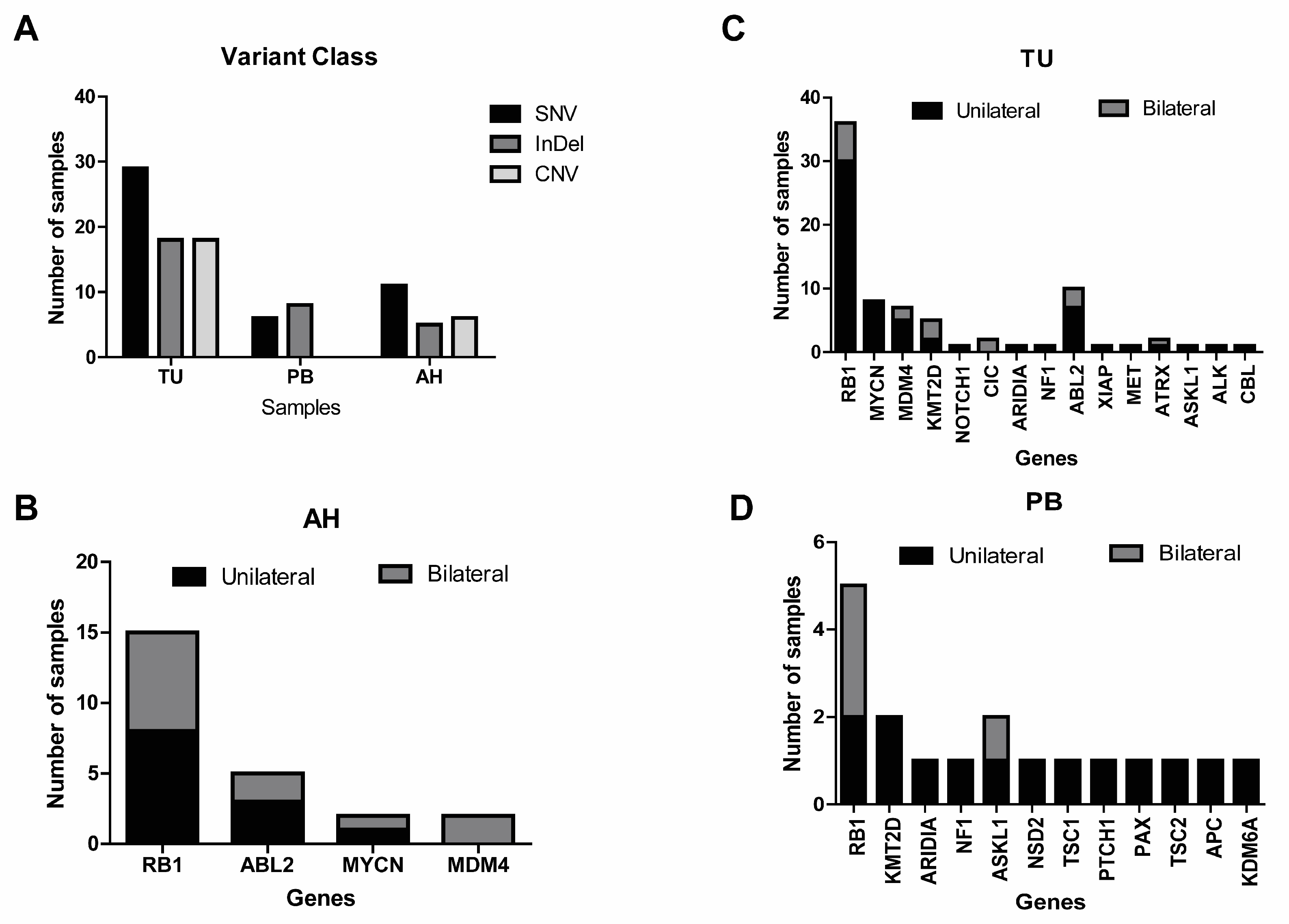
2.3. Identification of Genetic Alterations in Tumor and Peripheral Blood

A total of 125 samples, comprising 69 tumors (TU) and 56 peripheral blood (PB) samples from 76 patients, were comprehensively evaluated using the OCCRA® panel. Alterations were detected at an average rate of 0.7 per TU sample (range 0–3) and 0.3 per PB sample (range 0–2). The genomic landscape of RB patients can be observed in Supplementary Figure S1. No genetic alterations were detected in 61/125 samples (49%; 21 TU and 40 PB) using the OCCRA® panel. In the remaining 64 samples (51%), alterations were identified in 22 (Figure 1A,C,D), with a total of 48 genetic variants detected, comprising 19 SNV, 18 insertions/deletions (InDels), and 4 CNAs. When analyzing all altered genes, it was observed that 9 out of 15 genes in tumor samples and 10 out of 12 genes in peripheral blood samples were found exclusively in patients with unilateral retinoblastoma (Figure 1C).
Alterations in the RB1 gene were found in 32% of the samples (35 TU and 5 PB—Figure 1C), specifically, 16 SNV and 11 InDels (Table 3). The c.1333C>T and c.958>T variants were the most frequent in the TU samples (Table 3). Among the 35 tumors with RB1 gene alteration, 21 contained only RB1 variants, while 14 exhibited RB1 alterations along with one or two additional alterations in other genes (Supplementary Figures S1 and S2A). Co-occurring alterations in RB1 and seven genes were identified, with the most common being CNAs in MYCN and MDM4 (Supplementary Figure S2B).
In 12 tumors and 11 peripheral blood samples, from 21 patients, no RB1 alterations were detected, but other gene alterations were identified. The most common genes affected were MYCN and ABL2 genes in TU samples and ASKL1 and KMT2D in PB samples (Supplementary Table S1). In case RB66, where only a TU sample was available, three alterations were identified: one InDel in the gene CBL, and two CNAs in ABL2 and MDM4 (Supplementary Figure S1).

2.4. Genetic Analysis from Tumors, Aqueous Humor, and Peripheral Blood

A total of 29 cases (38%; 23 unilateral and 6 bilateral diseases) out of 76 cases had paired samples (TU, AH, and PB) (Figure 2). No genetic alterations were detected in six cases (21%). Mutation analysis identified 10 SNVs and 4 InDel in the RB1 gene across 55% (16/29) cases (Table 4). Among the 16 cases, RB1 gene alterations were observed in all samples from three patients (RB43, RB47, and RB65) (Table 4). In case RB47, with a diagnosis of bilateral disease and bilateral enucleation, AH from both eyes was evaluated, and just TU from the left eye. The evaluation revealed not only RB1 gene alterations but also amplification of the ABL2 and MDM4 genes in the TU and AH samples. Additionally, amplification of the MYCN gene was observed exclusively in the AH sample from the right eye (Supplementary Table S2). In the familial case RB65, RB1 gene alterations (specifically the c.1597G>T mutation) were detected consistently across all analyzed samples (TU, AH, and PB).
In 7 of the 16 cases with RB1 gene alterations, changes were observed exclusively in TU and AH samples (Table 4). Additionally, two cases (RB36 and RB37) showed CNA in ABL2 and MDM4 genes, with the latter alteration found only in the tumor sample (Figure 2 and Supplementary Table S2). Patient RB32 exhibited a deletion (c.4324_4325delCG) and an insertion (c.921_922insC) in the PTCH1 gene, which was identified exclusively in the PB sample (Supplementary Table S2).
In seven patients without RB1 alterations, variations were identified in nine other genes, with the ABL2 and MYCN being the most frequently altered (Supplementary Table S2). In patient RB69, alterations varied between TU and PB samples—a CNA in the MDM3 gene was observed in the TU, whereas two InDels in the ARID1A gene were identified in the PB sample.

3. Discussion

Aqueous humor (AH) is an important source of tumor-derived genetic material, underscoring its potential use in liquid biopsies for retinoblastoma, particularly for detecting mutations in the RB1 gene, as recently demonstrated [10,11,16,17,18]. In our study, genetic alterations in retinoblastoma samples were analyzed using a comprehensive next-generation sequencing (NGS) panel specific for pediatric neoplasms. A total of 164 samples (TU, PB, and AH) from a cohort of 76 patients were evaluated. Within this cohort, 60% of patients were diagnosed in group E retinoblastoma, the most advanced stage of retinoblastoma as classified by the IIRC [19].
As far as we know, this was the first study to employ genomic profiling using the OCCRA panel on different sample types (tumor, aqueous humor, and peripheral blood) from RB cases to investigate their molecular characteristics. The ion Torrent Oncomine Childhood Cancer Research Assay is a unique NGS-based tool designed for comprehensive genomic profiling of cancer affecting children and young adults. The assay is designed to provide researchers with sensitive and comprehensive sample amplification of relevant DNA mutations and fusion transcripts associated with childhood and young adult cancers in a single NGS run. The panel comprises 203 unique genes: 130 key DNA genes, 28 copy number variant targets, an expansive fusion panel of 90 driver genes with multiple partners, and 9 expression genes and controls. These cover the most relevant targets in the vast majority of all childhood and young adult oncology research samples [20]. For liquid biopsy, which involves low DNA concentrations, the method demonstrated the ability to detect genetic alteration in cell-free DNA (cfDNA) from AH, with high sensibility and coverage.
The findings revealed somatic genetic variants in 70% of TU samples, 29% of PB samples, and 44% of AH samples. A total of 22 altered genes were identified, with the most frequently detected variants being RB1, ABL2, MYCN, and MDM4 genes. Genetic alterations in the RB1 gene were identified in 33% of the samples. The most frequent genetic alterations, in our samples, were the pathogenic variants RB1: c.958C>T and c.1333C>T. The c958C>T mutation was detected in five cases of unilateral diseases. Interestingly, this mutation was found in both samples, TU and AH, of patient RB53. In the literature, this mutation has been reported in cases of unilaterally, being present in 5% of tumor samples analyzed [21]. It has also been described in familial RB cases as a mosaic mutation in 2–10% of the study’s cases [22,23]. The c.1333C>T mutation was detected in five cases, including four unilateral cases (four TU and one AH) and e one bilateral case (one AH). This mutation has already been described in four unilateral cases of patients from Singapore and four bilateral cases from India [22,24]. In the cases from India, three patients presented disease progression, and one required bilateral enucleation because he did not respond to treatment [24].
Previous research has demonstrated that somatic copy number alterations (SCNAs) may contribute to tumor progression in retinoblastoma cases [25]. CNAs in ABL2, MDM3, and MYCN genes were identified in 18 tumor samples. Among these, 4 out of 18 patients exhibited alterations in the RB1 gene as well as CNAs in MDM4 or MYCN genes. Two patients that had central nervous system recurrence and have died, have presented SNVs and InDELs in the RB1 gene and CNVs in ABL2 and MDM4, both.
The most recurrent SCNAs in RB include gains in 1q, 2p, 6p; losses in 13q e 16q; and MYCN gene amplifications [26,27]. The amplification of MDM2 and MDM4 genes may play an important role in retinoblastoma tumorigenesis, as these genes regulate p53 stability, as well as regulating the protein expression levels of the RB gene [28]. ABL2 amplification has been reported in primary solid and hematologic tumors and in medulloblastoma, the expression of ABL1 and ABL2 has been associated with shorter survival rates [29].
Liquid biopsy can assist in the diagnosis and prognosis of patients with retinoblastoma, contributing to treatment planning based on the results. As previously described, findings such as MYCN gene amplification may indicate the importance of enucleation, as this gene has been reported to be associated with disease progression [10].
A notable aspect of this study is the evaluation of paired TU, AH, and PB samples from 29 out of 76 patients. Concordance of molecular findings across the three sample types from the same patient, obtained in the same day, was observed in 10% of the cases (3/29). In two cases (RB43 e RB65), the same SNV in the RB1 gene was identified in all three sample types from each patient. In one third case, paired TU and AH samples from both eyes were analyzed. All five samples evaluated, from both eyes (TU and AH) and peripheral blood presented the same RB1 mutation, while the TUs and AHs (from left and right eyes) exhibited concordant CNVs in ABL2 and MDM4. Interestingly, a copy number gain of the MYCN gene was found exclusively in the right AH sample.
We found 28% of the TU and AH samples with molecular concordance. Specifically, six cases presented SNV or InDELs in the RB1 gene in TU and AH, but not in PB. Additionally, two cases (RB36 and RB37) exhibited identical molecular alterations across the compared sample types.
In conclusion, the OCCRA panel enabled the detection, in different samples, of molecular alterations in the RB1 gene, as well as CNAs in the MYCN, ABL2, and MDM4 genes. Limitations of aqueous humor (AH) were observed, primarily due to the small volume of material available and the consequently low concentration of cell-free DNA (cfDNA). However, as AH provides a viable alternative for analyzing tumors inaccessible to traditional biopsy methods, liquid biopsy holds significant potential to improve diagnostic accuracy and guide treatment strategies in retinoblastoma cases.

4. Methods

4.1. Patients and Samples

A total of 162 samples (tumor (TU), aqueous humor (AH), and peripheral blood (PB), from 76 patients diagnosed with RB at Pediatric Oncology Institute-Grupo de Apoio ao Adolescente e à Criança com Câncer/Federal University of São Paulo (IOP-GRAACC/UNIFESP), between 2002 and 2022, were used in this study. TU and AH samples were collected after enucleated eye surgery. All samples belong to the Pediatric Oncology Institute Biobank IOP/GRAACC/UNIFESP (National Commission of Ethics in Research—CONEP B-053). This study was approved by the Institutional Research Committee at the Federal University of Sao Paulo (n° 0715P/2021).

4.2. DNA and RNA Extraction from Tumor and Peripheral Blood

Total DNA and RNA from 68 TU and 55 PB samples were extracted using AllPrep DNA/RNA Mini Kit (QIAGEN, Dusseldorf, Germany). DNA and RNA were quantified using the Nanodrop 2000 and Qubit 3 fluorometer (Life Technologies, Carlsbad, CA, USA). Quality thresholds for DNA and RNA were A260/2680 ratios of 1.6–18.8 and 1.8–2.0, respectively.

4.3. Cell-Free DNA/RNA Isolation from Aqueous Humor

Cell-free DNA/RNA was extracted from 39 AH samples. A volume of 100 µL AH was centrifuged at 2000× g for 10 min at 4 °C, supernatant was transferred to a new tube and centrifuged at 1400× g for 10 min. Then, the supernatant was processed by the QIAamp ccfDNA/RNA kit (QIAGEN, Dusseldorf, Germany), according to the manufacturer’s instructions. The samples were measured using the Quantus Fluorometer (PROMEGA, Madison, WI, USA).

4.4. cDNA Synthesis

Sequencing libraries were prepared through complementary DNA (cDNA) synthesis. To isolate RNA specifically from aqueous humor (AH), 10 µL of previously extracted nucleic acid was processed using a genomic DNA elimination protocol with the QuantiTect Reverse Transcription Kit, following the manufacturer’s instructions.
For cDNA synthesis, 20 ng of total RNA isolated from TU, AH, and PB samples was used. The synthesis was performed using the SuperScript IV VILO Master Mix (Thermo Fisher Scientific, Waltham, MA, USA) according to the manufacturer’s protocol.

4.5. Library Preparation and Next-Generation Sequencing (NGS) Run with the OCCRA® Panel

A total of 20 ng DNA (TU, AH, and PB) and 20 ng cDNA (TU and PB), previously described, were used to prepare the sequencing libraries, of the Oncomine Childhood Cancer Research Assay (OCCRA) (Thermo Fisher Scientific, Waltham, MA, USA) according to the manufacturer’s protocol. Library construction was conducted in the Ion Chef System (Thermo Fisher Scientific).
Samples were sequenced and analyzed in the Ion S5 instrument, then Ion Reporter conducted the variant calling and fusion detection analysis, as previously described [30,31]. Sequencing libraries were prepared from 20 ng (15 μL) of DNA and 20 ng (13 μL) of cDNA from each tumor sample and performed on the Ion Chef TM System© (Thermo Fisher Scientific, Waltham, MA, USA), following the manufacturer’s protocol.

4.6. Oncomine Childhood Cancer Research Assay (OCCRA©) Sequencing Panel Analysis

At the end of the sequencing run, analysis of the raw data generated by large-scale sequencing was performed and the quality of each reading was evaluated. The readings obtained were aligned with the human reference genome hg19/GHCh37. The generated BAM files were analyzed using IGV (Integrative Genomics Viewer) software 5.20. The VCF (Variant Call Format) files were obtained and the interpretative analysis was performed using the software Ion Reporter© (Thermo Fisher Scientific, Waltham, MA, USA), through a specific platform designed for the OCCRA© panel. Genetic variants were classified according to type (SNVs, InDels, CNVs, fusions, deletions), functional effect (missense, nonsense, synonymous, and frameshift), and clinical significance (pathogenic, likely pathogenic, uncertain significance, likely benign, and benign). The variant classification was performed according to FATHMM (Functional Analysis through Hidden Markoy Model) prediction. In the CNVs analysis, only samples with a MAPD (Median of Absolute Values of all Pairwise Differences) ≤ 0.35 were considered. Variant characteristics were evaluated using COSMIC (Catalog of Somatic Mutations in Cancer), ClinVar (Clinically Relevant Variation), GeneCards (Human Gene Database), and Varsome (The Human Genomics Community) databases.

4.7. Data and Statistical Analysis

Data analysis was performed using GraphPad Prism version 7.0 (GraphPad Software, San Diego, CA, USA) for Windows. OS curves were generated by applying the Kaplan–Meier method with a 95% confidence interval (95% CI), and then compared by Log-rank test. Statistical significance was taken as p < 0.05 (5%).

Supplementary Materials

The following supporting information can be downloaded at: https://www.mdpi.com/article/10.3390/ijms26083523/s1.

Author Contributions

T.B.M. and I.D.O., writing—review and editing, writing—original draft, validation, methodology, investigation, funding acquisition, formal analysis, and conceptualization. F.T.G., review and editing, validation, supervision, methodology, investigation, and conceptualization. F.T.L., clinical assistance, clinical data supervision, review and editing, and conceptualization. B.F.S.C.M., clinical assistance, clinical data supervision, and review and editing. C.R.D.M., clinical assistance, clinical data supervision, review and editing, conceptualization, and resources. L.F.T., samples collection, clinical assistance, clinical data supervision review and editing, resources, and conceptualization. S.R.C.d.T., writing—review and editing, validation, supervision, project administration, methodology, investigation, funding acquisition, and conceptualization. All authors have read and agreed to the published version of the manuscript.

Funding

This work was supported by PRONON (Programa Nacional de Apoio à Atenção Oncológica, Brazil): 25000.019858/2018-14 and National Science and Technology Institute for Children’s Cancer Biology and Pediatric Oncology—INCT BioOncoPed, Brazil: 406484/2022-8.

Institutional Review Board Statement

The study was conducted in accordance with the Declaration of Helsinki, and approved by the Institutional Review Board (or Ethics Committee) of Pediatric Oncology Institute Biobank IOP/GRAACC/UNIFESP (National Commission of Ethics in Research—CONEP B-053) and Institutional Research Committee at the Federal University of Sao Paulo (n° 0715P/2021).

Informed Consent Statement

Informed consent was obtained from all subjects involved in the study.

Data Availability Statement

The data presented in this study are available within the article.

Acknowledgments

We thank the IOP-GRAACC/UNIFESP (Grupo de Apoio ao Adolescente e à Criança com Câncer, SP, Brazil), the PRONON (Programa Nacional de Apoio à Atenção Oncológica, Brazil), and the INCT (Instituto Nacional de Ciência e Tecnologia) for the financial support.

Conflicts of Interest

The authors have no conflicts of interest to declare.

References

  1. Dimaras, H.; Corson, T.; Cobrinik, D.; White, A.; Zhao, J.; Munier, F.L.; Abramson, D.H.; Shields, C.L.; Chantada, G.L.; Njuguna, F.; et al. Retinoblastoma. Nat. Rev. Dis. Primers 2015, 1, 15021. [Google Scholar] [CrossRef] [PubMed]
  2. Yun, J.; Li, Y.; Xu, C.T.; Pan, B.R. Epidemiology and Rb1 gene of retinoblastoma. Int. J. Ophthalmol. 2011, 4, 103–109. [Google Scholar] [CrossRef] [PubMed]
  3. Karcioglu, Z.A.; Gordon, R.A.; Karcioglu, G.L. Tumor seeding in ocular fine needle aspiration biopsy. Ophthalmology 1985, 92, 1763–1767. [Google Scholar] [CrossRef] [PubMed]
  4. Karcioglu, Z.A. Fine needle aspiration biopsy (FNAB) for retinoblastoma. Retina 2002, 22, 707–710. [Google Scholar] [CrossRef] [PubMed]
  5. Eide, N.; Walaas, L. Fine-needle aspiration biopsy and other biopsies in suspected intraocular malignant disease: A review. Acta Ophthalmol. 2009, 87, 588–601. [Google Scholar] [CrossRef]
  6. Eriksson, O.; Hagmar, B.; Ryd, W. Effects of fine-needle aspiration and other biopsy procedures on tumor dissemination in mice. Cancer 1984, 54, 73–78. [Google Scholar] [CrossRef]
  7. Joosse, S.A.; Pantel, K. Circulating DNA and Liquid Biopsies in the Management of Patients with Cancer. Cancer Res. 2022, 82, 2213–2215. [Google Scholar] [CrossRef]
  8. Munier, F.L.; Gaillard, M.-C.; Balmer, A.; Soliman, S.; Podilsky, G.; Moulin, A.P.; Beck-Popovic, M. Intravitreal chemotherapy for vitreous disease in retinoblastoma revisited: From prohibition to conditional indications. Br. J. Ophthalmol. 2012, 96, 1078–1083. [Google Scholar] [CrossRef]
  9. Munier, F.L.; Soliman, S.; Moulin, A.P.; Gaillard, M.-C.; Balmer, A.; Beck-Popovic, M. Profiling safety of intravitreal injections for retinoblastoma using an anti-reflux procedure and sterilisation of the needle track. Br. J. Ophthalmol. 2012, 96, 1084–1087. [Google Scholar] [CrossRef] [PubMed]
  10. Berry, J.L.; Xu, L.; Murphree, A.L.; Krishnan, S.; Stachelek, K.; Zolfaghari, E.; McGovern, K.; Lee, T.C.; Carlsson, A.; Kuhn, P.; et al. Potential of Aqueous Humor as a Surrogate Tumor Biopsy for Retinoblastoma. JAMA Ophthalmol. 2017, 135, 1221–1230. [Google Scholar] [CrossRef] [PubMed]
  11. Gerrish, A.; Stone, E.; Clokie, S.; Ainsworth, J.R.; Jenkinson, H.; McCalla, M.; Hitchcott, C.; Colmenero, I.; Allen, S.; Parulekar, M.; et al. Non-invasive diagnosis of retinoblastoma using cell-free DNA from aqueous humour. Br. J. Ophthalmol. 2019, 103, 721–724. [Google Scholar] [CrossRef]
  12. Berry, J.L.; Xu, L.; Polski, A.; Jubran, R.; Kuhn, P.; Kim, J.W.; Hicks, J. Aqueous Humor Is Superior to Blood as a Liquid Biopsy for Retinoblastoma. Ophthalmology 2020, 127, 552–554. [Google Scholar] [CrossRef]
  13. Im, D.H.; Pike, S.; Reid, M.W.; Peng, C.-C.; Sirivolu, S.; Grossniklaus, H.E.; Hubbard, G.B.; Skalet, A.H.; Bellsmith, K.N.; Shields, C.L.; et al. A Multicenter Analysis of Nucleic Acid Quantification Using Aqueous Humor Liquid Biopsy in Retinoblastoma: Implications for Clinical Testing. Ophthalmol. Sci. 2023, 3, 100289. [Google Scholar] [CrossRef]
  14. Ghose, N.; Kaliki, S. Liquid biopsy in Retinoblastoma: A review. Semin. Ophthalmol. 2022, 37, 813–819. [Google Scholar] [CrossRef] [PubMed]
  15. Raval, V.; Racher, H.; Wrenn, J.; Singh, A.D. Aqueous humor as a surrogate biomarker for retinoblastoma tumor tissue. J. Am. Assoc. Pediatr. Ophthalmol. Strabismus 2022, 26, 137.e1–137.e5. [Google Scholar] [CrossRef]
  16. Berry, J.L.; Xu, L.; Kooi, I.; Murphree, A.L.; Prabakar, R.K.; Reid, M.; Stachelek, K.; Le, B.H.A.; Welter, L.; Reiser, B.J.; et al. Genomic cfDNA Analysis of Aqueous Humor in Retinoblastoma Predicts Eye Salvage: The Surrogate Tumor Biopsy for Retinoblastoma. Mol. Cancer Res. 2018, 16, 1701–1712. [Google Scholar] [CrossRef] [PubMed]
  17. Xu, L.; Kim, M.E.; Polski, A.; Prabakar, R.K.; Shen, L.; Peng, C.-C.; Reid, M.W.; Chévez-Barrios, P.; Kim, J.W.; Shah, R.; et al. Establishing the Clinical Utility of ctDNA Analysis for Diagnosis, Prognosis, and Treatment Monitoring of Retinoblastoma: The Aqueous Humor Liquid Biopsy. Cancers 2021, 13, 1282. [Google Scholar] [CrossRef] [PubMed]
  18. Kletke, S.N.; Soliman, S.; Racher, H.; Mallipatna, A.; Shaikh, F.; Mireskandari, K.; Gallie, B.L. A typical anterior retinoblastoma: Diagnosis by aqueous humor cell-free DNA analysis. Ophthalmic Genet. 2022, 43, 862–865. [Google Scholar] [CrossRef] [PubMed]
  19. Linn Murphree, A. Intraocular retinoblastoma: The case for a new group classification. Ophthalmol. Clin. N. Am. 2005, 18, 41–53, viii. [Google Scholar] [CrossRef] [PubMed]
  20. Hiemenz, M.C.; Ostrow, D.G.; Busse, T.M.; Buckley, J.; Maglinte, D.T.; Bootwalla, M.; Done, J.; Ji, J.; Raca, G.; Ryutov, A.; et al. A Comprehensive Next-Generation Sequencing Panel for Pediatric Malignancies. J. Mol. Diagn. 2018, 20, 765–776. [Google Scholar] [CrossRef]
  21. Lohmann, D.R.; Gerick, M.; Brandt, B.; Oelschläger, U.; Lorenz, B.; Passarge, E.; Horsthemke, B. Constitutional RB1-gene mutations in patients with isolated unilateral retinoblastoma. Am. J. Hum. Genet. 1997, 61, 282–294. [Google Scholar] [CrossRef] [PubMed]
  22. Tomar, S.; Sethi, R.; Sundar, G.; Quah, T.C.; Quah, B.L.; Lai, P.S. Mutation spectrum of RB1 mutations in retinoblastoma cases from Singapore with implications for genetic management and counselling. PLoS ONE 2017, 12, e0178776. [Google Scholar] [CrossRef] [PubMed]
  23. Rushlow, D.; Piovesan, B.; Zhang, K.; Prigoda-Lee, N.L.; Marchong, M.N.; Clark, R.D.; Gallie, B.L. Detection of mosaic RB1 mutations in families with retinoblastoma. Hum. Mutat. 2009, 30, 842–851. [Google Scholar] [CrossRef] [PubMed]
  24. Gupta, H.; Malaichamy, S.; Mallipatna, A.; Murugan, S.; Jeyabalan, N.; Babu, V.S.; Ghosh, A.; Ghosh, A.; Santhosh, S.; Seshagiri, S.; et al. Retinoblastoma genetics screening and clinical management. BMC Med. Genom. 2021, 14, 188. [Google Scholar] [CrossRef]
  25. Kim, M.E.; Polski, A.; Xu, L.; Prabakar, R.K.; Peng, C.-C.; Reid, M.W.; Shah, R.; Kuhn, P.; Cobrinik, D.; Hicks, J.; et al. Comprehensive Somatic Copy Number Analysis Using Aqueous Humor Liquid Biopsy for Retinoblastoma. Cancers 2021, 13, 3340. [Google Scholar] [CrossRef] [PubMed]
  26. Kim, M.E.; Xu, L.; Prabakar, R.K.; Shen, L.; Peng, C.-C.; Kuhn, P.; Gai, X.; Hicks, J.; Berry, J.L. Aqueous Humor as a Liquid Biopsy for Retinoblastoma: Clear Corneal Paracentesis and Genomic Analysis. J. Vis. Exp. 2021, 175, e62939. [Google Scholar] [CrossRef]
  27. Sradhanjali, S.; Rout, P.; Tripathy, D.; Kaliki, S.; Rath, S.; Modak, R.; Mittal, R.; Chowdary, T.K.; Reddy, M.M. The Oncogene MYCN Modulates Glycolytic and Invasive Genes to Enhance Cell Viability and Migration in Human Retinoblastoma. Cancers 2021, 13, 5248. [Google Scholar] [CrossRef]
  28. Oliveira Reis, A.; Ribamar de Carvalho, I.N.S.; Damasceno, P.B.S. Influence of MDM2 and MDM4 on development and survival in hereditary retinoblastoma. Pediatr. Blood Cancer 2012, 59, 39–43. [Google Scholar] [CrossRef]
  29. Jones, J.K.; Zhang, H.; Lyne, A.M. ABL1 and ABL2 promote medulloblastoma leptomeningeal dissemination. Neuro-Oncol. Adv. 2023, 5, vdad095. [Google Scholar] [CrossRef]
  30. Cabral de Carvalho Corrêa, D.; Tesser-Gamba, F.; Dias Oliveira, I.; da Silva, N.S.; Capellano, A.M.; Alves, M.T.d.S.; Silva, F.A.B.; Dastoli, P.A.; Cavalheiro, S.; de Toledo, S.R.C. Molecular profiling of pediatric and adolescent ependymomas: Identification of genetic variants using a next-generation sequencing panel. J. Neurooncol. 2021, 155, 13–23. [Google Scholar] [CrossRef] [PubMed]
  31. Guimarães, G.M.; Tesser-Gamba, F.; Petrilli, A.S.; Donato-Macedo, C.; Alves, M.; de Lima, F.; Garcia-Filho, R.; Oliveira, R.; Toledo, S. Molecular profiling of osteosarcoma in children and adolescents from different age groups using a next-generation sequencing panel. Cancer Genet. 2021, 258–259, 85–92. [Google Scholar] [CrossRef] [PubMed]
Figure 1. Genetic alterations identified in samples. Frequency of variant class (A) and genetic alterations identified in aqueous humor (B), tumor (C), and peripheral blood (D).
Ijms 26 03523 g001
Figure 2. Genomic landscape of 29 RB cases paired samples (TU, AH, and PB).
Ijms 26 03523 g002
Table 1. Clinical demographics of 76 patients with retinoblastoma included in this study.
Retinoblastoma
UnilateralBilateralTrilateralAll
Number of patients (%)58 (77%)17 (22%)1 (1%)76 (100%)
Number of eyes5834294
Mean age diagnosis (Months)32201529
Gender
Male32 (55%)10 (59%)043 (57%)
Female28 (48%)7 (41%)1 (100%)33 (43%)
Familial RB02 (11%) 2 (3%)
Tumor
Intraocular48 (83%)11 (65%)1 (100%)60 (79%)
Extraocular10 (17%)6 (35%)016 (21%)
Primary treatment
Enucleation53 (91%)8 (47%)061 (80%)
Therapy5 (9%)9 (53%)1 (100%)15 (20%)
Recurrence4 (7%)7 (41%)1 (100%)12 (16%)
Mortality5 (9%)3 (18%)08 (10%)
IIRC group
A1 (2%)4 (12%)05 (5%)
B2 (3%)4 (12%)06 (6%)
C01 (3%)01 (1%)
D04 (12%)1 (50%)5 (5%)
E46 (79%)11 (32%)1 (50%)58 (62%)
Without group9 (15%)10 (29%)019 (21%)
Table 2. Types of genetic variants in RB1 gene in AH of 15 cases with retinoblastoma.
Patient IDLateralityInitial TreatmentGeneChromosomeCodingAminoacid ChangeVariant ClassificationGene ClassificationVariant ClassVariant EffectClinical Significance
RB32ULPERB113q14.2c.160G>Tp.Glu54TerDeletionLostSNVNonsensePathogenic
13q14.2c.1735C>Tp.Arg579TerDeletionLostSNVNonsensePathogenic
RB36ULPERB113q14.2c.1072C>Tp.Arg358TerDeletionLostSNVNonsensePathogenic
13q14.2c.1341_1342insAp.Leu448fsDeletionLostINDELFrameshift InsertionPatogenic
RB37ULPERB113q14.2c.1333C>Tp.Arg445TerDeletionLostSNVNonsensePatogenic
RB40BLIVCRB113q14.2c.1333C>Tp.Arg445TerDeletionLostSNVNonsensePatogenic
RB43BLIVCRB113q14.2c.361C>Tp.Gln121TerDeletionLostSNVNonsense
RB46BLIVCRB113q14.2c.1072C>Tp.Arg358TerDeletionLostSNVNonsensePatogenic
RB47 ODBLPERB113q14.2c.933delTp.Pro312fsDeletionLostINDELFrameshift DeletionPatogenic
RB47 OEBLPERB113q14.2c.933delTp.Pro312fsDeletionLostINDELFrameshift DeletionPatogenic
RB48BLPERB113q14.2c.2486delCp.Ser829TerDeletionLostINDELnonsensePatogenic
RB53ULPERB113q14.2c.958C>Tp.Arg320TerDeletionLostSNVNonsensePatogenic
RB54ULPERB113q14.2c.1318G>Tp.Glu440TerTruncating mutationLostSNVNonsensePatogenic
RB64ULPERB113q14.2c.1363C>Tp.Arg455TerTruncating mutationLostSNVNonsensePatogenic
RB65BLPERB113q14.2c.1597G>Tp.Glu533TerDeletionLostSNVNonsensePatogenic
RB70ULPERB113q14.2c.2478delTp.Pro827GlnfsTer6Truncating mutationLostINDELFrameshift DeletionPatogenic
RB73ULPERB113q14.2c.1654C>Tp.Arg552TerTruncating mutationLostSNVNonsensePatogenic
IVC = systemic intravenous chemotherapy. PE = primary enucleation.
Table 3. Genomic alterations identified in the RB1 gene in the samples TU and PB.
CodingAminoacid ChangeClassificação VarianteClassificação GeneVariant ClassVariant EffectClinical SignificanceTUPB
c.1654C>Tp.Arg552TerDeletionLostSNVNonsensePathogenic10
c.1333C>Tp.Arg455TerDeletionLostSNVNonsensePathogenic40
c.1363C>Tp.Arg455TerDeletionLostSNVNonsensePathogenic30
c.958C>Tp.Arg320TerDeletionLostSNVNonsensePathogenic50
c.1666C>Tp.Arg556TerDeletionLostSNVNonsensePathogenic21
c.1072C>Tp.Arg358TerDeletionLostSNVNonsensePathogenic30
c.1060C>Tp.Gln354TerDeletionLostSNVNonsensePathogenic10
c.844G>Tp.Glu282TerDeletionLostSNVNonsensePathogenic10
c.1735C>Tp.Arg579TerDeletionLostSNVNonsensePathogenic10
c.160G>Tp.Glu54TerDeletionLostSNVNonsensePathogenic10
c.526C>Tp.Gln176TerDeletionLostSNVNonsensePathogenic10
c.361C>Tp.Gln121TerTruncating mutationLostSNVNonsensePathogenic21
c.1318G>Tp.Glu440TerTruncating mutationLostSNVNonsensePathogenic10
c.1597G>Tp.Glu533TerTruncating mutationLostSNVNonsensePathogenic11
c.1330C>Tp.Gln444TerTruncating mutationLostSNVNonsensePathogenic11
c.2308C>Tp.Gln770TerTruncating mutationLostSNVNonsensePathogenic10
c.2532_2541delGTTCCAGAAAp.Phe845TerDeletionLostInDelNonsensePathogenic10
c.1942delTp.Ser648fsDeletionLostInDelFrameshift deletionPathogenic10
c.795delAp.Lys265fsDeletionLostInDelFrameshift deletionPathogenic10
c.733_739delCCCATTAp.Pro245fsDeletionLostInDelFrameshift deletionPathogenic10
c.660_661delATp.Val222fsDeletionLostInDelFrameshift deletionPathogenic10
c.1827delTp.Val610TerDeletionLostInDelNonsensePathogenic10
c.1341_1342insAp.Leu448fsDeletionLostInDelFrameshift insertionPathogenic10
c.933delTp.Pro312fsDeletionLostInDelFrameshift deletionPathogenic11
c.2486delCp.Ser829TerDeletionLostInDelNonsensePathogenic10
c.613_614insGAAGp.Val205fsDeletionLostInDelFrameshift insertionPathogenic10
c.2478delTp.Pro827GlnfsTer6Truncating mutationLostInDelFrameshift deletionPathogenic10
Table 4. Types of genetic variants in the RB1 gene in 16 cases with samples paired (TU, AH, and PB).
Patient IDGenderLateralitySampleNucleotide ChangeAmino acid ChangeVariant ClassificationGene ClassificationVariant ClassConsequences
RB 32MULTU/AHc.160G>Tp.Glu54TerDeletionLostSNVNonsense
c.1735C>Tp.Arg579TerDeletionLostSNVNonsense
RB 36MULTU/AHc.1072C>Tp.Arg358TerDeletionLostSNVNonsense
c.1341_1342insAp.Leu448fsDeletionLostInDelFrameshift insertion
RB 37MULTU/AHc.1333C>Tp.Arg445TerDeletionLostSNVNonsense
RB 43FBLTU/AH/PBc.361C>Tp.Gln121TerTruncating mutationLostSNVNonsense
RB 47MBLTU/AH/PBc.933delTp.Pro312fsDeletionLostInDelFrameshift deletion
RB 48FBLTU/AHc.2486delCp.Ser829TerDeletionLostInDelNonsense
RB 53FULTU/AHc.958C>Tp.Arg320TerTruncating mutationLostSNVNonsense
RB 55FULTUc.361C>Tp.Gln121TerTruncating mutationLostSNVNonsense
RB 56MULTUc.958C>Tp.Arg320TerTruncating mutationLostSNVNonsense
RB 58FULTUc.958C>Tp.Arg320TerTruncating mutationLostSNVNonsense
c.1072C>Tp.Arg358TerDeletionLostSNVNonsense
RB 59MULTUc.958C>Tp.Arg320TerTruncating mutationLostSNVNonsense
RB 64FULTU/PBc.1363C>Tp.Arg455TerTruncating mutationLostSNVNonsense
RB 65MBLTU/AH/PBc.1597G>Tp.Glu533TerTruncating mutationLostSNVNonsense
RB 68MULTU/PBc.1330C>Tp.Gln444TerTruncating mutationLostSNVNonsense
RB 70FULTU/AHc.2478delTp.Pro827GlnfsTer6Truncating mutationLostInDelFrameshift deletion
RB 71MULTUc.2308C>Tp.Gln770TerTruncating mutationLostSNVNonsense
Disclaimer/Publisher’s Note: The statements, opinions and data contained in all publications are solely those of the individual author(s) and contributor(s) and not of MDPI and/or the editor(s). MDPI and/or the editor(s) disclaim responsibility for any injury to people or property resulting from any ideas, methods, instructions or products referred to in the content.

